diagnosi prenatale integrata cariotipo + array-cgh rdi prenatale.pdf .pdf · v. pecile17,g.simoni2,...
TRANSCRIPT
DIAGNOSI PRENATALE INTEGRATA
CARIOTIPO + ARRAY-CGH La tecnica di array-CGH (Comparative Genomic Hybridization) ha visto recentemente uno sviluppo
massiccio, imponendosi prepotentemente come realtà diagnostica e strumento indispensabile nella
genetica medica. L'evoluzione scientifica e l'ottimizzazione sperimentale ha infatti consentito di snellire e
semplificare in maniera importante i protocolli analitici e di diminuire i costi, permettendo ad un numero
sempre crescente di laboratori di avvicinarsi a questo tipo di tecnica. I risultati ottenuti si sono dimostrati
incoraggianti soprattutto per quanto riguardo lo studio di pazienti con ritardo mentale idiopatico e
dismorfismi, per i quali la probabilità di positività al test è stata stimata intorno al 10-12% (Sagoo et al,
2009, Miller et al, 2010). Lo studio delle regioni coinvolte e del contenuto genico e il confronto con i dati
raccolti in banche dati ha permesso l'individuazione di regioni associate a sindromi note e, in molti casi,
aiutato nella correlazione genotipo-fenotipo prestandosi come valido supporto alla consulenza genetica. I
vantaggi addotti dall'utilizzo di questa nuova tecnologia hanno aperto nuovi scenari, fino al recente
dibattito riguardo un suo possibile utilizzo in diagnosi prenatale dove il cariotipo è considerato ad oggi la
tecnica di elezione. L'interrogativo principale risiede nel fatto che la citogenetica permette di individuare
alterazioni numeriche e strutturali ad un livello di risoluzione più basso (10 Mb circa) e in tempi maggiori
rispetto all'array-CGH.
Non esiste attualmente una regolamentazione che stabilisca in maniera precisa l'utilizzo dell'array CGH in
diagnosi prenatale. L’array permette di evidenziare con un unico saggio tutte le sindromi da
microdelezione/duplicazione note (diversamente da altri tests molecolari commerciali che si limitano
all’analisi di un limitato gruppo di patologie) tuttavia esisten la possibilità di individuare risultati dal
significato clinico incerto e senza alcun valore predittivo fetale. Ad oggi la percentuale di risultati dal
significato clinico incerto può essere stimata intorno al 1-3% contro dati con rilevanza patologica in
gravidanze senza indicazioni dello 0,1-0,3%.
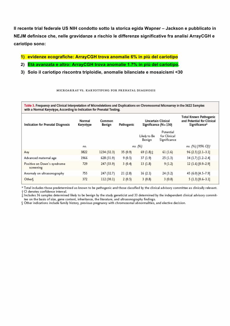
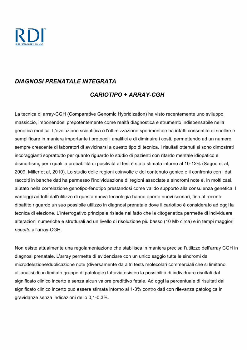
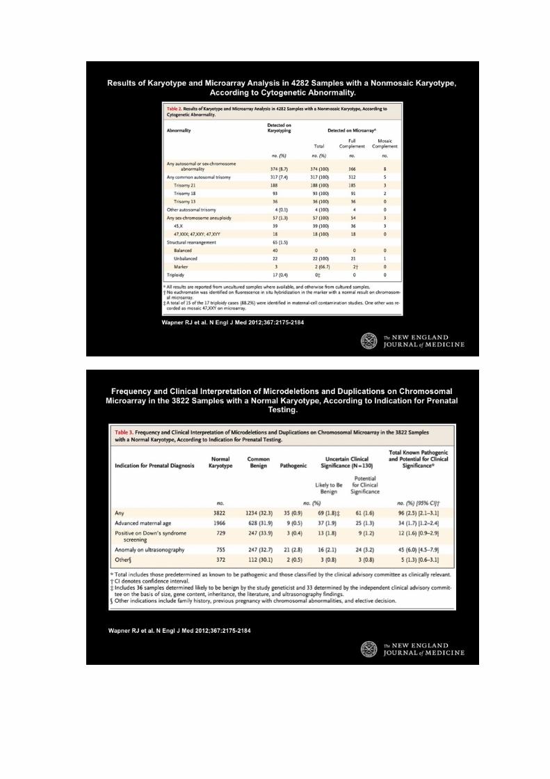
Il recente trial federale US NIH condotto sotto la storica egida Wapner – Jackson e pubblicato in
NEJM definisce che, nelle gravidanze a rischio le differenze significative fra analisi ArrayCGH e
cariotipo sono:
1) evidenze ecografiche: ArrayCGH trova anomalie 6% in più del cariotipo
2) Età avanzata e altro: ArrayCGH trova anomalie 1.7% in più del cariotipo.
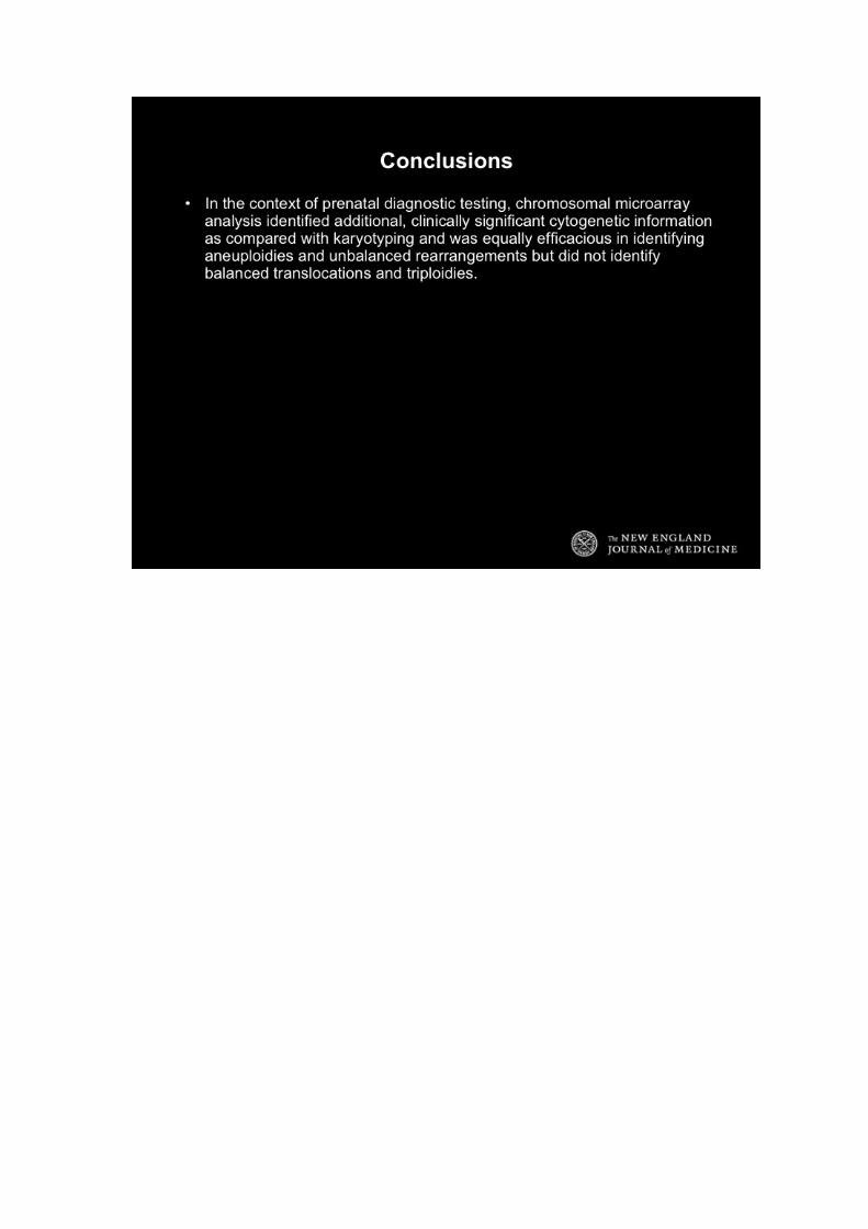
3) Solo il cariotipo riscontra triploidie, anomalie bilanciate e mosaicismi <30

La Società Italiana di Genetica Umana ha approvato e pubblicato una posizione ufficiale con le
indicazioni per l’uso di array-CGH in diagnosi prenatale solo assieme al cariotipo.
Il test di array CGH può e si consiglia venga proposto alla facoltà di scelta delle pazienti che
appartengono suesposte categorie a rischio (standard care practice) e venga considerato come
un esame integrante di secondo livello, che affianchi il cariotipo. In particolare:
• Caratterizzazione di riarrangiamenti cromosomici de novo apparentemente bilanciati • Presenza di anomalie ecografiche • Presenza di un microriarrangiamento strutturale bilanciato nei genitori • La età materna e presenza di test biochimici a rischio
I nostri laboratori, nell’intento di rispondere alle esigenze di medici e pazienti, sono oggi in grado di offrire
la diagnosi prenatale con la combinazione del cariotipo e array-CGH.
CARIOTIPO+ARRAY-CGH
La informativa al consenso per la indagine citogenetica prenatale vede inserita la clausola relativa alla
scelta della tecnica supplementare ArrayCGH offerta alla valutazione e sottoscrizione della paziente.
I colleghi medici e i pazienti interessati possono rivolgersi direttamente ai nostri laboratori per ulteriori
informazioni e modalità degli esami.
Campioni necessari: campione liquido amniotico o villi coriali, campione di sangue periferico in EDTA dei
genitori.


Ultrasound Obstet Gynecol 2012; 39: 384–388Published online in Wiley Online Library (wileyonlinelibrary.com). DOI: 10.1002/uog.11092
Microarray application in prenatal diagnosis: a positionstatement from the cytogenetics working group of the ItalianSociety of Human Genetics (SIGU), November 2011
A. NOVELLI1, F. R. GRATI2, L. BALLARATI3, L. BERNARDINI1, D. BIZZOCO4, L. CAMURRI5,R. CASALONE6, L. CARDARELLI7, P. CAVALLI8, R. CICCONE9, M. CLEMENTI10, L. DALPRA11,M. GENTILE12, G. GELLI13, P. GRAMMATICO14, M. MALACARNE15, A. M. NARDONE16,V. PECILE17, G. SIMONI2, O. ZUFFARDI9 and D. GIARDINO3
1Mendel Laboratory, Casa Sollievo della Sofferenza Hospital, IRCCS, San Giovanni Rotondo, Italy; 2TOMA Advanced Biomedical AssaysS.p.A., Busto Arsizio (VA), Italy; 3Laboratory of Medical Cytogenetics and Molecular Genetics, IRCCS Istituto Auxologico Italiano, Milan,Italy; 4Artemisia Fetal-Maternal Medical Centre, Rome, Italy; 5Genetics Department, RDI Rete Diagnostica Italiana, Padova, Italy; 6SSDipartimentale Genetica Azienda Osp. Universitaria Ospedale di Circolo e Fondazione Macchi, Varese, Italy; 7Laboratorio Citotest,Consorzio GENiMED, Sarmeola di Rubano, Padova, Italy; 8Servizio di Genetica, AO Istituti Ospitalieri, Cremona, Italy; 9BiologiaGenerale e Genetica Medica, Universita’ degli Studi di Pavia, Pavia, Italy; 10U.O. Complessa di Genetica Clinica, Dipartimento di Pediatria,Universita di Padova, Padova, Italy; 11Dipartimento di Neuroscienze e Biotecnologie Mediche, Universita di Milano-Bicocca, Monza, Italy;12Dipartimento di Genetica Medica, Ospedale Di Venere, ASL Bari, Italy; 13UOSD di Genetica Medica, Centro per la Salute della DonnaS.Anna, ASL Rome, Italy; 14Medical Genetics, Department of Molecular Medicine, Sapienza University, San Camillo-Forlanini Hospital,Rome, Italy; 15Laboratorio di Genetica, Ospedale Galliera, Genova, Italy; 16Laboratory of Medical Genetics, Fondazione PTV-PoliclinicoTor Vergata, Rome, Italy; 17S.C. Laboratorio di Genetica Medica, IRCCS Burlo Garofalo, Trieste, Italy
KEYWORDS: microarray; position statement; prenatal diagnosis; SIGU; ultrasound fetal abnormalities
ABSTRACT
A precise guideline establishing chromosomal microarrayanalysis (CMA) applications and platforms in the prenatalsetting does not exist. The controversial question iswhether CMA technologies can or should soon replacestandard karyotyping in prenatal diagnostic practice. Areview of the recent literature and survey of the knowledgeand experience of all members of the Italian Society ofHuman Genetics (SIGU) Committee were carried out inorder to propose recommendations for the use of CMA inprenatal testing. The analysis of datasets reported in themedical literature showed a considerable 6.4% incidenceof pathogenic copy number variations (CNVs) in thegroup of pregnancies with sonographically detected fetalabnormalities and normal karyotype. The reported CNVsare likely to have a relevant role in terms of nosologyfor the fetus and in the assessment of reproductiverisk for the couple. Estimation of the frequency ofcopy number variations of uncertain significance (VOUS)varied depending on the different CMA platforms used,ranging from 0–4%, obtained using targeted arrays, to9–12%, obtained using high-resolution whole genomesingle nucleotide polymorphism (SNP) arrays. CMAanalysis can be considered a second-tier diagnostic test
to be used after standard karyotyping in selected groupsof pregnancies, namely those with single (apparentlyisolated) or multiple ultrasound fetal abnormalities,those with de novo chromosomal rearrangements, evenif apparently balanced, and those with supernumerarymarker chromosomes. Copyright 2012 ISUOG.Published by John Wiley & Sons, Ltd.
BACKGROUND
In the last few years chromosomal microarray anal-ysis (CMA) technology (array comparative genomichybridization, aCGH; single nucleotide polymorphismarray, SNP array) has acquired increasing relevance,becoming a fundamental diagnostic tool in medical genet-ics. In fact, technological evolution and experimentaloptimization have resulted in a notable simplificationof analytic protocols, leading to a decrease in costs andenabling the progressive spread of this technology inmany laboratories all over the world. Encouraging results,in terms of detection rate, were obtained in patientsaffected by unexplained developmental delay/intellectualdisability (DD/ID), autism spectrum disorders (ASD)or multiple congenital anomalies (MCA), in whom
Correspondence to: Dr A. Novelli, Istituto CSS Mendel, Viale Regina Margherita, 261, 00198 Roma, Italy (e-mail: [email protected])
Accepted: 22 December 2011
Copyright 2012 ISUOG. Published by John Wiley & Sons, Ltd. POSITION STATEMENT

Position Statement: prenatal diagnosis of CMA 385
the diagnostic yield was improved over that obtainedby karyotyping by an estimated 10–20%1–3. Accu-rate evaluation of the gene content of the imbal-anced genomic regions, together with comparison withdata collections present in publicly available reposi-tory databases (DGV, http://projects.tcag.ca/variation/;DECIPHER, http://decipher.sanger.ac.uk/; OMIM, http://www.ncbi.nlm.nih.gov/omim), enabled detection of crit-ical regions related to known syndromes, allowing geno-type–phenotype correlations in several cases. For suchreasons, in 2010 the Italian Society of Human Genet-ics (SIGU) Committee proposed a national document inwhich, based on the literature and on the experience of allparticipating institutions, CMA was recommended as thefirst-tier diagnostic test in the postnatal setting for patientswith DD/ID, ASD or MCA (http://www.sigu.net).
The advantages offered by CMA technology haveopened up new avenues regarding its possible applicationin prenatal diagnosis, where traditional karyotyping is stillconsidered the gold standard method for all indications forinvasive testing. Compared with conventional karyotyp-ing, CMA can rapidly detect imbalances with a resolutionof up to a few Kb using standardized protocols4.
LITERATURE REVIEW
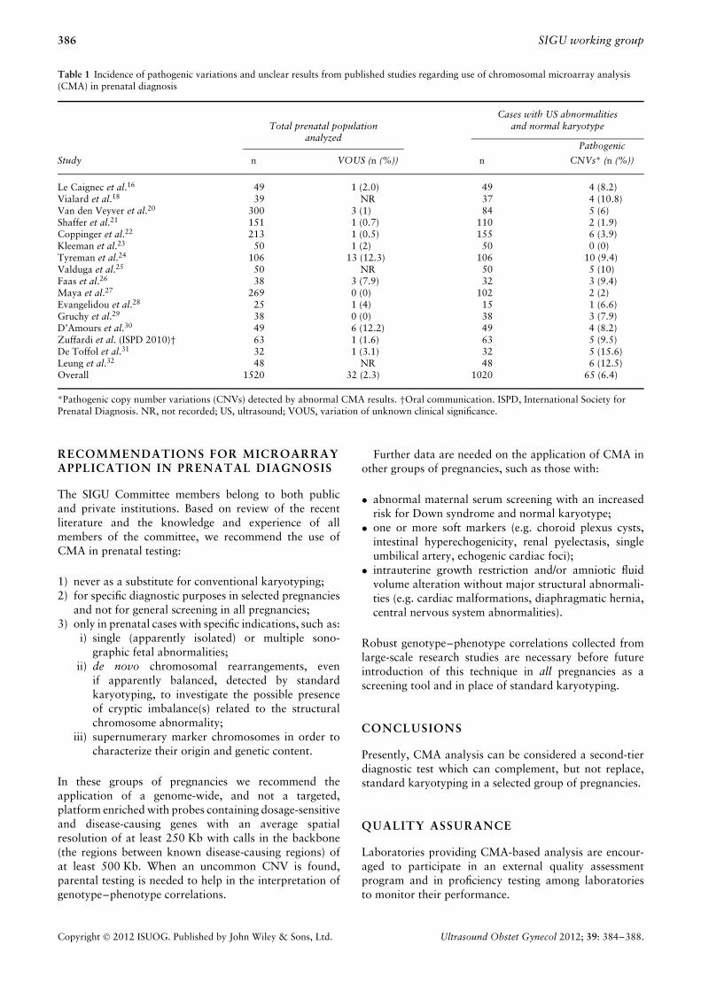
A precise guideline establishing CMA applications andplatforms in the prenatal setting does not exist and this sit-uation has led to debates and controversies5–11 concerningwhether CMA technology can or should replace standardkaryotyping in prenatal diagnostic practice. Consideringthe limited knowledge in this field, the SIGU Committeehas focused on disadvantages related to this technologyand currently advises against its unlimited and unselectedapplication in routine prenatal diagnosis. Without strictguidelines for the use of CMA in prenatal diagnosis, itcould potentially be more harmful than it is useful whenapplied during prenatal life, because of the unclear resultsit can provide. Current knowledge has gaps regarding theclinical interpretation of copy number variations (CNVs).This is because of the possibility of detecting an imbalancenot previously described, the lack of knowledge about thefunction of many genes, our relatively poor understand-ing of gene–gene and gene–environment interactions,and the role of epigenetic modifications in modulatingthe penetrance and expressivity of CNVs12–14. There areadditional questions related to the detection during theprenatal diagnostic period of variations of uncertain sig-nificance (VOUS), which have no known predictive valuewith regards to fetal and future health, and can thus causeincreased parental anxiety7,15. In addition, the diagnos-tic yield of CMA in the prenatal setting has not beenestablished clearly in all categories of indications becausethe majority of published papers included selected caseswith fetal abnormalities detected by ultrasound and anapparently normal karyotype. In this group of pregnan-cies the CMA detection rate is, on average, 6.4% (range,0–15.6%) (Table 1). Datasets reported in the medicalliterature clearly show a significant incidence of
pathogenic CNVs in this group of pregnancies and thesedetected CNVs are likely to have a relevant role in terms ofnosology for the fetus and for the assessment of reproduc-tive risks for the couple16–32. In cases with sonographicfetal abnormalities, the sum of the detection rates of con-ventional cytogenetic analyses (28% for chorionic villi and12% for amniotic fluid: ∼ 20% on average)33 and CMA(6.4%), i.e. combining the first-tier karyotype with thesecond-tier CMA, provide an overall detection of ∼ 27%.
Frequencies of VOUS seem to be difficult to assess dueto the different CMA platforms used in the various studies,and range from 0–4% when assessed by targeted arraysto ∼ 9–12% when assessed by high resolution wholegenome SNP arrays (Table 1)16–32. In contrast, the rate ofdetection of known, disability-causing pathogenic CNVsby CMA in all pregnant women has been estimated to bebetween 0.16% and 0.3%6. Analysis of the proportion ofambiguous findings compared to pathogenic CNVs showsthat using CMA technology in the prenatal setting withouta specific clinical indication is not justified at present.
Another important limitation related to the applica-tion of CMA as a first-tier test is represented by theimpossibility of detecting balanced rearrangements i.e.those without genetic losses or gains. This would lead tounderestimation of the risks of phenotypic consequencesrelated to: (i) disruption or modulation of the expressionof gene(s) located at the breakpoint(s); (ii) inactivation(position effect) of gene(s) at the breakpoint region(s);and (iii) missing the opportunity to investigate and detectuniparental disomy conditions related to imprinting syn-dromes in cases involving imprinted chromosomes34–36.SNP array has the advantage of being able to detectlong continuous stretches of homozygosity (LCSH), rep-resenting whole chromosomal or segmental uniparentalisodisomies (a duplicate of one chromosome from a par-ent and no chromosome from the other parent). It cannot,however, detect heterodisomies (the most common formof uniparental disomy, in which both chromosomes in apair are inherited from one parent) without testing parentsin conjunction with the fetal specimen. In addition, SNParray provides consanguinity information (occurrence ofincest) that raises important ethical issues; therefore, itsuse in terms of LCSH may be limited37. Finally, polyploi-dies and mosaicisms lower than 30%, that are relativelycommon findings in chorionic villi and amniotic fluidsamples33, cannot currently be detected by aCGH38,39.
On the other hand, CMA is useful to clarify abnormalkaryotype results. In cases with supernumerary markerchromosomes, CMA can aid in their classification andcharacterization, improving the diagnostic accuracy andallowing specific genetic counseling to be offered to thecouple40–42. The role of CMA prenatally in cases with denovo apparently balanced chromosomal rearrangementshas not been studied extensively; however, in postnataldatasets of patients with de novo apparently balancedchromosomal rearrangements and an abnormal pheno-type, CMA detects cryptic imbalances in 35–40% ofsamples with reciprocal translocations and in 72–75% ofsamples with complex rearrangements43–45.
Copyright 2012 ISUOG. Published by John Wiley & Sons, Ltd. Ultrasound Obstet Gynecol 2012; 39: 384–388.
386 SIGU working group
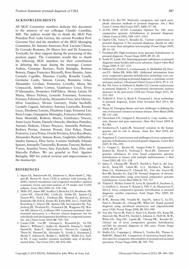
Table 1 Incidence of pathogenic variations and unclear results from published studies regarding use of chromosomal microarray analysis(CMA) in prenatal diagnosis
Total prenatal populationCases with US abnormalities
and normal karyotypeanalyzed
Pathogenic
Study n VOUS (n (%)) n CNVs* (n (%))
Le Caignec et al.16 49 1 (2.0) 49 4 (8.2)Vialard et al.18 39 NR 37 4 (10.8)Van den Veyver et al.20 300 3 (1) 84 5 (6)Shaffer et al.21 151 1 (0.7) 110 2 (1.9)Coppinger et al.22 213 1 (0.5) 155 6 (3.9)Kleeman et al.23 50 1 (2) 50 0 (0)Tyreman et al.24 106 13 (12.3) 106 10 (9.4)Valduga et al.25 50 NR 50 5 (10)Faas et al.26 38 3 (7.9) 32 3 (9.4)Maya et al.27 269 0 (0) 102 2 (2)Evangelidou et al.28 25 1 (4) 15 1 (6.6)Gruchy et al.29 38 0 (0) 38 3 (7.9)D’Amours et al.30 49 6 (12.2) 49 4 (8.2)Zuffardi et al. (ISPD 2010)† 63 1 (1.6) 63 5 (9.5)De Toffol et al.31 32 1 (3.1) 32 5 (15.6)Leung et al.32 48 NR 48 6 (12.5)Overall 1520 32 (2.3) 1020 65 (6.4)
*Pathogenic copy number variations (CNVs) detected by abnormal CMA results. †Oral communication. ISPD, International Society forPrenatal Diagnosis. NR, not recorded; US, ultrasound; VOUS, variation of unknown clinical significance.
RECOMMENDATIONS FOR MICROARRAYAPPLICATION IN PRENATAL DIAGNOSIS
The SIGU Committee members belong to both publicand private institutions. Based on review of the recentliterature and the knowledge and experience of allmembers of the committee, we recommend the use ofCMA in prenatal testing:
1) never as a substitute for conventional karyotyping;2) for specific diagnostic purposes in selected pregnancies
and not for general screening in all pregnancies;3) only in prenatal cases with specific indications, such as:
i) single (apparently isolated) or multiple sono-graphic fetal abnormalities;
ii) de novo chromosomal rearrangements, evenif apparently balanced, detected by standardkaryotyping, to investigate the possible presenceof cryptic imbalance(s) related to the structuralchromosome abnormality;
iii) supernumerary marker chromosomes in order tocharacterize their origin and genetic content.
In these groups of pregnancies we recommend theapplication of a genome-wide, and not a targeted,platform enriched with probes containing dosage-sensitiveand disease-causing genes with an average spatialresolution of at least 250 Kb with calls in the backbone(the regions between known disease-causing regions) ofat least 500 Kb. When an uncommon CNV is found,parental testing is needed to help in the interpretation ofgenotype–phenotype correlations.
Further data are needed on the application of CMA inother groups of pregnancies, such as those with:
• abnormal maternal serum screening with an increasedrisk for Down syndrome and normal karyotype;
• one or more soft markers (e.g. choroid plexus cysts,intestinal hyperechogenicity, renal pyelectasis, singleumbilical artery, echogenic cardiac foci);
• intrauterine growth restriction and/or amniotic fluidvolume alteration without major structural abnormali-ties (e.g. cardiac malformations, diaphragmatic hernia,central nervous system abnormalities).
Robust genotype–phenotype correlations collected fromlarge-scale research studies are necessary before futureintroduction of this technique in all pregnancies as ascreening tool and in place of standard karyotyping.
CONCLUSIONS
Presently, CMA analysis can be considered a second-tierdiagnostic test which can complement, but not replace,standard karyotyping in a selected group of pregnancies.
QUALITY ASSURANCE
Laboratories providing CMA-based analysis are encour-aged to participate in an external quality assessmentprogram and in proficiency testing among laboratoriesto monitor their performance.
Copyright 2012 ISUOG. Published by John Wiley & Sons, Ltd. Ultrasound Obstet Gynecol 2012; 39: 384–388.
Position Statement: prenatal diagnosis of CMA 387
ACKNOWLEDGMENTS
All SIGU Committee members dedicate this documentto the memory of our colleague Claudio Castellan,MD. The authors would like to thank the SIGU PastPresident Prof. Lidia Larizza, the current President Prof.Giovanni Neri and the members of the SIGU ManagingCommittee, Dr Antonio Amoroso, Prof. Luciana Chessa,Dr Corrado Romano, Dr Marco Seri and Dr FrancescaTorricelli, for their support during the processing of thisposition paper. The Committee would like to thankthe following SIGU members for their contributionin debating this issue during the meetings: CarmenArdisia, Giuseppe Barrano, Paola Battaglia, RominaBonora, Dagna Francesca Bricarelli, Rosa Busuito, AnnaCarmela Capalbo, Massimo Carella, Rossella Caselli,Girolama Casile, Simona Cavani, Bommina Celso,Alessandro Civolani, Domenico Coviello, Maria IdaCorpaccioli, Ambra Cortesi, Gianfranco Croci, ElviraD’Alessandro, Domenico Dell’Edera, Monia Letizia DiMaria, Marco Fichera, Lonardo Fortunato, FrancescaForzano, Fabrizia Franchi, Alessandra Friso, Sara Ghezzo,Silvia Guarducci, Silvana Guerneri, Nadia Iacobelli,Carmelo Lagana, Salvatrice Antonia Lauricella, RosettaLecce, Elisabetta Lenzini, Elisabetta Lippi, Ermanna Lisi,Ilaria Longo, Giuseppina Marseglia, Francesca Malvestiti,Anna Montaldi, Roberta Murru, Gianfranco Nocera,Anna Lucia Nutini, Daniela Orteschi, Marilena Pantaleo,Antonio Pedicini, Chiara Pescucci, Antonella Pettinari,Barbara Pivetta, Antonio Pizzuti, Gisa Police, DianaPostorivo, Lucia Prima, Orsola Privitera, Erica Racalbuto,Alessandra Renieri, Simona Rossi, Guglielmo Sabbadini,Sandra Santucci, Elisa Savin, Lorenzo Sinibaldi, IolandaSpasari, Antonella Tanzariello, Romano Tenconi, BarbaraTorres, Annalisa Vetro, Sara Zanchetti, Anna Zilio andMarcella Zollino. We are grateful to Prof. AgatinoBattaglia, MD for critical revision and improvements tothe manuscript.
REFERENCES
1. Sagoo GS, Butterworth AS, Sanderson S, Shaw-Smith C, Hig-gins JP, Burton H. Array CGH in patients with learning dis-ability (mental retardation) and congenital anomalies: updatedsystematic review and meta-analysis of 19 studies and 13,926subjects. Genet Med 2009; 11: 139–146.
2. Miller DT, Adam MP, Aradhya S, Biesecker LG, Brothman AR,Carter NP, Church DM, Crolla JA, Eichler EE, Epstein CJ,Faucett WA, Feuk L, Friedman JM, Hamosh A, Jackson L,Kaminsky EB, Kok K, Krantz ID, Kuhn RM, Lee C, Ostell JM,Rosenberg C, Scherer SW, Spinner NB, Stavropoulos DJ, Tep-perberg JH, Thorland EC, Vermeesch JR, Waggoner DJ, Wat-son MS, Martin CL, Ledbetter DH. Consensus statement: chro-mosomal microarray is a first-tier clinical diagnostic test forindividuals with developmental disabilities or congenital anoma-lies. Am J Hum Genet 2010; 86: 749–764.
3. Cooper GM, Coe BP, Girirajan S, Rosenfeld JA, Vu TH,Baker C, Williams C, Stalker H, Hamid R, Hannig V, Abdel-Hamid H, Bader P, McCracken E, Niyazov D, Leppig K,Thiese H, Hummel M, Alexander N, Gorski J, Kussmann J,Shashi V, Johnson K, Rehder C, Ballif BC, Shaffer LG, Eich-ler EE. A copy number variation morbidity map of develop-mental delay. Nat Genet 2011; 43: 838–846.
4. Shaffer LG, Bui TH. Molecular cytogenetic and rapid aneu-ploidy detection methods in prenatal diagnosis. Am J MedGenet C Semin Med Genet 2007; 145C: 87–98.
5. ACOG 2009. ACOG Committee Opinion No. 446: arraycomparative genomic hybridization in prenatal diagnosis.Obstet Gynecol 2002; 114: 1161–1163.
6. Ogilvie CM, Yaron Y, Beaudet AL. Current controversies inprenatal diagnosis 3: For prenatal diagnosis, should we offerless or more than metaphase karyotyping? Prenat Diagn 2009;29: 11–14.
7. Friedman JM. High-resolution array genomic hybridization inprenatal diagnosis. Prenat Diagn 2009; 29: 20–28.
8. South ST, Lamb AN. Detecting genomic imbalances in prenataldiagnosis: main hurdles and recent advances. Expert Opin MedDiagn 2009; 3: 227–235.
9. Hillman SC, Pretlove S, Coomarasamy A, McMullan DJ, Davi-son EV, Maher ER, Kilby MD. Additional information fromarray comparative genomic hybridization technology over con-ventional karyotyping in prenatal diagnosis: a systematic reviewand meta-analysis. Ultrasound Obstet Gynecol 2011; 37: 6–14.
10. Bui TH, Vetro A, Zuffardi O, Shaffer LG. Current controversiesin prenatal diagnosis 3: is conventional chromosome analysisnecessary in the post-array CGH era? Prenat Diagn 2011; 31:235–243.
11. Zuffardi O, Vetro A, Brady P, Vermeesch J. Array technologyin prenatal diagnosis. Semin Fetal Neonatal Med 2011; 16:94–98.
12. Sharp AJ. Emerging themes and new challenges in defining therole of structural variation in human disease. Hum Mutat 2009;30: 135–144.
13. Henrichsen CN, Chaignat E, Reymond A. Copy number vari-ants, diseases and gene expression. Hum Mol Genet 2009; 8:R1–R8.
14. Stankiewicz P, Lupski JR. Structural variation in the humangenome and its role in disease. Annu Rev Med 2010; 61:437–455.
15. Pergament E. Controversies and challenges of array comparativegenomic hybridization in prenatal genetic diagnosis. Genet Med2007; 9: 596–599.
16. Le Caignec C, Boceno M, Saugier-Veber P, Jacquemont S,Joubert M, David A, Frebourg T, Rival JM. Detection ofgenomic imbalances by array based comparative genomichybridisation in fetuses with multiple malformations. J MedGenet 2005; 42: 121–128.
17. Sahoo T, Cheung SW, Ward P, Darilek S, Patel A, del Gau-dio D, Kang SH, Lalani SR, Li J, McAdoo S, Burke A,Shaw CA, Stankiewicz P, Chinault AC, Van den Veyver IB,Roa BB, Beaudet AL, Eng CM. Prenatal diagnosis of chromo-somal abnormalities using array-based comparative genomichybridization. Genet Med 2006; 8: 719–727.
18. Vialard F, Molina Gomes D, Leroy B, Quarello E, Escalona A,Le Sciellour C, Serazin V, Roume J, Ville Y, de Mazancourt P,Selva J. Array comparative genomic hybridization in prenataldiagnosis: another experience. Fetal Diagn Ther 2009; 25:277–284.
19. Bi W, Breman AM, Venable SF, Eng PA, Sahoo T, Lu XY,Patel A, Beaudet AL, Cheung SW, White LD. Rapid prenataldiagnosis using uncultured amniocytes and oligonucleotidearray CGH. Prenat Diagn 2008; 28: 943–949.
20. Van den Veyver IB, Patel A, Shaw CA, Pursley AN, Kang SH,Simovich MJ, Ward PA, Darilek S, Johnson A, Neill SE, Bi W,White LD, Eng CM, Lupski JR, Cheung SW, Beaudet AL.Clinical use of array comparative genomic hybridization(aCGH) for prenatal diagnosis in 300 cases. Prenat Diagn2009; 29: 29–39.
21. Shaffer LG, Coppinger J, Alliman S, Torchia BA, Theisen A,Ballif BC, Bejjani BA. Comparison of microarray-based detec-tion rates for cytogenetic abnormalities in prenatal and neonatalspecimens. Prenat Diagn 2008; 28: 789–795.
Copyright 2012 ISUOG. Published by John Wiley & Sons, Ltd. Ultrasound Obstet Gynecol 2012; 39: 384–388.

388 SIGU working group
22. Coppinger J, Alliman S, Lamb AN, Torchia BS, Bejjani BA,Shaffer LG. Whole genome microarray analysis in prenatal spec-imens identifies clinically significant chromosome alterationswithout increase in results of unclear significance compared totargeted microarray. Prenat Diagn 2009; 29: 1156–1166.
23. Kleeman L, Bianchi DW, Shaffer LG, Rorem E, Cowan J,Craigo SD, Tighiouart H, Wilkins-Haug LE. Use of arraycomparative genomic hybridization for prenatal diagnosis offetuses with sonographic anomalies and normal metaphasekaryotype. Prenat Diagn 2009; 29: 1213–1217.
24. Tyreman M, Abbott KM, Willatt LR, Nash R, Lees C, Whit-taker J, Simonic I. High resolution array analysis: diagnosingpregnancies with abnormal ultrasound findings. J Med Genet2009; 46: 531–541.
25. Valduga M, Philippe C, Bach Segura P, Thiebaugeorges O,Miton A, Beri M, Bonnet C, Nemos C, Foliguet B, Jonveaux P.A retrospective study by oligonucleotide array-CGH analysisin 50 fetuses with multiple malformations. Prenat Diagn 2010;30: 333–341.
26. Faas BH, van der Burgt I, Kooper AJ, Pfundt R, Hehir-Kwa JY,Smits AP, de Leeuw N. Identification of clinically significant,submicroscopic chromosome alterations and UPD in fetuseswith ultrasound anomalies using genome-wide 250k SNP arrayanalysis. J Med Genet 2010; 47: 586–594.
27. Maya I, Davidov B, Gershovitz L, Zalzstein Y, Taub E, Cop-pinger J, Shaffer LG, Shohat M. Diagnostic utility of array-based comparative genomic hybridization (aCGH) in a prenatalsetting. Prenat Diagn 2010; 30: 1131–1137.
28. Evangelidou P, Sismani C, Ioannides M, Christodoulou C,Koumbaris G, Kallikas I, Georgiou I, Velissariou V, Pat-salis PC. Clinical application of whole-genome array CGHduring prenatal diagnosis: Study of 25 selected pregnancies withabnormal ultrasound findings or apparently balanced structuralaberrations. Mol Cytogenet 2010; 3: 24.
29. Gruchy N, Decamp M, Richard N, Jeanne-Pasquier C, BenoistG, Mittre H, Leporrier N. Array CGH analysis in high-riskpregnancies: comparing DNA from cultured cells and cell-freefetal DNA. Prenat Diagn 2011; DOI: 10.1002/pd.2861.
30. D’Amours G, Kibar Z, Mathonnet G, Fetni R, Tihy F, DesiletsV, Nizard S, Michaud J, Lemyre E. Whole-genome array CGHidentifies pathogenic copy number variations in fetuses withmajor malformations and a normal karyotype. Clin Genet 2012;8: 128–141.
31. De Toffol S, Marcato L, Malvestiti F, Chinetti S, Grimi B,Maggi F, Simoni G, Grati FR. Retrospective analysis of prenatalsamples with sonographic anomalies using genome-widebacterial artificial chromosome array comparative genomichybridization. Prenat Diagn 2010; 30 (Suppl. 1): S24 (Abstract9–4).
32. Leung TY, Vogel I, Lau TK, Chong W, Hyett JA, Petersen OB,Choy KW. Identification of submicroscopic chromosomalaberrations in fetuses with increased nuchal translucency andapparently normal karyotype. Ultrasound Obstet Gynecol2011; 38: 314–319.
33. Grati FR, Barlocco A, Grimi B, Milani S, Frascoli G, Di MecoAM, Liuti R, Trotta A, Chinetti S, Dulcetti F, Ruggeri AM,
De Toffol S, Clementi M, Maggi F, Simoni G. Chromosomeabnormalities investigated by non-invasive prenatal testingaccount for approximately 50% of fetal unbalances associatedwith relevant clinical phenotypes. Am J Med Genet A 2010;152A: 1434–1442.
34. Warburton D. De novo balanced chromosome rearrangementsand extra marker chromosomes identified at prenatal diagnosis:clinical significance and distribution of breakpoints. Am J HumGenet 1991; 49: 995–1013.
35. Kotzot D. Complex and segmental uniparental disomy updated.J Med Genet 2008; 45: 545–556.
36. Liehr T. Cytogenetic contribution to uniparental disomy (UPD).Mol Cytogenet 2010; 29: 8.
37. Beaudet AL. Ethical issues raised by common copy numbervariants and single nucleotide polymorphisms of certain anduncertain significance in general medical practice. Genome Med2010; 2: 42.
38. Ballif BC, Rorem EA, Sundin K, Lincicum M, Gaskin S, Cop-pinger J, Kashork CD, Shaffer LG, Bejjani BA. Detection oflow-level mosaicism by array CGH in routine diagnostic speci-mens. Am J Med Genet A 2006; 140: 2757–2767.
39. Robberecht C, Schuddinck V, Fryns JP, Vermeesch JR. Diag-nosis of miscarriages by molecular karyotyping: benefits andpitfalls. Genet Med 2009; 11: 646–654.
40. Trifonov V, Fluri S, Binkert F, Nandini A, Anderson J,Rodriguez L, Gross M, Kosyakova N, Mkrtchyan H, Ewers E,Reich D, Weise A, Liehr T. Complex rearranged small super-numerary marker chromosomes (sSMC), three new cases;evidence for an underestimated entity? Mol Cytogenet 2008;1: 6.
41. Tsuchiya KD, Opheim K, Hannibal M, Hing A, Glass IA,Raff M, Beattie C, Norwood T, Torchia B. Unexpected com-plexity of supernumerary marker chromosomes revealed bymicroarray comparative genomic hybridization. Mol Cytogenet2008; 1: 7.
42. Gruchy N, Lebrun M, Herlicoviez M, Alliet J, Gourdier D,Kottler ML, Mittre H, Leporrier N. Supernumerary markerchromosomes management in prenatal diagnosis. Am J MedGenet A 2008; 146A: 2770–2776.
43. Ciccone R, Giorda R, Gregato G, Guerrini R, Giglio S, Car-rozzo R, Bonaglia MC, Priolo E, Lagana C, Tenconi R,Rocchi M, Pramparo T, Zuffardi O, Rossi E. Reciprocaltranslocations: a trap for cytogenetists? Hum Genet 2005; 117:571–582.
44. Baptista J, Mercer C, Prigmore E, Gribble SM, Carter NP,Maloney V, Thomas NS, Jacobs PA, Crolla JA. Breakpointmapping and array CGH in translocations: comparison of aphenotypically normal and an abnormal cohort. Am J HumGenet 2008; 82: 927–936.
45. Feenstra I, Hanemaaijer N, Sikkema-Raddatz B, Yntema H,Dijkhuizen T, Lugtenberg D, Verheij J, Green A, Hordijk R,Reardon W, Vries BD, Brunner H, Bongers E, Leeuw ND, vanRavenswaaij-Arts C. Balanced into array: genome-wide arrayanalysis in 54 patients with an apparently balanced de novochromosome rearrangement and a meta-analysis. Eur J HumGenet 2011; 19: 1152–1160.
Copyright 2012 ISUOG. Published by John Wiley & Sons, Ltd. Ultrasound Obstet Gynecol 2012; 39: 384–388.