diabetic retinopathy · prevalenceof the types of diabetes xtype 2 – in 90% of diabetic patients...
TRANSCRIPT

DIABETIC RETINOPATHYGhanbari MD1391:10:18
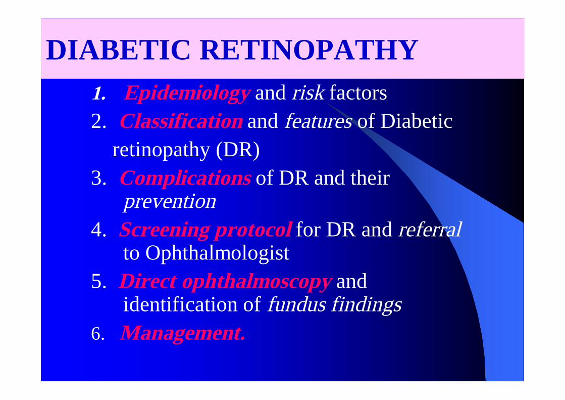
DIABETIC RETINOPATHY1. Epidemiology and risk factors2. Classification and features of Diabetic
retinopathy (DR)3. Complications of DR and their
prevention4. Screening protocol for DR and referral
to Ophthalmologist5. Direct ophthalmoscopy and
identification of fundus findings6. Management.

Epidemiology of DRRISK of developing DR:
Type I or IDDM – 70%
Type II or NIDDM - 39%
Type II on insulin – 70%

Prevalence of the types of Diabetes
Type 2 – in 90% of diabetic patients
• Hence Type 2 – in most of the DiabeticRetinopathy patients as well
Diabetic retinopathy - most common cause oflegal blindness between ages 20 and 74 years.

RISK FACTORS:1. Duration of diabetes2. Poor control of Diabetes3. Pregnancy4. Hypertension5. Nephropathy6. Obesity and hyperlipidemia7. Smoking

PathogenesisMicroangiopathy which has featuresof both microvascular leakage andocclusion.Larger vessels may also be involved
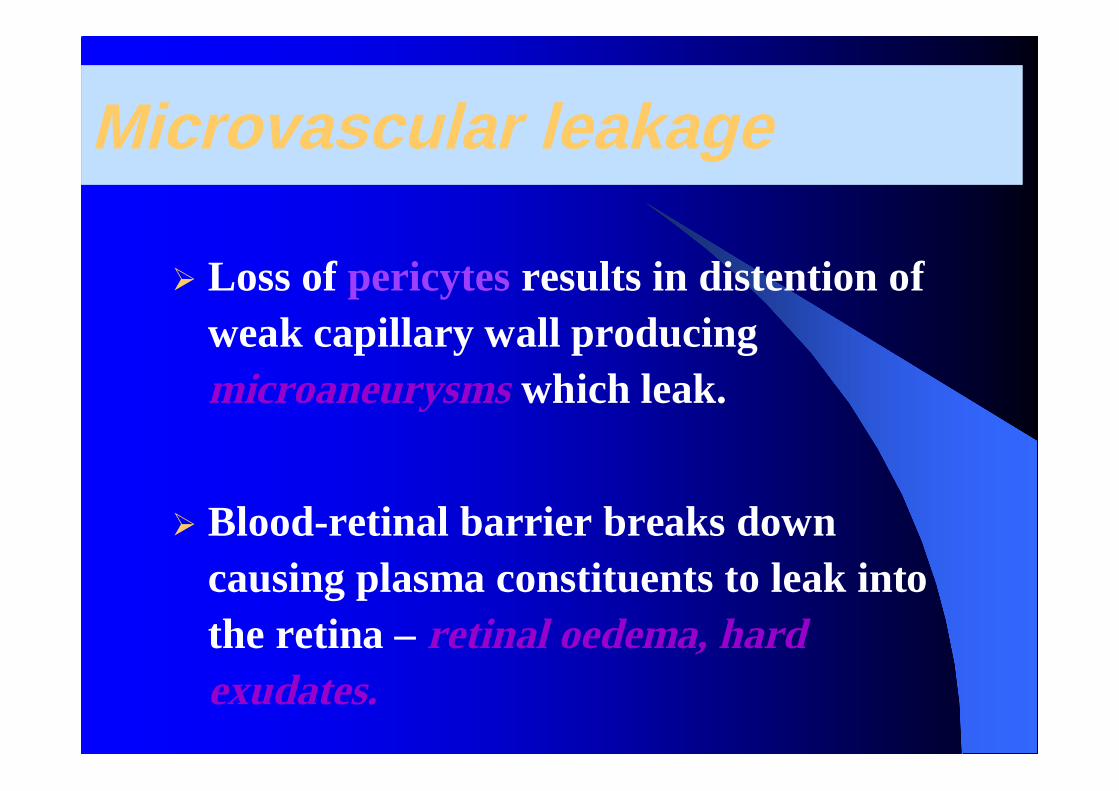
Microvascular leakage
Loss of pericytes results in distention ofweak capillary wall producingmicroaneurysms which leak.
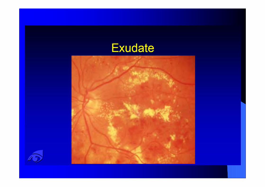
Blood-retinal barrier breaks downcausing plasma constituents to leak intothe retina – retinal oedema, hardexudates.

Microvascular occlusion
Basement membrane thickening,endothelial cell damage, deformed RBCs,platelet stickiness and aggregation.
Vascular Endothelial Growth Factor(VEGF) is produced by hypoxic retina.
VEGF stimulates the growth of shunt andnew vessels


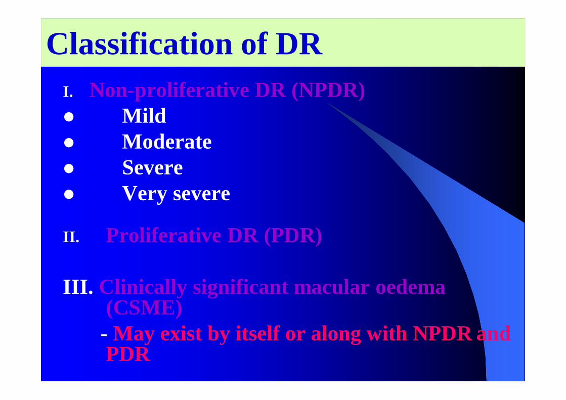
Classification of DRI. Non-proliferative DR (NPDR)
MildModerateSevereVery severe
II. Proliferative DR (PDR)
III. Clinically significant macular oedema(CSME)
- May exist by itself or along with NPDR andPDR

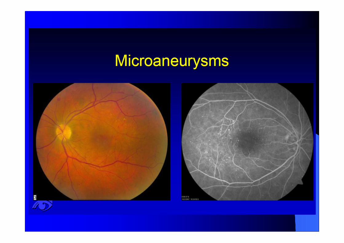
• At least one microaneurysm -earliest clinically detectable lesion
Retinal hemorrhages
Hard or soft exudates
Mild NPDR

Moderate NPDR• Microaneurysms and/or dot and blot
hemorrhages in at least 1 quadrant
• Soft exudates (Cotton wool spots)
• Venous beading or IRMA(intraretinal microvascularabnormalities)

Mild and Moderate Non- proliferative DRwas previously known as Background DR

Severe NPDRAny one of the following 3 features is present
Microaneurysms and intraretinal hemorrhagesin all 4 quadrants
Venous beading in 2 or more quadrants
Moderate IRMA in at least 1 quadrant
Known as the 4-2-1 rule

Very severe NPDR
Any two of the features of the 4-2-1 rule is present
Severe and Very severe Non-proliferative DRwas known as the Pre-proliferative DR

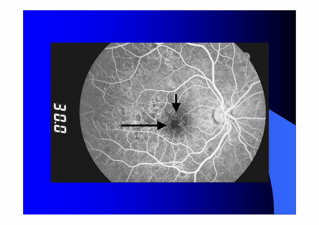
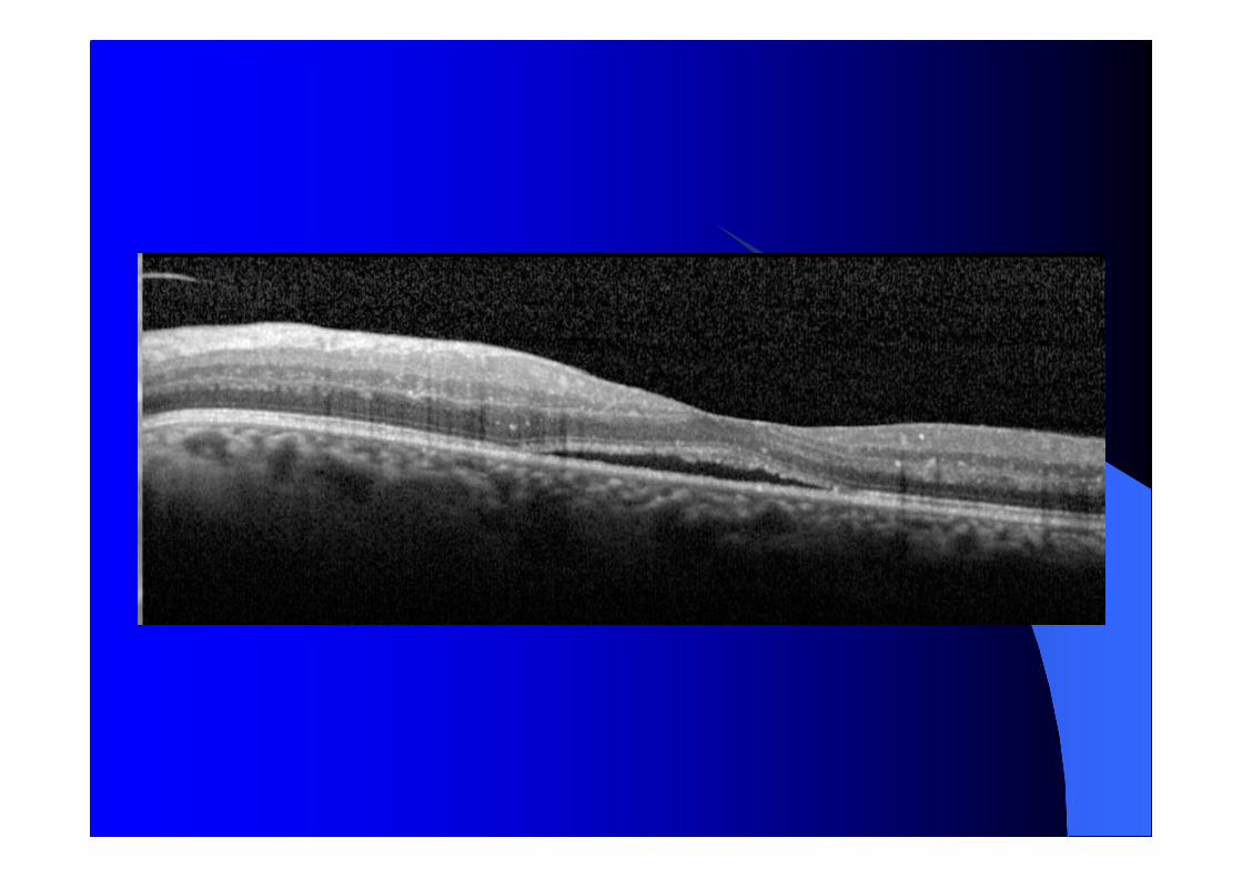
Clinically significant Macular edemaCSME
Retinal edema close to fovea
Hard exudates close to fovea
Presents with dimness of vision
By itself or along with NPDR or PDR

CSME – Hard exudates close to fovea andassociated retinal thickening
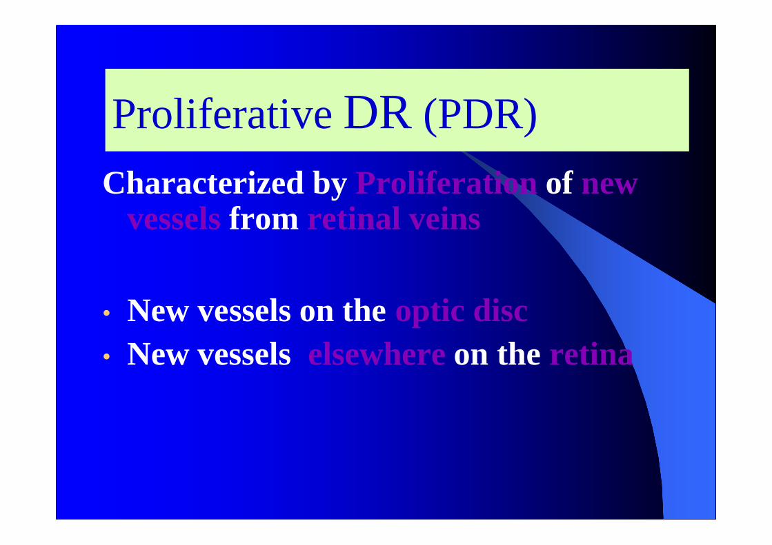
Proliferative DR (PDR)Characterized by Proliferation of new
vessels from retinal veins
• New vessels on the optic disc• New vessels elsewhere on the retina

Proliferative DR
NVD

COMPLICATIONS OF DIABETICRETINOPATHY
Vitreous hemorrhageTractional retinal detachmentRubeosis IridisGlaucomaBlindness
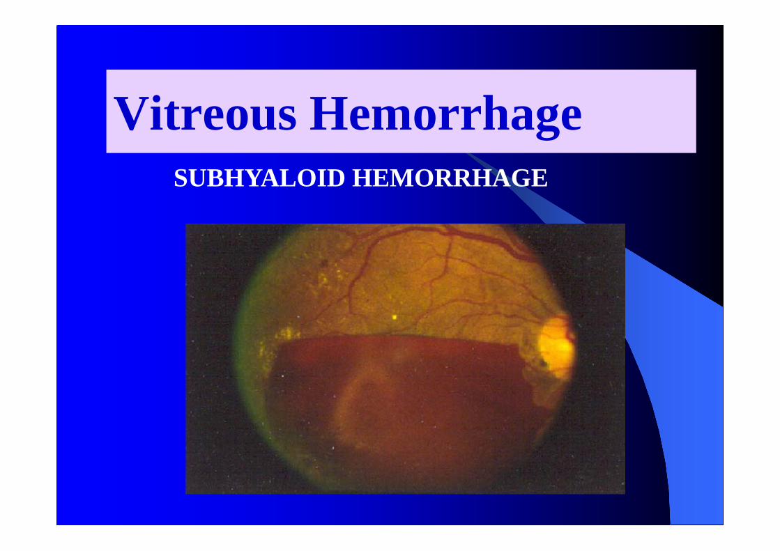
Vitreous HemorrhageSUBHYALOID HEMORRHAGE

Tractional retinal detachment

Rubeosis Iridis

Neovascular GlaucomaComplication of rubeosis iridis
New vessels cause angle closure
Mechanical obstruction to aqueous outflow
Intra ocular pressure rises
Pupil gets distorted as iris gets pulled
Eye becomes painful and red
Loss of vision

BlindnessNon-clearing vitreous hemorrhage
Neovascular glaucoma
Tractional retinal detachment
Macular ischemia

PREVENTION OF COMPLICATIONS
By early institution of appropriate treatment
This requires early detection of DR in itsasymptomatic treatable condition
By routine fundus examination of allDiabetics (cost effective screening)
And appropriate referral to ophthalmologist

Mild and Moderate NPDR
- No specific treatment for retinopathy- Good metabolic control to delay
progression- Control of associated Hypertension,
Anemia and Renal failure
Severe and very severe NPDR
• Close follow up by Ophthalmologist
Clinically significant macular oedema
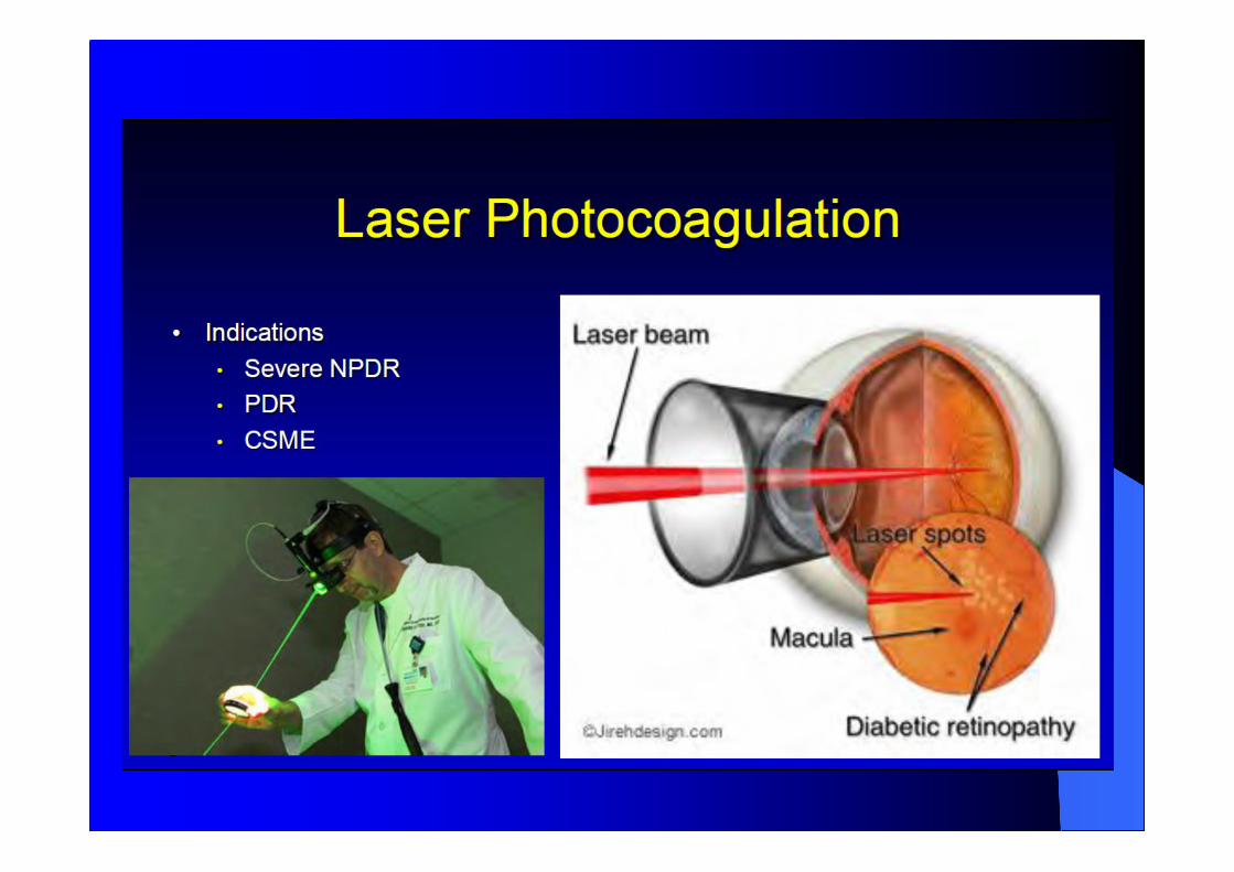
- Laser photocoagulation to minimise risk ofvisual loss.
Retinal laser photocoagulation as per thejudgment of ophthalmologist (in high risk eyes)
It converts hypoxic retina (which producesANGIOGENIC factors) into anoxic retina (whichcan’t)
Proliferative DR

Screening protocol for Diabeticretinopathy
1. Screening once in a 1 year
Diabetics with normal fundusMild NPDR
2. Screening once in 6 months
Moderate NPDR

Referral to OphthalmologistVisual Symptoms– Diminished visual acuity– Seeing floaters– Painful eye
Fundus findings- Macular oedema/hard exudates close to fovea- Proliferative DR- Vitreous hemorrhage- Moderate to severe and very severe NPDR- Retinal detachment- Cataract obscuring fundus view

Referral to Ophthalmologist
Presence of Risk Factors
- Pregnancy- Nephropathy

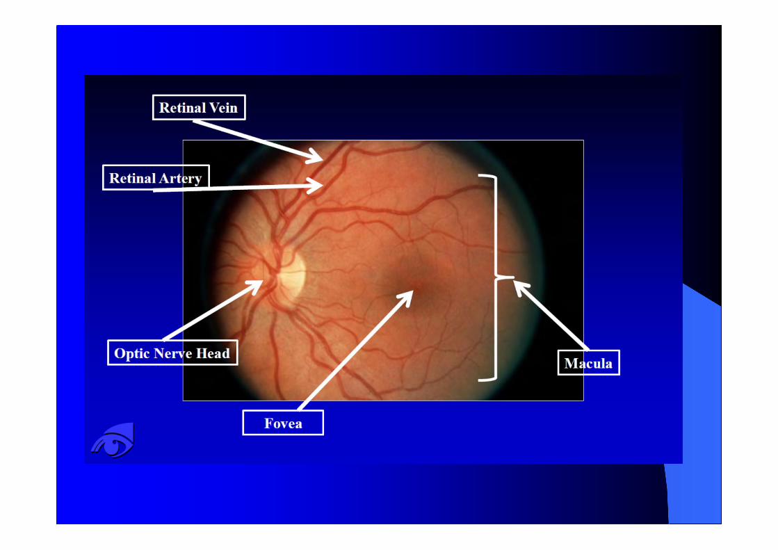
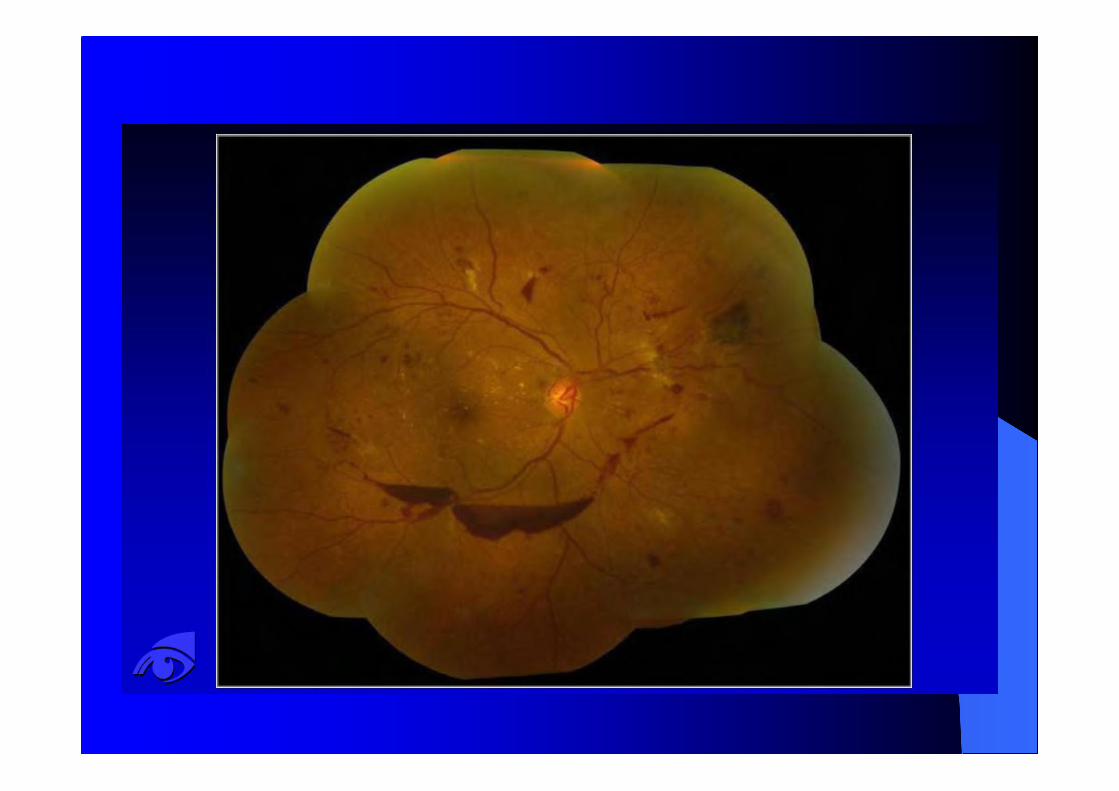
DIRECTOPHTHALMOSCOPY
Examination of the fundus of the eyeTo screen for Diabetic RetinopathyAfter dilatation of both eyes with 0.5%tropicamideFlashlight test, prior to dilatation todetect eyes with shallow ACProcedure will be demonstrated
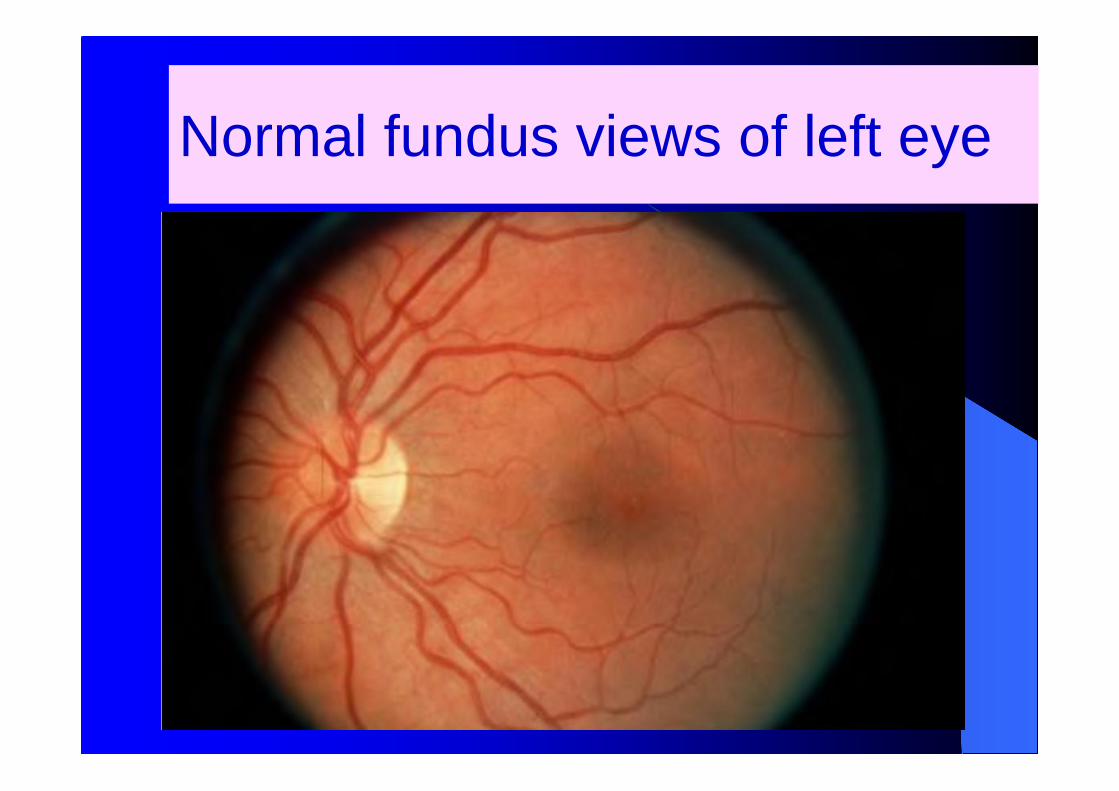
Normal fundus views of left eye

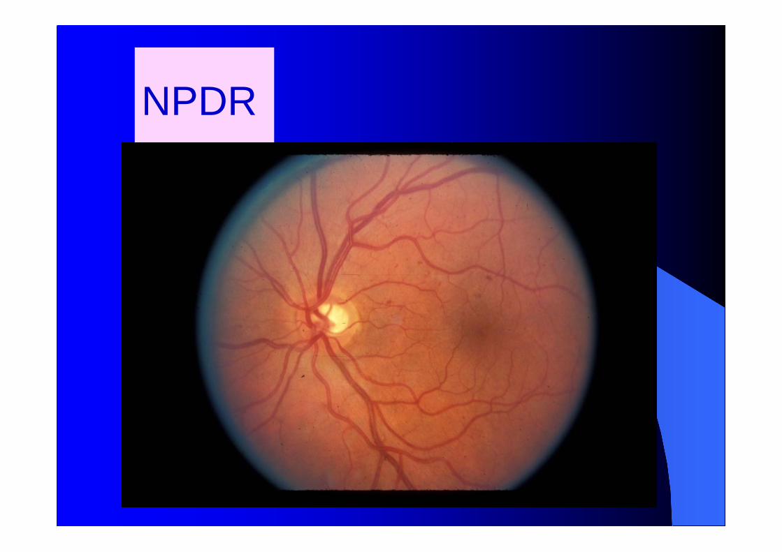
NPDR

Mild NPDR
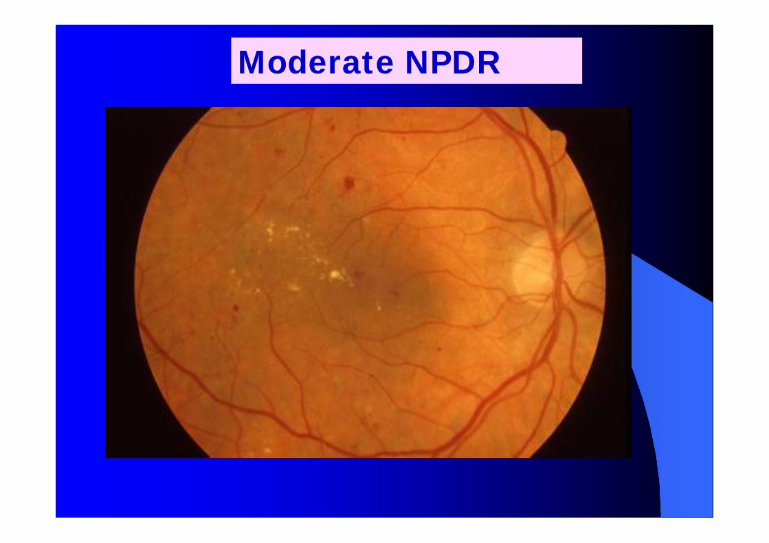
Moderate NPDR

Severe NPDR

Circinate retinopathy – Hard exudates in aring around leaking aneurysms

DRUSEN
Age related Macular degeneration: Note the drusen.Not to be confused with Hard exudates. There are nomicroaneurysms or dot/blot hemorrhages.

PDR – New vessels on disc

PDR – New vessels on disc and New vesselselsewhere on retina
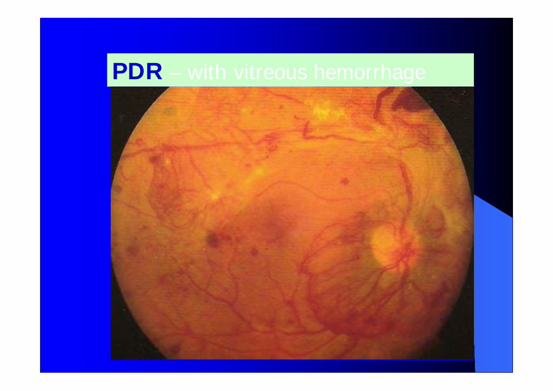
PDR – with vitreous hemorrhage
Vitreous bleed

Vitreous Hemorrhage

Tractional retinaldetachment Fibro-vascular
proliferation

MANAGEMENT
Control of DM. Will not prevent butdelays
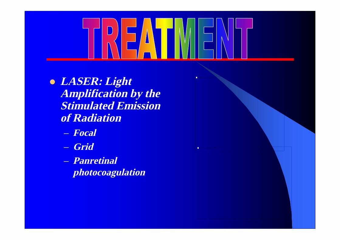
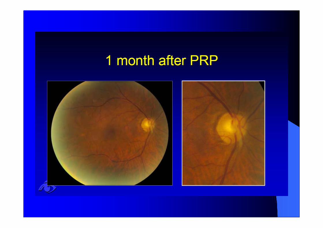
LASER: LightAmplification by theStimulated Emissionof Radiation– Focal– Grid– Panretinal
photocoagulation
This image cannot currently be displayed.
This image cannot currently be displayed.


0ZW0gaW


End-stage diabetic eye disease
This image cannot currently be displayed.
PHTHISISShrunken, soft eye withopaque vascularisedcornea and no visualPotential.


Thank you!
Any doubts?