diabetes mellitus in adults - aspects of incidence, autoimmunity and...
TRANSCRIPT

LUND UNIVERSITY
PO Box 117221 00 Lund+46 46-222 00 00
Diabetes mellitus in Adults - Aspects of Incidence, Autoimmunity and C-peptide
Thunander, Maria
2011
Link to publication
Citation for published version (APA):Thunander, M. (2011). Diabetes mellitus in Adults - Aspects of Incidence, Autoimmunity and C-peptide. Medicine(Lund).
General rightsCopyright and moral rights for the publications made accessible in the public portal are retained by the authorsand/or other copyright owners and it is a condition of accessing publications that users recognise and abide by thelegal requirements associated with these rights.
• Users may download and print one copy of any publication from the public portal for the purpose of private studyor research. • You may not further distribute the material or use it for any profit-making activity or commercial gain • You may freely distribute the URL identifying the publication in the public portalTake down policyIf you believe that this document breaches copyright please contact us providing details, and we will removeaccess to the work immediately and investigate your claim.
Download date: 18. Jul. 2019

1
Diabetes mellitus in Adults Aspects of Incidence, Autoimmunity
and C-peptide
Maria ThunanderDepartment of Clinical Sciences, Lund
Endocrinology and Diabetes Faculty of Medicine
Lund University
1
Diabetes mellitus in Adults Aspects of Incidence, Autoimmunity
and C-peptide
Maria ThunanderDepartment of Clinical Sciences, Lund
Endocrinology and Diabetes Faculty of Medicine
Lund University

ISSN 1652-8220ISBN 978-91-86871-02-4 Copyright © Maria ThunanderMedicinska fakulteten, Kliniska vetenskaper, LundLunds universitetTryckt av Media-Tryck, Lund 2011
3
Abstract
Diabetes mellitus in Adults – Aspects of Incidence, Autoimmunity and C-peptide
Type 1 and type 2 diabetes increase worldwide, leading to a heavy burden of disease and its complications. All 1666 adults aged 18-100 years with new onset diabetes in Kronoberg during 3 years were registered, and type of diabetes classified by pancreatic autoantibodies and C-peptide. Annual incidences of both type 1 and type 2 diabetes were higher than previously described (27.1/100 000 and 378/100 000). Type 1 incidence was bimodal with peaks in 0-19 and 50-80 years. Patients with latent auto-immune diabetes (LADA) were treated either with insulin or conventionally (diet ± oral hypoglycaemic agents). Beta cell function (glucagon-stimulated C-peptide) and metabolic control (HbA1c) were followed for 36 months. LADA patients treated with insulin did not demonstrate better preservation of beta cell function but had a better metabolic control compared to those on conventional treatment. Adult type 1 patients with long duration (n=40) were examined for pancreatic antibodies, residual C-peptide, and other autoantibodies and complications, with focus on complications from the connective tissues. After 20-30 years duration of diabetes, 20% of type 1 patients had detectable pancreatic autoantibodies, 23% had still detectable C-peptide. Complications from the connective tissues were as common as retinopathy and increased with duration.

4
Men strunt är strunt och snus är snus, om ock i gyllne dosor,
och rosor i ett sprucket krus
är ändå alltid rosor.
But trash is trash and snuff is snuff,
even when in golden case
and roses in a broken vase
will always still be roses.
IDEALISM OCH/AND REALISM
Swedish poet Gustaf Fröding, 1894
Till alla de som samarbetat med, stöttat och delat glädje med mig, inklusive alla patienter som ställt upp. To all those who have collaborated with, supported and rejoiced with me, including all the patients who participated.

5
Table of contents
Abstract.................................................................................................................... 3
Table of contents ..................................................................................................... 5
List of papers ........................................................................................................... 9
Abbreviations ......................................................................................................... 10
Introduction ............................................................................................................ 14
Historic perspective ....................................................................................... 14
The impact of diabetes .................................................................................. 16
This thesis – Diabetes in Adults .................................................................... 16
Definition of Diabetes............................................................................................. 17
Epidemiology ......................................................................................................... 19
Epidemiology – introduction .......................................................................... 19
Epidemiology of Diabetes.............................................................................. 19
A diabetes epidemic? ............................................................................................ 22
Incidence ............................................................................................................... 24
Type 1 – autoimmune diabetes ..................................................................... 24
Childhood type 1 diabetes............................................................................. 24
Summary ....................................................................................................... 27
Type 2 – non-autoimmune diabetes.............................................................. 29
Autoimmunity and Ketoacidoisis............................................................................ 32
Autoimmunity................................................................................................. 32
Ketoacidosis .................................................................................................. 33
Classification of Diabetes ...................................................................................... 34
Early observations ......................................................................................... 34
Classification in different age groups ............................................................ 34
Grounds for classification ...................................................................................... 35

6
Classification by age 1965 ............................................................................ 35
Latent Autoimmune Diabetes in Adults – LADA.................................................... 40
Definition – to be or not to be – that is the question?.................................... 40
Prognosis....................................................................................................... 41
Treatment ...................................................................................................... 41
Atypical forms of diabetes ..................................................................................... 42
Monogenic diabetes ...................................................................................... 42
Flatbush diabetes .......................................................................................... 42
Secondary diabetes....................................................................................... 43
Gestational diabetes...................................................................................... 43
Beta cells and beta cell function ............................................................................ 45
The beta cells of the pancreas ...................................................................... 45
Beta-cell function – a key issue..................................................................... 45
C – peptide ............................................................................................................ 47
C-peptide – definition..................................................................................... 47
C-peptide – function and use ........................................................................ 47
C-peptide and classification .......................................................................... 48
Metabolic control and HbA1c................................................................................. 49
Metabolic control ........................................................................................... 49
HbA1c............................................................................................................ 49
Interventions in autoimmune diabetes................................................................... 51
Prevention studies ......................................................................................... 51
Early intervention studies .............................................................................. 51
Body Mass Index – BMI and Obesity .................................................................... 53
BMI as risk factor and obesity epidemic........................................................ 53
Autoimmunity and autoimmune diseases.............................................................. 55
Autoimmunity in general ................................................................................ 55
Autoimmune diseases ................................................................................... 55
Autoimmune disease and type 1 diabetes .................................................... 57
Complications of diabetes ..................................................................................... 58
Complications in general ............................................................................... 58

7
Complications from the connective tissues – musculosceletal disorders ..... 58
Complications and autoimmune conditions of the gastrointestinal system... 59
Vascular conditions and antibodies, including ACL and ANCA .................... 59
Complications of pregnancy .......................................................................... 59
Long duration of type 1 diabetes ........................................................................... 60
Autoimmunity after long duration of type 1 diabetes ..................................... 60
Residual C-peptide and complications.......................................................... 60
Objectives of the thesis.......................................................................................... 61
The objectives of this work was..................................................................... 61
Methods ................................................................................................................. 62
Subjects and population, and ethical issues ................................................. 62
Collection of data........................................................................................... 64
Diagnosis and classification of diabetes, and study designs ........................ 65
Classification of diabetes............................................................................... 65
Study designs ................................................................................................ 66
Intervention, outcome measures and adverse events .................................. 66
Outcome measures and adverse events....................................................... 67
Laboratory methods....................................................................................... 67
Statistical methods ........................................................................................ 69
Results................................................................................................................... 71
Incidences ..................................................................................................... 71
Type 1 diabetes ............................................................................................. 71
Type 2 diabetes ............................................................................................. 74
Autoimmunity related to beta cells ................................................................ 75
Autoimmunity related to BMI ......................................................................... 77
Aspects related to autoimmunity – Acidosis, ketonuria and insulin treatment at onset.......................................................................................... 77
Autoimmunity in LADA patients during three year follow-up......................... 77
Pancreatic autoimmunity after long duration of type 1 diabetes ................... 78
C-peptide ....................................................................................................... 79
BMI, weight, adverse events and three-group-analysis ................................ 87
Classification ................................................................................................. 89

8
Metabolic control ........................................................................................... 91
Clinical complications, and surgery, of the connective tissues and musculosceletal disorders, and rheumatic antibodies Study IV.................... 93
Vascular clinical conditions and relations to complications of diabetes, and of pregnancy, and vascularly related antibodies Study IV ............................ 94
Hepatogastric and thyroid clinical conditions and related antibodies Study IV ................................................................................................... ..... 95
Discussion ............................................................................................................. 98
Incidence of type 1 diabetes.......................................................................... 98
Incidence of type 2 diabetes........................................................................ 100
Classification ............................................................................................... 104
Tools for classification ................................................................................. 104
The value/usefulness of classification......................................................... 108
LADA intervention........................................................................................ 108
Type 1 diabetes of long duration Study IV .................................................. 113
Conclusions ......................................................................................................... 118
Acknowledgements.............................................................................................. 119
References .......................................................................................................... 123
Populärvetenskaplig sammanfattning på svenska ……………………………….… 155

9
List of papers
This thesis is based on the following articles referred to in the text by their Roman numerals:
I Thunander, M., Petersson, C., Jonzon, K., Fornander, J., Ossiansson, B., Törn, C., Edvardsson, S., Landin-Olsson, M. Inci-dence of type 1 and type 2 diabetes in adults and children in Kronoberg, Sweden. Diabetes Research & Clinical Practice, 2008, 82; 247-55.
II Thunander, M., Törn, C., Petersson, C., Ossiansson, B., Fornander, J., Landin-Olsson, M. Levels of C-peptide, BMI and age, and their utility for classification of diabetes in relation to autoimmunity, in adults with newly diagnosed diabetes in Kronoberg, Sweden. Submitted
III Thunander, M., Thorgeirsson, H., Törn, C., Petersson, C., Landin- Olsson, M. Beta-cell function and metabolic control in early insulin vs conventional treatment – A three year follow-up. EuropeanJournal of Endocrinology, 2011, 164; 239-245.
IV Thunander, M., Törn, C., Landin-Olsson, M. Musculosceletal and other complications in relation to C-peptide and pancreatic, rheu-matic an other autoimmunity in diabetes of long duration. Submitted
9
List of papers
This thesis is based on the following articles referred to in the text by their Roman numerals:
I Thunander, M., Petersson, C., Jonzon, K., Fornander, J., Ossiansson, B., Törn, C., Edvardsson, S., Landin-Olsson, M. Inci-dence of type 1 and type 2 diabetes in adults and children in Kronoberg, Sweden. Diabetes Research & Clinical Practice, 2008, 82; 247-55.
II Thunander, M., Törn, C., Petersson, C., Ossiansson, B., Fornander, J., Landin-Olsson, M. Levels of C-peptide, BMI and age, and their utility for classification of diabetes in relation to autoimmunity, in adults with newly diagnosed diabetes in Kronoberg, Sweden. Submitted
III Thunander, M., Thorgeirsson, H., Törn, C., Petersson, C., Landin- Olsson, M. Beta-cell function and metabolic control in early insulin vs conventional treatment – A three year follow-up. EuropeanJournal of Endocrinology, 2011, 164; 239-245.
IV Thunander, M., Törn, C., Landin-Olsson, M. Musculosceletal and other complications in relation to C-peptide and pancreatic, rheu-matic an other autoimmunity in diabetes of long duration. Submitted matic an other autoimmunity in type 1 diabetes of long duration. Submitted

10
Abbreviations
Ab Antibody
ACL Anticardiolipin antibody
AD Autoimmune disease
ADA American Diabetes Association
AGA Antigliadin antibody
AMA Antimitochondrial antibody
ANA Antinuclear antibody
ANCA Antineutrophilic cytoplamic antibody
ANOVA Analysis of variance
APA Anti parietal cell antibody
APS Antiphospholipid syndrome
APS Autoimmune polyglandular syndrome
ATD Autoimmune thyroid disease
AUC Area under the curve
BMC Bio Medical Center
BMI Body mass index
CCT Complications from the connective tissues
CD8 Cluster of differentiation 8 (on surface of lymphocytes)
CHD Coronary heart disease
CI Confidence interval
C-peptide Connecting peptide
DCCT Diabetes Control and Complications Trial
DIAMAP A road map for diabetes research in Europe

11
DIAMOND Diabetesegister i ett antal länder
DMARD Disease modifying anti rheumatic drug
DNA Deoxyribonucleic acid
ELISA Enzyme-linked immunosorbent assay
EMA Endomyceal antibody
ENA Eno Nuclear antibody
END-IT European Nicotinamide Diabetes Intervention Trial
EURODIAB Europeiskt barndiabetesregister
FCP Fasting C-peptide
FPG Fasting plasma glucose
FTS Flexor tenosynovitis
GADA Glutamic acid decarboxylase antibodies
GCP Glucagon stimulated C-peptide
GDM Gestational diabetes mellitus
HbA1c Heamoglobin A1c
IAA Insulin autoantibodies
IA-2A Protein tyrosine phosphatase isoform antibody
ICA Islet cell antibodies
IDDM Insulin dependent diabetes mellitus
IDF International Diabetes Federation
IFCC International Federation of Clinical Chemistry
IFG Impaired fasting glucose
IGT Impaired glucose tolerance
HLA Histocompatibility leucocyte antigen
HNF Hepatic nuclear factor
KPD Ketosis Prone Diabetes
LADA Latent autoimmune diabetes in adults
LJM Limited joint mobility
MI Myocardial infarction

12
MIDD Mitochondrial inherited diabetes and deafness
MODY Maturity onset diabetes in the young
MPO Antimyeloperoxidase antibody
MSD Musculosceletal disorder
NHANES The National Health and Nutrition Examination Survey
NDDG National Diabetes Data Group
NIDDM Non-Insulin dependent diabetes mellitus
NOD-mouse Non-obese diabetic mouse
OGTT Oral glucose tolerance test
OHA Oral hypoglycaemic agent
PA Pernicious anemia
PAI Primary adrenal insufficiency
PG Plasma glucose
PHCC Public health care center
PPG Postprandial plasma glucose
RF Rheumatic factor
ROC Receiver operator characteristic curve
PNP Purine nucleoside phosphorylase
RPG Random plasma glucose
SC Shoulder capsulitis
SD Standard deviation
SLE Systemic lupus erythematosus
SMA Smooth muscle antibody
SS-A Sjögen´s syndrome antibody A
SS-B Sjögen´s syndrome antibody B
SU Sulfonylurea
TNF-alfa Tumor necrosis factor alfa
TPO Thyreoperoxidase antibody
TTG Tissue transglutaminase antibody

13
TTS Tokyo Study
T1D Type 1 diabetes
T2D Type 2 diabetes
UKPDS United Kingdom Prospective Diabetes Study
WC Waist circumference
WHO World Health Organisation
WHR Waist-hip ratio
ZnT8 Zinc transporter 8 antibody
14
Introduction
Diabetes mellitus is a heterogeneous group of metabolic disorders characterized by chronic hyperglycemia, that has both short, sometimes immediate, and long term consequences and complications (1, 2).
Historic perspective Diabetes has been known to humans since prehistoric times. The first descriptions have been attributed to the Egyptians ca 3000-1500 years BC (3), and also the Turkish physician Aretaeus, a disciple of Hippocrates, in the 2nd century, who described a state of immense thirst and massive urine discharge, and named it diabetes, after the Greek words dia – across, apart and bainein – to straddle, leading to diabétés, a siphon, referring to the massive discharge of urine (4). Diabetes mellitus will in the following be named diabetes, since the other possible type of diabetes, diabetes insipidus, involves a hypothalamic – pituitary condition of the vasopressin system regulating the balance of salts and fluids in the body (5) and will not be mentioned or discussed at all in this text. The name coincidence is due to the meaning of the word diabetes being the passage of large quantities of urine, a consequence of both DI and DM.
Diagnosis of diabetes was long based on the observation that the glucose-rich urine of the patients tasted like honey, thereby the name diabetes mellítus, mellitus meaning honey (3). Still diabetes was an uncommon diagnosis in the 19th century. Up until 1851 the diagnosis of diabetes was based on the taste of the urine, which may have curbed enthusiasm for screening, and self monitoring is said to have been occasionally recommended (6). Improved access to urine tests is described to probably account for the increased frequency of diagnosis from 1885 onwards. By 1923 tools for urinary glucose measurement were available in many US drug-stores, at a cost of 1 cent each (6).
Before the 20th century, and up to 1914, a diagnosis of diabetes was equal to a death sentence, at least after about 5 years duration of the disease, as the legendary Dr Elliott P Joslin of Boston, Massachusetts wrote in The Canadian Medical

15
Association Journal in 1924, except for “exceptional cases whose onset was above the age of fifty years who lived out their respective expectations of life” (7). In many parts of the world it still is equal to a death sentence, as for many of the affected in parts of Africa for instance (8, 9).
“In June 1914 the situation changed”, Joslin continues, and with “a regimen of undernutrition” in addition to the prevailing “conservatism” in every way, both the patients´ lifestyle and interventions by physicians, the life expectancy was described to have increased from 4.8 to six years (7).
The situation changed very quickly in 1922 after the discovery by Banting and Best, given full resources to work with it by professor McLeod, that treatment with insulin could correct the hyperglycemia and metabolic derangements of the patients, and increase survival (7, 10-12). For this the Nobel Prize was awarded already in 1923 (13). In his Nobel lecture Banting describes that actually Zuelzer et al treated 6 cases of diabetes with insulin obtained after extraction from pan-creas with alcohol with favourable outcome in 1908, but after Forschbach, in Minkowski´s clinic, repeated it with less favourable results, the line of treatment was abandoned by that group of investigators (13).
Early texts describe how treatment with insulin changed the extremely dehydrated, cachectic and terminal condition of the affected to normally thriving ordinary weight children so well that their identity among their comrades was now con-cealed (7). Life expectancy increased with 10-20+ years from the 1920:s up to the 1940s in adults with diabetes. A complete change from weakness and constant concomitant dangerous infections and early cardiovascular disease, to a healthy state of bodily efficiency and ability to work was brought on by treatment with insulin (12). With improving treatment the life expectancy increased further to almost, but not quite, normal life expectancy today (14, 15).
The development of the treatment of diabetes, and the research preceding the practices, during the 20th century, and continuing into the 21st century, have been immense and impressing, and according to Joslin in 1942 it “created respect also for scientific work in laboratories such as perhaps no other achievement had done” (12). The collaboration between clinical and laboratory work has for 150 years been, and still is, at the core of both diabetes research and clinical practice.

16
The impact of diabetes The significance of diabetes is illustrated by it being estimated to be the fifth leading cause of death globally, constituting a considerable cause of premature mortality, a situation which is predicted to worsen (16, 17). Other illustrations of the impact of diabetes are Swedish figures demonstrating that 2/3 of patients with acute myo-cardial infarction or stroke had pathologic glucose metabolism, 1/3 as had diabetes and 1/3 as IGT (18, 19). Also the still remaining excess mortality even in a well financed and distributed health care system such as the Swedish one remains a matter of concern (14, 15, 20).
This thesis – Diabetes in Adults This thesis deals primarily with some aspects of diabetes in adults, a group many times larger than that of affected children (21). Main subjects are the occurrence of diabetes, with focus on new onset diabetes, incidence, and conditions at the onset, with focus on autoimmunity and residual beta-cell function, measured as C-peptide, and their relations to age and to BMI. After the first phase of the disease, the question appears of what the conditions are after long duration of diabetes, why the last part deals with this, focusing on some less described aspects of clini-cal complications in relation to autoimmunity and residual C-peptide after long duration of diabetes. Musculosceletal and some aspects of hepatogastric compli-cations receive special attention. We had the opportunity to investigate these complications in parallel with a number of autoantibodies related to conditions of these organs.
Autoimmune diabetes in adults will receive special attention since this is the second largest group of diabetes in adults, and a suitable group for trying out new treatments in autoimmune diabetes. If such new modes of treatment are successful, the trials may serve as a model for new treatments also for children and adole-scents with diabetes. Progression of beta cell destruction is more rapid in children and thereby more difficult to study. Residual beta cell function is usually much larger in adults and thereby more suitable for intervention. Besides this the group of autoimmune diabetes in adults is large enough to deserve special attention and research in its own right, with the aim to find the best treatments for these large groups of patients (22-25).

17
Definition of Diabetes
Diabetes is diagnosed when p-glucose is measured to be higher than normal, fasting (FPG) ≥ 7.0 mmol/l, on two different occasions, or once non-fasting, or postprandial (after meal, PPG), or random plasma glucose (RPG), venous ≥11.1 mmol/l, capillary ≥12.2 mmol/l (26).
The only other conditions that need to be satisfied are that the person does not have rests of sugar on the finger tip, if capillary, for instance from eating fruit, or if venous, that a glucose containing fluid is not simultaneously being infused intravenously in the same arm as the venous sample is drawn from. Other exceptions that may need special attention are if the patient is receiving high doses of some drug known to affect the level of plasma glucose, usually by increasing insulin resistance, for instance cortisone steroid treatment. But on the other hand not everyone who receives steroids in high doses display plasma glucose levels in the diabetic range, so it may be more accurate to assume that an observation of elevated levels of plasma glucose in such a situation is demasquing a latent glucose intolerant or diabetic state, rather than a causal relationship (27).
In contrast to many other endocrine conditions where the diagnosis can be the challenge, this is seldom the case with diabetes. The challenges with diabetes are associated with treatment. The first challenge is keeping the patient alive, through avoiding fatal ketoacidosis, now more of a routine task, at least in affluent countries (3, 28). Then treatment needs to continue hour by hour, day by day, year by year, of a condition the patient often does not feel, and where strict both life style, and medical, regimens are needed to be kept, to avoid a number of quite serious long term complications (29, 30). The long term complications are prima-rily vascular. In type 1 diabetes the most prevalent complications are the micro-vascular as retinopathy, nephropathy and neuropathy. In type 2 diabetes the macrovascular complications such as myocardial infarction, stroke and peripheral artery disease including the risk of amputation, are by far the most prevalent, and the prevalence is directly related to the quality of treatment and care (31). The macrovascular complications pose threats also to patients with type 1 diabetes (32). All these types of complications and more are common in both type 1 and

18
type 2 diabetes, and especially in type 2 diabetes life is shortened by cardiac disease, myocardial infarction (MI) or cardiac insufficiency with or without previous MI (33). The references and examples are from Westernized countries with existing and fairly well functioning health care systems. In other parts of the world, such as Sub-Saharan Africa for instance, the present situation is much darker (9, 34).
The knowledge of diabetes has increased almost exponentially over the past decades. Diabetes is now known to entail a number of complex metabolic changes in many different organs of the body, affecting a number of them in special ways (28, 35-46). All stages of the disease are now under scrutiny in search of not only new and better ways of treatment, but also of prevention and cure (47-53).

19
Epidemiology
Epidemiology – introduction Epidemiology means the science of the distribution, etiology and course of di-seases. A central task in epidemiologic research is to quantify the occurrence of di-sease in populations (54). Epidemiology is still considered a young science that has mainly developed after the Second World War when the United States initiated many large-scale epidemiologic studies, one of the most notable being the Framingham Heart Study, initiated in 1949, and still continuing to produce valu-able information (55).
Physicians throughout recorded history from Hippocrates and onwards have consi-dered the causes of diseases. It was only when scientists began to measure the occurrence of disease, rather than merely reflect on what may have caused disease, that scientific knowledge about causation made progress (54). The basic building blocks for epidemiologic conclusions are incidence rates, which involves counting disease occurrence in relation to the population and time span in which they occur. Rothman and Greenland observed in Modern Epidemiology that “these data are not easy to obtain, since most diseases occur rarely in human populations, which means considerable time and effort are needed to make the basic measure-ments. Often epidemiologists need to obtain cooperation from numerous other people to make their observations, and concerns for the privacy of subjects” and budget concerns “are common limitations in the field, and it is not possible to manipulate study variables, as can be done in experimental science” (54).
Epidemiology of Diabetes
Determining factors
Diabetes is a chronic disease of long duration and the prevalence is the easiest to measure, but both disease duration and mortality affect the prevalence (31). Incidence rates have therefore been judged the most valuable for demonstrating whether the occurrence of a disease is increasing or not (56). To define incidence it is necessary to define when a disease starts.

20
Prevalence – definition
Unlike incidence measures, which focus on events, prevalence focuses on disease status. Prevalence may be defined as the proportion of a population that has a disease at a specific point in time (56).
Type 1 diabetes – starting point and frequency of ketoacidosis
In the case of type 1 diabetes it was previously not considered difficult to define the starting point since obvious symptoms, such as weight loss, fatigue, polyuria, polydipsia and blurred vision were common at disease onset, and in combination with increased levels of plasma glucose the diagnosis was easily made. With time, the disease has become more well known to the general public and increasing numbers of patients are diagnosed earlier with less obvious symptoms (57). The proportion of patients presenting with the feared life-threatening condition of ketoacidosis have decreased, especially in populations with higher incidences of type 1 diabetes, and 20-25% is considered a high frequency (58). In settings where good health care is available, but the public awareness of diabetes lower, as in Kuweit in the 1990:s a frequency of ketoacidosis at presentation as high as 50% was noted, but no cases were fatal (59). On the contrary in a recent registration from urban Kenya 30% of hospital cases of diabetic ketoacidosis were fatal (8). In a Swedish study of young adults less than 20% had ketoacidosis at the time of diagnosis of type 1 diabetes (28). Despite this type 1 diabetes is considered a con-dition not too difficult to define a beginning of, or time of diagnosis.
Type 2 diabetes – when does it start?
For type 2 diabetes the situation is different. From a number of countries it has been described that for every person diagnosed with type 2 diabetes there are one or several other affected persons still unaware of their diabetic condition (60, 61). The reason for this is a long prodromal period of successive deterioration of the metabolic situation (36). There is an intermediate condition between normal carbohydrate metabolism, or health, and fully developed diabetes (62). This was acknowledged when WHO in its Technical Report of an Expert Committee on Diabetes Mellitus in 1980 included a definition of an intermediate, or pre-diabetic, stage termed impaired glucose tolerance, IGT, the use of which has become wide-spread (63). In the WHO recommendations in 1999 this was taken further by incorporating also a definition of impaired fasting glucose, IFG (64). The definitions of diabetes and of IGT and IFG from 1999 were not changed in the WHO/IDF consultation in 2006 (65) .They are displayed in Table 1.

21
Normal
Glucose Tolerance
Impaired Fasting Glucose/ IFG
Impaired Glucose Tolerance/ IGT
Diabetes
Fasting < 6,1 6,1 - 6,9 ≥ 7,0
OGTT 2 h
(capillary)
< 8,9 8,9 - 12,1 ≥ 12,2
OGTT 2 h
(venous)
< 7,8 7,8 - 11,0 ≥ 11,1
Table 1. The 1999 WHO criteria for diabetes, IFG and IGT
WHO have published advice on definition, diagnosis and classification of diabetes since 1965. The criteria for defining diabetes are based on epidemiological studies demonstrating relationships with, and potential threshold values for, increased all-cause mortality, and development of microvascular, especially retinopathy, and cardiovascular complications (65). Increased availability of such data, demon-strating increased levels of risk for groups with lower values of PG and FPG were the reasons for the successive lowering of the levels that define the different categories of glucose intolerance, in 1985, and then in 1999.
WHO have examined a number of epidemiological studies examining the associa-tions between different levels of hyperglycemia, IGT and IFG, and how they identify groups with significantly increased premature mortality and increased risk of microvascular and cardiovascular complications. For example early studies of the Pima Indians demonstrated that retinopathy could be present already at diag-nosis of type 2 diabetes (66). Some later examples, apart from the studies listed by WHO, are a review by Laakso in 1999 demonstrating that the risk for cardio-vascular events increased practically linearly with increasing fasting blood glucose, also within the normal range (67), and an Australian study which found that adolescents with type 2 diabetes had some serious complications after only a short duration of disease, indicating that they probably had been under influence of hyperglycemia also before diagnosis (68). The increase in blood glucose is

22
continuous and the cut point can be discussed as stated by Edwin Gale with the words “nobody really knows where it begins” (69).
That significant numbers of persons unaware of their diabetic condition have been identified during screening studies underlines the difficulties in diagnosing a condition that does not clearly announce itself by obvious symptoms or signs (60, 61, 70).
The previous paragraphs demonstrate that it is difficult to define exactly when a person acquires type 2 diabetes, since it is a gradual development of decompen-sation from normoglycemia via IGT/IFG towards clear diabetes, and many are described to be unaware of also a fully developed diabetic condition, so the time point of origin of the disease for the purposes of studying incidence has to be equated to the occasion when diabetes is first diagnosed.
A diabetes epidemic? During the past decades the incidence and/or prevalence of both type 1 and type 2 diabetes have been reported to increase worldwide (71-76), and the trends continue (6, 77-80). Extensive and reliable epidemiological data were scarce before the 1980-90:s, and still are from many parts of the world (72, 77, 81, 82). The exceptions are the Nordic countries, and in particular Norway, where three surveys, one a questionnaire to doctors, and two retrospective surveys, recorded prevalence of diabetes during 1925-1954. The last two, from the cities of Oslo and Bergen, had the aim of documenting the effects of food rationing during World War II on the incidence of diabetes, finding that it decreased during the war time, and then increased again (83, 84).
In the United States two regions started early with registries. In a class of its own for epidemiological studies in general, over time, is the Rochester Epidemiological Project in Minnesota where the incidences and prevalences of a number of common diseases, not only diabetes, but also hypertension, rheumatic and other diseases have been recorded and analysed for over 40 years (85, 86). In Allegheny county, Pennsylvania, a registry of insulin-dependent diabetes was developed, and reported incidence of IDDM from 1965 onwards (87). The more the incidence of type 1 diabetes was recorded, the more it appeared that incidence rates were increasing, when data were compared worldwide (80).

23
Regarding type 2 diabetes, which is by far the major type of diabetes on the world, there was an increasing awareness of increasing prevalences first in the developed countries. After some international compilations under the initiative of WHO it became clear that diabetes posed a major health threat in many countries (72, 73). Awareness has grown this affects not only the populations of the affluent coun-tries, but that diabetes is increasing even more rapidly, and with even more devastating consequences also in the developing world (33, 88-90). In 2006 WHO declared diabetes to be one of the conditions to be fought with the highest priority in the field of global health (91). It was the first ever non-communicable disease to reach that status, preceded only by infectious diseases such as tuberculosis, small pox, malaria and HIV.
Initially there was no doubt that the prevalence of diabetes was increasing (33, 88, 92). Later, however, a discussion has appeared regarding whether the incidence has levelled out and increase in prevalence is due, at least in some areas, to in-creased life expectancy, occurring in parallel both in general populations and in patients with diabetes (93-95). The opinions differ depending on where and how it is examined. DIAMAP, “A road map for diabetes research in Europe” for 2010-2019, is a project funded by the European Commission, with the task to develop a guide for planning and funding research, the first of its kind for any area of research in Europe, published in 2010. It states that diabetes is a major health challenge of the 21st century, that it has reached epidemic proportions in all regions of the world and that prevalence of diabetes in both its major forms is expected to continue to rise inexorably if nothing is done to prevent it (96). It also states that while new drugs and a holistic approach to treatment have improved prognosis and quality of life for affected individuals, there is still no cure, and to develop novel prevention strategies for both type 1 and type 2 diabetes are the very first goals mentioned, labelled no 1.1 and 1.2 in the DIAMAP report, chapter 1 Genetics and Epidemiology (96).
24
Incidence
Type 1 – autoimmune diabetes The majority of epidemiological data regarding type 1 diabetes describes the situation in age groups 0-14 years, or 0-19 years, since the disease has mostly been regarded a condition appearing during childhood or adolescence (6, 82, 97). Today it is recognized that new type 1 or autoimmune diabetes can appear also in young and middle-aged adults (57, 98-102), but epidemiological data regarding type 1 in the oldest age groups, and especially incidence data in adults with new type 1 diabetes have been very scarce, (1, 102-104).
Childhood type 1 diabetes
The significance of registries and population-based studies
During the 1980:s the significance of registries for measuring the incidence of childhood type 1 diabetes was acknowledged (105, 106). The reasons for registries were not only to achieve a good picture of the distribution of the disease. By observation of geographic differences the understanding of the underlying mechanisms of the appearance of the disease could increase (54, 106). These observations could in turn lead to intervention trials aiming for the prevention of diabetes, both type 1 and, likewise, type 2 (47, 48, 107, 108). Preferably the registries would continue to measure incidence, so that also time trends regarding matters such as calendar year, age groups, and seasonality of year regarding both birth date and date of diagnosis of diabetes, could be analysed (105). This has been done, among others, in the Swedish pediatric age group, and in Lithuania (109, 110). A nationwide incidence registry was also started for the adolescent-young adult age-groups, 15-34 years, in Sweden (111). Important information may be gathered from follow-up of such registries, for instance regarding mortality, and even income for the affected patients compared to persons without diabetes (20, 111, 112). In Europe multinational collaborations have been undertaken, and produced valuable data regarding incidences in different parts of Europe, and also regarding trends in incidence, which have mainly been increasing, with mean 3% /year (97, 113-116). An international collaboration, the DIAMOND project has

25
also provided data regarding incidences of childhood type 1 diabetes worldwide (77, 117, 118).
The situation in Scandinavia with personal civic numbers and public health registries creates unique opportunities for these kind of long term follow-up investigations (119, 120). In Sweden for instance there are registries of mortality and cause of death, diagnoses of hospital discharges, cancer incidence, and births including congenital defects. Also Denmark and Finland have a number of public health registries. Norway has data even from the first half of the 20th century regarding prevalence of diabetes (6, 84).
In Great Britain several registries were initiated in the 1970-90:s, for ages 0-15 years in the Oxford region (121), in Devon and Cornwall (122), and in Yorkshire for ages 0-15 and 15-29 years (123). Also from Scotland epidemiological data have emanated for a long time (124). In Belgium there is a nationwide registry of type 1 diabetes stretching from age 0 to age 39 years (98). In the United States the previously mentioned Rochester Epidemiological Project in Minnesota, records all types of diabetes, and is based on a records linkage system (85, 86). From Allegheny county, Pennsylvania, incidence of IDDM was reported from 1965 on-wards (87).
Geographical patterns
For childhood onset type 1 diabetes there is a clear pattern of a north-south gradient in the figures of incidence. This is illustrated by the levels of annual incidences reported from the different geographical regions, especially in Europe where most of the available data are from. Notably, also within the British Isles the same pattern with higher annual incidences in the north, compared to the south, and within Scandinavia where Denmark displays lower incidences than Finland and Sweden.
North-western Europe, especially Finland, with 45 / 100 000 and year, and Sweden, the exception being Iceland, have reported the highest incidences of type 1 diabetes in children (80, 110, 125). From Mid-western Europe moderate levels of incidence rates have been reported. In Great Britain the southern parts reported medium levels of annual incidence of type 1 diabetes in 0-15 year-olds of 14-18, while from Scotland a figure of 24 was reported, displaying a north-south gradient within the country. From Germany national figures of 14-17 were reported from 1993-95, with the remark that these were about two-fold higher than estimates from the former Eastern Germany in the late 1980s (126). From Austria and

26
Switzerland incidence rates of about 10 were reported from the 1990s. From Poland medium incidence rates, rising from 9 to 15 from 1997-2002 were reported (127). From southern Europe the lowest incidences were reported, as 4-6 from Greece, and from Germany it was reported that immigrants from former Yugo-slavia, Italy and Greece had incidence rates closer to their country of origin than compared to the German population (128, 129). Eastern Europe had low incidences but seem to proceed from low to moderate (97, 130, 131). Greece was initially reported to have the lowest incidence rates in Europe, but a report from Crete illustrates increasing rates there two (132).
A recently published age-period cohort analysis of 1990-2003 from Italy reveals that the nationwide overall annual incidences have risen from ca 10 to ca 15, but that also in Italy there are large variations between geographical regions. Sardinia has among the highest recorded incidences in Europe of 41, compared to the earlier highest reported incidence of 45 from Finland. Trento in northern Italy stands out with 18.7. The rest of Italy has a south-north gradient of 9-12 (133). Previously rather low incidences were reported mainland Italy, but recently medium high incidences, representing about a 50% increase in incidence, have been reported. Other parts of Southern Europe have reported low incidences, except for Spain where medium high incidences were identified of 8-18 (134). From the other side of the Mediterranean, from Northern Africa, Libya and Sudan reported moderate levels (80, 97, 114, 116, 134-138).
Asia, Oceania, and South America have reported low incidences (80, 118). The pattern from Europe seems to be mimicked in a North-American intra-continental variation from a very low incidence of 0.6 in Mexico to 23.9- 35.9 in Canada (80, 139). From Brazil 1996 was reported 12 (140). See also Figure 1. Higher incidences of type 1 are described in Caucasians than those of other origins, when colocalized (129, 142). The very lowest incidence rates, have been reported from Asia, such as from Japan (143, 144) 1-3, even in Japanese populations abroad (142), from China < 1, but increasing (146), and Korea < 1 (118).
Australia reports continued increase from 12 to 23 in 1985-2002 (147), and are higher in children of Caucasian descent.

27
Summary
In summary patterns with the lowest incidences in the south, medium high inci-dences in the mid-Continent of Europe, and the highest incidences in the northern parts, especially in Scandinavia, which is situated the furthest north, have been reported (80, 148). It is interesting that within not only countries like Great Britain, but also within Italy seemingly the same type of south-north gradient has been observed (123, 124, 133). The same pattern seems to exist in North and Middle America. Lastly, and perhaps most interesting is the fact that the pattern seems to repeat itself in the Southern Hemisphere, with low rates of incidence reported from countries in northern-middle South America, and incidences in the medium high range from the southernmost places there (80, 117, 148, 149).
This has been illustrated in Figure 1. Lower incidences around the equator, and increasing incidences with latitude, whether north or south. Incidences of type 1 diabetes has not been as thoroughly or often examined in the countries of the Far East, but some investigations have been performed, and very low incidences were reported from China and Japan (80, 146). Those populations are very large, so the condition still affects many patients in the Asian region. Especially in Japan a lot of advanced research in the areas of Endocrinology and Diabetes have been performed for a long time, and efforts in China are quickly multiplying (143, 150), (31). Continuing increases (97, 109, 115, 123, 125, 146, 151), but also stable inci-dences of type 1 (152-154) have been reported. Increases in incidence of type 1 have been suggested to level off, especially in regions with the highest incidences (97, 121, 153, 155), while the highest rates of increases in incidence of T1D in children and adolescents have been reported to occur in the countries previously reporting the lowest incidences (113, 148). The overall annual increase in incidence rates in 17 European countries, in the EURODIAB register, in 1989-2003 was 3.9%, ranging from 0.6 to 9.3% (113).

28
From Italy, where much attention and research is devoted to diabetes, and T1D in children in particular the incidence is still reported to increase in linearly (31).
0
5
10
15
20
25
30
35
40
45
50
New
Fo
un
dla
nd
US
A
Mex
ico
Tie
rra
de
No
rwa
y
Sw
eden
Fin
lan
d
Da
nm
ark
Ba
ltic
Po
len
Sco
tlan
d
UK
Fra
nc
e
Italy
mai
n
Sa
rdin
ia
Lib
ya
Ku
we
it
Isra
el
Mau
riti
us
New
Ze
ala
nd
Jap
an
/100
00
0 an
d y
ear
Figure 1. Incidence of type 1 diabetes/100 000 and year in ages 0-14 yrs 1994-1999. (148)
Type 1 diabetes in adults
Some investigators have found that incidences of T1D were lower in the age group above compared to below age 20 years (101, 153). Lower incidences were found in the age group 20-30 years in a multinational European study (101), while a nationwide Belgian study found no differences in incidence rates between the 0-15-yr-olds vs the 15-39-yr-olds, or that slightly more of the adults were affected by new-onset type 1 than among the children. The results, or sparity of results, lead to the assumption that the incidence of what was still perceived as childhood diabetes, or diabetes of juvenile onset, simply tapered off in ages above age 40 years. There have been a few studies over the years that have recorded incidence of IDDM or type 1 diabetes in age groups above 30-40 years. The main earlier ones that did were the Rochester study where incidence of diabetes was recorded (82, 86, 156). An early Finnish study compared children and adults at the time of diagnosis of type 1 diabetes and found indications of a bimodal incidence rate (157). In Denmark a study of age groups above 30 years, with a population at risk

29
of more than 1 million inhabitants and a very high degree of ascertainment, > 99%, indicated that IDDM may develop at any age, and that the life-time risk of developing IDDM was higher than had been expected (158).
The three previously mentioned studies, from the 1960:s, 1970:s and 1980:s were the best available studies of incidence of type 1 diabetes, in adults when we planned and conducted our Kronoberg incidence study.
In the mid-1970s the possibility to analyse diabetes related pancreatic autoanti-bodies emerged and the methods were further developed during the 1980:s and 1990:s (159-162). Findings of more evidence of adult type 1, or autoimmune, diabetes, and of autoimmunity in what had been perceived as type 2 patients increased awareness of a “grey zone”, an increasing number of patients were it was not easy to know which type of diabetes they had (163). Did this matter? Was it wrong to try to classify all patients (164)? This subject is further discussed in the sections on Classification and LADA. Later it became clear that more types of specific, or semi-specific, diabetes exist, and this knowledge expands continuous-ly. For details of other, more unusual types of diabetes see the section Atypical diabetes.
Type 2 – non-autoimmune diabetes
In general
For type 2 diabetes it is often difficult to name the exact starting point of the disease, since it is precluded by a continuous process of developing derangements successively ending in a diabetic state (36). This state is not always obvious to the patient, until strong clinical symptoms occur, which is probably the reason why most studies on occurrence of type 2 diabetes study prevalence (71, 72), but also the fact that it is easier to count existing patients than identifying and registering patients prospectively.
For type 2 diabetes there are many reports on prevalence (61, 71, 72, 89, 165-167) but studies regarding incidence, which is considered the most informative measure of whether the occurrence of diabetes is truly increasing, are much fewer (56, 82). Contributing to this is probably larger difficulty in establishing date of onset, but also larger numbers of patients and more varying types of health care facilities (82).

30
Type 2 diabetes in adults
The incidence of type 2 diabetes has varied very much both geographically and over time (31). Often, especially in large comparative studies, no distinction is made between all types of diabetes, and type 2 diabetes, due to difficulties in classification in field situations, and in administrative situations of capturing registrations (72). Since after all in the larger perspective 90% of all diabetes has usually been found to be type 2, with hitherto methods, this can be accepted when it comes to comparisons on national and continental levels (31, 72). The past decade or more it has been clear that the prevalence of type 2 diabetes, has been rising in a very disturbing manner, by some described as catastrophic (33), and it obviously continues, at least or especially in the developing parts of the world, where China and India are believed to be the countries that will harbour the absolute largest numbers of patients with diabetes in the upcoming decades (31). In Scandinavia and Northern Europe a cautious discussion has started regarding whether the incidence of type 2 diabetes is actually still increasing, or whether it is mostly the effects of decreasing mortality in both the general and diabetic popu-lations, and when it comes to incidence also of earlier debut and prolonged duration of disease (31, 95, 168, 169).
Undiagnosed diabetes
Prevalence of diagnosed and undiagnosed type 2 in the US was reported to have increased from 8.8% in 1976-80 to 12.3% in 1988-94, and to be due in large part to an actual increase in type 2 and not to increased survival by patients with diabetes (160, 171). Several countries report many undiagnosed cases of type 2 (33, 73). In Asturias, Spain >50% were unaware of their condition (161). In a study from northern Sweden prevalence of previously unknown diabetes was 2.6% in men, women 2.7%, lower than the frequencies of diagnosed diabetes (166).
Our aim was to register the patients that were diagnosed with diabetes. To under-take to find every unknown person with hyperglycemia on the diabetic level would be to undertake a complete population screening as was done for instance in the Norwegian county of North Tröndelag (173). We did not set out to do this, the resources were not adjusted for that. Despite the often high rates of undiagnosed diabetes mentioned in international reports we do estimate that with the situation in Sweden regarding personal identification numbers, good access to health care and general education the number of missed cases, or undiagnosed diabetes should definitely not exceed the 20-25% found in the large Norwegian study.

31
Type 2 diabetes in children
Only ten to fifteen years ago type 2 diabetes was hardly believed to occur in children, other than as extremely rare occasions (174). Since then awareness has grown that the observed epidemic of obesity in both adults and youth have brought along a fast increase in the numbers of children and adolescents affected by diabetes (31, 175). When studying these more closely it has become apparent that a substantial proportion, especially in the United States, and particularly of the adolescents with diabetes, have type 2 diabetes (176). Along the way many have been treated with insulin and assumed to have type 1 diabetes, because of their age, and on the other hand, when antibodies have been analysed, the proportions of the different types of diabetes have not been exactly the same in the young as in adults (174). Still the increasing frequency of adolescents and even children with type 2 diabetes is taken very seriously (175). The data regarding exact figures of incidence or prevalence of type 2 diabetes in the young has been limited until recently, but the last years there has been increasing focus on the subject (31, 177, 178), although figures of incidence are still lacking from many parts of the world.

32
Autoimmunity and Ketoacidoisis
Autoimmunity The demonstration of islet cell antibodies, ICA, in the sera of patients with type 1 diabetes in the mid-1970s provided strong evidence for the autoimmune nature of type 1 diabetes (159, 160). Within 1-2 decades the use of the antibody assays became more spread, and were applied to increasing numbers of populations. It then became clear that autoimmune diabetes also appeared in adults. Some acquired classical type 1 diabetes, but it also became clear that there were patients who were not insulin dependent at diagnosis but had antibodies, and ICA:s started to be analysed also in groups of patients initially diagnosed with type 2 diabetes (103, 179, 180).
Initially these autoantibodies were believed to be more or less directly responsible for the destruction of the beta cells, but with time this has been questioned, and whether that is so, or they merely constitute markers of the autoimmune process, is still not clear (181).
First ICA, islet cell autoantibodies, were discovered (159, 160). Later glutamic acid decarboxylase antibodies, GADA, were identified, and it became clear that ICA were markers for more than one type of pancreatic autoantibody. The process of identifying GADA took some time (182). Later the pancreatic autoantibodies related to IA-2, tyrosine phosphatase, with isoforms, IA-2A and IA-β2A, and then recently the ZnT8 Ab, have been identified. It has been demonstrated that IA-2A, and IAA, insulin autoantibodies, do not add much information in adults (183, 184). The ZnT8 Ab appears mostly in recent-onset childhood type 1 diabetes, and tends to decline rapidly after onset of diabetes (185). Insulin autoantibodies IAA, occur in 30% and are more frequent in young patients. The limitation of this antibody assay is that it can not distinguish from insulin antibodies induced by insulin treatment (186-188). What is clear, however, is that GADA and ICA are much more common than IAA, IA-2A, and ZnT8 Ab in LADA (25).

33
For more information regarding autoimmunity in general see that section.
Ketoacidosis The proportion of patients presenting with ketoacidosis have decreased, especially in populations with higher incidences of type 1 diabetes (110). In a Swedish study of young adults less than 20% had ketoacidosis at the time of diagnosis of type 1 diabetes (28). In a recent very large multicenter study from Austria and Germany around 20% of childhood onset T1D presented with ketoacidosis (58). Even if ketoacidosis is still common, the majority are not that decompensated at presenta-tion today in the more affluent countries. In the developing parts of the world it is still more common, and mortality in the condition is also higher (189). After all even in the northern and western parts of the world type 1 diabetes was a fatal disease only a few generations ago, but this changed with the introduction of treatment with insulin (7, 11, 13). It is also well known that socioeconomic factors influence both the incidence and the outcome of diabetic ketoacidosis (8, 110, 189).

34
Classification of Diabetes
Early observations Gilchrist, Best and Banting in their early descriptions of insulin treatment described that the amounts of insulin available was so scarce that they always tried to see if the patients could do without it during periods, and only those who did not seem to be able to survive without it were offered continuous treatment (190). In 1934-39 Himsworth made observations and experiments leading to the con-clusion that there were two kinds of patients with diabetes, insulin sensitive and insulin insensitive (191). This was the ground for others, paired with their own clinical observations, to suggest in 1951 that there were two different types of dia-betes (192, 193). It seems reasonable to assume that many clinicians made these observations routinely in clinical practice, but it took time before someone did systematical observations and recorded and published them.
From this has sprung the major classification groups currently recommended by the WHO, type 1 and type 2 (26). The last decades it has become clear that dia-betes is indeed a number of heterogeneous conditions, all resulting in hyper-glycemia and with its detrimental consequences (194-197).
Classification in different age groups At diagnosis of diabetes classification is often difficult, which has been well recognized in young adults (57, 198) and in adults, for some time (99, 103, 180, 199, 200). Recently this has become a growing problem also in adolescents and children (174, 201-203), where prevalences of type 2 or non-autoimmune diabetes are increasing, due to the obesity epidemic observed during the past decade (150, 175, 178, 196). Diabetes is most prevalent in the very eldest age groups, and interest in the classification also of these groups is also increasing (184).

35
Grounds for classification
Classification by age 1965
The first WHO Technical Report on Diabetes was published in 1965. It was then concluded that classification by symptoms or need for treatment would give overlapping groups, which should be avoided. The only possible way was classi-fication by age of onset alone. The suggested groups were 1) 0-14 years, infantile or childhood diabetes; 2) 15-24 years, young diabetics; 25-64 years, adult diabe-tics; and over 65 years of age, elderly diabetics (204). (The word diabetics, now considered improper was used in the report).
Classification by insulin dependence 1980 and 1985
The next WHO Technical Report was published in 1980. It declared that research had brought new knowledge and that a new approach to classification was re-quired (63). It was observed that information regarding several areas such as HLA-genes, islet antibodies, different courses and frequency of complications, and other factors had started to become available, but that it was scarce, and seemed to differ in different populations regarding ethnicity, geographical distribution, age etc. It was concluded that it was an interesting area of future research, since the rele-vance of the information to clinical management and disease prevention was unclear.
Since the mentioned parameters could not be used at the time to construct a simple classification that was useful to all, an interim classification, initially prepared by the Diabetes Data Group of the National Institutes of Health, USA, (NDDG) was recommended (63, 205). This classified the diabetes patients according to insulin dependence. For the classes see Table 2.

36
I) Diabetes mellitus with groups
1) Insulin-dependent – type 1 (IDDM)
2) Non-insulin-dependent – type 2, (NIDDM) with
a) Non-obese and
b) Obese
3) Other types, diabetes associated with certain (defined) “conditions and syndromes”
II) Impaired glucose tolerance, IGT, with
a) Non-obese and
b) Obese
III) Gestational diabetes
Table 2. Classification of diabetes according to WHO 1980 and 1985
In 1985 WHO continued to recommend classification by treatment into IDDM and NIDDM (206). With time though it became clear that these were unclear de-finitions, since NIDDM patients became insulin treated, and insulin-dependent with time, and at a short glance it was not always obvious which type they had been from the beginning (207). In 1998 the American Diabetes Association (ADA) suggested, and WHO acknowledged, that a classification based on etiology of the different diabetic conditions would be more satisfactory, and such a classi-fication was officially recommended in 1999 (26, 64).
Classification by etiology 1999
The classification by etiological type results from improved understanding of the causes of diabetes (64). The terms type 1 and type 2 diabetes were reintroduced by WHO in 1999 (64). It has been increasingly acknowledged that diabetes mellitus is a heterogeneous group of metabolic conditions with partly or mainly the same consequences, but different etiologies. Some are of more clear genetic origin, as for example the different types of MODY, maturity onset diabetes in the young (208), or MIDD (Mitochondrial inherited diabetes and deafness) (209). Still the majority of cases of diabetes fall within the two major etiopathogenic categories now labelled type 1 and type 2 diabetes (1, 26, 194), Table 3.
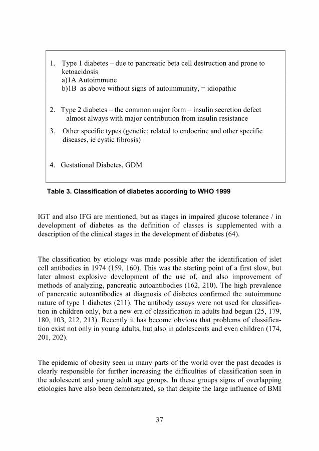
37
1. Type 1 diabetes – due to pancreatic beta cell destruction and prone to ketoacidosis a)1A Autoimmune b)1B as above without signs of autoimmunity, = idiopathic
2. Type 2 diabetes – the common major form – insulin secretion defect almost always with major contribution from insulin resistance
3. Other specific types (genetic; related to endocrine and other specific diseases, ie cystic fibrosis)
4. Gestational Diabetes, GDM
Table 3. Classification of diabetes according to WHO 1999
IGT and also IFG are mentioned, but as stages in impaired glucose tolerance / in development of diabetes as the definition of classes is supplemented with a description of the clinical stages in the development of diabetes (64).
The classification by etiology was made possible after the identification of islet cell antibodies in 1974 (159, 160). This was the starting point of a first slow, but later almost explosive development of the use of, and also improvement of methods of analyzing, pancreatic autoantibodies (162, 210). The high prevalence of pancreatic autoantibodies at diagnosis of diabetes confirmed the autoimmune nature of type 1 diabetes (211). The antibody assays were not used for classifica-tion in children only, but a new era of classification in adults had begun (25, 179, 180, 103, 212, 213). Recently it has become obvious that problems of classifica-tion exist not only in young adults, but also in adolescents and even children (174, 201, 202).
The epidemic of obesity seen in many parts of the world over the past decades is clearly responsible for further increasing the difficulties of classification seen in the adolescent and young adult age groups. In these groups signs of overlapping etiologies have also been demonstrated, so that despite the large influence of BMI

38
there are still groups of very obese young people who develop diabetes early and have clear signs of beta cell dysfunction (174, 203, 214).
Definition of type 1 diabetes and autoimmune diabetes – and LADA
Type 1 diabetes was previously called juvenile or juvenile-onset diabetes, which was a description of the fact that it had been observed that this disease was mainly diagnosed in children, adolescents and possibly young adults (215). The numbers who actually display ketoacidosis at onset of diabetes are fewer today, at least in high prevalence areas, which has been described, and attributed to greater knowledge of diabetes, and more of the affected youth are diagnosed before they reach such a decompensated state (189, 216). In a nationwide Swedish incidence study from 1992-93 of 15-34-yr-olds only 13% had ketoacidosis at onset of diabetes (28). The terminology has changed from juvenile diabetes, to IDDM after the 1980-85 WHO recommendation (206), and to type 1 diabetes in 1998 (26, 194) now, at least in research settings, often named classical type 1 diabetes (217).
With time it has been observed that (classical) type 1 diabetes, with or without ketoacidosis, with low or normal BMI, weight loss and insulin dependency direct-ly from the start is present also in adult patients (196). Despite sporadic such observations by many clinicians, the concept of classical type 1 diabetes has not been easy to establish in older ages. In diabetes epidemiology studies of the incidence of type 1 diabetes have focused on the childhood age groups of 0-14-yr-olds, sometimes stretching to 15-19-yr-olds (97, 148).
In the last two decades studies have emerged that investigate the proportions of patients with pancreatic autoantibodies among either those not insulin-requiring at diagnosis, or all patients with diabetes. The prevalences found have varied widely between studies, depending on the population studied (99, 103, 184, 200, 218, 219). Analysis of antibodies in larger cohorts of patients with diabetes that were not insulin requiring at diagnosis have lead to the identification of a number of patients that are antibody positive, without displaying the previously typical clinical picture of classical type 1 diabetes. Several terms have been used for this group, from antibody positive type 2 diabetes, and latent autoimmune diabetes in adults (LADA) to slowly progressive autoimmune diabetes in adults, (52, 200, 212, 220). According to the WHO recommendations from 1999 they are a variant of type 1, or autoimmune, diabetes (64). All this has lead to recent suggestions to rather use the term autoimmune diabetes for all such diabetes (221). This is further discussed under LADA.
39
Definition of type 2 diabetes and non-autoimmune diabetes
Type 2 diabetes is a diagnosis of exclusion, what is left when other more clearly defined conditions, such as autoimmune diabetes, MODY, or some other genetic or otherwise clearly specified types of diabetes, and secondary and gestational diabetes are excluded (64). See also Table 2.
Tools of classification
Different kinds of diabetes display different traits regarding heredity, treatment needs and patterns of complications, i.e. prognosis. In clinical practice general observations of clinical traits were the first tools used for distinguishing between diabetes types, present wherever there were patients with diabetes (192). Clinical traits are still used (184). In the 1970:s the knowledge of C-peptide in general evolved, and in the 1980:s it started to be used alone, and in combination with antibody analyses, for the classification of diabetes type mostly in research settings (103, 180, 222).
In the 1970:s and 1980:s the development of antibody analyses started a cascade of investigations to determine their prevalence, and thereby usefulness, in different patient populations, and also potentially in the general population, to predict the occurrence of autoimmune diabetes (99, 103, 104, 180, 182, 199, 200, 218, 222-224). The aims were to identify autoimmune diabetes and to predict beta cell destruction and insulin dependency. In research settings analysis of pancreatic antibodies has become standard for classification of diabetes. In affluent societies, especially in hospital settings and policlinics this is often, but not always, done, more often in adults than in children and adolescents. In primary care settings, however, and especially in less affluent localisations, this is far from the rule, and clinical basal criteria such as age and BMI have had to do as tools of classification. Antibodies are costly analyses, while C-peptide is a less expensive analysis.

40
Latent Autoimmune Diabetes in Adults – LADA
Definition – to be or not to be – that is the question? Antibodies in patients that had originally been diagnosed with type 2 diabetes were first described by Irvine and Bottazzo, and a decade later, when methods improved and new antibodies were discovered (182, 210, 225) the literature regarding autoantibody positive diabetes in adults increased (99, 103, 180, 199). The name LADA (Latent Autoimmune Diabetes in Adults) was suggested and has been widely used (22, 104, 163, 226). Mainly in Japan the term “slowly pro-gressive type 1 diabetes” has been used synonymously (227).
The first studies were clinical (103, 180, 199). They were followed also by more experimental investigations, and comparisons between LADA and both classical type 1 diabetes, and type 2 diabetes. One example was Botnia study of clinical and genetic characteristics of type 2 diabetes with and without GADA, describing both likenesses and differences between the groups (220, 228). A discussion soon developed regarding whether LADA was a form of type 1 diabetes, or of type 2, or neither (183, 212, 229-231). It also brought focus on the fact that the definition of LADA was not a solid consensus (24, 163, 212, 232). Some continued to describe it as type 2 diabetes with antibodies (200, 220), while others saw it more as a slowly developing variant of type 1 diabetes (229, 233), which has also been observed is the prevailing definition recommended by WHO (26, 194, 221). Suggestions have been made for joint definitions of LADA such as age ≥ 30 years, or ≥ 35 years, in combination with non-insulin-dependency at the time of diagnosis, but for how long has been different, 1 month? 3? 6 months? 12 months? Prevalence of pancreatic antibodies is a requisite, but which antibodies? Initially when that was the only one that could be analysed, it was ICA. Later GADA has been the more common to use, if not both, but some have analysed GADA + ICA. Some patients may be positive for 1 or more antibodies but not GADA (230, 232). In the UKPDS it was observed that the LADA group shared HLA and insulin genes with the type 1, but not with the type 2 group (234). Some investigators have seen differences on group level related to different levels of GADA titres and

41
suspected different subgroups of LADA (235, 236). These findings were confirmed by some, but not all investigators, not by the UKPDS for instance (237). Besides, the further division of LADA, which is already a subgroup, is not desirable either (230). All the indistinctness has lead back to a suggestion not to use LADA as a term, but to revert to calling all autoimmune diabetes just that –autoimmune diabetes (221).
Prognosis The need for a special definition of LADA patients was generated by the obser-vation that their prognosis was different. These Ab+ patients developed beta-cell failure, and become insulin dependent, more often, and more rapidly, than others who were not insulin dependent at diagnosis of diabetes (103, 200, 218). A few studies have also investigated the scope of complications developing in LADA patients (238, 239). Isomaa et al found that the frequency of microvascular com-plications were the same in both type 1, LADA and type 2 patients, while coronary heart disease (CHD) was as frequent among lADA as type 2 patients, but in the LADA patients CHD was related more to glycemic control than in T2D.
Treatment Most adults with autoimmune diabetes not insulin requiring at onset become so within 3-6 years (103, 200, 218, 226). The group is most often called LADA (104, 212), and has been identified as a suitable group for evaluation of new therapies in autoimmune, or, type 1 diabetes. It also constitutes a large group with need of evaluation of best therapy in its own right (22, 24, 213, 23). What is the best therapy for the LADA group has been, and still is, unknown (24, 233, 242).
Just as in type 1 diabetes there is an autoimmune attack on the beta-cells, resulting in destruction of the insulin producing cells and what is termed beta-cell failure, the inability to produce enough insulin to maintain glucometabolic homeostasis (221, 243). The effects of insulin on this autoimmune attack was first tested in animal studies of rodents (108, 244), and then in a pilot trial of ten Japanese LADA patients (245). Insulin has been suggested to be a possible candidate to promote halt or reversal of beta cell function due to several mechanisms (233). One is beta cell rest which experimental and clinical studies have demonstrated to be associated with increased residual beta-cell function, another is immuno-modulation (181, 246-248).
42
Atypical forms of diabetes
A number of conditions not primarily fitting into the usual descriptions of type 1 or type 2, or autoimmune vs non-autoimmune, diabetes have been described. The knowledge of them has increased recently, and was well summarized by Murphy, Ellard and Hattersley 2008 (195).
Monogenic diabetes Monogenic diabetes resulting from mutations that primarily reduce beta-cell function accounts for 1-2% of diabetes cases, although it is often misdiagnosed as either type 1 or type 2 diabetes. Diabetes diagnosed before 6 months of age fre-quently results from mutation of genes related to ATP-sensitive inward rectifier potassium channel, or sulphonylurea receptor 1 subunits of an ATP-sensitive potassium channel, and improved glycemic control can be achieved by treatment with high-dose sulphonylureas rather than insulin (195).
Individuals with familial, young-onset diabetes that does not fit with either type 1 or type 2 diabetes might have mutations in the transcription factors HNF-1α (hepatocyte nuclear factor 1α) or HNF-4α, and can be treated with sulphonylureas.
Extrapancreatic features, such as renal disease (caused by mutations in HNF-1β) or deafness (caused by a mitochondrial mutation) usually require early treatment with insulin (195).
Flatbush diabetes Black African Americans that came to hospital with typical ketoacidosis and diabetes, but later improved to a condition of non-insulin-dependent diabetes were first recognized in the New Your City suburb called Flatbush, thereby the name Flatbush diabetes (249). The phenomenon has been observed in several places in Africa, and other locations (9, 197). During stable conditions a type 2-like picture, with or without features of the metabolic syndrome, is displayed, but

43
when concurrent compromising conditions occur such as grave infection, or other concomitant disease, ketoacidosis and insulin dependence may develop, and later abate again. Presence of beta-cell dysfunction has been described (250). The phenomenon was recently described in a Caucasian in South America (251).
Secondary diabetes Secondary diabetes is the term used when diabetes is not the result of primary pancreatic or beta-cell related disease, or the metabolic syndrome combined with beta-cell dysfunction, but when some other condition is the cause. The classical example of secondary diabetes is after surgical removal of part of, or the whole pancreas, usually due to either malignant tumors or the late effects of advanced or repeated pancreatitis (252). The patient is left without any, or to small, production of insulin and becomes insulin-dependent, directly after the surgery.
These patients are characterized by somewhat lower risk of late complications of diabetes, perhaps due to the usually late time of debut of diabetes. They are also have a markedly enhanced sensitivity to administered insulin and are more sensitive to hypoglycemia, which may develop faster and be deeper, probably due to the lack also of alpha cells and of glucagon production (252). Another well known example of secondary diabetes are the cases affected by diabetes within the framework of another endocrine disorder, often Acromegaly or Cushing´s syn-drome, but it also happens in conjunction with other neuroendocrine tumors, and their treatment. Both pancreatectomy and Somatostatin analogs may interfere here. Except for the cases related to pancreatectomy, who are more prone to insulin sensitivity, these cases related to other hormonal disturbances are usually related to insulin resistance, are more common in patients with family history of T2D, and resemble type 2 diabetes, but they often resolve if the basic endocrine disturbance can be cured (253).
Gestational diabetes Gestational diabetes is when diabetes is diagnosed for the first time during preg-nancy. Many of these patients return to normal plasma glucose values after delivery, but again a majority of the women who have experienced gestational diabetes develop diabetes during their life span. About 10% usually do so within the first year postpartum, and earlier studies demonstrated cumulative incidences of up to 60% after ca 15 years. More recent studies have found about 35% cumulative incidence of diabetes after 10-15 years. It is possible that modern inter-vention policies can improve this figure. GDM is related to obesity and the

44
majority of the diabetes they develop is type 2. Investigations of autoimmunity in populations with GDM have demonstrated that about 5-10% (?) are antibody positive and develop type 1 diabetes either directly in connection with the GDM, or later (254).
New cases of secondary and gestational diabetes were excluded from the Kronoberg incidence study.

45
Beta cells and beta cell function
The beta cells of the pancreas The beta cells are situated in the so called islets of Langerhans in the pan-creas. Their embryonic origin was long believed to be ectodermal, related to the neural crest, but this hypothesis has lately been refuted (255). The prevailing opinion now is that both the exocrine and endocrine parts of the pancreas stem from endoderm. This has clinical interest since one of the lines of development of better treatments in diabetes deals with trials to develop differ-rentiated, insulin producing beta cells from embryonic stem cells (256). The beta cells constitute the majority of five types of endocrine, peptide hormone producing cells dispersed in tiny islets throughout the pancreas (256). Endocrine means their site of action is far removed from the site of secretion. Insulin is a hormone secreted from the beta cells which decreases blood glucose levels by facilitating utilization of glucose in the peripheral tissues, especially the skeletal muscles and the liver. Another cell type are the alpha cells that produce glucagon. The effect of glucagon is the exact opposite of the effect of insulin. The effect of glucagon is to maintain blood glucose levels during fasting by the mobilization of glucose from glycogen stores in peripheral tissues such as muscle and liver (252).
Beta-cell function – a key issue
Defects in beta cell function
Interest in beta-cell function and C-peptide has risen considerably the last years (49, 257). New insights from basic and clinical research suggest that the common denominator of all forms of diabetes is the lack of an adequate functional beta-cell mass (absolute in type 1 diabetes and relative in type 2 diabetes) and thereby insulin insufficiency leading to hyperglycemia (49, 256, 258). There is an in-creased awareness in general of the defect in beta cell function present in many patients with type 2 diabetes (214), and also an awareness that different types of treatment may affect beta cell function differently (24, 259, 260).

46
There has long been an awareness of the large defect in beta cell function that is often present at diagnosis of classical type 1 diabetes in children and adolescents (261). This also includes the awareness that potentially remaining beta cell function practically always has decreased further after diagnosis, despite start of treatment with insulin, and even if a remission period occurs (186, 261, 262).
Measurement of C-peptide became more common in research settings from the 1970:s and 1980:s onwards. Since then the focus has increasingly been on trying to preserve what is left of the beta-cell function at diagnosis, in both children and adults (245, 247, 248, 263, 264). The fact that more of the affected young type 1 patients are now discovered earlier in the process does not seem to have led to much larger proportions of patients with long term remaining residual beta-cell function, at least not until 2008 (258). Prediction in the general population is not impossible, but complicated. Since yet there is no good treatment or cure to apply before onset of autoimmune diabetes it is not considered ethical to screen before diagnosis other than when direct participation in trials attempting prevention is offered (181).
Preservation of beta cell function
The main reasons why preservation of beta cell function is the goal of the efforts are the following: There are some data suggesting that higher remaining levels of beta cell function seem to involve delayed or inhibited development of com-plications, and/or a diabetes more easily controlled. The main example of this is the result of the DCCT study, where even very modest improvements in preservation of beta-cell function, measured as C-peptide, were associated with decreased proportions of patients affected by retinopathy, and lower frequency of hypoglycemia (30). Hypoglycemia is the main obstacle in achieving the desired goal of better metabolic control, measured by levels of HbA1c. Actually both the risk of hypoglycemia, and the observation that near-normal levels of blood glucose seemed to be preferable for patients and for achieving desired clinical goals (then in the short term), were observed already very early on in the beginnings of the use of insulin treatment (11). To be able to decrease the frequency of hypoglycemia would not only achieve a safer treatment situation per se, but also enable other goals to be better achieved, such as lower frequency of vascular complications. The close connection between level of HbA1c and prevalence of complications, both micro- and macrovascular, is well established in both types of diabetes (30, 32, 39, 312, 266). Another goal intensively sought after in studies of autoimmune diabetes is the possibility to halt the autoimmune attack on the beta cells, with the result of either halting, or most-wished-for, reversing the impairment in beta cell function (267).
47
C – peptide
C-peptide – definition C-peptide was first described in 1967 by Steiner & Oyer (268). Early investi-gations attempted to define a physiological role for C-peptide but failed to do so. The discussion has continued as to the significance of C-peptide as a potentially biologically active endogeneous peptide hormone, and some interpret newer studies to indicate this (269). Nevertheless, quite early the interest in C-peptide became focused on its use as a marker for beta-cell secretory function (270).
C-peptide is secreted from the pancreas into the circulation in equimolar concen-trations as insulin, along with proinsulin and split proinsulins (intermediates) (268, 271). C-peptide is more stable, with longer half life, and by measuring this as a substitute for the more unstable insulin also avoids the problem of distin-guishing between endogenous and exogenous insulin, which the insulin assay cannot (271). These are the reasons why C-peptide has been used increasingly as a measure of the endogenous insulin production, usually termed beta-cell function.
C-peptide – function and use C-peptide has been identified as the appropriate outcome measure of residual beta cell function in intervention studies of autoimmune diabetes (272). It has for long been identified as the most important marker of failure or success in trials of type 1 diabetes in children, and later also of LADA, thereby in all autoimmune diabetes (263, 273). For some time now C-peptide has also emerged as one of the main goal parameters in trials and treatment of type 2 diabetes (49, 257, 260). This has been emphasized further by the new classes of drugs for treatment of type 2 diabetes, first the thiazoledinediones, and then the incretin enhancers and mime-tics, which in animal studies have demonstrated favourable effects on beta cell function, and the same has been suggested also in some human studies recently (259, 260, 274, 275).

48
C-peptide and classification When C-peptide was first discovered using it for classification was examined by some investigators (222, 276), but the use of autoantibodies in research settings soon overshadowed the use of C-peptide only, although many, but not all, investigations of different groups have analyzed both antibodies and C-peptide (197).

49
Metabolic control and HbA1c
Metabolic control Metabolic control, also named glycemic control, means to restore the metabolic derangements inflicted by the diabetic state, to, or to near, normal levels (11, 36). That this was desirable was observed already during the first year of the era of insulin treatment (11). Then the primary goal was the survival of the patient, the avoidance of ketosis and coma, very literally, and to restore in the short and middle term perspective the physical and mental strength to the patient, so that he or she was enabled to resume at least parts of the life lead before being stricken by diabetes (11, 12). The goal of clinical treatment is very much the same today, with the addition of keeping the levels of blood glucose at or near normal levels in order to avoid the gruesome long term complications now well known to threat the well-being and even survival of the patient in the long term perspective. As has been mentioned the development of these long term complications are now well known to be closely related to the levels of plasma glucose over time (39, 265). It has become evident the past decades that the nearer normal the mean levels of glucose are over time, the lower is the proportion of patients on group level that acquire the vascular complications (32, 278, 279).
HbA1c It is quite unique for a disease, as is the case with diabetes, to have a marker that allows the patients themselves and the care givers to closely monitor and evaluate how the levels of plasma glucose have been for a number of weeks back. This is the case with HbA1c as a marker of glycemic control in diabetes, and work is still ongoing to improve these means (280). With repeated measures of HbA1c it is possible to estimate the over all success or failure of the whole combination of treatment of a special patient. With this knowledge the advice and care offered to each patient can be individualised, regarding both recommendations of diet and physical exercise, as well as regarding relations to psychological and psychosocial stress, and, not least, suitable glucose lowering pharmacological treatment . The measure HbA1c can be used not only to aim at lowering levels in young patients to avoid future complications, but also to identify elderly patients with frequent low glucose levels and increased safety risks, ie hypoglycemia, to enable adjustments

50
of treatment (281). Recently a worldwide transition to harmonized HbA1c measurements has commenced, and Sweden joined this in the fall of 2010. The new standard is called IFCC, and there is a webb-based site were figures can be easily converted from the previously most used method in Sweden, Mono-S, to the new IFCC values, HbA1c.nu.
HbA1c in research
Last but not least, HbA1c is a very valuable parameter in research, both treatment trials and other kinds of research (39). In order to be able to compare the results of different studies it is necessary to have universal measurements of HbA1c. This has not been the case, but algorithms have been developed for overcoming this, and conversion to DCCT-standard (as in the DCCT trial) has been praxis, and algorithms for this developed (282). In the future however, also in research articles from now on the IFCC standard HbA1c values will be used.

51
Interventions in autoimmune diabetes
Prevention studies It has only been realistic to identify about 10% of those affected by autoimmune, or type 1 diabetes, namely is those with type 1 in a first degree relative, or several affected in the family (213). Also type 1 was mostly regarded as a juvenile disorder, so preventive measures have been focused on the groups of relatives of young patients with T1D that could be identified, and who were also motivated to participate in prevention trials, in relation to the size of their risk (50). Without success some large intervention trials in populations have also been conducted, as the END-IT, a multicenter trial of Nicotinamide to school children in Europe, aiming to reduce the proportion affected by type 1 diabetes (181, 283).
Early intervention studies Since about 90% of the affected by type 1 diabetes do not have close relatives with the disease, and have not been possible to identify, and there is still no known intervention that could change the course of the development of the disease, for decades there have been trials of interventions that are initiated at or shortly after the onset of T1D, parallel with the necessary insulin treatment (181, 217).
After the identification of islet autoantibodies in the mid 1970:s confirmed the autoimmune basis of type 1, trials of non-specific immunomodulating agents such as prednisone, and especially cyclosporine were conducted. Some effect could be demonstrated on beta cell function, but it was not long lasting, but ended when the therapy was ended, and the side effects in this situation were considered not acceptable (181, 263).
In the 1990:s focus moved to the adult LADA group which was identified as a suitable group for trying out interventions in autoimmune diabetes (22). The reasons for this were not only that it seemed more ethical to try a new intervention
52
in adults in general, before conducting trials in children, but also the fact that adults have been demonstrated to usually have larger remaining beta cells masses and functions at the time of diagnosis, both in classical type 1 diabetes, and in LADA (242, 261).
Studies from Japan
A lot of interest and research regarding autoimmune diabetes has come from Japan, despite the lower incidence rates of type 1 diabetes reported in children. The large size of the population means still many are affected. Type 2 diabetes affects many Japanese today (143). In early studies of NOD mice, rodents gene-tically designed to develop autoimmune diabetes, it was demonstrated that treat-ment with insulin could delay or postpone the development of diabetes (108, 244). Further it was demonstrated in a pilot study published in 1996 by Kobayashi that small doses of sc insulin to ten LADA patients achieved the same in humans (245). Later the Tokyo Study (TTS) was initiated (284).
After the pilot study by Kobayashi 1996, the first reports of treatment trials in LADA, especially designed for LADA patients, were the preliminary reports from the Tokyo Study (TTS) in 2002 and 2003, indicating that treatment with insulin was more favourable than SU (284, 285). Insulin was not only a means of keeping down glucose levels, which might lead to down regulation of the immune attack on the beta cells by way of so called beta cell rest (181). With other substances, with some side effects, such as diazoxide, beta cell rest has been demonstrated to have favourable effects on the preservation of beta cell function in newly diagnosed young T1D patients and children (248, 286). Insulin has also been described to have directly immunomodulating properties (181)
A Cochrane review
In 2007 a Cochrane review of treatment in LADA was published (24). Of the studies screened only 8 could fulfill the inclusion criteria. It was really only the Kobayashi pilot trial, the preliminary data from TTS and a Chinese small inter-vention study that were randomised trials designed for LADA patients. One of the studies was UKPDS, of newly diagnosed type 2 patients randomised to insulin vs SU, and where antibodies had been analysed along the way (200). Another was a Swedish study of type 2 patients investigating the effect of removal of SU (287), and one was a phase II GAD vaccination study (288). The general conclusions were that the definitions of LADA differed and the heterogeneity between studies made any conclusions unsure, that it might be that SU should be avoided but from the evidence available it was not possible to recommend any other treatment before the rest (24). The final result of TTS was published in 2008 (52).
53
Body Mass Index – BMI and Obesity
BMI as risk factor and obesity epidemic Obesity is a well known risk factor for diabetes, especially type 2 diabetes (289). That there are close relations between body weight, and build, and diabetes control, was well known already early in the 20th century, when it was observed that substantial weight loss might ameliorate diabetes, if it was not of the insulin-dependent-for-survival-type (11). It was noted during the World Wars in Europe, when food was scarce and there were years of rationing, that the incidence of diabetes declined, something that was later documented in Norway (84). The influence of weight and possibility of weight management for treatment was mentioned in the 1980 WHO recommendations regarding diabetes (63).
Besides description of a diabetes epidemic in the later parts of he 20th Century and into the 21st Century, an epidemic of obesity is also described, and that the appearance of the two are parallel, with obesity preceding diabetes (33, 176, 290). The term Diabesity has even been coined to describe the phenomenon (46). The causes are mainly attributed to the more sedentary Western lifestyle spreading not only within Western countries, but also increasingly to and within the developing parts of the world (46, 88, 290).
The epidemics of obesity and diabetes are also reported to occur in both adole-scents and children. The pediatric epidemic was first observed and described in the US, but reports have then accumulated from many parts of the world, as Europe, Australia and New Zealand, and Asia (176, 291-294). If young people are affected by diabetes, and now type 2 diabetes, at earlier stages in life, the time they are affected by hyperglycemia and its negative effects increases, and compli-cations will develop earlier as well. This is already seen, as in Australia (68).

54
Even if obesity is not the only risk factor for developing type 2 diabetes, it is an independent and important one, and when examining a population of patients with newly diagnosed diabetes it is strongly indicated to include measurement of BMI, both for the information per se, and to create possibilities to relate it to other factors of interest (214, 295).
Measurements of body weight and obesity
Normal weight in adults varies with sex, height and age, but instead of using complicated tables of criteria, overweight and obesity are recommended to be estimated by BMI, body mass index, in kg/m2. This is the established measure-ment recommended for overweight and obesity by WHO. It is easily obtained and relatively reliable (296). Later measurements of waist hip ratio (WHR) and waist circumference (WC), have been demonstrated to sometimes correlate even better to cardiovascular risk. The correlation between BMI and WC regarding asso-ciation with incident diabetes is however very good (297).
In all our studies the measurements of weight and height for BMI were recorded by a nurse, at the PHCC:s and hospitals, and not self reported, in order to increase the objectivity of the measurements.
55
Autoimmunity and autoimmune diseases
Autoimmunity in general It was recognized in the early 1900:s that our body’s defence mechanisms could recognize organs or tissues as foreign, and use their usual program to attack and destroy them. This was met with scepticism until in the 1950:s antinuclear (ANA) and anti-thyroid antibodies were demonstrated in patients with SLE and thyroiditis, and the concept of “autoimmunity” was accepted, auto meaning “self” (298). Autoimmune diseases (AD) are a heterogeneous group of diseases in which an adaptive immune response against self antigens causes damage to an organ or tissue (299). AD are characterised by an infiltration of the target organ with mono-nuclear cells (181). A core issue is whether cytotoxicity or apoptosis are most responsible for the cell death occurring in autoimmunity (300). The cytotoxicity is described to be either direct by autoreactive CD8+ T-cells, or by indirect cytotoxic mechanisms via proinflammatory cytokines from the infiltrating cells. Abnormal antibody production is the basis of specific endocrine diseases. The deranged immune system produces autoantibodies that react with the target organs at different levels, but usually at the receptor, where they can act on different levels of the hormone producing cell systems (301).
The beliefs have varied from time to other as to whether the so called autoanti-bodies are causative factors in the development of autoimmune diseases, or merely markers of the on-going process(es). From a belief of causation the perspective has developed more towards a marker perspective (181), but it may be that the last of this is not yet known.
Autoimmune diseases Autoimmune diseases as a group represent a diverse collection of diseases in terms of demographic profile and primary clinical manifestations. The commonality between them, however, is the damage to tissues and organs that arises from the response to self-antigens (299). Epidemiological studies have shown that genetic

56
as well as environmental factors trigger the development of autoimmune diseases. The genetic susceptibility alone would not be sufficient for disease expression (181). There is evidence of some common genetic and environmental risk factors across diseases. Autoimmune diseases are generally thought of as being relatively rare, but their effects on mortality and morbidity are quite high (299). Auto-immune diseases are among the leading causes of death among young women in ages below 65 years, at least in the United States (302). They are reported to affect at least 3-5% of the populations in Western countries (300).
Endocrine autoimmune diseases can be roughly divided into cell- and humoral-mediated diseases. Cell-mediated, or organ-specific, autoimmune disorders include type 1 diabetes, which is in many ways the best known, and the “model” of these diseases, and many believe that if the answer to how an islet beta cell is destroyed all the other tissues affected in other autoimmune diseases will benefit as well (181, 298).
The more common autoimmune diseases
The autoimmune diseases discussed in this thesis, and related autoantibodies, are listed in table 4. Other autoimmune disorders with loose associations to other autoimmune conditions are vitiligo of the skin, ulcerative colitis, Crohn´s disease, Myasthenia Gravis, vasculitides, and others.
Autoimmune polyglandular syndromes
The names are given to some common combinations of AD. The most common combinations are Autoimmune Thyroid disease (ATD) + Pernicious Anemia (PA). The next is T1D + ATD +/- PA. These, in combinations or one each, together with primary adrenal insufficiency (PAI) is not very common, but among T1D it occurs in 1-2% (303). On the other hand up to 40% of those with PAI are also affected, sooner or later, by T1D. Another common combination, is T1D + Celiac disease. This is perhaps the most frequently examined association of T1D and AD, especially in children, where it has been examined in many countries, but also in adults (304, 305). In adults some large studies regarding connections between T1D and PA have been conducted in Belgium, where up to 20% of T1D patients were positive (306).
The rarest combinations are those associated with the so-called APS I, with debut usually in the first two decades of life, and especially three unusual manifestations: chronic mucocandidiasis, hypoparathyroidism (hypocalcemia), and primary adrenal insufficiency. Alopecia is not uncommon, but they seldom have T1D (307). APS 1 is an inherited autosomal recessive disorder (308).

57
APS II is the most common of the APS. It is diagnosed when at least two of the following three are present: Autoimmune thyroid disease, Type 1 diabetes and primary adrenal insufficiency. Other potentially related disorders are: celiac disease, pernicious anemia, alopecia, vitiligo, serositis, hypogonadism, myasthenia gravis. Some authors have attempted to subdivide APS II into APS II and APS III, depending on which combinations of AD are present. However little information or further understanding is gained from this (308).
Epidemiology of Autoimmune diseases
In a recent summary the estimated incidences of autoimmune diseases other than T1D (all per 100 000 and year) varied from less than 1 per 100 000 person-years (e.g autoimmune primary adrenal insufficiency, PAI,=Addison´s disease, chronic active (autoimmune) hepatitis, CAH and myasthenia gravis) to more than 20 per 100 000 and person-years (Adult-onset Rheumatoid Arthritis, age >16 yrs 23.7, and Thyroiditis/Hypothyroidism, age>19 yrs 21.8 (299). Adding Graves disease/ Hyperthyroidism´, 13.9, means a yearly incidence of 35.7 for autoimmune thyroid disease (ATD). The prevalences of the different diseases are also affected by their different tendency for chronicity, leading to great differences in the prevalences.
Autoimmune disease and type 1 diabetes It is well known that autoimmune type 1 diabetes is associated with increased frequencies of a number of other autoimmune conditions (309). Actually as soon as one other such condition is present, some call it an APS (see above, type II). There have been many investigations examining these interrelationships. We did not attempt to draw a complete map of diabetes related autoimmune conditions. Our aim was to examine if there were increased prevalence of antibodies ordi narily related to inflammatory rheumatic diseases, or vascular antibodies also implicated in vascular disease. The reasons were an impression that musculo- sceletal disorders seemed common among the patients, that there were some with concomitant rheumatic disease, and that most investigations have focused on either presence of antibodies or clinical conditions (310, 311). Both musculo-sceletal disorders (MSD) and hepatogastric conditions, apart from celiac disease, receive a lot less attention than other complications of diabetes in clinical practice.

58
Complications of diabetes
Complications in general If the patient survives the short term perspective the real challenge and threat of the disease of diabetes are the long term complications. The most common complications of T1D usually described are the microvascular retinopathy, nephropathy and neuro-pathy (41). Also common are macrovascular complications such as myocardial infarct-tion, stroke and peripheral arterial insufficiency. Retinopathy has long been identified as the most common complication. Only a few decades ago blindness was not an unusual consequence of type 1 diabetes (45). Also the other most feared, lethal, compli-cation of nephropathy, has declined in prevalence (312). Both improvements are associated with improvements in the treatment. Increasing numbers of injections of insulin per day, and development of the insulins and insulin treatment, have enabled patients to keep a tighter control of their glucose levels (312). The DCCT (the Diabetes Control and Complications Trial) landmark study first clearly demonstrated the connection between more intensive insulin treatment, and lower levels of HbA1c, and lower prevalences of a number of complications (32, 313). As a consequence also mortality in general has declined in type 1 diabetes (283), but despite this there is still quite an excess mortality, in young persons with diabetes, even in societies with excellent health care systems (19, 108, 283).
Complications from the connective tissues – musculosceletal disorders Besides the life threatening complications also other less serious ones occur in the long term, as from the musculosceletal system, or connective tissues. The most common of these musculosceletal disorders (MSD), are limited joint mobility (LJM), flexor tenosynovitis (FTS) and shoulder capsulitis (SC). MSD from the upper extremity are described to be most common (314), (315, 316).
Rheumatology and diabetes – common ground?
While complications from the connective tissues in diabetes are being increasingly recognized (317, 318), the field of rheumatology has developed, regarding diagnostic possibilities with a number of antibody assays, and the especially regarding treatment

59
possibilities. The last decade new so-called DMARDS (Disease Modifying Anti Rheumatic Drugs) that are directly immunomodulationg, the TNF-α-blockers, have come into clinical use (319).
Complications and autoimmune conditions of the gastrointestinal system Complications from the gastrointestinal system have also been increasingly re-cognized. These include a number of problems and symptoms, from diarrhea to obstipation, some related to gastric neuropathy, gastroparesis and decreased mobility of the intestines, but also connections with autoimmune conditions such as celiac disease and pernicious anemia (320-324). Type 1 diabetes is mentioned tio be connected to autoimmune hepatitis, but there are not many investigations in that area (325).
Vascular conditions and antibodies, including ACL and ANCA Anticardiolipin antibodies are a type of autoantibodies directed to antiphospholipid autoantigens. Clinically they have been related to both arterial and venous thrombo-embolism, and also to recurrent pregnancy loss (326). The conditions, and corre-sponding antibodies, occur either as a separate syndrome, the antiphospholipid syn-drome, APS, or related to systemic lupus erythematosus, SLE (327). Since ACL are related to vascular pathologic conditions, and many complications of diabetes are vascular, a few investigators have studied the prevalence of ACL in groups of patients with diabetes, to search for possible associations. It has been associated with retino-pathy and with diabetic foot ulcers (328), (329). There have been a few reports of ANCA in association with diabetes, suggesting they might be associated with vascular complications (330).
Complications of pregnancy Complications of pregnancy, such as preeclampsia, are more common in women with diabetes than in the general population (331). One study has described an association between ACL and complications of pregnancy in type 1 diabetes (332).

60
Long duration of type 1 diabetes
Autoimmunity after long duration of type 1 diabetes There are a number of studies of the prevalences of different antibodies at and after the time of diagnosis of diabetes (103, 188, 333) but the conditions after long duration of type 1 diabetes are less known.
Residual C-peptide and complications Most patients affected by type 1 diabetes lose all measurable C-peptide within 1-3 years from time of diagnosis of diabetes (103, 226, 261). In the DCCT it was demonstrated that even small remaining amounts of C-peptide production could be beneficial, as it was associated with lower frequency of retinopathy, and of hypoglycemia (30). Hypoglycemia is the main obstacle when trying to achieve a near normal level of glycemic control, in the treatment of insulin dependent diabetes, especially autoimmune diabetes where also part of the counterregulatory system, with glucagon from the alpha cells of the pancreatic islets, are affected by the autoimmune process (256).
61
Objectives of the thesis
The objectives of this work was To prospectively estimate the incidence of diabetes, type 1 and type 2, apply-
ing modern criteria for diagnosis and classification, in the defined population of our region, the county of Kronoberg, primarily in all adult ages, 18-100 years, and with retrospective data from the childhood groups for comparison.
To describe the levels of C-peptide and BMI and their relationship to antibody status and age, and their accuracy as tools for classification of diabetes type, in adults of all ages, 18-100 years, at diagnosis of diabetes, in a defined popula-tion.
To evaluate if early introduction of insulin treatment to patients with LADA, Latent Autoimmune Diabetes in Adults, lead to better preservation of beta cell-function or metabolic control, compared to conventional treatment.
To describe the prevalence of pancreatic and other autoantibodies and corre-sponding clinical conditions and complications, and their relation to residual C-peptide, in patients with long duration of type 1 diabetes.
62
Methods
Subjects and population, and ethical issues
Health Care Organisation
In order to receive a true estimate of the incidence we studied the incidence of diabetes in the whole population of Kronoberg. All the 25 public health care centers (PHCC) participated and the two hospitals, Växjö Central Hospital and Ljungby Community Hospital, in the region of Kronoberg participated. At the time of the data collection private practices were very scarce and small in Kronoberg. The practices that might diagnose diabetes were contacted and agreed to participate, but reported no new cases during the study period.
Population at risk
The population at risk equalled the population of Kronoberg County. Statistical figures of all inhabitants, age-groups and sex, were collected from the Kronoberg Regional Administration, who keep continuous, yearly census. Figures for the four years of the study period were obtained, and means of the four years were used as denominators in the calculations of incidences. Figures were also divided into age groups, and sex groups, where relevant. The mean population of Kronoberg was 177 102 inhabitants during the study period. The mean adult population aged 18-100 years was 138 192 inhabitants. The variations between the years was small.
The method of opportunistic screening (334, 335) was applied meaning that all adult patients were tested during routine or acute contact with the medical care system, irrespective of reason for consulting.
Geographical considerations
Kronoberg is situated in a forest belt in Southern Sweden, with the two largest communities being Växjö, with 60 000 inhabitants, and Ljungby with 15 000 inhabitants. The rest of the population mainly lives in small rural commu-nities.

63
Age distribution of the population
Kronoberg is known to have among the oldest populations in Sweden, only surpassed by the county of Halland (situated directly west of Kronoberg). A comparison was therefore made with the whole population of Sweden at the time of the study, but there were no significant differences to be found between the age distribution of the two populations, and in this respect our data ought to be representative for Sweden. It is possible that comparison with only the three largest cities in Sweden might show differences, since they may have slightly younger populations, but with Sweden as a whole, there was no difference.
Our aim was to investigate the situation for adults, but after presenting data at an international meeting questions of comparisons with the pediatric age groups were raised, and for this purpose data from age groups 0-17 years could be included retrospectively, since there is only one pediatric department in Kronoberg where all pediatric cases of diabetes are known and treated. Combined with the thorough control in the Swedish society with unique personal registration numbers, at the time practically only public schools, where school health care is provided by the same pediatric department, and a national mortality register vouches for 100% ascertainment in that group.
Patients
The main incidence study was conducted during three years, from May 1st 1998 to April 31st 2001. All adult patients with a new diagnosis of diabetes in Kronoberg, in all the health care centers and at the departments of Internal Medicine in the two hospitals, were registered. Patients who receive a diagnosis of diabetes in any other department are automatically referred to either the departments of internal medicine, or a primary health care center.
All new cases of diabetes mellitus aged 18 yrs or older (excluding secondary and gestational) were registered. (Study I and II).
For Study III patients were recruited from those being positive to one or both of the antibodies ICA and/or GADA among the newly diagnosed in the Kronoberg area during the incidence registration for study I. Simultaneously, the Diabetes Laboratory at BMC, Lund was, and still is, offering to perform analyses of antibodies and C-peptide for better classification of diabetes to the health care facilities in the greater Lund region, and recruitment had started there. Patients

64
were offered to come for a screening visit to the main research centers in Lund or Växjö for further information and evaluation of exclusion and inclusion criteria.
For Study IV the patients were all recruited from those attending the outpatient Diabetes policlinic of the Department of Internal Medicine at the Central Hospital of Växjö, a secondary institution were almost all adult type 1 patients, aged ≥ 18 years, in the larger eastern part of Kronoberg are treated. All were aged 19-31 years at the time of data collection. Prevalences of antibodies and clinical complications were compared between those with shorter (3-10 yrs), intermediate (11-20 yrs) and long duration of type 1 diabetes.
Ethical issues
All the studies were approved by the Ethical committee of the Medical Faculty of Lund University. All patients received oral and written information regarding the studies, and provided informed consent. The studies were all performed in accordance with the Declaration of Helsinki.
Collection of data Blood samples were collected from 1626 of the 1666 newly diagnosed patients, 97.6%, either fasting or non-fasting, but with information of which, for further analysis. All blood analyses were centrally analysed at the Diabetes Laboratory, BMC, Lund University Hospital.
A questionnaire with clinical data was filled out by the treating physician or diabetes specialist nurse. This included body measurements, information regarding present and previous cardiovascular disease and/or risk factors, and some socio-economic information. The first three blood glucose measurements responsible for the diagnosis of diabetes were also recorded. (Study I and II)
Medical charts were reviewed for adults classified as type 1 to evaluate presence of acidosis and/or ketonuria, and of insulin treatment from onset, defined as within 4 weeks of diagnosis of diabetes. (Study I).
Data regarding the newly diagnosed 0-17-yr-olds was obtained retrospectively from the Pediatric Department. Some but not all children and adolescents had been tested for antibodies and C-peptide. (Study I)

65
The patients in the LADA study attended initial and yearly visits at the main research departments (Växjö and Lund) and many were followed there also for the regular follow-up visits every three months, with collection of blood samples and anthropometric data, and information regarding adverse events and decisions of changes in treatment. Those who were not followed at the main centers continued at their health care center, and came for yearly visits including glucagon tests for collection of samples of stimulated C-peptide. (Study III).
Blood samples for analysis of C-peptide and autoantibodies were collected, non-fasting, so C-peptide measurements are random, RCP, at regular visits for ordinary surveillance of diabetes at the out patient policlinic. Clinical information was collected by chart review. (Study IV).
Anthropometric measurements
Measurements for height and weight for calculation of BMI were taken and recorded by a nurse, and not self reported by the patients. Age was calculated by subtracting the date of diagnosis from date of birth.
Diagnosis and classification of diabetes, and study designs
Diagnosis of diabetes
Diabetes was diagnosed according to WHO/ADA criteria if FPG was ≥ 7.0 mmol/ L (equal to fasting blood glucose, FBG, ≥ 6.1 mmol/L, ≥ 126 mg/dl) at least twice, or if any random value was ≥ 12.2 (capillary) or ≥ 11.1 (venous) mmol/L (equal to FBG ≥ 11.1 mmol/L, ≥ 200 mg/dl) (64, 194). If the diagnosis was not confirmed by further testing, they were not included in the study. These cases were very few compared to those included. (Study I, II, II).
Classification of diabetes Patients positive to any of the antibodies ICA or GADA, or with a C-peptide value < 0.25 nmol/L were classified as type 1 diabetes, the last criteria only used in study 1. Patients without antibodies and with C-peptide 0.25 nmol/L were classified as type 2 diabetes (64). (Study I).
In study II the classification grounds were autoimmune or non-autoimmune dia-betes. Those positive to any of ICA and/or GADA were designated auto-immune, and the rest non-autoimmune patients with diabetes (221).

66
In study III only autoimmune patients were eligible for screening. Adult patients, aged ≥ 18 years, positive to at least one of GADA and/or ICA, and not insulin-dependent at the time of diagnosis of diabetes were eligible, that is they were classified as LADA patients (221).
In study IV all the patients had received their diagnosis of diabetes ≥ 3 years before study entrance, many several years back, and at the Pediatric department, and when other diagnostic criteria for blood glucose levels were used (2), but all were treated with insulin in multiple doses since ≥ 3 years, most had no residual C-peptide, and they were all dependent on insulin for survival.
Study designs The incidence study is a large, prospective, population-based cohort study with a high degree of ascertainment. It was performed during three consecutive years to avoid influence of particular conditions related to a calendar year, and also to increase the number of affected during the study period. (Study I and II).
Study IV is a retrospective cohort study, but of a smaller, defined group.
Study III was in practice an open randomized parallel controlled intervention study, with follow-up for three years. The intention was to randomize all the patients according to a prearranged system with preprepared closed envelopes in blocks of 8, and the majority of the patients were, but strict randomization was not possible because some patients simply refused insulin treatment before it was necessary. They were referred to the control group.
Intervention, outcome measures and adverse events
Intervention
The effect of early insulin treatment, (Intervention group, I) compared to treatment with diet only, or diet + oral hypoglycaemic agents, OHA, and insulin only when clinically indicated, that is “usual care” (Control group, C), on residual beta-cell function, measured by glucagon-stimulated C-peptide, and metabolic control,
67
HbA1c, was investigated in LADA patients during the first three years after diagnosis of diabetes. All patients were screened at one of the two main research departments either at Lund University Hospital, or Växjö Central Hospital, but thereafter they could either have their regular 3-monthly visits including blood samples for analysis of HbA1c, C-peptide and autoantibodies, and evaluation of treatment, at their primary health care center, or at one of the research depart-ments. All patients were evaluated at one of the two main centers, including with glucagon stimulation tests for GCP, at baseline, and after 12, 24 and 36 months. (Study III).
Outcome measures and adverse events The primary outcome variables were residual beta cell function, measured by glucagon-stimulated C-peptide, GCP, and metabolic control, evaluated by HbA1c, after 12, 24 and 36 months. Secondary outcome variables were adverse events in the form of major hypoglycemic events, defined as plasma glucose < 3.0 mmol/l or the need of another person to resolve it, and weight change during the study period. (Study III.)
Laboratory methods
Plasma glucose
At the primary health care centers capillary blood was analysed using the Hemo-Cue Glucose System (HemoCue AB, Angelholm, Sweden) and at the hospital departments blood glucose was checked by venous sample. All pathological values were confirmed by further testing meaning several PG samples, fasting and not fasting. At the time of the incidence study blood glucose values were used in Sweden, and then plasma glucose was introduced. Initially, as in Paper I we gave both values, and the mg/dl, but in the later work all the glucose values are given as plasma glucose. (Study I, II, III, IV)
C-peptide analyses
C-peptide was analysed by radioimmunoassay using a commercial kit (MD315, Euro-Diagnostica AB, Malmö, Sweden). Total variation (sum of intra and interassay variation) was 7%, reference range 0.25-1.0 nmol/L, detection limit 0.13 nmol/L. All C-peptide analyses were performed at one and the same central laboratory at the Department of Clinical Chemistry, Lund University Hospital, Lund, in cooperation with ass prof Anders Isaksson.
(Studies I, II, III, IV).
68
Analyses of pancreatic autoantibodies ICA and GADA
Islet cell antibodies, ICA, were analysed with an immunofluorescence assay (the detection limit was 9 JDF-U, sensitivity 100% and specificity 88%. Glutamic acid decarboxylase antibodies, GADA, were analysed with a radioimmunoprecipitation assay, (the lower reference limit was an index of 0,08 corresponding to 21.2 WHO-U/ml, sensitivity 70% and specificity 100%. Both analyses were standardized according to the Diabetes Antibody Standardization Program (187, 336). (Study I, II, III, IV.) All the ICA and GADA analyses were performed centrally at the Diabetes Laboratory, BMC, Lund.
Analyses of other autoantibodies
The rest of the autoantibodies were analysed at the Department of Clinical Microbiology at the Central Hospital in Växjö, in collaboration with Professor Gunnar Kahlmeter.
The non-pancreatic autoantibodies that were analysed were: RF, rheumatic factor (with particle agglutination), ANA (antinuclear ab) and ACL (anticardiolipin ab). dsDNA ab or ENA-screen (screens for more unusual rheumatic antibodies such as SS-A, SS-B, Jo-1, Sm-ag, RNP, anti-Scl-70 related to less common such condi-tions), CCP, AGA, EMA, TTG, APA, TPO, AMA, SMA, and ANCA (MPO- + PR3-).
The other methods were: For ANA, dsDNA, SMA, AMA, APA, EMA indirect immunofluorescence. For AGA, ACL, ENA and TPO enzyme immuno assay. TTG were analysed with an enzyme immuno assay (QuantaLite h-TG ELISA, Inova) and CCP Immunoscan CCPlus, Eurodiagnostica); MPO + PR3hs ANCA (Alegria, Orgentec).
HbA1c analysis and standardization
HbA1c was analysed by the Mono-S method. All HbA1c values have then been converted to DCCT standard (Study III) and DCCT + IFCC standard (Study IV) (282). The DCCT standard HbA1c values are about 1% higher than the Mono S values we have been used to see in Sweden.

69
Statistical methods
In all studies unless otherwise mentioned
Incidences were calculated assuming a binomial distribution. Given incidences are per 100 000 inhabitants and year (63). The incidence data are presented as raw data, but also standardized to the age distribution of the world population (337). A comparison was made between the populations of Kronoberg, and of all of Sweden, since the Kronoberg population is known to be one of the eldest in Sweden, but there were no significant differences between the two popu-
lations. (Study I).
Normally distributed descriptive data are given as either mean ± SD, non-normally distributed as median values and range, sometimes for clarity and where clinically relevant substituted by minimum and maximum values.
For normally distributed data differences between groups were calculated by Student´s T-test, or X2. For non-normally distributed data comparisons between groups were calculated using the non-parametric Kruskall-Wallis and the Mann-Whitney U-test. When multiple comparisons were done correction according to Bonferroni was performed (Study II).
For comparisons of C-peptide levels and HbA1c, considering the baseline values in study III, one way ANOVA was performed. (Study III).
A number of simple and multiple linear and logistic regression models were
applied, usually when applicable first forward, then backward Wald.
P-values <0.05 were considered significant, and all tests were two-sided. All given confidence intervals (CI) are 95%.
Stratification: For comparisons of different age groups the patients were stratified into ten year age groups, and for comparisons between different BMI groups the classical groups defining underweight BMI ≥ 20; normal weight BMI ≥ 20 < 25; overweight BMI ≥ 25 < 30; obese ≥30 < 35; and gravely obese ≥35 kg/m2 were used. (Study II).

70
For comparison of sensitivity and specificity of different parameters as diagnostic tools receiver operator characteristic curves, ROC, analysis were performed. (Study II).
Statistical analyses were carried out with SPSS (Statistical Package for the Social Sciences, Chicago, Illinois), version 13.0 (Study I), and version 17. (Studies II, III and IV).

71
Results
Incidences A mean of 555 (range 530-578) new adults were diagnosed with diabetes in Kronoberg each year. The incidence of all diabetes was 402 (CI 399-404) new patients /100 000 and year, 27.1 with type 1, and 378 with type 2 diabetes. During the 3 years a total of 1666 new diabetes patients were registered, and 1630 of them, 97.8% left blood samples for classification of diabetes type. For incidences per age group and sex see Table 1 in Paper I. As a second source for ascertainment of the adult cases electronic records of the two ophthalmology departments, + their data base of diabetes patients, were checked, and a contact was verified for 98% of the 97% of the adult patients still alive at the time of this control after a few years from initial registration. Of the missing 3% 2% were deceased by then, and 1% had moved away from Kronoberg.
Type 1 diabetes
Age
The incidence of type 1 diabetes was 27.1 (25.6-27.4) in adults, and peaked twice, in ages 0-9 years and in ages 50-80 years (Figure 2). Incidences in ages above 50 years did not in any decade differ from those in the 0-9-yr-olds. Incidences in age groups 20-49 years were lower than in the 0-9-yr-olds, (p0.09-< 0.001). Median age at diagnosis was 57.0 years in the adults, and of all type 1 patients in ages 0-100 years 57% (91/161) were diagnosed above age 40 years, of the adults aged 20-100 years it was 83% (91/109).
Children and Adolescents aged 0-19 years
Incidence in the age group 5-9 years was significantly higher than in 0-4, 10-14 and 15-19-yr-olds. Of the children and adolescents 92.5% (49/53) were diagnosed with type 1 diabetes.
Gender
There were no significant sex differences in incidences of type 1 diabetes within the whole adult population of Kronoberg, and neither in the whole pediatric

72
population aged 0-19 years. There were some smaller sex differences in the pediatric subgroups, but not within any of the adult age subgroups (Paper I).
Incidence of type 1 diabetes in Kronoberg
0
10
20
30
40
50
60
70
0-9 10-19 20-29 30-39 40-49 50-59 60-69 70-79 80+
Age group
Inci
den
ce /
100
000
an
d y
ear
Men
Women
Figure 2. Incidence of Type 1 diabetes in Kronoberg
Den övre bilden om möjligt lite större och lite längre ned är .
Tomt här, denna bild på nästa sida.
Ground for diagnosis of type 1 diabetes
30
25
20Only C-peptide<0.25 nmol/ln 15Min 1 Ab pos
10
5
020-2930-39 40-49 60-6970-79 80-8918-19 50-59 90+
Age group
Figure 3. Ground for diagnosis of Type 1 diabetes
72
population aged 0-19 years. There were some smaller sex differences in the pediatric subgroups, but not within any of the adult age subgroups (Paper I).
Incidence of type 1 diabetes in Kronoberg
0
10
20
30
40
50
60
70
0-9 10-19 20-29 30-39 40-49 50-59 60-69 70-79 80+
Age group
Inci
den
ce /
100
000
an
d y
ear
Men
Women
Figure 2. Incidence of Type 1 diabetes in Kronoberg
Ground for diagnosis of type 1 diabetes
30
25
20Only C-peptide<0.25 nmol/ln 15Min 1 Ab pos
10
5
020-2930-39 40-49 60-6970-79 80-8918-19 50-59 90+
Age group
Figure 3. Ground for diagnosis of Type 1 diabetes

Early insulin treatment in adults with new onset type 1 diabetes
0
5
10
15
20
25
30
18-19
20-29
30-39
40-49
50-59
60-69
70-79
80+
Age group
n
No information
No insulin
Early insulin treatment
Fig 4 a. All adults with new onset type 1 diabetes
Early insulin treatment among adults with new onset type 1 diabetes
0
2
4
6
8
10
12
18-19 20-29 30-39 40-49 50-59 60-69 70-79 80+Age group
n
Fig 4 b. Age distribution of new onset type 1 with early insulin treatment
0%
20%
40%
60%
80%
100%
18-19
20-29
30-39
40-49
50-59
60-69
70-79
80 +
No information
No insulin
Early insulin treatment
73
Early insulin treatment in adults with new onset type 1 diabetes
Den övre bilden om möjligt lite större och lite längre ned är .
Tomt här, denna bild på nästa sida.
Ground for diagnosis of type 1 diabetes
30
25
20Only C-peptide<0.25 nmol/ln 15Min 1 Ab pos
10
5
020-2930-39 40-49 60-6970-79 80-8918-19 50-59 90+
Age group
Figure 3. Ground for diagnosis of Type 1 diabetes
Fig 4 b. Age distribution of new onset type 1 with early insulin treatment .
Early insulin treatment in adults with new onset type 1 diabetes
0
5
10
15
20
25
30
18-19
20-29
30-39
40-49
50-59
60-69
70-79
80+
Age group
n
No information
No insulin
Early insulin treatment
Fig 4 a. All adults with new onset type 1 diabetes
Early insulin treatment among adults with new onset type 1 diabetes
0
2
4
6
8
10
12
18-19 20-29 30-39 40-49 50-59 60-69 70-79 80+Age group
n
Fig 4 b. Age distribution of new onset type 1 with early insulin treatment
0%
20%
40%
60%
80%
100%
18-19
20-29
30-39
40-49
50-59
60-69
70-79
80 +
No information
No insulin
Early insulin treatment
73
Early insulin treatment in adults with new onset type 1 diabetes
Den övre bilden om möjligt lite större och lite längre ned är .
Tomt här, denna bild på nästa sida.
Ground for diagnosis of type 1 diabetes
30
25
20Only C-peptide<0.25 nmol/ln 15Min 1 Ab pos
10
5
020-2930-39 40-49 60-6970-79 80-8918-19 50-59 90+
Age group
Figure 3. Ground for diagnosis of Type 1 diabetes
Fig 4 b. Age distribution of new onset type 1 with early insulin treatment .
Early insulin treatment all patients
0%
20%
40%
60%
80%
100%
18-19
20-29
30-39
40-49
50-59
60-69
70-79
80 +
No information
No insulin
Early insulin treatment

74
Type 2 diabetes
Age
The incidence of type 2 diabetes peaked in ages 65-85 years, and increased with age from 20 to 80 years (p < 0.001), with no further increases in ages above 80 years. Median age at diagnosis of type 2 diabetes in the adults was 67.0 years. Of all those with new type 2 diabetes 96% were diagnosed above age 40 years.
Children and Adolescents aged 0-19 years
Only 4 children, 8%, all aged 10-14 years, were diagnosed with type 2 diabetes in Kronoberg during the 3 years of the study.
Gender
The incidence of type 2 diabetes was 16% higher for males in all age groups above age 40 years (p<0.001). Two boys and 2 girls had type 2 diabetes.
0
200
400
600
800
1000
1200
0-9 10-19 20-29 30-39 40-49 50-59 60-69 70-79 80 +
Age group
Inci
den
ce /
100
000
and
yea
r
Men
Women
0
200
400
600
800
1000
1200
0-9 10-19 20-29 30-39 40-49 50-59 60-69 70-79 80 +
Age group
Inci
den
ce /
100
000
and
yea
r
Men
Women
0
200
400
600
800
1000
1200
0-9 10-19 20-29 30-39 40-49 50-59 60-69 70-79 80 +
Age group
Inci
den
ce /
100
000
and
yea
r
Men
Women
0
200
400
600
800
1000
1200
0-9 10-19 20-29 30-39 40-49 50-59 60-69 70-79 80 +
Age group
Inci
den
ce /
100
000
and
yea
r
Men
Women
Figure 5. Incidence of Type 2 diabetes in Kronoberg
0
200
400
600
800
1000
1200
0-9 10-19 20-29 30-39 40-49 50-59 60-69 70-79 80 +
Age group
Inci
den
ce /
100
000
and
yea
r
Men
Women
Age group Men Women0-9 0 0
10-19 6 620-29 20 2930-39 54 7040-49 237 17150-59 468 35760-69 746 65170-79 964 777
80 + 1000 879
0
200
400
600
800
1000
1200
0-9 10-19 20-29 30-39 40-49 50-59 60-69 70-79 80 +
Age group
Inci
den
ce /
100
000
and
yea
r
Men
Women
0
200
400
600
800
1000
1200
0-9 10-19 20-29 30-39 40-49 50-59 60-69 70-79 80 +
Age group
Men
Women

75
Standard World population vs Kronoberg population
0%
5%
10%
15%
20%
25%
0-9 10-19 20-29 30-39 40-49 50-59 60-69 70-79 80+
Age group
Kronoberg popStandard World pop
Figure 6. Age distribution of the standard world population vs the population of Kronoberg
Autoimmunity related to beta cells
Autoimmunity at the time for diagnosis of diabetes – Adults
Of all the adult patients 6.2% were positive for at least one of the antibodies ICA or GADA. Of these 90% were positive for GADA, 71% for ICA, and 61% for both antibodies. Only GADA had 29% and only ICA had 10%.
The majority of the Ab+ subjects were both GADA+ and ICA+, Figure 7A+B. The Ab+ proportion of newly diagnosed subjects was highest in the adult age groups below 60 years, with peak in the 20-29-yr-olds, Figure 7A. The proportion of Ab+
subjects among newly diagnosed was 3-5% in ages 60-100 years. The proportion of newly diagnosed Ab+ decreased with age, but the absolute numbers of Ab+
subjects was higher in the 50-80-yr-olds than in the younger age-groups, Figure 7B.
Autoimmunity at the time for diagnosis of diabetes –- Children and Adolescents
Some but not all children and adolescents had been tested for antibodies and C-peptide. All four diagnosed as type 2 diabetes were negative for GADA and IA-2A.

76
Antibody positivity in % by age group in adults with newly diagnosed diabetes
35%
10%
6%4%
3% 3%2%
31%
8%
6%4%
2%3%
2%
31%
10%
8%7%
3%5%
3%
35%
12%
8%7%
4% 5%
3%
0%
5%
10%
15%
20%
25%
30%
35%
40%
20-29 30-39 40-49 50-59 60-69 70-79 80 +
Age group
ICA +
Both +
GAD +
Any Ab +
Figure 7 A.
Antibody positivity by age in absolute numbers of newly diagnosed adults with diabetes
0
5
10
15
20
25
20-29 30-39 40-49 50-59 60-69 70-79 80 +
Age group
ICA +
Both +
GAD +
Any Ab +
Figure 7 B.
Figures 7 A+B. Prevalence of antibodies per age-group in percent (7A) and absolute numbers (7B) in adults with newly diagnosed diabetes.

77
Autoimmunity related to BMI When BMI was ≤ 20 kg/m2 15-20% were positive to at least one antibody, at BMI 20-25 5-7% were positive, and at BMI ≥ 25 less than 5% were positive.
Aspects related to autoimmunity – Acidosis, ketonuria and insulin treatment at onset
Acidosis / ketonuria
Of the patients diagnosed as type 1 diabetes the status regarding ketoacidosis and ketonuria was only known for 60%, by retrospective chart review. Of those 15% presented with documented ketoacidosis, and 60% with ketonuria (at least +2 of 3). It can be assumed however, that those whose charts were not available had only been to a PHCC, and not to hospital, for all the hospital records were available, but not all PHCC records retrospectively, due to a change to electronic records.
Insulin treatment from onset
The status regarding early insulin treatment was known for 84% of the adult patients diagnosed with type 1 diabetes. Of them 46% received early insulin treatment, within four weeks of diagnosis, and could be said to have classical type 1 diabetes. That would with this definition give a percentage of LADA of 54%. Extending the period of being free from insulin treatment to 3 or 6 months would change status for an occasional few patients, but in large not the general rate of about 50/50% classical T1D vs LADA.
Autoimmunity in LADA patients during three year follow-up At baseline there were no significant differences in the prevalences of either of the three antibodies between the two treatment groups, or between genders, different ages, duration of diabetes before study start, or levels of BMI or HbA1c.
Autoimmunity at baseline
At baseline prevalences of GADA were 94%, of ICA 67%, and 72% had both antibodies. GADA only had 22%, and 6% had only ICA, with no significant differences between the two treatment groups. Of the 32 patients for which IA-2A status was known 7 (22%) were positive, all of them were also positive to GADA, and all but one was positive to ICA. This means that 75% of all were positive to at least two antibodies. At baseline there were no significant differences in the prevalences of either of the three antibodies between the two treatment groups, or

78
between genders, different ages, duration of diabetes before study start, or levels of BMI or HbA1c.
Autoimmunity during follow-up
Most patients kept their antibody status during the three year period of follow-up. There were examples of antibodies lost, and appearing, even coming and going, and a few patients became negative to both antibodies. But the proportions with each type of change were all so small that no figures are given here.
Titres of autoantibodies
At baseline mean indexes of GADA were 0.78 (±0.39) in I, in C 0.78 (±0.48) (ns), and mean ICA titres were 29.4 (±40) in I, 72.4 (±119) JDF-U in C (ns). There were no significant differences between the groups in titres of GADA or ICA after 12, 24 or 36 months. The titres of GADA or ICA were not related to patient age, gender, BMI, diabetes duration, treatment or HbA1c at baseline or during follow-up. C-peptide at baseline or 36 months was not influenced by baseline titre of GADA, ICA or IA-2A.
Pancreatic autoimmunity after long duration of type 1 diabetes Prevalence of beta cell specific antibodies are displayed in Figure 8. Of young adults in the third decade of life with duration of type 1 diabetes of 11-20 years 46.2% (6/13) were still ICA+, and 30.8% (4/13) were still GADA+. With disease duration 21-30 years 14% (2/15) were still ICA+, and 20% (3/15) were still GADA+, that is 27% (4/15) were still positive for at least one of the antibodies. There were no significant gender differences regarding the prevalences of pan-creatic antibodies.

79
Figure 8. Prevalence of ICA and GADA at different durations of type 1 diabetes in young adults.N = 12, 13, 15 individuals in groups 3-10, 11-20 and 21-30 yrs of diabetes duration
C-peptideIf not stated otherwise C-peptide is fasting C-peptide, FCP, since in the incidence study 83.5% (1357/1626) of the C-peptide samples were collected fasting, and are the grounds for most of the data, except under Random C-peptide (RCP). Collec-ted random were 16.5% (269) of the samples. (Study I and II).
In the LADA intervention study both FCP and Glucagon stimulated C-peptide, GCP, were collected. GCP values are described, there were no significant differ- ences in the results of FCP compared with GCP in that study (Study III).
All C-peptide values were collected at regular visits, that is random, RCP, in the study of complications and antibodies in patients with type 1 diabetes of long duration (Study IV).
Fasting C-peptide in relation to age, gender and diabetes type at diagnosis of diabetes
In all the fasting subjects with newly diagnosed diabetes mean FCP was 1.36±0.8, range 0.13-8.30 nmol/l, lower than mean RCP (p<0.0001). Mean FCP was signi-ficantly lower in the newly diagnosed with autoimmune diabetes, than in the non-autoimmune group, (Table 1A, Paper 2). There were no significant sex differences in mean FCP in the autoimmune, but among the non-autoimmune the women had higher mean FCP, 1.44±0.8 vs 1.36±0.86, (p<0.01), and than the men. The level of FCP increased significantly with age at diagnosis in both non-autoimmune and autoimmune adults with new diabetes, (both p<0.0001), Figure 9. For every one-

80
one-year increase of age in the newly diagnosed FCP was increased with 0.02 nmol/l in both autoimmune and non-autoimmune diabetes, (both p<0.001); and RCP with 0.02 nmol/l (Ab+ p<0.002; Ab- p<0.001); for trend for increase of FCP per 10-year-age-group p<0.0001 for both diabetes types. For levels of FCP per diagnosis, age and BMI group see Tables 1 and 2, Paper 2 and Figure 9 A+B.
C-peptide in relation to BMI in newly diagnosed diabetes
FCP was increased with 0.04 nmol/l in autoimmune and with 0.03 nmol/l in non-autoimmune new diabetes, (both p<0.001), per 1 kg/m2 increase in BMI. The corresponding levels for RCP was 0.05 nmol/l in both new Ab+ and Ab- diabetes, (p<0.03-<0.001). Being Ab+ meant a 0.4 nmol/l lower FCP, or a 0.5 nmol/l lower RCP, at the same levels of age and BMI, compared to the Ab- subjects (p<0.001-0.03). In multiple regression analysis age, BMI and autoimmunity together explained 12% of level of FCP (R2 =0.116); and 17% of RCP (R2=0.172, p<0.001). Mean FCP increased with increasing BMI, and BMI-group, and also with age within each BMI-group, especially apparent in Ab- diabetes, Figure 9B. In Ab- diabetes the whole range of C-peptide levels were represented in the newly diagnosed within each BMI-group, Table 2, Paper II, and Figure 9B.
Combined influence of age and BMI on level of C-peptide at diabetes onset
Level of FCP at onset of diabetes was independently influenced by both age and BMI. Their combined influence on level of FCP was greatest in autoimmune diabetes, 40% (R2=0.388), compared to 11 % (R2=0.114) in non-autoimmune diabetes.

Figure 9 A+B. Mean fasting C-peptide per age group in autoimmune and non-autoimmune diabetes, 9A; and per BMI-group and age group in non-autoimmune diabetes, 9B
81
Random C-peptide (RCP) at diagnosis of diabetes
In all the non-fasting mean RCP was 1.81±1.2, range 0.13-8.10 nmol/l. Mean RCP differed significantly between new Ab+ vs Ab- diabetes, 0.95±0.25 vs 1.62±0.17 nmol/l, (p<0.01). Women had higher mean RCP, 2.27±1.36 vs 1.75±0.92 nmol/l, than the men (p<0.02). Subjects older than 70 yrs had significantly higher levels of RCP than the 30-59-yr-olds, 1.78-1.92±0.2 compared to 0.93-1.30±0.33-0.17; (p<0.01), otherwise there were no significant differences in RCP between the 10-year-age-groups.

82
**
**
Figure 10. Mean fasting C-peptide per antibody status in newly diagnosed diabetes patients in Kronoberg
C-peptide in relation to autoimmunity at diagnosis of diabetes in adults
Mean FCP levels in those negative to both antibodies were significantly higher than for those positive to both antibodies (p<0,0001; p<0.01 when multiple com-parisons), but with correction for multiple comparisons not significantly different from those positive to only one antibody, whether GADA or ICA, and there were no significant differences in levels of C-peptide between any of the Ab+ groups, Figure 10. C-peptide levels in those negative to both antibodies were significantly higher than for those positive to only ICA (p < 0,05), but this lost significance in multiple comparisons. The mean FCP in GADA+ only subjects, 1.09±0.59, was lower than in the Ab-, 1.40±0.78, but the difference did not reach significance, Figure 10 and Table 3, Paper II.
Children and Adolescents
Some but not all children and adolescents had been tested for antibodies and C-peptide. The mean random C-peptide of the four diagnosed as type 2 diabetes was 1.2 nmol/l.
C-peptide the first three years of either intervention with early insulin treat-ment or conventional treatment in LADA patients
For baseline characteristics of the LADA groups see table 4. Median duration of diabetes at inclusion in the study was 5.0 (quartiles 3.0-9.0) months. In both groups 90% of the patients, 18/20 in I and 15/17 in C, completed 36 months of

83
follow-up. Four patients withdrew consent. Among the conventionally treated (C) 30% (5/17) started insulin treatment due to clinical necessity within 6-6-12-18-24-30 months.
During follow-up mean C-peptide was 0,75(± 0,51), 0,67(±0,61), 0,61(±0,59) in the early insulin treated and 2,04(± 1,05), 1,46(±0,71), 1,40(±0,93) in the controls. The annual absolute -0,29(±0,37), -0,51(±0,33), -0,64(±0,44), vs 0,47(±1,20), -0,17(±0,63), -0,17(±0,92), and relative 0,39(±0,29), 0,45(±0,38), 0,39(±0,29) vs 0,88(±0,49), 0,95(±0,34), 0,88(±0,49) decrease in C-peptide level was not significantly different between the groups. The only factor significant in logistic regression for a residual C-peptide level of > 0,5 nmol/l after three years was initial C-peptide level, not age, level of HbA1c or treatment.
For levels of glucagon-stimulated C-peptide see figure 11. C-peptide levels were unchanged for four patients, increased by mean 0.73(±0.5) nmol/l for six, and declined for all others during 36 months. Mean glucagon-stimulated C-peptide decreased significantly in both groups during the 36 months (p <0.0001). There was a significant time trend for the decrease in C-peptide of 0.17 nmol/l / year, (p 0.03), over 36 months without any significant difference between the groups. In a repeated-measures-ANOVA with time as covariate, that analyses the changes in C-peptide over time with the levels at baseline taken into account, no differences could be found regarding mean stimulated C-peptide at any time point, although with the Mann-Whitney U-test the difference in C-peptide at baseline, Cp0, was signi-ficant, p 0.03. There were large variations in C-peptide levels between differrent individuals, at all time points, within both the groups, (p <0.0001), explaining all the variation between them, Figure 11 A+B. Cp0 explained 43% of level of C-peptide at 36 months, Cp36, R2
0.43 (p <0.0001). Furthermore, age was the only other factor that had a weak and non-significant influence on Cp36, explaining about 5% of Cp36, R2 0.049 (p 0.2). The influence of age was eliminated when included in a multiple regression model. Gender, baseline or end-of-study values of BMI, titres of GADA or ICA, HbA1c or diabetes duration before study start did not influence C-peptide at end-of-study.
The odds ratio (OR) for having a Cp36 ≥ 0.5 nmol/l was 2.4 for every increase in Cp0 with 0.10 nmol/l (p 0.02), and 1.06 for each increase in baseline age by one year, (p= 0.03). If correcting for Cp0 and age, the two factors found to have any influence on Cp36, the insulin treated had a non-significant OR of 2.5 (0.04-184) of having a Cp36 of ≥ 0.5 nmol/l.

84
The results for levels of fasting C-peptide, (FCP) when comparing the two treatment groups (data not shown) were in accordance with the described results of stimulated C-peptide.
All Intervention group
Control Group
p-value
Age (yrs) 54.1 (± 14.9) [30-80)
51.0 (± 14.3) [30-75]
57.8 (± 15.1) [31 – 80]
ns
Gender Men (%) 51.4 45.0 58.8 ns
Diabetes duration (months)
5.0 [1 - 24] 6.0 [1.5 - 24] 5.0 [1 - 22] ns
BMI (kg/m2) 27.8 (± 6.2) [19.4 - 49.5]
27.0 (± 6.9) [19.4 - 49.5]
28.7 (± 5.3) [22.0 - 42.2]
ns
C-peptide (nmol/l, stim)
1.45 (± 0.83) [0.29 – 3.4]
1.2 (± 0.73) [0.29 - 3.0]
1.7 (± 0.86) [0.65-3.4]
p 0.03
HbA1c (%) 7.2 (± 1.3) [5.2 - 10.1]
7.3 (± 1.3) [5.6 - 10.1]
7.0 (± 1.3) [5.2-9.5]
ns
TitreGADA (index, ref <0.08)
0.78 (± 0.43) [0.6-1.6]
0.78 (± 0.39) [0.06 -1.6]
0.78 (± 0.48) [0.12-1.6]
ns
Titre ICA 50.2 (± 89) [0.0-449]
29.4 (±40.5)
72.4 (±118)
ns
Table 4. Baseline characteristics of the LADA groups. Values are mean (± SD) [range], except duration, which is median

C peptide nmol/l Intervention group
0
0,5
1
1,5
2
2,5
3
3,5
4
cpep0 cpep12 cpep24 cpep36
C peptide nmol/l Control group
0
0,5
1
1,5
2
2,5
3
3,5
4
cpep0 cpep12 cpep24 cpep36
85
Figure 11 A + 11 B
Figure 11 A+ B. C-peptide (nmol/l) for individual LADA subjects, in grey (--), and mean for the group, black (--), at baseline and after 12, 24 and 36 months. The mean decline in C-peptide during the study was the same in both groups, r= 0.174 and r=0.174 (ns).
C-peptide after long duration of type 1 diabetes Study IV
Levels and frequency of residual C-peptide
Of all the young adults 77.5% had no detectable level of random C-peptide (RCP). Detectable levels were present in 22.5% (9/40), three in each group of diabetes duration.
Mean C-peptide level was 0.05±0.12, range 0.00-0.62 nmol/l, among those with detectable levels 0.22±0.15, range 0.14-0.62.
C-peptide and relations to antibody status and HbA1c
There was no significant difference in pancreatic antibody status between those who had vs did not have any detectable level of C-peptide, it was present in 23% (5/21) of the Ab-, 18.% (2/11) in those positive for both antibodies, 33% (2/6) of the only ICA+ and 2/2 of the only GADA+.
C�peptide�nmol/l���Control�group
0
0,5
1
1,5
2
2,5
3
3,5
4
cpep0 cpep12 cpep24 cpep36
C�peptide�nmol/l��Intervention�group
0
0,5
1
1,5
2
2,5
3
3,5
4
cpep0 cpep12 cpep24 cpep36

86
C-peptide after long duration of type 1 diabetes Study IV
Levels and frequency of residual C-peptide
Of all the young adults 77.5% had no detectable level of random C-peptide (RCP). Detectable levels were present in 22.5% (9/40), three in each group of diabetes duration.
Mean C-peptide level was 0.05±0.12, range 0.00-0.62 nmol/l, among those with detectable levels 0.22±0.15, range 0.14-0.62.
C-peptide and relations to antibody status and HbA1c
There was no significant difference in pancreatic antibody status between those who had vs did not have any detectable level of C-peptide, it was present in 23% (5/21) of the Ab-, 18.% (2/11) in those positive for both antibodies, 33% (2/6) of the only ICA+ and 2/2 of the only GADA+.
Figure 12. Mean and BMI, kg/m2, including 95% confidence intervals;

87
Antibody status did not explain whether C-peptide was detectable or not, (R2 0.02), nor did C-peptide level (R2 0.03), and neither did mean HbA1c level (R2 0.01), all ns.
C-peptide and relation to patient age, age at onset of diabetes and duration of diabetes
Duration of diabetes had a significant influence on level of C-peptide, with 0.004 nmol/l lower per year (p < 0.03). Age of onset of diabetes explained 12% of the level of C-peptide (R2 0.12, p<0.03), and diabetes duration explained about 13% of level of C-peptide (R2 0.13, p<0.03). In a multiple regression model including patient age, age of onset of diabetes and duration of diabetes only duration of diabetes remained significantly related to level of C-peptide. There was no relation between C-peptide level and having/not having had surgery for musculosceletal disorders.
BMI, weight, adverse events and three-group-analysis
BMI at onset of diabetes in adults Study II
Age, Gender, diabetes types
Mean BMI for all was 28.8±5.8, range 15.5-62.6 kg/m2. Mean BMI for the Ab+ was significantly lower, 26.4±5.0, range 16-46.4 kg/m2, than for the Ab-, 28.9±5.4, range 15.5-62.6 kg/m2 (p<0.0001). The women had higher mean BMI level than the men, both the autoimmune, 27.3±5.3 vs 24.6 kg/m2 (p<0.02) and the non-autoimmune, 29.3±5.6 kg/m2 versus 28.5±5.0 (p<0.02). For BMI in different age groups see Table 1, Paper II and Figure 12.
The newly diagnosed with non-autoimmune diabetes in the younger adult age groups, especially ages 30-50 years, had the highest mean levels of BMI, which then decreased with increasing age at onset, (for trend p<0.0001). Mean BMI indicated overweight or obesity in all age-groups for Ab- and Ab+, except for 20-39-yr-old Ab+ patients. The ranges of BMI ranged from underweight to very (Ab+) or morbidly (Ab-) obese in both diabetes types. The women were older than the men at diagnosis, 66.4±14.5, min 22, max 100 vs 64.1±14.3, min 21, max 94 years, (p<0.003).
Mean BMI in the Ab- did not differ significantly from neither the only GADA+, nor the only ICA+, but did from those positive to both antibodies (p<0.01). There were no significant differences in BMI between the different groups of Ab+.

88
Weight and weight change during intervention Study III
All the patients participating in the intervention with early insulin treatment were autoimmune LADA patients with newly or fairly newly diagnosed diabetes. Their mean age was 53.0 yrs (±16, range 19-80) and M/F ratio 19/17. Most subjects were overweight since both the I and C groups had mean BMI ≥ 27 kg/m2. Both weight at baseline, and changes in weight during the three year intervention and follow-up varied widely. This is demonstrated in Figure 13..
Mean weight at baseline was 77.4 (±14.5, range 57.8-110) kg in the intervention group (I), and 83.0 (±17.8, range 50.8-117) kg in the control group (C) (ns). At end-of-study mean weight was 79.3 (±12.4, 57.7-101) kg in I, 82.3 (±14.8, range 50.4-115) kg in C (ns).
Mean weight change during the study was 2.5 (±4.8, range -8.8- +9.3) kg in I; - 1.0 (±10.5, range -27.3-+16.4) kg in C (ns).
BMI was not registered or analysed in the patients with type 1 diabetes of long duration (Study IV).
Adverse events Study III
No episodes of major hypoglycemia were reported for any of the patients, and only a few minor ones. There were no significant differences in mean levels of weight at baseline or end-of study, or in weight change during the three year insulin intervention study.
Three group analysis and ever - vs never -insulin-treated Study III
For three groups, those treated with insulin from baseline, those never treated with insulin, and those who were originally treated with diet ± OHA, but had to start insulin treatment during the study, the influences of age, BMI, HbA1c, diabetes duration before study start, or antibody titres, were analyzed, again with no significant results (p 0.12-0.87), apart from the significant influence of Baseline C-peptide on C-peptide at 36months (p<0.0001).

89
Figure 13. Weight at baseline and weight change during 3 years in LADA patients.
Classification
Classification – general
Of the 1666 patients with newly diagnosed diabetes analyses of antibodies were available for 1626 (97,6%). For classification into autoimmune and non-autoimmune diabetes see under the chapter Autoimmunity (Study II).
In the publication of incidences we chose to classify into type 1 and type 2 diabetes, for purposes of comparison since practically all incidence studies worldwide are done regarding type 1 or type 2 diabetes, or all diabetes(72, 77, 338). Since it is known that not 100% of newly diagnosed with type 1 diabetes, even of autoimmune origin, display antibodies, patients with initial C-peptide below the lower reference limit, <0.25 nmol/l, were also classified as type 1 diabetes, in order to minimize misclassification. There were 26 patients with this low C-peptide level, but only 11 of them were antibody negative, meaning the maximum proportion of the whole material risking misclassification in this way was 0.7%. (Study I).

90
ROC-analysis
The capability of age, BMI and C-peptide to discriminate between autoimmune and non-autoimmune subjects, in the population-based study of patients with newly diagnosed diabetes of all adult ages, 18-100 years, was explored by ROC-analysis. (Study I-II).
ROC curves for the fasting subjects are displayed in Figure 14. Area under the curve, AUC, for fasting C-peptide was superior to those for age and BMI, for differentiating between Ab+ and Ab- subjects. Area under the curve, AUC, was 0.75 for C-peptide, 0.67 for age and 0.64 for BMI, (p<0.0001 for all). For sensi-tivity and specificity see Table 5. (Study II) again with no significant results (p 0.12-0.87), apart from the significant influence of Baseline C-peptide on C-peptide at 36months (p<0.0001).
Figure 14. ROC-analysis of C-peptide, BMI and age to identify subjects that are autoimmune
90
So the maximum proportion of the whole material risking misclassification in this way was 0.7%. (Study I).
ROC-analysis
The capability of age, BMI and C-peptide to discriminate between autoimmune and non-autoimmune subjects, in the population-based study of patients with newly diagnosed diabetes of all adult ages, 18-100 years, was explored by ROC-analysis. (Study I-II).
ROC curves for the fasting subjects are displayed in Figure 14. Area under the curve, AUC, for fasting C-peptide was superior to those for age and BMI, for differrentiating between Ab+ and Ab- subjects. Area under the curve, AUC, was 0.75 for C-peptide, 0.67 for age and 0.64 for BMI, (p<0.0001 for all). For sensi-tivity and specificity see table in Table 4. (Study II)
Figure 14. ROC-analysis of C-peptide, BMI and age to identify subjects that are autoimmune

91
Table 5. Sensitivity and specificity in ROC-analysis of C-peptide, BMI and age to identify autoimmune subjects
Metabolic controlMetabolic control at diagnosis of diabetes has not been investigated within the scope of this thesis (Study I and II).
Metabolic control during intervention in LADA patients Study III
Metabolic control within the groups
Among the controls the level of HbA1c had increased significantly at 36 months from 7.0 (±1.3)% at baseline to 7.5 (±1.5)% after 36 months, (p 0.006), Figure 11. In the intervention group there was no significant difference between HbA1c at baseline, 7.3 (±1.3)%, and after 36 months, 7.2 (±0.7)% (p 0.6), actually it was a slight decrease that did not reach significance, Figure 11.
Fasting C-peptide, nmol/l Sensitivity Specificity
0.50 96% 37%
0.60 93% 48%
0.70 88% 54%
0.80 83% 58%
0.90 74% 62%
1.00 65% 66%
Age, years
40 96% 20%
50 87% 44%
55 76% 53%
BMI kg/m2
23.0 90% 22%
24.0 84% 44%
25.0 76% 45%
Table 5. Sensitivity and specificity in ROC-analysis of C-peptide,

92
Metabolic control between the groups
The differences between the groups in absolute levels of HbA1c were not signi-ficant either at baseline or after 12, 24 or 36 months. For levels of HbA1c at baseline and during follow-up see Figure 15.
Multivariate analysis
The levels of HbA1c were not influenced by age, gender, BMI, antibody preva-lences or titres, or C-peptide levels.
*
*
Figure 15. Mean HbA1c levels, (%, DCCT-standard) for the two LADA groups during 36 months. *- * The stars indicate that the increase of 0.5% in HbA1c from baseline to 36 months within the control group was significant.
Patients with long duration of type 1 diabetes Study IV
Mean HbA1c for all the patients with type 1 diabetes of duration 3-30 years was 8.2±1.2, min 5.8, max 11.2% (IFCC 67±14, range 40-99 mmol/mol)
Those with complications from the connective tissues had significantly higher mean HbA1c levels, 8.6% vs 7.5%, (IFCC 70 vs 58 mmol/mol) than those without such complications. Those with FTS had higher HbA1c than those without diabetic complications from the connective tissues, 8.7% vs 8.0%, IFCC 72 vs 64 mmol/l. There was no relation between mean HbA1c and having had surgery for MSD.
93
There was a tendency for mean HbA1c to influence level of C-peptide.
Clinical complications, and surgery, of the connective tissues and musculosceletal disorders, and rheumatic antibodies Study IV Complications from the connective tissues appeared with increasing frequency with increasing duration of diabetes, and were most prevalent in those with long duration of disease, 64.3% (9/15). These complications were most prevalent in the women, 50% (2/4) with short duration, 60% (3/5) in those with moderate duration, and 71% (5/7) in young women with 21-30 years´ duration of diabetes, as prevalent as retinopathy in females of that group, 71% (5/7).
A number of the patients had other problems related to the connective tissues, not primarily diabetic or rheumatic. These conditions were mostly fractures, a few distortions and some undefined pain conditions, that were not typical of diabetes neuropathy. For details of the musculosceletal and the vascular complications see Table 6.
One each of the young patients with type 1 diabetes had psoriasis arthritis, reactive arthritis and Raynaud phenomena, but none of the young adults in this cohort had any other classic rheumatic inflammatory disease.
Surgery for musculosceletal disorders
It was very common for these still young patients to already have had surgery for conditions of the connective tissues, almost all for typical diabetes related conditions such as CTS and FTS. In groups A, B, and C 25%(3/12), 25%(3/12) and 43% (6/14) had had surgery, for the women in those groups 50%, 40% and 57%; for all it was 30% (12/40), more common in the women, 50%, than in the men 17%.
Neither level of C-peptide, nor mean HbA1c levels were related to whether the young patients had undergone surgery or not for diabetic complications of the connective tissues, or other musculosceletal disorders. Age of onset of diabetes had no influence, and neither did duration of diabetes. To have diagnosis of diabetic complications of the connective tissues was of course highly significantly

94
related to having experienced such surgery (p= 0.002), and to type of complication from the connective tissues (p <0.02).
Antibodies related to rheumatic diseases
Few of the young type 1 diabetes patients were positive for any of the rheumatic antibodies. One individual each, and not the same, was positive for RF (male, short duration), ANA (female, medium duration) and ACL (male, medium duration). No one was positive for CCP, dsDNA, or the ENA-screen. Neither was anyone one positive for MPO-ANCA or PR3-ANCA.
Vascular clinical conditions and relations to complications of diabetes, and of pregnancy, and vascularly related antibodies Study IV
Vascular clinical conditions and relations to complications of diabetes
Of the young adults with type 1 diabetes of long duration 80% (12/15) had retino-pathy of any type, for moderate duration it was 67% (8/12), and in shorter duration 42% (5/12).
Nephropathy of any kind, for the majority macroalbuminuria ≥100μg, was present in 33% (13/39) of all, one individual was receiving dialysis treatment.
Some other kind of vascular complication was present in 15% (6/40) of the patients. One individual had trombothic microangiopathy, but no ACL, the others had either hypertension ≥140/85 and/or cerebrovascular disease. Three of these individuals, or 20% (3/15) had long duration of diabetes, Table 6. None of the patients had suffered a myocardial infarction or symptomatic peripheral arterial disease.
Pregnancy
Complications of pregnancy were present in 3/16 (19%) of the women, two with medium and one with long duration of diabetes. Two of the women had suffered from 2 spontaneous abortions each, and one had experienced preeclampsia.

95
Antibodies related to vascular conditions
One individual each, and not the same, was positive for ACL (male, medium duration) , and one for ANA (female, medium duration). No one was positive for MPO-ANCA or PR3-ANCA.
None of the young women with type 1 diabetes affected by complications of pregnancy were positive for ACL, or for ANCA, no women were.
Hepatogastric and thyroid clinical conditions and related antibodies Study IV
Hepatic clinical conditions
Mild liver afflictions affected 3/40 (7,5%) of the patients. Two had Gilbert’s syndrome and one had had Hepatitis A.
Celiac disease
There was one patient with previously diagnosed celiac disease, and 3 were diagnosed as a consequence of the study, in total 10% (4/40).
Iron deficiency
Idiopathic iron deficiency with no other known cause (celiac disease, gynecho-logical explanation etc) affected 4/40 (10%).
Autoimmune thyroid disease (ATD)
In total 15% had autoimmune thyroid disease (ATD), 3/12 (25%), 2/13 (15%), 1/15 (7%) with short, moderate and long duration.
None of the clinical conditions were associated with duration of diabetes in this limited material.
Hepatogastric and thyroid autoantibodies
One individual was positive for SMA (2.5% of all). Noone was positive for mitochondrial ab.
Of the antibodies related to celiac disease AGA were present in 1/12 (8.%), 0 and 2/15 (13%); EMA in 0, 2/13 (15%) and TTG in none of the young patients with type 1 diabetes of short, medium and long duration. Of these 4/5 (80%) were positive only to one of AGA and EMA, and one individual to both of them. Of all

96
the young patients 5/40 (12.5%) were positive for any of the three antibodies related to celiac disease.
Parietal cell antibodies were present in two men, 2/40 (5%) of all, one with short and one with medium duration of diabetes.
Thyroid antibodies, TPO, were present in 3/12 (25%), 2/13 (15% and 3/15 (20%) of the young type 1 patients with short, medium and long duration of diabetes.

97
Table 6. Frequency of some complications of diabetes and other clinical
conditions in young patients in relation to duration of type 1 diabete

98
Discussion
Incidence of type 1 diabetes
Type 1 diabetes in Adults
Our findings confirmed that new cases of type 1 diabetes were diagnosed in all adult age groups. Incidence studies in type 1 diabetes in adults are rare. Those that exist have mostly only included subjects up to age 30 or 40 years (98, 101, 123). In the present study we included all age groups, up to 100 years (no one was older, if so they would have been included). We are aware of no other incidence study that have consequently included all the oldest age groups, and confirmed diabetes type by serologies in all patients in all age groups. In this way it was possible to confirm that the incidences in age groups from 50 to 80 years were as high as in the youngest, pediatric, age groups. This can explain why the incidences we found in adults were higher than in previous reports. We actually found that the actual numbers of patients with newly diagnosed autoimmune diabetes were twice as high in the oldest as in the pediatric age groups, which demonstrates a bimodal incidence with two peaks in different age groups. In our study the peaks were in childhood, especially ages 0-9 years, and again in ages above 50 years. Three other studies, using clinical classification, have found indications of bimodal incidence rates. They were the Rochester study in the 1960:s (156) and the second was a large Danish study in ages above 30 years in 1973-77, which confirmed that the incidence of type 1 diabetes in adults was higher than previously assumed (158). The third was a Finnish study from 1983-86 that compared childhood and adulthood type 1 (157). The peaks of incidence in those studies were all close to puberty and in or after the fifth decade, similar to our study. We found lower incidences in ages 20-49 years, similar to findings from Rochester, Denmark, and two later studies of young adults in Finland and in some European countries (101, 102, 156, 158, 339).

99
Type 1 diabetes in Children and Adolescents
The incidences we found in Kronoberg were among the highest reported for type 1 diabetes in children and teenagers. This is in accordance with previous studies from Sweden, Scandinavia, Europe and worldwide. In these Finland and Sweden appear to have the highest incidences of type 1 diabetes in children and young adults in the world (113, 148), only matched by Canada, especially the Avalon peninsula in Newfoundland (340), also geographically located at very northern latitudes, and Sardinia with 40.9, which is an island in the middle of the northern Mediterranean area (133). The high incidence of type 1 diabetes in Sardinia, breaking the north-south trend of decreasing incidences by latitude, in the Northern hemisphere, has intrigued many, and is believed to be attributed both to increased genetic risk and increase in some hitherto unidentified environmental exposure (133). The genetic discrepancy between Sardinia and peninsular Italy appears to be at least since the 3rd millennium BC (341).
As described in the methods section and in Paper 1 our case finding in the younger age groups is on good grounds believed to be a 100%, lending credibility to the incidence figures in the pediatric age groups. Even if higher our data also corresponds to data reported from other parts of Sweden from 1990-99 (148). In the Kronoberg study the incidence of T1D was higher in the 0-9-yr-olds than in the 10-19-yr-olds, opposite to the case in the early Rochester study (156). There have been a number of reports of incidence of childhood T1D shifting to a younger age group, especially the under-5-yr-olds (113, 121, 155). A recent very large Italian investigation, with population-at-risk comprising 40% of the Italian pediatric population, could not confirm this (133). We saw higher incidences in the 5-9-yr-olds compared to the 0-4-yr-olds, but this still does not rule out a shift to younger age at diagnosis in a wider age perspective, as recorded from Sweden in the 0-34-yr-olds (153). Increases in incidences of type 1 are suggested to level off, especially in regions with the highest incidences, as in Sweden (113, 342).
Gender and incidence of type 1 diabetes
The small gender differences we observed were restricted to some pediatric subgroups, but for the whole population 0-100 years, and also for 0-19 years and for 20-100 years, there were no gender differences, consistent with recent international findings (113, 148). None or male excess was identified in previous studies in Europe and in populations of European origin, if residing in other parts of the world, and a female excess in populations of African and Asian origin (343). The gender distribution in the oldest age groups has not been described previously (100).

100
Autoimmunity and Ketoacidosis at presentation of diabetes in adults
We found that 6.2% of the newly diagnosed adults had autoantibodies, and in addition to the 0.7% with low C-peptide levels a total of 6.9% were diagnosed with type 1 diabetes (344). Similar to this an Italian study of adult-onset diabetes, mean disease duration 8 years, found 7.1% autoantibody positive adults (184). Considering age distribution of the antibody prevalence also the UKPDS had similar findings with 11.6% autoantibody positive patients aged 25-65 yrs (200, 345). Another Swedish study, of prevalent cases, that examined 40-75-yr-olds and actually classified by serology found 9.4% type 1, in accordance with our findings if considering the age interval (219). It has previously been demonstrated, and our data support this, that clinical classification alone is not reliable and tends to underestimate the amount of autoimmune diabetes (99, 103, 199, 200, 212).
Ketoacidosis was present at diagnosis in 15% of the adults and in 17% of the children and adolescents with type 1diabetes, consistent with a study of Swedish young adults where 13% presented with ketoacidosis (28). This was a lower frequency than the 20-25% reported recently in children and adolescents from Germany and Austria (58). All these recent figures are many times lower than the frequencies seen 80-100 years ago. Before 1914 65% of the diabetes patients were described to die from diabetic coma (ie ketoacidosis), and with “undernutrition”, but before the insulin era in 1922 40% (346). This clearly demonstrates the progress made during the past century.
Incidence of type 2 diabetes
Incidence of type 2 diabetes in Adults
The incidence of type 2 diabetes of 378 that we found was among the highest published from Sweden. Covering all ages and a high level of case finding may have contributed to the high incidence of type 2 in adults found in Kronoberg compared to earlier reports. The first Laxå study found 346, in a follow-up increasing to 445 and the Skaraborg study 266 (347-349).
In our study incidence increased with age, in accordance with findings from Iceland and others (350). The only gender difference we found was that in ages above age 40 years 16% more men were diagnosed in every age group, which was similar to findings from northern Sweden (166).

101
International figures of incidence in type 2 diabetes
As mentioned incidence studies of type 2 diabetes are few, probably due to the difficulty in defining time of onset, but also a greater number of patients and more diverse health care systems (82). Figures have been published from Spain where an incidence of 500 was found in the Basque country in 2000 (351). In Cremona, Italy the incidence was estimated as negligible below age 30 years and 600 in ages above 50 years. In the US the Framingham Heart Study in Massachusetts found that incidence rates had doubled from the 1970:s to the 1990:s, and were higher than the Swedish ones (338). A Danish pharmacoepidemiological database study found an increasing prevalence of drug treated diabetes 1993-99, concluding that it was potentially reflecting earlier diagnosis and treatment, and longer survival of the affected (352), while a British database study of a 1.3 million population found an incidence of all diabetes of 221, which was a 25% increase from 1994 to 1998, concluding it an actual increase in incidence and neither due to the increase of elders in the population, nor better screening (95).
From Finland an alarming increase of in the incidence of type 2 diabetes in young adults was reported to have occurred during the 1990:s, the figures not very different from the Kronoberg findings, when considering time of registration, reported yearly increase, and similar increases with age (102). From England in a 2001 census prevalence of all diabetes was reported to be 4.4%, of which 7.7% was type 1, figures similar to the Kronoberg results (353).
The incidence of T2D that we found was high, but it has long been apparent that the prevalence of diabetes, which is more often reported than the incidence, is much higher in many other parts of the world compared to Sweden and Scandina-via. For example in Spain around 10%, a Polish study reported a rise in prevalence from 1986 to 2000 from 3.7% to 10.8% (354). In Australia prevalences >7% have been recorded and from the US at and above 10% (46, 355). Point prevalences of up to 14% of T2D have been reported from Brazil, urban India and Japan (89, 143, 356). These figures are higher than those in a recent global report with estimates for the prevalence in 2010, and projections for the year 2030. Sweden is already reported to have a prevalence of 7%, based on the MONICA figures from northern Sweden (166), with only a slight expected increase to 8% in 2030. This contrasts to higher figures in most European countries, and staggeringly high figures from countries such as the US, Mexico, the Middle East, and above all China with 4.5% affected today involving 43 000 000 people, estimated to increase to 62 000 000 in 2030 (5.8%), and India, 50 000 000 subjects today (7%), increasing to 8.6% giving 87 000 000 affected. This can be compared to the Swedish figures of 484 000

102
affected today, estimated to increase to 556 000 in 20 years (357). This was a recent Australian estimate, a European earlier one found a little more conservative, but still increasing, figures (21).
In several northern European countries immigrants from South East Asia have been found to exhibit much larger prevalences of diabetes, mainly type 2, than the Caucasian inhabitants. An area of Oslo, Norway with 15% non-western ethnic background, had diabetes prevalence for South Asian women of 27.5%, vs 2.9% in Western women. South Asian men had a diabetes prevalence of 14.3%, and Western men 5.9% (358). Another example of the frequently reported higher prevalences in minority and immigrant populations comes from an impoverished urban area in Great Britain with prevalences in Pakistanis of 33%, African-Caribbeans 22% and Europeans 20%, in ages 35-79 yrs (359). The same type of phenomenon was noted in a Japanese-Brazilian population (360). Another observation is that individuals of African descent display increasing incidences of T2D in the Caribbean and even higher in the US compared to in Africa (361).
Type 2 diabetes and gender
We found only one significant gender difference, at ages above 40 years 16% more men were diagnosed. A Spanish study found similar incidences in both sexes, ages ≥ 24 yrs (351). King et al, 1998, noted a pronounced female excess in diabetes prevalence in developed countries, but in developing countries equal numbers of men and women (72). According to a 2001 review almost all studies of type 2 from before mid-20th-century showed a clear female bias, while type 2 now is equally prevalent among men and women in most populations, with some evidence of male preponderance in early middle age (100). Madras, India 1989-2000, reported a shift towards female preponderance (89).
Undiagnosed diabetes
Several countries report many cases of undiagnosed diabetes (92), although there were also reports of decrease in such figures (363). This is of course a relevant subject in relation to our study. One very large Norwegian diabetes prevalence screening study found 20% previously undiagnosed diabetes (173). With high levels of access and resources of care in the Swedish health care system, similar to Norway, combined with the use of the method of opportunistic screening, we expect to have fewer undiagnosed cases than reported from other parts of the world (72, 355, 363). In a Spanish study for instance, more than half of the

103
prevalent cases of type 2 diabetes were unaware of their condition (355). We expect to have at the most the level described in the Norwegian study.
Type 2 diabetes in children and adolescents
Type 2 diabetes did occur in adolescents in Kronoberg, but in very few cases, and only among the 10-14-yr-olds. This is quite the opposite of reports from many parts of the world were type 2 diabetes is reported to have increased substantially (21, 33, 143, 175, 176, 291), not least in the US (292). A study from the Western Pacific region in youth with type 2 diabetes aged ≤18 yrs found median age 14.9 yrs and median duration 2.3 yrs (150). A large study of 8.8 million school children from Tokyo, Japan 1974-2002, based on prevalence of urinary glucose found an incidence of type 2 of 2.63, for junior high school students 6.43, similar to our findings (365). In the US 1999-2002 NHANES survey 0.5% of adolescents had diabetes, of which 29% was type 2 (292).
Incidence and prevalence of all diabetes – Sweden
The incidence of all diabetes of 402 that we found in was adults among the highest published from Sweden, and well in accordance with those published from Laxå. Where the standardized annual incidence was 346 in 1972-88, with active opportunistic screening from 1983 onwards (347). A recently published 30-year-follow-up concluded that diabetes incidence had not increased in Laxå during 30 years, an increase was seen up to 1988, to about 450, but from then until 2001 no further increase was seen (348). Laxå is a rural community with one PHCC, of ca 7-8000 inhabitants, the number of inhabitants decreasing during the years of observation, and with the tendency generally seen in this kind of community that the younger leave, and the older stay, which may explain slightly higher figures than from Kronoberg, where the main town Växjö has a growing university which has changed the trend to an influx of young individuals the past decades.
The Skaraborg Diabetes Registry reported annual incidence of type 1 diabetes of 14.7 and type 2 of 266 from a period of 1991-95 with estimation until 1998 (349). Skaraborg is a county, like Kronoberg, and with a slightly larger total population and 3 hospitals. A continuous registry was started in 1991, but findings depend on patients being reported to the register, and then checked by a capture-recapture method, initially a bird counting method much applied in diabetic epidemiological studies, where several sources, here three, are used and compared (367). The earlier period, but also different criteria for diagnosis (in Skaraborg WHO 1985) and different degree of case-finding /ascertainment may explain the differences

104
found. This is supported by a given point prevalence of all diabetes of 3.2% from Skaraborg in 1995, while we found 4.0% in such a point prevalence estimation in Kronoberg in 2000. The mean age of the Kronoberg patients with new T2D was 65.6 years, similar to in Skaraborg, 66.6 years (349). In a thorough investigation of prevalence of diabetes, being equalled to T2D, from northern Sweden, part of a WHO MONICA project in Västerbotten and Norrbotten, inviting population samples on several occasions for thorough health surveys including OGTT, with participation rates of 82-72%, higher in older groups, found overall diabetes prevalence in the ages 25-64 years, mean 45 years, to be about 3.3% , and the rate of undiagnosed diabetes around 2.2% (166). The age group was younger than our total age group, and it was noted that non-attenders were younger, and less seldom treated for hypertension. An initial male overrepresentation decreased to more equal sex distribution in the later par of the period, which was 1986-1999.
Classification The best way of classifying diabetes, and the usefulness of doing it, is still under debate (164, 197, 232, 240, 368). Meanwhile it has been shown previously that serologic tools, such as analysis of pancreatic autoantibodies and/or C-peptide can be of use besides more clinical parameters such as age, BMI, insulin dependency at diagnosis, occurrence of ketoacidosis etc which have been more readily available (103, 104, 197, 199).
Difficulties with classification
Previously the difficulties of classification, and thereby choosing the optimal treatment and follow-up for a patient, has been identified as mostly a problem of young adults possibly stretching into young middle age (57, 98, 369). Now it appears more and more a problem of all ages, not least among teenagers, but sometimes also in older adults and the elderly, where diabetes is most frequent (344).
Tools for classification In work no 2 we explored the utility of the parameters age and BMI, frequently used in clinical classification of diabetes, and C-peptide level, less frequently investigated systematically, to discriminate between autoimmune versus non-autoimmune diabetes.

105
C-peptide
By analysis with receiver operator characteristics, ROC-curves, we found that C-peptide (FCP) was a better discriminator than both age and BMI for identifying those positive to at least one of the two antibodies GADA and ICA. The sensitivity and specificity were not perfect, but the overall result of AUC for C-peptide of 0.75 was in level with the results of three of the four different classification schemes using combinations of antibodies and C-peptide, and BMI, in ketose prone diabetes, KPD, AUC 0.71-0.77, and also of the different approaches examined in a recent retrospective Japanese study of 158 type 2 patients on hospital admission, AUC 0.73-0.78. Both those studies may reflect more compromised patients since all of the examined were included in connection with hospital admissions, which was only the case for 1/3 of our patients. The KPD study examined prediction of absent or present beta-cell function, and prediction of insulin dependency, and the Japanese study examined prediction of future insulin use 6 months later (197, 371).
The strength of our study is that it is large and population-based, including adults of all ages, with both autoimmune and non-autoimmune diabetes and with newly diagnosed diabetes, most within 2 weeks from diagnosis, 90% within 3 months, compared to most studies examining levels of C-peptide that include patients with often long duration of diabetes, up to 5-10 years from diagnosis, describing a different stage of the diseases, or mixing patients with very different durations, despite the knowledge that C-peptide levels are dynamic and change with time (261). Another strength of the present study is that in this strong material we have compared with ROC analysis the usefulness of FCP alone, compared to the frequently used, but with proven limited value, age and BMI, and quantified all three.
In non-autoimmune diabetes the level of C-peptide was high in young and middle-aged adults at diagnosis, with no further increase with increasing age at diagnosis, probably due to the beta cells already being maximally stimulated.
Age
We found age to be of some, but clearly limited value in discriminating between the two major types of diabetes. Age has been used since the turn of the 19th century for approximating diabetes type, especially since the 1930-50s after the more outspoken opinions regarding two major different types of diabetes (191, 192). As also stated in previous sections autoimmune or type 1 diabetes was long considered mostly a condition with onset during childhood or adolescence,

106
possibly in young adults, and often age was used as the dividing criterion for classification of diabetes, as in the 1965 recommendations by WHO (204). As soon as some other criteria were at least partly found fit to use this was changed, in 1980, to the treatment related IDDM and NIDDM (63).
Awareness of the limitations of using age as criterion for classification has risen during the past decades, as evidence for autoimmune diabetes with onset in adult age groups has accumulated (25, 103, 226). Meanwhile it seems that the problem could be described as the metaphor of a candle burning in both ends. Not only has it been demonstrated that autoimmune diabetes has onset in all age groups, including the eldest, not least by our incidence study (344). The past decade, and the very recent years, evidence has emerged and is accumulating regarding increasing incidence of type 2 diabetes not only in youth and younger adults, but also in adolescents and now in children as well (175, 196, 201, 202). In the ROC analysis we found that age is not impossible to use, but probably works best combined with other criteria due to the limitations there are. In our analysis only adults were included. It is also necessary to be aware of the setting, since for instance in Japan the prevalence of type 2 far exceeds that of type 1 in children and adolescents, while in many populations type 2 is still rare in the pediatric age group compared to the incidences in older age groups, as it was in Kronoberg. The implication of a wrong diagnosis in individual cases can be detrimental. The reason for this is that misdiagnosed type 2 in children and adolescents are guaranteed to receive insulin treatment, whether warranted or not, and with the insulin resistant constitution present in T2D, and insulin having an inherent effect of promoting weight gain, this may lead to vicious circles of increased insulin resistance and need for increasing doses of insulin, if goals for metabolic control are to be met (177, 214). The same problem may of course appear in adults (372).
BMI
We found BMI to be clearly less useful than C-peptide alone for classification of autoimmune vs non-autoimmune diabetes in adults. The difference was not small, larger than between age and BMI. BMI was slightly less useful than age, according to the ROC analysis. In the 1980s and early 1990s it was often stated that not all patients diagnosed with type 2 diabetes were obese, but that about one third were usually reported to be lean (373). This occurred even when mean BMI was slightly elevated (373). The development of the obesity epidemic and increasing BMIs in many populations have probably moderated this in later years (290).

107
Autoantibodies
Autoantibodies have been the golden standard, used without or in combination with C-peptide measurements in many research settings the past decade (177, 277). Where availability is good it is also used when needed, but seldom always, in clinical settings. In clinical practice age and BMI are always readily available, and plenty used, for quick classification estimates (374). Autoantibodies are the most resource demanding of these parameters, and not available everywhere. This lead us to evaluate the other tools compared to the prevalence of at least one antibody, in ROC analysis. The antibodies we used, ICA and GADA, are the ones shown to be most valuable in discrimination between autoimmune and non-autoimmune diabetes in adults. It has been demonstrated that IA-2A and IAA are of little or no value in classification and prediction of insulin dependency in adults (25). We examined the prevalence of ICA and GADA on population level in a large investigation of new onset diabetes in adults of all ages. Many have studied the prevalence of autoantibodies in different populations, often clinic-based, and often including patients with very different and often long duration of diabetes (220, 373), but we are aware of no other study including all adult age groups, and especially the oldest, 70-100 years. We could confirm the presence of antibodies in all adult age groups, also in 80-100-yr-olds, thereby confirming that autoantibody positive new diabetes occurs also in those age groups, Figure 7, and as seen in Figure 3 direct insulin dependency and ketosis also appear in new diabetes in the eldest (344) (Study I and II).
Of all the antibody positive new patients with diabetes we found that 90% were GADA+, 71% were ICA+, and 61% were positive for both antibodies. Only GADA+ were 29%, only ICA+ were 10%. The prevalence of all antibodies that we found was in accordance with studies under similar, but not exactly the same circumstances (220, 345). The prevalences of GADA was about the same as that found in children with recent onset T1D, but the prevalence of ICA and of multiple antibodies was lower than what is usually seen in children, and young adults as a group (211, 376, 377).
Titres of GADA and ICA
Several investigators have suggested that the titres of GADA were important to the capacity of the GADA to predict future deterioration of beta cell function and/or appearing insulin dependency (235, 236). Also here it seems that what population is examined and under what circumstances, may be significant. In our LADA intervention study in a number of simple and multiple logistic regression analyses we did not find any hint of any significance of the GADA, or ICA titres to any of

108
the outcome parameters. This was in accordance with reports from the large UKPDS study (234, 378). Both these studies were intervention studies, where the intervention type and rate was controlled by protocols and physicians, as opposed to the conditions in more observational studies. The limited number of participants in our LADA intervention study might possibly account for the lack of significance found, but the figures were very insignificant, and the findings were the same in the much larger UKPDS study, that also included newly diagnosed patients (234).
The value/usefulness of classification A reliable classification of diabetes type can save time, work and worry, and thereby costs, for patients and health workers (372). It can increase patient security and minimize risks (277, 379). See also under age above, regarding the pediatric population (177).
It may be argued that some groups risking misclassification, if it is clinical, are not large, but this concerns patients, and mistakes and risks on the level of individual patients are generally not tolerated. We do not argue that all patients need to be serologically classified in general practice, but whenever there is doubt, or ought to be, and liberally since studies by us and others have shown that isolated observations can be misleading, for instance relying on age or BMI (99, 103, 199, 200). The autoimmune group has traits different from ordinary newly diagnosed non-insulin-dependent, or type 2 patients which has been described (220). In the latest classification scheme recommended by WHO LADA is considered a variant of autoimmune, type 1 diabetes (26) and many share that view (183, 221, 233), although this has also been discussed (163, 164, 230).
LADA intervention
Definition of LADA Study III
There has not been any general consensus definition of LADA which complicates comparisons and pooling of results of intervention studies. The most common denominators are adult age, positivity to at least one pancreatic autoantibody, and non-insulin-dependency at diagnosis (212). Age, BMI, duration of diabetes and of insulin-independency, which antibodies are analysed, GADAb titres, and expression of the essential outcome variable C-peptide vary (24, 52, 200, 212, 236, 264). A Cochrane review also noted the heterogeneity between studies, and the conclusion about early insulin treatment was uncertain (24). Our study included

109
patients aged ≥30 yrs, non-insulin-dependent at the times of diagnosis and inclusion, and positive to at least one pancreatic autoantibody, for 75% two antibodies, thereby fulfilling the main criteria for LADA (212).
Treatment for LADA patients Study III
Few prospective intervention studies have been conducted in LADA patients and there is still no general agreement on the best treatment aimed to preserve beta-cell function (24, 25, 242, 380). The results of the study indicated that none of the baseline parameters, except initial C-peptide level, significantly influenced out-come, eliminating the importance of several criteria in comparisons with other studies. In other studies patients aged >65 yrs have often been excluded, but LADA exists also in these older age groups (344). Differences in defining the LADA population regarding age, diabetes duration before start of intervention, antibody prevalences and GADAb titres, BMI, and, not least, baseline levels of C-peptide, if available, have contributed to difficulties in interpreting and comparing results of the few existing LADA intervention studies (24, 52, 212, 264, 381).
Metabolic control
We found in the three year follow-up of LADA patients that intervention with early insulin treatment resulted in a small decrease in mean level of HbA1c of 0.1% from baseline to 36 months, that did not reach significance. It was a better result than in the control group that received conventional treatment, and showed a significant deterioration (increase) of mean HbA1c level of 0.5% from baseline to 36 months, (p= 0.006). The results within the groups are an indication that early insulin treatment might be a better option for LADA patients (378). The number of participants in the study was not large, on the border of power to show a difference, according to calculations done for TTS (52), and this may have influenced the results. Unfortunately a direct significant difference between the two groups could not be demonstrated for the HbA1c levels at 36 months. The fact that the shape of the HbA1c curve for the control group in the LADA intervention study very much has the shape of the well known “UKPDS curve”, often considered to describe the natural course of deteriorating beta cell function in type 2 diabetes, is interesting (266), and might be the resulting outcome of the conventional type of treatment, perhaps in LADA as well.
Residual beta cell function
Mean GCP decreased significantly in both the groups during follow-up. This is the course usually seen in autoimmune diabetes (226, 261). No direct significant difference in the mean levels or changes of GCP levels, as markers of beta cell function, could be demonstrated. Actually the mean rate of decline of GCP levels

110
were exactly the same in the two groups, despite the significant difference in baseline GCP. Patient age, duration of diabetes, BMI, antibody prevalences and titres at baseline, and after 36 months, did not influence level of residual GCP after 36 months. The only factor that we found did have a very significant influence on the outcome level of GCP was the baseline level of GCP. Unfortunately, possibly due to problems with randomisation in some patients, as discussed in Paper III, there was a significant difference in difference in the mean baseline levels of GCP between the groups, (p= 0.03). The odds ratio (OR) for having a GCP at 36 months ≥ 0.5 nmol/l was 2.4 for every increase in baseline GCP with 0.10 nmol/l (p=0.02), and 1.06 for every increase in baseline age by 1 year, corresponding to the findings from Study II of C-peptide increasing substantially with age. There was also a non-significant OR of 2.5 (0.04-184) for the insulin treated to have a residual GCP of ≥ 0.5 nmol/l at 36 months. This was an indication of a tendency of more favourable outcome in the intervention group.
The decline in residual beta-cell function was progressive for the majority of our LADA patients, as is usual in autoimmune diabetes (212, 226, 261, 380). We observed great variation in the rates and magnitudes of beta-cell loss between patients, and between different time periods during the study, with no consistent patterns. Mechanisms such as more step-wise losses due to for instance partial remissions might explain this (37). The decline in C-peptide was irrespective of age, gender, BMI, antibody titres, HbA1c or treatment modality. The lack of influence of BMI, age, diabetes duration and baseline HbA1c on disease progression was also seen in a non-interventional observation study of LADA that followed 13 Ab-positive patients by stimulated C-peptide for 2 years (380). Similarly to UKPDS we found no association between GADA levels and disease progress (237), in contrast to observational studies that described this (235, 236). We could in a number of regression analyses not define any other factor, besides Cp0, that significantly influenced the level of Cp36. The significance of initial C-peptide level was also demonstrated in a large Swedish study of new-onset 15-34-year-olds, and in the Tokyo intervention study (52, 382). The length of our study may explain that some patients with initially higher levels of C-peptide, overrepresented in the control group, by 36 months had not yet lost enough beta-cell function to be clinically insulin-dependent. Some antibody-positive patients have been described to take up to 12 years to become insulin-dependent, but practically all eventually did (383).

111
Early insulin vs Conventional treatment – OHAs
No studies have directly compared treatment with metformin and insulin (or SU) in LADA patients. In our control group most patients were treated with metformin, and only a minority with SU, in contrast to the Tokyo study (TTS) where all control patients were SU treated. Our study was not designed to test the OHA treatments per se, but it is possible to speculate that this fact may have influenced the results. One way might be that this made the treatment our control patients received less negative to their beta cell function, compared to SU, for instance in the UKPDS (that did not report on C-peptide levels), and this might have contri-buted to less difference between our groups hampering possibility of significance. This does not ameliorate the limitation of the group size.
Many reports end with a general recommendation of insulin treatment in LADA, but the evidence has not been compelling. The 2007 Cochrane review on treatment in LADA commented on the lack of LADA studies with Metformin, and in general concluded that many pieces of information were missing, but one conclusion was that SU might promote insulin dependency and apart from not recommending SU no preference was found for any special type of treatment to the LADA group (24). In our study after three years 65% of the conventionally treated patients were not yet treated with insulin. In contrast to both UKPDS and the Tokyo Study, only 30% of our control patients were treated with SU which could be of importance for the beta-cell function.
Our study, as most prospective intervention studies of LADA, was not large. The Tokyo study with 60 patients, found a preference for insulin treatment vs SU, possibly due to the differences in treatment, but longer duration of diabetes, up to five years without insulin before inclusion, rendering a selection of patients with better endogenous insulin production from the start, would have excluded those who progressed earlier to insulin dependence, so the trial population differed from ours (52). Baseline level of C-peptide was an important independent predictor of the ability to preserve a sufficient amount of C-peptide over time, just as in our study.
The current evidence situation and trials of other substances
Since the publication of the Cochrane review the TTS results have been published (52). Also a couple of smaller studies from China, one testing rosiglitazone vs SU or insulin. Those with lower C-peptide levels received insulin +/- rosiglitazone. In the study, of 54 patients in four groups, the insulin-treated, all with low FCP, ≤0.3 nmol, received either insulin-alone, or combined with rosiglitazone (RGZ), and the

112
OHA-treated, all with FCP >0.3 nmol/l, received SU or RGZ (264). The results indicated that all who received RGZ better preserved their beta-cell function (264). Vitamin D3 in addition to insulin has also been tried in a small LADA intervention study, and was found favourable for preservation of residual C-peptide (381). Both studies were small, one lost many patients during follow-up. Both treatments were given in addition to insulin. The most interesting information is perhaps that they indicate that the outcome can be influenced.
Common definitions not only of the LADA group, or whatever groups are under trial, and also of the outcome measures, especially for C-peptide, would be very useful to the field. A suggestion is to use nmol/l, an SI unit, and to keep to clean measurements, and not different indexes in different studies, since that tends to obscure the results and make both direct comparisons, and pooling of results, difficult.
Should insulin be recommended? – and what´s next?
The information from all these studies, including ours, are not conclusive enough to definitely recommend one of the treatments before the others, with the exception that already the Cochrane review recommended to abstain from SU, which the TTS result, and to a little extent ours, support.
The TTS results and ours are an indication that insulin treatment may be the best recommendation today, but this needs confirmation. And other potential treat-ments, such as the incretin groups of new drugs, and possibly the new immuno-modulating interventions, need to be tried under controlled circumstances. LADA represents the second largest group of patients with diabetes and large, adequately powered and well controlled randomized trials are warranted in order to find out what is the best treatment for the group.
Another observation is that to identify suitable and willing LADA patients involves screening many patients, and in order to achieve good power of investigations collaboration in multicenter trials seems to be necessary. It should be noted that it has been announced that at least one such trial is under way (384).
A cure before or at diagnosis of type 1, or a “vaccination”, has for some time been considered feasible (385). Two substances for vaccination are presently being
113
tested, GAD65 – Diamyd® and Diapep 277 (53, 386). Other possible immune interventions are the monoclonal anti CD3 antibodies that have shown promising preliminary results, and are now under phase II and III trials, of which at least otelixizumab is still being investigated (51, 387). To actually test interventions in phase II and III trials is a principle that was tested in the 1980s and early 1990s with broader immunomodulating substances as azathioprine, prednisone and cyclosporine, and some effect could be demonstrated, but it did not have long lasting effects, and the side effects were considerable. So the new chapter of more specific immunomodulating therapies for intervention and possible prediction in autoimmune diabetes is promising, but still no information regarding any major positive effects have been made publicly available.
To our knowledge this is the first European LADA intervention study, designed for LADA patients, publishing results. Our aim was to describe a situation close to ordinary practice, with glucose goals the general recommendations at the time, and decisions of increasing or changing the treatment for the control patients receiving “usual care” were at the discretion of the treating physician.
In this study we examined the best treatment for LADA patients. At the time of study start this was not known, and a few small animal and human studies had indicated that insulin from the start could be favourable (388, 245). We found no clear evidence of this, but the study was small and there were randomisation and recruitment problems, since some patients were not inclined to be randomised to and accept insulin treatment when not proven clinically necessary. This is an observation in itself, and has been foreseen by others (22). We found a significantly better preservation of metabolic control in the early insulin treated. We also saw a non-significant OR favouring insulin treatment for preservation of beta-cell function, indicating the possibility that a larger study population and/or a longer period of follow-up might have demonstrated significant preference for early insulin treatment also regarding beta-cell function. The optimal treatment for new LADA patients still is not known, and warrants further study (241).
Type 1 diabetes of long duration Study IV
Pancreatic autoantibodies
The prevalence of GADab and ICA after long duration of type 1 confirms that about 20 % are still positive to GADab and/or ICA after long disease duration. This was in accordance with the few previous observations (390).

114
Rheumatic antibodies
Very few of our patients were positive for any of the rheuma related antibodies at all. A specific association between diabetes and Rheumatoid Arthritis was recently described in a Swedish study, but it was restricted to those positive for anti CCP antibodies, which none of our patients were (391)
Complications of the connective tissues / musculosceletal disorders
The proportions of patients with long duration of type 1 diabetes with com-plications from the connective tissues (CCT) was very high, 64 %, and in women with long duration as common as retinopathy, 71%, which is generally found to be the most frequent serious complication of type 1 diabetes. The proportion of patients that had had surgery for complications from the connective tissues was also high, 43%. There were no significant relationships between positivity to ICA or GAD after long duration of type 1 and any complications of the connective tissues.
As for the rheumatic types of antibodies ANA has been investigated before, and usually considered an autoimmune epiphenomenon (392). Despite the fact that CCT are being increasingly recognized, we found no significant correlation with any of the rheumatic antibodies. Other occasional patients with RA or SLE, often combined with more than 1 other autoimmune condition, have been observed among our type 1 diabetes patients outside of this cohort. ANA are example of non organ specific antibodies, whereas the organ specific antibodies, among which are those related to Rheumatoid Arthritis, are much more often being tested in patients with type 1 diabetes, due to the well known connections of different combinations of autoimmune polyglandular syndromes
CCT related to diabetes have been well examined especially in Finland by Arkkila who has investigated the major known diabetic complications of the upper extremity, sometimes even with some kind of serology, but not any rheuma related serologies (310, 393-395). As for CTS it was investigated in New Zealand where a lifetime risk of 85% after 54 years of type 1 diabetes was found (396). The CTS risk was related to duration of diabetes, but in patients with older onset of diabetes time to debut of CTS was considerably shorter.
We found an association with CCT and increasing duration of diabetes, but our material was too small to look into subgroups of diagnoses. In our young patients

115
with type 1 diabetes CCT were very common after duration of diabetes 20-30 years, especially in the women, where it was as common as retinopathy, usually considered the most prevalent complication of T1D. Being common after long duration was in accordance with previous studies that found associations with both micro- and macrovascular complications (394). The more unusual rheumatically related antibodies we had planned to investigate were not analysed since all the patients were negative for the ENA-screen. In conclusion CCT are very common in type 1 diabetes of all duration lengths, and deserve more attention, but they are clearly not related to any disguised inflammatory rheumatic disease, despite that diabetes and vascular disease in general have been more associated with inflame-mation in the past years (397)
Vascular complications and relations to antibodies related to vascular dis-orders
ACL and ANCA have in a few studies been implicated in complications of dia-betes. Only 1 of our patients was positive for ANA, and no one for ANCA. The size of our material was small, but if any of these vascular related antibodies had been associated with any of the many diabetic vascular complications perhaps it would have been expected to find a few more positive patients. Hypertension was not uncommon in our patients, and is a well known common manifestation of diabetes. It is worth noticing that none of our patients had experienced any serious macrovascular complication yet.
Complications of pregnancy
The proportion of women with some kind of more serious pregnancy complication was not small, but the numbers are small and must be interpreted with caution. None of our female patients were positive for ACL, despite their complications of pregnancy and that two had recurrent fetal loss, which is associated with ACL even in women without diabetes (332), (326). Our findings were in contrast to those of Boddi et al who found that 34% of 35 women with T1D were ACL positive, and that 50% of those positive, 6/12, in that investigation experienced either pregnancy induced hypertension or intra uterine growth retardation (332).
Hepatogastric antibodies and complications
The thyrogastric antibodies and related autoimmune conditions are among the most investigated of the autoimmune conditions appearing together with T1D. They were not our primary aim, but an investigation of this type seems incomplete without them. The hepatic related antibodies are less well investigated, and were

116
not prevalent in our patients either. Our findings were similar to a recent sero-logical study of prevalences of some organ specific antibodies in T1D comparing prevalences in children and adults. None of them had AMAs either, and only 2 had SMA (311). APA and the prevalence of pernicious anemia has been very well investigated in Belgium by De Block et al (322). Actually there were few of our patients that were APA positive, only 2/40 (5%). The prevalence is usually found to be about twice that.
Celiac disease and thyroid antibodies and complications
These are by far the two most and best investigated autoimmune conditions co-occurring with T1D. The prevalences of autoimmune thyroid disease and celiac disease in this cohort was in congruence with findings from other both Scandinavian and other investigators (304, 305).The frequency we found was not low, and using the antiendomyceal assay might increase detection (398) Also that 50% or more are asymptomatic or undetected and found by screening is common. The prevalence of TPO antibodies, and that 75% of those positive had clinical disease, was in line with the usual frequency (309).
Residual C-peptide levels, and relations to complications
The majority of our young T1D patients had no detectable residual beta cell function, and in the minority of 23% that did have it, there was no association with duration of diabetes. Our study was small, but the result indicates support to the hypothesis that the progressive decline in beta cell function is halted in some individuals after the initial assault, autoimmune or other. In classical T1D the majority of patients lose detectable C-peptide within a few years (261). In the DCCT trial a small remaining rest of endogeneous beta-cell function was associated with decreased prevalence of retinopathy and less hypoglycemia (30). There are not many investigations exploring these conditions. Another study that did explored potential differences in gender and prevalence of concomitant other autoimmune endocrine diseases, and found that mean C-peptide was undetectable in both 25-45yr-olds with age-at-onset of T1D 0-19yrs and mean duration 18yrs, and also in GADA+ adults aged 30-50yrs with onset-age 25-35yrs and mean duration 10yrs, while in 50-80-yr-olds with onset-age 45-75yrs and duration about 5yrs mean C-peptide levels were 0.30-0.45 nmol/l, meaning some individuals had some residual beta-cell function, and roughly similar to the levels that we found, in all our three age groups. This supports the speculations that although the course of beta-cell function in autoimmune diabetes hitherto has been an unavoidable decrease, there are signs that this is not the rule for every patient, which we saw in an intervention study of LADA patients (378), and this conveys hope that

117
successful interventions will eventually be found to halt the previously seemingly inevitable course of beta-cell destruction (261, 383). Such interventions have been sought for a long time but finally seem more realistic today (387).

118
Conclusions
In adults incidence of both type 1 and type 2 diabetes was higher than previously reported from Sweden.
New autoimmune type 1 diabetes was diagnosed in adult age groups.
C-peptide was better than both age and BMI in discriminating between autoimmune and non-autoimmune diabetes. The best method for classi-fication of diabetes type is a combination of analysis of antibodies and C-peptide.
The best treatment for new LADA patients remains to be determined, but we saw a tendency towards a better preservation of level of HbA1c in those who received early insulin treatment.
There is an urgent need for further studies in this field since the LADA group is as large or larger than the type 1 group in adults, and also since therapies useful in LADA can possibly be useful also in new type 1 diabetes, even in children.
Twenty per cent of those with type 1diabetes of duration more than twenty years were still positive for pancreatic autoantibodies, and as many had a detectable level of C-peptide.
Complications from the connective tissues were more common than previously thought in type 1 diabetes of long duration, especially in women, and for this a substantial number of patients needed surgery, mainly hand surgery.
No relation was seen between any of the rheumatic antibodies and the complications of the connective tissues.

119
Acknowledgements
I would like to express my sincere gratitude to all those who made this work possible, in many different ways.
Clinical science is nothing without patients, so first of all I would like to express my appreciation for all the patients who, mostly cheerfully, have participated in the different studies. To make a commitment to participate in a clinical study for several years, especially when it involves doing work you otherwise would not have to do, like taking insulin every day, is an act of confidence and generosity.
I also wish to sincerely thank all the diabetes nurses and physicians of Kronoberg who kept the whole project going by examining and including patients for three years, in order to gain more knowledge for us all.
And then first of all I would like to express my endless gratitude to my fantastic supervisor Mona Landin-Olsson for being it all, and giving it all, a truly fantastic scientist, clinician, tutor, companion, athlete, socialite, head of family (?) and much more. After ten years I still learn new things from you all the time, and with a sharp mind you get straight to the point, and usually have a good time while doing it. Thank you for introducing me into the world of diabetes science and guiding me all this way, always trusting and inspiring. And for all the good times along the way. Which sometimes includes your likewise both goodhearted and sharp husband, Håkan, the hunter.
Christer Petersson, not a co-supervisor but almost, for a sharp mind, always a keen interest, and very good collaboration with the Diabetes in Kronoberg – DIK Incidence study, for introducing and guiding me into the labyrinths of Primary Care, with its soul and all.
Along with Christer our coworkers during the first half of this “game”, Jan Fornander and Birger Ossiansson, for great interest and zeal when the whole project was to be put on its feet, and then for keeping it going for years, arranging meetings for all participating health care workers, running the steering committee, filling out applications for funds and what not. I began to learn a lot about the conditions in Primary Care then.
An enormous warm thank you to the statisticians Anna Lindgren and Per Nyberg, without you there would be no study results at all, and thank you for your patient tutoring into the fascinating world of statistics, and to Kronoberg County Council for providing this opportunity.

120
The final and largest enormous thank you to research assistant Dorthe Geisler for saving the entire show by grabbing the work and meticulously putting it into the correct template!
For early inspiration, collaboration and antibody analyses Gunnar Kahlmeter.
For excellent clinical tutoring in Endocrinology during my work periods at Lund University Hospital, for continuing this on distance in the Southern Health Care region, and for always being good spirited and keenly interested in what is the real problem, and what is the correct way of solving it? – Stig Valdemarsson. For insisting I had to do a research project as part of my training to become a specialist in Endocrinology.
My collegues and chief managers, all at different times, and friends, at the Department of Internal Medicine at Växjö Central Hospital. You have all in different ways contributed to making this possible (and perhaps sometimes impossible), thank you for that and for encouragement and interest. Having a clinical researcher on the staff is of course a square player who does not always fit the round hole, and is not always at the clinic. We have worked together for many years, and I can honestly say I have learnt things from you all, and laughed with you all - Stephan Quittenbaum, Mats Roman, Per-Henrik Nilsson, and now just recently Maria Wiltz, in the order you were our supervisor.
Siw Gustavsson, Kerstin Carlberg, Anette Tegnvallius, Lena Bergh, Marie Dahlman, Catharina Lindström, Eva Boo, Christina Ottosson, Eva-Lena Tillström, Lena Moberg – ni som arbetar varenda dag på Diabetes – och Endokrin-mottagningarna vid Medicinkliniken Växjö med våra patienter, och med oss doktorer. Tack för allt gott samarbete och all god samvaro. För övrigt har jag lärt mig mycket av er, och Siw lärde mig mina första stapplande steg om diabetes, om patienterna. Det är ingen ordning, man måste ju bara räkna upp namnen.
Och alla andra kollegor vid Medicinkliniken i Växjö, nuvarande och tidigare, doktorer, sköterskor, undersköterskor, sekreterare – ja alldeles särskilt sekreterarna på endo-diab Christina, Åsa, Agneta, Tanja, Inger, m fl. Så mycket tid har gått, så många kaffe- och te-koppar har tömts (eller kallnat), så mycket har avhandlats, att vi finns kvar… huset, kliniken, ja vi alla. Nämner inga fler, fast det finns många, för det är så svårt att veta var sluta....En särskild tanke till mina närmaste kollegor vid endo-diabsektionen som vi kämpar på för att alltid hålla flaggan högt för – och som dragit det tunga lasset när jag på senare tid varit forskningsledig (som det heter) Michael Anderzon och Karl Ljungström måste förstås nämnas särskilt liksom en glad hälsning till våra framtidshopp Cecilia Sandström och Elin A-K. Very warm thanks to Pavo Hedner, Jan Calissendorff and Erik Almquist for taking care of my patients!
Alla goda kollegor jag lärt mig mycket av, både kliniskt och ibland vetenskapligt, eller mänskligt, i Södra Regionen, under arbetstiden i Lund och även Malmö på 1990-talet, senare inom det dagliga kliniska regionsamarbetet, men också inom nätverket för – ja ni vet det där långa namnet med kirurgiska endokrina sjukdomar; inom prioriteringsgruppen som blev regionalt råd med flera. Alla tidiga kliniska kollegor, Berit Sternby, Hanne Prytz,

121
Eva Marie Erfurth, Birgitta Bulow, Bodil Eckert, Margareta Bramnert, Bengt Hallengren, Kerstin Berntorp Jan Appelquist, Anders Frid, m fl. Det är ganska många år vissa av oss samarbetat på avstånd nu.
Många även ute i Sverige har på olika sätt och vid olika tillfällen inspirerat och uppmuntrat till och i forskningsarbetet, kollegorna inom Svensk Förening för Diabetologi; Lena Landstedt, Ulf Adamsson och Michael Alvarsson från Stockholm; Hans Arnquist och Bertil Ekman från Linköping; Britt Eden-Engström från Uppsala (är du nöjd med mattan Britt?); Henrik Olsen- min mentor i Malmö; nu Helsingborg liksom tidigare Anders Nilsson; kollegorna i Sydöstra – Pär Wanby och Martin Carlsson i Kalmar med flera.
Det finns många fler, men utrymmet och tiden håller på att ta slut, det kom lite hastigt på det här.
Ett alldeles särskilt varmt och innerligt tack går till alla medarbetare vid FoU Kronoberg och Folkhälsoavdelningen, ingen nämnd och ingen glömd. Så välkom-nande, trevligt, glatt och varmt har jag aldrig upplevt det att komma till en arbetsplats förr, och vet ju att jag inte är ensam om det. Men det är ju faktiskt väldigt kul och trevligt också. Jämt! Tack för det! Vill egentligen börja med namn, men skulle inte kunna sluta….
Märker nu att jag övergått till svenska mitt i, får nog vara så.
Gamla och nya medarbetare i forskning och annorstädes, Carina Törn, alltid klar och tydlig, alltid snabbast att svara elektroniskt, vare sig du är i Skåne eller på andra sidan Atlanten, tack för gott samarbete Carina.
Alla kollegor i fackligt arbete under många år, liksom i olika ämnesgrupper inom Landstinget Kronoberg, Diabetes, Endokrinologi, Osteoporos, Fetma – ni vet vilka ni är……..det har varit trevligt, lärorikt och inspirerande, liksom samarbetet med allas vår diabetessamordnare Karin Johansson, vinnare av Vårdens IT-pris för automatöverföring till NDR – det ni!
Röster från förr, alltid lika kära, ni är viktiga, som Anna B, Lisa E, Christina L, Marie O´N, Anita S.
I want to thank my parents, no longer with us, for being truly curious, always seeking new knowledge, and passing this passion on to us children. I wish to thank my sister Kerstin for all the fantastic times we have together, for being always encouraging and for being there.
I also wish to thank my fantastic sister- and brother-in law, Ulla and Johan, and their spouses, Bengt and Annika, for being so cheerful, and inclusive, and damn right trevliga, for so many good times.
Our closest friends in Växjö – Matlaget – Olle, Eva, Lisa, Thomas med flera – vad gjorde vi utan matlaget?? And the new member – my colleague and friend Christina Sjödin – many thanks for all good laughs and fantastic adventures in nature.

122
Last, but foremost, I would like to express my deep gratitude to my family, Charlotta, Carolina and Hans, for being who you are and being there. Especially when most needed. And for bearing with me, when I have my periods of – work. I love you all.
How did the list get so long – I guess when you have been around for a while and been around here and there you have met many people and have had time to work with many people……..
This was incredibly long – and please, all not mentioned – you are included! I did not plan this, it came hastily close before printing, I have been busy with the science.

123
References
1. Bennett P, and Knowler, WC. 2005 Definition, Diagnosis and Classi-fication of Diabetes Mellitus and Glucose Homeostasis. In: Kahn ea (ed) Joslin´s Diabetes Mellitus. Lipincott, Williams & Wilkins, Boston; pp 331-39
2. WHO 1985 Diabetes Mellitus. Report of a WHO Study Group No 727. WHO, Geneva
3. Barnett DM, and Krall, L.P. 2005 The History of Diabetes. In: Kahn Cea (ed) Joslin´s Diabetes Mellitus. Lippincott, Williams & Wilkins, Boston; pp 1-17
4. YourDictionary.com The American Heritage © Dictionary of the English Language.2010 Houghton Mifflin Harcourt Publishing Company
5. Williams Textbook of Endocrinology. Eds Wilson, JD., Foster, MD., Kronenberg, HM., and Larsen, PR.9th Ed
6. Gale EAM The Rise of Childhood Type 1 Diabetes in the 20th Century. Diabetes 2002 51:3353-61.;
7. Joslin EP The Treatment of Diabetes Mellitus. Can Med Assoc J 1924 14:808-11;
8. Mbugua PK, Otieno CF, Kayima JK, Amayo AA, McLigeyo SO Diabetic ketoacidosis: clinical presentation and precipitating factors at Kenyatta National Hospital, Nairobi. East Afr Med J 2005 82:S191-6;
9. Gill GV, Mbanya JC, Ramaiya KL, Tesfaye S A sub-Saharan African perspective of diabetes. Diabetologia 2009 52:8-16;
10. Banting FG, Best CH, Collip JB, Campbell WR, Fletcher AA Pancreatic Extracts in the Treatment of Diabetes Mellitus. Can Med Assoc J 1922 12:141-6;
11. Banting FG, Campbell WR, Fletcher AA Further Clinical Experience with Insulin (Pancreatic Extracts) in the Treatment of Diabetes Mellitus. Br Med J 1923 1:8-12;
12. Joslin EP The Use of Insulin in its Various Forms in the Treatment of Diabetes. Bull N Y Acad Med 1942 18:200-16;
124
13. Banting FG An Address on Diabetes and Insulin: Being The Nobel Lecture Delivered at Stockholm on September 15th, 1925. Can Med Assoc J 1926 16:221-32;
14. Torn C, Ingemansson S, Lindblad U, Gudbjornsdottir S Excess mortality in middle-aged men with diabetes aged 15-34 years at diagnosis. Acta Diabetol 2011
15. Jansson SP, Andersson DK, Svardsudd K Mortality trends in subjects with and without diabetes during 33 years of follow-up. Diabetes Care 2011 33:551-6;
16. Roglic G, Unwin N Mortality attributable to diabetes: estimates for the year 2010. Diabetes Res Clin Pract 2010 87:15-9;
17. Roglic G, Unwin N, Bennett PH, Mathers C, Tuomilehto J, Nag S, Connolly V, King H The burden of mortality attributable to diabetes: realistic estimates for the year 2000. Diabetes Care 2005 28:2130-5;
18. Norhammar A, Tenerz, A., Nilsson, G., Hamsten, A., Efendic, S., Ryden, L., Malmberg, K. Glucose metabolism in patients with acute myocardial infarction and no previous diagnosis of diabetes mellitus: a prospective study. Lancet 2002 359:2140-4;
19. Farroukhnia N. ea Blood glucose in acute stroke, different therapeutic targets for diabetic and non-diabetic patients? Acta Neurol Scand 2005 112:81-87;
20. Dahlquist G, Kallen B Mortality in childhood-onset type 1 diabetes: a population-based study. Diabetes Care 2005 28:2384-7;
21. Wild S, Roglic G, Green A, Sicree R, King H Global prevalence of diabetes: estimates for the year 2000 and projections for 2030. Diabetes Care 2004 27:1047-53;
22. Pozzilli P, Di Mario U Autoimmune diabetes not requiring insulin at diagnosis (latent autoimmune diabetes of the adult): definition, characterization, and potential prevention. Diabetes Care 2001 24:1460-7;
23. Bjorklund A, Grill, V., Carlsson, S., Groop, L. [Diabetes half-way between type 1 and type 2. LADA--the second biggest diabetic group where best treatment is uncertain]. Lakartidningen 2008 105:1568-70;
24. Brophy S, Brunt, H., Davies, H., Mannan, S., Williams, R. Interventions for latent autoimmune diabetes (LADA) in adults. Cochrane Database Syst Rev 2007:CD006165;
25. Naik RG, Brooks-Worrell BM, Palmer JP Latent autoimmune diabetes in adults. J Clin Endocrinol Metab 2009 94:4635-44;

125
26. Alberti KG, Zimmet PZ Definition, diagnosis and classification of diabetes mellitus and its complications. Part 1: diagnosis and classification of diabetes mellitus provisional report of a WHO consultation. Diabet Med 1998 15:539-53;
27. Pandit M, Burke, J., Gustafson, AB., Minocha, A., Peiris, AN. Drug-induced Disorders of Glucose Tolerance. Annals of Internal Medicine 1993 118:529-539;
28. Ostman J, Landin-Olsson M, Torn C, Palmer J, Lernmark A, Arnqvist H, Bjork E, Bolinder J, Blohme G, Eriksson J, Littorin B, Nystrom L, Schersten B, Sundkvist G, Wibell L Ketoacidosis in young adults is not related to the islet antibodies at the diagnosis of Type 1 diabetes mellitus--a nationwide study. Diabet Med 2000 17:269-74;
29. Nordwall M, Hyllienmark L, Ludvigsson J Early diabetic complications in a population of young patients with type 1 diabetes mellitus despite intensive treatment. J Pediatr Endocrinol Metab 2006 19:45-54;
30. Steffes MW, Sibley, S., Jackson, M., Thomas, W. Beta-cell function and the development of diabetes-related complications in the diabetes control and complications trial. Diabetes Care 2003 26:832-6;
31. Bruno G, Landi A Epidemiology and costs of diabetes. Transplant Proc 2011 43:327-9;
32. Nathan DM, Cleary PA, Backlund JY, Genuth SM, Lachin JM, Orchard TJ, Raskin P, Zinman B Intensive diabetes treatment and cardiovascular disease in patients with type 1 diabetes. N Engl J Med 2005 353:2643-53;
33. Zimmet P The burden of type 2 diabetes: are we doing enough? Diabetes Metab 2003 29:6S9-18;
34. Levitt NS Diabetes in Africa: epidemiology, management and healthcare challenges. Heart 2008 94:1376-82;
35. Buschard K Diabetic animal models. Apmis 1996 104:609-14;
36. DeFronzo RA Pathogenesis of type 2 diabetes mellitus. Med Clin North Am 2004 88:787-835, ix;
37. Atkinson MA Thirty Years of Investigating the Autoimmune Basis for Type 1 Diabetes. Diabetes 2005 54:1253-63;
38. Forsen T, Eriksson J, Tuomilehto J, Reunanen A, Osmond C, Barker D The fetal and childhood growth of persons who develop type 2 diabetes. Ann Intern Med 2000 133:176-82;
39. Holman RR, Paul SK, Bethel MA, Matthews DR, Neil HA 10-year follow-up of intensive glucose control in type 2 diabetes. N Engl J Med 2008 359:1577-89;

126
40. Gaede P, Lund-Andersen H, Parving HH, Pedersen O Effect of a multifactorial intervention on mortality in type 2 diabetes. N Engl J Med 2008 358:580-91;
41. Pambianco G, Costacou T, Ellis D, Becker DJ, Klein R, Orchard TJ The 30-year natural history of type 1 diabetes complications: the Pittsburgh Epidemiology of Diabetes Complications Study experience. Diabetes 2006 55:1463-9;
42. Zgibor JC, Piatt GA, Ruppert K, Orchard TJ, Roberts MS Deficiencies of cardiovascular risk prediction models for type 1 diabetes. Diabetes Care 2006 29:1860-5;
43. Walsh MG, Zgibor J, Songer T, Borch-Johnsen K, Orchard TJ The socioeconomic correlates of global complication prevalence in type 1 diabetes (T1D): a multinational comparison. Diabetes Res Clin Pract 2005 70:143-50;
45. Nordwall M, Bojestig M, Arnqvist HJ, Ludvigsson J Declining incidence of severe retinopathy and persisting decrease of nephropathy in an unselected population of Type 1 diabetes-the Linkoping Diabetes Complications Study. Diabetologia 2004 47:1266-72;
46. Zimmet P, Alberti KG, Shaw J Global and societal implications of the diabetes epidemic. Nature 2001 414:782-7;
47. Tuomilehto J, Lindstrom J, Eriksson JG, Valle TT, Hamalainen H, Ilanne-Parikka P, Keinanen-Kiukaanniemi S, Laakso M, Louheranta A, Rastas M, Salminen V, Uusitupa M Prevention of type 2 diabetes mellitus by changes in lifestyle among subjects with impaired glucose tolerance. N Engl J Med 2001 344:1343-50;
48. Simpson RW, Shaw JE, Zimmet PZ The prevention of type 2 diabetes--lifestyle change or pharmacotherapy? A challenge for the 21st century. Diabetes Res Clin Pract 2003 59:165-80;
49. Palmer AJ, Roze S, Valentine WJ, Minshall ME, Lammert M, Oglesby A, Hayes C, Spinas GA What impact would pancreatic beta-cell preservation have on life expectancy, quality-adjusted life expectancy and costs of complications in patients with type 2 diabetes? A projection using the CORE Diabetes Model. Curr Med Res Opin 2004 20 Suppl 1:S59-66;
50. Skyler JS, Krischer JP, Wolfsdorf J, Cowie C, Palmer JP, Greenbaum C, Cuthbertson D, Rafkin-Mervis LE, Chase HP, Leschek E Effects of oral insulin in relatives of patients with type 1 diabetes: The Diabetes Prevention Trial--Type 1. Diabetes Care 2005 28:1068-76;
51. Keymeulen B, Vandemeulebroucke E, Ziegler AG, Mathieu C, Kaufman L, Hale G, Gorus F, Goldman M, Walter M, Candon S, Schandene L, Crenier L, De Block C, Seigneurin JM, De Pauw P, Pierard D, Weets I, Rebello P, Bird P, Berrie E, Frewin M, Waldmann H, Bach JF, Pipeleers D, Chatenoud L Insulin needs after

127
CD3-antibody therapy in new-onset type 1 diabetes. N Engl J Med 2005 352:2598-608;
52. Maruyama T, Tanaka S, Shimada A, Funae O, Kasuga A, Kanatsuka A, Takei I, Yamada S, Harii N, Shimura H, Kobayashi T Insulin intervention in slowly progressive insulin-dependent (type 1) diabetes mellitus. J Clin Endocrinol Metab 2008 93:2115-21;
53. Ludvigsson J, Faresjo M, Hjorth M, Axelsson S, Cheramy M, Pihl M, Vaarala O, Forsander G, Ivarsson S, Johansson C, Lindh A, Nilsson NO, Aman J, Ortqvist E, Zerhouni P, Casas R GAD treatment and insulin secretion in recent-onset type 1 diabetes. N Engl J Med 2008 359:1909-20;
54. Rothman K, and Greenland S The emergence of Modern Epidemiology. Modern Epidemiology 1998 2nd Edition:3-28;
55. Wei GS, Coady SA, Goff DC, Jr., Brancati FL, Levy D, Selvin E, Vasan RS, Fox CS Blood Pressure and the Risk of Developing Diabetes in African Americans and Whites: ARIC, CARDIA, and the Framingham Heart Study. Diabetes Care 2011
56. Rothman K, and Greenland, S. Measures of Disease Frequency. Modern Epidemiology 1998 2nd Edition:29-46;
57. Arnqvist HJ, Littorin B, Nystrom L, Schersten B, Ostman J, Blohme G, Lithner F, Wibell L Difficulties in classifying diabetes at presentation in the young adult. Diabet Med 1993 10:606-13;
58. Neu A, Hofer, S. E., Karges, B., Oeverink, R., Rosenbauer, J., Holl, R. W. Ketoacidosis at diabetes onset is still frequent in children and adolescents: a multicenter analysis of 14,664 patients from 106 institutions. Diabetes Care 2009 32:1647-8;
59. al Khawari M, Shaltout A, Qabazard M, Abdella N, al Moemen J, al-Mazidi Z, Mandani F, Moussa MA Incidence and severity of ketoacidosis in childhood-onset diabetes in Kuwait. Kuwait Diabetes Study Group. Diabetes Res Clin Pract 1997 35:123-8;
60. Harris MI, Flegal KM, Cowie CC, Eberhardt MS, Goldstein DE, Little RR, Wiedmeyer HM, Byrd-Holt DD Prevalence of diabetes, impaired fasting glucose, and impaired glucose tolerance in U.S. adults. The Third National Health and Nutrition Examination Survey, 1988-1994. Diabetes Care 1998 21:518-24;
61. Botas Cervero P. DAE, Castano Fernandez G., Diaz De Grenu C., Prieto Santiago J., Diaz Cadorniga FJ Prevalence of diabetes mellitus and glucose intolerance in the population aged 30-75 years in Asturias, Spain. Rev Clin Esp 2002 202:421-9;

128
62. Ferrannini E, Gastaldelli A, Miyazaki Y, Matsuda M, Mari A, DeFronzo RA Beta-Cell function in subjects spanning the range from normal glucose tolerance to overt diabetes: a new analysis. J Clin Endocrinol Metab 2005 90:493-500;
63. Organization WH WHO Expert Committee on Diabetes Mellitus: second report. World Health Organ Tech Rep Ser 1980 646:1-80;
64. WHO GC Definition, Diagnosis and Classification of diabetes mellitus and its complications. World Health Organization Press 1999 WHO/NCD/NCS/99:1-59;
65. WHO Definition and diagnosis of diabetes mellitus and intermediate hyperglycemia: report of a WHO/IDF consultation. WHO Press 2006:p 1-46;
66. Dorf A, Ballintine EJ, Bennett PH, Miller M Retinopathy in Pima Indians. Relationships to glucose level, duration of diabetes, age at diagnosis of diabetes, and age at examination in a population with a high prevalence of diabetes mellitus. Diabetes 1976 25:554-60;
67. Laakso M Hyperglycemia and cardiovascular disease in type 2 diabetes. Diabetes 1999 48:937-42;
68. Eppens MC, Craig ME, Cusumano J, Hing S, Chan AK, Howard NJ, Silink M, Donaghue KC Prevalence of diabetes complications in adolescents with type 2 compared with type 1 diabetes. Diabetes Care 2006 29:1300-6;
69. Gale EA The myth of the metabolic syndrome. Diabetologia 2005 48:1679-83;
70. Saudek CD, Herman WH, Sacks DB, Bergenstal RM, Edelman D, Davidson MB A new look at screening and diagnosing diabetes mellitus. J Clin Endocrinol Metab 2008 93:2447-53;
71. Amos AF, McCarty DJ, Zimmet P The rising global burden of diabetes and its complications: estimates and projections to the year 2010. Diabet Med 1997 14 Suppl 5:S1-85;
72. King H, Aubert RE, Herman WH Global burden of diabetes, 1995-2025: prevalence, numerical estimates, and projections. Diabetes Care 1998 21:1414-31;
73. King H, Rewers M Global estimates for prevalence of diabetes mellitus and impaired glucose tolerance in adults. WHO Ad Hoc Diabetes Reporting Group. Diabetes Care 1993 16:157-77;
74. Ruwaard D, Hirasing RA, Reeser HM, van Buuren S, Bakker K, Heine RJ, Geerdink RA, Bruining GJ, Vaandrager GJ, Verloove-Vanhorick SP Increasing incidence of type I diabetes in The Netherlands. The second nationwide study among children under 20 years of age. Diabetes Care 1994 17:599-601;

129
75. Schober E, Schneider U, Waldhor T, Tuomilehto J Increasing incidence of IDDM in Austrian children. A nationwide study 1979-1993. Austrian Diabetes Incidence Study Group. Diabetes Care 1995 18:1280-3;
76. Tuomilehto J, Rewers M, Reunanen A, Lounamaa P, Lounamaa R, Tuomilehto-Wolf E, Akerblom HK Increasing trend in type 1 (insulin-dependent) diabetes mellitus in childhood in Finland. Analysis of age, calendar time and birth cohort effects during 1965 to 1984. Diabetologia 1991 34:282-7;
77. Karvonen M, The Diamond Project Group. Incidence and trends of childhood Type 1 diabetes worldwide 1990-1999. Diabetic Medicine 2006 23:857-66;
78. Passa P Diabetes trends in Europe. Diabetes Metab Res Rev 2002 18 Suppl 3:S3-8;
79. Dahlquist G Diabetes accelererar bland barn i Europa. Läkartidningen 2008 105:1917;
80. Onkamo P, Vaananen, S., Karvonen, M., Tuomilehto, J. Worldwide increase in incidence of Type I diabetes--the analysis of the data on published incidence trends. Diabetologia 1999 42:1395-403;
81. Gale EA The discovery of type 1 diabetes. Diabetes 2001 50:217-26;
82. Warram J, and Krolewski, AS. 2005 Epidemiology of Diabetes Mellitus. In: Kahn ea (ed) Joslin´s Diabetes Mellitus. Lipincott, Williams&Wilkins, Boston; pp 341-54
83. Hanssen P Incidence of diabetes mellitus in Oslo, Norway, 1925 to 1954. Br J Prev Soc Med 1946 20:105-116;
84. Westlund K Incidence of Diabetes mellitus in Norway, 1925 to 1954. Br J Soc Med 1966 20:105-116;
85. St Sauver JL, Grossardt BR, Yawn BP, Melton Iii LJ, Rocca WA Use of a Medical Records Linkage System to Enumerate a Dynamic Population Over Time: The Rochester Epidemiology Project. Am J Epidemiol 2011
86. Melton LJ, 3rd, Palumbo PJ, Chu CP Incidence of diabetes mellitus by clinical type. Diabetes Care 1983 6:75-86;
87. LaPorte RE, Fishbein HA, Drash AL, Kuller LH, Schneider BB, Orchard TJ, Wagener DK The Pittsburgh insulin-dependent diabetes mellitus (IDDM) registry. The incidence of insulin-dependent diabetes mellitus in Allegheny County, Pennsylvania (1965-1976). Diabetes 1981 30:279-84;
88. Zimmet P Globalization, coca-colonization and the chronic disease epidemic: can the Doomsday scenario be averted? J Intern Med 2000 247:301-10;

130
89. Ramachandran A. SC, Vijay V., Epidemiology of type 2 diabetes in Indians. J Indian Med Assoc 2002 100:425-7;
90. Narayan Venkat KM, Zhang, P., Kanaya, A.M., Williams, D.E., Engelgau, M.M., Imperatore, G., Ramachandran, A. Diabetes: The Pandemic and Potential Solutions. Disease Control Priorities in Developing Countries 2006 2nd edition
91. Noauthorslisted WHO Research for Health: A Position Paper on WHO´s Role and Responsibilities in Health Research. 2006 ACHR45/0516 Rev. 1:1-28;
92. Zimmet P Diabetes epidemiology as a tool to trigger diabetes research and care. Diabetologia 1999 42:499-518;
93. Stovring H, Andersen M, Beck-Nielsen H, Green A, Vach W Rising prevalence of diabetes: evidence from a Danish pharmaco-epidemiological database. Lancet 2003 362:537-8;
94. Jansson SPO, Andersson DKG, Svardsudd K Prevalence and incidence rate of diabetes mellitus in a Swedish community during 30 years of follow-up. Diabetologia 2007 50:703-710;
95. Ryan R, Newnham, A., Khunti, K., Majeed, A. New cases of diabetes mellitus in England and Wales, 1994-1998: database study. Public Health 2005 119:892-9;
96. Project SrfFD DIAMAP, A Road map for Diabetes Research in Europe. 2010:3-65;
97. Green A, Patterson CC Trends in the incidence of childhood-onset diabetes in Europe 1989-1998. Diabetologia 2001 44 Suppl 3:B3-8;
98. Vandewalle CL, Coeckelberghs MI, De Leeuw IH, Du Caju MV, Schuit FC, Pipeleers DG, Gorus FK Epidemiology, clinical aspects, and biology of IDDM patients under age 40 years. Comparison of data from Antwerp with complete ascertainment with data from Belgium with 40% ascertainment. The Belgian Diabetes Registry. Diabetes Care 1997 20:1556-61;
99. Tuomi T, Groop, L. C., Zimmet, P. Z., Rowley, M. J., Knowles, W., Mackay, I. R. Antibodies to glutamic acid decarboxylase reveal latent autoimmune diabetes mellitus in adults with a non-insulin-dependent onset of disease. Diabetes 1993 42:359-62;
100. Gale EA, Gillespie KM Diabetes and gender. Diabetologia 2001 44:3-15;
101. Kyvik KO, Nystrom L, Gorus F, Songini M, Oestman J, Castell C, Green A, Guyrus E, Ionescu-Tirgoviste C, McKinney PA, Michalkova D, Ostrauskas R, Raymond NT The epidemiology of Type 1 diabetes mellitus is not the same in young adults as in children. Diabetologia 2004 47:377-84;

131
102. Lammi N, Taskinen O, Moltchanova E, Notkola IL, Eriksson JG, Tuomilehto J, Karvonen M A high incidence of type 1 diabetes and an alarming increase in the incidence of type 2 diabetes among young adults in Finland between 1992 and 1996. Diabetologia 2007 50:1393-400;
103. Landin-Olsson M, Nilsson, K. O., Lernmark, A., Sundkvist, G. Islet cell antibodies and fasting C-peptide predict insulin requirement at diagnosis of diabetes mellitus. Diabetologia 1990 33:561-8;
104. Zimmet PZ, Tuomi T, Mackay IR, Rowley MJ, Knowles W, Cohen M, Lang DA Latent autoimmune diabetes mellitus in adults (LADA): the role of antibodies to glutamic acid decarboxylase in diagnosis and prediction of insulin dependency. Diabet Med 1994 11:299-303;
105. LaPorte RE, Tajima N, Akerblom HK, Berlin N, Brosseau J, Christy M, Drash AL, Fishbein H, Green A, Hamman R, et al. Geographic differences in the risk of insulin-dependent diabetes mellitus: the importance of registries. Diabetes Care 1985 8 Suppl 1:101-7;
106. Geographic patterns of childhood insulin-dependent diabetes mellitus. Diabetes Epidemiology Research International Group. Diabetes 1988 37:1113-9;
107. Mordes JP, Schirf B, Roipko D, Greiner DL, Weiner H, Nelson P, Rossini AA Oral insulin does not prevent insulin-dependent diabetes mellitus in BB rats. Ann N Y Acad Sci 1996 778:418-21;
108. Atkinson MA, Maclaren NK, Luchetta R Insulitis and diabetes in NOD mice reduced by prophylactic insulin therapy. Diabetes 1990 39:933-7;
109. Dahlquist G, Mustonen L Analysis of 20 years of prospective registration of childhood onset diabetes time trends and birth cohort effects. Swedish Childhood Diabetes Study Group. Acta Paediatr 2000 89:1231-7;
110. Pundziute-Lycka A, Dahlquist, G., Urbonaite, B., Zalinkevicius, R. Time trend of childhood type 1 diabetes incidence in Lithuania and Sweden, 1983-2000. Acta Paediatr 2004 93:1519-24;
111. Waernbaum I, Blohme G, Ostman J, Sundkvist G, Eriksson JW, Arnqvist HJ, Bolinder J, Nystrom L Excess mortality in incident cases of diabetes mellitus aged 15 to 34 years at diagnosis: a population-based study (DISS) in Sweden. Diabetologia 2006 49:653-9;
112. Steen Carlsson K, Landin-Olsson M, Nystrom L, Arnqvist HJ, Bolinder J, Ostman J, Gudbjornsdottir S Long-term detrimental consequences of the onset of type 1 diabetes on annual earnings--evidence from annual registry data in 1990-2005. Diabetologia 2010 53:1084-92;

132
113. Patterson CC, Dahlquist GG, Gyurus E, Green A, Soltesz G Incidence trends for childhood type 1 diabetes in Europe during 1989-2003 and predicted new cases 2005-20: a multicentre prospective registration study. Lancet 2009 373:2027-33;
114. Green A The EURODIAB studies on childhood diabetes 1988-1999. Europe and Diabetes. Diabetologia 2001 44 Suppl 3:B1-2;
115. Variation and trends in incidence of childhood diabetes in Europe. EURODIAB ACE Study Group. Lancet 2000 355:873-6;
116. Green A, Gale EA, Patterson CC Incidence of childhood-onset insulin-dependent diabetes mellitus: the EURODIAB ACE Study. Lancet 1992 339:905-9;
117. Karvonen M, Viik-Karjander, M., Moltchanova, E., Libman, I., LaPorte, R., Tuomilehto, J., For the Diabetes Mondiale (DiaMond) Project Group Incidence of Childhod Type 1 Diabetes Worldwide. Diabetes Care 2000 23:1516-1526;
118. Karvonen M, Tuomilehto, J., Libman, I., LaPorte, R. A review of the recent epidemiological data on the worldwide incidence of type 1 (insulin-dependent) diabetes mellitus. World Health Organization DIAMOND Project Group. Diabetologia 1993 36:883-92;
119. Population registration in Sweden. Swedish Tax Agency 2008 Edition 4:www.skatteverket.se;
120. Rosén M National Health Data Registers: A Nordic heritage to public health. Scandinavian Journal of Public Health 2002 30:81-5;
121. Gardner SG, Bingley PJ, Sawtell PA, Weeks S, Gale EA Rising incidence of insulin dependent diabetes in children aged under 5 years in the Oxford region: time trend analysis. The Bart's-Oxford Study Group. Bmj 1997 315:713-7;
122. Zhao HX, Stenhouse E, Soper C, Hughes P, Sanderson E, Baumer JH, Demaine AG, Millward BA Incidence of childhood-onset Type 1 diabetes mellitus in Devon and Cornwall, England, 1975-1996. Diabet Med 1999 16:1030-5;
123. Feltbower RG, McKinney PA, Parslow RC, Stephenson CR, Bodansky HJ Type 1 diabetes in Yorkshire, UK: time trends in 0-14 and 15-29-year-olds, age at onset and age-period-cohort modelling. Diabet Med 2003 20:437-41;
124. Patterson CC, Smith PG, Webb J, Heasman MA, Mann JI Geographical variation in the incidence of diabetes mellitus in Scottish children during the period 1977-1983. Diabet Med 1988 5:160-5;
125. Tuomilehto J, Karvonen, M., Pitkaniemi, J., Virtala, E., Kohtamaki, K., Toivanen, L., Tuomilehto-Wolf, E. Record-high incidence of Type I (insulin-dependent) diabetes mellitus in Finnish children. The Finnish Childhood Type I Diabetes Registry Group. Diabetologia 1999 42:655-60;

133
126. Rosenbauer J, Icks A, Giani G Incidence and prevalence of childhood type 1 diabetes mellitus in Germany--model-based national estimates. J Pediatr Endocrinol Metab 2002 15:1497-504;
127. Pilecki O, Robak-Kontna K, Jasinski D, Bogun-Reszczynska Z, Bojko-Zbikowska M [Epidemiology of type 1 diabetes mellitus in Bydgoszcz region in the years 1997-2002]. Endokrynol Diabetol Chor Przemiany Materii Wieku Rozw 2003 9:77-81;
128. Bartsocas CS, Dacou-Voutetakis C, Damianaki D, Karayanni CH, Kassiou C, Qadreh A, Theodoridis CH, Tsoka H, Green A Epidemiology of childhood IDDM in Athens: trends in incidence for the years 1989-1995. Eurodiab ACE G1 Group. Diabetologia 1998 41:245-6;
129. Neu A, Willasch A, Ehehalt S, Kehrer M, Hub R, Ranke MB Diabetes incidence in children of different nationalities: an epidemiological approach to the pathogenesis of diabetes. Diabetologia 2001 44 Suppl 3:B21-6;
130. Kretowski A ea The large increase in incidence of Type 1 diabetes mellitus in Poland. Diabetologia 2001 44:B48-B50;
131. Bratina NU ea Incidence of childhood-onset Type 1 diabetes in Slovenia and the Tuzla region (Bosnia and Hrzegovina) in the period 1990-1998. Diabetologia 2001 44:B27-B31;
132. Mamoulakis D, Galanakis E, Bicouvarakis S, Paraskakis E, Sbyrakis S Epidemiology of childhood type I diabetes in Crete, 1990-2001. Acta Paediatr 2003 92:737-9;
133. Bruno G, Maule M, Merletti F, Novelli G, Falorni A, Iannilli A, Iughetti L, Altobelli E, d'Annunzio G, Piffer S, Pozzilli P, Iafusco D, Songini M, Roncarolo F, Toni S, Carle F, Cherubini V Age-period-cohort analysis of 1990-2003 incidence time trends of childhood diabetes in Italy: the RIDI study. Diabetes 2010 59:2281-7;
134. Morales-Perez FM, Barquero-Romero J, Perez-Miranda M Incidence of type I diabetes among children and young adults (0-29 years) in the province of Badajoz, Spain during 1992 to 1996. Acta Paediatr 2000 89:101-4;
135. Elamin A, Omer, M. I., Zein, K., Tuvemo, T. Epidemiology of childhood type I diabetes in Sudan, 1987-1990. Diabetes Care 1992 15:1556-9;
136. Levy-Marchal C Variation by age group and seasonality at diagnosis of childhood IDDM in Europe. Diabtologia 1995 38:823-30;
138. Kadiki O Incidence of type 1 diabetes in children (0-14 years) in Benghazi, Libya (1991-2000). Diabetes Metab 2002 28:463-7;

134
139. Leigh N, A High Incidence of Childhood Type 1 Diabetes in the Avalon Peninsula, Newfoundland, Canada. Diabetes Care 2004 27:885-888;
140. Lisboa HR, Graebin R, Butzke L, Rodrigues CS Incidence of type 1 diabetes mellitus in Passo Fundo, RS, Brazil. Braz J Med Biol Res 1998 31:1553-6;
142. Patrick SL, Kadohiro JK, Waxman SH, Curb JD, Orchard TJ, Dorman JS, Kuller LH, LaPorte RE IDDM incidence in a multiracial population. The Hawaii IDDM Registry, 1980-1990. Diabetes Care 1997 20:983-7;
143. Neville SE, Boye KS, Montgomery WS, Iwamoto K, Okamura M, Hayes RP Diabetes in Japan: a review of disease burden and approaches to treatment. Diabetes Metab Res Rev 2009 25:705-16;
144. Akazawa Y Prevalence and incidence of diabetes mellitus by WHO criteria. Diabetes Res Clin Pract 1994 24 Suppl:S23-7;
146. Li XH, Li TL, Yang Z, Liu ZY, Wei YD, Jin SX, Hong C, Qin RL, Li YQ, Dorman JS, Laporte RE, Wang KA A nine-year prospective study on the incidence of childhood type 1 diabetes mellitus in China. Biomed Environ Sci 2000 13:263-70;
147. Haynes A, Bower C, Bulsara MK, Jones TW, Davis EA Continued increase in the incidence of childhood Type 1 diabetes in a population-based Australian sample (1985-2002). Diabetologia 2004 47:866-70;
148. Incidence and trends of childhood Type 1 diabetes worldwide 1990-1999. Diabet Med 2006 23:857-66;
149. Tuomilehto J Incidence of childhood Type 1 diabetes worldwide 1990-99. IDF, Paris. Abstract, 2003
150. Urakami T, Kubota, S., Nitadori, Y., Harada, K., Owada, M., Kitagawa, T. Annual incidence and clinical characteristics of type 2 diabetes in children as detected by urine glucose screening in the Tokyo metropolitan area. Diabetes Care 2005 28:1876-81;
151. Mauny F, Grandmottet M, Lestradet C, Guitard J, Crenn D, Floret N, Olivier-Koehret M, Viel JF Increasing trend of childhood type 1 diabetes in Franche-Comte (France): analysis of age and period effects from 1980 to 1998. Eur J Epidemiol 2005 20:325-9;
152. Weets I, De Leeuw IH, Du Caju MV, Rooman R, Keymeulen B, Mathieu C, Rottiers R, Daubresse JC, Rocour-Brumioul D, Pipeleers DG, Gorus FK The incidence of type 1 diabetes in the age group 0-39 years has not increased in Antwerp (Belgium) between 1989 and 2000: evidence for earlier disease manifestation. Diabetes Care 2002 25:840-6;

135
153. Pundziute-Lycka A, Dahlquist G, Nystrom L, Arnqvist H, Bjork E, Blohme G, Bolinder J, Eriksson JW, Sundkvist G, Ostman J The incidence of Type I diabetes has not increased but shifted to a younger age at diagnosis in the 0-34 years group in Sweden 1983-1998. Diabetologia 2002 45:783-91;
154. Joner G, Stene LC, Sovik O Nationwide, prospective registration of type 1 diabetes in children aged <15 years in norway 1989-1998: no increase but significant regional variation in incidence. Diabetes Care 2004 27:1618-22;
155. Charkaluk ML, Czernichow P, Levy-Marchal C Incidence data of childhood-onset type I diabetes in France during 1988-1997: the case for a shift toward younger age at onset. Pediatr Res 2002 52:859-62;
156. Krolewski AS, Warram, J. H., Rand, L. I., Kahn, C. R. Epidemiologic approach to the etiology of type I diabetes mellitus and its complications. N Engl J Med 1987 317:1390-8;
157. Karjalainen J, Salmela, P., Ilonen, J., Surcel, H. M., Knip, M. A comparison of childhood and adult type I diabetes mellitus. N Engl J Med 1989 320:881-6;
158. Molbak AG, Christau, B., Marner, B., Borch-Johnsen, K., Nerup, J. Incidence of insulin-dependent diabetes mellitus in age groups over 30 years in Denmark. Diabet Med 1994 11:650-5;
159. Bottazzo GF, Florin-Christensen A, Doniach D Islet-cell antibodies in diabetes mellitus with autoimmune polyendocrine deficiencies. Lancet 1974 2:1279-83;
160. MacCuish AC, Irvine WJ, Barnes EW, Duncan LJ Antibodies to pancreatic islet cells in insulin-dependent diabetics with coexistent autoimmune disease. Lancet 1974 2:1529-31;
161. Olsson ML, Sundkvist G, Lernmark A Prolonged incubation in the two-colour immunofluorescence test increases the prevalence and titres of islet cell antibodies in type 1 (insulin-dependent) diabetes mellitus. Diabetologia 1987 30:327-32;
162. Bjork E, Kampe O, Andersson A, Karlsson FA Expression of the 64 kDa/glutamic acid decarboxylase rat islet cell autoantigen is influenced by the rate of insulin secretion. Diabetologia 1992 35:490-3;
163. Leslie RD, Kolb H, Schloot NC, Buzzetti R, Mauricio D, De Leiva A, Yderstraede K, Sarti C, Thivolet C, Hadden D, Hunter S, Schernthaner G, Scherbaum W, Williams R, Pozzilli P Diabetes classification: grey zones, sound and smoke: Action LADA 1. Diabetes Metab Res Rev 2008
164. Gale EAM Declassifying diabetes. Diabetologia 2006 49:1989-1995;

136
165. Zimmet PZ, McCarty DJ, de Courten MP The global epidemiology of non-insulin-dependent diabetes mellitus and the metabolic syndrome. J Diabetes Complications 1997 11:60-8;
166. Eliasson M, Lindahl B, Lundberg V, Stegmayr B No increase in the prevalence of known diabetes between 1986 and 1999 in subjects 25-64 years of age in northern Sweden. Diabet Med 2002 19:874-80;
167. Soderberg S, Zimmet, P., Tuomilehto, J., de Courten, M., Dowse, G. K., Chitson,P., Gareeboo, H., Alberti, K. G., Shaw, J. E. Increasing prevalence of Type 2 diabetes mellitus in all ethnic groups in Mauritius. Diabet Med 2005 22:61-8;
168. Gale EA Is there really an epidemic of type 2 diabetes? Lancet 2003 362:503-4;
169. Green A, Stovring H, Andersen M, Beck-Nielsen H The epidemic of type 2 diabetes is a statistical artefact. Diabetologia 2005 48:1456-8;
171. Leibson Cea Relative contribution of incidenceand survival to increasing prevalence of adult-onset diabetes mellitus: a population-based study. Am J Epidemiol 1997 146:12-22;
173. Midthjell K, Bjorndal A, Holmen J, Kruger O, Bjartveit K Prevalence of known and previously unknown diabetes mellitus and impaired glucose tolerance in an adult Norwegian population. Indications of an increasing diabetes prevalence. The Nord-Trondelag Diabetes Study. Scand J Prim Health Care 1995 13:229-35;
174. Gilliam LK, Brooks-Worrell BM, Palmer JP, Greenbaum CJ, Pihoker C Autoimmunity and clinical course in children with type 1, type 2, and type 1.5 diabetes. J Autoimmun 2005 25:244-50;
175. Vivian EM Type 2 diabetes in children and adolescents--the next epidemic? Curr Med Res Opin 2006 22:297-306;
176. Rosenbloom AL, Joe JR, Young RS, Winter WE Emerging epidemic of type 2 diabetes in youth. Diabetes Care 1999 22:345-54;
177. Tfayli H, Bacha F, Gungor N, Arslanian S Islet cell antibody-positive versus -negative phenotypic type 2 diabetes in youth: does the oral glucose tolerance test distinguish between the two? Diabetes Care 2010 33:632-8;
178. Rosenbloom AL, Silverstein JH, Amemiya S, Zeitler P, Klingensmith GJ Type 2 diabetes in children and adolescents. Pediatr Diabetes 2009 10 Suppl 12:17-32;
179. Irvine WJ, McCallum CJ, Gray RS, Duncan LJ Clinical and pathogenic significance of pancreatic-islet-cell antibodies in diabetics treated with oral hypoglycaemic agents. Lancet 1977 1:1025-7;

137
180. Groop LC, Bottazzo GF, Doniach D Islet cell antibodies identify latent type I diabetes in patients aged 35-75 years at diagnosis. Diabetes 1986 35:237-41;
181. Seissler J, Scherbaum WA Are we ready to predict and prevent endocrine/organ specific autoimmune diseases? Springer Semin Immunopathol 2002 24:273-95;
182. Velloso LA, Kampe O, Hallberg A, Christmanson L, Betsholtz C, Karlsson FA Demonstration of GAD-65 as the main immunogenic isoform of glutamate decarboxylase in type 1 diabetes and determination of autoantibodies using a radioligand produced by eukaryotic expression. J Clin Invest 1993 91:2084-90;
183. Palmer JP, Hampe CS, Chiu H, Goel A, Brooks-Worrell BM Is latent autoimmune diabetes in adults distinct from type 1 diabetes or just type 1 diabetes at an older age? Diabetes 2005 54 Suppl 2:S62-7;
184. Genovese S, Bazzigaluppi, E., Goncalves, D., Ciucci, A., Cavallo, M. G., Purrello, F., Anello M, Rotella CM, Bardini G, Vaccaro O, Riccardi G, Travaglini P, Morenghi E, Bosi E, Pozzilli P Clinical phenotype and beta-cell autoimmunity in Italian patients with adult-onset diabetes. Eur J Endocrinol 2006 154:441-7;
185. Vaziri-Sani F, Oak S, Radtke J, Lernmark K, Lynch K, Agardh CD, Cilio CM, Lethagen AL, Ortqvist E, Landin-Olsson M, Torn C, Hampe CS ZnT8 autoantibody titers in type 1 diabetes patients decline rapidly after clinical onset. Autoimmunity 2010 43:598-606;
186. Torn C, Landin-Olsson M, Lernmark A, Palmer JP, Arnqvist HJ, Blohme G, Lithner F, Littorin B, Nystrom L, Schersten B, Sundkvist G, Wibell L, Ostman J Prognostic factors for the course of beta cell function in autoimmune diabetes. J Clin Endocrinol Metab 2000 85:4619-23;
187. Verge CF, Stenger, D., Bonifacio, E., Colman, P. G., Pilcher, C., Bingley, P. J., Eisenbarth, G. S. Combined use of autoantibodies (IA-2 autoantibody, GAD autoantibody, insulin autoantibody, cytoplasmic islet cell antibodies) in type 1 diabetes: Combinatorial Islet Autoantibody Workshop. Diabetes 1998 47:1857-66;
188. Ludvigsson J, Hellstrom S Autoantibodies in relation to residual insulin secretion in children with IDDM. Diabetes Res Clin Pract 1997 35:81-9;
189. Otieno CF, Kayima JK, Omonge EO, Oyoo GO Diabetic ketoacidosis: risk factors, mechanisms and management strategies in sub-Saharan Africa: a review. East Afr Med J 2005 82:S197-203;
190. Gilchrist JA, Best CH, Banting FG Observations with Insulin on Department of Soldiers' Civil Re-Establishment Diabetics. Can Med Assoc J 1923 13:565-72;
191. Himsworth HP Insulin Deficiency and Insulin Inefficiency. Br Med J 1940 1:719-22;
138
192. Bronstein J, Lawrence RD Two types of diabetes mellitus, with and without available plasma insulin. Br Med J 1951 1:732;
193. Lister J, Nash J, Ledingham U Constitution and insulin sensitivity in diabetes mellitus. Br Med J 1951 1:376-9;
194. Diagnosis and classification of diabetes mellitus. Diabetes Care 2010 33 Suppl 1:S62-9;
195. Murphy R, Ellard S, Hattersley AT Clinical implications of a molecular genetic classification of monogenic beta-cell diabetes. Nat Clin Pract Endocrinol Metab 2008 4:200-13;
196. Craig ME, Hattersley A, Donaghue KC Definition, epidemiology and classification of diabetes in children and adolescents. Pediatr Diabetes 2009 10 Suppl 12:3-12;
197. Balasubramanyam A, Garza, G., Rodriguez, L., Hampe, CS., Lakshmi,G., Lernmark, A., Maldonado, MR. Accuracy and Predictive Value of Ketosis-Prone Diabetes. Diabetes Care 2006 29:2575-2579;
198. Torn C, Landin-Olsson M, Ostman J, Schersten B, Arnqvist H, Blohme G, Bjork E, Bolinder J, Eriksson J, Littorin B, Nystrom L, Sundkvist G, Lernmark A Glutamic acid decarboxylase antibodies (GADA) is the most important factor for prediction of insulin therapy within 3 years in young adult diabetic patients not classified as Type 1 diabetes on clinical grounds. Diabetes Metab Res Rev 2000 16:442-47;
199. Gottsater A, Landin-Olsson, M.,Lernmark, A., Fernlund, P., Sundkvist, G. Islet cell antibodies are associated with β−cell failure in obese adult onset diabetic patients. Acta Diabetol 1994 31:226-31;
200. Turner R. SI, Horton V., UKPDS: autoantibodies to islet-cell cytoplasm and glutamic acid decarboxylase for prediction of insulin requirement in type 2 diabetes. Lancet 1997 350:1288-1293;
201. Zachariah S, Sharfi MO, Nussey SS, Bano G Latent autoimmune diabetes in the young. Clin Med 2008 8:552-3;
202. Reinehr T, Schober E, Wiegand S, Thon A, Holl R Beta-cell autoantibodies in children with type 2 diabetes mellitus: subgroup or misclassification? Arch Dis Child 2006 91:473-7;
203. Greenbaum CJ, Anderson AM, Dolan LM, Mayer-Davis EJ, Dabelea D, Imperatore G, Marcovina S, Pihoker C Preservation of beta-cell function in autoantibody-positive youth with diabetes. Diabetes Care 2009 32:1839-44;

139
204. WHO Diabetes Mellitus Report of a WHO Expert Committee. World Health Technical Report Series. 1965
205. Classification and diagnosis of diabetes mellitus and other categories of glucose intolerance. National Diabetes Data Group. Diabetes 1979 28:1039-57;
206. Report of a WHO Study Group. World Health Organisation Tech Rep Ser no 727. 1985
207. Wilson RM, Van der Minne P, Deverill I, Heller SR, Gelsthrope K, Reeves WG, Tattersall RB Insulin dependence: problems with the classification of 100 consecutive patients. Diabet Med 1985 2:167-72;
208. Shields B, Hicks S, Shepherd MH, Colclough K, Hattersley AT, Ellard S Maturity-onset diabetes of the young (MODY): how many cases are we missing? Diabetologia 2010
209. de Andrade PB, Rubi B, Frigerio F, van den Ouweland JM, Maassen JA, Maechler P Diabetes-associated mitochondrial DNA mutation A3243G impairs cellular metabolic pathways necessary for beta cell function. Diabetologia 2006 49:1816-26;
210. Landin-Olsson M Precision of the islet-cell antibody assay depends on the pancreas. J Clin Lab Anal 1990 4:289-94;
211. Landin-Olsson M, Karlsson A, Dahlquist G, Blom L, Lernmark A, Sundkvist G Islet cell and other organ-specific autoantibodies in all children developing type 1 (insulin-dependent) diabetes mellitus in Sweden during one year and in matched control children. Diabetologia 1989 32:387-95;
212. Fourlanos S, Dotta, F., Greenbaum, CJ., Palmer, JP., Rolandsson, O., Colman, PG., Harrison, LC. Latent autoimmune diabetes in adults (LADA) should be less latent. Diabetologia 2005 48:2206-12;
213. Seissler J Latent (slowly progressing) autoimmune diabetes in adults. Curr Diab Rep 2008 8:94-100;
214. Burns N, Finucane FM, Hatunic M, Gilman M, Murphy M, Gasparro D, Mari A, Gastaldelli A, Nolan JJ Early-onset type 2 diabetes in obese white subjects is characterised by a marked defect in beta cell insulin secretion, severe insulin resistance and a lack of response to aerobic exercise training. Diabetologia 2007 50:1500-8;
215. Ludvigsson J, Safwenberg J, Heding LG HLA-types, C-peptide and insulin antibodies in juvenile diabetes. Diabetologia 1977 13:13-7;
216. Ludvigsson J Prevention of adverse events in juvenile diabetes. Minerva Pediatr 2004 56:277-90;

140
217. Landin-Olsson M Latent autoimmune diabetes in adults. Ann N Y Acad Sci 2002 958:112-6;
218. Gottsater A, Landin-Olsson, M., Fernlund, P., Lernmark, A., Sundkvist, G. Beta-cell function in relation to islet cell antibodies during the first 3 yr after clinical diagnosis of diabetes in type II diabetic patients. Diabetes Care 1993 16:902-10;
219. Wroblewski M, Gottsater, A., Lindgarde, F., Fernlund, P., Sundkvist, G. Gender, autoantibodies, and obesity in newly diagnosed diabetic patients aged 40-75 years. Diabetes Care 1998 21:250-5;
220. Tuomi T, Carlsson A, Li H, Isomaa B, Miettinen A, Nilsson A, Nissen M, Ehrnstrom BO, Forsen B, Snickars B, Lahti K, Forsblom C, Saloranta C, Taskinen MR, Groop LC Clinical and genetic characteristics of type 2 diabetes with and without GAD antibodies. Diabetes 1999 48:150-7;
221. Rolandsson O, Palmer JP Latent autoimmune diabetes in adults (LADA) is dead: long live autoimmune diabetes! Diabetologia 2010 53:1250-3;
222. Gjessing HJ, Matzen LE, Faber OK, Froland A Fasting plasma C-peptide, glucagon stimulated plasma C-peptide, and urinary C-peptide in relation to clinical type of diabetes. Diabetologia 1989 32:305-11;
223. Landin-Olsson M, Karlsson FA, Lernmark A, Sundkvist G Islet cell and thyrogastric antibodies in 633 consecutive 15- to 34-yr-old patients in the diabetes incidence study in Sweden. Diabetes 1992 41:1022-7;
224. Montana E, Goday A, Rosel P, Casamitjana R, Soler J, Gomis R Islet-cell antibodies: markers of a more severe insulin-dependent diabetes mellitus? Diabetes Res 1989 11:167-71;
225. Bjork E, Kampe O, Karlsson FA, Pipeleers DG, Andersson A, Hellerstrom C, Eizirik DL Glucose regulation of the autoantigen GAD65 in human pancreatic islets. J Clin Endocrinol Metab 1992 75:1574-6;
226. Stenstrom G, Gottsater, A., Bakhtadze, E., Berger, B., Sundkvist, G. Latent autoimmune diabetes in adults: definition, prevalence, beta-cell function, and treatment. Diabetes 2005 54 Suppl 2:S68-72;
227. Kobayashi T, Tamemoto K, Nakanishi K, Kato N, Okubo M, Kajio H, Sugimoto T, Murase T, Kosaka K Immunogenetic and clinical characterization of slowly progressive IDDM. Diabetes Care 1993 16:780-8;
228. Carlsson A, Sundkvist G, Groop L, Tuomi T Insulin and glucagon secretion in patients with slowly progressing autoimmune diabetes (LADA). J Clin Endocrinol Metab 2000 85:76-80;

141
229. Leslie RP, P. Type I diabetes masquerading as type II diabetes. Possible implications for prevention and treatment. Diabetes Care 1994 10:1214-19;
230. Gale EA Latent autoimmune diabetes in adults: a guide for the perplexed. Diabetologia 2005 48:2195-9;
231. Groop L, Tuomi T, Rowley M, Zimmet P, Mackay IR Latent autoimmune diabetes in adults (LADA)--more than a name. Diabetologia 2006 49:1996-8;
232. Brophy S, Yderstraede, K., Mauricio, D., Hunter, S., Hawa, M., Pozzilli, P., Schernthaner, G., Schoot, N., Buzzetti, R., Davies, H., Leslie, D., Williams, R. Time to insulin initiation cannot be used in defining latent autoimmune diabetes in adults. Diabetes Care 2008 31:439-41;
233. Leslie RD, Williams R, Pozzilli P Clinical review: Type 1 diabetes and latent autoimmune diabetes in adults: one end of the rainbow. J Clin Endocrinol Metab 2006 91:1654-9;
234. Desai M, Clark A Autoimmune diabetes in adults: lessons from the UKPDS. Diabet Med 2008 25 Suppl 2:30-4;
235. Lohmann T, Kellner, K., Verlohren, H. J., Krug, J., Steindorf, J., Scherbaum, W. A., Seissler, J. Titre and combination of ICA and autoantibodies to glutamic acid decarboxylase discriminate two clinically distinct types of latent autoimmune diabetes in adults (LADA). Diabetologia 2001 44:1005-10;
236. Radtke MA, Midthjell K, Nilsen TI, Grill V Heterogeneity of patients with latent autoimmune diabetes in adults: linkage to autoimmunity is apparent only in those with perceived need for insulin treatment: results from the Nord-Trondelag Health (HUNT) study. Diabetes Care 2009 32:245-50;
237. Desai M, Cull CA, Horton VA, Christie MR, Bonifacio E, Lampasona V, Bingley PJ, Levy JC, Mackay IR, Zimmet P, Holman RR, Clark A GAD autoantibodies and epitope reactivities persist after diagnosis in latent autoimmune diabetes in adults but do not predict disease progression: UKPDS 77. Diabetologia 2007 50:2052-60;
238. Isomaa B, Almgren P, Henricsson M, Taskinen MR, Tuomi T, Groop L, Sarelin L Chronic complications in patients with slowly progressing autoimmune type 1 diabetes (LADA). Diabetes Care 1999 22:1347-53;
239. Radtke MA, Lund Nilsen TI, Midthjell K, Grill V Urinary albumin excretion in latent autoimmune diabetes in adults (LADA) is more similar to type 2 than type 1 diabetes: results of the Nord-Trondelag Health Study 1995-1997. Diabetes Metab 2009 35:273-9;
242. Cernea S, Buzzetti R, Pozzilli P Beta-cell protection and therapy for latent autoimmune diabetes in adults. Diabetes Care 2009 32 Suppl 2:S246-52;

142
243. Maruyama T, Koyama A, Hampe CS Latent autoimmune diabetes in an adult. Ann N Y Acad Sci 2008 1150:267-9;
244. Gotfredsen CF, Buschard K, Frandsen EK Reduction of diabetes incidence of BB Wistar rats by early prophylactic insulin treatment of diabetes-prone animals. Diabetologia 1985 28:933-5;
245. Kobayashi T, Nakanishi, K., Murase, T., Kosaka, K. Small doses of subcutaneous insulin as a strategy for preventing slowly progressive beta-cell failure in islet cell antibody-positive patients with clinical features of NIDDM. Diabetes 1996 45:622-6;
246. Palmer JP Beta cell rest and recovery--does it bring patients with latent autoimmune diabetes in adults to euglycemia? Ann N Y Acad Sci 2002 958:89-98;
247. Bjork E, Berne C, Kampe O, Wibell L, Oskarsson P, Karlsson FA Diazoxide treatment at onset preserves residual insulin secretion in adults with autoimmune diabetes. Diabetes 1996 45:1427-30;
248. Ortqvist E, Bjork E, Wallensteen M, Ludvigsson J, Aman J, Johansson C, Forsander G, Lindgren F, Berglund L, Bengtsson M, Berne C, Persson B, Karlsson FA Temporary preservation of beta-cell function by diazoxide treatment in childhood type 1 diabetes. Diabetes Care 2004 27:2191-7;
249. Banerji MA, Chaiken RL, Huey H, Tuomi T, Norin AJ, Mackay IR, Rowley MJ, Zimmet PZ, Lebovitz HE GAD antibody negative NIDDM in adult black subjects with diabetic ketoacidosis and increased frequency of human leukocyte antigen DR3 and DR4. Flatbush diabetes. Diabetes 1994 43:741-5;
250. Banerji MA Impaired beta-cell and alpha-cell function in African-American children with type 2 diabetes mellitus--"Flatbush diabetes". J Pediatr Endocrinol Metab 2002 15 Suppl 1:493-501;
251. Rodacki M, Zajdenverg L, Lima GA, Cavalcante Nunes R, Milech A, de Oliveira JE [Case report: diabetes flatbush - from ketoacidosis to non pharmacological treatment]. Arq Bras Endocrinol Metabol 2007 51:131-5;
252. Habener JaK, JT. Glucagon and Glucagon-like Peptides. Joslin´s Diabetes Mellitus 2005 Chapter 11:179-193;
253. Resmini E, Minuto F, Colao A, Ferone D Secondary diabetes associated with principal endocrinopathies: the impact of new treatment modalities. Acta Diabetol 2009 46:85-95;
254. Nilsson C, Ursing D, Torn C, Aberg A, Landin-Olsson M Presence of GAD antibodies during gestational diabetes mellitus predicts type 1 diabetes. Diabetes Care 2007 30:1968-71;

143
255. Cleaver O, and Melton, DA Development of the Endocrine Pancreas. Joslin´s Diabetes Mellitus 2005 14th Ed:22-39;
256. Jorgensen MC, Ahnfelt-Ronne J, Hald J, Madsen OD, Serup P, Hecksher-Sorensen J An illustrated review of early pancreas development in the mouse. Endocr Rev 2007 28:685-705;
257. Marchetti P, Lupi R, Del Guerra S, Bugliani M, D'Aleo V, Occhipinti M, Boggi U, Marselli L, Masini M Goals of treatment for type 2 diabetes: beta-cell preservation for glycemic control. Diabetes Care 2009 32 Suppl 2:S178-83;
258. Nordwall M, Ludvigsson J Clinical manifestations and beta cell function in Swedish diabetic children have remained unchanged during the last 25 years. Diabetes Metab Res Rev 2008 24:472-9;
259. Zhou Z, Li X, Huang G, Peng J, Yang L, Yan X, Wang J Rosiglitazone combined with insulin preserves islet beta cell function in adult-onset latent autoimmune diabetes (LADA). Diabetes Metab Res Rev 2005 21:203-8;
260. Vilsboll T The effects of glucagon-like peptide-1 on the beta cell. Diabetes Obes Metab 2009 11 Suppl 3:11-8;
261. Tsai EB, Sherry, N. A., Palmer, J. P., Herold, K. C. The rise and fall of insulin secretion in type 1 diabetes mellitus. Diabetologia 2006 49:261-70;
262. Scholin A, Berne C, Schvarcz E, Karlsson FA, Bjork E Factors predicting clinical remission in adult patients with type 1 diabetes. J Intern Med 1999 245:155-62;
263. Cyclosporin-induced remission of IDDM after early intervention. Association of 1 yr of cyclosporin treatment with enhanced insulin secretion. The Canadian-European Randomized Control Trial Group. Diabetes 1988 37:1574-82;
264. Yang Z, Zhou, Z., Li, X., Huang, G., Lin, J. Rosiglitazone preserves islet beta-cell function of adult-onset latent autoimmune diabetes in 3 years follow-up study. Diabetes Res Clin Pract 2009 83:54-60;
266. Intensive blood-glucose control with sulphonylureas or insulin compared with conventional treatment and risk of complications in patients with type 2 diabetes (UKPDS 33). UK Prospective Diabetes Study (UKPDS) Group. Lancet 1998 352:837-53;
267. Raskin P, Mohan A Emerging treatments for the prevention of type 1 diabetes. Expert Opin Emerg Drugs 2011 15:225-36;
268. Steiner DF, Oyer PE The biosynthesis of insulin and a probable precursor of insulin by a human islet cell adenoma. Proc Natl Acad Sci U S A 1967 57:473-80;
269. Wahren J C-peptide: new findings and therapeutic implications in diabetes. Clin Physiol Funct Imaging 2004 24:180-9;

144
270. Polonsky KS, Rubenstein AH C-peptide as a measure of the secretion and hepatic extraction of insulin. Pitfalls and limitations. Diabetes 1984 33:486-94;
271. Cavaghan M, and Polonsky K 2005 Insulin secretion in vivo. In: Kahn R, et al (ed) Joslin´s Diabetes Mellitus, 14th ed. Lippincott, Williams & Wilkins, Boston; pp 109-124
272. Palmer JP, Fleming, G. A., Greenbaum, C. J., Herold, K. C., Jansa, L. D., Kolb, H., Lachin, J. M., Polonsky, K. S., Pozzilli, P., Skyler, J. S., Steffes, M. W. C-peptide is the appropriate outcome measure for type 1 diabetes clinical trials to preserve beta-cell function: report of an ADA workshop, 21-22 October 2001. Diabetes 2004 53:250-64;
273. Pozzilli P, Guglielmi C Immunomodulation for the prevention of SPIDDM and LADA. Ann N Y Acad Sci 2006 1079:90-8;
274. Zinman B, Gerich J, Buse JB, Lewin A, Schwartz S, Raskin P, Hale PM, Zdravkovic M, Blonde L Efficacy and safety of the human glucagon-like peptide-1 analog liraglutide in combination with metformin and thiazolidinedione in patients with type 2 diabetes (LEAD-4 Met+TZD). Diabetes Care 2009 32:1224-30;
275. Irwin N, McKinney JM, Bailey CJ, McClenaghan NH, Flatt PR Acute and Long-term Effects of Peroxisome Proliferator-activated Receptor-gamma Activation on the Function and Insulin Secretory Responsiveness of Clonal Beta-Cells. Horm Metab Res 2011 43:244-9;
276. Berger B, Stenstrom G, Sundkvist G Random C-peptide in the classification of diabetes. Scand J Clin Lab Invest 2000 60:687-93;
278. Bojestig M, Arnqvist HJ, Hermansson G, Karlberg BE, Ludvigsson J Declining incidence of nephropathy in insulin-dependent diabetes mellitus. N Engl J Med 1994 330:15-8;
279. Eeg-Olofsson K, Cederholm J, Nilsson PM, Zethelius B, Svensson AM, Gudbjornsdottir S, Eliasson B New aspects of HbA1c as a risk factor for cardiovascular diseases in type 2 diabetes: an observational study from the Swedish National Diabetes Register (NDR). J Intern Med 2010 268:471-82;
280. del Castillo E, Montes-Bayon M, Anon E, Sanz-Medel A Quantitative targeted biomarker assay for glycated haemoglobin by multidimensional LC using mass spectrometric detection. J Proteomics 2011 74:35-43;
281. Chen LK, Chen YM, Lin MH, Peng LN, Hwang SJ Care of elderly patients with diabetes mellitus: A focus on frailty. Ageing Res Rev
282. Hoelzel W, Weykamp, C., Jeppsson, J. O., Miedema, K., Barr, J. R., Goodall, I., Hoshino T, John, W. G., Kobold U, Little R, Mosca A, Mauri P, Paroni R,

145
Susanto F, Takei I, Thienpont L, Umemoto M, Wiedmeyer HM IFCC reference system for measurement of hemoglobin A1c in human blood and the national standardization schemes in the United States, Japan, and Sweden: a method-comparison study. Clin Chem 2004 50:166-74;
283. Gale EA, Bingley PJ, Emmett CL, Collier T European Nicotinamide Diabetes Intervention Trial (ENDIT): a randomised controlled trial of intervention before the onset of type 1 diabetes. Lancet 2004 363:925-31;
284. Kobayashi T, Maruyama T, Shimada A, Kasuga A, Kanatsuka A, Takei I, Tanaka S, Yokoyama J Insulin intervention to preserve beta cells in slowly progressive insulin-dependent (type 1) diabetes mellitus. Ann N Y Acad Sci 2002 958:117-30;
285. Maruyama T, Shimada A, Kanatsuka A, Kasuga A, Takei I, Yokoyama J, Kobayashi T Multicenter prevention trial of slowly progressive type 1 diabetes with small dose of insulin (the Tokyo study): preliminary report. Ann N Y Acad Sci 2003 1005:362-9;
286. Bjork E, Berne C, Karlsson FA Induction of beta-cell rest in type 1 diabetes. Studies on the effects of octreotide and diazoxide. Diabetes Care 1998 21:427-30;
287. Landstedt-Hallin L, Arner P, Lins PE, Bolinder J, Olsen H, Groop L The role of sulphonylurea in combination therapy assessed in a trial of sulphonylurea withdrawal. Scandinavian Insulin-Sulphonylurea Study Group Research Team. Diabet Med 1999 16:827-34;
288. Agardh CD, Cilio CM, Lethagen A, Lynch K, Leslie RD, Palmer M, Harris RA, Robertson JA, Lernmark A Clinical evidence for the safety of GAD65 immunomodulation in adult-onset autoimmune diabetes. J Diabetes Complications 2005 19:238-46;
289. Meigs JB Epidemiology of type 2 diabetes and cardiovascular disease: translation from population to prevention: the Kelly West award lecture 2009. Diabetes Care 2010 33:1865-71;
290. Obesity: preventing and managing the global epidemic. Report of a WHO consultation. World Health Organ Tech Rep Ser 2000 894:i-xii, 1-253;
291. Malecka-Tendera E, Erhardt E, Molnar D Type 2 diabetes mellitus in European children and adolescents. Acta Paediatr 2005 94:543-6;
292. Duncan GE Prevalence of diabetes and impaired fasting glucose levels among US adolescents: National Health and Nutrition Examination Survey, 1999-2002. Arch Pediatr Adolesc Med 2006 160:523-8;
293. Craig ME, Femia G, Broyda V, Lloyd M, Howard NJ Type 2 diabetes in Indigenous and non-Indigenous children and adolescents in New South Wales. Med J Aust 2007 186:497-9;
146
294. Eppens MC, Craig ME, Jones TW, Silink M, Ong S, Ping YJ Type 2 diabetes in youth from the Western Pacific region: glycaemic control, diabetes care and complications. Curr Med Res Opin 2006 22:1013-20;
295. Hillier TA, Pedula KL Characteristics of an adult population with newly diagnosed type 2 diabetes: the relation of obesity and age of onset. Diabetes Care 2001 24:1522-7;
296. Orzano AJ, Scott JG Diagnosis and treatment of obesity in adults: an applied evidence-based review. J Am Board Fam Pract 2004 17:359-69;
297. Vazquez G, Duval S, Jacobs DR, Jr., Silventoinen K Comparison of body mass index, waist circumference, and waist/hip ratio in predicting incident diabetes: a meta-analysis. Epidemiol Rev 2007 29:115-28;
298. Bottazzo GF, and Locatelli, M. Preface. Springer Semin Immunopathol 2002 24:229-230;
299. Cooper GS, Stroehla BC The epidemiology of autoimmune diseases. Autoimmun Rev 2003 2:119-25;
300. Franchi L, Testi R Is it cytotoxicity or apoptosis which causes cell death in autoimmunity? Springer Semin Immunopathol 2002 24:331-41;
301. Blecher M Receptors, antibodies, and disease. Clin Chem 1984 30:1137-56;
302. Walsh SJ, Rau LM Autoimmune diseases: a leading cause of death among young and middle-aged women in the United States. Am J Public Health 2000 90:1463-6;
303. De Block CE, De Leeuw IH, Vertommen JJ, Rooman RP, Du Caju MV, Van Campenhout CM, Weyler JJ, Winnock F, Van Autreve J, Gorus FK Beta-cell, thyroid, gastric, adrenal and coeliac autoimmunity and HLA-DQ types in type 1 diabetes. Clin Exp Immunol 2001 126:236-41;
304. Mahmud FH, Murray JA, Kudva YC, Zinsmeister AR, Dierkhising RA, Lahr BD, Dyck PJ, Kyle RA, El-Youssef M, Burgart LJ, Van Dyke CT, Brogan DL, Melton LJ, 3rd Celiac disease in type 1 diabetes mellitus in a North American community: prevalence, serologic screening, and clinical features. Mayo Clin Proc 2005 80:1429-34;
305. Maki M, Mustalahti K, Kokkonen J, Kulmala P, Haapalahti M, Karttunen T, Ilonen J, Laurila K, Dahlbom I, Hansson T, Hopfl P, Knip M Prevalence of Celiac disease among children in Finland. N Engl J Med 2003 348:2517-24;
306. De Block CE, De Leeuw IH, Van Gaal LF High prevalence of manifestations of gastric autoimmunity in parietal cell antibody-positive type 1 (insulin-dependent)

147
diabetic patients. The Belgian Diabetes Registry. J Clin Endocrinol Metab 1999 84:4062-7;
307. Betterle C, Greggio NA, Volpato M Clinical review 93: Autoimmune polyglandular syndrome type 1. J Clin Endocrinol Metab 1998 83:1049-55;
308. Greenspan FeG, D. ed Autoimmune Polyglandular Syndromes. Basic & Clinical Endocrinology 2007 Medical Books Lange:97 ff;
309. Van den Driessche A, Eenkhoorn V, Van Gaal L, De Block C Type 1 diabetes and autoimmune polyglandular syndrome: a clinical review. Neth J Med 2009 67:376-87;
310. Arkkila PE, Koskinen PJ, Kantola IM, Ronnemaa T, Seppanen E, Viikari JS Dupuytren's disease in type I diabetic subjects: investigation of biochemical markers of type III and I collagen. Clin Exp Rheumatol 2000 18:215-9;
311. Allen S, Huber J, Devendra D Prevalence of organ-specific autoantibodies in childhood- and adult-onset type 1 diabetes. Ann N Y Acad Sci 2008 1150:260-2;
312. Nordwall MA, H. J., Bojestig, M., Ludvigsson, J. Good glycemic control remains crucial in prevention of late diabetic complications - the Linkoping Diabetes Complications Study. Pediatr Diabetes 2009 10:168-176;
313. The effect of intensive treatment of diabetes on the development and progression of long-term complications in insulin-dependent diabetes mellitus. The Diabetes Control and Complications Trial Research Group. N Engl J Med 1993 329:977-86;
314. Arkkila PE, Gautier JF Musculoskeletal disorders in diabetes mellitus: an update. Best Pract Res Clin Rheumatol 2003 17:945-70;
315. Morén-Hybinette I, Moritz, U., Scherstén, B. The Clinical Picture of the Painful Diabetic Shoulder - Natural History, social Consequenses and Analysis of Concomitant Hand Syndrome. Acta Med Scand 1987 221:73-82;
316. Lekholm C, Sundkvist,G.,Lundborg, G., Dahlin, L. The diabetic hand- complications of diabetes. Läkartidningen 2001 98:306-12;
317. Lebiedz-Odrobina D, Kay J Rheumatic manifestations of diabetes mellitus. Rheum Dis Clin North Am 2010 36:681-99;
318. Bracewell C, Vila J, Narayanan K, Kelly C Common ground: the overlap between Endocrinology and Rheumatology. Eur J Intern Med 2009 20:569-71;
319. Chen YF, Jobanputra P, Barton P, Jowett S, Bryan S, Clark W, Fry-Smith A, Burls A A systematic review of the effectiveness of adalimumab, etanercept and infliximab for the treatment of rheumatoid arthritis in adults and an economic

148
evaluation of their cost-effectiveness. Health Technol Assess 2006 10:iii-iv, xi-xiii, 1-229;
320. Nilsson PH Diabetic gastroparesis: a review. J Diabetes Complications 1996 10:113-22;
321. Chaikomin R, Rayner CK, Jones KL, Horowitz M Upper gastrointestinal function and glycemic control in diabetes mellitus. World J Gastroenterol 2006 12:5611-21;
322. De Block CE, De Leeuw IH, Van Gaal LF Autoimmune gastritis in type 1 diabetes: a clinically oriented review. J Clin Endocrinol Metab 2008 93:363-71;
323. Jeha GS, Haymond M Understanding and interpreting laboratory test results in the clinical management of diabetes mellitus. Pediatr Endocrinol Rev 2007 5 Suppl 1:608-28;
324. Fisher AH, Lomasky SJ, Fisher MJ, Oppenheim YL Celiac disease and the endocrinologist: a diagnostic opportunity. Endocr Pract 2008 14:381-8;
325. Bell DS, Allbright E The multifaceted associations of hepatobiliary disease and diabetes. Endocr Pract 2007 13:300-12;
326. Lim W Antiphospholipid antibody syndrome. Hematology Am Soc Hematol Educ Program 2009:233-9;
327. Edwards CJ, Hughes GR Hughes syndrome (the antiphospholipid syndrome): 25 years old. Mod Rheumatol 2008 18:119-24;
328. Giusti C, Schiaffini R, Bosco D, Ciampalini P, Pantaleo A, Vingolo EM, Gargiulo P Lupus anticoagulant positivity in insulin dependent diabetic patients: an additional risk factor in the pathogenesis of diabetic retinopathy? Br J Ophthalmol 2000 84:531-3;
329. Alagozlu H, Bakici Z, Gultekin F, Yildirim B, Sezer H Anticardiolipin antibody positivity in diabetic patients with and without diabetic foot. J Diabetes Complications 2002 16:172-5;
330. Accardo-Palumbo A, Triolo G, Giardina E, Carbone MC, Ferrante A, Triolo G Detection of anti-myeloperoxidase antibodies in the serum of patients with type 1 diabetes mellitus. Acta Diabetol 1996 33:103-7;
331. Persson M, Norman M, Hanson U Obstetric and perinatal outcomes in type 1 diabetic pregnancies: A large, population-based study. Diabetes Care 2009 32:2005-9;
332. Boddi M, Prisco D, Fedi S, Cellai AP, Liotta AA, Parretti E, Mecacci F, Mello G, Abbate R Antiphospholipid antibodies and pregnancy disorders in women with insulin dependent diabetes. Thromb Res 1996 82:207-16;

149
334. Andersson D, Lundblad E, Svardsudd K A model for early diagnosis of type 2 diabetes mellitus in primary health care. Diabetic Medicine 1993 1993:167-73;
335. Authorsnolisted The cost-effectiveness of screening for type 2 diabetes. CDC Diabetes Cost-Effectiveness Study Group, Centers for Disease Control and Prevention. JAMA 1998 280:1757-63;
336. Bingley PJ, Bonifacio E, Mueller PW Diabetes Antibody Standardization Program: first assay proficiency evaluation. Diabetes 2003 52:1128-36;
337. Cancer Incidence in Five Continents. IARC, Lyon 2002 VIII
338. Fox CS, Pencina, M. J., Meigs, J. B., Vasan, R. S., Levitzky, Y. S., D'Agostino, R. B., Sr. Trends in the incidence of type 2 diabetes mellitus from the 1970s to the 1990s: the Framingham Heart Study. Circulation 2006 113:2914-8;
339. Lammi N Type 1 and type 2 diabetes among young adults in Finland: Incidence and perinatal exposures. Thesis 2009
340. Newhook LA, Curtis J, Hagerty D, Grant M, Paterson AD, Crummel C, Bridger T, Parfrey P High incidence of childhood type 1 diabetes in the Avalon Peninsula, Newfoundland, Canada. Diabetes Care 2004 27:885-8;
341. Vargiu R, Cucina A, Coppa A Italian populations during the Copper Age: assessment of biological affinities through morphological dental traits. Hum Biol 2009 81:479-93;
342. Berhan Y, Waernbaum I, Lind T, Mollsten A, Dahlquist G Thirty years of prospective nationwide incidence of childhood type 1 diabetes: the accelerating increase by time tends to level off in Sweden. Diabetes 2011 60:577-81;
343. Karvonen M, Pitkaniemi M,J Pitkaniemi, K Kohtamaki, N. Tajima,J Tuomilehto. Sex differences in the incidence of insulin-dependent diabetes mellitus: an analysis of the recent epidemiological data Diabete Metab Rev 13 (1997) 275-291.
344. Thunander M, Petersson, C., Jonzon, K., Fornander, J., Ossiansson, B., Torn, C., Edvardsson, S., Landin-Olsson, M. Incidence of type 1 and type 2 diabetes in adults and children in Kronoberg, Sweden. Diabetes Res Clin Pract 2008 82:247-55;
345. Davis TM WA, Mehta ZM, Cull CA, Stratton IM, Botazzo GF, Bosi E, Mackay IR, Holman RR. Islet autoantibodies in clinically diagnosed type 2 diabetes: prevalence and relationship with metabolic control (UKPDS 70). Diabetologia 2005 48:695-702;
346. Joslin EP Diabetes mellitus 1921-46. Practitioner 1946 157:413-9;

150
347. Andersson DK, Svardsudd K, Tibblin G Prevalence and incidence of diabetes in a Swedish community 1972-1987. Diabet Med 1991 8:428-34;
348. Jansson SP, Andersson DK, Svardsudd K Prevalence and incidence rate of diabetes mellitus in a Swedish community during 30 years of follow-up. Diabetologia 2007 50:703-10;
349. Berger B, Stenstrom G, Sundkvist G Incidence, prevalence, and mortality of diabetes in a large population. A report from the Skaraborg Diabetes Registry. Diabetes Care 1999 22:773-8;
350. Vilbergsson S, Sigurdsson, G., Sigvaldason, H., Hreidarsson, A. B. Sigfusson, N. Prevalence and incidence of NIDDM in Iceland: evidence for stable incidence among males and females 1967-1991--the Reykjavik Study. Diabet Med 1997 14:491-8;
351. Arteagoitia JM, Larranaga, M. I., Rodriguez, J. L., Fernandez, I., Pinies, J. A. Incidence, prevalence and coronary heart disease risk level in known Type 2 diabetes: a sentinel practice network study in the Basque Country, Spain. Diabetologia 2003 46:899-909;
352. Stovring Hea Rising prevalence of diabetes: evidence from a Danish pharmacoepidemiological database. Lancet 2003 362:537-38;
353. Ryan Rea New cases of diabetes mellitus in England and Wales, 1994-1998: Database study. Public Health 2005 119:892-99;
354. Szurkowska M ea Prevalence of type II diabetes mellitus in population of Krakow. Pol Arch Med Wewn 2001 106:771-9;
356. Passos VM, Barreto SM, Diniz LM, Lima-Costa MF Type 2 diabetes: prevalence and associated factors in a Brazilian community--the Bambui health and aging study. Sao Paulo Med J 2005 123:66-71;
357. Shaw JE, Sicree RA, Zimmet PZ Global estimates of the prevalence of diabetes for 2010 and 2030. Diabetes Res Clin Pract 2010 87:4-14;
358. Jenum AK, Holme I, Graff-Iversen S, Birkeland KI Ethnicity and sex are strong determinants of diabetes in an urban Western society: implications for prevention. Diabetologia 2005 48:435-9;
359. Riste L High Prevalence of Type 2 Diabetes in All Ethnic Groups, Including Europeans, in a British Inner City. Diabetes Care 2001 24:1377-83;
360. Gimeno SG, Ferreira SR, Franco LJ, Hirai AT, Matsumura L, Moises RS Prevalence and 7-year incidence of Type II diabetes mellitus in a Japanese-Brazilian population: an alarming public health problem. Diabetologia 2002 45:1635-8;

151
361. Rotimi CN, Cooper RS, Okosun IS, Olatunbosun ST, Bella AF, Wilks R, Bennett F, Cruickshank JK, Forrester TE Prevalence of diabetes and impaired glucose tolerance in Nigerians, Jamaicans and US blacks. Ethn Dis 1999 9:190-200;
363. Cowie CC, Rust KF, Ford ES, Eberhardt MS, Byrd-Holt DD, Li C, Williams DE, Gregg EW, Bainbridge KE, Saydah SH, Geiss LS Full accounting of diabetes and pre-diabetes in the U.S. population in 1988-1994 and 2005-2006. Diabetes Care 2009 32:287-94;
365. Urakami T ea Annual incidence and clinical characteristics of type 2 diabetes in children as detected by urine glucose screening in the Tokyo metropolitan area. Diabetes Care 2005 28:1876-81;
367. LaPorte R Counting Diabetes in the Next Millennium. Diabetes Care 1993 16:528-534;
368. Groop L, Tuomi T, Rowley M, Zimmet P, Mackay IR Latent autoimmune diabetes in adults (LADA)-more than a name. Diabetologia 2006 49:1996-8;
369. Borg H, Arnqvist HJ, Bjork E, Bolinder J, Eriksson JW, Nystrom L, Jeppsson JO, Sundkvist G Evaluation of the new ADA and WHO criteria for classification of diabetes mellitus in young adult people (15-34 yrs) in the Diabetes Incidence Study in Sweden (DISS). Diabetologia 2003 46:173-81;
371. Goto A, Takaichi M, Kishimoto M, Takahashi Y, Kajio H, Shimbo T, Noda M Body mass index, fasting plasma glucose levels, and C-peptide levels as predictors of the future insulin use in Japanese type 2 diabetic patients. Endocr J 2010 57:237-44;
372. Appel SJ, Wadas TM, Rosenthal RS, Ovalle F Latent autoimmune diabetes of adulthood (LADA): an often misdiagnosed type of diabetes mellitus. J Am Acad Nurse Pract 2009 21:156-9;
373. Clauson P, Linnarsson R, Gottsater A, Sundkvist G, Grill V Relationships between diabetes duration, metabolic control and beta-cell function in a representative population of type 2 diabetic patients in Sweden. Diabet Med 1994 11:794-801;
374. Fourlanos S, Perry C, Stein MS, Stankovich J, Harrison LC, Colman PG A clinical screening tool identifies autoimmune diabetes in adults. Diabetes Care 2006 29:970-5;
376. Jensen R, Gilliam L, Torn C, Landin-Olsson M, Palmer J, Akesson K, Kockum I, Lernmark B, Karlsson AF, Lynch KF, Breslow N, Lernmark A, Sundkvist G Islet cell autoantibody levels after the diagnosis of young adult diabetic patients. Diabet Med 2007 24:1221-8;

152
377. Pihoker C, Gilliam LK, Hampe CS, Lernmark A Autoantibodies in diabetes. Diabetes 2005 54 Suppl 2:S52-61;
378. Thunander M, Thorgeirsson H, Torn C, Petersson C, Landin-Olsson M Beta-cell function and metabolic control in latent autoimmune diabetes in adults with early insulin versus conventional treatment: a 3-year follow-up. Eur J Endocrinol 2011 164:239-45;
379. Lutgens MW, Meijer M, Peeters B, Poulsen ML, Rutten MJ, Bots ML, van der Heijden GJ, Soedamah-Muthu SS Easily obtainable clinical features increase the diagnostic accuracy for latent autoimmune diabetes in adults: an evidence-based report. Prim Care Diabetes 2008 2:207-11;
380. Chaillous L, Bouhanick B, Kerlan V, Mathieu E, Lecomte P, Ducluzeau PH, Delamaire M, Sonnet E, Maugendre D, Marechaud R, Rohmer V, Sai P, Charbonnel B Clinical and metabolic characteristics of patients with latent autoimmune diabetes in adults (LADA): absence of rapid beta-cell loss in patients with tight metabolic control. Diabetes Metab 2010 36:64-70;
381. Li X, Liao L, Yan X, Huang G, Lin J, Lei M, Wang X, Zhou Z Protective effects of 1-alpha-hydroxyvitamin D3 on residual beta-cell function in patients with adult-onset latent autoimmune diabetes (LADA). Diabetes Metab Res Rev 2009 25:411-6;
382. Jensen RA, Gilliam, L. K., Torn, C., Landin-Olsson, M., Karlsson, F. A., Palmer, J. P., Kockum, I., Akesson, K., Lernmark, B., Lynch, K., Breslow, N., Lernmark, A. Multiple factors affect the loss of measurable C-peptide over 6 years in newly diagnosed 15- to 35-year-old diabetic subjects. J Diabetes Complications 2007 21:205-13;
383. Borg H, Gottsater A, Fernlund P, Sundkvist G A 12-year prospective study of the relationship between islet antibodies and beta-cell function at and after the diagnosis in patients with adult-onset diabetes. Diabetes 2002 51:1754-62;
384. Brophy S, Davies, H., Bain, S., Stephens, J. W., Cheung, W. Y., Richards, K., Wareham, K., Beaverstock, C., Lloyd, J., Page, D, Williams, M., Russell, I., Williams, R. Randomized, controlled, parallel-group prospective study to investigate the clinical effectiveness of early insulin treatment in patients with latent autoimmune diabetes in adults. BMC Endocr Disord 2008 8:8;
385. Ludvigsson J Immune intervention at diagnosis--should we treat children to preserve beta-cell function? Pediatr Diabetes 2007 8 Suppl 6:34-9;
386. Eldor R, Kassem S, Raz I Immune modulation in type 1 diabetes mellitus using DiaPep277: a short review and update of recent clinical trial results. Diabetes Metab Res Rev 2009 25:316-20;

153
387. Kaufman A, Herold KC Anti-CD3 mAbs for treatment of type 1 diabetes. Diabetes Metab Res Rev 2009 25:302-6;
388. Zhang ZJ, Davidson L, Eisenbarth G, Weiner HL Suppression of diabetes in nonobese diabetic mice by oral administration of porcine insulin. Proc Natl Acad Sci U S A 1991 88:10252-6;
390. Lindholm E, Hallengren B, Agardh CD Gender differences in GAD antibody-positive diabetes mellitus in relation to age at onset, C-peptide and other endocrine autoimmune diseases. Diabetes Metab Res Rev 2004 20:158-64;
391. Liao KP, Gunnarsson M, Kallberg H, Ding B, Plenge RM, Padyukov L, Karlson EW, Klareskog L, Askling J, Alfredsson L Specific association of type 1 diabetes mellitus with anti-cyclic citrullinated peptide-positive rheumatoid arthritis. Arthritis Rheum 2009 60:653-60;
392. Helmke K, Otten A, Maser E, Wolf H, Federlin K Islet cell antibodies, circulating immune complexes and antinuclear antibodies in diabetes mellitus. Horm Metab Res 1987 19:312-5;
393. Arkkila PE, Kantola IM, Viikari JS, Ronnemaa T, Vahatalo MA Dupuytren's disease in type 1 diabetic patients: a five-year prospective study. Clin Exp Rheumatol 1996 14:59-65;
394. Arkkila PE, Kantola IM, Viikari JS Limited joint mobility in non-insulin-dependent diabetic (NIDDM) patients: correlation to control of diabetes, atherosclerotic vascular disease, and other diabetic complications. J Diabetes Complications 1997 11:208-17;
395. Arkkila PE, Kantola IM, Viikari JS, Ronnemaa T Shoulder capsulitis in type I and II diabetic patients: association with diabetic complications and related diseases. Ann Rheum Dis 1996 55:907-14;
396. Singh R, Gamble G, Cundy T Lifetime risk of symptomatic carpal tunnel syndrome in Type 1 diabetes. Diabet Med 2005 22:625-30;
397. Herder C, Peltonen M, Koenig W, Sutfels K, Lindstrom J, Martin S, Ilanne-Parikka P, Eriksson JG, Aunola S, Keinanen-Kiukaanniemi S, Valle TT, Uusitupa M, Kolb H, Tuomilehto J Anti-inflammatory effect of lifestyle changes in the Finnish Diabetes Prevention Study. Diabetologia 2009 52:433-42;
398. Picarelli A, Sabbatella L, Di Tola M, Vetrano S, Casale C, Anania MC, Porowska B, Vergari M, Schiaffini R, Gargiulo P Anti-endomysial antibody of IgG1 isotype detection strongly increases the prevalence of coeliac disease in patients affected by type I diabetes mellitus. Clin Exp Immunol 2005 142:111-5;


Populärvetenskaplig svensk sammanfattning
Diabetes är en livslång kronisk sjukdom som före upptäckten av insulin 1922 ledde till döden inom 0-5 år från debuten vid ungdomsdiabetes, och inom ca 5-20 år vid åldersdiabetes.Användningen av insulin ändrade detta helt. Istället blev långtidskomplikationer ett hot som förkortade och påverkade livet. Förutom svåra infektioner handlar det om småkärlskomplikationer drabbande bl a ögonen (retinopati) som ofta ledde till blindhet, livshotande njurpåverkan (nefropati) och nervpåverkan (neuropati) som bidrar till problem från flera kroppsdelar. Storkärlskomplikationer i form av hjärtinfarkt, stroke och om det drabbar benen ger risk för sår och amputation, är allra vanligast vid åldersdiabetes, men allt detta drabbar också många patienter med ungdomsdiabetes.
Båda typerna av diabetes beskrivs i ett världsperspektiv öka mycket kraftigt i hela världen. Samtidigt har från några få ställen, bl a norra Sverige och Danmark, rapporterats avstannande ökning. Trots detta drabbas ett stort antal individer och förutom mycket individuellt lidande medför det också höga sjukvårdskostnader, där vård finns att få. På grund av de stora konsekvenserna för folkhälsan förklarade WHO 2006 diabetes tillhöra de sjukdomar i världen som ska bekämpas, behandlas, och bedrivas forskning kring med allra högsta prioritet. Under 1970-talet upptäcktes att patienter med typ 1 diabetes hade antikroppar riktade mot de insulinproducerande cellerna i bukspottkörteln, som var typiska för den inflammatoriska processen i dessa celler, och utmärkande för sk ungdomsdiabetes eller typ 1. Mellan 80-90% av barnen som nyinsjuknar i diabetes har dessa pankreatiska (bukspottkörtelrelaterade)antikroppar ICA och/eller GADA. När man senare analyserade dessa antikroppar hos personer med typ 2 diabetes så visade det sig att 5-10% av dessa hade antikroppar som vid typ 1 diabetes. Denna typ av diabetes kallade man LADA (latent autoimmune diabetes in adults). Dessa patienter blev oftast insulinkrävande inom 3 år till skillnad från vanliga typ 2 patienter som klarar sig längre med kost och tabletter.
Det är generellt ont om studier kring insjuknande i diabetes hos vuxna, sk incidens, och därför gjorde vi en registrering av alla vuxna som nyinsjuknade i diabetes i Kronobergs län under 3 år. Detta avhandlingsarbete och studie I handlar mycket om hur stora andelar av vår befolkning i Kronoberg, jämfört med andra befolkningar, barn, ungdomar, vuxna, i olika åldersgrupper, i olika länder och delar av världen insjuknar årligen i all diabetes, och i någon av de stora huvudgrupperna, typ 1 (ungdoms-) och typ 2 (ålders-). Sådan information är viktig dels för att möta informationsflödet om stora ökningar i andra delar av Europa och världen, dels för att kunna planera resursbehov inom sjukvården nu och i framtiden, och på samhällsnivå behov av förebyggande åtgärder inom folkhälsoarbetet.
Det var 1666 personer i åldern 18-100 år, dvs i medeltal 555 personer varje år i Kronoberg som sjuknade i diabetes vilket innebär en årlig incidens hos vuxna på 402/100 000 invånare. Till det kom 15-20 nya fall per år i åldersgruppen 0-17 år, vilka inkluderades för jämförelser. Det var störst andel nyinsjuknade i de högsta åldersgrupperna. Denna incidens var högre än vad som tidigare beskrivits i Sverige. Det var 378 nya typ 2, och 27 nya typ1 patienter per 100 000 och år som insjuknade. Typ 2 diabetes är överallt allra vanligast. I Kronoberg, där vi även räknade de äldsta åldersgrupperna, var det totalt 93 % som hade typ 2 diabetes. Ca 7 % av de vuxna hade typ 1 diabetes (tidigare kallat ungdomsdiabetes). Helt nytt var att vi kunde visa att för varje barn som insjuknar i typ 1 diabetes insjuknar 2 vuxna i typ 1. Det är alltså en stor grupp även bland vuxna.
155

Typ 1 diabetes är vanligast bland barn och typ 2 diabetes är vanligare hos vuxna men det finns en stor gråzon där det inte är lätt att avgöra vilken typ av diabetes det rör sig om. Förutom antikroppar kan man också mäta den egna insulinproduktionen genom att analysera C-peptid. I typiska fall ligger C-peptid lågt hos typ 1 och högt hos typ 2. I det andra delarbetet har vi jämfört hur kliniska data som ålder och fetma beskrivet i BMI (kg/m2) jämfört med mätning av C-peptid kan identifiera de diabetespatienter som har en autoimmun diabetes. Det visar sig att C-peptid är bättre än både ålder och BMI, men att det fortfarande är många som har hög C-peptid trots att de har en autoimmun diabetes. Vi fann att de antikroppspositiva i genomsnitt hade lägre nivåer av C-peptid än de icke antikroppspositiva, och att de hade lite lägre genomsnittligt BMI, men fortfarande inom överviktsområdet, vilket betyder att en snabb blick på patienten ej kan avgöra diabetestyp. Slutsatsen är därför att man bör analysera C-peptid och ännu hellre GAD-antikroppar om man inte är helt säker på vilken typ av diabetes patienten har.
Djurförsök och två japanska rapporter har visat att om man sätter in insulinbehandling tidigt i förloppet av autoimmun diabetes så bevarar man bättre den egna kvarvarande insulinproduktionen. För att testa detta behandlades hälften av en grupp LADA-patienter med insulin medan resten fick konventionell behandling bestående av kost och motion med eller utan tablettbehandling. Vi följde dessa patienter under tre år med regelbunden testning av egen insulinproduktion och medelblodsockerkoncentration. Vi kunde under dessa tre år se att insulinproduktionen sjönk hos alla och vi kunde inte se någon skillnad hos de som hade fått tidig insulinbehandling. Dock var deras blodsockerreglering lite bättre jämfört med tablettbehandlade.
Sena diabeteskomplikationer som drabbar andra organ kommer efter ett antal år med diabetessjukdomen. Vanliga komplikationer drabbar ögon, njurar, nerver och blodkärl. Den sistnämnda komplikationen ger upphov till hjärtinfarkt, stroke och kallbrand i benen. Man vet också besvär från rörelseapparaten är vanligare hos diabetiker. I den fjärde studien undersökte vi detta hos unga patienter med typ 1 diabetes, inte äldre än 20-30 år, som haft sin sjukdom 3-30 år. Vi fann att det var lika vanligt att ha komplikationer från rörelseapparaten som att ha ögonbottenförändringar. Hos unga kvinnor som haft diabetes 20-30 år var frekvensen av båda dessa komplikationstyper 70%.
Våra slutsats är att insjuknandet i diabetes är högt i Sverige och högre än tidigre beskrivet. Antalet som har autoimmun diabetes dvs i egentlig mening typ 1 diabetes är dubbelt så många bland vuxna som bland barn. Insjuknandet i typ 1 hos vuxna kan ofta inte säkert diagnostiseras utan analys av antikroppar eftersom många inte skiljer sig åt kliniskt i början av sjukdomen. Dock vet man från andra studier att insulinproduktionen sjunker inom 3-6 år i de flesta fall, och insulinbehandling blir nödvändig. Tidigt insatt insulinbehadnling tycks inte påverka förloppet av hur insulinproduktionen sjunker. Däremot fann vi tecken på att det gav bättre sockerkontroll, något man vet är förknippat med minskad utveckling av diabeteskomplikationer. Komplikationsrisken är starkt beroende av hur lågt blodsockret är över tid, något som kan mätas med långtidsblodsockervärdet, HbA1c. Förutom redan kända sekundära senkomplikationer till diabetessjukdomen är besvär från rörelseapparaten vanligare än vad som varit känt hittills.
156