diabetes mellitus as the major risk factor for ... mycol 2017...p. rodr´ıguez-zulueta 1 and thomas...
TRANSCRIPT

Medical Mycology, 2017, 0, 1–16doi: 10.1093/mmy/myx017
Advance Access Publication Date: 0 2017Original Article
Original Article
Diabetes mellitus as the major risk factor for
mucormycosis in Mexico: Epidemiology,
diagnosis, and outcomes of reported cases
Dora E. Corzo-Leon1,2, Luis D. Chora-Hernandez1,3, Ana
P. Rodrıguez-Zulueta1 and Thomas J. Walsh4,∗
1Department of Epidemiology and Infectious Diseases, Hospital General Dr. Manuel Gea Gonzalez, MexicoCity, Mexico, 2Medical Mycology and Fungal immunology / Wellcome Trust Strategic Award Program.Aberdeen Fungal Group, University of Aberdeen, Aberdeen, UK, 3Department of Infectious Diseases.Hospital General No. 1 Instituto Mexicano del Seguro Social. Morelia Michoacan and 4Transplantation-Oncology Infectious Diseases Program and Infectious Diseases Translational Research Laboratory,Departments of Medicine, Pediatrics, and Microbiology & Immunology, Weill Cornell Medicine of CornellUniversity, New York, NY, USA∗To whom correspondence should be addressed. Dr. Thomas J. Walsh, 1300 York Ave., Rm A-421, New York, NY 10065,Tel: +212-746-6320 (main office); Fax: +212-746-8675; E-mail: [email protected]
Received 30 August 2016; Revised 26 December 2016
Abstract
Mucormycosis is an emerging infectious disease with high rates of associated mortal-ity and morbidity. Little is known about the characteristics of mucormycosis or ento-mophthoromycosis occurring in Mexico. A search strategy was performed of literaturepublished in journals found in available databases and theses published online at Uni-versidad Nacional Autonoma de Mexico (UNAM) library website reporting clinical casesor clinical case series of mucormycosis and entomophthoromycosis occurring in Mexicobetween 1982 and 2016. Among the 418 cases identified, 72% were diabetic patients, andsinusitis accounted for 75% of the reported cases. Diabetes mellitus was not a risk factorfor entomophthoromycosis. Mortality rate was 51% (125/244). Rhizopus species were themost frequent isolates (59%, 148/250). Amphotericin B deoxycholate was used in 89% ofcases (204/227), while surgery and antifungal management as combined treatment wasused in 90% (172/191). In diabetic individuals, this combined treatment approach wasassociated with a higher probability of survival (95% vs 66%, OR = 0.1, 95% CI, 0.02–0.43’ P = .002). The most common complications were associated with nephrotoxicityand prolonged hospitalization due to IV antifungal therapy. An algorithm is proposed toestablish an early diagnosis of rhino-orbital cerebral (ROC) mucormycosis based on stan-dardized identification of warning signs and symptoms and performing an early directmicrobiological exam and histopathological identification through a multidisciplinarymedical and surgical team. In summary, diabetes mellitus was the most common risk
C© The Author 2017. Published by Oxford University Press on behalf of The International Society for Human and Animal Mycology.All rights reserved. For permissions, please e-mail: [email protected]
1

2 Medical Mycology, 2017, Vol. 00, No. 00
factor for mucormycosis in Mexico; combined antifungal therapy and surgery in ROCmucormycosis significantly improved survival.
Key words: Mucormycosis, Mexico, Epidemiology, Treatment, Diagnostic Algorithm.
Introduction
Mucormycosis is an emerging infectious disease, which af-fects mainly immunocompromised patients. Mucormycosisis associated with high mortality (>50%) and disabilityrates.1–3 Early diagnosis and initiation of therapy signifi-cantly improves survival and decreases morbidity.4–8 Ad-vances in clinical laboratory methods also may provide anearlier diagnosis.4 Recognition of risk factors is a key ele-ment for early clinical diagnosis of mucormycosis.
Risk factors for mucormycosis may vary considerably bycountry. Studies from France, United States, and Greece in-dicate a shift from diabetes mellitus to hematological malig-nancies as a leading risk factor.3,6,9,10 French investigatorsalso describe trauma as an important emerging risk factorfor mucormycosis in their country.11 By comparison, re-ports from Iran and India describe diabetes mellitus as thepredominant risk factor for mucormycosis.12–14
Little is known about the risk factors for mucormy-cosis in Latin American countries. Given the estimatedpopulation of Latin American countries of more than500 million people (https://www.census.gov/en.html),including approximately 120 million in Mexico(http://www.inegi.org.mx), understanding the risk factorsfor this uncommon but frequently lethal mycosis is im-portant. We therefore conducted a systematic review ofthe epidemiology, clinical manifestations, and outcome ofcases of mucormycosis in Mexico. To our knowledge thereare no prior comprehensive reviews about mucormycosisin the country of Mexico.
Methods
Objectives
The aims of this study are (1) to characterize the epidemiol-ogy and clinical manifestations of mucormycosis in Mexicobased on a comprehensive literature review of all reportedcases and (2) to propose a clinical diagnostic algorithm formucormycosis in patients with diabetes based on the obser-vations made from literature reviewed in this study.
Literature search and identification of cases publishedin journals.A search strategy in the English and Spanish literaturewas performed using the databases and search engine tools
LILACS, Scielo, EMBASE, MEDLINE, Google scholar, andMedigraphic. Terms used for search were “phycomycosis,”“zygomycosis,” “mucormycosis, “Zygomycetes,” “Mu-cor,” “Phycomycosis,” “Rhizopus,” “Rhizomucor,” “Mu-corales,” “Apophysomyces,” “Cunninghamella,” “Ab-sidia,” “Lichtheimia” (and also with the correspondingSpanish term) and the filter term: “Mexico,” “Mexican,”“Latin America”; see Figure 1.
Literature search and identification of cases fromtheses and meetings.We also searched on the central database of the NationalUniversity of Mexico (UNAM) library (http://bibliotecacentral.unam.mx), and medical literature reported in inter-national (Workshop of the ECMM/ISHAM Working groupon Zygomycetes Utrecht, The Netherlands 2013) and na-tional medical annual meetings (Mexican Society of Infec-tious Diseases Annual Meeting 2013) with the same searchcriteria. We included only literature reporting cases of mu-cormycosis from Mexican hospitals.
Inclusion of cases of mucormycosis.All cases of mucormycosis in Mexico published between1970 and March 2016 who fulfilled EORTC/MSG consen-sus group criteria for proven and probable invasive fungalinfection5 were included in this report. These criteria wereapplied in a manner similar to that of the different popu-lations at risk for mucormycosis as recently described.6,11
Exclusion criteria included infections not occurring in Mex-ico, information not available online, and reports that wereredundantly duplicated in different databases (Fig. 1). Clin-ical information including comorbidities, age, sex, meth-ods for laboratory diagnosis, isolates, clinical presentationand site of infection, complications, year of publication,management received, and outcome (mortality, time untiltreatment, resolution) were registered in the database.
Statistical analysis and softwareExcel (Microsoft, Redmond, Washington, US) was used tobuild the database. Data are presented as proportions andpercentages. When the performance of diagnostic tests wasanalyzed, the performance was reported also by type of cen-ter doing the diagnosis. Types of center were categorizedas specialized and nonspecialized. A specialized center wasdefined as having a mycology laboratory only focused on

Corzo-Leon et al. 3
Figure 1. Results of the search strategy. This figure summarizes the literature excluded and included for this study. The search strategy was done inEnglish and Spanish literature using several online resources.
the diagnosis and identification of fungi and which is knownalso to be providing technical training to other parties. Dif-ferences in proportion for mortality were analyzed by χ2
(InStat, GraphPad Software, San Diego, CA).Logistic regression analysis for mortality risk factors was
performed. For this analysis, only individuals with diabeteswere included based on complete information about out-come, risk factors for mortality, and treatment. Only vari-ables with a P value ≤ .1 in univariate analysis were in-cluded in the logistic regression model. Sex and age wereincluded in the model as control variables although their Pvalue was >.1. Odds ratio and 95% confidence intervalswere estimated. The regression was performed used SPSSInc 21 software (IBM corporation, New York, US).
Clinical diagnostic algorithmThis clinical diagnostic algorithm was developed based onfindings in this study to include the most frequent type of in-fection, population affected, complications, diagnostic testsperformed, disciplines involved in diagnosis and manage-ment. This algorithm also was aligned with internationalguidelines for management of mucormycosis,15 especiallyas Mexican guidelines for treatment of mucormycosis arenot available.
Results
Documents reviewed
A total of 221 documents were found; these include 28 the-ses from the Universidad Nacional Autonoma de Mexico(UNAM) and 193 published papers from which 13 thesesand 39 papers were included following the inclusion criteria(Fig. 1). In addition, cases reported in one International andone Mexican annual meeting were included. A total of 414cases of mucormycosis and four of entomophthoromycosiswere found among these documents. The first report avail-able was published in 1990 and included cases occurringfrom 1982 to 2015.
Demographic characteristics
The main characteristics are summarized in Table 1. Slightlymore cases occurred in males (54%). The median age was42 years (0–80 years old). Diabetes mellitus was the morefrequent underlying condition (72%). This preponderanceof cases was maintained since 1982 until 2016; however,the number of cases of mucormycosis reported among in-dividuals with neoplastic diseases increased during the lastdecade (Fig. 2).

4 Medical Mycology, 2017, Vol. 00, No. 00
Table 1. Demographic and clinical characteristics: differences between populations with diabetes and malignancy.
CharacteristicDiabetesN = 302/418 (72%)
Malignancy∗[77/418 (18%)Hematological 72/77 (93%)]
Total population∗∗N = 418 (%)
Age, years (median, IIR) 50 (38–60) 26 (18–43) 42 (0–80)Sex (Male) 96/187 (51) 11/27 (41) 225 (54)Mortality rate
N = 246 (%)a101/192 (53) 11/25 (44)
11/23 (48)127/246 (52)
Type of infectionb N = 181 (%) N = 31 (%) N = 418 (%)Sinus (overall)
Palatine infectionSinocerebral/cerebral
159 (88)39/159 (24)85/159 (53)
11 (35)2/11 (18)3/11 (27)
315 (75)45/315 (14)210/315 (66)
Pulmonary 8 (4) 11 (35) 26 (6)Cutaneous 9 (5) 2 (6) 28 (6.5)Disseminated+ 2 (1) 4 (13) 23 (5.5)Unspecified∗∗∗ 1 (0.5) 0 19 (4.5)Abdominal++ 1 (0.5) 3 (10) 5 (1)Cerebral & 1 (0.5) 0 2 (0.5)
IIR, Interquartile interval range.aMortality rate estimated with the available information of 246 individuals.bType of infection was estimated depending on the number of individuals with available information: 181 with diabetes mellitus and 31 with malignancy.Although an overall estimation was possible for some variables among the 418 cases, in some reports only the site of infection was reported without mention ofthe underlying disease.∗ Five individuals with malignancy had diabetes mellitus as comorbidity∗∗ Includes cases without underlying condition or without diabetes and without malignancy (N = 39, 9.3%). This group had mortality rate in 52% (15/29).Ten individuals with no underlying condition (19/39, 49%) did not have information available and eight were reported as previously healthy. Prior trauma waspresent in eight of 39 (20%), of these five individuals had no other associated condition. Five individuals had autoimmune disease (5/39, 13%), three with HIV(human immunodeficiency virus) infection (3/39, 8%), other prior conditions were drug toxicity (2/39, 5%), post-surgery (2/39, 5%). Drug toxicities consisted inagranulocytosis or neutropenia due to drugs. Numbers reported in populations with diabetes and malignancy vary due to the availability of the information.∗∗∗ Unspecified: refers to information of the site/type of infection was unavailable+2 or more sites affected.++Only abdominal infection. These were gastric, renal, hepatic, splenic, and intestinal presentation.&Only due to trauma and postsurgical process, no sinus infection.
Information about the metabolic status of diabetes mel-litus was available for 176 cases of which 74 (42%) pre-sented ketoacidosis state at the time of the diagnosis ofmucormycosis. Among the 77 patients with malignancies,
information about the type of malignancy was available for68 individuals. Among these 68 patients, 37 (54%) werereported to have acute leukemia, followed by 14 (20%)cases with acute lymphoblastic leukemia, 6 (9%) with acute
Figure 2. Cases of mucormycosis and entomophthoromycosis in Mexico reported in the literature. Bar chart showing the raw number of casesreported over the time and by medical condition.

Corzo-Leon et al. 5
myeloid leukemia, 5 (7%) with solid tumor, and 4 (6%)with non-Hodgkin’s lymphoma.
Clinical presentation and mortality rate
Sinus infection was the predominant type of infection in75%, followed by pulmonary disease in 6% (Table 1). Asinformation about clinical presentation by host populationwas not given by all the reports, the analysis of clinical pre-sentation by host population was performed only with theinformation available. For the diabetes population, infor-mation corresponding to clinical presentation was availablefor 181 patients, among whom 159 (88%) cases had sinusinfections. This contrasts with the population with neo-plastic conditions (n = 31 available), where pulmonary andsinus presentations were similar (Table 1 and Fig. 3).
The median time between the beginning of symptomsof mucormycosis and initiation of antifungal therapy was15 days (IQR 7–30 days). No differences in mortality rateswere noted between the delay of management at day 7or 14. Among the 188 cases, 97 (52%) had documentedprogressive mucormycosis despite antifungal therapy.
Complications
The main complications of mucormycosis reported in thestudy patient population are summarized in Tables 2 and 3.These complications are (1) those caused by mucormycosisitself, including but not limited to cavernous sinus thrombo-sis, disseminated infection, periorbital destruction, palatineulcers, osteomyelitis, and death (2) those secondary to an-tifungal management, including but not limited to nephro-toxicity, hypokalemia, prolonged hospitalization (15–30days due to IV antifungal management and surgeries), bac-terial superinfection and (3) those resulting in functionalimpairment or anatomical deformities, such as orbital exen-teration, requiring reconstructive surgeries, prosthesis, andchronic rehabilitation after debulking surgery.
Mortality
Overall mortality in this population with mucormycosiswas 52% (127 of 246 cases). No differences were foundbetween subpopulations (diabetes [101/192, 53%], malig-nancy [11/25, 44%] and others [15/29, 52%], P = .7).Among diabetic patients with mucormycosis, those pre-senting with ketoacidosis had a higher rate of mortality(64% [36/56] vs 43% [33/75], P = .02) than those nonke-totic patients with diabetes mellitus by univariate analysis.However, ketoacidosis was not an independent risk factorfor mortality by logistic regression analysis (Table 4). Post
Figure 3. Clinical presentations of mucormycosis and entomoph-thoromycosis in Mexico. Pie charts showing the clinical presentationsby underlying condition using proportions.
mortem diagnosis was established in 16 cases, 7 of themhad disseminated mucormycosis.
Laboratory Diagnosis
The laboratory diagnostic procedures by which the mu-cormycosis was identified were histopathology, direct

6 Medical Mycology, 2017, Vol. 00, No. 00
Table 2. Published case reports in Mexico.
AuthorYear and place ofpublication reported Summary of study Complication
Gutierrez-Delgado EM44 2016 Monterrey NL Describes characteristic of chronicmucormycosis
Tubulointerstitialnephritis associatedwith amphotericin Btreatment
Alvarado-Lezama JA45 2015 Puebla Reports emphysematous gastritisdue to mucormycosis
Death due to septicshock
Hernandez Mendez Het al.46
2015 Mexico City Describes dental procedures doneone year after mucormycosis.
After mucormycosis,the main complicationwas breathing by mouthonly
Rodriguez-Gutierrez Get al.47
2015 Mexico City Syncephalastrum racemosum ascausative agent of pulmonarymucormycosis in a non-Hodgkin Tcell lymphoma diagnosed by PCR.
Not mentioned
Bonifaz A et al.48 2014 Mexico City Apophysomyces mexicanusreported as new species causingcutaneous mucormycosis.Cutaneous mucormycosis afterneck lacerations and T12 vertebralfracture after car accident.
Death due tomucormycosis
Valdez-Geraldo CM49 2014 La Paz, BajaCalifornia Sur
ROC mucormycosis and diabeticketoacidosis diagnosed for firsttime in an 11 year-old girl
3 months of antifungalmanagement andprotracted hospitallength-stay
Plowes et al.30 2014 Mexico City Use of sinonasal endoscopy as keysurgical approach for ROC
Thrombosis ofcavernous sinus
Bonifaz A et al.21 2014 Mexico City Children with diabetes also are inrisk of ROC mucormycosis, 15/22(68%) cases in this report had it.
High rate of mortality(>70%) in pediatricpopulation
Camara-Lemarrow et al.22 2014 Monterrey, NL. Fourteen cases. Higher survival inyounger individuals
6 of 14 individuals withhypokalemia andkidney injury.
Zamora-de la Cruz D50 2013 Guadalajara,Jalisco
Mucormycosis in an individualwith aplastic anaemia managedwith combined antifungal andsurgery
Ocular prosthesis couldnot be available before6 months
Ramirez-Dovala et al.51 2012 Mexico City Identification intraoperative withKOH. PCR for final identification.
Death due to septicshock
Gonzalez-Ramos LA52 2012 Hermosillo,Sonora
Reports endocarditis due tomucormycosis in a 3 year-old boy
Death due todisseminated infection
Telich-Tarriba et al.53 2012 Mexico City Early diagnosis withintraoperative biopsy. Recoveryafter 2 weeks of combinedmanagement.
Necrotizing fasciitis.
Garcia-Romero MT et al.54 2011 Mexico City Role of nasal endoscopy indiagnosis and follow up of ROCmucormycosis
Delays in diagnosis andtreatment.
Ayala-Gaytan et al.55 2010 Monterrey, NL. After surgery and at least 15 daysof IV antifungal management, fullrecovery
Not mentioned
Arce-Salinas CA.56 2010 Mexico City Red flags/ warning signs leadingthe diagnosis: ocular pain, anddiplopia in SLE on high doses ofcorticosteroids.
nerve paralysis andpalatal ulcer

Corzo-Leon et al. 7
Table 2. Continued
AuthorYear and place ofpublication reported Summary of study Complication
Cortez-Hernandez et al. 2010 Culiacan, Sinaloa Pulmonary mucormycosis is a5 year-old child. Trans-surgicaldiagnosis. Survival after 28 daysof IV antifungal therapy.
Not mentioned
Ramirez et al.57 2009 Mexico City Mucormycosis after neutropeniaassociated with drugs (metimazol).Recovery after surgery, 1 monthof antifungal therapy andreconstructive surgery∗∗∗
Long hospitallength-stay
Salinas-Lara et al.58 2008 Mexico City Extremely rare pituitary apoplexydue to mucormycosis
Pituitary apoplexy
Bonifaz A. et al.59 2008 Mexico City Symptoms began 9 days beforediagnosis and palatine ulcer 5 daysbefore. Complete response 4/21(19%). Direct exam using KOHidentified 21/21 cases of ROC.Antifungal therapy IV treatmentwas used for 12–25 days.
Not mentioned
Ameen et al.23 2007 Mexico City Mucormycosis after venepuncture.Multidisciplinary approach andmanagement
Not mentioned
Robledo-Ogazon et al.60 2007 Mexico City Consider mucormycosis in deepinfections after trauma
Dead due to septicshock
Torres-Chavez T et al.61 2007 Mexico City Mucormycosis after dentalextraction
Dead due tocardiovascular failure
Papadakis-Solis M et al.62 2007 Mexico City Necrotic nasal tissue, and palatineulcer as red flags/ warning signs.Using 3-phase bone scan todetermine extension of surgicalresection.
Not mentioned
Zurita R. 2006 Monterrey, NL Describes diabetic foot infectiondue to mucormycosis in a renaltransplant recipient
Nephrotoxicity andamputation
Carrillo-Esper et al.63 2006 Mexico City Renal mucormycosis in alymphoblastic leukaemia patient.Combined management withcaspofungin and amphotericin Bdeoxycholate
Not mentioned
Alvarez-Leyva MA64 2005 Oaxaca Describes a case of ROC indiabetes
Difficult metaboliccontrol
Martin-Mendez HMet al.65
2005 Mexico City Orbital apex syndrome as a redflag/ warning sign for diagnosis
Sinus thrombosis andstroke as causes ofdeath.
Carrada-Bravo T.66 2004 Irapuato, Gto. Rhinosinusitis Mucormycosis in aHIV person. Three months ofantifungal therapy and surgery.
Long hospital length ofstay
Castillo-Garcıa A67 2004 Mexico City Reports 8 of 13 cases of ROC dueto Mucor sp in diabetic patients
8 of 13 deceased.
Lara-Torres et al.68 2004 Mexico City Mucormycosis necrotizing gastroenterocolitis. Highlights thegastrointestinal infection seenfrequently among children
Deceased

8 Medical Mycology, 2017, Vol. 00, No. 00
Table 2. Continued
AuthorYear and place ofpublication reported Summary of study Complication
Fujarte Victorio AS et al.69 2003 Mexico City Mucormycosis co-diagnosed withsmall cell lung carcinoma.Management with 30 days of IVAmphotericin B deoxycholate.
Not mentioned
Hernandez-Magana Ret al.70
2001 Mexico City Nosocomial acquisition ofcutaneous mucormycosis in a6-month old child
Loss of soft tissue ofmalar region nasaldestruction
Barron-Soto et al.71 2001 Mexico City 14/24 (42%) individuals requiredorbital exenteration. Reports highmortality rate (54%) among 24individuals with diabetes.
High mortality rate
Romero-Zamora JL et al.72 2000 Mexico City 12 diabetic individuals received atleast 20 days of IV antifungaltherapy. Red flags/ warning signs:palatine ulcer, cranial nerveparalysis. Mortality of 25%.
Super bacterialinfection, andketoacidosis as cause ofdeath.
Salazar-Flores M.73 2000 Mexico City Disseminated mucormycosis afterpneumonia. Diagnosis premortemwas not possible due to lack ofcultures during their hospital stay.
Death due to septicshock.
Bross-Soriano D et al.74 1998 Mexico City Describes four acute cases ofmucormycosis and new diagnosisof diabetes mellitus
Death due to septicshock.
Rangel-Guerra RA et al.75 1996 Monterrey, NL Eleven out of 22 patients withROC and diabetes requiredmaxillectomy and orbitalexenteration (50%). All fivedisseminated mucormycosis caseswere diagnosed post-mortem
Not mentioned
Escobar et al.76 1990 Mexico City Brain abscess in SLE bymucormycosis could have a distantorigin, such as skin venepuncture.
Deceased
ROC, Rhino-orbito-cerebral mucormycosis. SLE, Systemic lupus erythematous.
cytology/smear, and cultures. Information abouthistopathology was available for 223 cases, and amongthese, 197 (88%) were positive. Among 234 cases report-ing the results of direct cytology/smear, 231 cases werepositive (98%). Among these same 234 cases, the culturepositivity was 76% (n = 179). The overall performancerate for culture was 71% (262 positive cultures among 369cases available to evaluate). However, the performancerate of cultures reported by specialized mycology centers(142/158 [90%]) was higher than those for cases reportedby nonspecialized centers (120/211 [57%]) (P < .0001),Table 5.
Mycological identification
Among 250 reported isolates, the most frequent organismsrecovered were Rhizopus spp. (59%) followed by Mucor
spp. (28%), and Rhizomucor spp. (4%) (Table 6). Whileculture remains the most common method for identifica-tion, yield of cultures continues to be low in mucormycosisand mold infections in general. In Mexico, more recent re-ports, since 2012, described the use of molecular methodsincluding newly described species such as Apophysomycesmexicanus (Tables 2 and 3).
Management
Information about management was available for 244 pa-tients, and the information about the type of antifungalagent administrated for 227 individuals. Amphotericin Bdeoxycholate (AmBD) was used in 89% of cases, and 23cases (11%) received AmBD plus another antifungal agent.Antifungal therapy without surgery was documented in 29(13%) of 227 individuals (Table 7). Two hundred and nine

Corzo-Leon et al. 9
Table 3. Doctoral theses written and published online at Universidad Nacional Autonoma de Mexico (UNAM) library website.
Author of thesis Year Number of cases Summary of the study
Pedroza-Teran LG77 2016 11 Reports cases between 2011 and 2015. Evaluatein detail main complications associated with themanagement of mucormycosis such asnosocomial infections and acute renal injury.
Alamilla-Azpeitia RS78 2015 1 Reports a nurse healthcare program aftermucormycosis.
Tepox-Padron A79 2014 36 Cases since 1987 to 2013. Prior steroidtreatment was found in 47%. Bivariate analysisdid not find mortality risk factors.
Rodriguez-Gutierrez G80 2013 14 Cases since 1994–2013. PCR and sequencingare useful diagnostic tools.
Chavez-Martinez S. 2011 4 Describes maxillofacial rehabilitation aftertreatment and reconstructive surgeries
Avila-Cardoso C81 2010 19 Cases reported 2004–2009. Describes the mainaffected structures in ROC mucormycosis.
Gomez-Castelan A82 2007 1 Case diagnosed in 2001. Details rehabilitationwith ocular prosthesis 5 years aftermucormycosis.
Garcıa-Luis D83 2005 1 Describes evolution of mucormycosis in adiabetic patient.
Rodriguez.Osorio C84 2004 15 Reports cases between 1987 and 2004.Describes side effects related with AmphotericinB deoxycholate and also describes a mean lengthof hospital stay of 41 days.
Acuna-Ayala H85 2002 24 Reports cases from 1982 to 2000. Only 8/24cases are included due to the rest were publishedelsewhere.
Luna Olguın L86 2000 2 Describes two cases diagnosed in 1999Melendez Rivera L87 1999 31 Reports cases diagnosed between 1986 and
1998. Post mortem diagnosis made in four cases.Only nine with diabetic ketoacidosis.
Martinez, Martinez M88 1996 1 Reports a case with diabetic ketoacidosis
ROC, Rhino-orbito-cerebral mucormycosis.
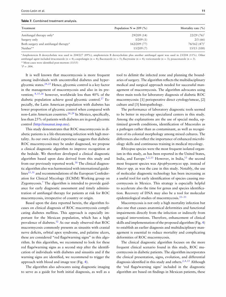
individuals with the complete information about treatmentand outcome were selected and analyzed. Surgery and anti-fungal management were used in 77% of these (162/209),and management with only an antifungal agent was used in14% (29/209). Those who received combined managementwith surgery and one antifungal agent had lower mortalityrate when compared to those who received antifungal ther-apy only (76/162, 47% vs 22/29, 76%, respectively, P =.004), (Table 7).
Logistic regression analysis
In logistic regression analysis, which only included dia-betic individuals with complete information to be analyzed(N = 91), we also found higher survival for those receivingsurgery and antifungal agent as management strategy whencompared with antifungal use only (70% [53/76] vs 20%[3/15]). Combined treatment approach was associated with
a higher probability of survival (95% vs 66%, OR = 0.1,CI 95% 0.02–0.43, P = .002) independently of sex, age,and ketoacidosis status (Table 4).
Discussion
This study underscores that the preponderance of mu-cormycosis in Mexico occurs in patients with diabetic mel-litus. This finding is important as most reports in more eco-nomically developed countries are reporting patients withhematological malignancies, and transplant recipients as thepredominant and increasing population in relation to a de-creasing proportion of cases with diabetes mellitus.6,8,16
Unlike previous studies, in which higher mortality rate inpatients with hematological malignancies17 in comparisonto those with diabetes mellitus has been reported, our study,found a similar rate of mortality between these two groups.However, when we compare mortality rate of diabetic

10 Medical Mycology, 2017, Vol. 00, No. 00
Table 4. Risk factors associated with death in 91 diabetic individuals assessed by multivariate logistic regression analysis.
Vital status Univariate Multivariate analysis
Characteristic Survived N = 56(%) Dead N = 35(%) P value OR 95% CI P value
Sex (Female) 26 (41) 24 (50) .36 1.57 0.60–4.24 .36Age 48 ± 16 47 ± 15 .53 0.98 0.95–1.02 .43Ketoacidosis 20 (33) 18 (38) .68 1.91 0.67–5.41 .22Surgery and antifungal therapy∗ 53 (95) 23 (66) .001 0.10 0.02–0.43 .002Cutaneous infection 5 (9) 3 (9) .71Pulmonary infection 5 (9) 3 (9) .71ROC infection 46 (83) 29 (83) .92Abdominal 0 3 (8) .99Disseminated 0 1 (3) .99
CI, confidence interval. OR, odds ratio.∗Surgery and antifungal therapy vs antifungal therapy only.X2 value 17.02, P = .002.
Table 5. Performance of diagnostic testing.a
Diagnostic tool used Reports from nonspecialized centers Reports from specialized center Overall
Positive direct smear/cytology 73/ 76 (95%) 158/158 (100%) 231/234 (98%)Positive culture 120/211 (57%) 142/158 (90%) 262/369 (71%)
a158 (41%) of 369 cases were reported by a specialized center of diagnostic mycology, while 211 cases were reported by nonspecialized centers.
Table 6. Clinical isolates reported from 250 patients.
Organism isolated Total population n = 250 (%)
Rhizopus species 148 (59)Rhizopus oryzae/R. arrhizus 108/148 (73)Rhizopus sp. 34/148 (23)Rhizopus rhizopodiformis 2/148 (1.3)Rhizopus microsporus 2/148 (1.3)Rhizopus azygosporus 1/148 (0.6)Rhizopus pusillus 1/148 (0.6)Mucor species 71 (28)Mucor sp. 66/71 (93)Mucor circinelloides 5/71 (7)Rhizomucor sp. 10 (4)Lichtheimia corymbifera 8 (3)Cunninghamella sp. 4 (1.5)Syncephalastrum racemosum 3 (1)Basidiobolus sp. 3 (1)Conidiobolus sp. 1 (0.5)Apophysomyces mexicanus 1 (0.5)Absidia sp. 1 (0.5)
population in our report and other previous reports,17 therate of mortality was higher in our population. This studyfurther demonstrated that among diabetic patients with mu-cormycosis, those presenting with ketoacidosis had a higherrate of mortality by univariate analysis. However, ketoaci-dosis was not an independent risk factor for mortality bylogistic regression analysis. Treatment with both antifungaltherapy and surgery improved the probability of success.
These findings are similar to prior reports of ROC mu-cormycosis,18,19 especially in the diabetic population,15,20
and those with localized disease.16 One should note, how-ever, that since patients with disseminated mucormyco-sis, which carries a poor prognosis, would not usually becandidates for surgery, the benefits of combined surgeryand antifungal therapy pertain to patients with localizeddisease.
As a quality control for verification of the underlyingdiseases and clinical manifestations of mucormycosis in thiscases series, we reviewed the distribution of these variablesin several reports from large medical centers in Mexico.A review of 22 cases of mucormycosis in children from asingle center in Mexico found that the rhinocerebral formwas the main clinical presentation (77%), followed by pri-mary cutaneous and pulmonary patterns.21 Consistent withthe overall findings in the study reported herein, diabetesmellitus was present 68% of these patients and hemato-logic diseases in 28%. Another study reported the clini-cal features and outcome of mucormycosis among 14 casesfrom a tertiary-care teaching hospital in northern Mexico.22
Compatible with the data of our report, 10 patients haddiabetes mellitus, and 6 had a hematological malignancy(acute leukemia) as underlying diseases. Finally, Ameenet al. reported five cases of mucormycosis diagnosed at a ter-tiary referral center for medical mycology (Dr Manuel GeaGonzalez General Hospital in Mexico City).23 ROC mu-cormycosis in that study was diagnosed in four of five cases;four of these patients had diabetic ketoacidosis.
Corzo-Leon et al. 11
Table 7. Combined treatment analysis.
Treatment Population N = 209 (%) Mortality rate (%)
Antifungal therapy only∗
Surgery onlyBoth surgery and antifungal therapy∗
Neither∗∗
29/209 (14)3/209 (1)
162/209 (77)15/209 (7)
22/29 (76)†
2/3 (66)76/162 (47)†
15/15 (100)
∗Amphotericin B deoxycholate was used in 204/227 (89%); amphotericin B deoxycholate plus another antifungal agent was used in 23/204 (11%). Otherantifungal agent included itraconazole (n = 4); caspofungin (n = 4); fluconazole (n = 5); flucytosine (n = 4); voriconazole (n = 3); posaconazole (n = 3).∗∗Most cases were identified post-mortem (11/15)†P = .004.
It is well known that mucormycosis is more frequentamong individuals with uncontrolled diabetes and hyper-glycemic states.24,25 Hence, glycemic control is a key factorin the management of mucormycosis and also in its pre-vention; 8,15,26 however, worldwide less than 40% of thediabetic population achieve good glycemic control.27 Es-pecially, the Latin American population with diabetes haslower proportion of glycemic control when compared withnon–Latin American countries.28,29 In Mexico, specifically,less than 25% of patients with diabetes are in good glycemiccontrol (http://ensanut.insp.mx).
This study demonstrates that ROC mucormycosis in di-abetic patients is a life-threatening infection with high mor-tality. As our own clinical experience suggests that diabeticROC mucormycosis may be under diagnosed, we proposea clinical diagnostic algorithm to improve recognition atthe bedside. We therefore developed a clinical diagnosticalgorithm based upon data derived from this study andfrom our previously reported work.30 The clinical diagnos-tic algorithm also was harmonized with international guide-lines15,31 and recommendations of the European Confeder-ation for Clinical Mycology (ECMM) Working group onZygomycosis.7 The algorithm is intended to provide guid-ance for early diagnostic assessment and timely adminis-tration of antifungal therapy for patients at risk for ROCmucormycosis, irrespective of country or origin.
Based upon the data reported herein, the algorithm fo-cuses on clinical diagnosis of ROC mucormycosis compli-cating diabetes mellitus. This approach is especially im-portant for the Mexican population, which has a highprevalence of diabetes.32 As our study observed that ROCmucormycosis commonly presents as sinusitis with cranialnerve deficits, orbital apex syndrome, and palatine ulcers,these are considered “red flags/warning signs” in this algo-rithm. In this algorithm, we recommend to look for thesered flags/warning signs as a second step after the identifi-cation of individuals with diabetes and sinusitis and if thewarning signs are identified, we recommend to trigger theapproach with blood and image test (Fig. 4).
The algorithm also advocates using diagnostic imagingto serve as a guide for both initial diagnosis, as well as a
tool to delimit the infected zone and planning the bound-aries of surgery. The algorithm reflects the multidisciplinarymedical and surgical approach needed for successful man-agement of mucormycosis. The algorithm advocates usingthree main tools for laboratory diagnosis of diabetic ROCmucormycosis: [1] perioperative direct cytology/smear, [2]culture and [3] histopathology.
The performance of laboratory diagnostic tools seemedto be better in mycology specialized centers in this study.Among the explanations are the use of special media, op-timized growth conditions, identification of Mucorales asa pathogen rather than as contaminant, as well as recogni-tion of its colonial morphology among mixed cultures. Thedifferences also reflect the importance of enhanced microbi-ology skills and continuous training in medical mycology.
Rhizopus species were the most frequent isolated organ-ism in this study, as has been reported in the United States,India, and Europe.1,2,13 However, in India,13 the secondmost frequent species was Apophysomyces spp. instead ofMucor spp. as was the case in this study. Notably, the useof molecular diagnostic technology has been increasing asa useful tool for early identification of species causing mu-cormycosis in Mexico. This strategy is especially helpfulto accelerate also the time for genus and species identifica-tion. Recovery of DNA may also be a tool for molecularepidemiological studies of mucormycosis.33–35
Mucormycosis is not only a high mortality infection butalso one that causes anatomical deformities and functionalimpairments directly from the infection or indirectly fromsurgical interventions. Therefore, enhancement of clinicalskills and implementation of the proposed algorithm (Fig. 4)to establish an earlier diagnosis and multidisciplinary man-agement is essential to reduce mortality and complicatingdeformities of ROC mucormycosis.
The clinical diagnostic algorithm focuses on the morefrequent clinical scenario found in this study, ROC mu-cormycosis in diabetic patients. The algorithm incorporatesthe clinical presentation, signs, evolution, and differentialdiagnosis identified in this study and others.2,8,15 Althoughthe ‘red flags/warning signs’ included in the diagnosticalgorithm are based on findings in Mexican patients, these

12 Medical Mycology, 2017, Vol. 00, No. 00
Figure 4. An algorithm for diagnosis and treatment of rhino-orbital-cerebral mucormycosis in patients with diabetes mellitus. Diabetes meansany type of diabetes (1 or 2), controlled or noncontrolled, with or without DKA. Individuals with noncontrolled diabetes, hyperglycemia, and/orDKA have higher risk of infections and worse outcomes. Following discharge, we recommend a close follow-up of at least 72 h that includes aclinical and biochemical reevaluation, as well as a determination of the need, for additional surgery. For patients with diabetes mellitus who arereceiving amphotericin B, particularly those with diabetic nephropathy, ambulatory monitoring of serum creatinine is important, as renal functionmay deteriorate over time. PAS: periodic acid Schiff, H&E: hematoxylin and eosin, GMS: Grocott-Gomori methenamine silver
findings have been also reported in the international litera-ture.36–38 Some of these clinical manifestations, such as theorbital apex syndrome, have been associated with worstprognosis and advanced infection.38 The orbital apex syn-drome caused by mucormycosis represents a surgical emer-gency that usually warrants orbital exenteration. As theorbital apex syndrome involves infarcted neurovascular tis-sue and threatens extension into the cavernous sinus, urgentsurgical intervention is needed; medical therapy alone isseldom effective for this complication of orbital mucormy-cosis. Despite the advances in antifungal therapy with theadvent of amphotericin B lipid formulations, isavuconazole,and posaconazole, the neurovascular infarction and threatto the cavernous sinus warrant urgent surgical resection.
Perioperative and intraoperative direct exam, KOHsmear, and cytology performed during surgery may acceler-ate diagnosis and early treatment. Also, perioperative directmicroscopic exam was suggested in the algorithm due to thefavourable performance of KOH smear found in this study,
clinical experience in our institution, and prior reports in in-ternational literature.13,15,39 We preferred KOH in our in-stitution, because for stains such as calcofluor white stain, afluorescence microscope is necessary and not always presentin Mexican hospitals. Certainly, if a fluorescence micro-scope is available, then calcofluor white stain should be per-formed when feasible. Moreover, as has been recommendedin a recent prospective study in India and by the British So-ciety for Medical Mycology, further histopathological ex-aminations with hematoxylin and eosin (H&E), periodicacid-Schiff (PAS), and Grocott-Gomori methenamine sliver(GMS) stains should be performed together with culturesof infected tissues.13,31
AmB formulations are considered the first line of treat-ment for mucormycosis. Lipid formulations of ampho-tericin B are associated with less renal dysfunction than thatof AmBD and are effective as initial therapy for mucormy-cosis.40 Although lipid formulations of AmB are not readilyavailable in Mexico, they should be used where possible in

Corzo-Leon et al. 13
patients with diabetes mellitus, diabetic nephropathy, andconcomitant nephrotoxic agents, where AmBD may inducerenal failure.
Isavuconazole is another option for primary therapy ofmucormycosis. Treatment with isavuconazole has shownsimilar outcomes to those of contemporaneously treated ex-ternal control patients receiving amphotericin B when usedas primary and salvage therapy.6,41 Isavuconazole has beenwell tolerated, even for individuals with mild to moderateliver impairment.42 For patients with underlying diabeticnephropathy, isavuconazole would be preferable to avoidfurther deterioration of renal function.
Following discharge, we recommend close follow-upwithin 72 h that includes a clinical and biochemical reeval-uation, as well as a determination of the need for addi-tional surgery. For patients with diabetes mellitus who arereceiving amphotericin B, particularly those with diabeticnephropathy, ambulatory monitoring of serum creatinineis important, as renal function may deteriorate over time.
Ambulatory monitoring of sinus mucormycosis by en-doscopy is a sensitive tool by which to assess antifungal re-sponse. Subtle changes in mucosal integrity and resolutionof eschars can be accurately assessed by endoscopic evalu-ation. By comparison, assessment by CT scan of anatomicchanges of sinus infiltrate caused by mucormycosis may lackthe detection of finer detail afforded by nasal endoscopy.
Although exenteration is a standard of care for theorbital apex syndrome, more conservative surgical mea-sures aimed at preserving the eye intact are pursued formanagement of mucormycosis of the orbit. Most cases ofsino-orbital mucormycosis can be managed following theprinciples of antifungal therapy, reversal of immunosup-pression, and surgical resection of infected tissue withoutloss of the eye. Sino-orbital mucormycosis can usually bemanaged without orbital exenteration providing that earlydiagnosis is established and that timely initiation of therapyis implemented. By comparison, a delay in exenteration fororbital apex syndrome may be complicated by intracranialextension and cavernous sinus thrombosis.
Reversal of immunosuppression or diabetic ketoacido-sis is essential for a favorable outcome of sino-orbitalmucormycosis. However, surgery and antifungal therapyare also important components of management, whereineach patient is managed individually by a multidisciplinaryteam.
This study has several limitations. As the data from casesand series reports is acquired retrospectively, informationfor individual patients was not always complete and couldbe heterogeneous. Since we could not exclude in this study apotential publication bias, even when the findings herein areconsistent with previous reports, a prospective multicenter
study should be performed to confirm these observationsand conclusions. The data depend upon the publication ofcases and therefore likely underestimate the true magni-tude and scope of mucormycosis in Mexico. Nonetheless,this report represents the most comprehensive review ofmucormycosis in Mexican patients and therefore advancesour understanding in this population of more than 120 mil-lion people.43 While the diagnostic algorithm proposed wasbuilt based upon clinical experience in a major medical cen-ter of Mexico and upon observations reported in the litera-ture reviewed in this study, its use should be prospectivelyvalidated.
In summary, ROC mucormycosis in the population withdiabetes mellitus is still the most frequently reported clinicalpresentation of this infection in Mexico. Combined man-agement with antifungal therapy and surgery improves theoutcome in mucormycosis when compared with antifungaltherapy alone and is cornerstone in management of ROCmucormycosis. Early clinical diagnosis, specialized trainingin laboratory diagnostic mycology, multidisciplinary man-agement, and timely antifungal therapy are important forsuccessful management of mucormycosis.
Acknowledgments
To all authors of the published reports and thesis for their inter-est in communicate their clinical experience. Dr. Walsh serves as aScholar in Mucormycosis of the Henry Schueler Foundation, an In-vestigator of Emerging Infectious Diseases of the Save Our Sick KidsFoundation, and the Sharp Family Foundation Scholar in InfectiousDiseases.
Dr. Walsh was supported for this work as a Scholar in Mucormy-cosis of the Henry Schueler Foundation.
Declaration of interest
Dr. Walsh has received research grants for experimental and clini-cal antifungal pharmacotherapeutics from Astellas, Novartis, Merck,and Pfizer. He has served as consultant in antifungal pharmacother-apeutics to Astellas, Drais, iCo, Novartis, Pfizer, Methylgene, andSigmaTau.
Dr. Rodriguez-Zulueta has been speaker on behalf of Pfizer,Stendhal, and MSD. Dr. Corzo-Leon and Dr. Chora-Hernandez haveno further disclosures.
References
1. Skiada A, Pagano L, Groll A et al. Zygomycosis in Europe:Analysis of 230 cases accrued by the registry of the EuropeanConfederation of Medical Mycology (ECMM) Working Groupon Zygomycosis between 2005 and 2007. Clin Microbiol Infect.2011;17: 1859–1867.

14 Medical Mycology, 2017, Vol. 00, No. 00
2. Roden MM, Zaoutis TE, Buchanan WL et al. Epidemiology andoutcome of zygomycosis: a review of 929 reported cases. ClinInfect Dis. 2005; 41: 634–653.
3. Lanternier F, Dannaoui E, Morizot G et al. A global analysisof mucormycosis in France: The RetroZygo study (2005–2007).Clin Infect Dis. 2012; 54 (Suppl. 1): 35–43.
4. Millon L, Herbrecht R, Grenouillet F et al. Early diagnosis andmonitoring of mucormycosis by detection of circulating DNAin serum: retrospective analysis of 44 cases collected throughthe French Surveillance Network of Invasive Fungal Infections(RESSIF). Clin Microbiol Infect. 2016; 22: 810.e1–810.e8.
5. De Pauw B, Walsh TJ, Donnelly JP et al. Revised definitionsof invasive fungal disease from the European Organization forResearch and Treatment of Cancer/Invasive Fungal InfectionsCooperative Group and the National Institute of Allergy andInfectious Diseases Mycoses Study Group (EORTC/MSG) C.Clin Infect Dis. 2008; 46: 1813–1821.
6. Marty FM, Ostrosky-Zeichner L, Cornely OA et al. Isavucona-zole treatment for mucormycosis: a single-arm open-label trialand case-control analysis. Lancet Infect Dis. 2016; 16: 828–837.
7. Walsh TJ, Skiada A, Cornely OA et al. Development of newstrategies for early diagnosis of mucormycosis from bench tobedside. Mycoses. 2014; 57: 2–7.
8. Kontoyiannis DP, Lewis RE. How I treat mucormycosis. Blood.2011;118: 1216–1224.
9. Bitar D, Lortholary O, Le Strat Y et al. Population-based analysisof invasive fungal infections. Emerg Infect Dis. 2014; 20: 1149–1155.
10. Gamaletsou MN, Drogari-Apiranthitou M, Denning DW et al.An estimate of the burden of serious fungal diseases in Greece.Eur J Clin Microbiol Infect Dis. 2016; 35: 1115–1120.
11. Lelievre L, Garcia-Hermoso D, Abdoul H et al. Posttraumaticmucormycosis. Medicine (Baltimore). 2014; 93: 373–382.
12. Vaezi A, Moazeni M, Rahimi MT et al. Mucormycosis in Iran:a systematic review. Mycoses. 2016: 59: 402–415.
13. Bala K, Chander J, Handa U et al. A prospective study of mu-cormycosis in north India: experience from a tertiary care hos-pital. Med Mycol. 2015; 53: 248–257.
14. Gopalakrishnan R. Mucormycosis in patients without cancer: acase series from a tertiary care hospital in South India. J AssocPhysicians India. 2013; 61: 305–308.
15. Cornely OA, Arikan-Akdagli S, Dannaoui E et al. ESCMID andECMM joint clinical guidelines for the diagnosis and manage-ment of mucormycosis 2013. Clin Microbiol Infect. 2014; 20(S3): 5–26.
16. Kyvernitakis A, Torres HA, Jiang Y et al. Initial use of com-bination treatment does not impact survival of 106 patientswith haematologic malignancies and mucormycosis: a propen-sity score analysis. Clin Microbiol Infect. 2016; 22: 811.e1–811.e8.
17. Spellberg B, Kontoyiannis DP, Fredricks D et al. Risk factors formortality in patients with mucormycosis. Med Mycol. 2012; 50:611–618.
18. Kasapoglu F, Coskun H, Ozmen OA et al. Acute invasive fungalrhinosinusitis: Evaluation of 26 patients treated with endonasalor open surgical procedures. Otolaryngol - Head Neck Surg.2010; 143: 614–620.
19. Sims CR, Ostrosky-Zeichner L. Contemporary treatment andoutcomes of zygomycosis in a non-oncologic tertiary care center.Arch Med Res. 2007; 38: 90–93.
20. Di Carlo P, Pirrello R, Guadagnino G et al. Multimodal surgicaland medical treatment for extensive rhinocerebral mucormycosisin an elderly diabetic patient: a case report and literature review.Case Rep Med. 2014; 2014:527062.
21. Bonifaz A, Tirado-Sanchez A, Calderon L et al. Mucormycosisin children: a study of 22 cases in a Mexican hospital. Mycoses.2014; 57 (s3): 79–84.
22. Camara-Lemarroy CR, Gonzalez-Moreno EI, Rodrıguez-Gutierrez R et al. Clinical features and outcome of mucormyco-sis. Interdiscip Perspect Infect Dis. 2014; 2014: 1–5.
23. Ameen M, Arenas R, Martinez-luna E et al. The emergence ofmucormycosis as an important opportunistic fungal infection:five cases presenting to a tertiary referral center for mycology.Int J Dermatol. 2007; 46: 380–384.
24. Waldorf AR, Ruderman N, Diamond RD. Specific susceptibil-ity to mucormycosis in murine diabetes and bronchoalveolarmacrophage defense against Rhizopus. J Clin Invest. 1984; 74:150–160.
25. Ibrahim AS, Kontoyiannis DP. Update on mucormycosis patho-genesis. Curr Opin Infect Dis. 2013; 26: 508–515.
26. Shirazi F, Farmakiotis D, Yan Y et al. Diet modification andmetformin have a beneficial effect in a fly model of obesity andmucormycosis. PLoS One. 2014; 9: 1–9.
27. Chan JCN, Gagliardino JJ, Baik SH et al. Multifaceted determi-nants for achieving. Diabetes Care. 2009; 32: 227–233.
28. Linetzky B, Curtis B, Frechtel G et al. Challenges associated withinsulin therapy progression among patients with type 2 diabetes:Latin American MOSAIc study baseline data. Diabetol MetabSyndr. 2016; 8: 41.
29. Brath H, Paldanius PM, Bader G et al. Differences in glycemiccontrol across world regions: a post-hoc analysis in patients withtype 2 diabetes mellitus on dual antidiabetes drug therapy. NutrDiabetes. 2016; 6: e217.
30. Plowes Hernandez O, Prado Calleros HM, Soberon MarmissolleDaguerre GS et al. Rhino-orbito-cerebral mucormycosis. Man-agement strategies to avoid or limit intracraneal affection andimprove survival. Acta Otorrinolaringol Esp. 2015; 66: 348–352.
31. Schelenz S, Barnes RA, Barton RC et al. British Society for Med-ical Mycology best practice recommendations for the diagnosisof serious fungal diseases. Lancet Infect Dis. 2015; 15: 461–474.
32. Encuesta Nacional de Salud, Internet. 2016. Available from:http://ensanut.insp.mx.
33. Alastruey-Izquierdo A, Mellado E, Pelaeez T et al. Population-based survey of filamentous fungi and antifungal resistance inSpain (FILPOP study). Antimicrob Agents Chemother. 2013;57: 3380–3387.
34. Vitale RG, De Hoog GS, Schwarz P et al. Antifungal suscep-tibility and phylogeny of opportunistic members of the orderMucorales. J Clin Microbiol. 2012; 50: 66–75.
35. Chowdhary A, Kathuria S, Singh PK et al. Molecular characteri-zation and in vitro antifungal susceptibility of 80 clinical isolatesof mucormycetes in Delhi, India. Mycoses. 2014; 57 (s3): 97–107.

Corzo-Leon et al. 15
36. Koc Z, Koc F, Yerdelen D et al. Rhino-orbital-cerebral mu-cormycosis with different cerebral involvements: infarct, hemor-rhage, and ophthalmoplegia. Int J Neurosci. 2007; 117: 1677–1690.
37. Jiang N, Zhao G, Yang S et al. A retrospective analysis of elevencases of invasive rhino-orbito-cerebral mucormycosis presentedwith orbital apex syndrome initially. BMC Ophthalmol. 2016;16: 10.
38. Gamaletsou MN, Sipsas NV, Roilides E et al. Rhino-orbital-cerebral mucormycosis. Curr Infect Dis Rep. 2012; 14: 423–434.
39. McDermott NE, Barrett J, Hipp J et al. Successful treatment ofperiodontal mucormycosis: report of a case and literature review.Oral Surgery, Oral Med Oral Pathol Oral Radiol Endodontol-ogy. 2010; 109: e64–e69.
40. Shoham S, Magill SS, Merz WG et al. Primary treatment of zy-gomycosis with liposomal amphotericin B: analysis of 28 cases.Med Mycol. 2010; 48: 511–517.
41. Graves B, Morrissey CO, Wei A et al. Isavuconazole as salvagetherapy for mucormycosis. Med Mycol Case Rep. 2016; 11: 36–39.
42. Desai A, Schmitt-Hoffmann A-H, Mujais S et al. Populationpharmacokinetics of isavuconazole in subjects with mild or mod-erate hepatic impairment. Antimicrob Agents Chemother. 2016;60: 3025–3031.
43. Instituto Nacional de Estadistica y Geografia (INEGI), Internet.2016. Available from: http://www.inegi.org.mx
44. Gutierrez-Delgado EM, Trevino-Gonzalez JL, Montemayor-Alatorre A et al. Chronic rhino-orbito-cerebral mucormycosis:a case report and review of the literature. Ann Med Surg. 2016;6: 87–91.
45. Alvarado-Lezama J, Espinosa-Gonzalez O, Garcıa-Cano E et al.Gastritis enfisematosa secundaria a mucormicosis gastrica. CirCir. 2015; 83: 56–60.
46. Mendez HH, Hidalgo AR. Endodontic patient with rhinocere-bral mucormycosis: clinical case report. Paciente endodonticocon mucormicosis rinocerebral: reporte de un caso. Rev Odon-tologica Mex. 2015; 19: 121–126.
47. Rodrıguez-Gutierrez G, Carrillo-Casas EM, Arenas R et al. Mu-cormycosis in a non-hodgkin lymphoma patient caused by Syn-cephalastrum racemosum: case report and review of literature.Mycopathologia. 2015; 180: 89–93.
48. Bonifaz A, Stchigel AM, Guarro J et al. Primary cutaneous mu-cormycosis produced by the new species Apophysomyces mexi-canus. J Clin Microbiol. 2014; 52: 4428–4431.
49. Valdez-Geraldo CM, Zavala-Ruiz MG, Collado-Castro I et al.Mucormicosis rinocerebral: reporte de caso en escolar con ce-toacidosis diabetica. Rev Mex Neurocienc. 2014; 15: 229–233.
50. la Cruz D, Dıaz-Salazar MX, Aguilera-Partida JA et al. Mu-cormycosis in a medullar aplasia patient: case report. Rev MexOftalmol. 2013; 87: 165–170.
51. Ramırez-dovala S, Sierra-tellez D, Contreras-rodrıguez D et al.Caso clınico mucormicosis rino-orbito-cerebral causada porRhizomucor pusillus en paciente diabetico descompensado. Der-matol rev Mex. 2012; 56: 132–136.
52. Gonzalez-ramos LA, Martınez-carballo EM, Rascon-alcantar A.Endocarditis por mucormicosis. Bol Clin Hosp Edo Son. 2012;29: 77–80.
53. Telich-Tarriba JE, Perez-Ortiz AC, Telich-Vidal J. Necrotizingfasciitis caused by cutaneous mucormycosis: a case report. CirCir. 2012; 80: 462–465.
54. Garcıa-Romero MT, Garcıa-Mendez J, Arenas R et al. Zygomy-cosis in two hematologic cases. Case Rep Infect Dis. 2011; 2011:181782.
55. Jacobo AGJ, Santiago PM, Elena GLC et al. Cutaneous zygomy-cosis in immunocompetent patients in Mexico. Mycoses. 2010;53: 538–540.
56. Arce-Salinas CA, Perez-Silva E. Mucormycosis complica-tions in systemic lupus erythematosus. Lupus. 2010; 19:985–988.
57. Trejo S, Contreras R. Mucormicosis en un paciente con agranu-locitosis secundaria a metimazol. Informe de un caso. Gac MedMex. 2009; 145: 235–238.
58. Salinas-Lara C, Rembao-Bojorquez D, de la Cruz E et al. Pitu-itary apoplexy due to mucormycosis infection in a patient withan ACTH producing pulmonary tumor. J Clin Neurosci. 2008;15: 67–70.
59. Bonifaz A, Macias B, Paredes-Farrera F et al. Palatal zygomyco-sis: experience of 21 cases. Oral Dis. 2008; 14: 569–574.
60. Robledo-ogazon F, Lizaola-perez B, Mier-giraud F et al. Mu-cormicosis de la pared abdominal. Informe de un caso. Cir Ciruj.2007; 75: 465–469.
61. Torres-Chavez T, Araiza Santibanez J, Sanchez P et al. Mu-cormicosis por Rhizopus azygosporus en paciente con diabetesmellitus tipo 2 y bocio or. Rev Endocrinol y Nutr. 2007; 15:109–114.
62. Papadakis-Solis M, Larrea y Richerand E, Carrasco-Rueda CAet al. Mucormicosis rinoorbitaria y centellografia osea de tresfases. Descripcion de un caso. Rev Inst Nal Enf Resp Mex. 2007;20: 210–212.
63. Carrillo-esper R, Elizondo-argueta S, Vicuna-gonzalez M.Mucormicosis renal aislada. Gac Med Mex. 2006; 142:511–514.
64. De I, Bernardino GH, Jarquın PZ et al. Mucormicosis rinocere-bral. Informe de un caso atıpico. Dermatologia C. 2005; 3 :272–275.
65. Manuel MH, Esther MD. Sındrome de apex orbitario causadopor mucormicosis orbitocerebral cronica e indolente: reporte dedos casos. AN ORL MEX. 2005; 50: 64–68.
66. Bravo TC. Mucormicosis Rinoorbital En Un Paciente ConSındrome De Inmunodeficiencia Adquirida ( SIDA ) Y. Rev MexOftalmol. 2004; 78: 193–195.
67. Adrian L, Garcıa C. Experiencia de mucormicosis en el HospitalJuarez de Mexico. Rev Hosp Jua Mex. 2004; 71: 3–13.
68. HR L-T. Un caso de gastroenterocolitis necrosante por zigomi-cosis. Rev Mex Pediatr. 2004; 71: 283–285.
69. Fujarte-Victorio AS, Casillas-Suarez C, Flores-Colin I et al. Mu-cormicosis pulmonar en un caso de carcinoma broncogenico decelulas pequenas con diabetes mellitus tipo 2. Rev Inst NAl EnfResp Mex. 2003; 16: 169–172.
70. Hernandez-Magana R, Gomez-Barreto D, Puente A et al. mu-cormicosis rinoorbitaria nosocomial causada Rhizopus oryzaeen un lactante desnutrido. Bol Med Hosp Infant Mex. 2001; 58:35–47.
71. Barron-soto AMA, Campos-navarro DLA, Antonio M et al.Morbilidad y mortalidad del paciente con mucormicosis

16 Medical Mycology, 2017, Vol. 00, No. 00
rinorbitaria posterior al tratamiento medico quirurgico opor-tuno. 2001; 11: 8–11.
72. Romero-zamora JL, Bonifaz A, Sanchez CJ et al. Mucormicosisrinocerebral. Reporte de doce casos. Rev Medica del Hosp GenMexico. 2000; 63: 178–184.
73. Flores MS, Eugenia M, Manrıquez V et al. Mucormicosis pul-monar diseminada. Informe de dos casos. Rev Inst Nal Enf RespMex. 2000; 13: 227–232.
74. Bross-Soriano D, Prado Calleros HM, Arrieta-Gomez J et al.Mucormicosis de la nariz y los senos paranasales.pdf. An Medi-cos Hosp ABC. 1998; 43: 95–100.
75. Rangel-Guerra RA, Martınez HR, Senz C et al. Rhinocerebraland systemic mucormycosis: clinical experience with 36 cases. JNeurol Sci. 1996; 143: 19–30.
76. Escobar A, Del Brutto OH. Multiple brain abscesses from iso-lated cerebral mucormycosis. J Neurol Neurosurg Psychiatry.1990; 53: 431–433.
77. Pedroza-Teran LG. Mortalidad en pacientes con mucormicosisinvasiva en el Hospital de Especialidades, Centro Medico Na-cional Siglo XXI, reporte de casos. UNAM 2016.
78. Alamilla-Azpeitia RS. Aplicado a paciente con “mucormicosiscutanea” basado en el modelo de Dorothea Orem en el HospitalGeneral de Mexico Dr. Eduardo Liceaga. UNAM 2015.
79. Tepox-Padron A. Caracterısticas clınicas y desenlace clınico dela mucormicosis en un hospital de tercer nivel de la ciudad deMexico. UNAM 2014.
80. Rodriguez-Gutierrez G. Diagnostico molecular de mucormi-cosis e identificacion de los distintos agentes causales.UNAM 2013.
81. Avila-Cardoso C. Prevalencia y datos epidemiologicos de la mu-cormicosis en el servicio de otorrinolaringologıa adultos. UNAM2010.
82. Gomez-Castelan A. Rehabilitacion de un paciente excenteradopor mucormicosis con protesis orbitaria implantosoportada: re-porte de caso clinico. UNAM 2007.
83. Garcıa Luis D. Diagnostico de mucormicosis, sus manifesta-ciones bucales y tratamiento; reporte de un caso clinico. UNAM2005.
84. Rodriguez-Osorio C. Mucormicosis. Experiencia en el InstitutoNacional de Ciencias Medicas y Nutricion “Salvador Zubiran.”UNAM 2004.
85. Hilda AA. Diabetes, mucormicosis y sus manifestaciones bu-cales. UNAM 2002.
86. Luna-Olguin O. Presentacion de dos casos clinicos de mucormi-cosis del servicio de micologia del Hospital General de Mexicoy revision de la literatura de 1980 a 1998. UNAM 2000.
87. Melendez-Rivera L. Mucormicosis, revision de la literatura y so-brevida del paciente en el Hospital de Especialidades del CentroMedico Nacional Siglo XXI, I.M.S.S. UNAM 1999.
88. Martinez-Martinez M. Mucormicosis en la unidad de cuidadosintensivos, reporte de un caso y revision de la literatura. UNAM1996.