developments in the production of biological and synthetic binders
TRANSCRIPT
Trends Trends in Analytical Chemistry, Vol. 30, No. 2, 2011
Developments in the productionof biological and synthetic bindersfor immunoassay and sensor-baseddetection of small moleculesTerry Fodey, Paul Leonard, John O�Mahony, Richard O�Kennedy,
Martin Danaher
The need for chemical and biological entities of predetermined selectivity and affinity towards target analytes is greater than ever, in
applications such as environmental monitoring, bioterrorism detection and analysis of natural toxin contaminants in the food chain.
In this review, we focus on advances in the production of specific binders, in terms of both natural entities (e.g., antibodies) and
synthetic binders (e.g., molecularly-imprinted polymers). We discuss the potential of emerging technologies for integration into
immunoassay and sensing techniques. We place special emphasis on use of these technologies in bioanalytical applications.
ª 2010 Elsevier Ltd. All rights reserved.
Keywords: Antibody; Aptamer; Binding protein; Biorecognition; Immunoassay; Molecular-imprinted polymer; Nanobody; Protein scaffold; Single-
chain variable fragment; Synthetic binder
Abbreviations: AC-DON, Acetyl deoxynivalenol; ALP, Alkaline phosphatase; ASP, Amnesic shellfish poisoning; ATP, Adenosine triphosphate;
AuNP, Gold nanoparticle; C domain, Constant domain; cDNA, Complementary deoxyribonucleic acid; CDR, Complementarity-determining region;
DARPin, Designed ankyrin repeat protein; DC, Deoxycortisol; DNA, Deoxyribonucleic acid; DON, Deoxynivalenol; E. coli, Escherichia coli; ELISA,
Enzyme-linked immunosorbent assay; Fab, Fragment-antigen binding; Fc-PEI, Ferrocene-appended poly(ethyleneimine); fg, femtogram (10�15 g);
FPA, Fluorescence-polarization assay; IC50, Concentration of inhibitor that is required for 50% inhibition of its target; IEMA, Immunoenzymometric
assay; Ig, Immunoglobulin; kDa, kiloDalton; LOD, Limit of detection; mAb, Monoclonal antibody; MC, Microcystin; MG, Malachite green; MIP,
Molecularly-imprinted polymer; mRNA, Messenger ribonucleic acid; NECEEM, Non-equilibrium capillary electrophoresis of equilibrium mixtures;
ng, nanogram (10�9 g); OBP, Odorant-binding protein; OS-IA, Open-sandwich immunoassay; OTA, Ochratoxin A; OTC, Oxytetracycline; PCR,
Polymerase-chain reaction; pg, picogram (10�12 g); PMP, Pinacolyl methylphosphate; PVC, Polyvinyl chloride; QCM, Quartz-crystal microbalance;
RNA, Ribonucleic acid; scFv, Single-chain variable fragment; sdAb, Single-domain antibody; SELEX, Systematic evolution of ligands by exponential
enrichment; SPE, Solid-phase extraction; SPR, Surface-plasmon resonance; ssDNA, Single-stranded deoxyribonucleic acid; TNT, Trinitrotoluene;
V domain, Variable domain; VH, Variable heavy chain; VL, Variable light chain; VOC, Volatile organic compound; ZEN, Zearalenone; lg,
microgram (10�6)
Terry Fodey
Institute of Agri-Food and Land Use, School of Biological Sciences, Queen’s University Belfast,
Belfast, Northern Ireland
Paul Leonard, Richard O’Kennedy
School of Biotechnology and National Centre for Sensor Research, Dublin City University, Dublin 9,
Ireland, and Biomedical Diagnostics Institute, Dublin City University, Dublin 9, Ireland
John O’Mahony, Martin Danaher*
Teagasc, Ashtown Food Research Centre, Ashtown, Dublin 15, Ireland
*Corresponding author.
Tel.: +353 1 8059500;
Fax: +353 1 8059550;
E-mail:
254 0165-9936/$ - see front matter ª 2010 Elsev
1. Introduction
Biological and synthetic binders are at theheart of the majority of modern diagnosticscreening assays. Traditionally, polyclonalantibodies and monoclonal antibodies(mAbs) have been the popular choice forsmall molecules. Subsequently, molecu-larly-imprinted polymers were developedas an alternative with applications mainlyin the area of sample preparation. In thepast decade, groups have begun to developalternative biological and synthetic bindersfor use in detection systems for laboratory
ier Ltd. All rights reserved. doi:10.1016/j.trac.2010.10.011

Trends in Analytical Chemistry, Vol. 30, No. 2, 2011 Trends
and field-based applications. In the area of antibodyproduction, alternative technologies have emerged;some are based on the modification of material that hasbeen produced in vivo, with others relying on an in vitroapproach eliminating the need to use animals.
In this article, we discuss three alternatives to con-ventional IgG antibodies – (i) nanobodies [1], (ii) apta-mers [2,3] and (iii) protein scaffolds [4]. We investigatetheir current state of development and their applicationin assays designed to detect small molecules. We reviewrecent developments in the area of synthetic binders,along with a range of applications including chemicalwarfare agents [5], toxins [6] and drugs of abuse [7]. Weconclude with a short review on the applications ofsingle-chain variable fragment (scFv) antibodies, whichnow comprise a more mature technology and are start-ing to be applied in working assays.
2. Evolution of antibodies as biorecognitionelements for haptens
Biomolecular recognition, based on non-covalent bind-ing, is central to all biological interactions. In addition toionic, hydrogen-bonding and hydrophobic interactions,shape complementarity plays a pivotal role in the processof biorecognition. Over a century ago, Paul Ehrlich firstrecognized the function of a special class of proteins andcalled them ‘‘antibodies’’. With the discovery came thehypothesis that such substances, termed ‘‘magicbullets’’, would seek their targets of their own accord [8].Since their discovery, antibodies, and more recently non-immunoglobulin (non-Ig) scaffold proteins, have beenexploited for scientific needs, such as analytical detec-tion. To understand the fundamental principles of anti-body-based detection systems, knowledge of antibodystructure and function is required. This section describesthe structure of antibodies, and antibody types anddiscusses the structural components of the antibodyinvolved in binding to a target analyte.
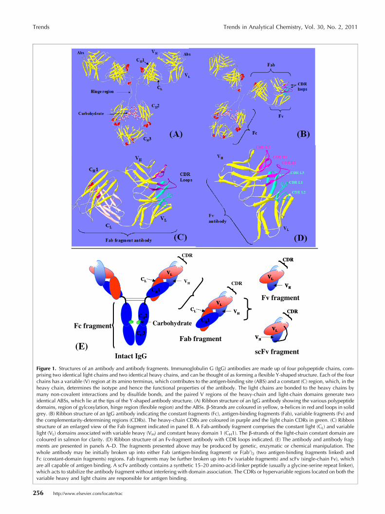
Antibodies are highly-soluble serum glycoproteins in-volved in the defense mechanism of the immune system.All antibodies are constructed in the same way, frompaired heavy and light polypeptide chains, and thegeneric term ‘‘immunoglobulin’’ (Ig) is used for all suchprotein molecules (Fig. 1). The discrete globular domainsof Ig chains fall into two distinct structural categories,corresponding to V and C domains. Each Ig domain isconnected in tandem and has a characteristic ‘‘Ig fold’’composed of two roughly parallel layers of anti-parallelb-pleated sheets connected with an intra-chain disulfidebond. The positions of the two cysteine residues in the b-strands that form the disulfide bond are highly conserved[9]. The tertiary structure of the Ig domains promotesinteraction between the faces of the b-sheets and sub-sequently allows the correct folding into a functional
antibody molecule. Linking the b-strands are loops ofamino acids that are more variable in sequence. Se-quence flexibility in these hypervariable loops makes itpossible to generate antibodies against a very broadrange of antigens including conformationally intact ordenatured proteins, short peptides, carbohydrates, drugs,hormones and low-molecular-weight compounds [10].
Antigens (any foreign entity or target for antibodyproduction) used for immunization may be highly-puri-fied preparations containing a single molecular species, amixture of molecules, or an extremely complex ‘‘anti-gen’’ (e.g., microorganism or cell). Haptens are too smallto elicit an immune response, so generation of anti-hapten antibodies requires conjugation to a larger mol-ecule (e.g., a protein, termed a carrier protein). Irre-spective of the antigen used for immunization, all of theantibodies that a given B-cell clone secretes have exactlythe same specificity and affinity for antigen and arecalled mAbs. However, when the immune system ischallenged with antigen, many different B cells respondand secrete antibodies. They can all potentially recognizethe antigen, but in slightly different ways, with differentspecificities, affinities and cross-reactivities. This is calleda polyclonal response and antibodies derived from thechallenged immune system are known as polyclonalantibodies. While these antibodies are easier to produce,their batch-to-batch variation and tendency to cross-react with conformationally similar compounds limittheir use for defined hapten-specific assays.
The discovery of hybridoma technology, first describedby Milstein and Kohler in 1975, revolutionized theproduction of truly antigen-specific antibodies [11].Primed B cells obtained from experimental animals (e.g.,mice and rats) immunized with the desired antigen canbe fused to myeloma cells, forming hybridoma cellscapable of producing antibody of the desired specificity.Thus, mAbs provide reagents with single-epitope speci-ficity and potentially limitless amounts of identicalantibody. Even though, the production of mAbs ofmurine or rat origin is now a routine procedure, easilywithin the capabilities of most investigators, it can alsobe a time-consuming and expensive process, so seriousthought should be given to the necessity of embarkingupon a program of mAb production.
The emergence of recombinant antibody phage-dis-play technology, developed during the past two decades,has transformed the way in which we generate anti-bodies for the specific detection of a chosen analyte.Recombinant antibodies can be generated by PCR fromcDNA derived from hybridoma cells or from the spleensof immunized animals and assembling scFv or Fabfragments by genetic engineering or by a combinatorialapproach. This is usually achieved by constructing anantibody-gene library by PCR amplification of the rear-ranged antibody genes from B lymphocytes. Thecombinatorial libraries can be generated from the B cells
http://www.elsevier.com/locate/trac 255
CDR
VH
CH3
CH1
CH2
VL
CL
VH
CH1
VL
CL
CDR VH
VL
CDR
VH
VL
CDR
Intact IgG
Fc fragment
Fv fragment
Fab fragment
scFv fragment
Carbohydrate
CH3
CH1
CH2
VL
CL
VH
CH1
VL
CL
CH1
VL
VH
VL
CDR
VH
VL
VH
VL
VH
VL
CDR
VH
VL
(E)
Figure 1. Structures of an antibody and antibody fragments. Immunoglobulin G (IgG) antibodies are made up of four polypeptide chains, com-prising two identical light chains and two identical heavy chains, and can be thought of as forming a flexible Y-shaped structure. Each of the fourchains has a variable (V) region at its amino terminus, which contributes to the antigen-binding site (ABS) and a constant (C) region, which, in theheavy chain, determines the isotype and hence the functional properties of the antibody. The light chains are bonded to the heavy chains bymany non-covalent interactions and by disulfide bonds, and the paired V regions of the heavy-chain and light-chain domains generate twoidentical ABSs, which lie at the tips of the Y-shaped antibody structure. (A) Ribbon structure of an IgG antibody showing the various polypeptidedomains, region of gylcosylation, hinge region (flexible region) and the ABSs. b-Strands are coloured in yellow, a-helices in red and loops in solidgrey. (B) Ribbon structure of an IgG antibody indicating the constant fragments (Fc), antigen-binding fragments (Fab), variable fragments (Fv) andthe complementarity-determining regions (CDRs). The heavy-chain CDRs are coloured in purple and the light chain CDRs in green. (C) Ribbonstructure of an enlarged view of the Fab fragment indicated in panel B. A Fab-antibody fragment comprises the constant light (CL) and variablelight (VL) domains associated with variable heavy (VH) and constant heavy domain 1 (CH1). The b-strands of the light-chain constant domain arecoloured in salmon for clarity. (D) Ribbon structure of an Fv-fragment antibody with CDR loops indicated. (E) The antibody and antibody frag-ments are presented in panels A–D. The fragments presented above may be produced by genetic, enzymatic or chemical manipulation. Thewhole antibody may be initially broken up into either Fab (antigen-binding fragment) or F(ab 0)2 (two antigen-binding fragments linked) andFc (constant-domain fragments) regions. Fab fragments may be further broken up into Fv (variable fragments) and scFv (single-chain Fv), whichare all capable of antigen binding. A scFv antibody contains a synthetic 15–20 amino-acid-linker peptide (usually a glycine-serine repeat linker),which acts to stabilize the antibody fragment without interfering with domain association. The CDRs or hypervariable regions located on both thevariable heavy and light chains are responsible for antigen binding.
Trends Trends in Analytical Chemistry, Vol. 30, No. 2, 2011
256 http://www.elsevier.com/locate/trac

Trends in Analytical Chemistry, Vol. 30, No. 2, 2011 Trends
of immunized animals (immune libraries) or from non-immunized animals (naıve libraries) [12]. Due to therandom light-chain and heavy-chain combinations,combinatorial antibody libraries may contain antibodyfragments not found in nature, so libraries can be used toisolate antibodies directed against antigens not suited toimmunization (e.g., recognizing highly toxic substancesor self-antigens). Having a direct link between theexperimental phenotype and its encapsulated genotype,phage display allows the evolution of selected bindersinto optimized molecules [13]. The power of phageselection to choose those ligands having the desiredbiological properties permits us to mimic the immunesystem and synthesize ‘‘tailor-made’’ antibodies for usein diagnosis, immunotherapy or immunoassay develop-ment.
Affinity selection of scFv by panning (Fig. 2) andsubsequent reinfection into E. coli greatly enhances thenumber of specific, strong-binding scFvs. During pan-ning, the phage library is incubated in an immunotubecoated with specific antigen, unbound phage particlesare washed away and the bound phages are then re-moved under strict elution conditions. Successive roundsof panning are carried out to enrich antigen binders andto ensure that antibody fragments with the strongestbinding affinities are isolated.
Re-infect E.coamplify phage
Allow binding and wash away unbound phage
Elute bound phage
Figure 2. The affinity-selection bio-panning process. Phage-displaying scFvinterest. Non-binding-phage scFv are washed away and antigen-specific phThe process (bio-panning) is repeated several times, with an enrichment ofbio-panning.
Antibodies are indisputably the most successful bind-ing molecules in biomedical science with establishedanalytical, diagnostic and therapeutic applications.However, limitations of antibodies have also beenuncovered, based largely on their biophysical propertiesand their complicated molecular composition [14].These limitations inspired a growing trend towardsengineering alternative non-Ig-binding proteins andmolecules. In the next section, we describe some of theseemerging biorecognition elements in detail.
3. Nanobodies
The sera of camels (Camelus dromedaries and Camelusbactrianus) and llamas (Lama glama, Lama pacos, Lamaguanicoe and Lama vicugna) not only contain the con-ventional IgG molecule comprising two heavy chainsand two light chains but also IgG molecules comprisingonly two heavy chains that lack the CH1 domain [15].The antigen-binding site of conventional four-chainantibodies is formed by combining the variable domainsof a heavy chain (VH) and a light chain (VL). Heavy-chain-only antibodies possess a binding site comprising asingle variable domain known as VHH (Fig. 3). The lackof the variable light chain and the variability that
li and s
Repeat binding and wash away unbound phage
antibodies are incubated in immunotubes coated with the antigen ofage eluted and subsequently re-infected into E. coli for amplification.antigen-specific phage-scFv antibodies observed after each round of
http://www.elsevier.com/locate/trac 257

Conventional four
chain IgG molecule Heavy chain only
molecule with long hinge
Heavy chain only
molecule with short hinge
VL
CL
CH1 VH
CH2
CH3
VHH
CH2
CH3
VHH
CH2
CH3
Figure 3. The normal four-chain molecule forms an antigen-binding site (VH–VL) through combination of the variable domains of a heavy chain(VH) and a light chain (VL). However, the heavy-chain antibodies, lacking the CH1 domain and interaction with a light chain, form antigen-bind-ing sites comprising a single domain, referred to as VHH.
Trends Trends in Analytical Chemistry, Vol. 30, No. 2, 2011
provides for antigen-binding capacity is compensated forby extension and increased exposure of one or more ofthe complementarity-determining region (CDR) loops onthe VHH domain [16]. The VHH domain can be isolated asa fragment from the IgG of an immunized animalthrough filamentous phage display, and this individualfragment can also be called a single-domain antibody(sdAb) or nanobody.
It had been shown that heavy-chain antibodies pro-duced in camelidae as a result of an immunogenicchallenge are functional and possess substantial bindingcapacity to protein antigens [17]. It was thought thatVHH domains may not be able to generate a crevice, suchas that found in the cleft of the VL–VH interface, whichconventional antibodies use to bind small haptens. In-deed, heavy-chain antibodies from llamas did not bindthe low-molecular-weight hapten, clenbuterol, but diddisplay affinity towards the BSA-carrier protein [18].However, VHH domains were produced in llamas thatwere capable of binding the azo dyes RR6 [19] and RR1[20]. Recombinant-antibody techniques were used tocreate phage-displayed libraries of variable-region frag-ments of anti-caffeine heavy-chain antibodies producedin a llama [21]. Caffeine-specific VHH fragments wereselected and confirmed by a positive reaction in a caf-feine ELISA that could be used to bind the stimulant at70�C. The same fragment was able to recover its reac-tivity after exposure to temperatures up to 90�C. Anti-bodies were also produced in llamas to the explosivetrinitrotoluene (TNT) and were fractionated into theirthree sub-classes [22]. The heavy-chain-only antibodiespossessed titers 10 times lower than the conventionalIgG1 but displayed similar selectivity and were morethermally stable. While the titers of IgG2 and IgG3 weretoo low to develop effective immunoassays, the IgG1 sub-class was implemented in a successful competitive assay,
258 http://www.elsevier.com/locate/trac
although it did not perform as well as a commercially-available mAb. However the authors concluded that theproduction of heavy-chain-only antibodies able to bind asmall hapten justifies efforts to produce an sdAb phage-display library from the mRNA of the animals.
The same workers produced single-domain antibodies(sdAbs) to the widely available toxin ricin [1]. Thebinders were selected from a phage-display library de-rived from the mRNA of heavy-chain antibodies ob-tained from lymphocytes of immunized llamas. ThesdAbs were found to bind three different epitopes of ricin,allowing them to function as capture and tracer ele-ments in a sandwich-assay format. The sdAbs wereincorporated into fluid-array immunoassays, providing alimit of detection (LOD) of 1.6 ng/mL. One sdAb pairdisplayed better specificity to ricin than a polyclonalantibody, and the binders were able to regain theirbinding ability after being heated to 85�C for an hour.The introduction of a range of food matrices had littleeffect on detection ability compared to that of the buffersystems. The authors concluded that the sdAbs selectedfrom the immune-derived library provided superior toxindetection compared to those that they had previouslyobtained from a semi-synthetic naive library but thatboth approaches showed potential for the production ofspecific, robust recognition elements.
Doyle et al. also used llamas to produce heavy-chain-only antibodies to the tricothecene mycotoxins, 15-acetyl-deoxynivalenol (15-AC-DON) [23]. A phagemidlibrary was constructed from amplified cDNA and spe-cific nanobodies were selected by panning against 15-acetyl-deoxynivalenol. The dominant clone (NAT-267)was expressed in E. coli and purified as an sdAbmonomer (mNAT-267) and was also used to generate apentamer (pNAT-267) version. A competitive inhibitionfluorescence polarization assay (FPA) determined IC50
Trends in Analytical Chemistry, Vol. 30, No. 2, 2011 Trends
values of 426 ng/mL, 169 ng/mL and 480 ng/mL for themonomer, pentamer and polyclonal llama sera, respec-tively. No cross-reactivity was found with structurallysimilar tricothecenes 3-acetyl-deoxynivalenol anddeoxynivalenol.
Anti-caffeine VHH antibody [24] has been produced bygrafting the complementarity-determining sequences ofa previously generated VHH fragment [21] onto an anti-RNase A antibody scaffold, followed by expression inE. coli. The antibody was found to bind other methyl-xanthines (theophylline, theobromine, and paraxan-thine) with lower affinity but sufficient to performaffinity chromatographic separation of the group ofcompounds, albeit in buffer conditions.
It is not yet fully understood how sdAbs recognize smallmolecules compared to conventional antibodies that usetwo variable domains (VH and VL). Changes in heatcapacity upon binding and size-exclusion chromatogra-phy were used to investigate the interaction between ananti-caffeine sdAbs and caffeine [25]. It was found thatthere is a non-conventional binding stoichiometry inwhich the final complex includes two VHH domains forevery caffeine molecule. It was suggested that dimeriza-tion of the sdAbs, induced by hapten binding, creates arelatively high affinity. The binding profiles of three caf-feine metabolites, theophylline, theobromine, and para-xanthine, were also investigated. Each ligand maintaineda 2:1 stoichiometry while displaying an approximate 50-fold range of observed binding affinities.
4. Aptamers
Aptamers are oligonucleotides (RNA or ssDNA), or shortnucleic-acid strands, which bind to target moleculeswith high specificity and sensitivity, achieved by virtueof their three-dimensional shape. The systematic evolu-tion of ligands by exponential enrichment (SELEX) is thein vitro method that has been developed over recentyears for production and selection of aptamer moleculesfor targets ranging from small molecules to proteins[26]. The success of the SELEX method depends uponlibraries containing large numbers (1013–1015) ofrandom oligonucleotides that can be screened relativelyquickly. Combinatorial chemistry techniques allow low-cost, straightforward manufacture of the random-sequence oligonucleotides by repeatedly duplicating thenatural 3 0–5 0 linkage [3].
Berezovski et al. described an alternative selectionprocess, whereby repetitive rounds of partitioning areperformed without amplification [2]. The process isreportedly more rapid than SELEX with completionachieved in one week. The process, non-equilibriumcapillary electrophoresis of equilibrium mixtures(NECEEM), also facilitates monitoring of bulk affinity ofenriched libraries at every step of partitioning and
screening of individual clones for their affinity to thetarget.
There have been many reports recently describingaptamer assays using adenosine or adenosine triphos-phate (ATP) as a model-target compound to demonstratethe ability to detect small molecules. Many of theseemploy a structure-switching design, whereby the targetaptamer initially forms a duplex structure with labeledcomplementary DNA; introduction and binding of thetarget breaks the duplex structure and causes a changein signal as the labeled DNA is released. This technique[27] was used in an electrochemical biosensor, and asimilar approach was adopted by Deng et al. [28] in abifunctional electrochemical sensor, where the aptamerscomprising the duplex were able to detect adenosine andlysozyme. The sensitivity of both these assays was en-hanced by labeling the aptamers with gold nanoparticles(AuNPs), which allowed increased loading of the[Ru(NH3)6]3+ electroactive complex.
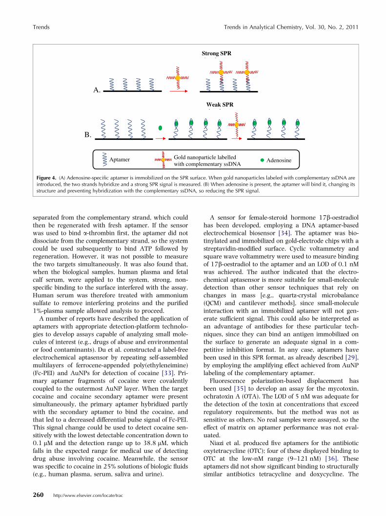
Alternative formats to structure switching have beendescribed. Wang and Zhou used a surface-plasmon res-onance (SPR) biosensor with the amplifying effect ofAuNPs to detect adenosine via surface-inhibition detec-tion [29]. The adenosine aptamer was immobilized on aSPR gold film and then hybridized with complementaryss-DNA that had been tagged with AuNPs, resulting in alarge change of SPR signal. However, the adenosineaptamer adopted a tertiary structure after adenosine wasadded to the SPR cell. In this formation, the aptamercould no longer hybridize with the tagged complemen-tary ss-DNA, so the SPR signal decreased proportionallyto the concentration of the small molecule (Fig. 4).
Successful integration of aptamers into a sensor hasalso been accomplished by using the two separated DNAstrands of an adenosine aptamer individually immobi-lized on AuNPs [30]. Interaction with adenosinereassembles the two aptamers causing aggregation of theAuNPs and a subsequent measurable color change. Xuand Lu designed a sensor comprising unbound malachitegreen (MG) and a double aptamer comprising MG andadenosine aptamers partially hybridized with a bridgingstrand [31]. Without adenosine, the affinity of the apt-amer for the MG is inhibited by the bridging strand.When adenosine is introduced, the aptamer binds to it,thereby dislodging the bridging strand. This allows theaptamer also to bind MG, leading to enhancement offluorescence.
While several model assays have been described, fewdemonstrate the effect of a real sample matrix on theperformance of the aptamers. A multi-functional, reus-able and label-free electrochemical assay has been re-ported, along with an investigation of sample-matrixeffects [32]. A mixed aptamer capable of binding ATPand a-thrombin formed a duplex with a partly comple-mentary strand, which was immobilized on a gold-elec-trode surface. After binding ATP, the mixed aptamer
http://www.elsevier.com/locate/trac 259
A.
Strong SPR i l
B.
Weak SPR i l
AdenosineGold nanoparticle labelled with complementary ssDNA
Aptamer
Figure 4. (A) Adenosine-specific aptamer is immobilized on the SPR surface. When gold nanoparticles labeled with complementary ssDNA areintroduced, the two strands hybridize and a strong SPR signal is measured. (B) When adenosine is present, the aptamer will bind it, changing itsstructure and preventing hybridization with the complementary ssDNA, so reducing the SPR signal.
Trends Trends in Analytical Chemistry, Vol. 30, No. 2, 2011
separated from the complementary strand, which couldthen be regenerated with fresh aptamer. If the sensorwas used to bind a-thrombin first, the aptamer did notdissociate from the complementary strand, so the systemcould be used subsequently to bind ATP followed byregeneration. However, it was not possible to measurethe two targets simultaneously. It was also found that,when the biological samples, human plasma and fetalcalf serum, were applied to the system, strong, non-specific binding to the surface interfered with the assay.Human serum was therefore treated with ammoniumsulfate to remove interfering proteins and the purified1%-plasma sample allowed analysis to proceed.
A number of reports have described the application ofaptamers with appropriate detection-platform technolo-gies to develop assays capable of analyzing small mole-cules of interest (e.g., drugs of abuse and environmentalor food contaminants). Du et al. constructed a label-freeelectrochemical aptasensor by repeating self-assembledmultilayers of ferrocene-appended poly(ethyleneimine)(Fc-PEI) and AuNPs for detection of cocaine [33]. Pri-mary aptamer fragments of cocaine were covalentlycoupled to the outermost AuNP layer. When the targetcocaine and cocaine secondary aptamer were presentsimultaneously, the primary aptamer hybridized partlywith the secondary aptamer to bind the cocaine, andthat led to a decreased differential pulse signal of Fc-PEI.This signal change could be used to detect cocaine sen-sitively with the lowest detectable concentration down to0.1 lM and the detection range up to 38.8 lM, whichfalls in the expected range for medical use of detectingdrug abuse involving cocaine. Meanwhile, the sensorwas specific to cocaine in 25% solutions of biologic fluids(e.g., human plasma, serum, saliva and urine).
260 http://www.elsevier.com/locate/trac
A sensor for female-steroid hormone 17b-oestradiolhas been developed, employing a DNA aptamer-basedelectrochemical biosensor [34]. The aptamer was bio-tinylated and immobilized on gold-electrode chips with astreptavidin-modified surface. Cyclic voltammetry andsquare wave voltammetry were used to measure bindingof 17b-oestradiol to the aptamer and an LOD of 0.1 nMwas achieved. The author indicated that the electro-chemical aptasensor is more suitable for small-moleculedetection than other sensor techniques that rely onchanges in mass [e.g., quartz-crystal microbalance(QCM) and cantilever methods], since small-moleculeinteraction with an immobilized aptamer will not gen-erate sufficient signal. This could also be interpreted asan advantage of antibodies for these particular tech-niques, since they can bind an antigen immobilized onthe surface to generate an adequate signal in a com-petitive inhibition format. In any case, aptamers havebeen used in this SPR format, as already described [29],by employing the amplifying effect achieved from AuNPlabeling of the complementary aptamer.
Fluorescence polarization-based displacement hasbeen used [35] to develop an assay for the mycotoxin,ochratoxin A (OTA). The LOD of 5 nM was adequate forthe detection of the toxin at concentrations that exceedregulatory requirements, but the method was not assensitive as others. No real samples were assayed, so theeffect of matrix on aptamer performance was not eval-uated.
Niazi et al. produced five aptamers for the antibioticoxytetracycline (OTC); four of these displayed binding toOTC at the low-nM range (9–121 nM) [36]. Theseaptamers did not show significant binding to structurallysimilar antibiotics tetracycline and doxycycline. The

Trends in Analytical Chemistry, Vol. 30, No. 2, 2011 Trends
aptamers were not applied to a detection assay or eval-uated for the effects of sample matrix.
Others have used an aptamer for neomycin B to de-velop a SPR-based biosensor assay [37]. The antibioticwas immobilized onto an activated gold-sensor surfaceand interaction with the aptamer created a measurableresponse. The competitive assay allowed quantificationbetween 10 nM and 100 lM with the prediction that theassay would allow detection of neomycin B in animalsamples, despite the fact that the effect of sample matrixwas not investigated.
An RNA-aptamer-based fluorescence assay to detectMG and the leucomalachite metabolite in fish [38] hasalso been developed. The assay was capable of detectingMG residues to well below 2 lg/kg in test samples. Furtheradvantages of the technique are the high tolerance of theaptamer to solvent and its stability when stored at 4�C.
5. Protein scaffolds
The idea to use protein scaffolds as molecular binders wasto copy what nature had already designed but withimprovements, where possible. The structure of IgG mol-ecules allows antigen recognition due to the combinationof a structurally-conserved framework supporting a spa-tially-defined binding site composed of peptide segmentsthat are hypervariable in both sequence and conforma-tion [39]. By selection of a very stable protein structurethat can tolerate substitution, insertion or deletion ofvariable domains, specific binding regions can thereforebe selected through recombinant-protein techniques. Theprotein scaffolds chosen are usually smaller than IgG,more robust, easily modified and less expensive to pro-duce. Although over 50 scaffolds have been identified,their long-term applicability still needs to be proved.
Affibodies are binding proteins that make use of pro-tein A as the scaffold; the 6-kDa structure comprises 58amino acids with 13 randomized to create the bindingdomain [40]. While their ability to bind peptides hasbeen demonstrated [41], they have yet to be shown tobind small molecules.
The same reservation applies to the designed ankyrin-repeat protein (DARPin) scaffold [42]. The 14-kDa bin-der has been shown to bind proteins [43] but not smallmolecules. DARPins have displayed some potential torival and to surpass antibody-based approaches to drugsfor therapeutic use [44].
By contrast, the protein lipocalin, which acts as abiological transporter of steroids, lipids and bilin, hasbeen employed as a binder, termed anticalin, of smallmolecules [45]. The bilin-binding protein from the LargeWhite butterfly, Pieris brassicae, was structurally ad-justed to bind the steroid digoxigenin [46]. A total of 17amino-acid substitutions within the binding siteproduced structural changes in the four loops that form
the entrance to the ligand pocket on top of the structur-ally conserved b-barrel framework, allowing complexa-tion with digoxigenin. Anticalins possess considerablymore amino acids (160–180) than affibodies andDARPins. The binding domain comprises 16 randomizedamino acids producing four loops with few disulfidebonds. It is the relative complexity of the binding domainthat presumably allows small-molecule interaction.
Ramoni et al. investigated the binding capacities offour forms of lipocalin odorant-binding protein (OBP) forthe detection of low-molecular-weight components ofexplosives (e.g., diphenylamine, dimethylphthalate, res-orcinol and dinitrotoluene) [47]. The OBP bound thesecompounds with affinity constants in the range 80 nM–10.6 mM, indicating (according to the authors) that OBPcould be used as a probe for the realization of a biosensorto sense explosive compounds.
Kim et al. [48] engineered human lipocalin, whichnaturally scavenges bacterial siderophores (iron chelat-ing compounds), to bind rare-earth and related metalions specifically as chelate complexes with [(R)-2-amino-3-(4-aminophenyl)propyl]-trans-(S,S)-cyclohexane-1,2-diaminepentaacetic acid (p-NH2-Bn-CHX-A00-DTPA).The anticalin produced was able to recognize benzyl-substituted cyclohexyl-DTPA-chelate complexes of yt-trium (Y3+) and related lanthanide ions. The moleculardocking module for chelated trivalent metal ions,including radioactive rare-earth elements, displays po-tential use in the field of nuclear medicine.
6. Synthetic alternatives to antibody-basedmolecular recognition
As well as the biological-based binders for small mole-cules described above, much research has focused on thepotential for artificial molecules and materials to achieveselective recognition of molecules ranging in size fromsmall organics up to proteins. The advantages offered byselective artificial materials over their biological coun-terparts often include ease of production, greater chem-ical and thermal stability, and greater batch-to-batchreproducibility. Synthetic binders may also be suitablefor specific recognition of smaller organic molecules,which may not provoke an adequate immunogenic re-sponse in order to obtain suitable antibodies. We discusshere some sensor applications wherein synthetic substi-tutes for antibodies are employed.
Careful design of synthetic molecules represents onemeans of generating species with a predetermined selec-tivity, which can then be incorporated into a sensingapplication. An excellent example of this is the array ofchemoresponsive dyes, based on metalloporphyrins,developed by Suslick and co-workers [49]. This demon-strated lg/kg sensitivity towards a number of volatileorganic compounds (VOCs), including amines, carboxylic
http://www.elsevier.com/locate/trac 261
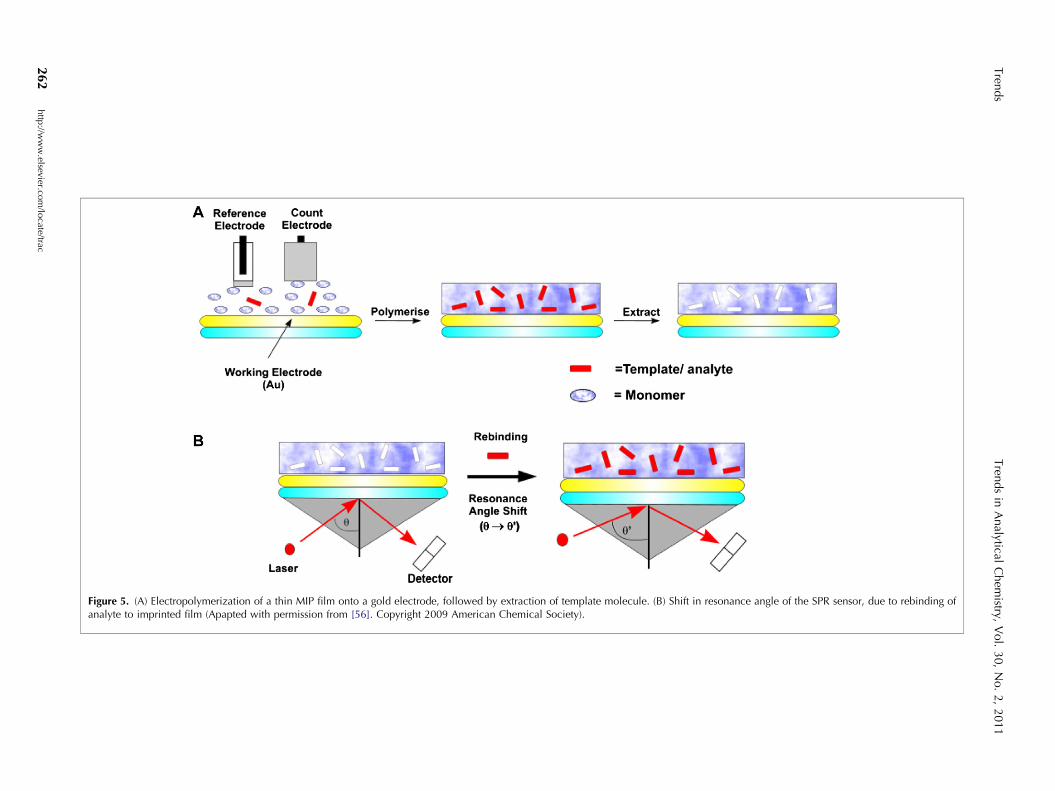
Figure 5. (A) Electropolymerization of a thin MIP film onto a gold electrode, followed by extraction of template molecule. (B) Shift in resonance angle of the SPR sensor, due to rebinding ofanalyte to imprinted film (Apapted with permission from [56]. Copyright 2009 American Chemical Society).
Tren
ds
Tren
ds
inA
nalytical
Chem
istry,V
ol.
30,
No.
2,
2011
262
http
://ww
w.elsevier.co
m/lo
cate/trac
Trends in Analytical Chemistry, Vol. 30, No. 2, 2011 Trends
acids and thiols. The technique was applied to the anal-ysis of VOCs in complex mixtures, including beers.
An azo-dye-reporter group for amines has also beenintegrated into a dendrimer to create a monomolecularimprinted species [50]. Changes in absorbance responseof the dendrimer could then be used as a means ofdetecting the presence of individual amine compounds.
Dendritic polymers have also been used for detectionof chemical-warfare agents, based on a fluorescent re-sponse to binding [51].
The sensing of heavy metals (e.g., mercury and silver)using calixarene-bridged binuclear phthalocyanines hasshown promise [52].
Molecularly Imprinted Polymers (MIPs) offer anaccessible method of creating substrate-specific materi-als. Their use as selective sorbents in chromatographicapplications has been extensively exploited for a widerange of analytes, particularly in SPE [53]. Some of themore novel applications have included the developmentof MIP-based sensing protocols [54].
Piezoelectric sensing using MIPs has frequently beenexamined, and a review of this area has been published[55]. However, in general, transduction of the bindingevent into a detectable signal requires careful MIP design,which may account for the fewer examples in the liter-ature of MIP-based sensing applications than applicationsof MIPs in separation methods. We here focus on three ofthe more salient areas of analyte detection – naturaltoxins, chemical warfare agents, and drugs of abuse.
SPR has proved to be one of the more adaptable tech-niques in the area of MIP-based sensing (Fig. 5), and hasbeen used to determine the presence of the mycotoxin,zearalenone (ZEN) [56]. Electropolymerization of pyrroleonto the surface of a gold chip in the presence of ZENproduced a highly specific SPR-sensing assay, with alinear range of 0.3–3000 ng/mL. The imprinted film dis-played selectivity efficiency of 100% towards ZEN and 15–27% for a number of structurally analogous compounds.
Alternatively, a photo-initiator can be immobilized onthe gold surface of the chip, to produce a thin polymerlayer (40 nm) suitable for SPR measurements. Thisapproach was employed by Lotierzo and co-workers toproduce a SPR assay specific for neurotoxin domoic acid,which can cause amnesic shellfish poisoning (ASP). Theassay had a detection range of 5–1000 lg/L in buffer [57].
Detection of endocrine-disrupting compound bisphenolA was achieved using a piezoelectric approach. Self-assembled monolayers of 2-aminoethanethiol were usedas a supporting substrate on the gold surface of a QCM chipfor a methacrylate polymer selective for bisphenol A [58].
Novel synthetic binders to OTA have been reported forthe preparation of affinity-based ‘‘clean-up’’ procedures.Giraudi et al. reported on the combinational synthesis ofa hexapeptide binder for the isolation of OTA from dif-ferent wines [59]. The limit of quantitation of themethod was 0.1 lg/L.
MIPs are well-suited to selective recognition of small,structurally-defined molecules {e.g., organophosphatepesticides [60] and chemical-warfare agents [61]}. Dueto the safety implications of working directly withchemical-warfare agents, methods have typically focusedon model compounds, namely less toxic analogues [62]or degradation products [63].
The MIPs technique has also proved amenable todetection of nitroaromatics [64], another significant setof analytes for counter-terrorism.
Prathish et al. developed a selective potentiometricsensor for methylphosphonic acid, a degradation productof various organophosphate-nerve agents [62]. Amethacrylate polymer was imprinted with methylphos-phonic acid, and the ground polymer was incorporatedinto a PVC-membrane sensor. The authors reported anLOD of 4.8 ng/mL. The sensor displayed a cross-reactivity of 0% up to 45% when exposed to compounds,including buffers and other organophosphates.
A different approach to generating an electro-chemical response via a MIP-recognition element wasdeveloped by Xie and co-workers for organophosphatepesticide chlorpyrifos [60]. Glassy-carbon electrodeswere modified with AuNPs, and, on this surface, elec-tropolymerizable p-aminothiophenol molecules wereassembled; the chlorpyrifos template was allowed tointeract with the monolayer of aminothiophenol,which was then electropolymerized, creating theimprint. Sensitivity of the method was of the order of�2000 ng/mL.
As an alternative to conventional methacrylate poly-mers, Taraneker et al. developed a dendrimer-basedmolecular recognition assay for detecting pinacolylmethylphosphonate (PMP), which is a breakdownproduct and analogue of toxic nerve agents [65]. Resi-dues could be detected at nM and pM levels using SPRand electrochemical detection, respectively.
Prathish and co-workers developed a novel monolithicMIP membrane for the electrochemical detection of di-ethyl chlorophosphate, which is a less toxic analogue ofnerve agents [66]. The LOD of the assay was 170 lg/L inaqueous solution.
A technical challenge in the use of conventionalmethacrylate-imprinted polymers is obtaining a suitableformat for sensor application. Bulk polymers, as typicallycreated for chromatographic use, are unsuitable, due todifficulty in immobilizing these polymers and poormass-transfer properties for the rapid binding/transduc-tion processes required in a sensing approach.
For piezoelectric sensing {e.g., the QCM work per-formed by Dickert and co-workers [67]}, polystyrene andpolyurethane layers have been spin-coated onto theQCM chip.
Suspensions of polymeric particles can also be sprayedonto the QCM chip, as described in a MIP-based TNT-sensing application [64].
http://www.elsevier.com/locate/trac 263
(B)
Direct immobilisation of MBP-VL
Add sample/ VH-AP
(A)
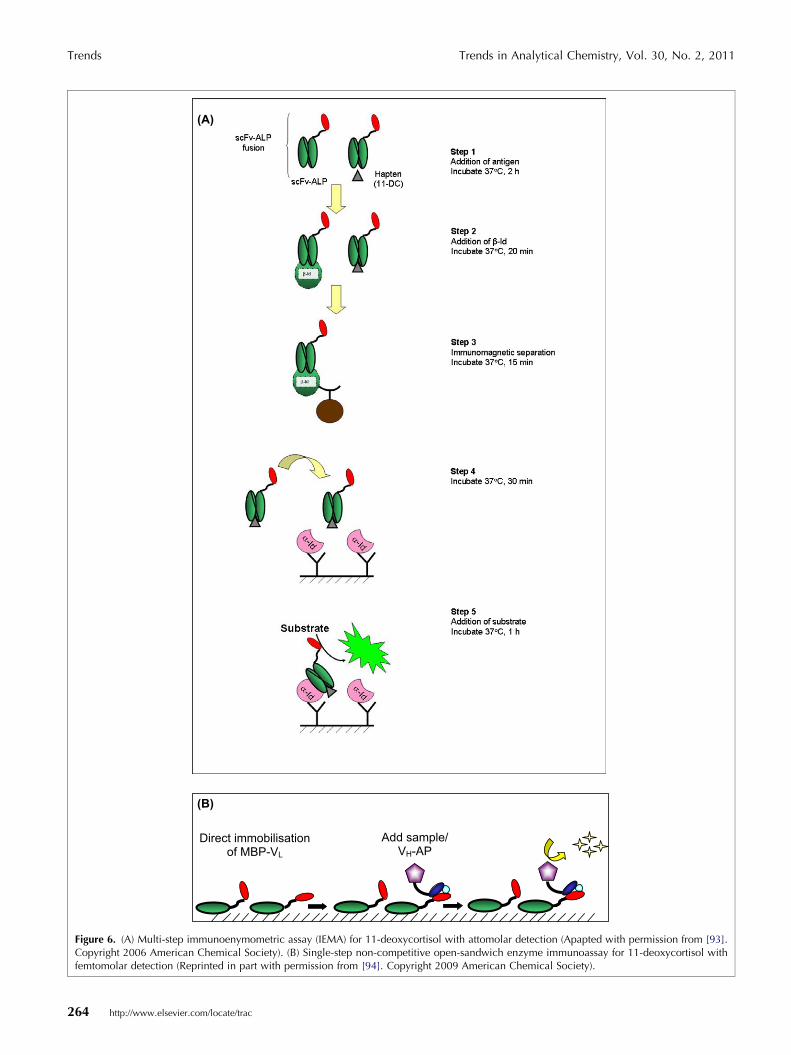
Figure 6. (A) Multi-step immunoenymometric assay (IEMA) for 11-deoxycortisol with attomolar detection (Apapted with permission from [93].Copyright 2006 American Chemical Society). (B) Single-step non-competitive open-sandwich enzyme immunoassay for 11-deoxycortisol withfemtomolar detection (Reprinted in part with permission from [94]. Copyright 2009 American Chemical Society).
Trends Trends in Analytical Chemistry, Vol. 30, No. 2, 2011
264 http://www.elsevier.com/locate/trac

Trends in Analytical Chemistry, Vol. 30, No. 2, 2011 Trends
Use of biological recognition elements is particularlyprevalent in rapid screening assays for the detection ofdrugs in blood, including drugs of abuse [68]. However,the complex nature of the sample matrix in biologicalsamples can pose a problem for the analyst, and selectiveclean up of samples using tailored stationary phasesoffers an attractive alternative to conventional analyticalstrategies.
One of the more commonly addressed analyticalchallenges in this field is the detection of morphine, anopioid. A means of colorimetric detection of morphineusing a methacrylate MIP [69] has been reported.Rebinding of morphine to the polymer, in the presence ofK+, Fe3+ and [Fe(CN)6]3�, leads to the bound morphineacting as a reducing agent and thus causing precipita-
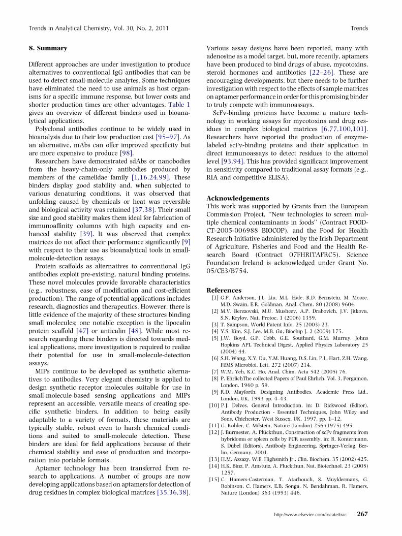
Table 1. Overview of the characteristics of different binders and their per
Binder Performance examples Means of product
Polyclonalantibody
Biacore-based assaywith LOD of 972 pg/mLfor morphine-3-glucuronide [95],3 ng/mL for aflatoxin B1[96] and 5 ng/mL forbenzimidazoles in milk[97].
Collection of serumimmunized host anlow-molecular-weigmolecule must beconjugated to carrie
Monoclonalantibody
Biosensor assay withLOD of 4 ng/mL warfarinin plasma ultrafiltrate[98]
Fusion of antibody-producing cells (splfrom immunized anwith myeloma-cell
Nanobody LOD for ricin =1.6 ng/mL [1];IC50 for 15-AC-DON = 1.24–0.5 lM[23]
Lymphocytes fromimmunized animalsto prepare sdAb libbinders selected byphage display
MIP LOD forzearalenone = 0.3 ng/g[56]; LOD for domoicacid = 5 lg/L [57].
Co-polymerizationmonomeric speciespresence of templattemplate analogue.
Aptamer LOD forcocaine = 0.1 lM [33];LOD foroestradiol = 0.1 nM [34];LOD for OTA = 5 nM[35]
Selected by SELEX ulibraries of largenumbers of randomoligonucleotides
Proteinscaffolds:AffibodyDARPinAnticalin
Anticalins for explosives:affinityconstants 80 nM–10.6 mM [47];Anticalins for lanthanide(III) chelates: affinityconstants2.77–95 nM [48]
Isolated from largeprotein libraries byphage display; yeastwo-hybrid; rationadesign; ribosome di
scFv Biosensor assay withLOD of 195 pg/mL freemorphine [100] and390 pg/mL aflatoxin B1[101].
Antibody genes clofrom immunized mand scFv-antibodyfragments selectedphage display
tion of Prussian Blue and generating a colorimetricresponse. The technique permitted detection of morphinedown to <3 ng/mL. An amperometric approach todesigning a morphine sensor incorporating a MIP, byYeh and Ho [7], offered an LOD of <6 ng/mL.
Detection of methamphetamine down to 1 mg/mL wasachieved by Romero Guerra et al. by immobilizing a MIPfilm on a QCM chip, employing a monomer mixturedesigned computationally [70].
7. ScFvs and their selected applications
In this section, we discuss the application of scFvs in thearea of trace analysis. Several scFvs have been developed
formance in bioanalytical applications
ion Advantages Disadvantages
fromimal;ht
r
Relatively inexpensive toproduce
Requirement ofconjugation may becomplicated, dependingon structure; productionof binder is in vivo; nocontrol over specificity;
een)imalline
Highly specific toantigen
in vivo production;expensive to produce
usedrary,
Robust to sample matrixeffects [1]; thermallystable [21,22]
in vivo production (butonly blood samplerequired); time-consuming production
ofine or
Inexpensive; chemicallyand thermally robust.
Materials can display arange of bindingaffinities towards targetspecies; less compatiblewith water than withorganic solvent.
sing Production is in vitro;tolerance to solvent [37]
Problematic conversionof binding event intodetectable signal; maybe susceptible to samplematrix effects [32,33](inconclusive evidence)
tlsplay
Production is in vitro Not many developed tobind small molecules,with notable exceptionof anticalins
nedice
by
Prokaryotic production,high-level expression,affinity tags incorporatedfor purification anddetection
Are usually less stablethan full-length IgG andhave a tendency toaggregate
http://www.elsevier.com/locate/trac 265

Trends Trends in Analytical Chemistry, Vol. 30, No. 2, 2011
to small molecules since the late 1990s. Some earliersuccessful applications were reported in recent reviewson immunoassays for aflatoxins [71] and sulfonamides[72]. As a result, these classes of molecules are not dis-cussed in this section, which covers mycotoxins, marinetoxins, hormones and agrochemicals.
The production of an scFv to deoxynivalenol (DON)and comparison with its parent mAb was reported [73].The binding affinity of the scFv was found to be weakerthan the mAb but dissociation from the chip surface ofan SPR biosensor was relatively fast.
Wang et al. reported the development of an anti-DONscFv, which had IC50 values of 8.2 ng/mL, 98.5 ng/mLand 153 ng/mL for DON, 3-AC-DON and 15-AC-DON,respectively [6]. No cross-reactivity was seen towardsnivalenol or T-2 toxin. The antibody showed improvedsensitivity to 3-AC-DON (�12%) and 15-AC-DON (27%)when compared to the parent mAb.
A comprehensive evaluation of the anti-3-AC-DONscFv was carried out through application to spiked andnaturally-contaminated samples. The results achievedcorrelated well against a commercial ELISA kit. Previ-ously, the production of an anti-ZEN scFv had IC50s of14 ng/mL and 17–54 ng/mL, for ZEN and four ZENanalogs, respectively [74]. Sensitivity was improved by afactor of three to nine through production of the scFv.However, this improvement was offset by the lower tol-erance of the scFv to methanol.
Other groups have subsequently produced anti-ZENscFvs and optimized the process to increase the yield ofbinding protein [75,76]. Wang et al. recently developeda bispecific scFv antibody through linking anti-DON andanti-ZEN scFvs [77]. The resulting antibody allowed thesimultaneous analysis of DON and ZEN residues after asingle extraction. The bispecific scFv was compared tothe original scFvs through application to a range ofnaturally-contaminated wheat samples. The productionof an anti-fumonisin scFv was accomplished, with thefragments displaying affinity towards B1 and B2 analogs[78].
Min et al. recently reported the development of ananti-fumonisin scFv [79]. However, the scFv showed 12times less binding affinity than its mAb and it wasconcluded that it required further optimization prior topractical application in working assays.
The development of binding proteins for detection ofmicrocystin (MC) toxins [80] offers good examples of theuse of phage-display libraries for the production of anti-MC scFvs [81,82]. Drake et al. recently reported theproduction of recombinant anti-MC binders usingtransgenic tobacco plants [83]. The anti-MC bindingprotein was suitable for the detection of microcystin-LRto <1 lg/L (WHO guideline level for water). An anti-domoic acid scFv was produced after pre-immunizationof chicken [84]. The scFv was improved 10-fold throughengineering to give an IC50 value of 150 ng/mL. The
266 http://www.elsevier.com/locate/trac
scFv was subsequently immobilized on a mesoporoussilica support to further the development of a biosensorfor environmental monitoring [85]. Garet and co-work-ers produced an scFv to the potent marine toxin, paly-toxin, using phage-display libraries [86]. The IC50 valuefor the scFv was 100 ng/mL. A sensitive procedure wasdeveloped to allow detection of palytoxin at levels downto 2 lg/g in clams and mussels.
There have been a limited number of applications inagrochemical-residue analysis. Huet et al. reported theevaluation of different polyclonal antibodies and anengineered antibody to fluoroquinolone antibiotics [87].The best results were achieved using a polyclonal anti-body. Wang and co-workers produced a highly-specificanti-carbofuran scFv [88]. No cross-reactivity wasshown to other carbamate residues, but sensitivity wasexcellent with an IC50 of 1 ng/mL. The generation ofalternatives to costly mass spectrometry-based assays forgrowth-promoting agents continues with the develop-ment of novel binders for application in screening as-says. Pan and co-workers produced an anti-clenbuterolscFv using phage-display libraries, showing that sensi-tivity of the scFv (IC50 = 0.78 ng/mL) could be signifi-cantly better than that of the mAb (IC50 = 1.34 ng/mL)[89].
A number of applications have been developed in thearea of clinical chemistry. These also have potential foruse in the areas of veterinary drug-residue analysis andsports forensics. An anti-17b-estradiol scFv was recentlydeveloped [90], as were artificial single-domain frag-ments (sdAbs) [91] through antibody engineering. Thebinding affinity of the scFv (Ka = 2.6 · 108/M) wasfound to be higher than the sdAb (Ka = 1 · 106/M). Inthe subsequent work, the affinity of the scFv(Ka = 6.3 · 108/M) was further improved [92]. Initialscreening showed that 16-fold improvement in sensi-tivity could be demonstrated with the scFv(IC50 = 0.56 ng) compared to the mAb (IC50 = 9.0 ng).Working assays developed showed that higher sensitiv-ities with IC50s of 21 pg and 0.47 pg could be achievedon radioimmunoassay (RIA) and competitive ELISAformats, respectively. In other work, the same groupdeveloped novel immunoassays for the detection of thecorticosteroid 11-deoxycortisol (11-DC) using an scFvwith high binding affinity (Ka = 1.3 · 1010/M) [93]. Theanti-11-DC binding protein was genetically linked withthe enzyme alkaline phosphatase (ALP) to generate anenzyme-antibody fusion protein. This approach alloweddevelopment of a multi-step, non-competitive, immuno-enzymometric assay (IEMA) with an LOD of 20 attomol(Fig. 6a). The assay has a wide working range of 10 fg–100 ng per microwell. The suitability of the assay wasevaluated through analysis of fortified serum samples.The same group subsequently reported the developmentof an simple one-step ‘‘open sandwich’’ immunoassay(OS-IA) for 11-DC (Fig. 6b) [94].
Trends in Analytical Chemistry, Vol. 30, No. 2, 2011 Trends
8. Summary
Different approaches are under investigation to producealternatives to conventional IgG antibodies that can beused to detect small-molecule analytes. Some techniqueshave eliminated the need to use animals as host organ-isms for a specific immune response, but lower costs andshorter production times are other advantages. Table 1gives an overview of different binders used in bioana-lytical applications.
Polyclonal antibodies continue to be widely used inbioanalysis due to their low production cost [95–97]. Asan alternative, mAbs can offer improved specificity butare more expensive to produce [98].
Researchers have demonstrated sdAbs or nanobodiesfrom the heavy-chain-only antibodies produced bymembers of the camelidae family [1,16,24,99]. Thesebinders display good stability and, when subjected tovarious denaturing conditions, it was observed thatunfolding caused by chemicals or heat was reversibleand biological activity was retained [37,38]. Their smallsize and good stability makes them ideal for fabrication ofimmunoaffinity columns with high capacity and en-hanced stability [39]. It was observed that complexmatrices do not affect their performance significantly [9]with respect to their use as bioanalytical tools in small-molecule-detection assays.
Protein scaffolds as alternatives to conventional IgGantibodies exploit pre-existing, natural binding proteins.These novel molecules provide favorable characteristics(e.g., robustness, ease of modification and cost-efficientproduction). The range of potential applications includesresearch, diagnostics and therapeutics. However, there islittle evidence of the majority of these structures bindingsmall molecules; one notable exception is the lipocalinprotein scaffold [47] or anticalin [48]. While most re-search regarding these binders is directed towards med-ical applications, more investigation is required to realizetheir potential for use in small-molecule-detectionassays.
MIPs continue to be developed as synthetic alterna-tives to antibodies. Very elegant chemistry is applied todesign synthetic receptor molecules suitable for use insmall-molecule-based sensing applications and MIPsrepresent an accessible, versatile means of creating spe-cific synthetic binders. In addition to being easilyadaptable to a variety of formats, these materials aretypically stable, robust even to harsh chemical condi-tions and suited to small-molecule detection. Thesebinders are ideal for field applications because of theirchemical stability and ease of production and incorpo-ration into portable formats.
Aptamer technology has been transferred from re-search to applications. A number of groups are nowdeveloping applications based on aptamers for detection ofdrug residues in complex biological matrices [35,36,38].
Various assay designs have been reported, many withadenosine as a model target, but, more recently, aptamershave been produced to bind drugs of abuse, mycotoxins,steroid hormones and antibiotics [22–26]. These areencouraging developments, but there needs to be furtherinvestigation with respect to the effects of sample matriceson aptamer performance in order for this promising binderto truly compete with immunoassays.
ScFv-binding proteins have become a mature tech-nology in working assays for mycotoxins and drug res-idues in complex biological matrices [6,77,100,101].Researchers have reported the production of enzyme-labeled scFv-binding proteins and their application indirect immunoassays to detect residues to the attomollevel [93,94]. This has provided significant improvementin sensitivity compared to traditional assay formats (e.g.,RIA and competitive ELISA).
AcknowledgementsThis work was supported by Grants from the EuropeanCommission Project, ‘‘New technologies to screen mul-tiple chemical contaminants in foods’’ (Contract FOOD-CT-2005-006988 BIOCOP), and the Food for HealthResearch Initiative administered by the Irish Departmentof Agriculture, Fisheries and Food and the Health Re-search Board (Contract 07FHRITAFRC5). ScienceFoundation Ireland is acknowledged under Grant No.05/CE3/B754.
References[1] G.P. Anderson, J.L. Liu, M.L. Hale, R.D. Bernstein, M. Moore,
M.D. Swain, E.R. Goldman, Anal. Chem. 80 (2008) 9604.
[2] M.V. Berezovski, M.U. Musheev, A.P. Drabovich, J.V. Jitkova,
S.N. Krylov, Nat. Protoc. 1 (2006) 1359.
[3] T. Sampson, World Patent Info. 25 (2003) 23.
[4] Y.S. Kim, S.J. Lee, M.B. Gu, Biochip J. 2 (2009) 175.
[5] J.W. Boyd, G.P. Cobb, G.E. Southard, G.M. Murray, Johns
Hopkins APL Technical Digest, Applied Physics Laboratory 25
(2004) 44.
[6] S.H. Wang, X.Y. Du, Y.M. Huang, D.S. Lin, P.L. Hart, Z.H. Wang,
FEMS Microbiol. Lett. 272 (2007) 214.
[7] W.M. Yeh, K.C. Ho, Anal. Chim. Acta 542 (2005) 76.
[8] P. EhrlichThe collected Papers of Paul Ehrlich, Vol. 3, Pergamon,
London, 1960 p. 59.
[9] R.D. Mayforth, Designing Antibodies, Academic Press Ltd.,
London, UK, 1993 pp. 4–43.
[10] P.J. Delves, General Introduction, in: D. Rickwood (Editor),
Antibody Production - Essential Techniques, John Wiley and
Sons, Chichester, West Sussex, UK, 1997, pp. 1–12.
[11] G. Kohler, C. Milstein, Nature (London) 256 (1975) 495.
[12] J. Burmester, A. Pluckthun, Construction of scFv fragments from
hybridoma or spleen cells by PCR assembly, in: R. Kontermann,
S. Dubel (Editors), Antibody Engineering, Springer-Verlag, Ber-
lin, Germany, 2001.
[13] H.M. Azzazy, W.E. Highsmith Jr., Clin. Biochem. 35 (2002) 425.
[14] H.K. Binz, P. Amstutz, A. Pluckthun, Nat. Biotechnol. 23 (2005)
1257.
[15] C. Hamers-Casterman, T. Atarhouch, S. Muyldermans, G.
Robinson, C. Hamers, E.B. Songa, N. Bendahman, R. Hamers,
Nature (London) 363 (1993) 446.
http://www.elsevier.com/locate/trac 267
Trends Trends in Analytical Chemistry, Vol. 30, No. 2, 2011
[16] S. Muyldermans, C. Cambillau, L. Wyns, Trends Biochem. Sci. 26
(2001) 230.
[17] A. Desmyter, K. Decanniere, S. Muyldermans, L. Wyns, J. Biol.
Chem. 276 (2001) 26285.
[18] I.G. Lange, A. Daxenberger, H.H. Meyer, Vet. Immunol. Immu-
nopathol. 83 (2001) 1.
[19] R. van der Linden, B. de Geus, W. Stok, W. Bos, D. van
Wassenaar, T. Verrips, L. Frenken, J. Immunol. Methods 240
(2000) 185.
[20] S. Spinelli, M. Tegoni, L. Frenken, C. van Vliet, C. Cambillau, J.
Mol. Biol. 311 (2001) 123.
[21] R.C. Ladenson, D.L. Crimmins, Y. Landt, J.H. Ladenson, Anal.
Chem. 78 (2006) 4501.
[22] G.P. Anderson, E.R. Goldman, J. Immunol. Methods 339 (2008)
47.
[23] P.J. Doyle, M. Arbabi-Ghahroudi, N. Gaudette, G. Furzer, M.E.
Savard, S. Gleddie, M.D. McLean, C.R. Mackenzie, J.C. Hall, Mol.
Immunol. 45 (2008) 3703.
[24] E.J. Franco, G.J. Sonneson, T.J. DeLegge, H. Hofstetter, J.R. Horn,
O. Hofstetter, J. Chromatogr., B 878 (2010) 177.
[25] G.J. Sonneson, J.R. Horn, Biochem 48 (2009) 6693.
[26] R. Stoltenburg, C. Reinemann, B. Strehlitz, Biomol. Eng. 24
(2007) 381.
[27] S. Zhang, J. Xia, X. Li, Anal. Chem. 80 (2008) 8382.
[28] C. Deng, J. Chen, L. Nie, Z. Nie, S. Yao, Anal. Chem. 81 (2009)
9972.
[29] J. Wang, H.S. Zhou, Anal. Chem. 80 (2008) 7174.
[30] F. Li, J. Zhang, X. Cao, L. Wang, D. Li, S. Song, B. Ye, C. Fan,
Analyst (Cambridge, UK) 134 (2009) 1355.
[31] W. Xu, Y. Lu, Anal. Chem. 82 (2009) 574.
[32] Y. Du, B. Li, H. Wei, Y. Wang, E. Wang, Anal. Chem. 80 (2008)
5110.
[33] Y. Du, C. Chen, J. Yin, B. Li, M. Zhou, S. Dong, E. Wang, Anal.
Chem. 82 (2010) 1556.
[34] Y.S. Kim, H.S. Jung, T. Matsuura, H.Y. Lee, T. Kawai, M.B. Gu,
Biosens. Bioelectron. 22 (2007) 2525.
[35] J.A. Cruz-Aguado, G. Penner, J. Agric. Food Chem. 56 (2008)
10456.
[36] J.H. Niazi, S.J. Lee, Y.S. Kim, M.B. Gu, Bioorgan. Med. Chem. 16
(2008) 1254.
[37] N. de-los-Santos-Alvarez, M.J. Lobo-Castanon, A.J. Miranda-
Ordieres, P. Tunon-Blanco, Biosens. Bioelectron. 24 (2009)
2547.
[38] S.L. Stead, H. Ashwin, B. Johnston, J.A. Tarbin, M. Sharman, J.
Kay, B.J. Keely, Anal. Chem. 82 (2010) 2652.
[39] A. Skerra, Curr. Opin. Biotechnol. 18 (2007) 295.
[40] P.A. Nygren, FEBS J. 275 (2008) 2668.
[41] C. Gronwall, A. Jonsson, S. Lindstrom, E. Gunneriusson, S. Stahl,
N. Herne, J. Biotechnol. 128 (2007) 162.
[42] H.K. Binz, M.T. Stumpp, P. Forrer, P. Amstutz, A. Pluckthun, J.
Mol. Biol. 332 (2003) 489.
[43] J. Li, A. Mahajan, M.D. Tsai, Biochemistry 45 (2006) 15168.
[44] M.T. Stumpp, H.K. Binz, P. Amstutz, Drug Discov. Today 13
(2008) 695.
[45] S. Schlehuber, G. Beste, A. Skerra, J. Mol. Biol. 297 (2000) 1105.
[46] I.P. Korndorfer, S. Schlehuber, A. Skerra, J. Mol. Biol. 330
(2003) 385.
[47] R. Ramoni, S. Bellucci, I. Grycznyski, Z. Grycznyski, S. Grolli, M.
Staiano, G. De Bellis, F. Micciulla, R. Pastore, A. Tiberia, V. Conti,
E. Merli, A. Varriale, M. Rossi, S. D�Auria, J. Phys. Condens. Mat.
19 (2007) 395012.
[48] H.J. Kim, A. Eichinger, A. Skerra, J. Am. Chem. Soc. 131 (2009)
3565.
[49] K.S. Suslick, N.A. Rakow, A. Sen, Tetrahedron 60 (2004)
11133.
[50] E. Mertz, S.L. Elmer, A.M. Balija, S.C. Zimmerman, Tetrahedron
60 (2004) 11191.
268 http://www.elsevier.com/locate/trac
[51] C. Hartmann-Thompson, D.L. Keeley, J.R. Rousseau, P.R. Dvor-
nic, J. Polym. Sci., Part A: Polym. Chem. 47 (2009) 5101.
[52] S. O�Malley, B. Schazmann, D. Diamond, K. Nolan, Tetrahedron
Lett. 48 (2007) 9003.
[53] E. Caro, R.M. Marce, F. Borrull, P.A.G. Cormack, D.C. Sherring-
ton, Trends Anal. Chem. 25 (2006) 143.
[54] E.L. Holthoff, F.V. Bright, Anal. Chim. Acta 594 (2007) 147.
[55] Y. Uludag, S.A. Piletsky, A.P.F. Turner, M.A. Cooper, FEBS J. 274
(2007) 5471.
[56] S.W. Choi, H.J. Chang, N. Lee, J.H. Kim, H.S. Chun, J. Agric. Food
Chem. 57 (2009) 1113.
[57] M. Lotierzo, O.Y.F. Henry, S. Piletsky, I. Tothill, D. Cullen, M.
Kania, B. Hock, A.P.F. Turner, Biosens. Bioelectron. 20 (2004)
145.
[58] N. Tsuru, M. Kikuchi, H. Kawaguchi, S. Shiratori, Thin Solid
Films 499 (2006) 380.
[59] G. Giraudi, L. Anfossi, C. Baggiani, C. Giovannoli, C. Tozzi, J.
Chromatogr., A 1175 (2007) 174.
[60] C. Xie, H. Li, S. Li, J. Wu, Z. Zhang, Anal. Chem. 82 (2009) 241.
[61] L. Malosse, P. Buvat, D. Ades, A. Siove, Analyst (Cambridge, UK)
133 (2008) 588.
[62] K.P. Prathish, K. Prasad, T.P. Rao, M.V.S. Suryanarayana,
Talanta 71 (2007) 1976.
[63] S. Le Moullec, A. Begos, V. Pichon, B. Bellier, J. Chromatogr., A.
1108 (2006) 7.
[64] G. Bunte, J. Hurttlen, H. Pontius, K. Hartlieb, H. Krause, Anal.
Chim. Acta 591 (2007) 49.
[65] P. Taranekar, A. Baba, J.Y. Park, T.M. Fulghum, R. Advincula,
Adv. Funct. Mater. 16 (2006) 2000.
[66] K.P. Prathish, V. Vishnuvardhan, T.P. Rao, Electroanalysis (NY)
21 (2009) 1048.
[67] F.L. Dickert, P. Lieberzeit, S.G. Miarecka, K.J. Mann, O. Hayden,
C. Palfinger, Biosens. Bioelectron. 20 (2004) 1040.
[68] K. Lachenmeier, F. Musshoff, B. Madea, Forensic Sci. Int. 159
(2006) 189.
[69] H.C. Hsu, L.C. Chen, K.C. Ho, Anal. Chim. Acta 504 (2004)
141.
[70] M.R. Guerra, I. Chianella, E.V. Piletska, K. Karim, A.P.F. Turner,
S.A. Piletsky, Analyst (Cambridge, UK) 134 (2009) 1565.
[71] P. Li, Q. Zhang, W. Zhang, Trends Anal. Chem. 28 (2009) 1115.
[72] H. Zhang, S. Wang, J. Immunol. Methods 350 (2009) 1.
[73] G.H. Choi, D.H. Lee, W.K. Min, Y.J. Cho, D.H. Kweon, D.H. Son,
K. Park, J.H. Seo, Protein Expres. Purif. 35 (2004) 84.
[74] Q. Yuan, J.R. Clarke, H.R. Zhou, J.E. Linz, J.J. Pestka, L.P. Hart,
Appl. Environ. Microbiol. 63 (1997) 263.
[75] H.J. Chang, S.W. Choi, H.S. Chun, Biotechnol. Lett. 30 (2008)
1801.
[76] S.H. Wang, X.Y. Du, L. Lin, Y.M. Huang, Z.H. Wang, World J.
Microb. Biotechnol. 24 (2008) 1681.
[77] S. Wang, C. Zheng, Y. Liu, H. Zheng, Z. Wang, J. Genet. Genom.
35 (2008) 313.
[78] B. Lauer, I. Ottleben, H.J. Jacobsen, T. Reinard, J. Agric. Food
Chem. 53 (2005) 899.
[79] W.K. Min, Y.J. Cho, J.B. Park, Y.H. Bae, E.J. Kim, K. Park, Y.C.
Park, J.H. Seo, Bioproc. Biosys. Eng. 33 (2010) 109.
[80] J. McElhiney, L.A. Lawton, Toxicol. Appl. Pharmacol. 203
(2005) 219.
[81] J. McElhiney, M. Drever, L.A. Lawton, A.J. Porter, Appl. Environ.
Microbiol. 68 (2002) 5288.
[82] J. McElhiney, L.A. Lawton, A.J. Porter, FEMS Microbiol. Lett. 193
(2000) 83.
[83] P.M.W. Drake, T. Barbi, M.R. Drever, C.J. van Dolleweerd, A.J.R.
Porter, J.K.C. Ma, FASEB J. 24 (2010) 882.
[84] W.J.J. Finlay, L. Shaw, J.P. Reilly, M. Kane, Appl. Environ.
Microbiol. 72 (2006) 3343.
[85] X. Hu, S. Spada, S. White, S. Hudson, E. Magner, J.G. Wall, J.
Phys. Chem. B 110 (2006) 18703.

Trends in Analytical Chemistry, Vol. 30, No. 2, 2011 Trends
[86] E. Garet, A.G. Cabado, J.M. Vieites, A. Gonzalez-Fernandez,
Toxicon 55 (2010) 1519.
[87] A.C. Huet, C. Charlier, G. Singh, S.B. Godefroy, J. Leivo, M.
Vehniaeinen, M.W.F. Nielen, S. Weigel, P. Delahaut, Anal. Chim.
Acta 623 (2008) 195.
[88] H. Wang, J. Yang, X. Liu, Y. Liang, H. Lei, Y. Shen, X. Xu, Y.
Sun, Z. Xu, Y. He, Biotechnol. Prog. 25 (2009) 1018.
[89] K. Pan, H. Wang, H.B. Zhang, H.W. Liu, H.T. Lei, L.I. Huang,
Y.M. Sun, J. Agric. Food Chem. 54 (2006) 6654.
[90] N. Kobayashi, Y. Kato, H. Oyama, S. Taga, T. Niwa, P. Sun, M.
Ohtoyo, J. Goto, Steroids 73 (2008) 1485.
[91] N. Kobayashi, H. Oyama, M. Nakano, T. Kanda, E. Banzono, Y.
Kato, T. Karibe, T. Nishio, J. Goto, Anal. Biochem. 387 (2009)
257.
[92] N. Kobayashi, H. Oyama, Y. Kato, J. Goto, E. Soderlind, C.A.K.
Borrebaeck, Anal. Chem. 82 (2010) 1027.
[93] N. Kobayashi, K. Iwakami, S. Kotoshiba, T. Niwa, Y. Kato, N.
Mano, J. Goto, Anal. Chem. 78 (2006) 2244.
[94] M. Ihara, T. Suzuki, N. Kobayashi, J. Goto, H. Ueda, Anal. Chem.
81 (2009) 8298.
[95] P.P. Dillon, S.J. Daly, B.M. Manning, R. O�Kennedy, Biosens.
Bioelectron. 18 (2003) 217.
[96] S.J. Daly, G.J. Keating, P.P. Dillon, B.M. Manning, R. O�Kennedy,
H.A. Lee, M.R. Morgan, J. Agric. Food Chem. 48 (2000) 5097.
[97] J. Keegan, M. Whelan, M. Danaher, S. Crooks, R. Sayers, A.
Anastasio, C. Elliott, D. Brandon, A. Furey, R. O�Kennedy, Anal.
Chim. Acta 654 (2009) 111.
[98] B. Fitzpatrick, R. O�Kennedy, J. Immunol. Methods 291 (2004)
11.
[99] P.J. Doyle, H. Saeed, A. Hermans, S.C. Gleddie, G. Hussack, M.
Arbabi-Ghahroudi, C. Seguin, M.E. Savard, C.R. MacKenzie, J.C.
Hall, J. Biol. Chem. 284 (2009) 35029.
[100] J. Brennan, P. Dillon, R. O�Kennedy, J. Chromatogr., B 786
(2003) 327.
[101] L. Dunne, S. Daly, A. Baxter, S. Haughey, R. O�Kennedy,
Spectrosc. Lett. 38 (2005) 229.
http://www.elsevier.com/locate/trac 269