development of the face, palate & nasal cavity donna forbes, ph.d. university of minnesota...
TRANSCRIPT
Development of the Face, Palate & Nasal Cavity
Donna Forbes, Ph.D.Donna Forbes, Ph.D.
University of MinnesotaUniversity of Minnesota
Medical School DuluthMedical School Duluth
Applied Anatomy: Med 6505Applied Anatomy: Med 6505
Fall, 2007Fall, 2007
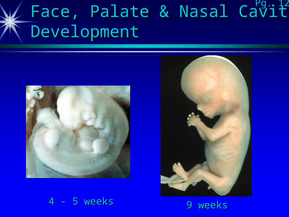
Face, Palate & Nasal CavityDevelopmentFace, Palate & Nasal CavityDevelopment
4 - 5 weeks4 - 5 weeks 9 weeks 9 weeksPg. 11 notes
Pg. 12
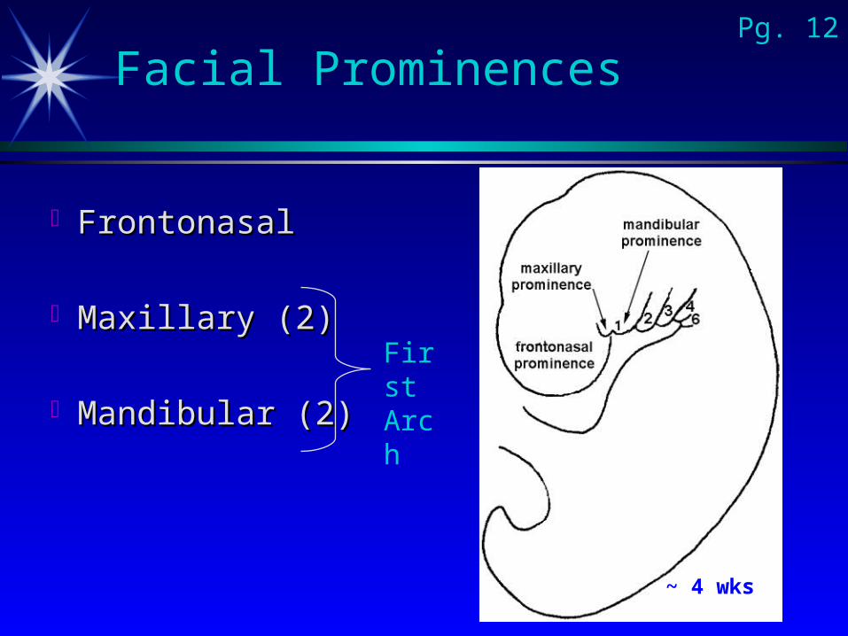
Facial Prominences
FrontonasalFrontonasal
Maxillary (2)Maxillary (2)
Mandibular (2)Mandibular (2)
First Arch
Pg. 12
~ 4 wks
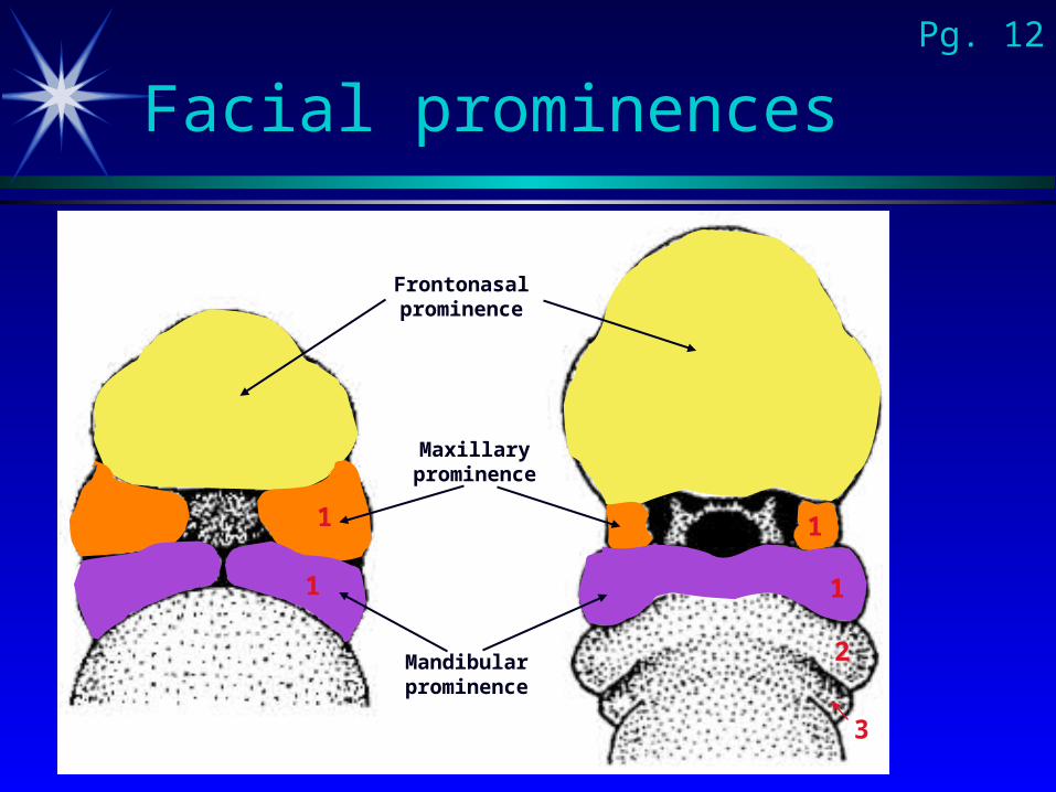
Facial prominences
Frontonasal prominence
Maxillary prominence
Mandibular prominence
2
1
1
1
1
3
Pg. 12
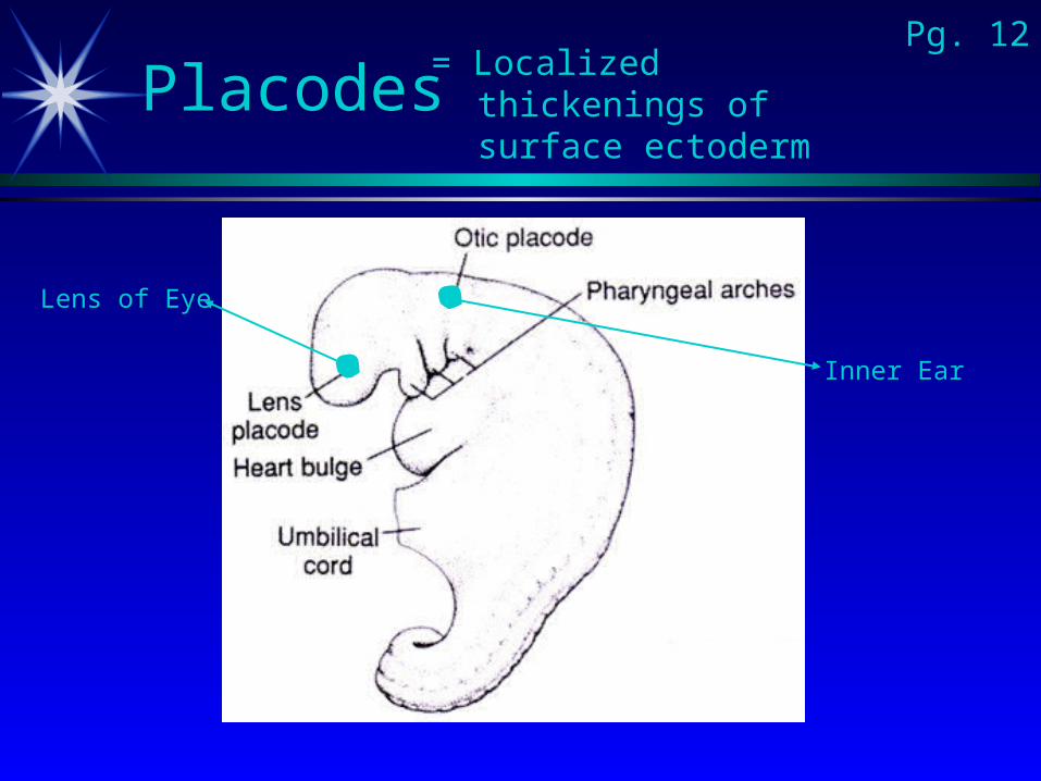
Placodes
Lens of Eye
Inner Ear
= Localized thickenings of surface ectoderm
Pg. 12

Nasal Placodes
Pg. 12
Placodes
Stomodeum
& Stomodeum
Similar to M&P fig 9-26
[10-26]
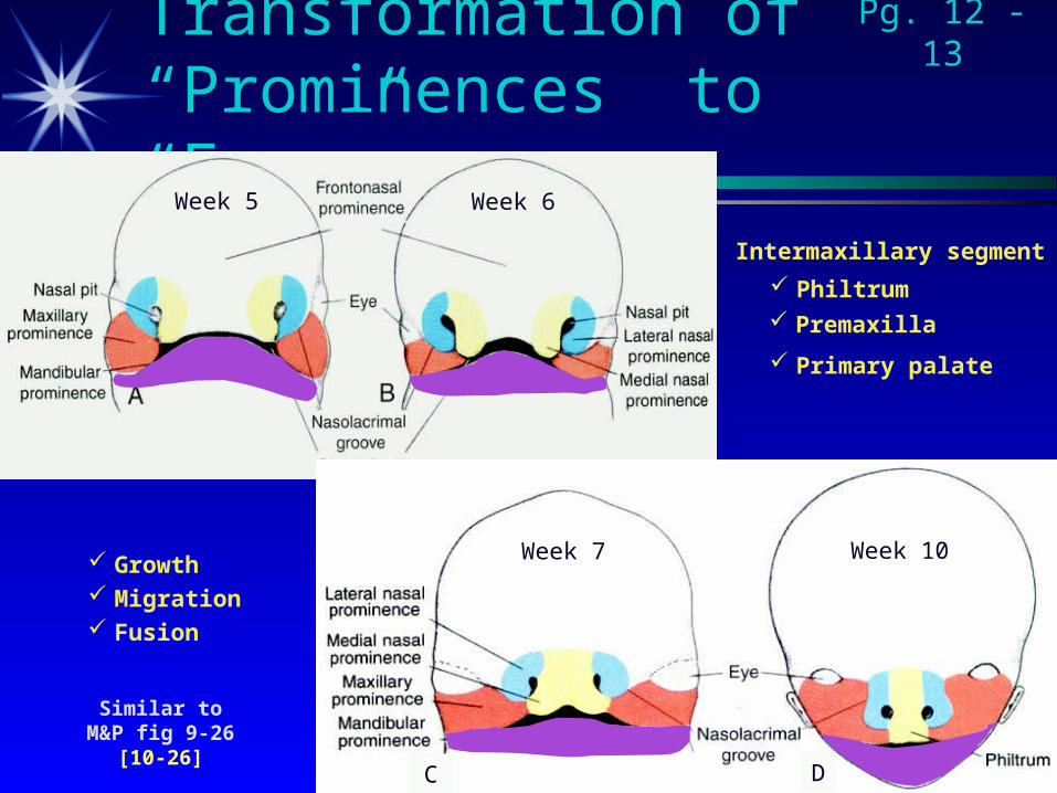
Transformation of “Prominences” to “Face”
Intermaxillary segment
Philtrum
Primary palate
Premaxilla
Pg. 12 - 13
C D
Growth Migration Fusion
Week 5 Week 6
Week 7 Week 10
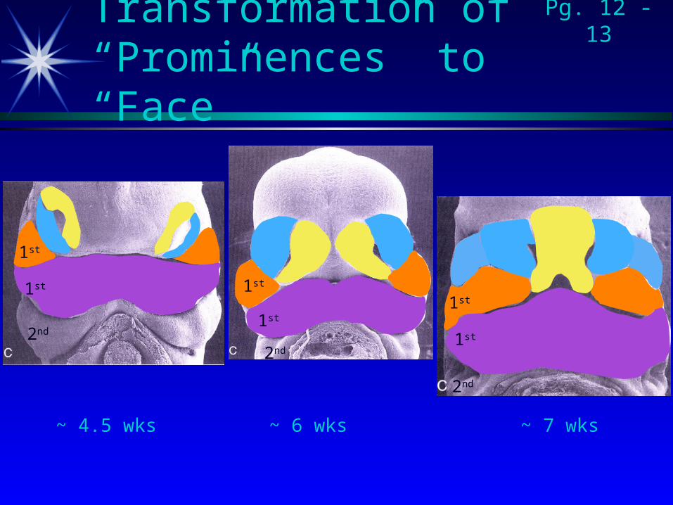
Transformation of “Prominences” to “Face”
~ 4.5 wks ~ 6 wks ~ 7 wks
Pg. 12 - 13
1st
2nd1st
1st
2nd
2nd
1st
1st1st
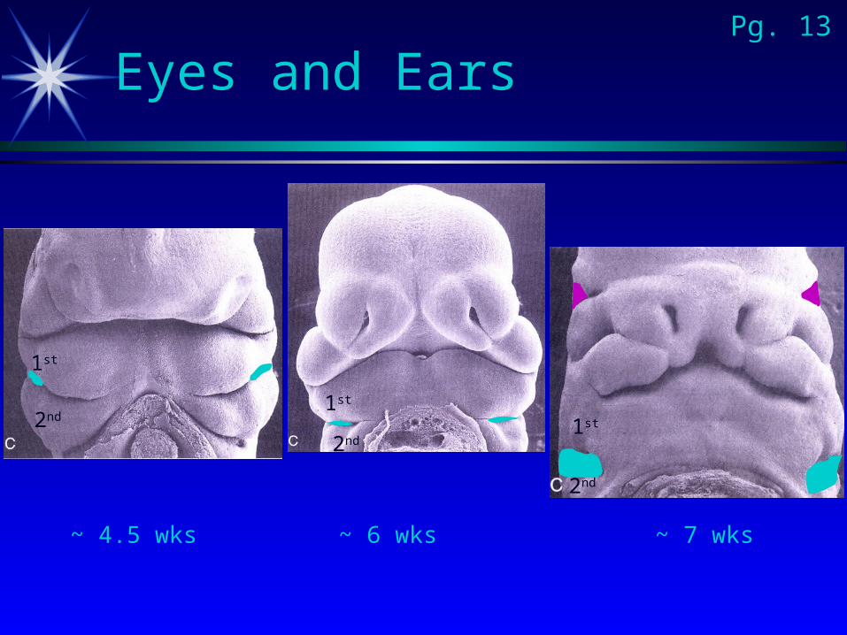
Eyes and Ears
1st
2nd1st
1st
2nd
2nd
Pg. 13
~ 4.5 wks ~ 6 wks ~ 7 wks
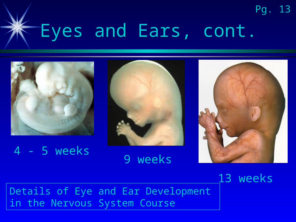
Eyes and Ears, cont.
9 weeks4 - 5 weeks
13 weeks
Pg. 13
Details of Eye and Ear Development in the Nervous System Course
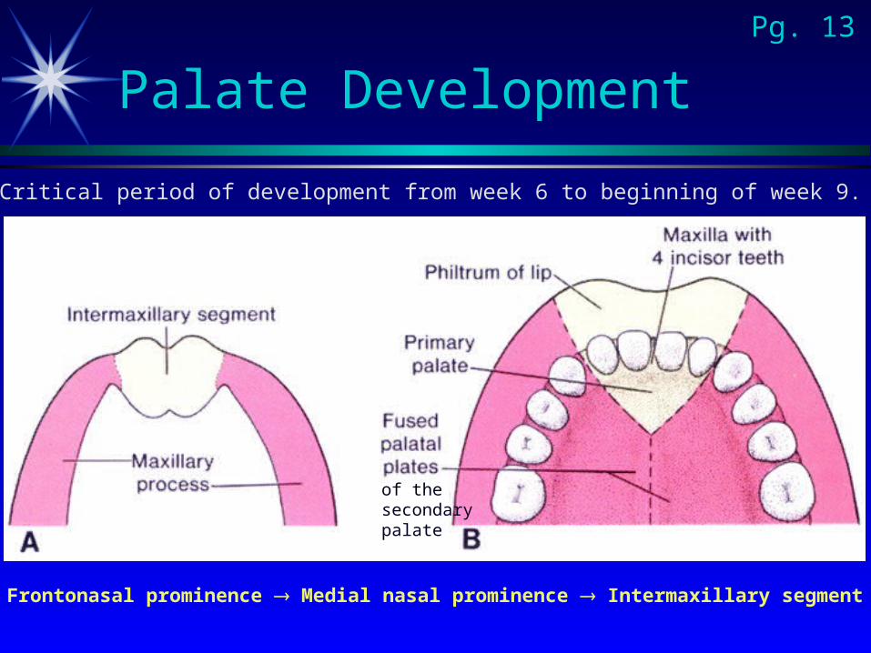
Palate Development
Frontonasal prominence Medial nasal prominence Intermaxillary segment
Critical period of development from week 6 to beginning of week 9.
Pg. 13
of the secondary palate
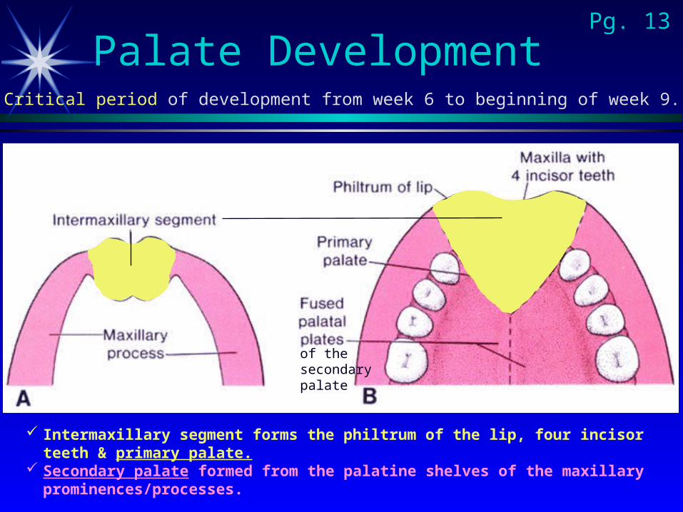
Palate Development
Intermaxillary segment forms the philtrum of the lip, four incisor teeth & primary palate.
Secondary palate formed from the palatine shelves of the maxillary prominences/processes.
Critical period of development from week 6 to beginning of week 9.
Pg. 13
of the secondary palate
Pg. 13
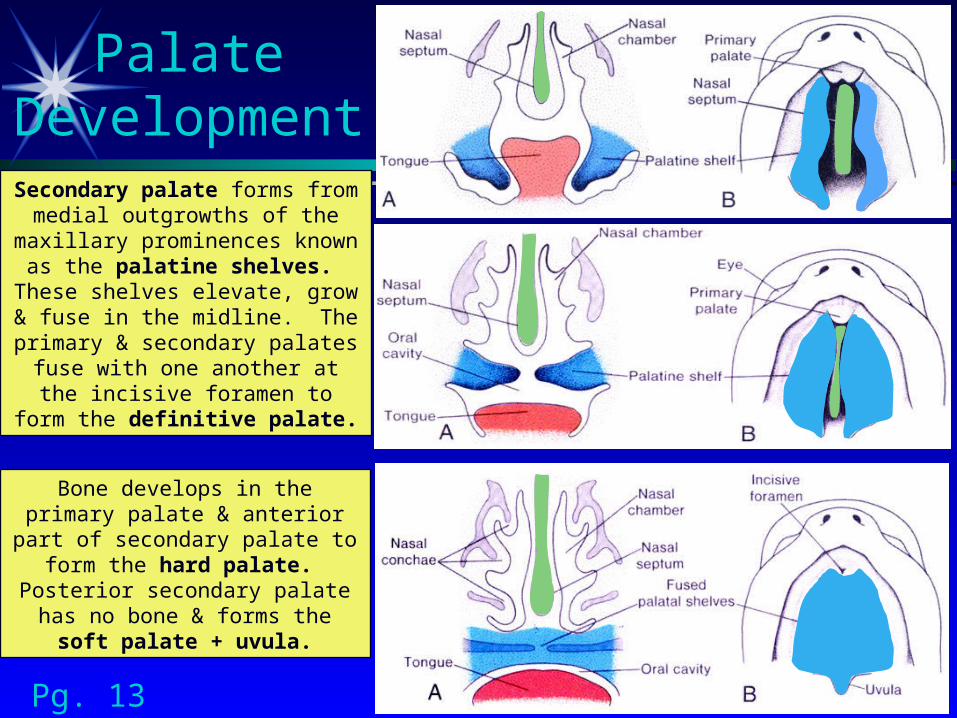
Palate DevelopmentSecondary palate forms from
medial outgrowths of the maxillary prominences known as the palatine shelves. These shelves elevate, grow & fuse in
the midline. The primary & secondary palates fuse with one another at the incisive foramen to form the definitive palate.
Bone develops in the primary palate & anterior part of
secondary palate to form the hard palate. Posterior
secondary palate has no bone & forms the soft palate + uvula.
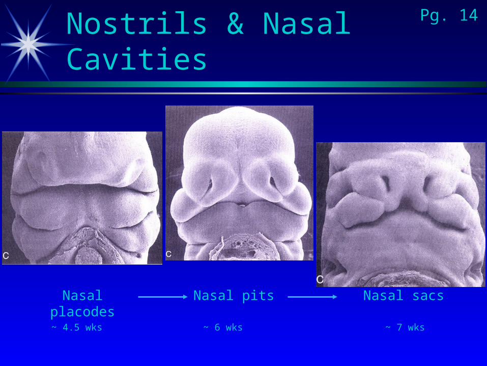
Nostrils & Nasal CavitiesPg. 14
~ 4.5 wks ~ 6 wks ~ 7 wks
Nasal placodes Nasal pits Nasal sacs

Nasal Cavities
5 weeks
12 weeks7weeks
6 weeks
Pg. 14
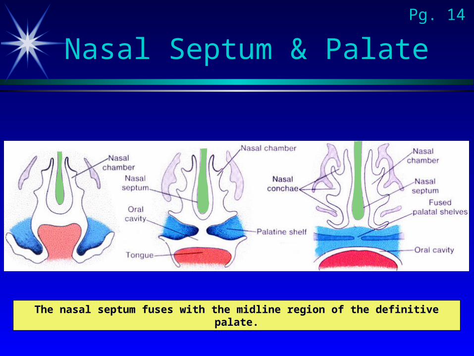
Nasal Septum & PalatePg. 14
The nasal septum fuses with the midline region of the definitive palate.
Changes in shape of face
Paranasal air sinuses
Nasal conchae
Tooth development
Pg. 14
Anterior defectsAnterior defectsInvolve upper lip, alveolar part of maxilla & anterior or Involve upper lip, alveolar part of maxilla & anterior or
primary palateprimary palate
Posterior defectsPosterior defectsInvolve hard and soft palate (secondary palate)Involve hard and soft palate (secondary palate)
Combination anterior & posterior defectsCombination anterior & posterior defects
Congenital AbnormalitiesPg. 15
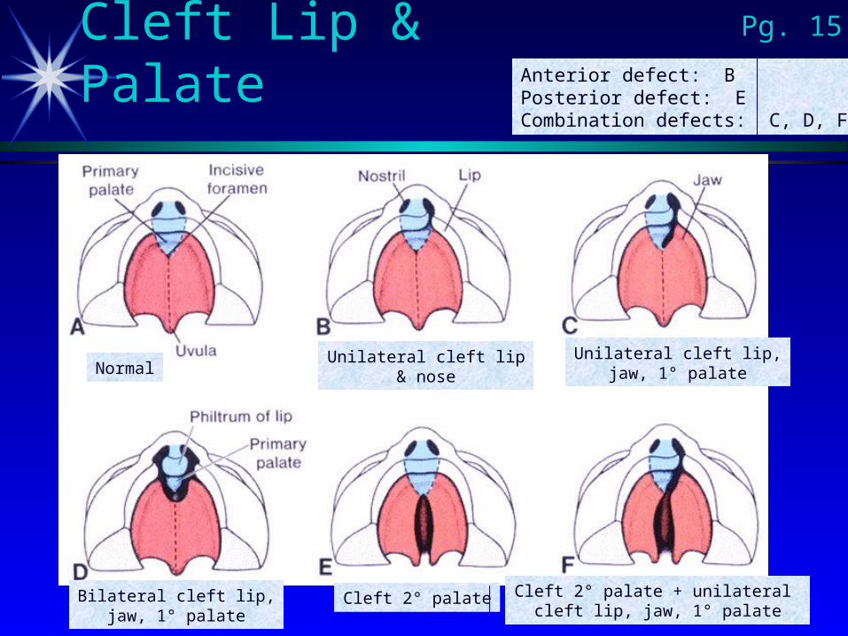
Cleft Lip & Palate
NormalUnilateral cleft lip
& noseUnilateral cleft lip,
jaw, 1° palate
Bilateral cleft lip,jaw, 1° palate
Cleft 2° palate Cleft 2° palate + unilateral cleft lip, jaw, 1° palate
Anterior defect: BPosterior defect: ECombination defects: C, D, F
Pg. 15
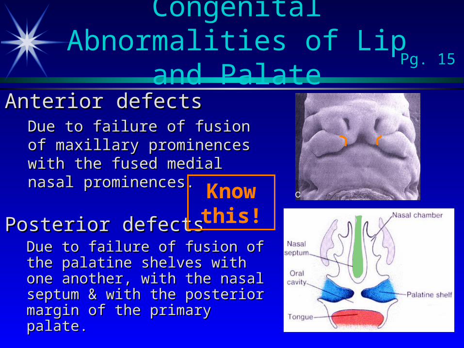
Anterior defectsAnterior defectsDue to failure of fusion of Due to failure of fusion of maxillary prominences with the maxillary prominences with the fused medial nasal fused medial nasal prominences.prominences.
Congenital Abnormalities of Lip and Palate Pg. 15
Posterior defectsPosterior defectsDue to failure of fusion of the Due to failure of fusion of the palatine shelves with one palatine shelves with one another, with the nasal septum another, with the nasal septum & with the posterior margin of & with the posterior margin of the primary palate.the primary palate.
Know this!
Teratogens interfere with Teratogens interfere with migration of neural crestmigration of neural crest so that there so that there is too little mesenchyme to work with. Examples include:is too little mesenchyme to work with. Examples include:
Anticonvulsants such as DilantinAnticonvulsants such as Dilantin Vitamin A; isotretinoin (Accutane)Vitamin A; isotretinoin (Accutane)
GeneticGenetic Chromosomal syndromes (e.g. Trisomy 13)Chromosomal syndromes (e.g. Trisomy 13) Single gene mutationsSingle gene mutations
Other:Other: Excessive cell death during the formation of the maxillary & Excessive cell death during the formation of the maxillary &
nasal prominencesnasal prominences Failure of palatal shelves to elevate at the proper timeFailure of palatal shelves to elevate at the proper time Excessively wide headExcessively wide head
Congenital Abnormalities: Etiology is Multifactorial Pg. 15
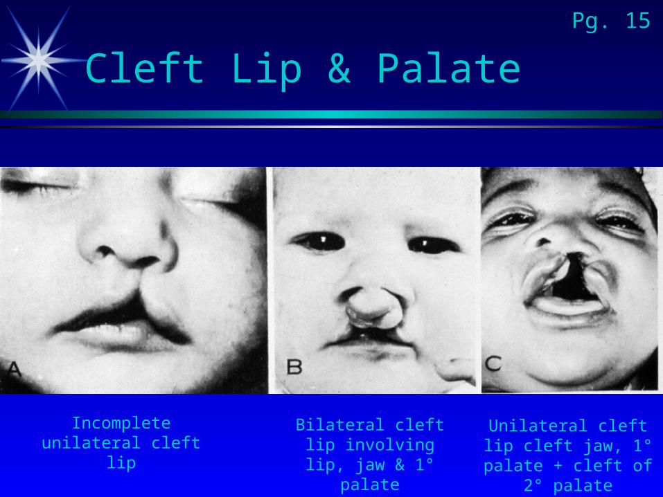
Cleft Lip & PalatePg. 15
Incomplete unilateral cleft lip
Bilateral cleft lip involving lip, jaw &
1° palate
Unilateral cleft lip cleft jaw, 1° palate +
cleft of 2° palate
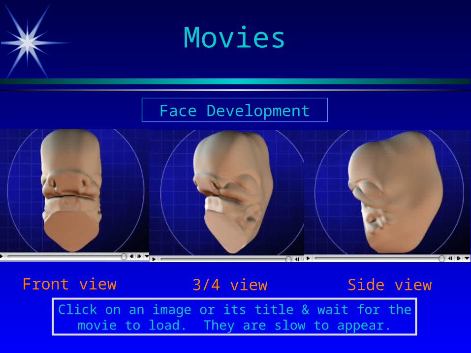
Movies
Face Development
Front view 3/4 view Side view
Click on an image or its title & wait for the movie to load. They are slow to appear.
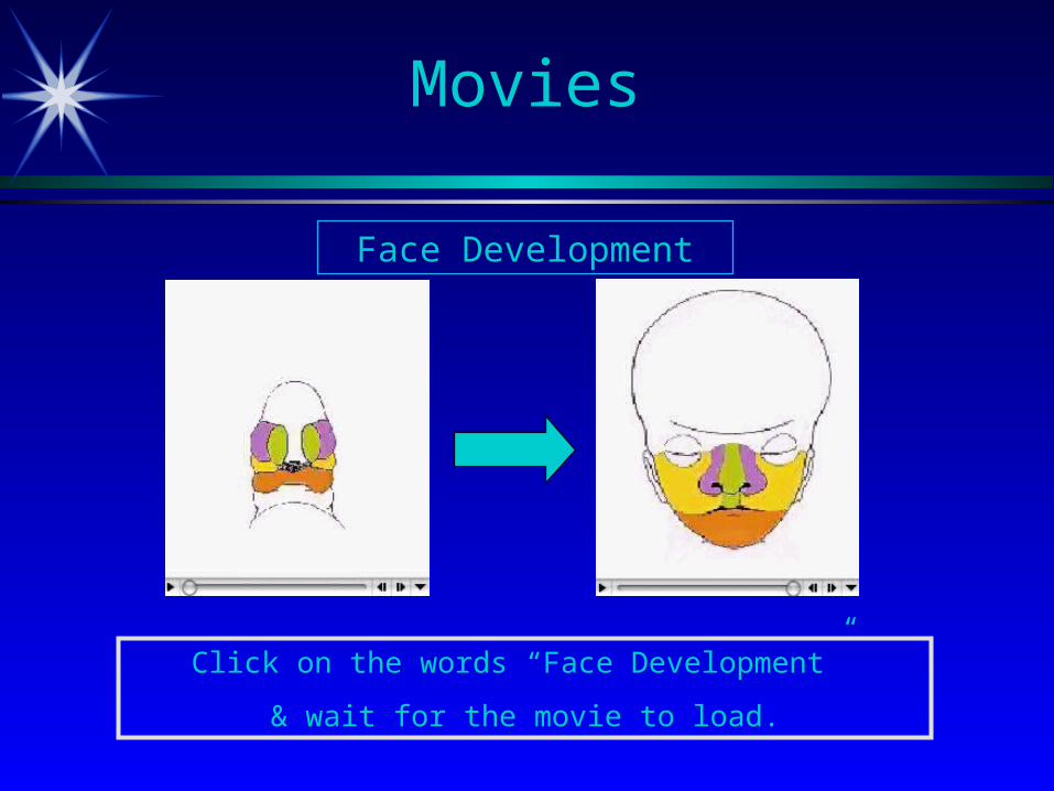
Movies
Face Development
Click on the words “Face Development”
& wait for the movie to load.
“Cases to Consider”
Discussion of these cases will be sent out as an email after you have some time to
consider them.
Pg. 16

Now for the “T-Quiz”!
Thymus
Thyroid
Tongue
Tonsil
Trachea
Trigeminal nerve
Tympanic cavity
Tympanic membrane
Origin:

Answers to this
“T-Quiz” are on the last slide.

Questions?
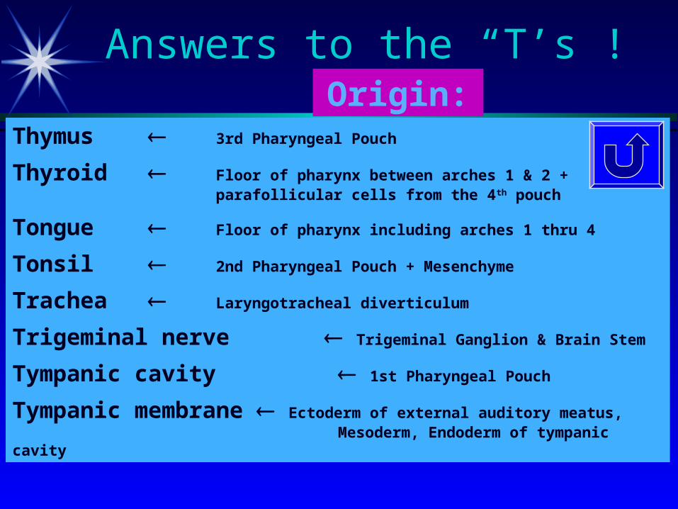
Answers to the “T’s”!
Thymus 3rd Pharyngeal Pouch
Thyroid Floor of pharynx between arches 1 & 2 + parafollicular cells from the 4th pouch
Tongue Floor of pharynx including arches 1 thru 4
Tonsil 2nd Pharyngeal Pouch + Mesenchyme
Trachea Laryngotracheal diverticulum
Trigeminal nerve Trigeminal Ganglion & Brain Stem
Tympanic cavity 1st Pharyngeal Pouch
Tympanic membrane Ectoderm of external auditory meatus, Mesoderm, Endoderm of tympanic cavity
Origin: