development of recombinant antibody technology for
TRANSCRIPT
Development of Recombinant Antibody Technology for
Application in Plant Pathogen Diagnosis
Remko Griep
Promotor: Dr. W.B. van Muiswinkel Hoogleraar in de Celbiologie en Immunologic, Departement Dierwetenschappen, Landbouwuniversiteit Wageningen.
Co-promotor: Dr. Ir. A. Schots Hoofd van het Laboratorium voor Monoklonale Antistoffen, Departement Plantenwetenschappen, Landbouwuniversiteit Wageningen.
IJ / '
Development of Recombinant Antibody Technology for
Application in Plant Pathogen Diagnosis
Remko Griep
Proefschrift ter verkrijging van de graad van doctor
op gezag van de rector magnificus van de Landbouwuniversiteit Wageningen,
Dr. CM. Karssen, in het openbaar te verdedigen op woensdag 17 maart 1999
des namiddags te half twee in de Aula.
The work presented in this thesis was carried out at the Laboratory for Monoclonal Antibodies, Wageningen Agricultural University, Binnenhaven 12, 6709 PD Wageningen, The Netherlands.
Financial support for this thesis was kindly provided by grants from the Program Committee for Agriculture and Biotechnology (PCLB), the Dutch Potato Industries and E.C. grants AIR3-CT94-1046 and FAIR1-CT95-0905.
CIP-DATA Koninklijke Bibliotheek, Den Haag
Griep, R.A. ISBN 90-5808-030-7
Development of recombinant antibody technology for application in plant pathogen diagnosis Remko Albert Griep
Thesis Wageningen Agricultural University - with references - with summaries in English and Dutch. Laboratory for Monoclonal Antibodies, P.O. Box 8123, 6700 ES Wageningen NL.
Key-words: Phage display, single-chain-Fv, Ralstonia solancearum, Beet necrotic yellow vein virus, tomato spotted wilt virus, green fluorescent protein, fluobodies.
Bibliographic abstract
This thesis describes the applicability of the novel phage display technique in selecting single-chain Fv antibodies from combinatorial antibody libraries that are specific for plant-pathogens such as Ralstonia solanacearum, beet necrotic yellow vein virus and tomato spotted wilt virus. Several of the retrieved antibodies are applicable as coating and detection reagents in a double antibody sandwich ELISA or in immunofluorescence. This shows the potential of the phage display system in obtaining antibodies that are suitable for routine diagnosis without the use of laboratory animals.
Druk: Ponsen & Looijen BV Wageningen
&3LIOTHEEK LANDBOUWUNIVERSITETT
WAGENINGEN
STELLINGEN
1. Het selecteren van antilichamen met behulp van de "phage display" techniek vormt een goed en tevens diervriendelijk altematief voor de hybridomatechmek.
Dit proefschrift.
2. Gezien de milioenen jaren evolutionaire ontwikkeling van het immuunsysteem is het nai'ef om te veronderstellen dat een combinatorial antibody library die geconstrueerd is uit B-lymfocyten van niet-gei'mmuniseerde donoren inderdaad nai'ef is.
Vaughan, T.J. et al., (1996) Human antibodies with sub-nanomolar affinities isolated from a large non-immunized phage display library. Nature Biotechnology 14:309-314.
3. De "phage display" techniek is bij uitstek geschikt voor het signaal-transductie onderzoek.
4. Het baseren van een diagnostische toets op slechts een monoklonaal antilichaam is even riskant als het spelen van Russische roulette.
5. De afkorting SIP voor het faagselectiesyteem met behulp van "selectively infective particles", weerspiegelt ook de gemoedstoestand van de onderzoekers die dit systeem van de grand proberen te krijgen.
Duenas, M. and Borrebaeck, C.A.K. (1994) Clonal selection and amplification of phage displayed antibodies by linking antigen recognition and phage replication. Bio/Technology 12: 999-1002.
6. Het met succes tot expressie brengen van eiwitten in een heteroloog systeem berust meer op geluk dan op wijsheid.
Levitt, M. et al., (1997) Protein folding: the endgame. Annual Review of Biochemistry 66: 549-579.
7. Waar cholera heerst groeien bananen, hetgeen deze dan ook tot een uitstek end 'adjuvants' maken voor het ontwikkelen van een oraal vaccin.
8. Het werken met kortlopende projecten is in de wetenschap een onderschatte vorm van geld- en kennisvernietiging.
9. Het verbod op het aanbrengen van reclame in proefschriften strookt niet met de commercialisering van de universiteiten.
10. Daar het tot nu toe niet gelukt is om het natuurlijke reservoir te vinden van diverse 'dierlijke virussen' is het, gezien de grote overeenkomst met 'plantenvirussen', aanbevelenswaardig om eens in het plantenrijk te gaan zoeken.
Wijkamp, I. et al., (1993) Multiplication of tomato spotted wilt virus in its insect vector. Journal of General Virology 74:341-349 and Le Guenno, B. (1997) Haemorrhagic fevers and ecological pertubations. Archives of Virology 13:191-199.
Stellingen behorende bij het proefschrift:
Development of Recombinant Antibody Technology for
Application in Plant Pathogen Diagnosis
Remko Griep, Wageningen, 17 maart 1999.

.Against all odds...'

Contents
Chapter 1 General introduction 1
Chapter 2 Development of specific recombinant monoclonal antibodies against the lipopolysaccharide of Ralstonia solanacearum race 3 19
Chapter 3 Selection of beet necrotic yellow vein virus specific single-chain Fv antibodies from a semi-synthetic combinatorial antibody library 37
Chapter 4 pSKAP/S: an expression vector for the production of single-chain Fv alkaline phosphatase fusion proteins 51
Chapter 5 Application of phage display in selecting tomato spotted wilt virus specific single-chain Fvs for sensitive diagnosis in ELISA 63
Chapter 6 Fluobodies: Green fluorescent single-chain Fv fusion proteins 81
Chapter 7
Discussion, summary and concluding remarks 95
Samenvatting 103
Curriculum vitae 107
List of publications 109
Chapter 1
General introduction
In the early days of agriculture crops were small, biodiversity was high and trade was limited. This situation gave plant pathogens only a limited chance to spread and to cause significant damage. However, as time past by, crops were covering larger areas. This, while the genetic homogeneity within the crops was increasing. In combination with the increasing trade, the application of monocultures in today's agriculture has resulted in the outbreak of many devastating plant diseases. The economic damage of diseases and pests, caused by viroids, viruses, bacteria, insects, fungi and plant-parasitic nematodes is gigantic. Indirect losses are difficult to assess because the presence of certain pathogens or plague organisms in productive soils discourages the cultivation of economically important plants.
Pathogens can be carried on, or in plant propagation material and a disease can become manifest when the crop starts to grow. Even when transmission rates are low, sufficient inoculum may be present for an epidemic outbreak. Certification of healthy plant propagation material is of major importance to avoid the spread of various diseases to regions where a particular plant-pathogen is absent. Moreover, the information obtained from general surveys, on the presence of plant-pathogens in or on crops or in soil, can be used for advisory systems with regard to crop rotation, cultivar selection, pesticide application, harvest dates, post harvest handling, etc.
Detection and identification The earliest form of plant-pathogen diagnosis was based on the recognition of specific symptoms in crop plants. Because many pathogens do not produce specific symptoms in their hosts or are latently present, the development of alternative methods to confirm the identity of the causal agent is required. These assays should be robust, uncomplicated and fast. Transmission to indicator host plants, plating on selective media, microscopy, chromatography and electrophoresis often provide valuable information and are still widely used as methods for the identification and detection of plant-pathogens. However, they are laborious and it may take up to weeks before a result is obtained. Modern molecular techniques such as the polymerase chain reaction [58] and nucleic acid sequence based amplification [35] are fast and can be used as a reliable tool to detect various plant-pathogens [62,77]. Although both are sensitive methods, they are not suited yet for testing the large numbers that have to be tested annually (over 2 million samples in the Netherlands alone) for the certification of plant propagation material. In contrast, serological assays form a good compromise between robustness, sensitivity, specificity on one hand and ease and expense of application on the other hand.
Serological assays The first report describing the use of serology for the identification of a plant-pathogenic bacterium was published in 1918 [31], when Jensen used an antiserum in an agglutination test showing that a strain of Agrobacterium tumefaciens from Denmark could be differentiated from a strain from the United States. During the 1930s, agglutination assays rapidly became popular for identification of plant-pathogenic bacteria and viruses, and remain so today. After the introduction of the agar double diffusion test by Ouchterlony [51], agar precipitin tests became widely used during the 1950s for identification of medically important bacteria. However, Ouchterlony double diffusion was not applied for identification of plant-pathogenic bacteria until 1960, when Lovrekovich and Klement [39] reported that species-specific antigens of Pseudomonas tabaci could be detected in Ouchterlony double diffusion tests but not in agglutination assays. Immunofluorescent (IF) staining was suggested in 1943 [52] for identification of bacteria in plants. However, no further interest was shown until 1965 when Morton [45] reported the superiority of direct IF over agglutination tests for rapid identification of the bacterium Xanthomonas vesicatoria. The development of the enzyme-linked immunosorbent assay (ELISA) by Van Weemen and co-workers [71] started a new era in direct detection of infectious agents. A few years after its development, this technique was also applied for the diagnosis of plant viruses [14].
The development of antisera against plant viruses has been very successful. In 1984, over 300 different virus species could be detected with specific immunoassays [69,70]. However, raising antisera specific for plant-parasitic nematode species, plant-pathogenic fungi and bacteria proved more difficult. Cross-reactions were generally observed with other nematode

species [78], saprophytic fungi [9] and closely related bacteria [44,68]. These problems can be ascribed to the variation in antibodies within a polyclonal antiserum.
Immune selection and the generation of polyclonal antibodies The ability to respond to an apparently unlimited array of foreign antigens is one of the
most remarkable features of the vertebrate immune system. Almost all antibody molecules contain a unique stretch of amino acids in their variable region (Fig. 1.1). This diversity is generated during B-lymphocyte differentiation (Fig. 1.2) in the bone marrow, where antibody encoding gene segments are randomly shuffled by a carefully regulated dynamic genetic system [27].
Fab''-
Light chain
FRl FR2
CDR1 CDR2
FR3
CDR3
FR4
Antigen binding
£' ^ B site
Fv
Figure 1.1. A model for the structure of a mouse antibody molecule, composed of two light and two heavy polypeptide chains. Two identical antigen-binding sites, formed by the variable regions of the heavy chain (VH) and light chain (VL), are located at one side of the antibody molecule. The constant regions of the light chain (CL), the constant domains of the heavy chain (CH1, CH2, CH3), interchain and intrachain disulfide bonds (S-S) are indicated. The Fv and Fab fragments of the antibody molecule are surrounded by dotted lines, whereas the organization of the variable domains in framework regions (FR) and complementarity determining regions (CDR) is shown in more detail within the box.
Germ line DNA
L1 V1 L250 V250 D 1-10 J 1-4 C|i C§
Rearranged DNA L V D J 2-4 Cli C8
Primary RNA transcript
L V J 2-4
4 1 * Cu C5
Messenger RNA DJ C^
lAAAAAAA
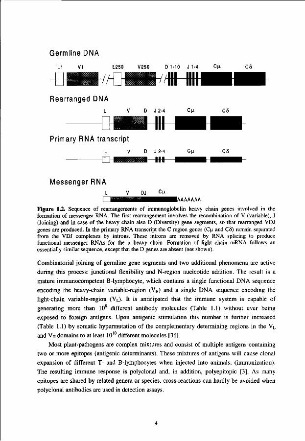
Figure 1.2. Sequence of rearrangements of immunoglobulin heavy chain genes involved in the formation of messenger RNA. The first rearrangement involves the recombination of V (variable), J (Joining) and in case of the heavy chain also D (Diversity) gene segments, so that rearranged VDJ genes are produced. In the primary RNA transcript the C region genes (Cu and C8) remain separated from the VDJ complexes by introns. These introns are removed by RNA splicing to produce functional messenger RNAs for the u heavy chain. Formation of light chain mRNA follows an essentially similar sequence, except that the D genes are absent (not shown).
Combinatorial joining of germline gene segments and two additional phenomena are active during this process: junctional flexibility and N-region nucleotide addition. The result is a mature immunocompetent B-lymphocyte, which contains a single functional DNA sequence encoding the heavy-chain variable-region (VH) and a single DNA sequence encoding the light-chain variable-region (VL). It is anticipated that the immune system is capable of generating more than 108 different antibody molecules (Table 1.1) without ever being exposed to foreign antigens. Upon antigenic stimulation this number is further increased (Table 1.1) by somatic hypermutation of the complementary determining regions in the VL
and VH domains to at least 1010 different molecules [36].
Most plant-pathogens are complex mixtures and consist of multiple antigens containing two or more epitopes (antigenic determinants). These mixtures of antigens will cause clonal expansion of different T- and B-lymphocytes when injected into animals, (immunization). The resulting immune response is polyclonal and, in addition, polyepitopic [3]. As many epitopes are shared by related genera or species, cross-reactions can hardly be avoided when polyclonal antibodies are used in detection assays.
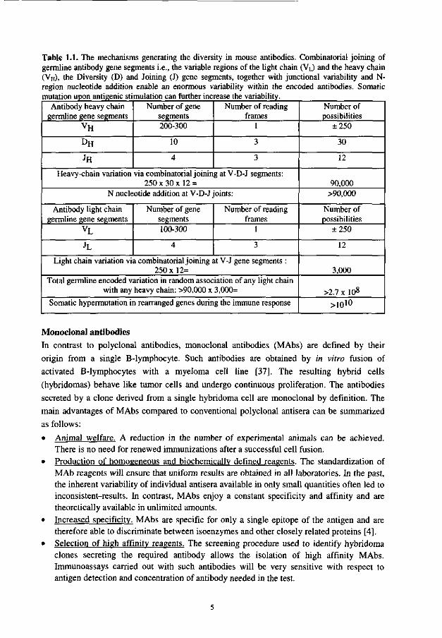
Table 1.1. The mechanisms generating the diversity in mouse antibodies. Combinatorial joining of germline antibody gene segments i.e., the variable regions of the light chain (VL) and the heavy chain (VH), the Diversity (D) and Joining (J) gene segments, together with junctional variability and N-region nucleotide addition enable an enormous variability within the encoded antibodies. Somatic mutation upon antigenic stimulation can further increase the variability.
Antibody heavy chain germline gene segments
vH
DH
JH
Number of gene segments 200-300
10
4
Number of reading frames
1
3
3
Heavy-chain variation via combinatorial joining at V-D-J segments: 250x30x12 =
N nucleotide addition at V-D-J joints:
Antibody light chain germline gene segments
vL
JL
Number of gene segments 100-300
4
Number of reading frames
1
3
Light chain variation via combinatorial joining at V-J gene segments : 250x12=
Total germline encoded variation in random association of any light chain with any heavy chain: >90,000 x 3,000=
Somatic hypermutation in rearranged genes during the immune response
Number of possibilities
±250
30
12
90,000 >90,000
Number of possibilities
±250
12
3,000
>2.7 x 10⁸
>ioio
Monoclonal antibodies In contrast to polyclonal antibodies, monoclonal antibodies (MAbs) are defined by their origin from a single B-lymphocyte. Such antibodies are obtained by in vitro fusion of activated B-lymphocytes with a myeloma cell line [37], The resulting hybrid cells (hybridomas) behave like tumor cells and undergo continuous proliferation. The antibodies secreted by a clone derived from a single hybridoma cell are monoclonal by definition. The main advantages of MAbs compared to conventional polyclonal antisera can be summarized as follows:
• Animal welfare. A reduction in the number of experimental animals can be achieved. There is no need for renewed immunizations after a successful cell fusion.
• Production of homogeneous and biochemically defined reagents. The standardization of MAb reagents will ensure that uniform results are obtained in all laboratories. In the past, the inherent variability of individual antisera available in only small quantities often led to inconsistent-results. In contrast, MAbs enjoy a constant specificity and affinity and are theoretically available in unlimited amounts.
• Increased specificity. MAbs are specific for only a single epitope of the antigen and are therefore able to discriminate between isoenzymes and other closely related proteins [4].
• Selection of high affinity reagents. The screening procedure used to identify hybridoma clones secreting the required antibody allows the isolation of high affinity MAbs. Immunoassays carried out with such antibodies will be very sensitive with respect to antigen detection and concentration of antibody needed in the test.
Limitations in generating MAbs Although, the number of MAbs directed against bacteria, fungi and nematodes is rapidly expanding [15,17,59,61,67], the many advantages should not obscure the fact that generation of MAbs against plant-pathogens is often laborious and troublesome. Upon immunization the immune response is triggered after recognition of foreign antigens by lymphocytes. As it is difficult to isolate pure plant viruses, a high percentage of the obtained hybridomas is often directed against the contaminating plant material that appears to be immunodominant [20]. For bacteria, fungi and plant-parasitic nematodes the choice of immunogen is difficult, as it is generally unknown which epitope is unique for a certain species. Moreover, another prerequisite is that this particular epitope has to be recognized by the immune system.
In addition, the hybridoma technique of Kohler and Milstein [37] does not satisfactory exploit the immune repertoire. While the antibody repertoire is estimated to consist of over 108 different antibodies (Table 1.1), only a few thousand antibodies are obtained, of which on average less than 1% are antigen-binding. With an optimized fusion protocol, e.g. electrofusion, a ten times higher fusion efficiency can be obtained [18,49], while antigenic in
vitro stimulation or proliferation of affinity selected B-lymphocytes before fusion can yield an improved degree of antigen-specific hybridomas in some cases [66]. However, the problems of immunodominance and the enormous variability in epitopes present within the complex plant-pathogens also diminish the chance that a useful MAb will be obtained when these optimized procedures are applied.
Another major drawback of the hybridoma technique is that it is best suited for the generation of murine antibodies, while the main interest has always been in human antibodies for medical applications, e.g. tumor therapy and tumor imaging. In these applications murine MAbs offer no alternative, because they can give rise to an undesired immune response in humans [79]. Unfortunately, considerable difficulties in making human hybridomas exist [10], and moreover, they cannot be directed against human tumor antigens as these are usually regarded as self and do not elicit an immune response. Fortunately, it was realized that the problems associated with generating human B-lymphocytes could be circumvented through immortalizing antibody genes by recombinant DNA technology, rather than by immortalizing the cells themselves.
Antibody technology The modular nature and conserved domain structure of antibodies [33] makes them attractive candidates for protein engineering and since 1984, recombinant DNA technology has revolutionized the production of MAbs. The initial recombinant antibodies were made by grafting the variable domains (VL and VH) of murine hybridomas on human constant domains [7,47]. Later, mouse complementary determining regions, the regions responsible for antigen binding, were put in a human framework [32,57,73] and expression of the chimaeric antibody genes in mammalian cells yielded humanized antibodies.
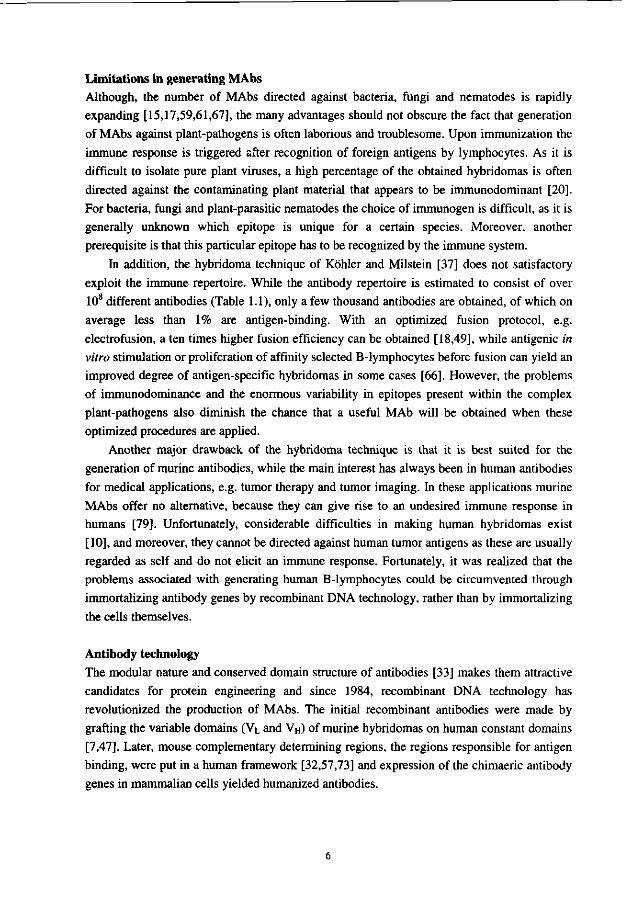
As yet, there are no reports of efficient expression of whole antibodies in bacteria, but subfragments (Fig. 1.1) such as Fabs (VHCH1 + VLCL) or Fvs (VH + VL) can be expressed well in Escherichia coli [5,55,63]. Spontaneous assembly of functional Fab fragments is observed when VHCH1 and VLCL domains are expressed as separate molecules. This association is in part driven by the hydrophobic interface between heavy and light chain residues. An interchain disulfide bond will stabilize assembled Fabs [22], but this is not a prerequisite for Fabs to be functional [26]. The key to expression of functional antibody fragments in E. coli appears to be export from the reducing environment of the cytoplasm into the oxidizing environment of the periplasm [56]. In contrast to Fab fragments, Fv fragments show a tendency to dissociate, particularly at low concentrations [21]. This can be overcome by connecting the C-terminus of one domain to the N-domain of the other with a flexible linker peptide [6,30] and expressing VH and VL domains as single-chain Fv (scFv) fusion proteins (Fig. 1.3).
Escherichia coli
Antigen
scFv protein
Figure 1.3. The variable domains VH and VL are joined by a linker peptide (L) and single-chain Fv (scFv) proteins are expressed and secreted into the periplasm of Escherichia coli after induction of the promoter (P) with a suitable inducer. In the scFv-encoding vector, the location of the signal peptide, the scFv gene, an ampicillin resistance gene (AMP) and an origin of replication (E. coli ori) are indicated.
Combinatorial antibody libraries of randomly assembled VH and VL genes Initially, expression of antibody fragments in E. coli was achieved by cloning the unique heavy- and light-chain encoding cDNA sequences from hybridoma cells of known specificity. This cloning was simplified after the introduction of the polymerase chain reaction [58] that allowed specific amplification of known DNA sequences. Although the nucleotide sequences
encoding the complementary-determining regions are highly variable, the flanking regions have relatively conserved sequences [33]. In fact, the polymerase chain reaction provides a means to amplify and clone the VH and VL antibody repertoire encoding sequences directly from B-lymphocytes of immunized mice [38,50,60,75], thereby omitting the need for the generation of hybridomas. The resulting libraries were combinatorial as the amplified VH and VL genes were randomly recombined irrespective of their original pairing. Within biased combinatorial libraries (derived from activated B-lymphocytes of hyper-immunized mice) the number of antigen-specific V genes is high, at best < 1/500 [46] and more usually < 1/5000 [11,54]. These libraries can therefore be relatively small (105-106) to regain original VH and VL pairings.
While it is difficult and time consuming to identify MAbs produced by hybridomas, the ease by which E. coli can be manipulated reduces the time required for antibody production and characterization. Bacterial expression of diverse combinatorial antibody libraries allows screening of a larger population of antibody fragments (50,000 per filter) than could be tested previously using conventional methods [28,64]. However, this screening process was still very laborious, especially in view of the huge number of clones generated using combinatorial libraries. This difficulty in isolation of fragments with binding activity has been largely overcome by the development of techniques for the display of antibody fragments on the surface of filamentous bacteriophage.
Mimicking the immune system by display of antibody fragments on bacteriophage Filamentous bacteriophage were first used to display small peptides on the minor coat
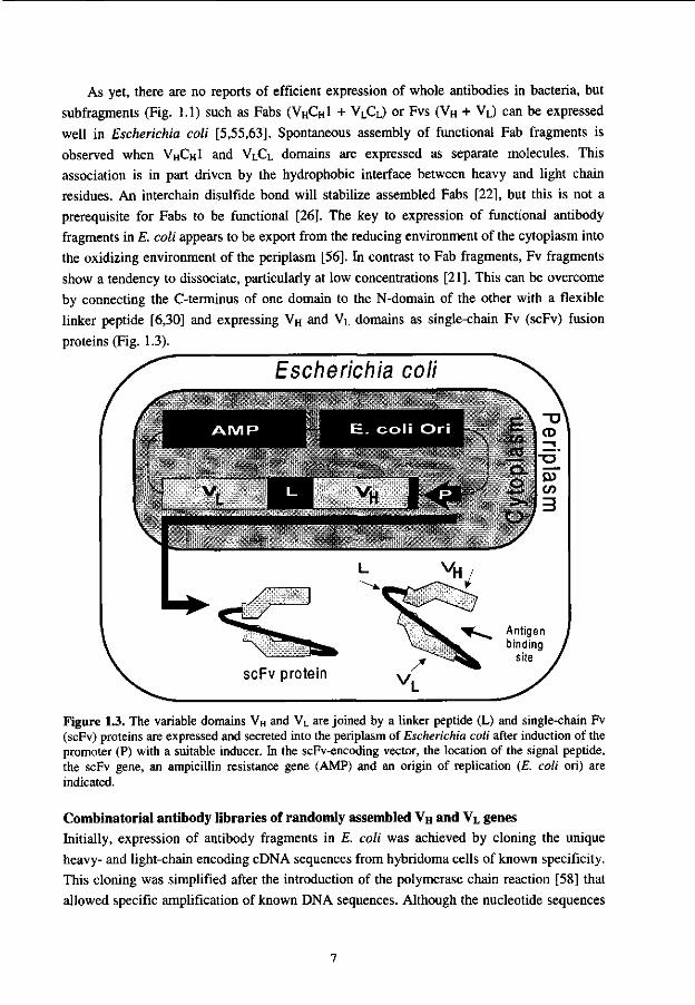
protein pill of bacteriophage Fd, and very rare peptides could be isolated [65] through sequential cycles of phage growth and selection by binding to antibody. When it appeared that larger proteins, among which scFv-antibodies [43] or Fab fragments [25], could also be expressed functionally on the surface of bacteriophage (Fig. 1.4), a powerful selection system was created to obtain specific MAbs from combinatorial phage-antibody libraries [13].
Figure 1.4. Expression of a scFv-antibody fragment on bacteriophage through genetic fusion of a scFv gene with the gene of minor coat protein pill. In the scFv-encoding phagemid vector, the scFv gene, an ampicillin resistance gene (AMP), a packaging signal (Ml3 ori) and an origin of replication (E. coli ori) are indicated. During phage assembly the scFv-pIII encoding phagemid is packaged into the phage particle and the scFv-pIII fusion protein is expressed on the phage surface. Consequently, the phenotype and genotype are linked.

In the immune system the B-lymphocytes represent self-replicating packages. They contain the antibody genes that encode the antibody displayed at their surface. Filamentous bacteriophage expressing functional antibody fragments on their surface, thus, mimic the B-lymphocyte by linking genotype and phenotype.
The phage display system (Fig. 1.5) allows direct selection of antibodies with rare specificities from combinatorial phage display antibody libraries through successive rounds of phage growth and selection for antigen binding. For instance, Bradbury and co-workers [8] showed the feasibility of this technique by selecting antigen binding phages from a pool of non-binding phages, even at a ratio of one binding in 109 irrelevant phages.
Presently a variety of formats for antibody phage display exist. Antibody fragments can be cloned directly into the genomes of filamentous bacteriophage or, alternatively, phagemid vectors can be used. These plasmid-based vectors have sequences derived from the intergenic regions of filamentous phage, which enable them to replicate as a single stranded DNA in E.
coli. These phagemids are packaged into filamentous phage particles (rescued) when cells harboring them are co-infected with helper phage such as M13K07 [74], that provide all the phage proteins but due to a defective origin they are themselves poorly packaged in competition with the phagemids.
Phage rescue
Combinatorial antibody library
f Escherichia coli
f , v p . scFv pill
\
'J t
Infection of E. coli
t target-antigen
Elution
Figure 1.5. Schematic representation of the selection of antigen-specific single-chain antibody fragments (scFvs) from a combinatorial antibody library using phage display. Helper phage, which contain the entire phage genome but lack an efficient packaging signal, are used to 'rescue' phagemids from a combinatorial library. When both helper phage and phagemid are present within the same bacterium, phage-antibodies (PhAbs) are assembled that carry fusion proteins of scFv and minor coat protein pill on their surface and contain the scFv-encoding phagemid vector. In order to select for antigen specificity, PhAbs rescued from a combinatorial antibody library are allowed to bind to immobilized antigen (Panning). Washing removes the PhAbs that lack affinity for the antigen. Bound PhAbs are eluted, and the selected PhAbs are enriched by sequential rounds of panning until the desired affinity is obtained.
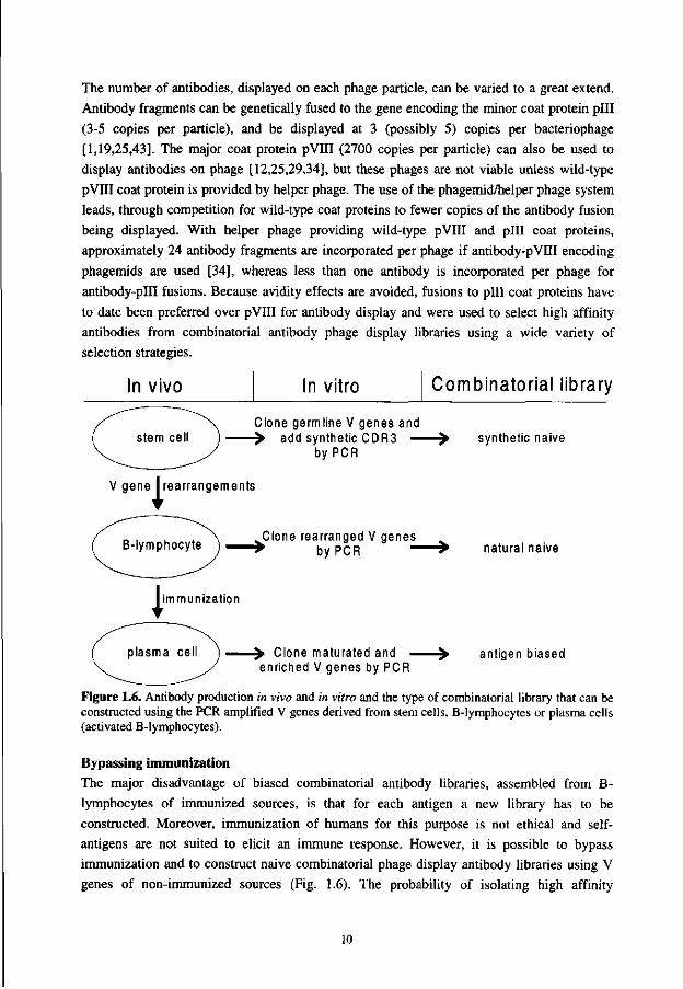
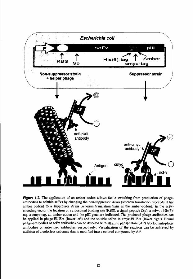
The number of antibodies, displayed on each phage particle, can be varied to a great extend. Antibody fragments can be genetically fused to the gene encoding the minor coat protein pill (3-5 copies per particle), and be displayed at 3 (possibly 5) copies per bacteriophage [1,19,25,43]. The major coat protein pVIII (2700 copies per particle) can also be used to display antibodies on phage [12,25,29,34], but these phages are not viable unless wild-type pVIII coat protein is provided by helper phage. The use of the phagemid/helper phage system leads, through competition for wild-type coat proteins to fewer copies of the antibody fusion being displayed. With helper phage providing wild-type pVIII and pill coat proteins, approximately 24 antibody fragments are incorporated per phage if antibody-pVIII encoding phagemids are used [34], whereas less than one antibody is incorporated per phage for antibody-pill fusions. Because avidity effects are avoided, fusions to pill coat proteins have to date been preferred over pVIII for antibody display and were used to select high affinity antibodies from combinatorial antibody phage display libraries using a wide variety of selection strategies.
In vivo In vitro Combinatorial library
Clone germ line V genes and add synthetic CDR3 —
by PCR synthetic naive
'i' V gene Irearrangements
Clone rearranged V genes by PCR - natural naive
1' immunization
Clone maturated and -enriched V genes by PCR
antigen biased
Figure 1.6. Antibody production in vivo and in vitro and the type of combinatorial library that can be constructed using the PCR amplified V genes derived from stem cells, B-lymphocytes or plasma cells (activated B-lymphocytes).
Bypassing immunization The major disadvantage of biased combinatorial antibody libraries, assembled from B-lymphocytes of immunized sources, is that for each antigen a new library has to be constructed. Moreover, immunization of humans for this purpose is not ethical and self-antigens are not suited to elicit an immune response. However, it is possible to bypass immunization and to construct naive combinatorial phage display antibody libraries using V genes of non-immunized sources (Fig. 1.6). The probability of isolating high affinity
10
antibodies towards every imaginary antigen from such a repertoire is dependent on the diversity of the library [53]. Therefore, these naive antibody phage display libraries have to be as large as possible. Naive human combinatorial antibody phage display libraries have been constructed that are comparable to the natural immune repertoire with a diversity of approximately 108 scFv-antibodies [41] or even more diverse, with 1010 scFv-antibodies [72]. Using an in vivo recombination technique, which applied the Cre-enzyme of phage PI [76], construction of a naive combinatorial Fab phage display library with a diversity of 1012 was achieved [23].
Alternatives for the use of rearranged V repertoires from a non-immunized source are cloned human germline VH and VL sequences. Using these genes, amplified with a specific 5' end and a synthetic CDR3 primer (Fig. -1.6), large (>108) synthetic combinatorial antibody libraries [2,26,48] were constructed. In addition, a synthetic Fab phage display library (108) was constructed in which the natural structure of the CDR3 was retained [16].
Selection of specific antibodies from combinatorial antibody phage display libraries Naive and synthetic combinatorial antibody phage display libraries encode antibodies of considerable diversity and can theoretically be used to select antibodies against many different antigens. In addition, the phage display system may also prove to be a more universal method to obtain specific MAbs, since the antibody specificity is not biased towards immunodominant epitopes as immunization procedures are omitted. Phages, encoding displayed antibody fragments, can be affinity selected from combinatorial antibody phage display libraries for binding to a particular antigen by passing the phage-antibodies over immobilized antigen (panning). Several formats have been used, such as immobilized antigen on a column matrix [43], antigen-coated plastic tubes or dishes [41], biotinylated antigen in solution followed by capture on streptavidin coated beads [24] and whole cells [16,42].
Phages that are bound to antigen are retained on the surface. Non-binding phage-antibodies are removed by washing. Bound phage-antibodies can be eluted by aspecific elution using acid [1] or alkaline buffers [40] or more specifically using competition with soluble antigen [13], proteolysis of spacers located between phage and antibody or reduction of disulfide bonds in biotinylated antigens. The selected (eluted) phages are used to infect E.
coli and can be applied in several sequential rounds of phage growth and selection, allowing even rare phage-antibodies to be isolated.
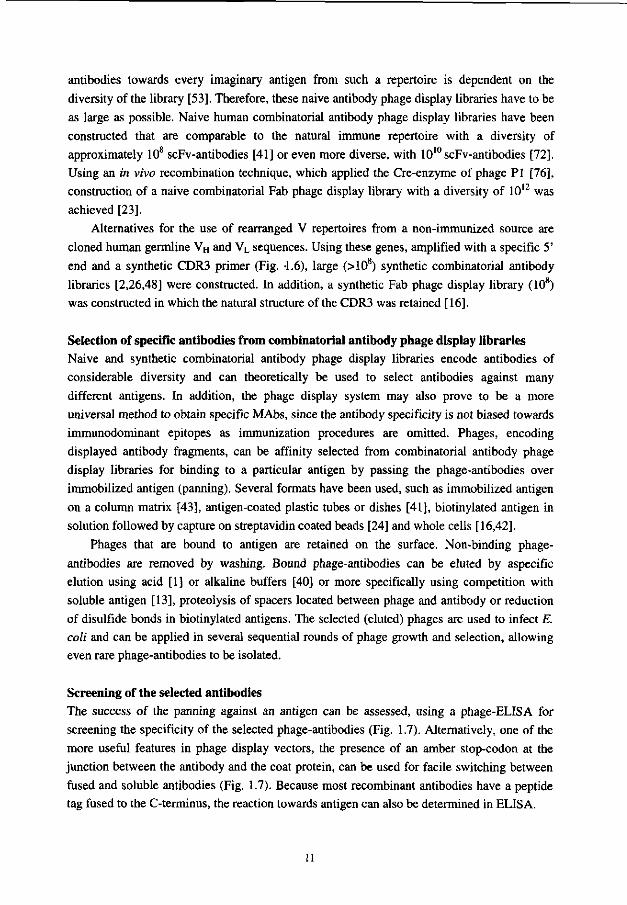
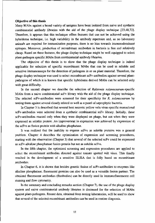
Screening of the selected antibodies The success of the panning against an antigen can be assessed, using a phage-ELISA for screening the specificity of the selected phage-antibodies (Fig. 1.7). Alternatively, one of the more useful features in phage display vectors, the presence of an amber stop-codon at the junction between the antibody and the coat protein, can be used for facile switching between fused and soluble antibodies (Fig. 1.7). Because most recombinant antibodies have a peptide tag fused to the C-terminus, the reaction towards antigen can also be determined in ELISA.
11
Escherichia coli
iC t t
RBS S p His(6)-tag I Amber
cmyc-tag t
Non-suppressor strain + helper phage
Suppressor strain
Figure 1.7. The application of an amber codon allows facile switching from production of phage-antibodies to soluble scFvs by changing the non-suppressor strain (wherein translation proceeds at the amber codon) to a suppressor strain (wherein translation halts at the amber-codon). In the scFv-encoding vector the location of a ribosomal binding site (RBS), a signal peptide (Sp), a scFv, a His(6)-tag, a cmyc-tag, an amber codon and the pill gene are indicated. The produced phage-antibodies can be applied in phage-ELISA (lower left) and the soluble scFvs in cmyc-ELISA (lower right). Bound phage-antibodies or scFv antibodies can be detected with alkaline phosphatase (AP) labeled anti-phage antibodies or anti-cmyc antibodies, respectively. Visualization of the reaction can be achieved by addition of a colorless substrate that is modified into a colored compound by AP.
12
Objective of this thesis Many MAbs against a broad variety of antigens have been isolated from naive and synthetic combinatorial antibody libraries with the aid of the phage display technique [23,48,72]. Therefore, it appears that this technique offers features that can not be achieved using the hybridoma technique, i.e. high variability in the antibody repertoire and, as no laboratory animals are required for immunization purposes, there is no bias towards immunodominant epitopes. Moreover, production of recombinant antibodies in bacteria is fast and relatively cheap. Based on these features, the phage display technique might be well equipped to select plant-pathogen specific MAbs from combinatorial antibody libraries.
The objective of this thesis is to show that the phage display technique is indeed applicable for selection of specific recombinant MAbs that can be used in reliable and sensitive immunoassays for the detection of pathogens in or on plant material. Therefore, the phage display technique was used to select recombinant scFv-antibodies against several plant-pathogens of which it is known that specific hybridoma derived MAbs can be selected only with great difficulty.
In the second chapter we describe the selection of Ralstonia solanacearum-specific
MAbs from a naive combinatorial scFv library with the aid of the phage display technique. The selected scFv-antibodies were screened for their specificity for R. solanacearum by testing them against several closely related as well as a panel of saprophytic bacteria.
In Chapter 3 is described that several beet necrotic yellow vein virus-specific monoclonal scFv-antibodies were selected from a synthetic combinatorial scFv library. Initially, these scFv-antibodies reacted only when they were displayed on phage, but not when they were expressed as soluble protein. An improvement in expression was achieved by expression of the scFvs as fusion protein with alkaline phophatase.
It was realized that the inability to express scFvs as soluble proteins was a general problem. Chapter 4 describes the optimization of expression and screening procedures, starting with the observation (Chapter 3) that several of the selected scFv could be expressed as scFv-alkaline phosphatase fusion protein but not as soluble scFvs.
In the fifth chapter, the optimized screening and expression procedures are applied to select the recombinant antibodies directed against tomato spotted wilt virus. This finally resulted in the development of a sensitive ELISA that is fully based on recombinant antibodies.
In Chapter 6, it is shown that besides genetic fusion of scFv-antibodies to enzymes like alkaline phosphatase, fluorescent proteins can also be used as a versatile fusion partner. The obtained fluorescent antibodies (fluobodies) can be directly used in immunofluorescent cell staining and flow cytometry.
In the summary and concluding remarks section (Chapter 7), the use of the phage display system and naive combinatorial antibody libraries is discussed for the selection of MAbs against plant-pathogens. Recent data, obtained from testing laboratories, will be used to show that several of the selected recombinant antibodies can be used in routine diagnosis.
13
REFERENCES
1. Barbas, C.F., Kang, A.S., Lerner, R.A. and Benkovic, SJ. (1991) Assembly of combinatorial antibody libraries on phage surfaces: the gene III site. Proc. Natl. Acad. Sci. USA 88: 7978-7982.
2. Barbas, C.F., Bain, J.D. Hoekstra, D.M. and Lerner, R.A. (1992) Semisynthetic combinatorial antibody libraries: a chemical solution to the diversity problem. Proc. Natl. Acad. Sci. USA 89: 4457-4461.
3. Benjamin, D.C. Berzofsky, J.A., East, I.J., Gurd, F.R., Hannum, C , Leach, S.J., Margoliash, E., Michael, J.G., Miller, A. and Prager, E.M. (1984) The antigenic structure of proteins: a reappraisal. Annu. Rev. Immunol. 2: 67-101.
4. Berzofsky, J.A., Hicks, G., Fedorko, J. and Minna, J. (1980) Properties of monoclonal antibodies specific for determinants on a protein antigen, myoglobin. J. Bio. Chem. 255: 188-191.
5. Better, M., Chang, C.P., Robinson, R.R. and Horwitz, A.H. (1988) Escherichia coli secretion of an active chimeric antibody fragment. Science 240: 1041-1043.
6. Bird, R.E., Hardman, K.D., Jacobson, J.M., Johnson, S., Kaufman, B.M., Lee, S.M., Lee, T., Pope, S.H., Riordan, G.S. and Whitlow, M. (1988) Single-chain antigen-binding protein. Science 242: 423-426.
7. Boulianne, G.L., Hozumi, N. and Shulman, M.J. (1984) Production of functional chimaeric mouse/human antibody. Nature 312: 643-646.
8. Bradbury, A., Persic, L., Werge, T. and Cattaneo, A. (1993) Use of living columns to select specific phage antibodies. Biotechnology 11: 1565-1596.
9. Brill, L.M., McClary, R.D. and Sinclair, J.B. (1994) Analysis of two ELISA formats and antigen preparations using polyclonal antibodies directed against Phomopsis longicolla. Phytopathology 84, 173-179.
10. Carson, D.A. and Freimark, B.D. (1986) Human lymphocyte hybridomas and monoclonal antibodies. Adv. Immunol. 38: 275-311.
11. Caton, A.J. and Koprowski, H. (1990) Influenza virus hemagglutinin-specific antibodies isolated from a combinatorial expression library are closely related to the immune response of the donor. Proc. Natl. Acad. Sci. USA 87: 6450-6454.
12. Chang, C.N., Landolfi, N.F. and Queen, C. (1991) Expression of antibody Fab domains on bacteriophage surfaces. J. Immunol. 147: 3610-3614.
13. Clackson, T., Hoogenboom, H.R., Griffiths, A.D. and Winter, G. (1991) Making antibodies using phage display libraries. Nature 352: 624-628.
14. Clark, M.F. and Adams, A.M. (1977) Characteristics of the microplate method of enzyme-linked immunosorbent assay for the detection of plant viruses. J. Gen. Virol. 34: 475-483.
15. De Boer, J.M., Smant, G., Goverse, A. Davis, E.L., Overmars, H.A., Pomp, H., Van Gent-Peltzer, M., Zilvernentant, J.F., Stokkermans, J.P.W.G., Hussey, R.S., Gommers, FJ., Bakker, J. and Schots, A. (1996) Secretory granule proteins from the esophageal glands of potato cyst nematode identified by monoclonal antibodies to a protein fraction from second stage juveniles. Mol. Plant. Microbe Interact. 9: 39-46.
16. De Kruif, J., De Boel, E. and Logtenberg, T. (1995) Selection and application of human single chain Fv antibody fragments from a semi-synthetic phage antibody display library with designed CDR3 regions. J. Mol. Biol. 248: 97-105.
17. Dewy, F.M. (1992) Detection of plant-invading fungi by monoclonal antibodies. In: Duncan J.M. and Torrance L. (eds) Techniques for the rapid detection of plant pathogens, Blackwell Scientific Publications, Oxford, pp 47-62.
18. Foung, S.K.H. and Perkins, S. (1989) Electric field-induced cell fusion and human monoclonal antibodies. J. Immunol. Methods. 116:117-122.
19. Garrard, LJ., Yang, M. O'Connel, M.P., Kelley, R.F. and Henner, D.J. (1991) Fab assembly and enrichment in a monovalent phage display system. Bio/Technology 9: 1373-1377.
14
20. George, R.A. and Converse, R.H. (1988) Methods for the enrichment of desired B-cell populations before anti-cauliflower mosaic virus hybridoma formation. Phytopathology 78: 1631-1636.
21. Glockshuber, R., Malia, M., Pfitzinger, I. and Pliickthun, A. (1990) A comparison of strategies to stabilize immunoglobulin Fv-Fragments. Biochemistry 29: 1362-1367.
22. Glockshuber, R., Schmidt, T. and Pliickthun, A. (1992) The disulfide bonds in antibody variable domains: effects on stability, folding in vitro, and functional expression in Escherichia coli. Biochemistry 31: 1270-1279.
23. Griffiths, A.D., Williams, S.C., Hartley, O., Tomlinson, I.M., Waterhouse, P., Crosby, W.L., Kontermann, R.E., Jones, P.T., Low, N.M., Allison, T.J., Prospero, T.D., Hoogenboom, H.R., Nissim, A., Cox, J.P.L., Harrison, J.L., Zaccolo, M., Gherardi, E. and Winter, G. (1994) Isolation of high affinity antibodies directly from large synthetic repertoires. EMBO J. 13:3245-3260.
24. Hawkins, R.E., Russell, S.J. and Winter, G. (1992) Selection of phage antibodies by binding affinity: mimicking affinity maturation. J. Mol. Biol. 226: 889-896.
25. Hoogenboom, H.R., Griffiths, A.D., Johnson, K.S., Chiswell, D.J., Hudson, P. and Winter, G. (1991) Multi-subunit proteins on the surface of filamentous phage: methodologies for displaying antibody (Fab) heavy and light chains. Nucleic Acids Res. 15: 4133-4137.
26. Hoogenboom, H.R. and Winter, G. (1992) Bypassing immunization. Human antibodies from synthetic repertoires of germline VH gene segments rearranged in vitro. J. Mol. Biol. 227: 381-388.
27. Hozumi, N. and Tonegawa, S. (1976) Evidence for somatic rearrangement of immunoglobulin genes coding for variable and constant regions. Proc. Natl. Acad. Sci. USA 73: 3628-3632.
28. Huse, W.D., Sastry, L., Iverson, S.A., Kang, A.S., Alting-Mees, M., Burton, D.R., Benkovic, SJ. and Lerner, R.A. (1989) Generation of a large combinatorial library of the immunoglobulin repertoire in phage lambda. Science 246: 1275-1281.
29. Huse, W.M., Stinchcombe, T.J., Glaser, S.M. Starr, L. McClean, M. Hellstrom, K.E. and Yelton, D.E. (1992) Application of a filamentous phage pVIII fusion protein system suitable for efficient production screening, and mutagenesis of F(ab) antibody fragments. J. Immunol. 149: 3914-3920.
30. Huston, J.S., Levinson, D., Mudgett-Hunter, M., Tai, M., Novotny, J., Margolies, M.N., Ridge, RJ., Bruccoleri, R.E., Haber, E., Crea, R. and Oppermann, H. (1988) Protein engineering of antibody binding sites: Recovery of specific activity in an anti-digoxin single-chain Fv analogue produced in Escherichia coli. Proc. Natl. Acad. Sci. USA. 85: 5879-5883.
31. Jensen, CO. (1918) Undersogelser vedrorende nogle svulstilignende dannelser hos planter. Serum laboratorium Meddelelser fra den Kgl. veterinaer-og landbohojskoles aarskrift. Copenhagen: Nielsen and Lydiche (eds.) 143-pp.
32. Jones, P.T., Dear, P.H., Foote, J., Neuberger, M.S. and Winter, G. (1986) Replacing the complementarity-determining regions in a human antibody with those of a mouse. Nature 321: 522-525.
33. Kabat, E., Wu, T.T., Reid-Miller, M., Perry, H.M., Gottesman, K.S. and Foeller, C. (1992) Sequences of proteins of interest, Ed. 5, US Department of Health and Human services, USA.
34. Kang, A.S., Barbas, C.F., Janda, K.D., Benkovic, S.J. and Lerner, R.A. (1991) Linkage of recognition and replication functions by assembling combinatorial antibody Fab libraries along phage surfaces. Proc. Natl. Acad. Sci. USA. 88: 4363-4366.
35. Kieviets, T. (1991) NASBA™ isothermal enzymatic in vitro nucleic acid amplification optimized for the diagnosis of HIV-1 infection. J. Virol. Meth. 35: 273-286.
36. Kim, S., Davis, E., Sinn, E., Patten, P. and Hood, L. (1981) Antibody diversity: somatic hypermutation of rearranged VH genes. Cell 27: 573-581.
37. Kohler, G. and Milstein, C. (1975) Continuous cultures of fused cells secreting antibody of predefined specificity. Nature 256: 495-497.
38. Larrick, J.W., Danielson, L. Brenner, C.A., Wallace, E.F., Abrahamson, M., Fry, K.E. and Borrebaeck, C.A.K. (1989) Polymerase chain reaction using mixed primers: cloning of
15
human monoclonal antibody variable region genes from single hybridoma cells. Biotechnology 7: 934-939.
39. Lovrekovich, L. and Klement, Z. (1965) Species-specific antigens of Pseudomonas tabaci. Acta Microbiol. Acad. Sci. Hung. 8: 303-310.
40. Marks, J.D., Hoogenboom, H.R., Bonnert, T.P., McCafferty, J., Griffiths, A.D. and Winter, G. (1991) By-passing immunization: Human antibodies from V-gene libraries displayed on phage. J. Mol. Biol. 222: 581-597.
41. Marks, J.D., Hoogenboom, H.R., Griffiths, A.D. and Winter, G. (1992) Molecular evolution of proteins on filamentous phage: mimicking the strategy of the immune system. J. Biol. Chem. 267: 1-4.
42. Marks, J.D., Ouwehand, W.H., Bye, J.M., Finnern, R., Gorrick, B.D., Voak, D., Thorpe, S., Hughes-Jones, N.C. and Winter, G. (1993) Human antibody fragments specific for human bloodgroup antigens from a phage display library. Bio/Technology 11:1145-1149.
43. McCafferty, J., Griffiths, A.D., Winter, G. and Chiswell, D.J. (1990) Phage-antibodies: filamentous phage displaying antibody variable domains. Nature 348: 552-554.
44. Miller, HJ. (1984) Cross-reactions of Corynebacterium sepedonicum antisera with soil bacteria associated with potato tubers. Neth. J. Plant. Pathol. 90: 23-28.
45. Morton, DJ. (1965) Comparison of three serological procedures for identifying Xanthomonas vesicatoria in pepper leaves. Phytopathology 55: 1191-1193.
46. Mullinax, R.L.,Gross, E.A., Amberg, J.R., Hay, B.N., Hogreve, H.H., Kubitz, M.M., Greener, A., Alting, M.M., Ardouel, D. and Short, J.M. (1990) Identification of human antibody fragment clones specific for tetanus toxoid in a bacteriophage lambda immunoexpression library. Proc. Natl. Acad. Sci. USA 87: 6450-6454.
47. Neuberger, M.S., Williams, G.T., Mitchell, E.B., Jouhal, S.S., Flanagan, J.G. and Rabbits, T.H. (1985) A hapten-specific chimaeric IgE antibody with human physiological effector function. Nature 314: 268-270.
48. Nissim, A., Hoogenboom, H.R., Tomlinson, I.M., Flynn, G., Midgley, C , Lane, D. and Winter, G. (1994) Antibody fragments from a single pot phage display library as immunochemical reagents. EMBO J. 13: 692-698.
49. Ohnishi, K., Chiba, J., Goto, Y. and Tokunaga, T. (1987) Improvement in the basic technology of electrofusion for generating of antibody-producing hybridomas. J. Immunol. Methods. 100: 181-189.
50. Orlandi, O., Gussow, D.H., Jones, P.T. and Winter, G. 1989. Cloning immunoglobulin variable domains for the expression by the polymerase chain reaction. Proc. Natl. Acad. Sci. USA 86: 3833-3837.
51. Ouchterlony, O. (1948) In vitro method for testing the toxin production capacity of diptheria bacteria. Acta Pathol. Microbiol. Scan. 25: 186-191.
52. Paton, A.M. (1943) The adaptation of the immunofluorescence technique for the use in bacterial investigations of plant tissues. J. Appl. Bacteriol. 27: 337
53. Perelson, A.S., and Oster, G.F. (1979) Theoretical studies of clonal selection: Minimal antibody repertoire size and reliability of self non-self discrimination. J. Theor. Biol. 81: 645-670.
54. Persson, M.A.A., Caothien, R.H. and Burton, D.R. (1991) Generation of diverse high-affinity human monoclonal antibodies by repertoire cloning. Proc. Natl. Acad. Sci. USA 88: 2432-2436.
55. Pliickthun, A. and Skerra, A. (1989) Expression of of functional Fv and Fab fragments In E. coli. Meth. Enzymol. 178: 497-515.
56. Pliickthun, A. (1991) Antibody engineering: advances from the use of Escherichia coli expression systems. Bio/Technology 9: 545-551.
57. Queen, C , Schneider, W.P., Selick, H.E., Payne, P.W., Landolfi, N.F., Duncan, J.F., Avdalovic, N.M., Levitt, M., Junghans, R.P. and Waldmann, T.A. (1989) A humanized antibody that binds to the interleukin 2 receptor. Proc. Natl. Acad. Sci. USA 86: 10029-10033.
58. Saiki, R.K. (1985) Enzymatic amplification of genomic beta-globin genomic sequences and restriction site analysis for diagnosis of sickle cell anemia. Science 230: 1350-1354.
16
59. Salinas, J. and Schots, A. (1994) Monoclonal antibodies-based test for detection of conidia of Botrytus cineria. Phytopathology 84: 351-356.
60. Sastry, L., Alting-Mees, M., Huse, W.D., Short, J.M., Sorge, J.A., Hay, B.H., Janda, K.D., Benkovic, SJ. and Lerner, R.A. (1989) Cloning of the immunological repertoire in Escherichia coli for generation of monoclonal catalytic antibodies: Construction of a heavy chain variable region-specific cDNA library. Proc. Natl. Acad. Sci. USA. 86: 5728-5732.
61. Schots, A., Hermsen, T., Schouten, S., Gommers, F.J. and Egberts, E. (1989) Serological differentiation of the potato-cyst nematodes Globodera pallida and G. rostochiensis: II. Preparation and characterisation of species specific monoclonal antibodies. Hybridoma 8: 401-413.
62. Seal, S.E., Jackson, L.A., Young, J.P.W. and Daniels, MJ. (1993) Differentiation of P. solanacearum, P. syzygii, P. picketii and the blood disease bacterium by partial 16S rRNA sequencing: construction of oligonucleotide primers for sensitive detection by polymerase chain reaction. J. Gen. Microbiol. 139: 1587-1594.
63. Skerra, A. and Pliickthun, A. (1988) Assembly of a functional immunoglobulin Fv fragment in Escherichia coli. Science 240: 1038-1040.
64. Skerra, A., Dreher, M.L. and Winter, G. (1991) Filter screening of antibody Fab fragments secreted from individual bacterial colonies: specific detection of antigen binding with a two-membrane system. Anal. Biochem. 196: 151-155.
65. Smith, G.P. (1985) Filamentous fusion phage: novel expression vectors that display cloned antigens on the virion surface. Science 228: 1315-1317.
66. Steenbakkers, P.G., Van Wezenbeek, P.M. and Olijve, W. (1993) Immortilization of antigen selected B cells. J. Immunol. Methods. 163: 33-40.
67. Van der Wolf, J.M., Van Beckhoven, J.R.C.M., De Boef, E., and Roozen, N.J.M. 1993. Serological characterization of fluorescent Pseudomonas strains cross-reacting with antibodies against Erwinia chrysanthemi. Neth. J. Plant Pathol. 99: 51-60.
68. Van de Koppel, M.M. and Schots, A. (1995) Monoclonal antibody-based double-antibody sandwich-ELISA for detection of Verticillium spp. in ornamentals. Phytopathology 85: 608-612.
69. Van Regenmortel, M.H.V. (1982) Serology and immunochemistry of plant viruses. Academic Press, Newyork. 302 pp.
70. Van Regenmortel, M.H.V. (1984) Recent advances in immunodiagnosis of viral diseases in crops . In Applied Virology, E. Kurstak (Ed) Academic Press, New York, pp 463-477.
71. Van Weemen, B.K. and Schuurs, A.H.W.M. (1971) Immunoassay using antigen-enzyme conjugates. FEBS Lett. 15: 232-236.
72. Vaughan, T.J., Williams, A J., Pritchard, K., Osbourn, J.K., Pope, A.R., Earnshaw, J.C., McCafferty, J., Hodits, R.A., Wilton, J. and Johnson, K. (1996) Human antibodies with sub-nanomolar affinities isolated from a large non-immunized phage display library. Nature Biotechnology 14:309-314.
73. Verhoeyen, M., Milstein, C. and Winter, G. (1988) Reshaping human antibodies: grafting an anti-lysozyme activity. Science 239: 1534-1536.
74. Vieira, J. and Messing, J. (1987) Production of single stranded plasmid DNA. Methods. Enzymol. 153: 3-11.
75. Ward, E.S., Gussow, D.H., Griffiths, A., Jones, P.T. and Winter, G. (1989) Binding activities of a repertoire of single immunoglobulin variable domains secreted from Escherichia coli. Nature 341: 544-546.
76. Waterhouse, P., Griffiths, A.D., Johnson, K.S. and Winter, G. (1993) Combinatorial infection and in vivo recombination. Nucleic. Acids. Res. 21: 2265-2266.
77. Weekes, R.J., Mumford, R.A., Barker, I. and Wood, K.R. (1996) Diagnosis of tospoviruses by reverse transcription polymerase chain reaction. Acta Hort. 431: 159-166.
78. Wharton, R.J., Storey, R.MJ. and Fox P.C. (1983) The potential of some immunochemical and biochemical approaches to the taxonomy of potato cyst-nematodes. In: Systemics Association Special Volume No. 22, "concepts in Nematode Systematics", Stone, A.R., Piatt, H.M. and Khalil, L.K. (Eds), pp. 235-248. Academic Press, London, New york.
79. Winter, G. and Milstein, C. (1991) Man-made antibodies. Nature 349: 293-299.
17
Chapter 2
Development of specific recombinant monoclonal antibodies against the lipopolysaccharide of Ralstonia solanacearum race 3
ABSTRACT
Recombinant single-chain antibodies (scFvs) against the lipopolysaccharide of Ralstonia
solanacearum (biovar 2, race 3) were successfully selected by phage display from a large
combinatorial antibody library. Characterization with regard to cross-reaction and use in
routine immunoassays showed that the selected antibodies had improved characteristics when
compared to the polyclonal antiserum which is currently used for 'brown rot' diagnosis of
potato in the Netherlands. The isolated monoclonal scFv antibodies reacted both in ELISA
and in immunofluorescence cell staining with all race 3 strains tested but only with some
strains belonging to other races. Furthermore, only a few cross-reactions with saprophytic
bacteria that cross-reacted with polyclonal antisera were observed. One of the recombinant
antibodies detected as few as 5 x 10³ bacteria in potato tuber extracts in ELISA. Therefore,
this antibody is potentially useful for detection off. solanacearum race 3.
This chapter has been published in a modified form as: Griep, R.A., Van Twisk, C, Van Beckhoven, J.R.C.M., Van der Wolf, J.M. and Schots, A. (1998) Development of specific recombinant monoclonal antibodies against the lipopolysaccharide of Ralstonia solanacearum race 3. Phytopathology 88 (8): 795-803.
19
INTRODUCTION
Ralstonia solanacearum is the causal organism of bacterial wilt and can infect over 450 different plant species, including many economically important crops [7]. The genus R. solanacearum (formerly Pseudomonas solanacearum) can be classified into 3 races (designated according to host range specificity) or grouped into 5 biovars on the basis of utilization of disaccharides and hexose alcohols. Race 3, nearly synonymous with biovar 2, forms a homogeneous group [26] and its host range is restricted to potato, eggplant and tomato. Race 3 is more adapted to temperate regions and is responsible for recent outbreaks of potato brown rot disease in different countries in Europe [27].
Quarantine measures have been taken in many countries to minimize the risk of introduction and spreading of the potato brown rot pathogen. Serological techniques, such as ELISA, immunofluorescence cell-staining (IF) and immunofluorescence colony staining (IFC), are commonly used to monitor the presence of the pathogen in potatoes and in the environment. In general, they are considered to be a good compromise between sensitivity and specificity of detection, and ease and expense of application [5, 22, 25].
So far, only polyclonal antibodies (PAbs) against whole or glutaraldehyde-fixed cells of R. solanacearum race 3 have been used in certification and risk assessment programs [5, 11]. However, the occurrence of false-positive reactions due to the presence of cross-reacting saprophytic bacteria in the extracts is considered a serious drawback to the use of PAbs. In IF cell staining, up to 3% false-positive reactions were recorded [11] while in ELISA the frequency of false-positive reactions can sometimes exceed this level (Elphinstone, personal communication). Consequently, positive reactions need to be followed by confirmation assays, for which often tedious and time-consuming bioassays have to be used.
In contrast to PAbs, hybridoma-derived monoclonal antibodies (MAbs) recognize only one epitope and thus a higher level of specificity is feasible. Attempts to produce MAbs against R. solanacearum were successful [1, 2, 8, 22]. However, the derived MAbs were not applicable for brown rot diagnosis in potatoes. The species-specific IgM MAb Psl, raised by Alvarez et al [1,2], reacted with extracellular polysaccharide-I in a very sensitive immunoassay [17] but failed to react with afluidal mutants [2] which can still be pathogenic [31], despite the phenotype conversion. Moreover, MAb Psl is not applicable for IFC in agarose plates for its large molecular mass of 900 kDa reduces gel diffusion and slows down staining and washing procedures. The MAbs developed by He [8], reacted with race 1 and 2, but not with race 3 strains [8]. Strain-specific MAbs that do not cross-react with the most closely related species (R. picketti, R. syzygii, Burkholderia cepacia or the banana blood disease bacterium) were developed successfully by Robinson et al [22]. However, these MAbs were not useful for certification of potato propagation material, as they did not react with 11 of 23 R. solanacearum strains tested, including two race 3 strains. In addition, they did not react in IF-techniques and a 100-fold decrease in sensitivity was observed in ELISA when PAbs were replaced by MAbs.
20

The difficulties mentioned above, in developing a R. solanacearum race 3 specific MAb which is suited for potato brown rot diagnosis using ELISA, IF and IFC techniques, might be caused by inefficient exploitation of the immune repertoire in the hybridoma technique. After all, whereas the available (mouse) antibody repertoire is estimated to include over 108
different antibodies, only a few thousand different hybridoma clones are obtained, of which on average less than 1% produce antigen binding antibodies. Thus, the probability of selecting MAbs of sufficient specificity and affinity for use in a diagnostic assay is low.
These diversity and efficiency problems were solved by recent advances in molecular immunology (Chapter 1). Forced cloning of antibody variable heavy (VH) and light (VL) chain genes by RT-PCR [20, 24] allowed amplification of the DNA encoding the antibody repertoire. Coupling VH and VL domains with a flexible peptide linker [10] enabled expression of variable antibody fragments (Fv) in Escherichia coli as single-chain Fv (scFv) molecules. Cloning of a pool of scFv encoding genes, in which the VH and VL domains were randomly combined, allowed the generation of large combinatorial antibody libraries. Today combinatorial libraries of over 1010 different antibodies have been constructed [6, 30]. This, in combination with the display of functional antigen binding fragments on the tips of filamentous phage created a powerful system to select specific MAbs [6,9,16,30] without the need for immunization i.e. without the use of experimental animals.
In this study, the versatility of the phage display system was challenged for the selection of R. solanacearum specific MAbs. To achieve this, phages derived from a large naive human combinatorial antibody library [30] were panned against purified lipopolysaccharides (LPS) of R. solanacearum and the expressed scFv-antibodies were characterized by ELISA and IF.
MATERIALS AND METHODS
Bacterial strains and growth conditions E. coli strains used for the isolation of recombinant antibodies, were TGI (K12, A(lac-pro),
supE, thi, hsdDS/F' traD36, proA+B+, lacf, /acZAM15) for selection of specific phage-
antibodies and HB2151 (K12, ara, Mac-pro), thi/F'/proA+B+, lacPZAMIS) for production of
soluble scFv-antibody fragments.
Strains of R. solanacearum and strains of cross-reacting bacteria were preserved at -80°C
on beads (Protect Bacterial Preservers, Biotrading Benelux, Wilnis, NL) in 15% glycerine, 8
mg/ml LabLemco broth (Oxoid, CM15). For each experiment, bacteria were grown for 48 to
72 h at 27°C on plates of Trypticase Soy Broth (30 g/1; BBL), and prior to use transferred to
slopes of growth factor agar (2.3 mM K2HP04, 0.2 mM MgS04.7H20, 1.7 mM NaCl, 4.3 mM
NH4H2P04, 5 mM glucose, 0.5% (w/v) yeast and 1.5 % (w/v) agar, pH 7.2) and grown for 24
h at 27°C. Characterization of cross-reacting bacteria by fatty acid analysis, using the
Microbial Identification System (Microbial ID, Newark, Delaware, USA), was kindly done by
Dr. J.G. Elphinstone [Central Science Laboratory (CSL), York, UK].
21

Purification of lipopolysaccharides The purification of lipopolysaccharides (LPS) from R. solanacearum (strain 1609: race 3,
biovar 2) was performed according to De Weger et al [4].
Panning procedure Selection of phage-antibodies (phages expressing functional scFvs on their surface) from the
human combinatorial antibody library was performed according to Vaughan et al [30] with a
few modifications. Immunosorbent tubes (Maxi-sorb, Nunc) were coated with 500 ug LPS
(125 ug/ml in 50 mM NaHC03, pH 9.8) for 18 h at 4°C. The tubes were washed twice with
PBS and blocked with PBM-2% (PBS containing 2% skimmed milk powder) for 30 min at
room temperature. Simultaneously, 2 ml of a stock derived from the Vaughan library [30],
containing 2.5 x 10¹³ phages displaying scFv antibodies (PhAbs), was mixed with 2 ml of
PBM-4% and preincubated for 30 min. After removing the blocking solution from the tubes
and washing with PBS, 4 ml of PhAbs was added to the tubes. PhAbs were allowed to bind to
LPS for 30 min on a roller bench and for another 90 min without rotation. Free PhAbs were
removed by washing the tubes 10 times with PBS containing 0.1% Tween-20 and for another
10 times with PBS to remove the detergent. Bound PhAbs were eluted by adding 1 ml 0.1 M
triethylamine (TEA) and subsequent incubation for 10 min on a roller bench. After collecting
the TEA eluent, the pH was neutralized by addition of 0.5 ml 1 M Tris/HCl pH 7.4. One ml of
the eluted PhAbs was used to infect 5 ml of E. coli TGI bacteria (or HB2151 in the final
panning round), which were freshly grown to an OD^o of 0.5 in 2 x tryptone yeast broth
(2TY, [23]). After infection (30 min, water bath at 37°C without shaking to allow optimal
infection), the E. coli cells were pelleted (3,000 x g, 10 min) and subsequently resuspended in
1 ml of 2TY broth containing 100 ug/ml ampicillin and 2% glucose (2TYB-AMP-2%Glu). To
establish the number of eluted PhAbs, 50 (al was taken from this suspension and serial
dilutions were plated on 2TY agar plates containing 100 ug/ml ampicillin and 2% glucose
(2TYA-AMP-2%Glu). The remaining 950 ul was plated separately on 225 X 225 mm 2TYA-
AMP-2%Glu plates, and the bacteria were grown for 18 h at 30°C.
This procedure was carried out four times in succession, using PhAbs prepared as
described below for the second, third and fourth panning round. To increase the specificity
and to reduce the background the stringency was increased after the second panning round.
The amount of antigen that was used for coating was then decreased from 125 ug/ml to 10
ug/ml in the third and to 1 ug/ml in the final panning round. Simultaneously, the stringency
was further enhanced by increasing the number of washings from 20 times after the second
panning round to 40 times in the subsequent rounds of selection.
22
Preparation of phage-antibodies for subsequent panning rounds The bacteria, derived from a previous panning round, were scraped from the plate (225 x 225 mm) and resuspended in 15 ml of 2TYB-AMP-2%Glu. From these bacteria 14.5 ml was used to prepare a freezer stock [22] and 500 ul was used to inoculate 50 ml of 2TYB-AMP-l%Glu and the bacteria were grown at 37°C with shaking (250 rpm). When an OD60o of 0.5 was reached, 1011 helper phages (M13K07, Pharmacia, Uppsala, Sweden) were added (multiplicity of infection 20). The 15 ml tube containing the mixture was put in a water bath at 37°C, without shaking to allow for optimal infection. After 30 min the bacteria were pelleted (2,100 x g, 10 min) and resuspended in 25 ml 2TYB-AMP and 25 ug/ml kanamycin. The bacteria were transferred to a 250 ml Erlenmeyer flask, and grown for 18 h at 30°C with shaking (250 rpm). The 25 ml overnight culture was harvested and the bacteria were removed by centrifugation (2,100 x g, 20 min). The PhAbs in the supernatant were precipitated by adding 5 ml of 20 % PEG-6000, 2.5 M NaCl and thoroughly mixed for 1 h at 4°C. The precipitated PhAbs were pelleted (2,100 g, 20 min) and resuspended in 1 ml of sterile PBS. Usually 5 x 10¹² PhAbs/ml were produced, as assayed by infection of strain TGI and plating for ampicillin resistance. The derived PhAbs were stored at 4°C until use in the next panning round or in phage-ELISA.
Mval-fingerprinting To analyze the diversity of the selected PhAbs, restriction fragment length polymorphism (RFLP) fingerprinting was performed on PCR amplified scFv DNA. Single colonies were picked and grown for 4 h in 2TYB-Amp-2%Glu. From these suspensions, 2 ul was taken and added to a 48 ul PCR mix containing 2.5 uM dNTPs; 0.25 U Super Taq DNA polymerase (HT Biotechnology, Cambridge, UK); 10 uM forward (5'-AGG AAA CAG CTA TGA CCA TGA TTA CGC CAA G-3') and 10 uM reverse (5'-GCC CAA TAG GAA CCC ATG TAC CGT AAC ACT G-3') primers; 2 mM MgCl2 and 50 mMTris/HCl, pH 8. In a thermal cycler (Perkin Elmer), 25 cycles (1 min 94°C; 2 min 72°C) were performed. From the PCR mix, 20 ul was added to 37.5 ul H20 and 6.5 ul of buffer H (Boehringer, Mannheim, Germany). After mixing, 10 U Mval (Boehringer) was added and the mixture was incubated for 18 h at 37 °C. The Mval digestion patterns were analyzed on a 3% FMC Metaphor agarose gel (Epicentre Technologies, Madison, USA).
Production and Purification of scFvs HB2151 bacteria were cultured according to Kerschbaumer et al [12] and pelleted. After incubation (5 min, 0°C) of the bacteria with 1/20 volume (referring to the original culture size) 50 mM Tris/HCl, pH 8 buffer containing 30% sucrose and 1 mM EDTA, the scFvs were extracted (45 min, 0°C) from the periplasm with 1/20 volume (referring to the original culture size) of 5 mM MgS04. Secreted scFvs were purified from the periplasmic fraction using immobilized metal affinity chromatography (IMAC) according to Lindner et al [15].
23
Routine protocol for the extraction of potato tubers The extraction of potatoes was carried out according to the legislation of the European
Community (97/647/EG). Briefly, maceration buffer (4.26 g Na2HP04 en 2.72 g KH2P04 (50
mM PO4) per liter of distilled water, pH 7.0) was added till 200 heel ends were fully
submerged. The sample was incubated during 18 h at 6°C while shaking (75 rpm) and filtrated
through a 40 to 100 um filter. Filtrate was collected after washing the filter with maceration
buffer and centrifuged during 15 min at 10,000 g at 6°C. The pellet was resuspended in 1 ml
of 10 mM phosphate buffer (2.7 g/1 of Na2HP04.12H20 and 0.4 g/1 of NaH2P04.2H20, pH
7.2). Glycerin was added to a final concentration of 15% and samples were stored at -70°C.
ELISA A phage-ELISA was used to assess the specificity of the phage-antibodies which were derived
from the for panning rounds, according to standard methods [3, 28], in which the plates were
washed four times with PBST (PBS containing 0.1% Tween-20) between each incubation
step. Briefly, a 96-well microtiter plate was coated with LPS (2 ug/ml in 0.1 M NaCOs), for 2
h at 37°C at 100 ul/well. After blocking with PBSTM-5% (PBS containing 0.1% Tween-20
and 5% skimmed milk powder) at 200 ul/well, the plates were incubated for 1 h with PhAbs
in PBSTM-1% at 100 ul/well. After that, the plates were incubated at 100 ul/well with anti-
M13 monoclonal antibodies (MAb 12E4, diluted to 5 ug/ml in PBSTM-1%) and finally with
rat anti-mouse PAbs conjugated to alkaline phophatase (RaAM-AP, Jackson Immuno
Research Laboratories Inc., Westgrove, PA), diluted 1:2,000 in PBSTM-1%. The reaction
with LPS was visualized by incubation with p-nitrophenyl phosphate substrate (p-NPP).
An indirect ELISA was used to assess the specificity of the recombinant scFv-antibodies,
according to standard methods [3, 28], in which the plates were washed three times with
PBST (PBS containing 0.1% Tween-20) between each incubation step. Briefly, a 96-well
microtiter plate was coated, either with LPS (2 ug/ml in 0.1 M NaC03) or with bacteria
(OD60o of 0.1 in 0.1 M NaC03), for 2 h at 37°C at 100 ul/well. After blocking with PBSTM-
5% at 200 ul/well, the plates were incubated for 1 h with PAbs PcA-9523 (a polyclonal
antiserum which was produced against whole cells of R. solanacearum race 3 strain 1609,
diluted 1:2000 in PBSTM-1%; PAb-ELISA) or with scFvs (cMyc-ELISA) diluted in PBSTM-
1% at 100 ul/well. After that, the plates were incubated at 100 ul/well, with goat anti-rabbit
PAbs conjugated to alkaline phosphatase (GaR-AP, Sigma, 1:2,000; PAb-ELISA) or anti-
cMyc monoclonal antibodies (MAb 9E10 [18], diluted to 1 ug/ ml in PBSTM-1%; cMyc-
ELISA [9] and finally with RaAM-AP (Jackson), diluted 1:2,000 in PBSTM-1%. The reaction
with LPS was visualized by incubation with p-NPP.
The detection limits of the scFv-antibodies and polyclonal antiserum PcA-9523 for R.
solanacearum were determined with an indirect double antibody sandwich ELISA, in which
the plates were washed three times with PBST between each subsequent incubation step.
Briefly, ELISA plates (Greiner, intermediate binding capacity: cat. no. 655001) were coated
24

with PAbs PcA-9523 (diluted 1:2,000 in 0.1 M NaC03) at 100 ul/well for 2 h at 37°C. After
blocking with PBSTM-5% for 30 min at 200 |il/well, the plates were incubated with ten-fold
serial dilutions of R. solanacearum bacteria (diluted in either PBS or in potato tuber extract) at
100 ul/well for 2 h at 37°C. Subsequently, the plates were incubated with either PcA-9523-AP
conjugate (diluted 1:2,000 in PBM) or with scFv anti-LPS 12 (diluted to 5 ug/ml in PBM-2%)
at 100 ul/well for 1 h at 37°C. This was followed by subsequent incubations with anti-cMyc
MAb 9E10 (1 jig/ml in PBM-2%) and RaAM-AP (1:2,000 in PBM-2%) at 100 ul/well for 1
h. The reactions were visualized by incubation with either p-NPP substrate or with
fluoresceindihosphate (FDP) substrate. Optical densities at 405 nm were measured with an
Anthos ELISA-reader and fluorescence was measured at 510 nm with a fluorometer (Perkin
Elmer, 7700).
Western blotting Purified LPS (0.5 ug/lane) was separated by SDS-PAGE and transferred to nitrocellulose
membranes as described by Van der Wolf et al [29]. Reactive groups on the membrane were
blocked with PBMT-5%. The reactivity of the scFv towards LPS was shown by subsequent
incubations of the blot with anti-LPS scFv, anti-cMyc MAb 9E10 (10 ug/ml in PBM-2%),
RaAM-AP (1:2,000 in PBM-2%) and BCEP substrate.
Immunofluorescence cell staining IF was performed according to Van der Wolf et al [29]. For IF cell-staining the bacteria were
coated on microscope slides and subsequently incubated with anti-LPS scFv (12,5 to 125
ug/ml PBS) and anti-cMyc MAb 9E10 (30 ug/ml PBS), which was conjugated with
fluorescein isothiocyanate (FITC).
RESULTS
Selection of LPS binding clones from a combinatorial antibody library From the first panning round in which 2.5 x 10¹³ PhAbs (expressing 1.4 x 10¹⁰ different
scFvs) were applied to the LPS coated immunosorbent tube, approximately 1.5 x 10⁶ PhAbs
were recovered (Fig. 2.1 A). In the subsequent rounds of selection, enrichment factors of 100
to 1000-fold were obtained, as the stringency during the selection was gradually increased.
Increasing signals in phage-ELISA (Fig. 2. IB) paralleled the increased efficiencies of
recovery during panning. This result indicates that phages binding to LPS were enriched
during the selection procedure.
25

B 10
^ > o o <D i—
c 0
a.
10
10
10
111
10
10
10
10"7
•
'
' ^ v
.v
V
. V
1.40
1.20
1.00 in ° 0.80
Q O 0.60
0.40
0.20
0.00
' ^M control PhAbs
• |X3 anti-LPS PhAbs
s ^
N
;
^
v 1 2 3 4 Number of panning rounds
1 2 3 4 Number of panning rounds
Figure 2.1. Recovery and specificity of LPS-binding phage-antibodies in 4 sequential rounds of panning. PhAbs were applied to LPS coated tubes and allowed to bind. Bound PhAbs were eluted after washing and used to infect E. coli. After phage rescue, with helper phage, the PhAbs (anti-LPS enriched) were used for a new round of panning. The number of applied and recovered PhAbs was counted and the recovery was plotted for each subsequent panning round (A). A phage-ELISA was done (B) with equal amounts of the selected anti-LPS PhAbs and compared to comparable amounts of control PhAbs (derived from a panning against another antigen) to show that the binding was LPS specific.
1 2 3 4
Figure 2.2. Characterization of four anti-LPS clones by Mval-RFLP finger printing of the scFv encoding DNA: lane 1, anti-LPS 3; lane 2, anti-LPS 8; lane 3, anti-LPS 12 and anti-LPS 13, lane 4.
Characterization of monoclonal LPS-binding scFv
Four bacterial clones, which produced scFvs with high activity in the cMyc-ELISA
(designated anti-LPS 3, anti-LPS 8, anti-LPS 12 and anti-LPS 13), were further characterized.
The variability within the scFv genes was established using RFLP of PCR amplified DNA.
Four different Mval-restriction patterns were observed (Fig. 2.2). Although the patterns of
anti-LPS 12 and anti-LPS 13 looked quite similar, nucleotide DNA sequencing of plasmid
DNA revealed that these scFvs were different (data not shown). The four different scFvs were
purified by immobilized metal affinity chromatography. Anti-LPS 3 could only be purified in
low amounts, 50 u,g/l maximum yieldscore, and SDS-PAGE indicated that the bulk of this
was degraded, as it did not have the expected molecular mass of 27 kDa (data not shown).
This was in contrast to the scFvs anti-LPS 8, 12 and 13 that yielded up to 1.5, 3 and 2.5 mg of
intact scFv-antibody per liter, respectively.
26

Purified scFvs anti-LPS 8, 12 and 13 gave high signals when tested in cMyc-ELISA with
purified LPS (Fig. 2.3) and with R. solanacearum cells (data not shown), whereas a weak
reaction was found with scFv anti-LPS 3. Similar results were obtained with IF. The scFvs
anti-LPS 8, 12 and 13 but not scFv 3 gave bright fluorescent staining of R. solanacearum at
12.5 |ig/ml (Fig. 2.4A) and brilliant fluorescence at 125 ug/ml (data not shown). It was
confirmed that the scFvs indeed reacted with LPS of R. solanacearum by Western blotting,
whereas no reaction with LPS from R. picketti was found (Fig. 2.4B).
Figure 2.3. Reaction of purified monoclonal scFv antibodies (anti-LPS 3, anti-LPS 8, anti-LPS 12 and anti-LPS 13) with LPS in ELISA. ELISA plates were coated with LPS derived from R. solanacearum. ScFv antibodies were applied and the binding to LPS was detected by subsequent incubations with anti-cMyc MAbs, Rat anti-Mouse PAbs conjugated with alkaline phosphatase and para-nitrophenylphosphate.
1.8 1.6
1.4
§1.2 gl.0 W 0.8
0.6 0.4
0.2
1 'J '
• anti-LPS 3
E 3 anti-LPS 8 H anti-LPS 12
1 J \
r-E»>
F—\ anti-LPS 13
1 Hst r - J s l
20 X 200 X 2,000 X 20,000 X Dilution factor
B 1
• i • > / • • > •
••• b* ' i f * *
•
Figure 2.4. Immunofluorescence staining of whole R. solanacearum bacteria (A), showing that a surface epitope is recognized by scFv anti-LPS 12 (12.5 ug/ml) and a Western blot (B), showing that the epitope which is recognized is located on the LPS of R. solanacearum. Purified LPS, derived from R. solancearum (tracks 1 and 3) and R. picketti (tracks 2 and 4) was blotted onto nitrocellulose and stained with silver (tracks 1 and 2) or with scFv anti-LPS 12 (tracks 3 and 4), respectively.
27
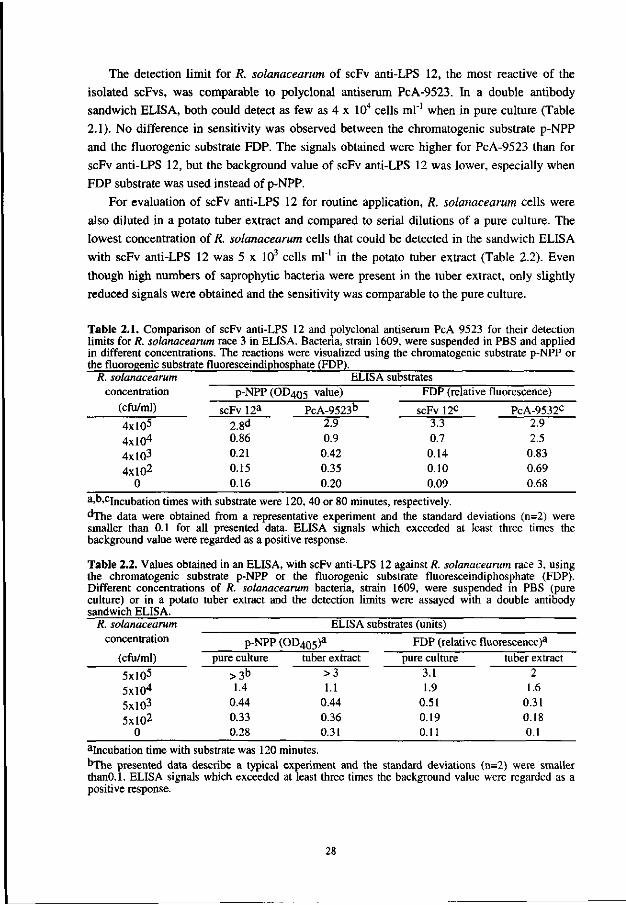
The detection limit for R. solanacearum of scFv anti-LPS 12, the most reactive of the
isolated scFvs, was comparable to polyclonal antiserum PcA-9523. In a double antibody
sandwich ELISA, both could detect as few as 4 x 10⁴ cells ml"1 when in pure culture (Table
2.1). No difference in sensitivity was observed between the chromatogenic substrate p-NPP
and the fluorogenic substrate FDP. The signals obtained were higher for PcA-9523 than for
scFv anti-LPS 12, but the background value of scFv anti-LPS 12 was lower, especially when
FDP substrate was used instead of p-NPP.
For evaluation of scFv anti-LPS 12 for routine application, R. solanacearum cells were
also diluted in a potato tuber extract and compared to serial dilutions of a pure culture. The
lowest concentration of R. solanacearum cells that could be detected in the sandwich ELISA
with scFv anti-LPS 12 was 5 x 10³ cells ml"1 in the potato tuber extract (Table 2.2). Even
though high numbers of saprophytic bacteria were present in the tuber extract, only slightly
reduced signals were obtained and the sensitivity was comparable to the pure culture.
Table 2.1. Comparison of scFv anti-LPS 12 and polyclonal antiserum PcA 9523 for their detection limits for R. solanacearum race 3 in ELISA. Bacteria, strain 1609, were suspended in PBS and applied in different concentrations. The reactions were visualized using the chromatogenic substrate p-NPP or the fluorogenic substrate fluoresceindiphosphate (FDP).
R. solanacearum concentration
(cfu/ml)
4xl05
4xl04
4xl03
4xl02
0
p-NPP
scFv 12a
2.8d
0.86 0.21 0.15 0.16
ELISA substrates (OD4Qg value)
PcA-9523b
2.9 0.9 0.42 0.35 0.20
FDP (relative
scFv 12c
3.3 0.7 0.14 0.10 0.09
fluorescence)
PcA-9532c
2.9 2.5
0.83 0.69 0.68
a.b.cjucubauon times with substrate were 120, 40 or 80 minutes, respectively. dThe data were obtained from a representative experiment and the standard deviations (n=2) were smaller than 0.1 for all presented data. ELISA signals which exceeded at least three times the background value were regarded as a positive response.
Table 2.2. Values obtained in an ELISA, with scFv anti-LPS 12 against R. solanacearum race 3, using the chromatogenic substrate p-NPP or the fluorogenic substrate fluoresceindiphosphate (FDP). Different concentrations of R. solanacearum bacteria, strain 1609, were suspended in PBS (pure culture) or in a potato tuber extract and the detection limits were assayed with a double antibody sandwich ELISA.
R. solanacearum concentration
(cfu/ml)
5xl05
5xl04
5xl03
5xl02
0
p-NPP pure culture
>3b 1.4
0.44 0.33 0.28
ELISA substrates (units)
(OD405)a
tuber extract >3 1.1
0.44 0.36 0.31
FDP (relative fluorescence)3
pure culture 3.1 1.9
0.51 0.19 0.11
tuber extract 2
1.6 0.31 0.18 0.1
aIncubation time with substrate was 120 minutes. ^The presented data describe a typical experiment and the standard deviations (n=2) were smaller thanO.l. ELISA signals which exceeded at least three times the background value were regarded as a positive response.
28
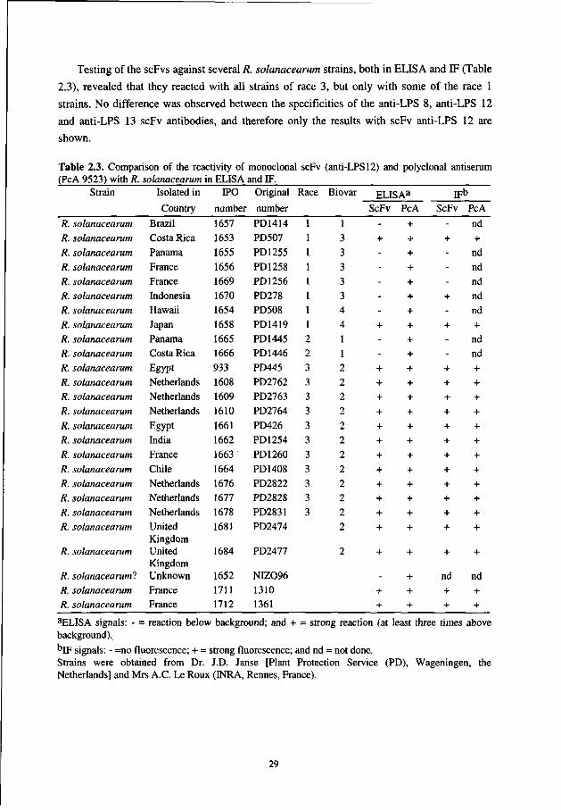
Testing of the scFvs against several R. solanacearum strains, both in ELISA and IF (Table
2.3), revealed that they reacted with all strains of race 3, but only with some of the race 1
strains. No difference was observed between the specificities of the anti-LPS 8, anti-LPS 12
and anti-LPS 13 scFv antibodies, and therefore only the results with scFv anti-LPS 12 are
shown.
Table 2.3. Comparison of the reactivity of monoclonal scFv (anti-LPS 12) and polyclonal antiserum (PcA 9523) with R. solanacearum in ELISA and IF.
Strain
R. solanacearum R. solanacearum R. solanacearum R. solanacearum R. solanacearum R. solanacearum R. solanacearum R. solanacearum R. solanacearum R. solanacearum R. solanacearum R. solanacearum R. solanacearum R. solanacearum
R. solanacearum R. solanacearum R. solanacearum R. solanacearum R. solanacearum R. solanacearum R. solanacearum R. solanacearum
R. solanacearum
R. solanacearum! R. solanacearum R. solanacearum
Isolated in
Country
Brazil Costa Rica Panama France France Indonesia Hawaii Japan Panama Costa Rica Egypt Netherlands Netherlands Netherlands Egypt India France Chile Netherlands Netherlands Netherlands United Kingdom United Kingdom Unknown France France
IPO
number
1657 1653 1655 1656 1669 1670 1654 1658 1665 1666 933 1608 1609 1610 1661 1662 1663 1664 1676 1677 1678 1681
1684
1652 1711 1712
Original
number
PD1414 PD507 PD1255 PD1258 PD1256 PD278 PD508 PD1419 PD1445 PD1446 PD445 PD2762 PD2763 PD2764 PD426 PD1254 PD1260 PD1408 PD2822 PD2828 PD2831 PD2474
PD2477
NIZ096 1310 1361
Race
2 2 3 3 3 3 3 3 3 3 3 3 3
Biovar
1 3 3 3 3 3 4 4 1 1 2 2 2 2 2 2 2 2 2 2 2 2
2
ELISAa
ScFv
-+ -----+ --+ + + + + + + + + + + +
+
-+ +
PcA
+ + + + + + + + + + + + + + + + + + + + + +
+
+ + +
jpb
ScFv
-+ ---+ -+ --+ + + + + + + + + + + +
+
nd + +
PcA
nd + nd nd nd nd nd + nd nd + + + + + + + + + + + +
+
nd + +
aELISA signals: - = reaction below background; and + = strong reaction (at least three times above background),
bjF signals: - =no fluorescence; + = strong fluorescence; and nd = not done. Strains were obtained from Dr. J.D. Janse [Plant Protection Service (PD), Wageningen, the Netherlands] and Mrs A.C. Le Roux (INRA, Rennes, France).
29
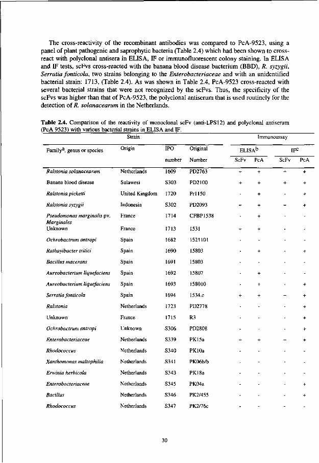
The cross-reactivity of the recombinant antibodies was compared to PcA-9523, using a panel of plant pathogenic and saprophytic bacteria (Table 2.4) which had been shown to cross-react with polyclonal antisera in ELISA, IF or immUnofluorescent colony staining. In ELISA and IF tests, scFvs cross-reacted with the banana blood disease bacterium (BBD), R. syzygii, Serratia fonticola, two strains belonging to the Enterobacteriaceae and with an unidentified bacterial strain: 1713, (Table 2.4). As was shown in Table 2.4, PcA-9523 cross-reacted with several bacterial strains that were not recognized by the scFvs. Thus, the specificity of the scFvs was higher than that of PcA-9523, the polyclonal antiserum that is used routinely for the detection of R. solanacearum in the Netherlands.
Table 2.4. Comparison of the reactivity of monoclonal scFv (anti-LPS12) and polyclonal antiserum (PcA 9523) with various bacterial strains in ELISA and IF.
Family3 , genus or species
Ralstonia solanacearum
Banana blood disease
Ralstonia picketti
Ralstonia syzygii
Pseudomonas marginalis pv. Marginales Unknown
Ochrobactrum antropi
Rathayibacter tritici
Bacillus macerans
Aureobacterium liquefaciens
Aureobacterium liquefaciens
Serratia fonticola
Ralstonia
Unknown
Ochrobactrum antropi
Enterobacteriaceae
Rhodococcus
Xanthomonas maltophilia
Erwinia herbicola
Enterobacteriaceae
Bacillus
Rhodococcus
Strain
Origin
Netherlands
Sulawesi
United Kingdom
Indonesia
France
France
Spain
Spain
Spain
Spain
Spain
Spain
Netherlands
France
Unknown
Netherlands
Netherlands
Netherlands
Netherlands
Netherlands
Netherlands
Netherlands
IPO
number
1609
S303
1720
S302
1714
1713
1682
1690
1691
1692
1693
1694
1723
1715
S306
S339
S340
S341
S343
S345
S346
S347
Original
Number
PD2763
PD2100
P r l l 5 0
PD2093
CFBP1538
1531
1521101
15803
15803
15807
158010
1534.C
PD2778
R3
PD2808
PK15a
PKlOa
PK06b/b
PK18a
PK04a
PK2/455
PK2/76c
Immunoassay
ELISA b I F 0
ScFv PcA ScFv
+ + +
+ + +
+
+ + +
+
+ +
-
+
-
+
+
+ + +
-
-
-
+ + +
-
-
-
-
-_
PcA
+
+
+
+
-
-
+
-
-
+
+
+
+
+
+
-
-
-
+
+
_
30
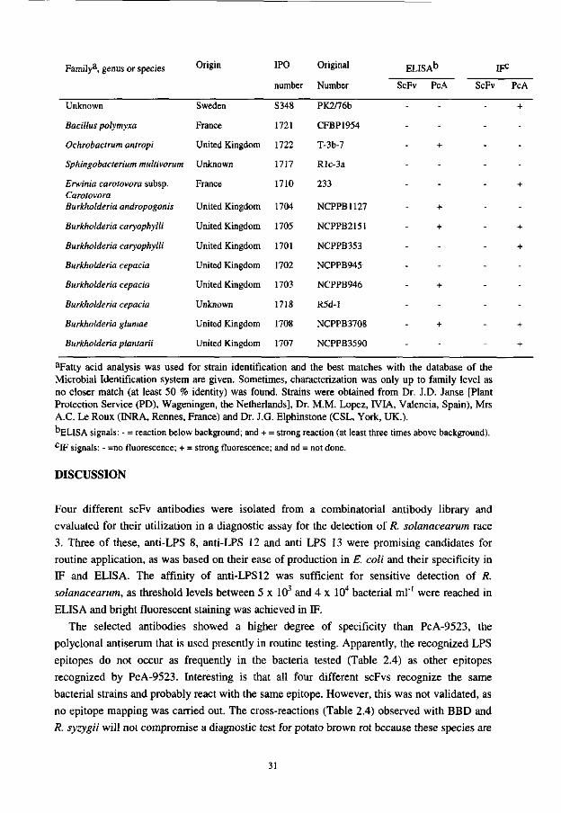
Family3, genus or species
Unknown
Bacillus polymyxa
Ochrobactrum antropi
Sphingobacterium multivorum
Erwinia carotovora subsp. Carotovora Burkholderia andropogonis
Burkholderia caryophylli
Burkholderia caryophylli
Burkholderia cepacia
Burkholderia cepacia
Burkholderia cepacia
Burkholderia glumae
Burkholderia plantarii
Origin
Sweden
France
United Kingdom
Unknown
France
United Kingdom
United Kingdom
United Kingdom
United Kingdom
United Kingdom
Unknown
United Kingdom
United Kingdom
IPO
number
S348
1721
1722
1717
1710
1704
1705
1701
1702
1703
1718
1708
1707
Original
Number
PK2/76b
CFBP1954
T-3b-7
Rlc-3a
233
NCPPB1127
NCPPB2151
NCPPB353
NCPPB945
NCPPB946
R5d-1
NCPPB3708
NCPPB3590
ELISAb
ScFv PcA
-
-
+
-_
+
+
-
-
+
-
+
-
IFc
ScFv PcA
+
-
-
-
+
-
+
+
-
-
-
+
+
aFatty acid analysis was used for strain identification and the best matches with the database of the Microbial Identification system are given. Sometimes, characterization was only up to family level as no closer match (at least 50 % identity) was found. Strains were obtained from Dr. J.D. Janse [Plant Protection Service (PD), Wageningen, the Netherlands], Dr. M.M. Lopez, rVIA, Valencia, Spain), Mrs A.C. Le Roux (INRA, Rennes, France) and Dr. J.G. Elphinstone (CSL, York, UK.).
"ELISA signals: - = reaction below background; and + = strong reaction (at least three times above background). CIF signals: - =no fluorescence; + = strong fluorescence; and nd = not done.
DISCUSSION
Four different scFv antibodies were isolated from a combinatorial antibody library and
evaluated for their utilization in a diagnostic assay for the detection of R. solanacearum race
3. Three of these, anti-LPS 8, anti-LPS 12 and anti LPS 13 were promising candidates for
routine application, as was based on their ease of production in E. coli and their specificity in
IF and ELISA. The affinity of anti-LPS 12 was sufficient for sensitive detection of R.
solanacearum, as threshold levels between 5 x 10³ and 4 x 10⁴ bacterial ml"1 were reached in
ELISA and bright fluorescent staining was achieved in IF.
The selected antibodies showed a higher degree of specificity than PcA-9523, the
polyclonal antiserum that is used presently in routine testing. Apparently, the recognized LPS
epitopes do not occur as frequently in the bacteria tested (Table 2.4) as other epitopes
recognized by PcA-9523. Interesting is that all four different scFvs recognize the same
bacterial strains and probably react with the same epitope. However, this was not validated, as
no epitope mapping was carried out. The cross-reactions (Table 2.4) observed with BBD and
R. syzygii will not compromise a diagnostic test for potato brown rot because these species are
31
not present in temperate areas where this test will be used. In contrast, the cross-reactions observed with the four other saprophytic bacteria from potato and soil can possibly cause false-positive results because they have been isolated in temperate areas. However, as the number of cross-reacting species is reduced relative to the PAbs in PcA-9523, it is to be anticipated that the use of these antibodies in seed testing will result in a decreased number of false positives.
The sensitivity observed in ELISA, comparable to high quality PAbs, is promising. In IF relatively high quantities of scFv-antibody were required to obtain the same signal as the PAbs. This is probably because the scFv-antibodies have one antigen-binding site and bind monovalently to the antigen. In contrast, full size antibodies have two antigen binding sites that can bind with two epitopes simultaneously. According to Neri et al [19], this avidity effect can increase binding up to 100-fold and results in an improved performance in immunoassays. Single-chain antibodies can be engineered in a format allowing bivalent binding [21]. Bivalent molecules can also be established through a genetic fusion with E. coli
alkaline phosphatase, a dimeric enzyme [13, 14]. Thus, if one of these avidity effects is added, it may further increase the sensitivity of the immunoassay. Alkaline phosphatase may be the most convenient choice for detection purposes. The production of scFv-enzyme fusion proteins may result in an improved reproducibility for the detection of antigens in various immunoassays since the variation due to repeated antibody enzyme conjugation is avoided.
Engineering the format of scFvs might also be advantageous for the development of routine assays for the detection of R. solanacearum. Single-chain antibodies are small molecules that are often inactivated upon coating to ELISA plates [12]. Fusion to large proteins like gluthatione-S-transferase, amphipathic helices or to other immunoglobulin domains may enhance the coating efficiency while retaining the full binding activity [12].
The monoclonal recombinant scFvs described here are applicable in routine assays for R.
solanacearum race 3, even though some cross-reactions with other species were observed. The selection of other anti-LPS scFvs that do not cross-react with these strains remains an option. The difficulty lies in the nature of the LPS of the cross-reacting bacteria. Although lipopolysaccharides are highly variable molecules, the possibility can not be excluded that LPS epitopes of cross-reacting bacteria are at least partially identical to the target bacteria. For the isolation of even more specific antibodies consideration may be given to outer membrane proteins or extracellular polysaccharides as antigens. We used a straightforward selection protocol that resulted in the most specific antibodies found thus far for R. solanacearum
(biovar 2, race 3). However, phage display allows easy adaptation of the selection protocols, such as the inclusion of cycles of negative (against unwanted bacteria) and positive selection (for different races). In addition, the present antibody libraries cover the complete immune repertoire, in contrast to the hybridoma technology where only a part of the immune repertoire is tested. Antibody phage display in combination with protein engineering is highly promising for the selection of tailor-made monoclonal antibodies and can be combined with the design of improved immunoassays for phytopathogenic bacteria.
32
ACKNOWLEDGMENTS
We thank Dr. H. Hoogenboom for allowing us to work at his laboratory (CESAME, Academic Hospital Maastricht, The Netherlands) with the Vaughan antibody library, developed by Cambridge Antibody Technology. We also thank Prof. Dr. W.B. van Muiswinkel (Wageningen Institute of Animal Sciences, Cell Biology and Immunology Group, Wageningen Agricultural University) for critically reading the manuscript.
REFERENCES
1. Alvarez, A.M. and Benedict, A.A. (1990) Monoclonal antibodies for the identification of plant pathogenic bacteria: potential applications to Ralstonia solanacearum. Pages 26-31 in: Middleton, K. J., and Hayward, A. C. (eds), Bacterial Wilt of Groundnut. Proceedings of an ACIAR/ICRISAT collaborative research-planning meeting held at Genting Highlands, Malaysia, 18-19 March 1990. ACIAR Proceedings 31, ACIAR, Canberra, Australia.
2. Alvarez, A.M., Berestecky, J.I., Stiles, J.I. Ferreira, S.A. and Benedict, A.A. (1993) Serological and molecular approaches to identification of Pseudomonas solanacearum strains from Heliconia. Pages 62-69 in: Bacterial Wilt; International Conference (1992), Kaoshiuing Taiwan. ACIAR Proceedings 45, ACT, Canberra, Australia.
3. Clark, M.F. and Adams, A.N. (1977) Characteristics of the microplate method of enzyme-linked immunosorbent assay for the detection of plant viruses. J. Gen. Virol. 34: 475-483.
4. De Weger, L.A., Jann, B., Jann, K. and Lugtenberg, B. (1987) Lipopolysaccharides of Pseudomonas spp. that stimulate plant growth: composition and use for strain identification. J. Bacterid. 169: 1441-1446.
5. Elphinstone, J.G., Hennessy, J., Wilson, J.K. and Stead, D.K. (1996) Sensitivity of different methods for the detection of Ralstonia solanacearum (Smith) Smith in potato tuber extract. OEPP/EPPO Bull. 26: 663-678.
6. Griffiths, A.D., Williams, S.C., Hartley, O., Tomlinson, I.M., Waterhouse, P., Crosby, W.L., Kontermann, R.E., Jones, P.T., Low, N.M., Allison, T.J., Prospero, T.D., Hoogenboom, H.R., Nissim, A., Cox, J.P.L., Harrison, J.L., Zaccolo, M., Gherardi, E. and Winter, G. (1994) Isolation of high affinity antibodies directly from large synthetic repertoires. EMBO J. 13: 3245-3260.
7. Hayward, A.C. (1991) Biology and epidemiology of bacterial wilt, caused by Ralstonia solanacearum. Annu. Rev. Phytopathol. 29: 65-87.
8. He, L.Y. (1986) Bacterial wilt in the People's Republic of China. Pages 40-48 in: Persley, G.J. (ed.), Bacterial Wilt Disease and the South Pacific. ACIAR Proceedings 13, ACIAR, Canberra, Australia.
9. Hoogenboom, H.R., Griffiths, A.D., Johnson, K.S., Chiswell, D.J., Hudson, P. and Winter, G. (1991) Multi-subunit proteins on the surface of filamentous phage: methodologies for displaying antibody (Fab) heavy and light chains. Nucleic Acids Res. 15: 4133-4137.
10. Huston, J.S., Levinson, D., Mudgett-Hunter, M., Tai, M., Novotny, J., Margolies, M. N., Ridge, R.J., Bruccoleri, R.E., Haber, E., Crea, R. and Oppermann, H. (1988) Protein engineering of antibody binding sites: Recovery of specific activity in an anti-digoxin single-chain Fv analogue produced in Escherichia coli. Proc. Natl. Acad. Sci. USA. 85: 5879-5883.
11. Janse, J. D. (1988) A detection method for Ralstonia solanacearum in symptomless potato tubers and some data on its sensitivity and specificity. OEPP/EPPO Bull. 18: 343-351.
12. Kerschbaumer, R.J., Hirschl, S., Kaufman, A., Ibl, M., Koenig, R. and Himmler, G. (1997) Single-chain Fv fusion proteins suitable as coating and detecting reagents in a double antibody sandwich enzyme-linked immunosorbent assay. Anal. Biochem. 249: 219-227.
33
13. Kerschbaumer, R.J., Hirschl, S., Schwager, C, Ibl, M. and Himmler, G. (1996) PDAP2: A vector for construction of alkaline phosphatase fusion-proteins. Immunotechnology 2: 145-150.
14. Kohl, J., Riiker, F., Himmler, G. and Katinger, H. (1990) Cloning and expression of an HIV-1 specific single-chain Fv region fused to Escherichia coli alkaline phosphatase. Ann. New York Acad. Sci. 646: 106-114.
15. Lindner, P., Guth, B., Wuelfing, C , Krebber, C, Steipe, B., Mueller, F. and Pluckthun, A. (1992) Purification of native proteins from the cytoplasm and periplasm of Escherichia coli using IMAC and histidine tails: A comparison of proteins and protocols. Methods 4: 41-56.
16. McCafferty, J., Griffiths, A.D., Winter, G. and Chiswell, D.J. (1990) Phage-antibodies: filamentous phage displaying antibody variable domains. Nature 348: 552-554.
17. McGarvey, J.A., Bell, C.J., Denny, T.P. and Schell, M.A. (1998) Analysis of extracellular polysaccaride-I in culture and in planta using immunological methods: New insights and implications. Pages 157-163 in: Prior, P., Allen, C , and Elphinstone, J. (eds), Bacterial wilt disease: molecular and ecological aspects, Springer-Verlag, Berlin.
18. Munro, S. and Pelham, H.R.B. (1986) An Hsp-70 like protein in the ER: Identity with the 78 kD glucose-regulated protein and immunoglobulin heavy chain binding protein. Cell 46: 291-300.
19. Neri, D., Momo, M., Prospero, T. and Winter, G. (1995) High-affinity antigen binding by chelating recombinant antibodies (CRABS). J. Mol. Biol. 246: 367-373.
20. Orlandi, O., Gussow, D.H., Jones, P.T. and Winter, G. (1989) Cloning immunoglobulin variable domains for the expression by the polymerase chain reaction. Proc. Natl. Acad. Sci. USA 86: 3833-3837.
21. Pack, P. and Pluckthun, A. (1992) Miniantibodies: use of amphipathic helices to produce functional, flexibly linked dimeric Fv fragments with high avidity in Escherichia coli. Biochemistry 31: 1579-1584.
22. Robinson-Smith, A., Jones, P., Elphinstone, J.G. and Forde, S.M.D. (1995) Production of antibodies to Ralstonia solanacearum, the causative agent of bacterial wilt. Food Agric. Immunol. 7: 67-79.
23. Sambrook, J., Fritch, E.F. and Maniatis, T. (1990) Molecular cloning: A laboratory manual. Cold Spring Harbour Laboratory, New York.
24. Sastry, L., Alting-Mees, M., Huse, W.D., Short, J.M., Sorge, J.A., Hay, B.H., Janda, K.D., Benkovic, S.J. and Lerner, R.A. (1989) Cloning of the immunological repertoire in Escherichia coli for generation of monoclonal catalytic antibodies: Construction of a heavy chain variable region-specific cDNA library. Proc. Natl. Acad. Sci. USA. 86: 5728-5732.
25. Seal, S.E. and Elphinstone, J.G. (1994) Advances in identification and detection of Ralstonia solanacearum. Pages 35-58 in: Hayward, A.C., and Hartman, G.L. (eds.), Bacterial Wilt: The disease and its causative agent, Ralstonia solanacearum. CAB International, Wallingford, UK.
26. Smith, J.J., Offord, L.C., Hoderness, M. and Saddler, G.S. (1995) Genetic diversity of Burkholderia solanacearum (synonym Ralstonia solanacearum) race 3 in Kenya. App. Environ. Microbiol. 61: 4263-4268.
27. Stead, D.E. (1996) Bacterial diseases of potatoes-future problems? Pages 172-177 in: Proceedings Crop Protection in Northern Britain 1996. University of Dundee, 19-21 March 1996.
34

28. Tijsen, P. (1985) Preparation of enzyme-antibody or other enzyme-macromolecule conjugates. Laboratory Techniques in Biochemistry and Molecular Biology. Vol 15. Practice and Theory of Enzyme Immunoassays. Burdon R. H., and P. H. van Knippenberg (eds), Elsevier Publishing, Amsterdam.
29. Van der Wolf, J.M., Van Beckhoven, J.R.C.M., De Boef, E. and Roozen, N.J.M. (1993) Serological characterization of fluorescent Pseudomonas strains cross-reacting with antibodies against Erwinia chrysanthemi. Neth. J. Plant Pathol. 99: 51-60.
30. Vaughan, T.J., Williams, A.J., Pritchard, K., Osbourn, J.K., Pope, A.R., Earnshaw, J.C., McCafferty, J., Hodits, R.A., Wilton, J. and Johnson, K., (1996) Human antibodies with sub-nanomolar affinities isolated from a large non-immunized phage display library. Nature Biotechnology 14: 309-314.
31. Woods, A.C. (1984) Moko disease: atypical symptoms induced by afluidal variants of Pseudomonas solanacearum in banana plants. Phytopathology 74: 972-976.
35
Chapter 3
Selection of beet necrotic yellow vein virus specific single-chain Fv antibodies from a semi-synthetic combinatorial antibody Library
ABSTRACT
Methods for the generation of monoclonal antibodies against plant viruses are limited because
current hybridoma techniques do not allow efficient exploitation of the immune repertoire.
Moreover, the immunization procedures often lead to a bias towards an immunodominant
contaminant in the immunogen preparation and not to the plant virus itself. The selection of
six different single-chain antibody variable fragments (scFv) against beet necrotic yellow vein
virus from a semi-synthetic human combinatorial antibody library showed the feasibility of
the phage display system. No bias towards minor contaminants in the purified virus
preparation was observed in ELISA, as all the selected scFvs reacted only with beet necrotic
yellow vein virus infected plant homogenates. In addition, two of the isolated beet necrotic
yellow vein virus-specific scFvs could be produced in E. coli as a scFv fusion protein with
alkaline phosphatase, and were applied in ELISA as specific ready to use antibody-enzyme
conjugates. Because of their specificity, these antibodies have potential to be used in sensitive
diagnostic assays for routine testing for beet necrotic yellow vein virus in sugar beets.
This chapter has been accepted for publishing in a modified form by the European Journal of Plant Pathology: Griep, R.A., Van Twisk, C. and Schots, A. Selection of beet necrotic yellow vein virus specific single-chain Fv antibodies from a semi-synthetic combinatorial antibody library.
37
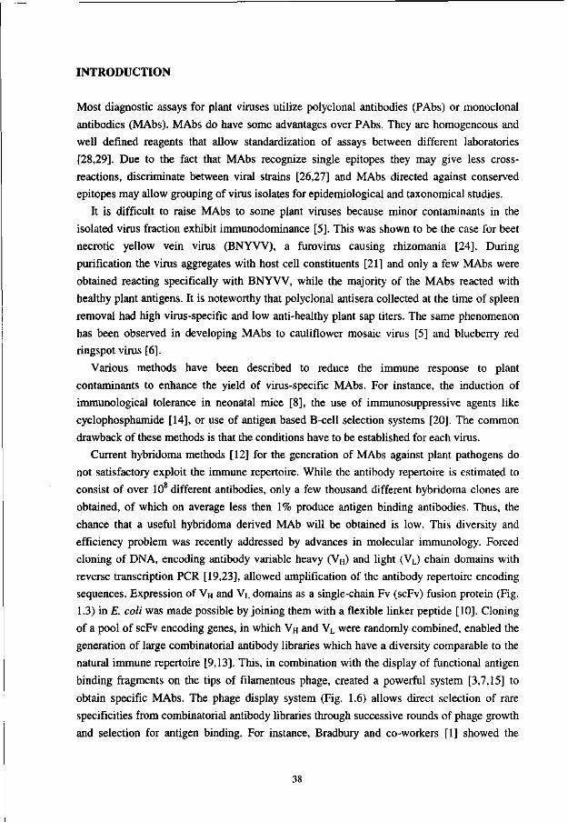
INTRODUCTION
Most diagnostic assays for plant viruses utilize polyclonal antibodies (PAbs) or monoclonal
antibodies (MAbs). MAbs do have some advantages over PAbs. They are homogeneous and
well defined reagents that allow standardization of assays between different laboratories
[28,29]. Due to the fact that MAbs recognize single epitopes they may give less cross-
reactions, discriminate between viral strains [26,27] and MAbs directed against conserved
epitopes may allow grouping of virus isolates for epidemiological and taxonomical studies.
It is difficult to raise MAbs to some plant viruses because minor contaminants in the
isolated virus fraction exhibit immunodominance [5]. This was shown to be the case for beet
necrotic yellow vein virus (BNYVV), a furovirus causing rhizomania [24]. During
purification the virus aggregates with host cell constituents [21] and only a few MAbs were
obtained reacting specifically with BNYVV, while the majority of the MAbs reacted with
healthy plant antigens. It is noteworthy that polyclonal antisera collected at the time of spleen
removal had high virus-specific and low anti-healthy plant sap titers. The same phenomenon
has been observed in developing MAbs to cauliflower mosaic virus [5] and blueberry red
ringspot virus [6].
Various methods have been described to reduce the immune response to plant
contaminants to enhance the yield of virus-specific MAbs. For instance, the induction of
immunological tolerance in neonatal mice [8], the use of immunosuppressive agents like
cyclophosphamide [14], or use of antigen based B-cell selection systems [20]. The common
drawback of these methods is that the conditions have to be established for each virus.
Current hybridoma methods [12] for the generation of MAbs against plant pathogens do
not satisfactory exploit the immune repertoire. While the antibody repertoire is estimated to
consist of over 108 different antibodies, only a few thousand different hybridoma clones are
obtained, of which on average less then 1% produce antigen binding antibodies. Thus, the
chance that a useful hybridoma derived MAb will be obtained is low. This diversity and
efficiency problem was recently addressed by advances in molecular immunology. Forced
cloning of DNA, encoding antibody variable heavy (VH) and light (VL) chain domains with
reverse transcription PCR [19,23], allowed amplification of the antibody repertoire encoding
sequences. Expression of VH and VL domains as a single-chain Fv (scFv) fusion protein (Fig.
1.3) in E. coli was made possible by joining them with a flexible linker peptide [10]. Cloning
of a pool of scFv encoding genes, in which VH and VL were randomly combined, enabled the
generation of large combinatorial antibody libraries which have a diversity comparable to the
natural immune repertoire [9,13]. This, in combination with the display of functional antigen
binding fragments on the tips of filamentous phage, created a powerful system [3,7,15] to
obtain specific MAbs. The phage display system (Fig. 1.6) allows direct selection of rare
specificities from combinatorial antibody libraries through successive rounds of phage growth
and selection for antigen binding. For instance, Bradbury and co-workers [1] showed the
38
feasibility of this technique by selecting antigen binding phages from a pool of non binding
phages, even at a ratio of one binding in 109 irrelevant phages. In addition, the phage display
system may also prove to be a more universal method to obtain specific MAbs, since the
antibody specificities are not biased towards immunodominant epitopes.
To study the versatility of the phage display system, we have applied it to BNYVV. This
virus was chosen as a model system because it is a classic example of the difficulties outlined
in raising specific MAbs. We examined the feasibility of the phage display system in isolating
a diverse repertoire of BNYVV-specific antibody fragments from a semi-synthetic
combinatorial antibody library [18].
MATERIALS AND METHODS
Bacterial strains
E. coli strains used for the isolation of recombinant antibodies, were TGI (K12, Mlac-pro),
supE, thi, hsdD5/F'traD36, proA+B+, lacf, /acZAM15) for selection of specific phage-
antibodies and HB2151 (K12, ara, Mac-pro), thilF'/proA+B+, lacfZAMIS) for expression of
soluble scFv-antibody fragments. HB2151 bacteria were grown at 30°C in 2TY containing
100 ug ml"1 ampicillin and 2% (w/v) glucose [22]. When the OD60o reached 0.5, the bacteria
were pelleted and the supernatant was discarded. The bacteria were resuspended in 2TY +
AMP (100 ug/ml), induced with 1 mM IPTG and grown for 18 h at 16°C.
Purification of beet necrotic yellow vein virions Beet necrotic yellow vein virus (Dutch isolate) was purified from mechanically inoculated
Chenopodium quinoa according to a slightly modified protocol from Putz and Kuszala [21].
Infected C. quinoa leaves (200 g) were homogenized in a mixture of 400 ml CCU and 800 ml
extraction buffer (0.1 M Tris/HCl buffer pH 9 containing 0.14 M NaCl; 5% (v/v) ethanol) and
2% (w/v) polyvinylpyrrolidon. The mixture was stirred for 15 min at room temperature (RT)
and then centrifuged for 15 min at 5,000 x g (GSA rotor, Sorvall). To the collected
supernatant, 5% (w/v) PEG-8000 was added and stirred at RT for 15 min and subsequently at
4°C for 90 min. The virions were pelleted at 16,000 x g (GSA, Sorvall) for 20 min. After
resuspending the pellets in 100 ml extraction buffer and stirring overnight at 4°C, sucrose was
added to 3.3% (w/v) and the mixture was centrifuged at 27,500 x g for 20 min (SW28,
Beckman) to remove large aggregates. Subsequently the virions were pelleted at 110,000 x g
(SW28, Beckman) for 2 h through a 3 cm layer of 20% (w/v) sucrose in extraction buffer. The
pellets were resuspended in 50 ml extraction buffer and, after stirring for 30 min at RT,
centrifuged for 10 min at 1,600 x g to remove insoluble precipitates. Half of the supernatant
was adjusted to a density of 1.1 g/cm3, half to 1.3 g/cm3 with CsCl2. The two halves were
layered 1:1 and centrifuged at 154,000 x g (SW41Ti, Beckman) for 18 h. The virions
containing band was collected and dialyzed overnight against 0.1 M Tris/HCl buffer pH 9
39

containing 0.14 M NaCl and then centrifuged for 5 min at 12,000 x g to remove aggregates.
The virion concentration was determined with a Beckman photospectrometer using a formula
[(1552 x A2so) + (-757.3 x A260)], described by Warburg and Christian [30], which gives the
protein concentration in ug ml"1. The concentration was adjusted to 1 mg ml'1 with 0.1 M
Tris/HCl buffer pH 9 containing 0.14 M NaCl and 1 ml aliquots were frozen in liquid nitrogen
and stored at -74°C until use.
Expansion of the antibody library From a stock of the combinatorial library, which was constructed by Nissim et al [18], 50 ul
was plated on a 240 x 240 mm minimal plate [22] to which ImM thiamin was added. The
bacteria were grown overnight at 30°C, scraped from the plate and resuspended in 50 ml of
2TY containing 100 ug ml"1 ampicillin and 1% (w/v) glucose. A freezer stock [22] was
prepared from 10 ml of these bacteria. One bacterial OD60o unit was taken from the remaining
40 ml, the volume adjusted to 10 ml by adding of 2TY containing 100 ug ml"1 ampicillin and
1% (w/v) glucose, and the bacteria were grown at 37°C while shaking (250 rpm). When an
OD6oo of 0.5 was reached, 1011 helper phages (M13K07, Pharmacia, Uppsala, Sweden) were
added. The 15 ml tube containing the mixture was put in a waterbath without shaking to allow
for optimal infection. After 30 min the bacteria were pelleted (2,100 x g, 10 min) and
resuspended in 25 ml 2TY containing 100 ug ml"1 ampicillin and 25 jag ml"1 kanamycin. The
bacteria were transferred to a 1 liter Erlenmeyer, with 225 ml of prewarmed (37°C) 2TY
containing 100 ug ml"1 ampicillin and 25 ug ml"1 kanamycin, and were grown for 18 h at 30°C
while shaking (250 rpm).
Preparation of phage-antibodies for panning (large-scale production) The 250 ml overnight culture was harvested and the bacteria were removed by centrifugation
(10,000 x g, 20 min, GSA, Sorvall). The phages in the supernatant were precipitated by
adding 50 ml of 20 % (w/v) PEG-6000/ 2.5 M NaCl and mixed thoroughly for 1 h at 4°C. The
precipitated phages were pelleted (8,000 x g, 40 min) and resuspended in 20 ml of sterile H20.
Precipitation was repeated by adding 4 ml of PEG/NaCl and mixed for 20 min at 4°C. The
phages were pelleted (17,000 x g, 10 min; SS34, Beckman), resuspended in 2.5 ml sterile H20
and the stock was stored at 4°C for further use. Usually 5 x 10¹³ phages were produced, as was
established by plating TGI bacteria on selective ampicillin plates after infection with serial
dilutions of the phage suspension.
Panning procedure Immunosorbent tubes (Maxi-sorb, Nunc) were coated with 40 ug BNYVV (10 ug ml"1 in 50
mM NaHC03, pH 9.5) for 2 h on a rollerbench at 25°C and for another 18 h at 4°C. The tubes
were washed 3 times with PBS and blocked with PBM (PBS containing 2% (w/v) skimmed
milk powder) for 30 min. Simultaneously, 2 ml of phage-antibodies was mixed with 2 ml of 2
40

x PBM and preincubated for 30 min. After removing the blocking solution from the tubes, 4
ml of the phage-antibodies containing PBM was added to an antigen-coated tube. Phage-
antibodies were allowed to bind to BNYVV for 30 min on a roller bench and for another 90
minutes without rotation. Free phages were removed by washing the tubes 20 times with 4 ml
PBS containing 0.1% (v/v) Tween-20 and for another 20 times with 4 ml PBS to remove the
detergent. After washing the tubes were rinsed with 4 ml 0.1 M Tris/HCl pH 8.9 containing
0.14 M NaCl. Bound phages were eluted by adding 1 ml of a 0.5 mg ml"1 BNYVV solution in
0.1 M Tris/HCl pH 8.9 containing 0.14 M NaCl to the tube and incubated for 45 min with
rotation, followed by a wash with 0.75 ml PBS. The BNYVV-eluted phages and phages in the
PBS washing were pooled. One ml aliquot, of the BNYVV-eluted phages, was used to infect
9 ml of E. coli TGI bacteria, for 30 min at 37°C in a water bath. The infected E. coli cells
were pelleted (2,100 x g, 10 min) and subsequently resuspended in 1 ml of 2TY, containing
100 ug ml"1 ampicillin and 1% (w/v) glucose. From these suspensions 50 ul aliquots were
taken and plated in serial dilutions on 2TY agar plates containing 100 ug ml"1 ampicillin and
1% (w/v) glucose to establish the number of eluted phages and to make freezer stocks [22] of
TGI bacteria which were growing in single colonies. The remaining 950 ul was plated
separately on 240 x 240 mm minimal plates [22] to which 1 mM thiamin was added, and
grown for 18 h at 30°C. Further enrichment of BNYVV-specific phage-antibodies was
achieved by three additional panning rounds, which were performed according to figure 1.6.
Preparation of phage-antibodies for ELISA (medium-scale production) From a streak of the bacterial freezer stocks, prepared during the panning procedure, single
colonies were tooth picked and plated on minimal plates [22] to which ImM thiamin was
added. The bacteria were grown for 18 h at 30°C, scraped from the plate and resuspended in 2
ml of 2TY containing 100 ug ml"1 ampicillin and 1% (w/v) glucose. One bacterial OD60o unit
was adjusted to 1 ml by adding of 2TY containing 100 ug ml"' ampicillin and 1% (w/v)
glucose, and 2 x 10¹⁰ helper phages (M13K07, Pharmacia). The 1.5 ml Eppendorf tube
containing the mixture was put in a waterbath for 30 min without shaking to allow for optimal
infection at 37°C. The bacteria were pelleted (8,000 x g, 10 min) and resuspended in 750 ul
2TY containing 100 ug ml"' ampicillin and 50 ug ml"' kanamycin. The bacteria were
transferred to a 24-well culture plate and grown for 18 h at 30°C while shaking (100 rpm).
The 750 ul overnight culture was harvested and the bacteria were removed by centrifugation
(8,000 x g, 10 min). If necessary, the phages in the supernatant were precipitated by adding
150 ul of 20% (w/v) PEG-6000/2.5 M NaCl and mixing well for 1 h at 4°C. The precipitated
phage-antibodies were pelleted (8,000 x g, 10 min), resuspended in 250 ul of PBM and stored
at 4°C until further use.
41
Preparation of phage-antibodies for ELISA (small-scale production) After each panning round, 100 single colonies were picked and inoculated in 100 ul of 2TY
containing 100 ug ml"1 ampicillin and 1% (w/v) glucose in a well of a 96-well culture plate.
To study the effect of the panning also 100 single colonies were picked from the original
library before any selection was carried out (Panning round 0). The bacteria were grown
shaking (200 rpm) for 18 h at 30°C. From these overnight cultures 5 ul was taken, inoculated
in a new 96-well plate well, containing 100 ul of 2TY with 100 ug ml"1 ampicillin and 1%
(w/v) glucose and grown shaking for 1 h at 37°C. To allow super-infection, 50 ul aliquots of
2TY containing 100 ug ml"1 ampicillin, 1% (w/v) glucose and 2.5 X 10⁹ helper phages
(M13K07, Pharmacia) were added to each well and incubated for 30 min without shaking and
for 1 h with shaking (200 rpm) at 37°C. The bacteria were pelleted (2,800 x g, 10 min) and
resuspended in 200 ul 2TY containing 100 ug ml"1 ampicillin and 50 ug ml"1 kanamycin. The
bacteria were grown shaking (200 rpm) at 30°C for 18 h, removed by centrifugation (2,800 x
g, 10 min) and the phage-antibody containing supernatant was stored at 4°C until further use.
Phage-ELISA The phage-ELISA was essentially carried out according to Clark and Adams [4] and to Tijsen
[25]. Briefly, ELISA plates (Labstar, Costar, Cambridge, UK) were coated overnight with 100
Hi of polyclonal rabbit-anti-BNYVV antibodies (3 ug IgG ml"1 in 50 mM NaHC03, pH 9.5) at
4°C. The plates were washed with PBST (PBS containing 0.1% (v/v) Tween-20) and blocked
with PBM for 30 min at 37°C at 200 ul per well. The microtiter plates were washed twice
with PBST and incubated for 1 h at RT with 100 ul/well of a C. quinoa leaf homogenate (50
mg dry leaf material ml"1 in PBM), with or without BNYVV lesions. After each incubation
step the microtiter plates were washed four times with PBST. The plates were subsequently
incubated for 1 h at RT with phage samples, diluted 1:1 in PBM; with polyclonal mouse-anti-
M13 antibodies, diluted 1:10,000 in PBM; and finally with polyclonal rat-anti-mouse
antibodies conjugated to alkaline phosphatase (Jackson Immuno-Research Laboratories, Inc.,
Westgrove, PA) diluted 1:2500 in PBM at 100 ul per well. The ELISA was developed by
adding 100 u I p-nitrophenylphosphate substrate per well and absorbance readings were made
at 405 nm, usually within 60 min. A reaction in phage-ELISA was considered to be positive if
the threshold value (mean of the background + three times the standard deviation) was
exceeded.
Mval-fingerprinting Fingerprinting was performed on PCR amplified scFv-DNA using the restriction enzyme Mval. Single colonies were tooth picked and grown for 4 h in 2TY containing 100 ug/ul ampicillin and 1% (w/v) glucose. The bacteria were boiled for 3 min and pelleted at 14.000 x g for 10 min. From the supernatant 2 ul was added to a 48 ul PCR mix containing 2.5 uM dNTPs; 0.25 U Super Taq DNA polymerase (HT Biotechnology, Cambridge, UK); 10 uM
42
Forward primer (5'-AGG AAA CAG CTA TGA CCA TGA TTA CGC CAA G-3') and 10 uM Backward primer (5'-GCC CAA TAG GAA CCC ATG TAC CGT AAC ACT G-3'); 2 mM MgCl2 and 50 mM Tris/HCl, pH 8 and 25 cycles (1 min 94°C; 2 min 72°C) were performed on a thermal cycler (Perkin Elmer). From the PCR mix, 20 pi was added to 36.5 pi H20 and 6,5 ul of buffer H (Boehringer, Mannheim, Germany). After mixing 2 ul (lU/jil) Mval
(Boehringer) was added and incubated 18 h at 37 °C. The Mval digestion patterns were analyzed on a 3% FMC Metaphor agarose gel (Epicentre Technologies, Madison, USA).
Cloning of scFvs into pDAP2/S The genes encoding the scFvs, BNY-8 and BNY-10, were PCR amplified using the forward and backward primers, HindUi and Notl digested, gel purified, ligated into HindWNotl digested pDAP2/S vector [11] and transfected to E. coli TGI bacteria. Transformed bacteria were picked and tested for the expression of scFv-alkaline phosphatase fusion-proteins through incubations with p-nitrophenyl phosphate.
Production of scFv-AP/S fusion-proteins Cultures were grown and induced as described by Kerschbaumer and co-workers [11]. Pelleted bacteria (5,000 rpm in a GSA rotor during 20 min at 4°C) were resuspended in 1/20 volume (referring to the original culture size) of a 50 mM Tris/HCl pH 8 buffer (containing 30% sucrose and 1 mM EDTA). After incubation for 5 min at 0°C the bacteria were pelleted (10,000 rpm in a GSA rotor during 20 min at 4°C) and the produced proteins were extracted by an osmotic shock from the periplasm with 1/20 volume (referring to the original culture size) of 5 mM MgS04 and incubation for 45 min at 0CC. The bacterial debris was removed by centrifugation.
ELISA An antigen coated plate (ACP)-ELISA was used to assess the specificity of the recombinant scFv-AP2/S fusion proteins in the osmotic shock fractions according to standard methods, in which the plates were washed four times between each incubation. Briefly, the wells of a 96 well microtiter plate were coated with the appropriate antigen in 0.1 M NaC03 pH 9.6) for 2 h at 37°C. After blocking with PBMT-5% (PBS containing 5% skimmed milk powder and 0.1% Tween-20) at 200 ul/well for 30 min at 37°C, the wells were incubated with scFv-AP2/S diluted in 100 ul of PBMT-1% for 1 h at 37°C. The reaction was visualized by a subsequent incubation with p-nitrophenylphosphate.
43

RESULTS
Selection of BNYW binding clones from a phage-antibody library To select BNYVV-specific monoclonal antibodies, scFv expressing phages were rescued from a semi-synthetic human antibody library containing 108 different antibody encoding phagemids [18], and subjected to panning. The rescued phages, containing 1013 phage-antibodies (PhAbs), were allowed to bind to immobilized BNYW particles. Bound PhAbs, were eluted by specific elution with competing antigen. To monitor the efficiency of selection during the panning experiments, the phage recovery was measured after each panning round. Determination of the phage recovery (Fig. 3.1 A) showed a clear enhancement. Especially in the third round where a strong increase in phage recovery, about 1,000 fold, was found. Because the fourth panning round did not show a further increase in phage recovery, a fifth selection round was not applied.
A B C
1 2 3 4 Panning round
0 1 2 3 4 Panning round
0 1 2 3 4 Panning round
Figure 3.1. Recovery and specificity of BNYVV-binding phage-antibodies in four sequential rounds of panning. A) The number of recovered PhAbs was counted and the recovery was plotted for each subsequent panning round. B) Phage-antibodies were produced (medium-scale production) and comparable amounts were tested in polyclonal phage-ELISA. Phage-antibodies that were rescued from a stock of the antibody library before any selection had been applied served as negative control (panning round 0). C) In addition the percentage of BNYVV-specific phage-antibody producing clones (small-scale production) was shown by a monoclonal phage-ELISA. Colonies were counted positive in case the derived signal exceeded the mean (n=3) of the negative control pHen (non-scFv expressing phage) plus three times the standard deviation.
To verify if any of the selected phage-antibodies were BNYVV-specific, a polyclonal sandwich phage-ELISA was carried out with samples taken from the stocks of phage-antibodies which were prepared by large-scale production for each panning round. In this BNYVV-specific phage-ELISA, the polyclonal PhAbs showed increasing signals for subsequent rounds of panning (data not shown). The same stocks of polyclonal PhAbs were also tested for their reactivity using a phage-ELISA format in which purified BNYW was coated directly to the plate. Although the signals from the sandwich ELISA were stronger than those obtained from the ACP phage-ELISA (data not shown), both ELISAs showed a similar increase. To ensure that differences in ELISA signals could be contributed to enhancements in specificity and not to degradation of the oldest samples during storage, and to allow the PhAbs obtained from the fourth selection round to be included in the ELISA as well, PhAbs were
44
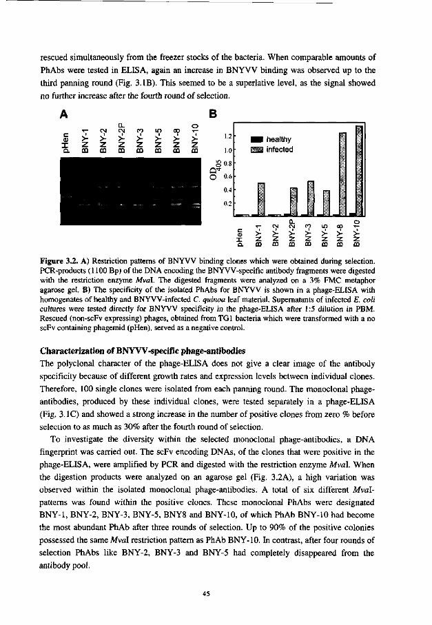
rescued simultaneously from the freezer stocks of the bacteria. When comparable amounts of PhAbs were tested in ELISA, again an increase in BNYVV binding was observed up to the third panning round (Fig. 3.IB). This seemed to be a superlative level, as the signal showed no further increase after the fourth round of selection.
A B
c <D
I Q .
• * -
>-z CO
<N > z CO
Q. CM >-z m
CO
> Z CO
m >-z CO
00 >-Z CO
o > 7 CO
1.2
1.0
g0 .8
a* O 0.6
0.4
0.2 •
H healthy — infected
(V T Q.
T—
> z m
CM > Z. CO
Q. eg >-7 CO
CO >-Z. CO
IT)
> 7 m
co > 7 CO
o
>-7 CD
Figure 3.2. A) Restriction patterns of BNYVV binding clones which were obtained during selection. PCR-products (1100 Bp) of the DNA encoding the BNYVV-specific antibody fragments were digested with the restriction enzyme Mval. The digested fragments were analyzed on a 3% FMC metaphor agarose gel. B) The specificity of the isolated PhAbs for BNYVV is shown in a phage-ELISA with homogenates of healthy and BNYVV-infected C. quinoa leaf material. Supernatants of infected E. coli cultures were tested directly for BNYVV specificity in the phage-ELISA after 1:5 dilution in PBM. Rescued (non-scFv expressing) phages, obtained from TGI bacteria which were transformed with a no scFv containing phagemid (pHen), served as a negative control.
Characterization of BNYVV-specific phage-antibodies The polyclonal character of the phage-ELISA does not give a clear image of the antibody specificity because of different growth rates and expression levels between individual clones. Therefore, 100 single clones were isolated from each panning round. The monoclonal phage-antibodies, produced by these individual clones, were tested separately in a phage-ELISA (Fig. 3.1C) and showed a strong increase in the number of positive clones from zero % before selection to as much as 30% after the fourth round of selection.
To investigate the diversity within the selected monoclonal phage-antibodies, a DNA fingerprint was carried out. The scFv encoding DNAs, of the clones that were positive in the phage-ELISA, were amplified by PCR and digested with the restriction enzyme Mval. When the digestion products were analyzed on an agarose gel (Fig. 3.2A), a high variation was observed within the isolated monoclonal phage-antibodies. A total of six different Mval-patterns was found within the positive clones. These monoclonal PhAbs were designated BNY-1, BNY-2, BNY-3, BNY-5, BNY8 and BNY-10, of which PhAb BNY-10 had become the most abundant PhAb after three rounds of selection. Up to 90% of the positive colonies possessed the same Mval restriction pattern as PhAb BNY-10. In contrast, after four rounds of selection PhAbs like BNY-2, BNY-3 and BNY-5 had completely disappeared from the antibody pool.
45
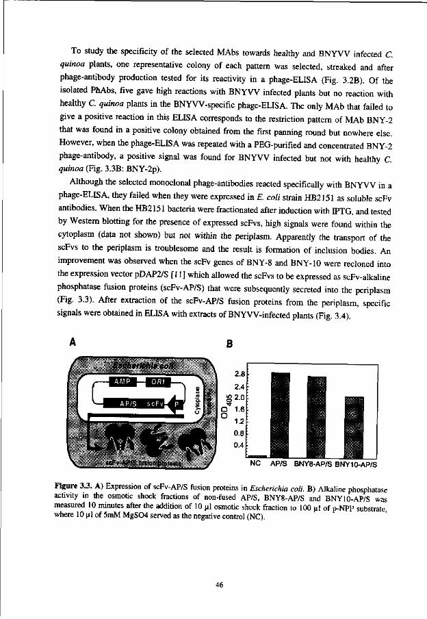
To study the specificity of the selected MAbs towards healthy and BNYVV infected C.
quinoa plants, one representative colony of each pattern was selected, streaked and after
phage-antibody production tested for its reactivity in a phage-ELISA (Fig. 3.2B). Of the
isolated PhAbs, five gave high reactions with BNYVV infected plants but no reaction with
healthy C. quinoa plants in the BNYVV-specific phage-ELISA. The only MAb that failed to
give a positive reaction in this ELISA corresponds to the restriction pattern of MAb BNY-2
that was found in a positive colony obtained from the first panning round but nowhere else.
However, when the phage-ELISA was repeated with a PEG-purified and concentrated BNY-2
phage-antibody, a positive signal was found for BNYVV infected but not with healthy C.
quinoa (Fig. 3.3B: BNY-2p).
Although the selected monoclonal phage-antibodies reacted specifically with BNYVV in a
phage-ELISA, they failed when they were expressed in E. coli strain HB2151 as soluble scFv
antibodies. When the HB2151 bacteria were fractionated after induction with IPTG, and tested
by Western blotting for the presence of expressed scFvs, high signals were found within the
cytoplasm (data not shown) but not within the periplasm. Apparently the transport of the
scFvs to the periplasm is troublesome and the result is formation of inclusion bodies. An
improvement was observed when the scFv genes of BNY-8 and BNY-10 were recloned into
the expression vector pDAP2/S [11] which allowed the scFvs to be expressed as scFv-alkaline
phosphatase fusion proteins (scFv-AP/S) that were subsequently secreted into the periplasm
(Fig. 3.3). After extraction of the scFv-AP/S fusion proteins from the periplasm, specific
signals were obtained in ELISA with extracts of BNYVV-infected plants (Fig. 3.4).
B
Escherichia coll •
•
':
^^—_
3* p "HA
^
"V,
-"-
• • - . . -
NC AP/S BNY8-AP/S BNY10-AP/S
Figure 3.3. A) Expression of scFv-AP/S fusion proteins in Escherichia coli. B) Alkaline phosphatase activity in the osmotic shock fractions of non-fused AP/S, BNY8-AP/S and BNY10-AP/S was measured 10 minutes after the addition of 10 ul osmotic shock fraction to 100 ul of p-NPP substrate, where 10 pi of 5mM MgS04 served as the negative control (NC).
46
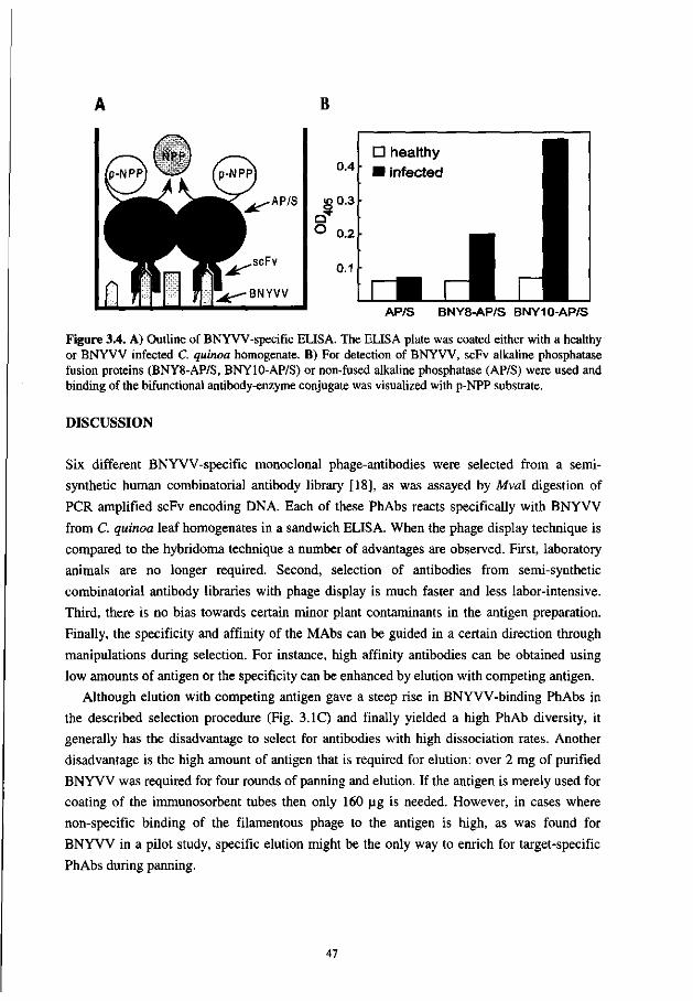
B
r^( (p-NPPJ X
nlr
SIPP)
-—- (p-NPPJ
^ ^ ^ ^ - A P / S
#JK-"scFv
^ - - B N Y V V
AP/S BNY8-AP/S BNY10-AP/S
Figure 3.4. A) Outline of BNYVV-specific ELISA. The ELISA plate was coated either with a healthy or BNYVV infected C. quinoa homogenate. B) For detection of BNYVV, scFv alkaline phosphatase fusion proteins (BNY8-AP/S, BNY10-AP/S) or non-fused alkaline phosphatase (AP/S) were used and binding of the bifunctional antibody-enzyme conjugate was visualized with p-NPP substrate.
DISCUSSION
Six different BNYVV-specific monoclonal phage-antibodies were selected from a semi
synthetic human combinatorial antibody library [18], as was assayed by Mval digestion of
PCR amplified scFv encoding DNA. Each of these PhAbs reacts specifically with BNYVV
from C. quinoa leaf homogenates in a sandwich ELISA. When the phage display technique is
compared to the hybridoma technique a number of advantages are observed. First, laboratory
animals are no longer required. Second, selection of antibodies from semi-synthetic
combinatorial antibody libraries with phage display is much faster and less labor-intensive.
Third, there is no bias towards certain minor plant contaminants in the antigen preparation.
Finally, the specificity and affinity of the MAbs can be guided in a certain direction through
manipulations during selection. For instance, high affinity antibodies can be obtained using
low amounts of antigen or the specificity can be enhanced by elution with competing antigen.
Although elution with competing antigen gave a steep rise in BNYVV-binding PhAbs in
the described selection procedure (Fig. 3.1C) and finally yielded a high PhAb diversity, it
generally has the disadvantage to select for antibodies with high dissociation rates. Another
disadvantage is the high amount of antigen that is required for elution: over 2 mg of purified
BNYVV was required for four rounds of panning and elution. If the antigen is merely used for
coating of the immunosorbent tubes then only 160 ug is needed. However, in cases where
non-specific binding of the filamentous phage to the antigen is high, as was found for
BNYVV in a pilot study, specific elution might be the only way to enrich for target-specific
PhAbs during panning.
47
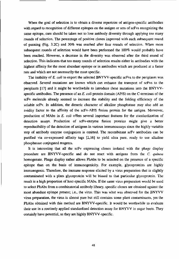
When the goal of selection is to obtain a diverse repertoire of antigen-specific antibodies
with regard to recognition of different epitopes on the antigen or sets of scFvs recognizing the
same epitope, care should be taken not to lose antibody diversity through applying too many
rounds of selection. The percentage of positive clones improved with each subsequent round
of panning (Fig. 3.2C) and 30% was reached after four rounds of selection. When more
subsequent rounds of selection would have been performed the 100% would probably have
been reached. However, a decrease in the diversity was observed after the third round of
selection. This indicates that too many rounds of selection results either in antibodies with the
highest affinity for the most abundant epitope or in antibodies which are produced at a faster
rate and which are not necessarily the most specific.
The inability of E. coli to export the selected BNYVV-specific scFvs to the periplasm was
observed. Several mutations are known which can enhance the transport of scFvs to the
periplasm [17] and it might be worthwhile to introduce these mutations into the BNYVV-
specific antibodies. The presence of an E. coli protein domain (AP/S) on the C-terminus of the
scFv molecule already seemed to increase the stability and the folding efficiency of the
soluble scFv. In addition, the dimeric character of alkaline phosphatase may also add an
avidity factor to the affinity of the scFv-AP/S fusion protein for the antigen. Moreover,
production of MAbs in E. coli offers several important features for the standardization of
detection assays. Production of scFv-enzyme fusion proteins might give a better
reproducibility of the detection of antigens in various immunoassays since the highly variable
step of antibody enzyme conjugation is omitted. The recombinant scFv antibodies can be
purified via co-expressed affinity tags [2,16] to yield ultra pure, ready to use alkaline
phosphatase conjugated reagents.
It is interesting that all the scFv expressing clones isolated with the phage display
procedure are BNYVV-specific and do not react with antigens from the C. quinoa
homogenate. Phage display rather allows PhAbs to be selected on the presence of a specific
epitope than on the basis of immunogenicity. For example, glycoproteins are highly
immunogenic. Therefore, the immune response elicited by a virus preparation that is slightly
contaminated with a plant glycoprotein will be biased to that particular glycoprotein. The
result is a high proportion of host-specific MAbs. If the same virus preparation would be used
to select PhAbs from a combinatorial antibody library, specific clones are obtained against the
most abundant epitope present; i.e., the virus. This was what was observed for the BNYVV
virus preparation, the virus is almost pure but still contains some plant contaminants, yet the
PhAbs obtained with this method are BNYVV-specific. It would be worthwhile to evaluate
their use in a routinely applied standardized detection assay for BNYVV in sugar beets. They
certainly have potential, as they are highly BNYVV-specific.
48
ACKNOWLEDGMENTS
This work was supported by a grant from the Program Committee on Agricultural Biotechnology (PcLB) and by E.C. grant AIR3-CT94-1046. We thank prof. dr. W.B. van Muiswinkel (Department of Experimental Animal Morphology and Cell Biology (EDC), Wageningen Agricultural University, The Netherlands) for critically reading the manuscript, dr. ir. D. Peters (Department of Virology, Wageningen Agricultural University, The Netherlands) for his kind gift of polyclonal Rabbit anti-BNYVV serum and G. Winter (MRC Laboratory of Molecular Biology, Cambridge, UK.) for the permission to use the Nissim library.
REFERENCES
1. Bradbury, A., Persic, L., Werge, T. and Cattaneo, A. (1993) Use of living columns to select specific phage antibodies. Biotechnology 11: 1565-1596.
2. Casey, J.L., Keep, P.A., Chester, K.A. and Robson, L. (1995) Purification of bacterially expressed single-chain Fv antibodies for clinical applications using metal chelate chromatography. J. Immunol. Methods. 179: 105-116.
3. Clackson, T., Hoogenboom, H.R., Griffiths, A.D. and Winter, G. (1991) Making antibodies using phage display libraries. Nature 352: 624-628.
4. Clark, M.F. and Adams, A.N. (1977) Characteristics of the microplate method of enzyme-linked immunosorbent assay for the detection of plant viruses. J. Gen. Virol. 34: 475-483.
5. George, R.A. and Converse, R.H. (1988) Methods for the enrichment of desired B-cell populations before anti-cauliflower mosaic virus hybridoma formation. Phytopathology 78: 1631-1636.
6. Hepp, R.F. and Converse, R.H. (1986) Blueberry ringspot detection in crude sap of highbush blueberry plants. Plant Disease 71: 536-539.
7. Hoogenboom, H.R., Griffiths, A.D., Johnson, K.S., Chiswell, D.J., Hudson, P. and Winter, G. (1991) Multi-subunit proteins on the surface of filamentous phage: methodologies for displaying antibody (Fab) heavy and light chains. Nucleic Acids Res. 15: 4133-4137.
8. Hsu, H.T., Wang, Y.C., Lawson, R.H., Wang, M. and Gonsalves, D. (1990) Splenocytes of mice with induced immunological tolerance to plant antigens for construction of hybridomas secreting tomato spotted wilt virus-specific antibodies. Phytopathology 80: 158-162.
9. Huse, W.D., Sastry, L., Iverson, S.A., Kang, A.S., Alting-Mees, M., Burton, D.R., Benkovic, S.J. and Lerner, R.A. (1989) Generation of a large combinatorial library of the immunoglobulin repertoire in phage lambda. Science 246: 1275-1281.
10. Huston, J.S., Levinson, D., Mudgett-Hunter, M., Tai, M., Novotny, J., Margolies, M.N., Ridge, R.J., Bruccoleri, R.E., Haber, E., Crea, R. and Oppermann, H. (1988) Protein engineering of antibody binding sites: Recovery of specific activity in an anti-digoxin single-chain Fv analogue produced in Escherichia coli. Proc. Natl. Acad. Sci. USA 85: 5879-5883.
11. Kerschbaumer, R.J., Hirschl, S., Kaufman, A., Ibl, M., Koenig, R. and Himmler, G. (1997) Single-chain Fv fusion proteins suitable as coating and detecting reagents in a double antibody sandwich enzyme-linked immunosorbent assay. Anal. Biochem. 249: 219-227.
12. Kohler, G. and Milstein, C. (1975) Continuous cultures of fused cells secreting antibody of predefined specificity. Nature 256: 495-497.
13. Marks, J.D., Hoogenboom, H.R., Bonnert, T.P., McCafferty, J., Griffiths, A.D. and Winter, G. (1991) By-passing immunization: Human antibodies from V-gene libraries displayed on phage. J. Mol. Biol. 222: 581-597.
14. Matthew, W.D. and Sandrock, A.W. Jr. (1987) Cyclophosphamide treatment used to manipulate the immune response for the production of monoclonal antibodies. J. Immunol. Methods. 100: 73-82.
49

15. McCafferty, J., Griffiths, A.D., Winter, G. and Chiswell, D.J. (1990) Phage-antibodies: filamentous phage displaying antibody variable domains. Nature 348: 552-554.
16. Neri, D., Delalla, C , Petrul, H., Neri, P. and Winter, G. (1995) Calmodulin as a versatile tag for antibody fragments. Biotechnology 13: 373-377.
17. Nieba, L., Honegger, A., Krebber, C. and Pliickthun, A. (1997) Disrupting the hydrophobic patches at the antibody variable/constant domain interface: improved in vivo folding and physical characterization of an engineered scFv fragment. Protein-Eng. 10: 435-440.
18. Nissim, A., Hoogenboom, H.R., Tomlinson, I.M., Flynn, G., Midgley, C , Lane, D. and Winter, G. (1994) Antibody fragments from a single pot phage display library as immunochemical reagents. EMBO J. 13: 692-698.
19. Orlandi, O., Gussow, D.H., Jones, P.T. and Winter, G. (1989) Cloning immunoglobulin variable domains for the expression by the polymerase chain reaction. Proc. Natl. Acad. Sci. USA 86: 3833-3837.
20. Parks, D.R., Bryan, V.M., Oi, V.T. and Herzenberg, L.A. (1979) Antigen-specific identification and cloning of hybridomas with a fluorescence-activated cell sorter. Proc. Natl. Acad. Sci. USA 76: 1962-1966.
21. Putz, C. and Kuszala, M. (1978) La rhizomanie de la betterave sucriere en Alsace. Recherche d'une nouvelle m6thode de purification du "Beet Necrotic Yellow Vein Virus". Ann. Phytopathol. 10: 247-262.
22. Sambrook, J., Fritch, E.F. and Maniatis, T. (1990) Molecular cloning: A laboratory manual. Cold Spring Harbour Laboratory, New York.
23. Sastry, L., Alting-Mees, M., Huse, W.D., Short, J.M., Sorge, J.A., Hay, B.H., Janda, K.D., Benkovic, S.J. and Lerner, R.A. (1989) Cloning of the immunological repertoire in Escherichia coli for generation of monoclonal catalytic antibodies: Construction of a heavy chain variable region-specific cDNA library. Proc. Natl. Acad. Sci. USA 86: 5728-5732.
24. Tamada, T. and Baba, T. (1973) Beet necrotic yellow vein virus from rhizomania-affected sugar beet in Japan. Ann. Phytopath. Soc. Japan 39: 325-332.
25. Tijsen, P. (1985) Preparation of enzyme-antibody or other enzyme-macromolecule conjugates. Laboratory Techniques in Biochemistry and Molecular biology. Vol 15. Practice and Theory of Enzyme Immunoassays. R. H. Burdon and P. H. van Knippenberg (eds.), Elsevier Publishing, Amsterdam.
26. Torrance, L., Larkins, A.P. and Butcher, G.W. (1986) Characterization of monoclonal antibodies against potato virus X and comparison of serotypes with resistance groups. J. Gen. Virol. 67: 57-67.
27. Torrance, L. (1995) Use of monoclonal antibodies in plant pathology. Eur. J. of Plant Path. 101:351-363.
28. Van Regenmortel, M.H.V. (1986) The potential for using monoclonal antibodies in the detection of plant viruses. In: Jones RAC and Torrance L (eds) Developments and applications in virus testing (pp 89-101) Wellesbourne: Association of Applied Biologists.
29. Van Regenmortel, M.H.V. (1990) Plant viruses. In: Van Regenmortel MHV and Neurath AR (eds) Immunochemistry of Viruses, II. The basis for Serodiagnosis and Vaccines (pp 505-515) Elsevier, Amsterdam, The Netherlands.
30 . Warburg, O. and Christian, W. (1942) Isolierung und Kristallisation des Garungsferments Enolase. Biochem. Z. 310: 384-421.
50
Chapter 4
pSKAP/S: an expression vector for the production of single-chain Fv alkaline phosphatase fusion proteins
ABSTRACT
The vector pSKAP/S, was constructed to enable overexpression of single-chain variable
fragment antibody (scFv)-alkaline phosphatase fusion proteins. In pSKAP/S, the scFv were
genetically fused to the mutated Escherichia coli PhoA/S gene that encodes an alkaline
phosphatase with increased specific activity. The restriction sites incorporated into pSKAP/S
allowed the scFv genes to be easily transferred from pUC119-derived phagemid vectors, that
are used frequently in phage display antibody library technology. Strong transcriptional control
of expression was achieved using the tetracycline promoter, and induction of different
individual clones with anhydrotetracycline resulted in secretion of most of the scFv-alkaline
phosphatase fusion proteins into the culture medium. Although some of the clones secreted
fusion proteins that were retained in the periplasm, these proteins could be isolated with a
simple extraction procedure. Increased amounts of a scFv-alkaline phosphatase fusion protein
were obtained when expressed in the pSKAP/S vector compared with expression in a vector
incorporating the lac promoter. Testing for binding of the scFv-alkaline phosphatase fusion
proteins to antigen was possible in an ELISA without the need for additional enzyme-
conjugated antibodies. The pSKAP/S vector was successfully used to obtain scFv fragments
from a preparation of phage-antibody clones after sub-cloning and expression of individual
clones as scFv-alkaline phosphatase fusions, whereas fewer clones (and clones with different
properties) were obtained from the same phage-antibody preparations when expressed as
soluble scFv fragments. Therefore, the pSKAP/S vector was shown to be useful in extending
the range of scFv obtained from phage display libraries.
This chapter has been accepted for publication in a modified form by the Journal Protein Expression and Purification: Griep, R.A., Van Twisk, C, Kerschbaumer, R.J., Harper, K., Torrance, L., Himmler, G., Van der Wolf, J.M. and Schots, A. pSKAP/S: an expression vector for the production of single-chain Fv alkaline phosphatase fusion proteins.
51
INTRODUCTION
Recombinant antibodies with unique binding specificities can be selected from large
combinatorial phage display single-chain variable fragment (scFv) antibody libraries. The
phage-antibodies are selected by successive rounds of binding to antigen, elution and growth
of the eluted phage in Escherichia coli without the need for animal immunization
[6,9,20,22,27]. Usually, binding of recombinant antibodies to antigen is detected by means of
linked peptide-tags (for example, c-myc [9], Flag [10], E-tag, Pharmacia). However,
sometimes inconsistencies are observed upon analysis of the soluble scFv obtained. For
example, soluble scFv-c-myc antibodies, selected against beet necrotic yellow vein virus
(BNYVV: Chapter 3) and potato leafroll virus (PLRV), failed to bind to their respective
antigens (Unpublished results). Yet, various scFvs, derived from the same selection process,
bound specifically to their antigens when expressed on filamentous phage.
Single-chain Fv comprise the variable domains of the heavy and light chains of
immunoglobulin molecules connected by a short peptide sequence (c. 10-15 amino acid
residues). It may be that some scFv-c-myc fusion proteins do not fold properly [21] because
the hydrophobic residues of the variable/constant domain interface, that are normally shielded
in the whole antibody molecule by the constant domains of the heavy and light chains, are
exposed in the scFv molecule. Fusion of scFv to the pUI coat protein of filamentous phage may
mimic the shielding effect of the hydrophobic residues. In addition, several scFvs may be
expressed on a single phage particle, thereby increasing the avidity of binding to antigen, in
contrast to the monomeric scFv-c-myc.
The expression of scFv-alkaline phosphatase (scFv-AP) fusion proteins promotes the
formation of bivalent antibodies, due to the dimeric nature of active alkaline phosphatase (AP.,
[1,4,7,12,13,15,26,28,29]). Moreover, the use of an improved alkaline phosphatase enzyme
(AP/S) with a reported 35-fold higher specific activity [18] compared to the E. coli wild-type
protein, increases the absorbance values obtained in ELISA [12].
In addition, increased levels of expression of scFv are possible through improvements to
the expression vector. Although the lac promoter is used frequently, it is known to be leaky,
and cell lysis often occurs prior to induction of protein expression by IPTG [24]. In contrast,
the TetA tetracycline promoter/operator of the TnlO TcR gene (reviewed by Hillen and Berens,
[8]) is strongly controlled and has been used to express antibody fragments in E. coli [25].
Therefore, the vector pSKAP/S was constructed to enable strongly regulated expression of
scFv-AP/S fusion proteins, under the transcriptional control of the TetA promoter in E. coli.
The results presented in this paper demonstrate the utility of application of pSKAP/S to the
expression of a variety of scFv.
52
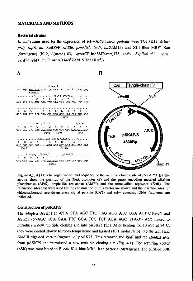
MATERIALS AND METHODS
Bacterial strains
E. coli strains used for the expression of scFv-AP/S fusion proteins were TGI (K12, A(lac-
pro), supE, thi, hsdD5/F'traD36, proA+B+, lacP, facZAM15) and XLl-Blue MRF' Kan
(Stratagene) (K12, A(mcM)183, A(mcrCB-hsdSMR-mrr)173, endAl SupE44 thi-l recAl
gyrA96 relAl, lac F' proAB lacfZAMIS Tn5 (Kanr)).
pSKAP/S t c t a g t aa^i rtt g c a t g c a a a t t c t a t t t c a ag g ag
H i n d l l l / PEL-B l e a d e r
M K Y L L P T A A a c a g t c a t a ATG AAA TAC CTA TTG CCT ACG GCA GCC
A G L L L L A A Q P A M GCT GGA TTG TTA TTA CTC GCG GCC CAG CCG GCC ATG
S f i l
. . . / P o l y l i n k e r / . . . S p a c e r . . A Q V Q L Q A R L A A A
GCC CAG GTG CAG CTG CAS GCG CGC CTG GCG GCC GCA P s t I Asc I NotI
. . . / A l k a l i n e p h o s p h a t a s e / A R A P L G D I A H H
GCC COG GCA CCA CTG GGG GAT ATC GCA CAC CAT Xmal EcoRV
His-tag. ./STOP/ pSKAP/S H H H H -
CAC CAT CAC CAT TAA GGA TCC gaa ttc tga get tga BanHI
B
'amHI
Figure 4.1. A) Genetic organization, and sequence of the multiple cloning site of pSKAP/S. B) The arrows show the position of the TetA promoter (P) and the genes encoding mutated alkaline phosphatase (AP/S), ampicillin resistance (AMP11) and the tetracycline repressor (TetR). The restriction sites that were used for the construction of this vector are shown and the insertion sites for chloramphenicol acetyltransferase signal peptide (CAT) and scFv encoding DNA fragments are indicated.
Construction of pSKAP/S The adapters ASK51 (5'-CTA GTA AGC TTC TAG AGG ATC CGA ATT CTG-3') and
ASK52 (5'-AGC TCA GAA TTC GGA TCC TCT AGA AGC TTA-3') were mixed to
introduce a new multiple cloning site into pASK75 [25]. After heating for 10 min at 94CC,
they were cooled slowly to room temperature and ligated (10:1 molar ratio) into the Xbal and
HindUl digested vector fragment of pASK75. This removed the Xbal and the HindUl sites
from pASK75 and introduced a new multiple cloning site (Fig. 4.1). The resulting vector
(pSK) was transfected to E. coli XLl-blue MRF' Kan bacteria (Stratagene). The purified pSK
53
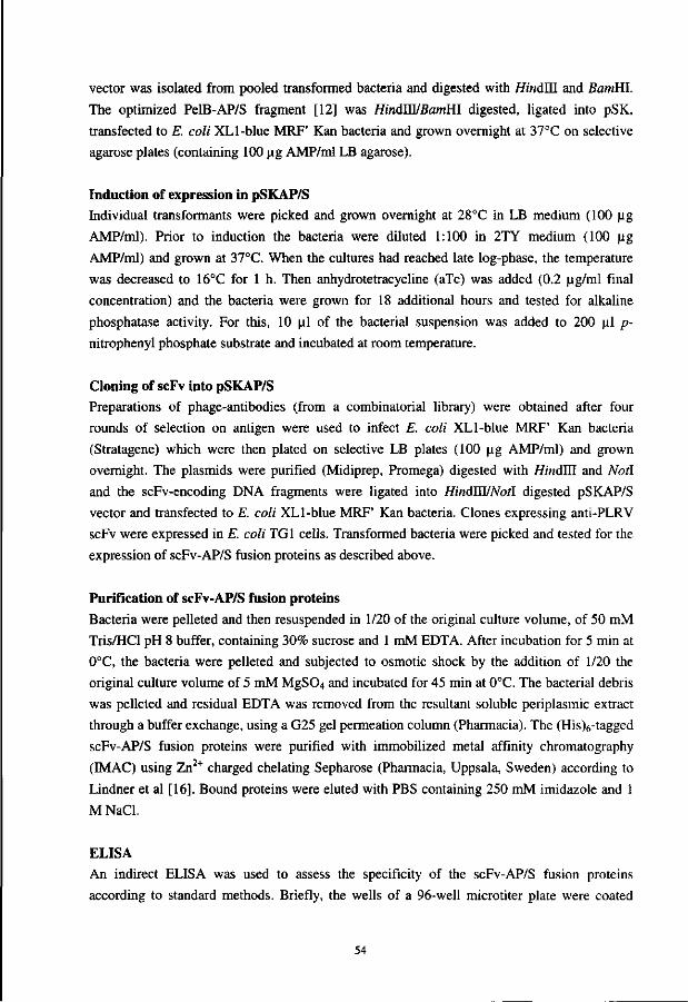
vector was isolated from pooled transformed bacteria and digested with HindlE and BamHl.
The optimized PelB-AP/S fragment [12] was HindWBamHl digested, ligated into pSK,
transfected to E. coli XLl-blue MRF' Kan bacteria and grown overnight at 37°C on selective
agarose plates (containing 100 |ig AMP/ml LB agarose).
Induction of expression in pSKAP/S Individual transformants were picked and grown overnight at 28°C in LB medium (100 ug
AMP/ml). Prior to induction the bacteria were diluted 1:100 in 2TY medium (100 ug
AMP/ml) and grown at 37°C. When the cultures had reached late log-phase, the temperature
was decreased to 16°C for 1 h. Then anhydrotetracycline (aTc) was added (0.2 ug/ml final
concentration) and the bacteria were grown for 18 additional hours and tested for alkaline
phosphatase activity. For this, 10 ul of the bacterial suspension was added to 200 ul p-
nitrophenyl phosphate substrate and incubated at room temperature.
Cloning of scFv into pSKAP/S Preparations of phage-antibodies (from a combinatorial library) were obtained after four
rounds of selection on antigen were used to infect E. coli XLl-blue MRF' Kan bacteria
(Stratagene) which were then plated on selective LB plates (100 ug AMP/ml) and grown
overnight. The plasmids were purified (Midiprep, Promega) digested with HindTH and Notl
and the scFv-encoding DNA fragments were ligated into HindWNotl digested pSKAP/S
vector and transfected to E. coli XLl-blue MRF' Kan bacteria. Clones expressing anti-PLRV
scFv were expressed in E. coli TGI cells. Transformed bacteria were picked and tested for the
expression of scFv-AP/S fusion proteins as described above.
Purification of scFv-AP/S fusion proteins Bacteria were pelleted and then resuspended in 1/20 of the original culture volume, of 50 mM
Tris/HCl pH 8 buffer, containing 30% sucrose and 1 mM EDTA. After incubation for 5 min at
0°C, the bacteria were pelleted and subjected to osmotic shock by the addition of 1/20 the
original culture volume of 5 mM MgS04 and incubated for 45 min at 0°C. The bacterial debris
was pelleted and residual EDTA was removed from the resultant soluble periplasmic extract
through a buffer exchange, using a G25 gel permeation column (Pharmacia). The (His)6-tagged
scFv-AP/S fusion proteins were purified with immobilized metal affinity chromatography
(IMAC) using Zn2+ charged chelating Sepharose (Pharmacia, Uppsala, Sweden) according to
Lindner et al [16]. Bound proteins were eluted with PBS containing 250 mM imidazole and 1
M NaCl.
ELISA An indirect ELISA was used to assess the specificity of the scFv-AP/S fusion proteins
according to standard methods. Briefly, the wells of a 96-well microtiter plate were coated
54
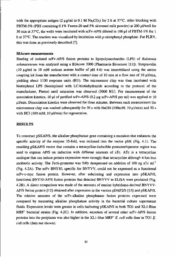
with the appropriate antigen (2 ug/ml in 0.1 M Na2C03) for 2 h at 37°C. After blocking with
PBTM-5% (PBS containing 0.1% Tween-20 and 5% skimmed milk powder) at 200 ul/well for
30 min at 37°C, the wells were incubated with scFv-AP/S diluted in 100 ul of PBTM-1% for 1
h at 37°C. The reaction was visualized by incubation withp-nitrophenyl phosphate. For PLRV,
this was done as previously described [7].
BIAcore measurements Binding of isolated scFv-AP/S fusion proteins to lipopolysaccharides (LPS) of Ralstonia
solanacearum was analyzed using a BIAcore 2000 (Pharmacia Biosensor [11]). Streptavidin
(10 ug/ml in 10 mM sodium acetate buffer of pH 4.6) was immobilized using the amine
coupling kit from the manufacturer with a contact time of 10 min at a flow rate of 10 ul/min,
yielding about 1100 response units (RU). The microsensor chip was then incubated with
biotinylated LPS (biotinylated with LC-biotinhydrazide according to the protocol of the
manufacturer, Pierce) until saturation was observed (5000 RU). For measurement of the
association kinetics, 10 ul of purified scFv-AP/S (0.2 ug scFv-AP/S per ml) was applied at 10
ul/min. Dissociation kinetics were observed for three minutes. Between each measurement the
microsensor chip was washed subsequently for 30 s with NaOH (lOOmM; 10 ul/min) and 30 s
with HC1 (100 mM; 10 ul/min) for regeneration.
RESULTS
To construct pSKAP/S, the alkaline phosphatase gene containing a mutation that enhances the
specific activity of the enzyme 35-fold, was recloned into the vector pSK (Fig. 4.1). The
resulting pSKAP/S vector that contains a tetracycline-inducible promoter/operator region was
used to express AP/S on induction with different amounts of aTc. ATc is a tetracycline
analogue that can induce protein expression more strongly than tetracycline although it has less
antibiotic activity. The 7efA-promoter was fully derepressed on addition of 100 ng aTc ml"1
(Fig. 4.2A). The scFv BNY10, specific for BNYVV, could not be expressed as a functional
scFv-c-myc fusion protein. However, after subcloning and expression into pSKAP/S,
functional BNY10-AP/S fusion proteins that detected BNYVV in ELISA were produced (Fig.
4.2B). A direct comparison was made of the amounts of murine hybridoma-derived BNYVV-
AP/S fusion protein [12] obtained after expression in the vectors pDAP2/S [13] and pSKAP/S.
The relative amounts of the scFv-alkaline phosphatase fusion proteins expressed were
compared by measuring alkaline phosphatase activity in the bacterial culture supernatant
fluids. Expression levels were greater in cells harboring pSKAP/S in both TGI and XLl-Blue
MRF' bacterial strains (Fig. 4.2C). In addition, secretion of several other scFv-AP/S fusion
proteins into the periplasm was also higher in the XL 1-blue MRF' E. coli cells than in TGI E.
coli cells (data not shown).
55

»2 o « * • O
o
1
Q BNY10-AP/S
• AP/s 1 L
.-jij i i i 10 20 50 100 150 200 400
aTc concentration (ng/ml)
B C
[ I Healthy • [""I PDAP2/S 04 • • • •
^ Infected H • pSKAP/S H
O'ui AP/S BNY10-AP/S
Monoclonal antibody TG1 XL1 TG1 XL1
E. coli strain
Figure 4.2. A) Induction of pSKAP/S with various amounts of anhydrotetracycline (aTc). The pSKAP/S vector produces either non-fused alkaline phosphatase (AP/S) or a fusion protein of AP/S with a single-chain variable fragment (BNY10-AP/S) directed against beet necrotic yellow vein virus (BNYVV). B) ELISA for testing of AP/S and BNY10-AP/S using either healthy or BNYVV-infected plant extracts for coating microtiter plates. C) Comparison of the expression (as judged by alkaline phophatase activity within the supernatant) of a murine scFv-AP/S fusion protein in the expression vectors pSKAP/S and pDAP2/S and the influence of the E. coli TGI and XLl-Blue MRF' Kan strains which were used for overexpression.
Table 4.1. Reactivity (number and percentage) of single-chain variable fragments (scFv) derived from a selection against potato leafroll virus (PLRV). ScFv were expressed on phage, as scFv-c-myc or as scFv-alkaline phosphatase fusion proteins (scFv-AP/S).
scFv expression Total number Number PLRV-reactive Percentage PLRV-reactive
ScFv-Phage ScFv-c-Myc ScFv-AP/S
48 96 48
48 0 46
100 0 95
The advantages of pSKAP/S for the expression of antibody fragments were demonstrated
with scFv selected against PLRV by antibody phage display technology (Table 4.1). ScFv from
the fourth and fifth rounds of selection against antigen were expressed on the surface of phage
and a high percentage reacted specifically with PLRV in phage-ELISA (Table 4.1). However,
when scFv from this selection were expressed as soluble scFv-c-myc fusion proteins, no
reaction with PLRV was obtained (Table 4.1). In contrast, when scFv from the fourth round of
selection were recloned into pSKAP/S and expressed as scFv-alkaline phosphatase fusion
proteins, a high proportion detected PLRV in ELISA (Table 4.1).
Similar results were also obtained after overexpression of Ralstonia solanacearum
specific scFv in pSKAP/S. Only four scFvs were isolated [5] on screening for scFv-c-myc
fusion proteins binding to R. solanacearum, amongst which was the scFv designated anti-
LPS12. The same phage preparation used to isolate the soluble scFvs was then recloned into
pSKAP/S, and a further ten different R. solanacearum-binding scFv were isolated (data not
shown). In some clones scFv-AP/S fusion proteins were found mainly in the periplasm
whereas others were mainly secreted into the culture fluid. This was determined by measuring
56
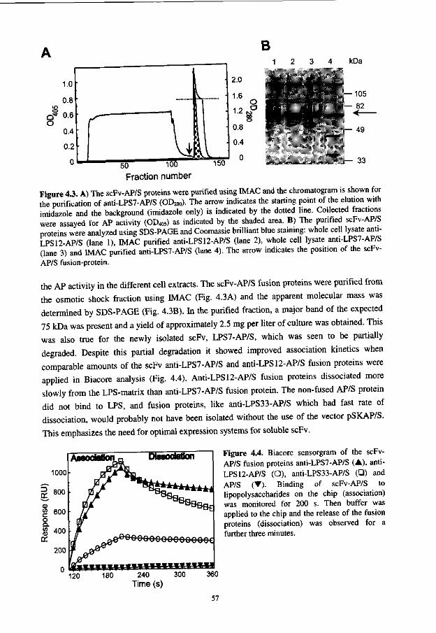
B
1.0
0.8 10
Q * 0.6 O
0.4
0.2
0
-
r I 50 100
Fraction number
s '•
1
11 ' 150
2.0
1.6 O
1.2 M ° OO O
0.8
0.4
0
r '
kDa
— 105 — 82
— 49
— 33
Figure 4.3. A) The scFv-AP/S proteins were purified using IMAC and the chromatogram is shown for the purification of anti-LPS7-AP/S (OD28o). The arrow indicates the starting point of the elution with imidazole and the background (imidazole only) is indicated by the dotted line. Collected fractions were assayed for AP activity (OD405) as indicated by the shaded area. B) The purified scFv-AP/S proteins were analyzed using SDS-PAGE and Coomassie brilliant blue staining: whole cell lysate anti-LPS12-AP/S (lane 1), IMAC purified anti-LPS12-AP/S (lane 2), whole cell lysate anti-LPS7-AP/S (lane 3) and IMAC purified anti-LPS7-AP/S (lane 4). The arrow indicates the position of the scFv-AP/S fusion-protein.
the AP activity in the different cell extracts. The scFv-AP/S fusion proteins were purified from
the osmotic shock fraction using MAC (Fig. 4.3A) and the apparent molecular mass was
determined by SDS-PAGE (Fig. 4.3B). In the purified fraction, a major band of the expected
75 kDa was present and a yield of approximately 2.5 mg per liter of culture was obtained. This
was also true for the newly isolated scFv, LPS7-AP/S, which was seen to be partially
degraded. Despite this partial degradation it showed improved association kinetics when
comparable amounts of the scFv anti-LPS7-AP/S and anti-LPS12-AP/S fusion proteins were
applied in Biacore analysis (Fig. 4.4). Anti-LPS12-AP/S fusion proteins dissociated more
slowly from the LPS-matrix than anti-LPS7-AP/S fusion protein. The non-fused AP/S protein
did not bind to LPS, and fusion proteins, like anti-LPS33-AP/S which had fast rate of
dissociation, would probably not have been isolated without the use of the vector pSKAP/S.
This emphasizes the need for optimal expression systems for soluble scFv.
Figure 4.4. Biacore sensorgram of the scFv-AP/S fusion proteins anti-LPS7-AP/S (A), anti-LPS12-AP/S (O), anti-LPS33-AP/S (•) and AP/S (•). Binding of scFv-AP/S to lipopolysaccharides on the chip (association) was monitored for 200 s. Then buffer was applied to the chip and the release of the fusion proteins (dissociation) was observed for a further three minutes.
57
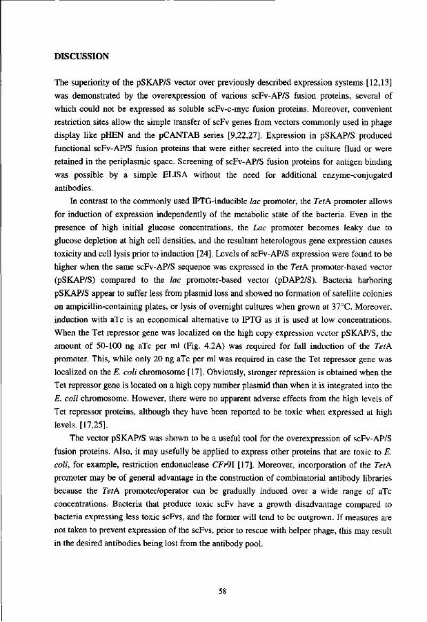
DISCUSSION
The superiority of the pSKAP/S vector over previously described expression systems [12,13]
was demonstrated by the overexpression of various scFv-AP/S fusion proteins, several of
which could not be expressed as soluble scFv-c-myc fusion proteins. Moreover, convenient
restriction sites allow the simple transfer of scFv genes from vectors commonly used in phage
display like pHEN and the pCANTAB series [9,22,27]. Expression in pSKAP/S produced
functional scFv-AP/S fusion proteins that were either secreted into the culture fluid or were
retained in the periplasmic space. Screening of scFv-AP/S fusion proteins for antigen binding
was possible by a simple ELISA without the need for additional enzyme-conjugated
antibodies.
In contrast to the commonly used IPTG-inducible lac promoter, the TetA promoter allows
for induction of expression independently of the metabolic state of the bacteria. Even in the
presence of high initial glucose concentrations, the Lac promoter becomes leaky due to
glucose depletion at high cell densities, and the resultant heterologous gene expression causes
toxicity and cell lysis prior to induction [24]. Levels of scFv-AP/S expression were found to be
higher when the same scFv-AP/S sequence was expressed in the TetA promoter-based vector
(pSKAP/S) compared to the lac promoter-based vector (pDAP2/S). Bacteria harboring
pSKAP/S appear to suffer less from plasmid loss and showed no formation of satellite colonies
on ampicillin-containing plates, or lysis of overnight cultures when grown at 37GC. Moreover,
induction with aTc is an economical alternative to IPTG as it is used at low concentrations.
When the Tet repressor gene was localized on the high copy expression vector pSKAP/S, the
amount of 50-100 ng aTc per ml (Fig. 4.2A) was required for full induction of the TetA
promoter. This, while only 20 ng aTc per ml was required in case the Tet repressor gene was
localized on the E. coli chromosome [17]. Obviously, stronger repression is obtained when the
Tet repressor gene is located on a high copy number plasmid than when it is integrated into the
E. coli chromosome. However, there were no apparent adverse effects from the high levels of
Tet repressor proteins, although they have been reported to be toxic when expressed at high
levels. [17,25].
The vector pSKAP/S was shown to be a useful tool for the overexpression of scFv-AP/S
fusion proteins. Also, it may usefully be applied to express other proteins that are toxic to E.
coli, for example, restriction endonuclease CFr9\ [17]. Moreover, incorporation of the TetA
promoter may be of general advantage in the construction of combinatorial antibody libraries
because the TetA promoter/operator can be gradually induced over a wide range of aTc
concentrations. Bacteria that produce toxic scFv have a growth disadvantage compared to
bacteria expressing less toxic scFvs, and the former will tend to be outgrown. If measures are
not taken to prevent expression of the scFvs, prior to rescue with helper phage, this may result
in the desired antibodies being lost from the antibody pool.
58
From the selected scFv-AP/S fusion proteins produced in E. coli, several showed partial
degradation when analyzed by SDS-PAGE, for example, LPS7-AP/S (Fig. 4.3B). Degradation
is frequently observed on expression of foreign genes in E. coli and may be the result of the
formation of inclusion bodies in the cytoplasm, or aggregates in the periplasm (both are known
to induce the expression of proteases). It is possible that human antibody sequences may not be
recognized efficiently by the folding machinery in E. coli cells. This may result in the improper
folding of the scFv and the subsequent formation of inclusion bodies. The bacterial strains
used for expression may be influential and indeed we observed higher levels of secretion with
the XL 1-blue MRF' strain, as previously reported [2]. However, others have shown expression
of Fab fragments to be independent of the bacterial strain [25]. Nevertheless, expression of
scFv-AP/S fusion proteins with the aid of pSKAP/S in XLl-blue MRF' bacteria allowed
several specific scFv to be isolated and characterized. These will now be further developed
into higher affinity, stable immunoassay reagents, for example by site directed mutagenesis
[21] or chain shuffling [19].
The correct folding of scFv has been shown to be dependent on the presence of some key
amino acid residues [21,14]. In addition, Freund and co-workers [3] have shown the existence
of an intermediate folding state between the correct and the misfolded states of scFv in the
periplasm. Fusion of scFv to E. coli AP/S may cause the heterologous protein to be recognized
by the chaperonins of E. coli, and furthermore, it may stabilize the scFv-AP/S fusion protein
by shielding the hydrophobic residues on the surface of the scFv, thereby allowing a higher
proportion of the expressed scFvs to fold properly. As the construction of all scFvs leads to the
exposure of the residues of the former variable/constant domain interface, this strategy might
lead to a generally applicable method for the expression of scFv fragments.
ACKNOWLEDGMENTS.
The sequence of pSKAP/S has been deposited at GenBank (accession number AF083070). This work was supported by E.C. grant AIR3-CT94-1046. We thank Prof. Dr. W.B. van Muiswinkel (Wageningen Institute of Animal Sciences, Cell Biology and Immunology Group, Wageningen Agricultural University, The Netherlands) for critically reading the manuscript. We also like to acknowledge Dr Renate Koenig and Dr Andrea Kaufmann (both "Biologische Bundesanstalt', Braunschweig, Germany) for providing the murine hybridoma derived anti-BNYVV scFv gene and finally Cambridge Antibody Technology for the use of the Nissim library and the Vaughan library, which supplied us with the scFv-antibodies used in this study.
REFERENCES
1. Ducancel, F., Gillet, D., Carrier, A., Lajeunesser, E., Menez, A. and Boulain, J.C. (1993) Recombinant colorimetric antibodies: construction and characterization of a bifunctional F(ab)2/alkaline phosphatase conjugate produced in Escherichia coli. Bio/Technology 11: 601-605.
2. Duenas, M., Vazquez, J., Ayala, M., SSderlind, E., Ohlin, M., Perez, L., Borrebaeck, C.A.K. and Gavilondo, J.V. (1994) Intra and extracellular expression of a scFv antibody
59
fragment in E. coli: Effect of bacterial strains and pathway engineering using GroES/L chaperonins. BioTechniques 16: 476-483.
3. Freund, C , Honegger, A., Hunziker, P., Holak, T.A. and Pliickfhun, A. (1996) Folding nuclei of the scFv fragment of an antibody. Biochemistry 35: 8457-8464.
4. Gandecha, A.R., Owen, M.R.L., Cockburn, B. and Whitelam, G.C. (1992) Antigen detection using recombinant, bifunctional single-chain Fv fusion proteins synthesized in Escherichia coli. Protein Express. Purific. 5: 361-365.
5. Griep, R.A., Van Twisk, C , Van der Wolf, J.M., Van Beckhoven, J.R.C.M. and Schots, A. (1998) Selection of Ralstonia solanacearum race 3 specific monoclonal antibodies from combinatorial libraries in "Bacterial wilt disease: molecular and ecological aspects" (Prior, P., Allen, C , and Elphinstone, J., Eds.), pp. 140-145, Springer Verlag, Berlin.
6. Griffiths, A.D., Williams, S.C., Hartley, O., Tomlinson, I.M., Waterhouse, P., Crosby, W.L., Kontermann, R.E., Jones, P.T., Low, N.M., Allison, T.J., Prospero, T.D., Hoogenboom, H.R., Nissim, A., Cox, J.P.L., Harrison, J.L., Zaccolo, M., Gherardi, E. and Winter, G. (1994) Isolation of high affinity antibodies directly from large synthetic repertoires. EMBO J. 13: 3245-3260.
7. Harper, K., Kerschbaumer, R.J., Ziegler, A., Macintosh, S.M., Cowan, G.H., Himmler, G. and Mayo, M.A. (1997) A scFv-alkaline phosphatase fusion-protein which detects potato leafroll luteovirus in plant extracts by ELISA. J. of Virolol. methods 63: 237 -242.
8. Hillen, W. and Berens, C. (1994) Mechanisms underlying expression of TnlO encoded tetracycline resistance. Annu. Rev. Microbiol. 48: 345-369.
9. Hoogenboom, H.R., Griffiths, A.D., Johnson, K.S., Chiswell, D.J., Hudson, P. and Winter, G. (1991) Multi-subunit proteins on the surface of filamentous phage: methodologies for displaying antibody (Fab) heavy and light chains. Nucleic Acids Res. 15: 4133-4137.
10. Hopp, T.P., Pricket, K.S., Price, V.L., Libby, R.T., March, C.J., Cerretti, D.P., Urdal, D.L. and Conlon, P.J. (1988) A short polypeptide marker sequence for recombinant protein identification and purification. Bio/Technology 6: 1204-1204.
11. Jonsson, U., Fagerstam, L., Ivarsson, B., Johnsson, B., Karlsson, R., Lundh, K„ Lofas., S., Persson, B., Roos, H., Ronnberg, I. and Malmqvist, M. (1991) Real-time biospecific interaction analysis using surface plasmon resonance and a sensor chip technology. Biotechniques 11: 620-627.
12. Kerschbaumer, R.J., Hirschl, S., Kaufman, A., Ibl, M., Koenig, R. and Himmler, G. (1997) Single-chain Fv fusion proteins suitable as coating and detecting reagents in a double antibody sandwich enzyme-linked immunosorbent assay. Anal. Biochem. 249: 219-227.
13. Kerschbaumer, RJ., Hirschl, S., Schwager, C , Ibl, M. and Himmler, G. (1996) PDAP2: A vector for construction of alkaline phosphatase fusion proteins. Immunotechnology 2: 145-150.
14. Knappik, A. and Pluckthun, A. (1995) Engineered turns of a recombinant antibody improve its in vivo folding. Prot. Eng. 8: 81-89.
15. Kohl, J., Riiker, F., Himmler, G. and Katinger, H. (1990) Cloning and expression of an HIV-1 specific single-chain Fv region fused to Escherichia coli alkaline phosphatase. Ann. New York Acad. Sci. 646: 106-114.
16. Lindner, P., Guth, B., Wuelfing, C , Krebber, C , Steipe, B., Mueller, F. and Pluckthun, A. (1992) Purification of native proteins from the cytoplasm and periplasm of Escherichia coli using IMAC and histidine tails: A comparison of proteins and protocols. Methods 4: 41-56.
17. Lutz, R. and Bujard H. (1997) Independent and tight regulation of transcriptional units in Escherichia coli via the LacR/O and the AraC/Il-I2 regulatory elements. Nucleic Acid. Res. 25: 1203-1210.
18. Mandecki, W., Shallcross, M.A., Sowadski, J. and Tomazic-Allen, S. (1991) Mutagenesis of conserved residues within the active site of Escherichia coli alkaline phosphatase yields enzymes with increased Kca,. Prot. Eng. 4: 801-804.
60
19. Marks, J.D., Griffiths, A.D., Malmqvist, M., Clackson, T.P., Bye, J.M. and Winter, G. (1992) By-passing immunization: building high affinity antibodies by chain shuffling. Biotechnology 7: 779-783.
20. McCafferty, J., Griffiths, A.D., Winter, G. and Chiswell, DJ. (1990) Phage-antibodies: filamentous phage displaying antibody variable domains. Nature 348: 552-554.
21. Nieba, L., Honegger, A., Krebber, C. and Pliickthun, A. (1997) Disrupting the hydrophobic patches at the antibody variable/constant domain interface: improved in vivo folding and physical characterization of an engineered scFv fragment. Protein-Eng. 10: 435-44.
22. Nissim, A., Hoogenboom, H.R., Tomlinson, I.M., Flynn, G., Midgley, C , Lane, D. and Winter, G. (1994) Antibody fragments from a single pot phage display library as immunochemical reagents. EMBO J. 13: 692-698.
23. Oehmichen, R., Klock, G., Altschmied, L. and Hillen, W. (1984) Construction of an E. coli strain overproducing the TnlO-encoded Tet repressor and its use for large scale purification. EMBO J. 3: 539-543.
24. Pliickthun, A. and Skerra, A. (1989) Expression of functional Fv and Fab fragments in Escherichia coli. Methods-Enzymol. 178: 497-515.
25. Skerra, A. (1994) Use of tetracycline promoter for the tightly regulated production of a murine antibody fragment in Escherichia coli. Gene 151, 131-135.
26. Susi, P., Ziegler, A. and Torrance, L. (1998) Selection of single-chain variable fragment antibodies to black currant reversion associated virus from a synthetic phage display library. Phytopathology 88: 230-233.
27. Vaughan, T.J., Williams, A.J., Pritchard, K., Osbourn, J.K., Pope, A.R., Earnshaw, J.C., McCafferty, J., Hodits, R.A., Wilton, J. and Johnson, K. (1996) Human antibodies with sub-nanomolar affinities isolated from a large non-immunized phage display library. Nature Biotechnology 14: 309-314.
28. Weiss, E. and Orfanoudakis, G. (1994) Application of an alkaline phosphatase fusion-protein system suitable for efficient screening and production of Fab-enzyme conjugates in Escherichia coli. J. Biotechnol. 33: 43-53.
29. Wels, W., Harwerth, I., Zwickl, M., Hardman, N., Groner, B. and Hynes, N.E. (1992) Construction, bacterial expression and characterization of a bifunctional single-chain antibody-phosphatase fusion protein targeted to the human Erbb-2 receptor. Bio/Technology 10: 1128-1132.
61
Chapter 5
Application of phage display in selecting tomato spotted wilt virus-specific single-chain Fvs for sensitive diagnosis in ELISA
ABSTRACT
A panel of recombinant single-chain antibodies (scFvs) against structural proteins of tomato
spotted wilt virus was retrieved from a human combinatorial scFv antibody library using the
novel phage display technique. After subcloning the encoding DNA sequences in the
expression vector pSKAP/S, which allowed the scFvs to be expressed as alkaline phosphatase
fusion proteins, seventeen different scFv antibodies were obtained. Twelve of these scFvs
were directed against the nucleoprotein (N) and five against the glycoproteins (G1/G2). Five
of the N-specific antibodies cross-reacted with two other tospoviruses TCSV and GRSV, but
none recognized the more distantly related tospoviruses INSV, WSMV, IYSV or PSMV. The
successful use of one of the retrieved antibodies as coating and detection reagent in a double
antibody sandwich ELISA showed the potential of the phage display system in obtaining
antibodies for routine TSWV diagnosis.
This chapter has been submitted in a modified form for publication: Griep, R.A., Prins, M., Van Twisk, C, Kerschbaumer, R.J., Kormelink, R., Goldbach, R.W. and Schots, A. Application of phage display in selecting tomato spotted wilt virus-specific single-chain Fvs for sensitive diagnosis in ELISA.
63
INTRODUCTION
Tomato spotted wilt virus (TSWV), the representative of the Tospovirus genus within the
family Bunyaviridae, is the causal agent [28] of a major viral disease in many crops. With an
estimated annual crop loss of over one billion US $, TSWV ranks among the top-ten of most
detrimental plant viruses worldwide [8,23]. The spread of this plant pathogen is difficult to
control, for it has a wide host range of over 1000 plant species including mono- and
dicotyledons of more than 80 different families. Tospoviruses are transmitted in a persistent
manner by thrips, which over the recent years have become resistant against most applied
insecticides. The spherical tospoviral particle (70-110 nm in diameter) contains three
structural proteins: a 28 kDa nucleoprotein (N) which is tightly associated with three genomic
RNA segments and two glycoproteins of 78 kDa (Gl) and 58 kDa (G2) which are associated
with the viral envelope. Furthermore low amounts (10-20 copies per virion) of the viral
polymerase (331 kDa) are present in the virus particle.
Hardly any durable resistance genes against tospoviruses are available for conventional
breeding purposes [23]. TSWV resistance has been obtained by transgenic expression of the
nucleoprotein or movement protein (NSM) gene sequences of TSWV into tobacco [24] and
tomato plants [33]. However, the use of transgenic plants for production of crops has been
limited to date and epidemics of tospoviruses are still prevented by sanitary measures in
combination with surveillance of disease development. Therefore, to prevent the spread of
tospoviruses, diagnostic assays like ELISA or PCR have to be developed which are suitable
for rapid detection of infected plants and, moreover, can be used for certification of plant
propagation materials.
High quality polyclonal antisera have been raised against several established members of
the genus Tospovirus (Table 5.1) including TSWV [7], groundnut ringspot virus (GRSV) [5],
impatiens necrotic spot virus (INSV) [20], tomato chlorotic spot virus (TCSV) [5] and
watermelon silverleaf mottle virus (WSMV) [36]. They are now widely applied for tospovirus
detection and with the aid of a double antibody sandwich (DAS)-ELISA [9, 35] they form a
good compromise between sensitivity, ease and expense of application. However, the applied
polyclonal antisera are generally based on N-specific antibodies and recognize mainly the
species against which they were raised. Thus far, members of the genus Tospovirus have been
classified into four distinct serogroups [1,6] (Table 5.1) which are a direct reflection of the
sequence homologies (50-80%) of the N protein within the different tospovirus species [5].
The glycoproteins, on the other hand, are more conserved as they share common epitopes, and
polyclonal antibodies raised against the glycoproteins of one virus species also detect others
[2]. However, as the N protein is by far the most abundant viral protein in infected plants and
tospoviruses moreover tend to lose their envelop during several passages in plants [25], this
protein remains the target of interest for serological detection purposes.
64
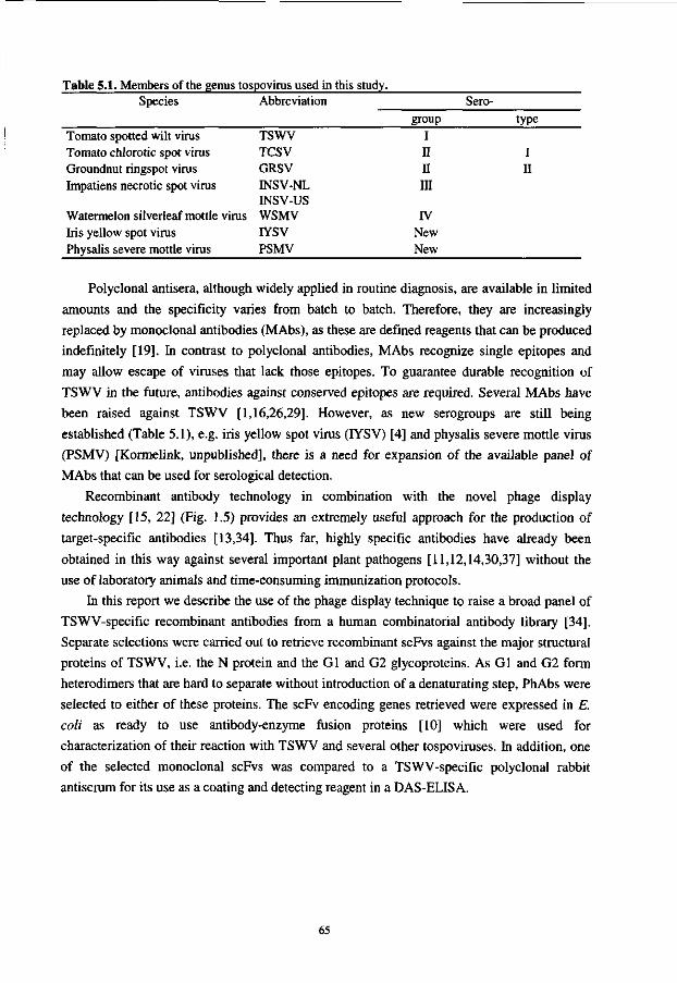
Table 5.1. Members of the genus tospovirus used in this study. Species
Tomato spotted wilt virus Tomato chlorotic spot virus Groundnut ringspot virus Impatiens necrotic spot virus
Watermelon silverleaf mottle virus Iris yellow spot virus Physalis severe mottle virus
Abbreviation
TSWV TCSV GRSV INSV-NL INSV-US WSMV IYSV PSMV
Sero-group
I II II III
IV New New
type
I II
Polyclonal antisera, although widely applied in routine diagnosis, are available in limited
amounts and the specificity varies from batch to batch. Therefore, they are increasingly
replaced by monoclonal antibodies (MAbs), as these are defined reagents that can be produced
indefinitely [19]. In contrast to polyclonal antibodies, MAbs recognize single epitopes and
may allow escape of viruses that lack those epitopes. To guarantee durable recognition of
TSWV in the future, antibodies against conserved epitopes are required. Several MAbs have
been raised against TSWV [1,16,26,29]. However, as new serogroups are still being
established (Table 5.1), e.g. iris yellow spot virus (IYSV) [4] and physalis severe mottle virus
(PSMV) [Kormelink, unpublished], there is a need for expansion of the available panel of
MAbs that can be used for serological detection.
Recombinant antibody technology in combination with the novel phage display
technology [15, 22] (Fig. 1.5) provides an extremely useful approach for the production of
target-specific antibodies [13,34]. Thus far, highly specific antibodies have already been
obtained in this way against several important plant pathogens [11,12,14,30,37] without the
use of laboratory animals and time-consuming immunization protocols.
In this report we describe the use of the phage display technique to raise a broad panel of
TSWV-specific recombinant antibodies from a human combinatorial antibody library [34].
Separate selections were carried out to retrieve recombinant scFvs against the major structural
proteins of TSWV, i.e. the N protein and the Gl and G2 glycoproteins. As Gl and G2 form
heterodimers that are hard to separate without introduction of a denaturating step, PhAbs were
selected to either of these proteins. The scFv encoding genes retrieved were expressed in E.
coli as ready to use antibody-enzyme fusion proteins [10] which were used for
characterization of their reaction with TSWV and several other tospoviruses. In addition, one
of the selected monoclonal scFvs was compared to a TSWV-specific polyclonal rabbit
antiserum for its use as a coating and detecting reagent in a DAS-ELISA.
65

MATERIALS AND METHODS
Bacterial strains E. coli strain TGI was used for the panning experiments. Expression of scFv-AP/S fusion
proteins was performed in XLl-Blue MRF' Kan bacteria (Stratagene).
Purification of tomato spotted wilt virus Infectious virus particles of TSWV Brazilian strain (BR-01) were isolated from systematically
infected Nicotiana rustica var. America leaves according to Gonsalves and Trujillo [9].
Purified virions were finally collected from a 20-40% sucrose gradient, subsequently pelleted,
resuspended in water and stored at -70°C. Concentration of viral protein was measured using
the Biorad protein assay (Biorad).
Purification of viral nucleocapsids Nicotiana rustica var. America plants were inoculated with TSWV (BR-01). After 8-18 days,
systemically infected leaves were harvested and homogenized in 4 ml/g TAS-E buffer (freshly
prepared: 0.1 M Tris-HCl; 0.01 M EDTA; 0.01 M Na2S03; 0.1% Cysteine, pH 8.0). The
homogenate was filtered through 2 layers of cheesecloth and put on ice. The supernatant was
centrifuged for 10 min at 3,000 rpm at 4°C in a GSA rotor, subsequently transferred to Ti45
tubes and centrifuged a second time for 30 min at 20,000 rpm at 4°C in a Ti45 rotor. The
pellet obtained, containing viral nucleocapsids, was resuspended in 0.4 ml/g TAS-R buffer
(freshly prepared: 0.01 M Tris-HCl; 0.01 M EDTA; 0.01 M Na2S03; 0.1% Cysteine; 0.01 M
Glycine; 1% Nonidet P40, pH 7.9) while gently stirring for 1-2 h at 4°C. A volume of 10 ml
of the nucleocapsid material was enlarged to 30 ml using TAS-R buffer and layered on a 30
ml 30% sucrose cushion (30% w/v sucrose in TAS-R buffer). After centrifuging for 2.5 h at
28,000 rpm at 4°C in a Ti45 rotor, the supernatant was decanted, the nucleocapsid pellet
resuspended overnight in PBS and stored at -70°C until further use. The concentration of N
protein was measured using the Biorad protein assay.
Preparation of virus-infected plant material for ELISA-assays Approximately 2 weeks after inoculation of Nicotiana benthamiana plants with tospoviruses
GRSV (SA-05), INSV (US-01), INSV (NL-07), IYSV, PSMV, TCSV (BR-03), TSWV (BR-
01) or WMSV (Table 1), systemically infected leaves were harvested and homogenized 1:10
w/v in extraction buffer (1 g leaf material/10 ml PBS 2% polyvinylpyrolidon) for use in
ELISA.
Selection of recombinant antibodies from a combinatorial antibody library Selection of target specific phage-antibodies (PhAbs, i.e. phages expressing functional scFv
on their surface) from the human combinatorial antibody library [34] was performed
66
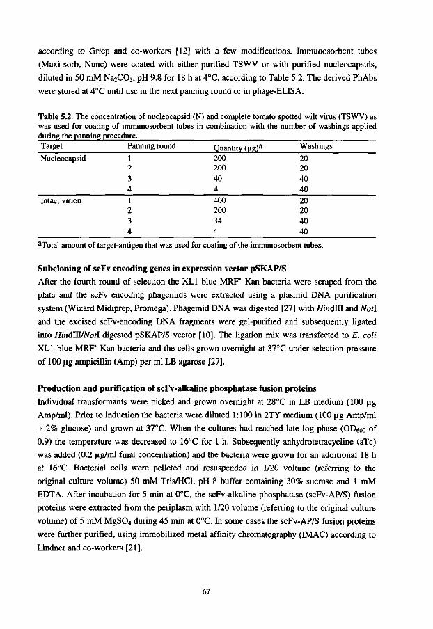
according to Griep and co-workers [12] with a few modifications. Immunosorbent tubes
(Maxi-sorb, Nunc) were coated with either purified TSWV or with purified nucleocapsids,
diluted in 50 mM Na2C03, pH 9.8 for 18 h at 4°C, according to Table 5.2. The derived PhAbs
were stored at 4°C until use in the next panning round or in phage-ELISA.
Table 5.2. The concentration of nucleocapsid (N) and complete tomato spotted wilt virus (TSWV) as was used for coating of immunosorbent tubes in combination with the number of washings applied during the panning procedure. Target Panning round Quantity (ug)a Washings Nucleocapsid 1 200 20
2 200 20 3 40 40 4 4 40
Intact virion 1 400 20 2 200 20 3 34 40 4 4 40
aTotal amount of target-antigen that was used for coating of the immunosorbent tubes.
Subcloning of scFv encoding genes in expression vector pSKAP/S After the fourth round of selection the XL1 blue MRF' Kan bacteria were scraped from the
plate and the scFv encoding phagemids were extracted using a plasmid DNA purification
system (Wizard Midiprep, Promega). Phagemid DNA was digested [27] with HindUl and Notl
and the excised scFv-encoding DNA fragments were gel-purified and subsequently ligated
into HindJU/Notl digested pSKAP/S vector [10]. The ligation mix was transfected to E. coli
XLl-blue MRF' Kan bacteria and the cells grown overnight at 37°C under selection pressure
of 100 ug ampicillin (Amp) per ml LB agarose [27].
Production and purification of scFv-alkaline phosphatase fusion proteins
Individual transformants were picked and grown overnight at 28°C in LB medium (100 ug
Amp/ml). Prior to induction the bacteria were diluted 1:100 in 2TY medium (100 ug Amp/ml
+ 2% glucose) and grown at 37°C. When the cultures had reached late log-phase (OD6oo of
0.9) the temperature was decreased to 16°C for 1 h. Subsequently anhydrotetracycline (aTc)
was added (0.2 ug/ml final concentration) and the bacteria were grown for an additional 18 h
at 16°C. Bacterial cells were pelleted and resuspended in 1/20 volume (referring to the
original culture volume) 50 mM Tris/HCl, pH 8 buffer containing 30% sucrose and 1 mM
EDTA. After incubation for 5 min at 0°C, the scFv-alkaline phosphatase (scFv-AP/S) fusion
proteins were extracted from the periplasm with 1/20 volume (referring to the original culture
volume) of 5 mM MgS04 during 45 min at 0°C. In some cases the scFv-AP/S fusion proteins
were further purified, using immobilized metal affinity chromatography (IMAC) according to
Lindner and co-workers [21].
67
Mval-fingerprinting To analyze the diversity of the recloned scFvs, restriction fragment length polymorphism
(RFLP) fingerprinting was performed on PCR amplified scFv-DNA. Single colonies were
picked and grown for 4 h in 2TY (100 (ig Amp/ml). From these suspensions, 2 ul was taken
and added to a 48 ul PCR mix containing 2.5 uM dNTPs; 0.25 U Super Taq DNA polymerase
(HT Biotechnology, Cambridge, UK); 10 uM pSK-forward (5'-ACT CTA TCA TTG ATA
GAG TTA TTT TAC CAC TCC C-3') and 10 uM pSK-reverse (5'-TTT ATC GCT AAG
AGA ATC ACG CAG AGC GGC G-3') primers; 2 mM MgCl2 and 50 mM Tris/HCl, pH 8.
Amplification was performed in a thermal cycler (Perkin Elmer), 25 cycles (1 min 94°C; 2
min 72°C) were performed. After PCR-amplification, 20 ul of the mix was digested with 10 U
of restriction enzyme Mval (Boehringer) for 18 h at 37 °C. Digestion patterns were analyzed
on a 3% FMC Metaphor agarose gel (Epicentre Technologies, Madison, USA).
Phage-ELISA The phage-ELISA was essentially carried out according to Clark and Adams [3] and to Tijsen
[32]. After each incubation step the microliter plates were washed 4 times with PBST (PBS
containing 0.1% (v/v) Tween-20). In brief, ELISA plates (Labstar, Costar, Cambridge, UK)
were coated overnight at 4°C with 100 ul/well purified viral nucleocapsids, diluted to 2 ug/ml
in 0.1 M Na2C03/pH 9.8. Plates were blocked with 200 ul/well PBSTM-5% (PBST
containing 5% skimmed milk) for 30 min at 37°C, washed twice with PBST and subsequently
incubated for 1 h at RT with 100 ul phage sample dilutions, nucleoprotein-phage-antibody
complexes were detected with monoclonal mouse-anti-M13 antibodies (MAb 12E4, 1 |ag/ml
in PBSTM-2%), and finally with polyclonal rat-anti-mouse antibodies conjugated to alkaline
phosphatase (Jackson Immuno-Research Laboratories, Inc., Westgrove, PA) diluted 1:2500 in
PBSTM-2%. The ELISA was developed by adding 100 ul /?-nitrophenyl phosphate (p-NPP)
substrate per well and scored for positive reactions by absorbance reading at 405 nm, usually
within 60 min.
Antigen coated plate ELISA An antigen-coated plate (ACP)-ELISA was used to assess the specificity of the recombinant
scFv-antibodies, according to standard methods [3, 32], in which the plates were washed four
times with PBST between each incubation step. In brief, ELISA plates were coated with either
TSWV virions or with purified nucleocapsids, both diluted to 2 ug/ml in 0.1 M Na2C03. After
blocking with PBSTM-5% for 30 min at 37°C, the plates were incubated for 1 h at 37°C with
recombinant anti-TSWV scFv alkaline phosphatase fusion proteins, diluted 1:1 in PBTM-4%
or with PcA-BROl-AP (alkaline phosphatase conjugated polyclonal Rabbit antibodies, raised
against the nucleocapsid of TSWV), diluted 1:2000 in PBSTM-2%. The reactions were
visualized by incubation with p-NPP substrate and optical densities were measured at 405 nm
with an ELISA-reader.
68
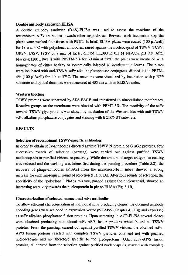
Double antibody sandwich ELISA A double antibody sandwich (DAS)-ELISA was used to assess the reactions of the
recombinant scFv-antibodies towards other tospoviruses. Between each incubation step the
plates were washed four times with PBST. In brief, ELISA plates were coated (100 ul/well)
for 18 h at 4°C with polyclonal antibodies, raised against the nucleocapsid of TSWV, TCSV,
GRSV, INSV, IYSV or a mix of these, diluted 1:1,000 in 0.1 M Na2C03, pH 9.8. After
blocking (200 ul/well) with PBSTM-5% for 30 min at 37°C, the plates were incubated with
homogenates of either healthy or systemically infected N. benthamiana leaves. The plates
were incubated with anti-TSWV scFv alkaline phosphatase conjugates, diluted 1:1 in PBTM-
4% (100 ul/well) for 1 h at 37°C. The reactions were visualized by incubation with p-NPP
substrate and optical densities were measured at 405 nm with an ELISA-reader.
Western blotting TSWV proteins were separated by SDS-PAGE and transferred to nitrocellulose membranes.
Reactive groups on the membrane were blocked with PBMT-5%. The reactivity of the scFv
towards TSWV glycoproteins was shown by incubation of the Western blot with anti-TSWV
scFv alkaline phosphatase conjugates and staining with BCIP/NBT substrate.
RESULTS
Selection of recombinant TSWV-specific antibodies In order to obtain scFv-antibodies directed against TSWV N protein or G1/G2 proteins, four
successive rounds of selection (panning) were carried out against purified TSWV
nucleocapsids or purified virions, respectively. While the amount of target antigen for coating
was reduced and the washing was intensified during the panning procedure (Table 5.2), the
recovery of phage-antibodies (PhAbs) from the immunosorbent tubes showed a strong
increase for each subsequent round of selection (Fig. 5.1 A). After four rounds of selection, the
specificity of the "polyclonal" PhAbs mixture, panned against the nucleocapsid, showed an
increasing reactivity towards the nucleoprotein in phage-ELISA (Fig. 5.IB).
Characterization of selected monoclonal scFv antibodies To allow efficient characterization of individual scFv producing clones, the obtained antibody
encoding genes were recloned in expression vector pSKAP/S (Chapter 4, [10]) and expressed
as scFv alkaline phosphatase fusion proteins. Upon screening in ACP-ELISA several clones
were obtained producing monoclonal scFv-AP/S fusion proteins which bound to TSWV
proteins. From the panning, carried out against purified TSWV virions, the obtained scFv-
AP/S fusion proteins reacted with complete TSWV particles only and not with purified
nucleocapsids and are therefore specific to the glycoproteins. Other scFv-AP/S fusion
proteins, all derived from the selection against purified nucleocapsids, reacted with complete
69
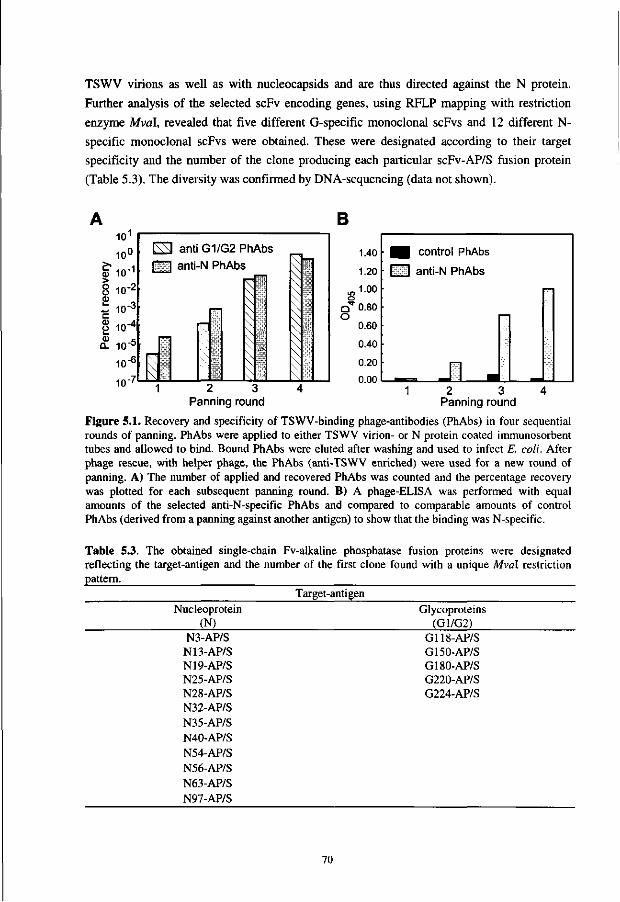
TSWV virions as well as with nucleocapsids and are thus directed against the N protein.
Further analysis of the selected scFv encoding genes, using RFLP mapping with restriction
enzyme Mval, revealed that five different G-specific monoclonal scFvs and 12 different N-
specific monoclonal scFvs were obtained. These were designated according to their target
specificity and the number of the clone producing each particular scFv-AP/S fusion protein
(Table 5.3). The diversity was confirmed by DNA-sequencing (data not shown).
B 10'
10°
§Tio-1
8 io-2
i IO"3
8 10-4
CL 10"°
10"6
7
L\M anti G1/G2 PhAbs I | anti-N PhAbs
^^^
I
' 1 \
^
*
\ V
xx
s \
1.40
1.20
»1-°° cT 0.80 O
0.60 0.40
0.20 n nn
• B r—]
l u 1 2 3 4 1 P an nir igrc ur id
control PhAbs
anti-N PhAbs
2 3 Panning round
I—1
4
Figure 5.1. Recovery and specificity of TSWV-binding phage-antibodies (PhAbs) in four sequential rounds of panning. PhAbs were applied to either TSWV virion- or N protein coated immunosorbent tubes and allowed to bind. Bound PhAbs were eluted after washing and used to infect E. coli. After phage rescue, with helper phage, the PhAbs (anti-TSWV enriched) were used for a new round of panning. A) The number of applied and recovered PhAbs was counted and the percentage recovery was plotted for each subsequent panning round. B) A phage-ELISA was performed with equal amounts of the selected anti-N-specific PhAbs and compared to comparable amounts of control PhAbs (derived from a panning against another antigen) to show that the binding was N-specific.
Table 5.3. The obtained single-chain Fv-alkaline phosphatase fusion proteins were designated reflecting the target-antigen and the number of the first clone found with a unique Mval restriction pattern.
Target-antigen Nucleoprotein
(N) N3-AP/S N13-AP/S N19-AP/S N25-AP/S N28-AP/S N32-AP/S N35-AP/S N40-AP/S N54-AP/S N56-AP/S N63-AP/S N97-AP/S
Glycoproteins (G1/G2)
G118-AP/S G150-AP/S G180-AP/S G220-AP/S G224-AP/S
70
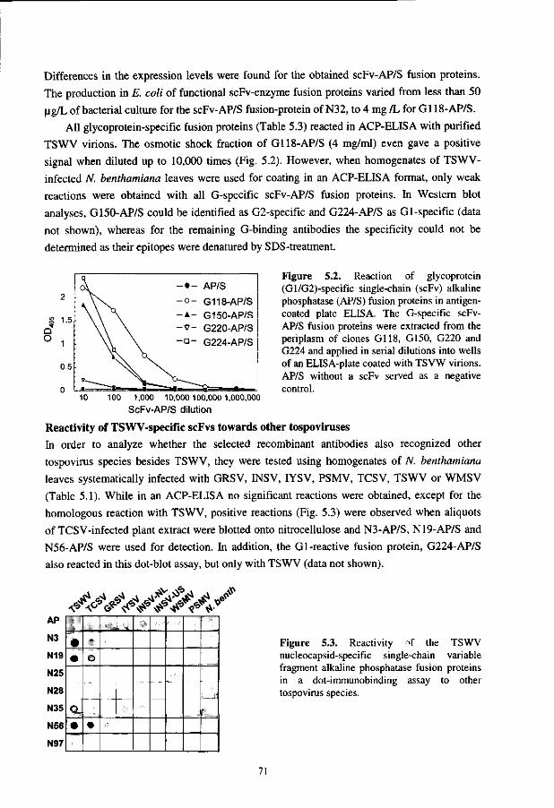
Differences in the expression levels were found for the obtained scFv-AP/S fusion proteins.
The production in E. coli of functional scFv-enzyme fusion proteins varied from less than 50
ug/L of bacterial culture for the scFv-AP/S fusion-protein of N32, to 4 mg /L for Gl 18-AP/S.
All glycoprotein-specific fusion proteins (Table 5.3) reacted in ACP-ELISA with purified
TSWV virions. The osmotic shock fraction of Gl 18-AP/S (4 mg/ml) even gave a positive
signal when diluted up to 10,000 times (Fig. 5.2). However, when homogenates of TSWV-
infected N. benthamiana leaves were used for coating in an ACP-ELISA format, only weak
reactions were obtained with all G-specific scFv-AP/S fusion proteins. In Western blot
analyses, G150-AP/S could be identified as G2-specific and G224-AP/S as Gl-specific (data
not shown), whereas for the remaining G-binding antibodies the specificity could not be
determined as their epitopes were denatured by SDS-treatment.
Q O
2
1.5
1
0.5
n
°V
v. "~^n •
- • - AP/S
- o - G118-AP/S - A - G150-AP/S - v - G220-AP/S - a - G224-AP/S
• I M ^ ^ Q - . m
Figure 5.2. Reaction of glycoprotein (Gl/G2)-specific single-chain (scFv) alkaline phosphatase (AP/S) fusion proteins in antigen-coated plate ELISA. The G-specific scFv-AP/S fusion proteins were extracted from the periplasm of clones G118, G150, G220 and G224 and applied in serial dilutions into wells of an ELISA-plate coated with TSVW virions. AP/S without a scFv served as a negative control.
10 100 1,000 10,000 100,000 1,000,000
ScFv-AP/S dilution
Reactivity of TSWV-specific scFvs towards other tospoviruses
In order to analyze whether the selected recombinant antibodies also recognized other
tospovirus species besides TSWV, they were tested using homogenates of N. benthamiana
leaves systematically infected with GRSV, INSV, IYSV, PSMV, TCSV, TSWV or WMSV
(Table 5.1). While in an ACP-ELISA no significant reactions were obtained, except for the
homologous reaction with TSWV, positive reactions (Fig. 5.3) were observed when aliquots
of TCSV-infected plant extract were blotted onto nitrocellulose and N3-AP/S, N19-AP/S and
N56-AP/S were used for detection. In addition, the Gl-reactive fusion protein, G224-AP/S
also reacted in this dot-blot assay, but only with TSWV (data not shown).
AP
N3
N19
N25
N28
N35
N56
N97
• •
Q-
•
o
• i t
Figure 5.3. Reactivity ->f the TSWV nucleocapsid-specific single-chain variable fragment alkaline phosphatase fusion proteins in a dot-immunobinding assay to other tospovirus species.
71
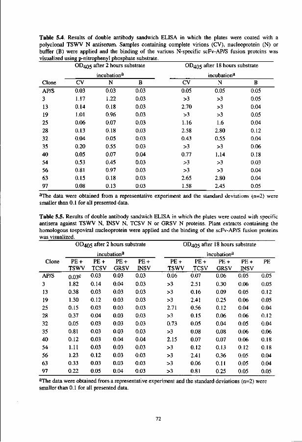
Table 5.4. Results of double antibody sandwich ELISA in which the plates were coated with a polyclonal TSWV N antiserum. Samples containing complete virions (CV), nucleoprotein (N) or buffer (B) were applied and the binding of the various N-specific scFv-AP/S fusion proteins was visualized using p-nitrophenyl phosphate substrate.
Clone
AP/S 3 13 19 25 28 32 35 40 54 56 63 97
OD405
CV
0.03 1.17 0.14 1.01 0.06 0.13 0.04 0.20 0.05 0.53 0.81 0.15 0.08
after 2 hours substrate
incubation3
N
0.03 1.22 0.18 0.96 0.07 0.18 0.05 0.55 0.07 0.45 0.97 0.18 0.13
B
0.03 0.03 0.03 0.03 0.03 0.03 0.03 0.03 0.04 0.03 0.03 0.03 0.03
OD 4 0 5
CV
0.05 >3
2.70 >3 1.16 2.58 0.43 >3
0.77 >3 >3
2.65 1.58
after 18 hours substrate
incubationa
N
0.05 >3 >3 >3 1.6
2.80 0.55 >3 1.14 >3 >3
2.80 2.45
B
0.05 0.05 0.04 0.05 0.04 0.12 0.04 0.06 0.18 0.03 0.04 0.04 0.05
aThe data were obtained from a representative experiment and the standard deviations (n=2) were smaller than 0.1 for all presented data.
Table 5.5. Results of double antibody sandwich ELISA in which the plates were coated with specific antisera against TSWV N, 1NSV N, TCSV N or GRSV N proteins. Plant extracts containing the homologous tospoviral nucleoprotein were applied and the binding of the scFv-AP/S fusion proteins was visualized.
Clone
AP/S
3 13 19 25 28 32 35 40 54 56 63 97
OD405 after 2 hours substrate
PE + TSWV
0.03c
1.82 0.38 1.30 0.15 0.37 0.05 0.81 0.12 1.11 1.23 0.33 0.22
incubation3
PE + TCSV 0.03
0.14 0.03 0.12 0.03 0.04 0.03 0.03 0.03 0.03 0.12 0.03 0.05
PE + GRSV 0.03
0.04 0.03 0.03 0.03 0.03 0.03 0.03 0.04 0.03 0.03 0.03 0.04
PE + HVSV
0.03
0.03 0.03 0.03 0.03 0.03 0.03 0.03 0.04 0.03 0.03 0.03 0.03
OD405 after 18 hours substrate
PE + TSWV 0.06
>3 >3 >3 2.71 >3 0.73 >3 2.15 >3 >3 >3 >3
incubation3
PE + TCSV
0.07
2.51 0.16 2.41 0.56 0.15 0.05 0.08 0.07 0.12 2.41 0.06 0.81
PE + GRSV 0.06
0.30 0.09 0.25 0.12 0.06 0.04 0.08 0.07 0.13 0.36 0.11 0.25
PE + INSV
0.05
0.06 0.05 0.06 0.04 0.06 0.05 0.06 0.06 0.12 0.05 0.05 0.05
PE
0.05
0.05 0.12 0.05 0.04 0.12 0.04 0.06 0.18 0.18 0.04 0.04 0.05
3The data were obtained from a representative experiment and the standard deviations (n=2) were smaller than 0.1 for all presented data.
72
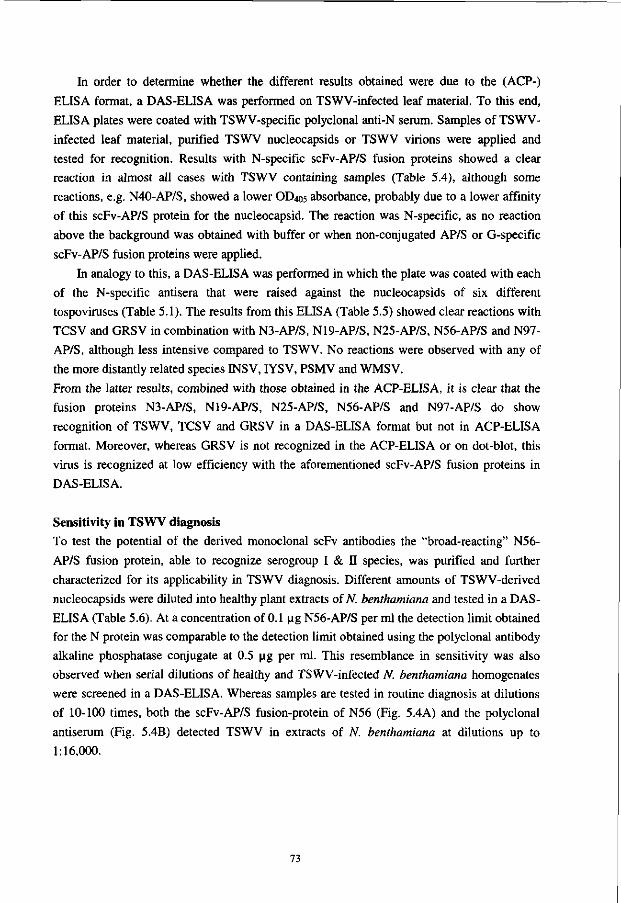
In order to determine whether the different results obtained were due to the (ACP-)
ELISA format, a DAS-ELISA was performed on TSWV-infected leaf material. To this end,
ELISA plates were coated with TSWV-specific polyclonal anti-N serum. Samples of TSWV-
infected leaf material, purified TSWV nucleocapsids or TSWV virions were applied and
tested for recognition. Results with N-specific scFv-AP/S fusion proteins showed a clear
reaction in almost all cases with TSWV containing samples (Table 5.4), although some
reactions, e.g. N40-AP/S, showed a lower OD405 absorbance, probably due to a lower affinity
of this scFv-AP/S protein for the nucleocapsid. The reaction was N-specific, as no reaction
above the background was obtained with buffer or when non-conjugated AP/S or G-specific
scFv-AP/S fusion proteins were applied.
In analogy to this, a DAS-ELISA was performed in which the plate was coated with each
of the N-specific antisera that were raised against the nucleocapsids of six different
tospoviruses (Table 5.1). The results from this ELISA (Table 5.5) showed clear reactions with
TCSV and GRSV in combination with N3-AP/S, N19-AP/S, N25-AP/S, N56-AP/S and N97-
AP/S, although less intensive compared to TSWV. No reactions were observed with any of
the more distantly related species INSV, IYSV, PSMV and WMSV.
From the latter results, combined with those obtained in the ACP-ELISA, it is clear that the
fusion proteins N3-AP/S, N19-AP/S, N25-AP/S, N56-AP/S and N97-AP/S do show
recognition of TSWV, TCSV and GRSV in a DAS-ELISA format but not in ACP-ELISA
format. Moreover, whereas GRSV is not recognized in the ACP-ELISA or on dot-blot, this
virus is recognized at low efficiency with the aforementioned scFv-AP/S fusion proteins in
DAS-ELISA.
Sensitivity in TSWV diagnosis To test the potential of the derived monoclonal scFv antibodies the "broad-reacting" N56-
AP/S fusion protein, able to recognize serogroup I & II species, was purified and further
characterized for its applicability in TSWV diagnosis. Different amounts of TSWV-derived
nucleocapsids were diluted into healthy plant extracts of N. benthamiana and tested in a DAS-
ELISA (Table 5.6). At a concentration of 0.1 ug N56-AP/S per ml the detection limit obtained
for the N protein was comparable to the detection limit obtained using the polyclonal antibody
alkaline phosphatase conjugate at 0.5 ug per ml. This resemblance in sensitivity was also
observed when serial dilutions of healthy and TSWV-infected N. benthamiana homogenates
were screened in a DAS-ELISA. Whereas samples are tested in routine diagnosis at dilutions
of 10-100 times, both the scFv-AP/S fusion-protein of N56 (Fig. 5.4A) and the polyclonal
antiserum (Fig. 5.4B) detected TSWV in extracts of N. benthamiana at dilutions up to
1:16,000.
73
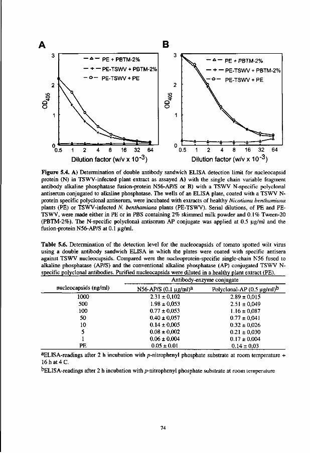
B - A - PE + PBTM-2%
— + — PE-TSWV + PBTM-2%
- o - PE-TSWV+ PE
Q O
•
,- 4 -
- * - PE + PBTM-2%
. — + — PE-TSWV + PBTM-2%
V - o - PE-TSWV+ PE
^^3 0.5 1 2 4 8 16 32 64
Dilution factor (w/v x 10"3)
0.5 1 2 4 8 16 32 64
Dilution factor (w/v x 10"^)
Figure 5.4. A) Determination of double antibody sandwich ELISA detection limit for nucleocapsid protein (N) in TSWV-infected plant extract as assayed A) with the single chain variable fragment antibody alkaline phosphatase fusion-protein N56-AP/S or B) with a TSWV N-specific polyclonal antiserum conjugated to alkaline phosphatase. The wells of an ELISA plate, coated with a TSWV N-protein specific polyclonal antiserum, were incubated with extracts of healthy Nicotiana benthamiana plants (PE) or TSWV-infected N. benthamiana plants (PE-TSWV). Serial dilutions, of PE and PE-TSWV, were made either in PE or in PBS containing 2% skimmed milk powder and 0.1% Tween-20 (PBTM-2%). The N-specific polyclonal antiserum AP conjugate was applied at 0.5 |ig/ml and the fusion-protein N56-AP/S at 0.1 ug/ml.
Table 5.6. Determination of the detection level for the nucleocapsids of tomato spotted wilt virus using a double antibody sandwich ELISA in which the plates were coated with specific antisera against TSWV nucleocapsids. Compared were the nucleoprotein-specific single-chain N56 fused to alkaline phosphatase (AP/S) and the conventional alkaline phosphatase (AP) conjugated TSWV N-specific polyclonal antibodies. Purified nucleocapsids were diluted in a healthy plant extract (PE).
Antibody-enzyme conjugate nucleocapsids (ng/ml) N56-AP/S (0.1 |ig/ml)a Polyclonal-AP (0.5 ng/ml)b
1000 500 100 50 10 5 1
PE
2.31 ±0,102 1.98 ±0,053 0.77 ± 0,053 0.40 ± 0,057 0.14 ±0,005 0.08 ± 0,002 0.06 ± 0,004 0.05 ± 0.01
2.89 ±0,015 2.51 ±0,049 1.16 ±0,087 0.77 ±0,041 0.32 + 0,026 0.21 ±0,030 0.17 ±0,004 0.14 ±0,03
aELISA-readings after 2 h incubation with p-nitrophenyl phosphate substrate at room temperature + 16 h at 4 C.
"ELIS A-readings after 2 h incubation with p-nitrophenyl phosphate substrate at room temperature
74
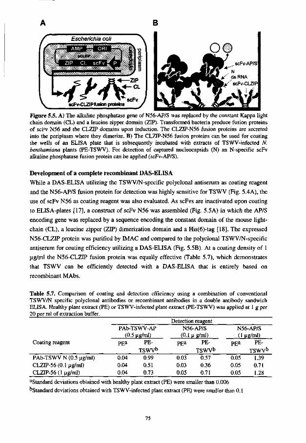
B
Escherichia coli
scFv-CLZIPfusion proteins
-ZIP - C L
scFv
Figure 5.5. A) The alkaline phosphatase gene of N56-AP/S was replaced by the constant Kappa light chain domain (CL) and a leucine zipper domain (ZIP). Transformed bacteria produce fusion proteins of scFv N56 and the CLZIP domains upon induction. The CLZIP-N56 fusion proteins are secreted into the periplasm where they dimerize. B) The CLZFP-N56 fusion proteins can be used for coating the wells of an ELISA plate that is subsequently incubated with extracts of TSWV-infected N. benthamiana plants (PE-TSWV). For detection of captured nucleocapsids (N) an N-specific scFv alkaline phosphatase fusion protein can be applied (scFv-AP/S).
Development of a complete recombinant DAS-ELISA
While a DAS-ELISA utilizing the TSWV/N-specific polyclonal antiserum as coating reagent
and the N56-AP/S fusion protein for detection was highly sensitive for TSWV (Fig. 5.4A), the
use of scFv N56 as coating reagent was also evaluated. As scFvs are inactivated upon coating
to ELISA-plates [17], a construct of scFv N56 was assembled (Fig. 5.5A) in which the AP/S
encoding gene was replaced by a sequence encoding the constant domain of the mouse light-
chain (CL), a leucine zipper (ZIP) dimerization domain and a His(6)-tag [18], The expressed
N56-CLZIP protein was purified by M A C and compared to the polyclonal TSWV/N-specific
antiserum for coating efficiency utilizing a DAS-ELISA (Fig. 5.5B). At a coating density of 1
ug/ml the N56-CLZIP fusion protein was equally effective (Table 5.7), which demonstrates
that TSWV can be efficiently detected with a DAS-ELISA that is entirely based on
recombinant MAbs.
Table 5.7. Comparison of coating and detection efficiency using a combination of conventional TSWV/N specific polyclonal antibodies or recombinant antibodies in a double antibody sandwich ELISA. Healthy plant extract (PE) or TSWV-infected plant extract (PE-TSWV) was applied at 1 g per 20 per ml of extraction buffer.
Detection reagent
Coating reagent
PAb-TSWV-AP (0.5 ug/ml)
N56-AP/S (0.1 ug/ml)
N56-AP/S (1 Ug/ml)
PEa PE-
TSWV0 PE* PE-
TSWV0 PEa PE-
TSWV0
PAb-TSWV N (0.5 ug/ml) CLZff-56 (0.1 ug/ml) CLZIP-56 (1 ug/ml)
0.04 0.04 0.04
0.99 0.51 0.73
0.03 0.03 0.05
0.57 0.36 0.71
0.05 0.05 0.05
1.39 0.71 1.28
aStandard deviations obtained with healthy plant extract (PE) were smaller than 0.006 DStandard deviations obtained with TSWV-infected plant extract (PE) were smaller than 0.1
75
DISCUSSION
With the aid of the phage display system, a series of 17 different TSWV-specific scFvs was
retrieved from the Vaughan combinatorial scFv-antibody library [34], utilizing expression as
scFv-alkaline phosphatase fusion protein in E. coli. Twelve of these scFv-AP/S fusion
proteins were identified as N-specific and five were directed against the glycoproteins of
TSWV (Table 5.3).
From the series of N-specific scFv-AP/S fusion proteins, N56-AP/S, at a concentration of
0.1 ug/ml could detect as little as 1 ng (Table 5.6) of N protein in a DAS-ELISA. In addition it
was shown that the CLZTP fusion protein of scFv N56 can be used as an effective coating
reagent (Table 5.7), suggesting that this recombinant scFv represents a promising candidate
for development of a complete recombinant diagnostic assay for routine detection of TSWV
in plant material.
Extensive ELISA and Western immunoblot analyses have demonstrated that the obtained
antibodies recognized at least five epitopes of the three structural TSWV proteins. Fusion
proteins G150-AP/S and G224-AP/S were found to recognize continuous epitopes on G2 or
Gl, respectively, whereas G118-AP/S most likely recognized a discontinuous epitope, as
reactivity was absent after mild denaturation of the glycoproteins by SDS-treatment. From the
N-specific scFv-AP/S, some reacted with the N-protein of TSWV, whereas others reacted
with an epitope that was not only present on the N-protein of TSWV, but also on those of
TCSV and GRSV. The N encoding genes of the latter isolates have been sequenced and share
around 78% sequence identity [5], but thus far, no scFvs have been observed recognizing a
common epitope within the N protein of all different tospovirus species. N56-AP/S is one of
few selected MAbs that recognizes up to three different tospoviruses, i.e. TSWV, GRSV and
TCSV, and must therefore be directed against a common epitope.
Polyclonal TSWV N-specific antisera, and now also some scFv, have been shown to
cross-react with TCSV and GRSV isolates. For a universal tospovirus diagnosis system a scFv
is required that recognizes all tospoviruses, even those that currently still escape from
detection. However, instead of searching for one single universal scFv, it is recommended to
pool a set of broad-reacting MAbs and thus to obtain a specific "polyclonal antiserum" with a
defined character and with continuous specificity for all the species within the genus
Tospovirus.
The data presented in this report have demonstrated the feasibility of the phage display
technique to select a large panel of TSWV-specific MAbs and is in strong support for the
identification of new series of scFv-antibodies. Some of which likely will have a broader
tospoviral detection range. As the phage display technique allows to alternate selections
between N (or G1/G2) proteins obtained from different isolates, it may now be possible to
guide selection of MAbs towards shared epitopes. After all, selection of MAbs by phage
display is not biased to a certain epitope on basis of immunogenicity but merely by the
76
frequency of occurrence. This means that selection would be for specificity against the most
prominent conserved epitopes within the N (or G1/G2) proteins.
Since the tospoviral N protein is involved in many viral processes, such as packaging of
RNA, replication, movement and transmission, an antibody that disrupts epitopes involved in
one of these processes may be successful in conferring resistance upon expression in plants.
The principle of such "plantibody"-mediated resistance has been shown with a recombinant
MAb against artichoke crinkled mottle virus by Tavladoraki and co-workers [31]. As the
genes encoding the TSWV-specific scFv-antibodies were obtained using the phage display
technique, they can be readily transferred into plants. Moreover, if later on mutants of TSWV
evolve which potentially could overcome the antibody-mediated resistance, new inhibiting
antibodies can be raised to ensure durable resistance, and in addition, to safeguard detection in
plants. After all, the power of the phage display system is the selection of large panels of
target-specific scFvs from naive combinatorial libraries, as was shown in this report in the
case of TSWV.
ACKNOWLEDGMENTS
This research was supported by a grant from the Program Committee on Agricultural Biotechnology (PcLB) and E.C. grants AIR3-CT94-1046 and FAIR1-CT95-0905.
We thank Dr. H. Hoogenboom for allowing us to work at his laboratory (CESAME, Academic Hospital Maastricht, The Netherlands) with the Vaughan antibody library, developed by Cambridge Antibody Technology. We also thank Prof. Dr. W.B. van Muiswinkel (Wageningen Institute of Animal Sciences, Cell Biology and Immunology Group, Wageningen Agricultural University) for critically reading the manuscript.
REFERENCES
1. Adam, G., Peters, D. and Goldbach, R.W. (1996) Serological comparison of tospovirus isolates using polyclonal and monoclonal antibodies. Acta Hort. 431: 135-158.
2. Adam, G., Roggero, P. Malavsi, F. and Papa, G. (1995) An approach for a general tospovirus assay by antibodies against natively purified tomato spotted wilt virus G proteins. EPPO Bull. 25: 247-257.
3. Clark, M.F. and Adams, A.N. (1977) Characteristics of the microplate method of enzyme-linked immunosorbent assay for the detection of plant viruses. J. Gen. Virol. 34: 475-483.
4. Cortes, I., Livieratos, I. C, Derks, A., Peters, D. and Kormelink, R. (1998) Molecular and serological characterization of iris yellow spot virus, a new and distinct Tospovirus species. Phytopathology 88: 1276-1282.
5. De Avila, A.C., De Haan, P., Kormelink, R., Resende, R., de O., Goldbach, R.W. and Peters, D. (1993) Classification of tospoviruses based on phylogeny of nucleoprotein gene sequences. J. Gen. Virol. 74: 153-159.
6. De A vila, A.C., Huguenot, C, Resende, R., de O., Kitajima, E.W., Goldbach, R.W. and Peters, D. (1990) Serological differentiation of of 20 isolates of tomato spotted wilt virus. J. Gen. Virol. 71: 2801-2807.
77
7. De Haan, P., Wagemakers, L., Goldbach, R. and Peters, D. (1989) Tomato spotted wilt virus, a new member of the Bunyaviridae? In: Genetics and pathogenicity of negative-strand viruses, Kolakofski, D. and Mahy, B.W.J.(eds.) Elseviers Scientific Publishers, Amsterdam, The Netherlands, pp. 287-290.
8. Goldbach, R. and Peters, D. (1994) Possible causes of the emergence of tospovirus diseases. Seminars in Virology 5: 113-120.
9. Gonsalves, D. and Trujillo, E.E. (1986) Tomato spotted wilt virus in papaya and detection of the virus in ELISA. Plant Dis. 70: 501-506.
10. Griep, R.A., Van Twisk, C , Kerschbaumer, R.J., Harper, K., Torrance, L., Himmler, G., Van der Wolf, J.M. and Schots, A. pSKAP/S: an expression vector for single-chain Fv alkaline phosphatase fusion proteins. Prot. Express. Purific, in press.
11. Griep, R.A., Van Twisk, C. and Schots, A. Selection of Beet Necrotic Yellow Vein Virus Specific Monoclonal Antibodies from a Semi-synthetic Combinatorial Antibody Library. Eur. J. Plant. Pathol., in press.
12. Griep, R.A., Van Twisk, C , Van der Wolf, J.M., Van Beckhoven, J.R.C.M. and Schots, A. (1998) Selection of Ralstonia solanacearum race 3 specific monoclonal antibodies from combinatorial libraries. Phytopathology 88: 795-803.
13. Griffiths, A.D., Williams, S.C., Hartley, O., Tomlinson, I.M., Waterhouse, P., Crosby, W.L., Kontermann, R.E., Jones, P.T., Low, N.M., Allison, T.J., Prospero, T.D., Hoogenboom, H.R., Nissim, A., Cox, J.P.L., Harrison, J.L., Zaccolo, M., Gherardi, E. and Winter, G. (1994) Isolation of high affinity antibodies directly from large synthetic repertoires. EMBO J. 13: 3245-3260.
14. Harper, K., Kerschbaumer, R.J., Ziegler, A., Macintosh, S.M., Cowan, G.H., Himmler, G. and Mayo, M.A. (1997) A scFv-alkaline phosphatase fusion-protein which detects potato leafroll luteovirus in plant extracts by ELISA. J. Virol. Methods 63: 237-242.
15. Hoogenboom, H.R., Griffiths, A.D., Johnson, K.S., Chiswell, D.J., Hudson, P. and Winter, G. (1991) Multi-subunit proteins on the surface of filamentous phage: methodologies for displaying antibody (Fab) heavy and light chains. Nucleic Acids Res. 15: 4133-4137.
16. Huguenot, C , Van den Dobbelsteen, G., de Haan, P., Wagemakers, C.A.M., Drost, G.A. Osterhaus, A.D.M.E. and Peters, D. (1990) Detection of tomato spotted wilt virus using monoclonal antibodies and riboprobes. Arch. Virol. 110: 47-62.
17. Kerschbaumer, R.J., Hirschl, S., Kaufman, A., Ibl, M., Koenig, R. and Himmler, G. (1997) Single-chain Fv fusion proteins suitable as coating and detecting reagents in a double antibody sandwich enzyme-linked immunosorbent assay. Anal. Biochem. 249: 219-227.
18. Kerschbaumer, RJ. and Himmler, G. Dedicated expression vectors for the production of diagnostic reagents in recombinant antibody-application. In: Harper, K. and Ziegler, A. Plant Sciences & and Plant Technology, Taylor and Francis, London, in press.
19. Kohler, G. and Milstein, C. (1975) Continuous cultures of fused cells secreting antibodies of predefined specificity. Nature 256: 495-497.
20. Law, M.D. and Moyer, J.W. (1990) A tomato spotted wilt-like virus with a serologically distinct N protein. J. Gen. Virol. 71: 933-938.
21. Lindner, P., Guth, B., Wuelfing, C, Krebber, C , Steipe, B., Mueller, F. and Pliickthun, A. (1992) Purification of native proteins from the cytoplasm and periplasm of Escherichia coli using IMAC and histidine tails: A comparison of proteins and protocols. Methods 4: 41-56.
22. McCafferty, J., Griffiths, A.D., Winter, G. and Chiswell, D.J. (1990) Phage-antibodies: filamentous phage displaying antibody variable domains. Nature 348: 552-554.
23. Prins, M. and Goldbach, R. (1998) The emerging problem of tospovirus infection and nonconventional methods of control. Trends in Microbiol. 6: 31-35.
24. Prins, M., Resende, R., de O., Anker, C, Van Scheppen, A., De Haan, P. and Goldbach, R. (1996) Engineered RNA-mediated resistance to tomato spotted wilt virus is sequence specific. Mol. Plant. Microb. Interact. 9: 416-418.
78
2 5. Resende, R de O., De Haan, P., Avila, A.C. de, Kitajima, E.W., Kormelink, R., Goldbach, R. and Peters, D. (1991) Generation of envelope and defective interfering RNA mutants of tomato spotted wilt virus by mechanical passage. J. Gen.Virol. 72: 2375-2383.
26. Roggero, P., Ogliara, P., Dellavalle, G., Lisa, V., Malavsi, F. and Adam, G. (1996) A general tospovirus assay using monoclonal antibodies against tomato spotted wilt virus glycoproteins. ActaHort. 431: 167-175.
27. Sambrook, J., Fritch, E.F. and Maniatis, T. (1990) Molecular cloning: A laboratory manual. Cold Spring Harbour Laboratory, New York.
28. Samuel, G., Bald, J.G. and Pittman, H.A. (1930) Investigations on spotted wilt of tomato. Commonwealth of Australia, Council for Scientific and Industrial Research, bulletin 44.
29. Sherwood, J.L., Sanborn, M.R., Keyser, G.C. and Meyers, L.D. (1989) Use of monoclonal antibodies in detection of tomato spotted wilt virus. Phytopathology 79: 61-64.
30. Susi, P., Ziegler, A. and Torrance, L. (1998) Selection of single-chain variable fragment
antibodies to black currant reversion associated virus from a synthetic phage display library.
Phytopathology 88: 230-233.
31. Tavladoraki, P.,Benvenuto, E.,Trinca, S., De Martinis, D., Cattaneo, A. and Galeffi, P.
(1993) Transgenic plants expressing a functional single-chain Fv antibody are specifically
protected from virus attack. Nature 366: 469-472.
32. Tijsen, P. (1985) Preparation of enzyme-antibody or other enzyme-macromolecule
conjugates. Laboratory Techniques in Biochemistry and Molecular Biology. Vol 15.
Practice and Theory of Enzyme Immunoassays. Burdon R. H., and P. H. van Knippenberg
(eds), Elsevier Publishing, Amsterdam.
33. Ultzen, T., Gielen, J.J.L. Venema, F., Westerbroek, A., De Haan, P., Tan, M.L.,
Schram, A., Van Grinsven, M.QJ.M. and Goldbach, R. (1995) Resistance to tomato
spotted wilt virus in tomato hybrids. Euphitica 85: 159-168.
34. Vaughan, T.J., Williams, A.J., Pritchard, K., Osbourn, J.K., Pope, A.R., Earnshaw, J.
C , McCafferty, J., Hodits, R.A., Wilton, J. and Johnson, K. (1996) Human antibodies
with sub-nanomolar affinities isolated from a large non-immunized phage display library.
Nature Biotechnology 14: 309-314.
35. Wang, M. and Gonsalves, D. (1990) ELISA detection of of various tomato spotted wilt
isolates using specific antisera to structural proteins of the virus. Plant Dis. 74: 154-158.
36. Yeh, S.D. and Chang, T.F. (1995) Nucleotide sequence of the N gene of watermelon silver
mottle virus, a proposed new member of the genus tospovirus. Phytopathology 85: 58-64.
37. Ziegler A., Torrance, L., Macintosh, S.M., Cowan, G.H. and Mayo, M.A. (1995)
Cucumber mosaic cucumovirus antibodies from a synthetic phage display library. Virology
214: 235-238.
79
Chapter 6
Fluobodies: green fluorescent single-chain Fv fusion proteins
ABSTRACT
Expression of single-chain Fv antibodies and green fluorescent protein as fusion proteins
(fluobodies) in Escherichia coli could facilitate the diagnosis of various diseases in single step
assays based upon immunofluorescence and/or flow-cytometry. They might even replace
some reagents frequently used in immunology such as chemically produced antibody-
fluorochrome conjugates. Therefore, an expression system (pSKGFP) was designed that
allows the expression of single-chain variable fragments as fusion proteins with modified
green fluorescent proteins. The resulting green fluorescent single-chain Fv fusion proteins can
be produced by expression in E. coli and purified in a single step via metal affinity
chromatography. Two different single-chain variable fragment antibodies, both directed
against the lipopolysaccharide of the bacterium Ralstonia solanacearum have been genetically
fused to a red-shifted green fluorescent protein and the produced fusion protein was tested for
usefulness in flow-cytometry and immunofluorescent cell staining. The fluobodies could be
produced in cultures of bacterial cells and purified using immobilized metal affinity
chromatography. They work well in flow-cytometry and immunofluorescent cell staining, are
specific for their target antigens and unlike FTTC-conjugated antibodies they do not fade upon
illumination. The expression system is compatible to frequently used phage display vectors.
Genes encoding single-chain antibody fragments can be easily transferred from these vectors
into pSKGFP that allows single-step characterization of the selected recombinant antibodies
by flow-cytometry or fluorescent cell staining.
This chapter has been submitted for publication in a modified form: Griep, R.A., van Twisk, C, van der Wolf, J.M. and Schots A. Fluobodies: green fluorescent single-chain Fv fusion proteins.

INTRODUCTION
Immunofluorescence (EF) [8,24] and phenotyping by flow-cytometry are useful techniques for
localization studies or for the diagnosis of various diseases [18,23]. The fluorochrome
fluorescein isothiocyanate (FITC) has been used extensively for this purpose. However, this
fluorochrome is very sensitive to photobleaching by illumination. Moreover, conjugation to
antibodies is not reproducible and if it occurs within the antigen binding site partial or
complete loss of the antigen binding capacity might occur [23]. After all, lysine, the
conjugation target for FITC is found frequently within antigen-binding sites [16]. Therefore,
new alternative fluorochromes with comparable excitation and emission spectra but lacking
the disadvantages of FITC have to be explored.
The green fluorescent protein (GFP) from the jellyfish, Aequorea victoria, was recently
described [3,14,22] and is a promising fluorescent probe with an emission peak at the same
wavelength as FITC (510 nm). The wild-type GFP has two excitation peaks, a major one at
395 nm and a minor at 470 nm [32]. It was found to be very stable when excited at 480 nm
[19,31]. Mutagenesis of wild-type GFP [4,5,6,10] led to improved versions of GFP. One of
these mutants, GFPuv, had an increased expression level of GFP in bacteria and better
solubility of the produced protein. Another mutant, GFPmutl [4], had one single red-shifted
excitation peak at 488 nm and contained two amino acid substitutions (PheM to Leu and Ser65
to Thr). It was found to fluoresce 35-fold more intensely than the wt-GFP when excited at 488
nm. Therefore, with the fluorochrome characteristics of FITC and an increased stability, this
GFPmutl spectral variant seems suited for application in IF and flow-cytometry.
Genetic fusion between antibody and GFP encoding genes would be ideal. It gives a 1:1
ratio between antibody and fluorochrome without the chance of inactivation. Recent progress
in molecular immunology [9,13,21,26,30] has allowed cloning and expression of antibody
encoding genes in E. coli. In this study, we describe the genetic fusion of the red-shifted
(mutl) mutant of GFP to two recombinant, Ralstonia solanacearum (biovar 2, race 3)
specific, single-chain variable fragment (scFv) antibodies [7,8] and the overexpression in E.
coli. The use of these fluobodies was evaluated in IF and flow-cytometry.
MATERIAL AND METHODS
Mutating, cloning and production of a red shifted green fluorescent protein To change the excitation peak of wild-type GFP or GFPuv (Clontech) from 396 nm to 488 nm
[4], two amino acid changes were introduced by PCR (Fig. 6.1). Phe64 was replaced by Leu
and Ser65 by Thr.
82
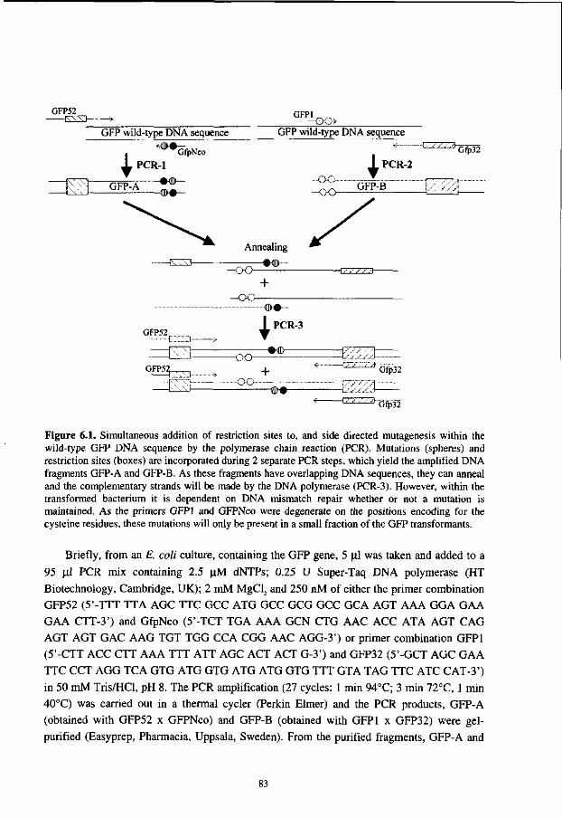
GFP52 GFPI ^ _. - P C
GFP wild-type DNA sequence GFP wild-type DNA sequence
Gfp32
Figure 6.1. Simultaneous addition of restriction sites to, and side directed mutagenesis within the wild-type GFP DNA sequence by the polymerase chain reaction (PCR). Mutations (spheres) and restriction sites (boxes) are incorporated during 2 separate PCR steps, which yield the amplified DNA fragments GFP-A and GFP-B. As these fragments have overlapping DNA sequences, they can anneal and the complementary strands will be made by the DNA polymerase (PCR-3). However, within the transformed bacterium it is dependent on DNA mismatch repair whether or not a mutation is maintained. As the primers GFPI and GFPNco were degenerate on the positions encoding for the cysteine residues, these mutations will only be present in a small fraction of the GFP transformants.
Briefly, from an E. coli culture, containing the GFP gene, 5 jul was taken and added to a
95 ill PCR mix containing 2.5 nM dNTPs; 0.25 U Super-Taq DNA polymerase (HT
Biotechnology, Cambridge, UK); 2 mM MgCl2 and 250 nM of either the primer combination
GFP52 (5'-TTT TTA AGC TTC GCC ATG GCC GCG GCC GCA AGT AAA GGA GAA
GAA CTT-3') and GfpNco (5'-TCT TGA AAA GCN CTG AAC ACC ATA AGT CAG
AGT AGT GAC A AG TGT TGG CCA CGG AAC AGG-3') or primer combination GFPI
(5'-CTT ACC CTT AAA TTT ATT AGC ACT ACT G-3') and GFP32 (5'-GCT AGC GAA
TTC CCT AGG TCA GTG ATG GTG ATG ATG GTG TTT GTA TAG TTC ATC CAT-3')
in 50 mM Tris/HCl, pH 8. The PCR amplification (27 cycles: 1 min 94°C; 3 min 72°C, 1 min
40°C) was carried out in a thermal cycler (Perkin Elmer) and the PCR products, GFP-A
(obtained with GFP52 x GFPNco) and GFP-B (obtained with GFPI x GFP32) were gel-
purified (Easyprep, Pharmacia, Uppsala, Sweden). From the purified fragments, GFP-A and
83
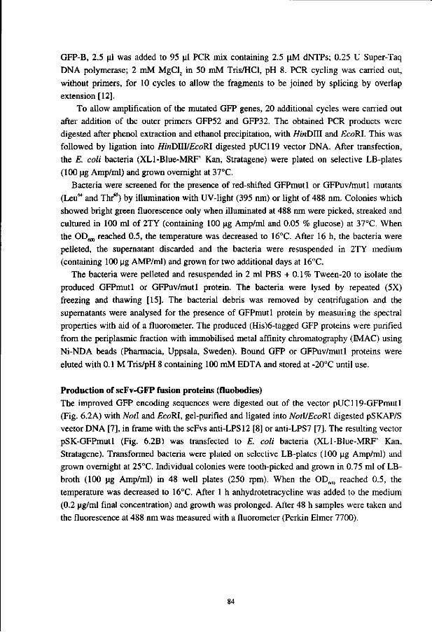
GFP-B, 2.5 ul was added to 95 ul PCR mix containing 2.5 uM dNTPs; 0.25 U Super-Taq DNA polymerase; 2 mM MgCl2 in 50 mM Tris/HCl, pH 8. PCR cycling was carried out, without primers, for 10 cycles to allow the fragments to be joined by splicing by overlap extension [12].
To allow amplification of the mutated GFP genes, 20 additional cycles were carried out after addition of the outer primers GFP52 and GFP32. The obtained PCR products were digested after phenol extraction and ethanol precipitation, with HinDlII and EcoRI. This was followed by ligation into HinDHVEcoRl digested pUC119 vector DNA. After transfection, the E. coli bacteria (XLl-Blue-MRF' Kan, Stratagene) were plated on selective LB-plates (100 |ig Amp/ml) and grown overnight at 37°C.
Bacteria were screened for the presence of red-shifted GFPmutl or GFPuv/mutl mutants (Leu64 and Thr65) by illumination with UV-light (395 nm) or light of 488 nm. Colonies which showed bright green fluorescence only when illuminated at 488 nm were picked, streaked and cultured in 100 ml of 2TY (containing 100 ug Amp/ml and 0.05 % glucose) at 37°C. When the OD^ reached 0.5, the temperature was decreased to 16°C. After 16 h, the bacteria were pelleted, the supernatant discarded and the bacteria were resuspended in 2TY medium (containing 100 ug AMP/ml) and grown for two additional days at 16°C.
The bacteria were pelleted and resuspended in 2 ml PBS + 0.1% Tween-20 to isolate the produced GFPmutl or GFPuv/mutl protein. The bacteria were lysed by repeated (5X) freezing and thawing [15]. The bacterial debris was removed by centrifugation and the supernatants were analysed for the presence of GFPmutl protein by measuring the spectral properties with aid of a fluorometer. The produced (His)6-tagged GFP proteins were purified from the periplasmic fraction with immobilised metal affinity chromatography (IMAC) using Ni-NDA beads (Pharmacia, Uppsala, Sweden). Bound GFP or GFPuv/mutl proteins were eluted with 0.1 M Tris/pH 8 containing 100 mM EDTA and stored at -20°C until use.
Production of scFv-GFP fusion proteins (fluobodies) The improved GFP encoding sequences were digested out of the vector pUC119-GFPmutl (Fig. 6.2A) with Notl and £coRI, gel-purified and ligated into NotVEcoRl digested pSKAP/S vector DNA [7], in frame with the scFvs anti-LPS12 [8] or anti-LPS7 [7]. The resulting vector pSK-GFPmutl (Fig. 6.2B) was transfected to E. coli bacteria (XLl-Blue-MRF' Kan, Stratagene). Transformed bacteria were plated on selective LB-plates (100 ug Amp/ml) and grown overnight at 25°C. Individual colonies were tooth-picked and grown in 0.75 ml of LB-broth (100 ug Amp/ml) in 48 well plates (250 rpm). When the OD6IK) reached 0.5, the temperature was decreased to 16°C. After 1 h anhydrotetracycline was added to the medium (0.2 ug/ml final concentration) and growth was prolonged. After 48 h samples were taken and the fluorescence at 488 nm was measured with a fluorometer (Perkin Elmer 7700).
84
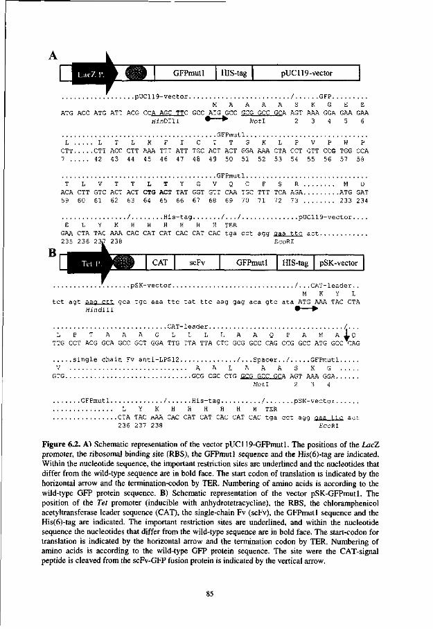
GFPmutl HIS-tag pUC119-vector
.pUC119-vector / GFP M A A A A S K G E E
ATG ACC ATG ATT ACG CCA AGC TTC GCC^TG^GCC GCG GCC GCA AGT AAA GGA GAA GAA " 2 3 4 5 6 Hi n D i l i Not I
GFPmut1 L L T L K F I C T T G K L P V P W P
CTT CTT ACC CTT AAA TTT ATT TGC ACT ACT GGA AAA CTA CCT GTT CCG TGG CCA 7 42 43 44 45 46 47 48 49 50 51 52 53 54 55 56 57 58
G F P m u t l . . T L V T T L T Y G V Q C
ACA CTT GTC ACT ACT CTG ACT TAT GGT GTT CAA TGC 59 60 61 62 63 64 65 66 67 68 69 70
F S R M D TTT TCA AGA ATG GAT 71 72 73 233 234
/ H i s - t a g / . . . / p U C 1 1 9 - v e c t o r . . E L Y K H H H H H H TER
GAA CTA TAC AAA CAC CAT CAT CAC CAT CAC t g a c c t agg g aa t t c a c t 235 236 23J 238 EccRI
CAT scFv GFPmutl HIS-tag pSK-vector
. . p S K - v e c t o r / . . . C A T - l e a d e r . . M K Y L
t c t a g t a a a c t t g c a t g c a a a t t c t a t t t c a ag gag a c a g t o a t a ATG AAA TAC CTA ffindlll • •
C A T - l e a d e r L P T A A A G L L L L A
7TG CCT ACG GCA GCC GCT GGA TTG TTA TTA CTC GCG A Q P A M A I Q
GCC CAG CCG GCC ATG GCC CAG
s i n g l e c h a i n Fv a n t i - L P S 1 2 / . . . S p a c e r . . / GFPmut l . V A A L A A A S K G .
GTG GCG CGC CTG GCG GCC GCA AGT AAA GGA. . Not! 2 3 4
.GFPmu t l . / H i s - t a g / p S K - v e c t o r L Y K H H H H H H TER
.CTA TAC AAA CAC CAT CAT CAC CAT CAC t g a c c t a gg a a a t t c a c t 236 237 238 EcoRI
Figure 6.2. A) Schematic representation of the vector pUC119-GFPmutl. The positions of the LacZ promoter, the ribosomal binding site (RBS), the GFPmutl sequence and the His(6)-tag are indicated. Within the nucleotide sequence, the important restriction sites are underlined and the nucleotides that differ from the wild-type sequence are in bold face. The start codon of translation is indicated by the horizontal arrow and the termination-codon by TER. Numbering of amino acids is according to the wild-type GFP protein sequence. B) Schematic representation of the vector pSK-GFPmutl. The position of the Tet promoter (inducible with anhydrotetracycline), the RBS, the chloramphenicol acetyltransferase leader sequence (CAT), the single-chain Fv (scFv), the GFPmutl sequence and the His(6)-tag are indicated. The important restriction sites are underlined, and within the nucleotide sequence the nucleotides that differ from the wild-type sequence are in bold face. The start-codon for translation is indicated by the horizontal arrow and the termination codon by TER. Numbering of amino acids is according to the wild-type GFP protein sequence. The site were the CAT-signal peptide is cleaved from the scFv-GFP fusion protein is indicated by the vertical arrow.
85
Purification of fluobodies Cultures were grown, induced and pelleted as described above. After incubation (5 min, 0°C)
of the bacteria with 1/20 volume (referring to the original culture size) of a 50 mM Tris/HCl
pH 8 buffer containing, 30% sucrose and 1 mM EDTA, the produced proteins were extracted
(45 min, 0°C) from the periplasm with 1/20 volume of 5 mM MgS04. The produced (His)6-
tagged proteins were purified from the periplasmic fraction with immobilized metal affinity
chromatography (MAC) using Ni-NDA beads (Pharmacia, Uppsala, Sweden). Bound
proteins were eluted with 0.1 M Tris/pH 8 containing 100 mM EDTA and stored at 4°C.
SDS-PAGE and Western blotting Purified GFP and fluobodies were loaded on a 12.5 % SDS polyacryl amide gel and blotted
onto nitro-cellulose according to Towbin and co-workers [27]. After blocking for 30 min with
PBS (containing 5% skimmed milk powder and 0.1 % Tween-20) the Western blot was
incubated (1.5 h, room temperature) with GFP specific polyclonal rabbit antibodies (Clontech)
diluted 1:25,000 in PBS (containing 2% skimmed milk powder and 0.1 % Tween-20). After
washing (4 x, PBS 0.1 % Tween-20) the blot was incubated for 1 h with alkaline phosphatase
conjugated Goat anti-Rabbit polyclonal antibodies (diluted 1:5,000 in PBS containing 2%
skimmed milk powder and 0.1 % Tween-20). Finally, staining with 5-bromo-4-chloro-3-
indoylphosphate/nitroblue tetrazolium substrate (BCIP/NBT) was used to visualize the bands
on the Western blot.
Flow-cytometry Purified GFPmutl or scFv LPS12-GFPmutl fluobodies were added to suspensions of
Ralstonia solanacearum or Ralstonia picketii (107 bacteria/ml PBS, containing 0.1% Tween-
20 and 5% FCS) and incubated for 1 h while rotating. Bacteria were washed twice with PBS +
0.1 Tween-20, resuspended in 1.5 ml PBS and analyzed for fluorescent staining. From each
sample 10,000 bacteria were screened, using a fluorescent activated cell sorter.
Immunofluorescence IF was performed according to Van der Wolf and co-workers [29]. For IF cell-staining the
bacteria were coated on microscope slides and incubated with GFPmutl, scFv LPS7-
GFPmutl fluobodies or with a R. solanacearum-specific FITC-conjugated polyclonal rabbit
antiserum.
DNA Sequencing The nucleotide sequences of the GFP inserts in pUC119 were verified by the dideoxy chain termination method [25] on an A.L.F. DNA sequencer (Pharmacia).
86

RESULTS
The Aequorea wt-GFP gene was modified by PCR to generate the GFPmutl spectral mutant
with a single excitation peak at 488 nm. In addition, a his-tag was added to the C-terminus of
the GFP protein and convenient restriction sites for cloning into the pUC119 vector were
added. Moreover, an attempt was carried out to change the cysteine residues on positions 48
and 70 by PCR, as it was anticipated these that would interfere with secretion of functional
scFv-GFP fusion proteins into the periplasmic space.
Several bacteria obtained from transfection of the pUC119-GFPmutl vector DNA (Fig.
6.2A) showed fluorescence only when illuminated at 488 nm (i.e. production of red-shifted
GFP). The GFPmutl proteins could be separated from the extracted cytoplasmic fraction by
immobilized metal chromatography and yielded up to 2 mg of GFPmutl protein per liter of
bacterial culture. The GFPmutl encoding plasmids were isolated from the most intensely
fluorescing colonies and the two amino acid changes in the GFPmutl region, PheMtoLeu and
Ser65 to Thr, were confirmed by DNA-sequencing. The cysteine residues on positions 48 and
70 were conserved, despite the attempt to replace them.
kDa Figure 6.3. Western blot on GFP containing samples. Proteins in total lysates of GFPmutl (lane
-105 1) or scFv-GFPmutl (lane 2) producing bacteria, ~ °2 molecular weight markers (lane 3) or purified - 49 scFv-GFPmutl fraction (lane 4) were separated by - scFv-GFP SDS-PAGE, blotted to nitrocellulose and stained
33 with GFP-specific antibodies. GFP- 28
19
The GFPmutl encoding genes were digested from the GFPmutl plasmids (Fig. 6.2A) and genetically fused to a gene encoding the scFv anti-LPS12 [8] (Fig. 6.2B) which is highly specific for the lipopolysaccharide of the bacterium ft solanacearum (biovar2, race 3). Upon induction with anhydrotetracycline clear green fluorescence could be measured intracellularly bacteria with the aid of a fluorometer. Further analysis showed that a green fluorescent compound was present in the periplasmic fraction. Purification of scFv-GFPmutl fusion proteins from the periplasmic fraction by immobilized metal affinity chromatography and subsequent analysis by Western blotting with GFP-specific antibodies showed an intensive band (Fig. 6.3, lane 4). With a molecular mass of approximately 45 kDa it was larger than the 30 kDa observed for non-fused GFPmutl protein (Fig. 6.3, lane 1) but smaller than the 53 kDa which was calculated using the scFv LPS12-GFP protein sequence. This phenomenon was probably the result of the formation of a functional chromatophore within the scFv-GFP
87

fusion protein and has been observed previously [2]. Support for this assumption was obtained
as on Western blot, within a total lysate of the scFv-GFP producing bacteria, a major band of
55 kDa was observed together with two additional bands of 47 and 45 kDa. The observed
molecular masses correspond to 3 products of the scFv-GFPmutl fusion protein: scFv-
GFPmutl, preceded by the chloramphenicol acetyl transferase (CAT)-signal peptide (55 kDa);
scFv-GFPmutl, preceded by the CAT-signal peptide with a functional chromatophore (47
kDa) and the desired periplasmic secretion product scFv-GFPmutl, with an active
fluorochrome but without the CAT-signal peptide (45 kDa).
The quantity of the produced scFv-GFPmutl proteins was on average 100 ug per liter of
bacterial culture. Replacement of the GFPmutl encoding sequence by GFPuv/mutl did not
result in the expected improvement of expression and enhanced secretion of the scFv-GFP
fusion protein to the periplasm, but rather lowered the yield.
A B
20-
CD
-o 10-
JQ
1111 mi l i 11 mini i 11 i iwf
10" 10' 102 103
Green fluorescence 10' 10" 10
Green fluorescence
10 10 Green fluorescence
n i l Hill i i | I I I I Mil 1 ' 2 ' •?
10 10 10 Green fluorescence
Figure 6.4. Flow-cytometry analysis of the binding of GFPmutl to A) Ralstonia solanacearum bacteria and B) R. picketii bacteria and of the binding of fluobody scFv LPS 12-GFPmut 1 to C) R. solanacearum and D) R. picketii.
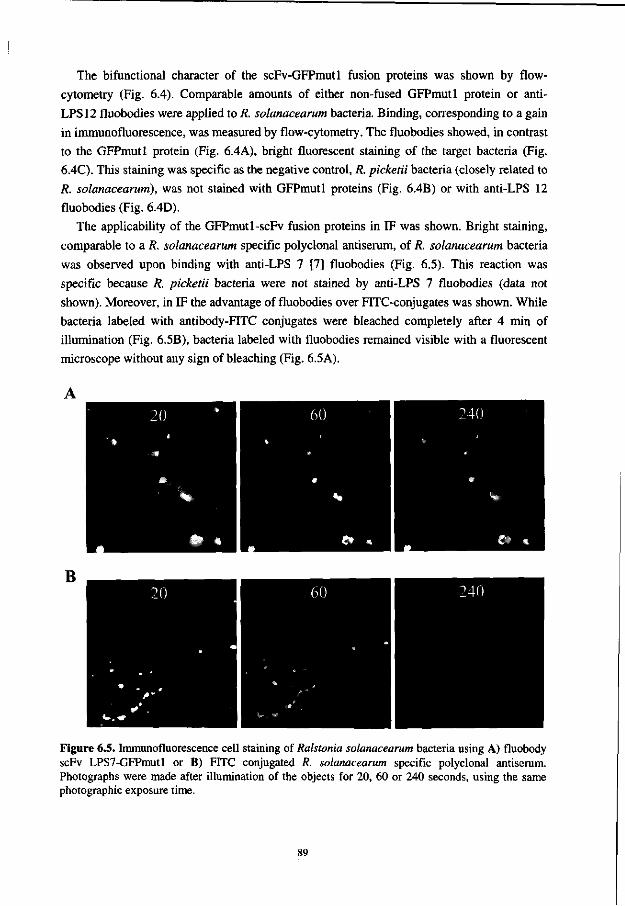
The Afunctional character of the scFv-GFPmutl fusion proteins was shown by flow-
cytometry (Fig. 6.4). Comparable amounts of either non-fused GFPmutl protein or anti-
LPS12 fluobodies were applied to R. solanacearum bacteria. Binding, corresponding to a gain
in immunofluorescence, was measured by flow-cytometry. The fluobodies showed, in contrast
to the GFPmutl protein (Fig. 6.4A), bright fluorescent staining of the target bacteria (Fig.
6.4C). This staining was specific as the negative control, R. picketii bacteria (closely related to
R, solanacearum), was not stained with GFPmutl proteins (Fig. 6.4B) or with anti-LPS 12
fluobodies (Fig. 6.4D).
The applicability of the GFPmutl-scFv fusion proteins in IF was shown. Bright staining,
comparable to a R. solanacearum specific polyclonal antiserum, of R. solanacearum bacteria
was observed upon binding with anti-LPS 7 [7] fluobodies (Fig. 6.5). This reaction was
specific because R. picketii bacteria were not stained by anti-LPS 7 fluobodies (data not
shown). Moreover, in IF the advantage of fluobodies over FITC-conjugates was shown. While
bacteria labeled with antibody-FITC conjugates were bleached completely after 4 min of
illumination (Fig. 6.5B), bacteria labeled with fluobodies remained visible with a fluorescent
microscope without any sign of bleaching (Fig. 6.5A).
B
Figure 6.5. Immunofluorescence cell staining of Ralstonia solanacearum bacteria using A) fluobody scFv LPS7-GFPmutl or B) FITC conjugated R. solanacearum specific polyclonal antiserum. Photographs were made after illumination of the objects for 20, 60 or 240 seconds, using the same photographic exposure time.
89

DISCUSSION
Fluorescent antibodies, which can be applied for direct labeling in flow-cytometry or in
immunofluorescence experiments, can be obtained from in vivo expression in E. coli bacteria.
The restriction sites in the vector allow easy transfer of scFv-genes from phage display
vectors like pHenl [11] and pCantab series (Pharmacia).
The general yield of obtained fluobodies is not high. While for non-fused scFvs the
maximum yield score of 3 mg per liter bacterial culture can be reached in shaker flasks under
non-optimal conditions [8], the genetic fusion with GFPmutl drastically reduced the yield.
This while under the same conditions the non-fused GFPmutl protein, when produced in the
cytoplasm (i.e. without secretion to the periplasm), reached a yield of 2 mg per liter. In
addition, when the same GFPmutl sequence was preceded by a signal sequence like pelB
[17], the fluorescence dropped to a similar level as if a scFv was present. The use of another
red-shifted GFP mutant (GFPuv/mutl) sequence, optimized for expression in E. coli,
increased the yield when expressed as a non-fused protein in the cytoplasm. But when
preceded by either a signal peptide alone or a signal peptide in combination with a scFv the
yield also dropped, even to levels below those obtained with the GFPmutl protein.
Apparently, the fast folding characteristics of the GFPuv/mutl mutant further lowers the
ability of the protein to be secreted.
It was anticipated that problems could arise after secretion of the scFv-GFP fusion proteins
into the periplasmic space, as 2 cysteine residues are present in the wt-GFP protein. These
cysteine residues normally do not form disulfide bonds in the wt-GFP but they might cause
cross-links [28] with the cysteine residues of the scFv-domain. Therefore, the primer
sequences were designed to replace those cysteine residues by serine, glycine or arginine.
However, in all cases, DNA-sequencing revealed that both cysteine residues were conserved.
As no non-fluorescing clones were sequenced it was not proved that both cysteine residues are
required for the formation of a functional fluorochrome. It appeared that no internal disulfide
bonds were formed between the scFv- and GFP-domains as the produced protein was able to
bind to the target antigen and simultaneously to function as a highly stable fluorescent dye.
High expression of scFv-GFP fusion proteins in the cytoplasm of E. coli might be possible
as high yields were described for a genetic fusion of GFP with protein A [2]. This fusion
protein was extracted by total disruption of the bacteria through sonic oscillation. For scFv-
GFP fusion proteins this is not favorable as the required disulfide bonds are not formed in the
cytoplasm and thus extracted scFv would be unstable. However, secretion of GFP fusion
proteins is possible, as was demonstrated by Oker-Blom and co-workers [20] for the genetic
fusion of GFP with streptavidin. This GFP-streptavidin fusion protein was expressed at high
yields in insect cells and appeared to be secreted into granules. Therefore, expression of
fluobodies might give a better yield in insect cells or in other eukaryotic expression systems in
which the folding and secretion of proteins is guided by more adequate chaperone proteins.
90
Although the GFP-streptavidin and GFP-Protein A fusion proteins have expression
advantages over the scFv-GFP fusion protein, they require multiple incubation steps in
immunoassays whereas scFv-GFP fusion proteins can be used in a single-step assay.
Together, the data show that fluobodies are a promising tool as fluorescent reagents in
immunoassays. Especially, since exploitation of fluobodies with other spectral characteristics
might facilitate multiple labeling [1,33] of different epitopes simultaneously.
ACKNOWLEDGMENTS
This research was supported by a grant from the Program Comittee on Agricultural Biotechnology (PCLB) and grants AIR-CT94-1046 and FAIR 1-CT95-0905. We thank Prof. Dr. W.B. van Muiswinkel (Wageningen Institute of Animal Sciences, Cell Biology and Immunology Group, Wageningen Agricultural University) for critically reading the manuscript.
REFERENCES
1. Anderson, M.T., Tjioe, I.M., Lorincz, M.C., Parks, D.R., Herzenberg, L.A. and Nolan, G.P. Simultaneous fluorescence-activated cell sorter analysis of two distinct transcriptional elements within a single cell using engineered green fluorescent proteins (1996) Proc. Natl. Acad. Sci. USA. 93: 8508-8511.
2. Aoki, T., Takahashi, Y., Koch, K.S. Leffert, H.L. and Watabe, H. (1996) Construction of a fusion protein between protein A and green fluorescent protein and its application to western blotting. FEBS lett. 384, 193-197.
3. Chalfie, M„ Tu, Y., Euskirchen, G., Ward, W.W. and Prasher, D.C. (1994) Green fluorescent protein as a marker for gene expression Science 363, 802-805.
4. Cormack, B.P., Valdivia, R. and Falkow, S. (1996) FACS-optimized mutants of the green fluorescent protein (GFP). Gene 173: 33-38.
5. Crameri, A., Whitehorn, E.A., Tate, E. and Stemmer, W.P.C. (1996) Improved green fluorescent protein by molecular evolution using DNA shuffling Nature Biotechnology 14: 315-319.
6. Delagrave, S., Hawtin, R.E., Silva, CM., Yang, M.M. and Youvan, D.C. (1995) Red-shifted excitation mutants of the green fluorescent protein Bio/Technology 13: 151-154.
7. Griep, R.A., Van Twisk, C, Kerschbaumer, R.J., Harper, K., Torrance, L., Himmler, G., Van der Wolf, J.M. and Schots, A. pSKAP/S: an expression vector for single-chain Fv alkaline phosphatase fusion proteins. Prot. Express. Purific. in press.
8. Griep, R.A., Van Twisk, C, Van der Wolf, J.M., Van Beckhoven, J.R.C.M. and Schots, A. (1998) Selection of Ralstonia solanacearum race 3 specific monoclonal antibodies from combinatorial libraries. Phytopathology 88: 795-803.
9. Griffiths, A.D., Williams, S.C., Hartley, O., Tomlinson, I.M., Waterhouse, P., Crosby, W.L., Kontermann, R.E., Jones, P.T., Low, N.M., Allison, T.J., Prospero, T.D., Hoogenboom, H.R., Nissim, A., Cox, J.P.L., Harrison, J.L., Zaccolo, M., Gherardi, E., and Winter, G. (1994) Isolation of high affinity antibodies directly from large synthetic repertoires. EMBO J. 13: 3245-3260.
10. Heim, R. and Tsien, R.Y. (1996) Engineering green fluorescent protein for improved brightness, longer wavelengths and fluorescence resonance energy transfer. Current Biology 6: 178-182
11. Hoogenboom, H.R., Griffiths, A.D., Johnson, K.S., Chiswell, D.J., Hudson, P. and Winter, G. (1991) Multi-subunit proteins on the surface of filamentous phage: methodologies for displaying antibody (Fab) heavy and light chains. Nucleic Acids Res. 15: 4133-4137.
91
12. Horton, R.M., Hunt, H.D., Ho, S.N., Pullen, J.K. and Pease, L.R. (1989) Engineering hybrid genes without the use of restriction enzymes. Gene 77: 61-68.
13. Huston, J.S., Levinson, D., Mudgett-Hunter, M., Tai, M., Novotny, J., Margolies, M.N., Ridge, RJ., Bruccoleri, R.E., Haber, E., Crea, R. and Oppermann, H. (1988) Protein engineering of antibody binding sites: Recovery of specific activity in an anti-digoxin single-chain Fv analogue produced in Escherichia coli. Proc. Natl. Acad. Sci. U.S.A. 85: 5879-5883.
14. Inouye, S. and Tsuji, F.I. (1994) Expression of the gene and fluorescence characteristics of the recombinant protein. FEBS lett 341: 277-280.
15. Johnson, B. H. and Hecht, M.H. (1994) Recombinant proteins can be isolated from E. coli cells by repeated cycles of freezing and thawing. Bio/Technology 12: 1357-1360.
16. Rabat, E.A., Wu, T.T., Reid-Miller, M., Perry, H.M. and Gottesmann, K.S. (1991) Sequences of Proteins of immunological interest. 5th edition (U.S. Department of health and human services, Public Health Service National Institutes of Health, Washington.
17. Lei, S., Lin, H., Wang, S., Callaway, J. and Wilcox, G. (1987) Characterization of the Erwinia carotovora pelB gene and its product pectate lyase. J. Bacteriol. 169: 4379-4383.
18. Lingenfelter, B., Fuller, T.C., Hartung, L., Hunter, J. and Wittwer, C. (1995) HLA-B27 screening by flow cytometry. Cytometry 22: 146-149.
19. Niswender, K.D., Blackman, S.M., Rohde, L., Magnuson, M.A. and Piston, D.W. (1995) Quantitative imaging of green fluorescent protein in cultured cells: comparison of microscopic techniques, use in fusion proteins and detection limits. J. Microbiol. 180: 109-116.
20. Oker-Blom, C , Orellana, A. and Keinanen, K. (1996) Highly efficient production of GFP and its derivatives in insect cells for visual in vitro applications.FEBS lett. 389: 238-243.
21. Orlandi, O., Gussow, D.H., Jones, P.T. and Winter, G. (1989) Cloning immunoglobulin variable domains for the expression by the polymerase chain reaction. Proc. Natl. Acad. Sci. U.S.A. 86: 3833-3837.
22. Prasher, D.C., Eckenrode, V.K., Ward, W.W., Prendergast, F.G. and Cormier, M.J. (1992) Primary structure of the Aequorea victoria green-fluorescent protein. Gene 111: 229-233.
23. Reimann, K.A., Waite, B.C., Lee-Parritz, D.E., Lin, W., Uchanska-Ziegler, B., O'Connell, MJ. and Letvin, N.L. (1994) Use of human leukocyte-specific monoclonal antibodies for clinically immunophenotyping lymphocytes of rhesus monkeys. Cytometry 17, 102-108.
24. Salinas, J. and Schots, A. (1994) Microscopical studies of the infection of gerbera flowers by Botrytis cinerea. Phytopathology 84: 351-356.
25. Sanger, F., Nicklen, S. and Coulson, A.R. (1977) DNA sequencing with the chain-terminating inhibitors^ Proc. Natl. Acad. Sci. USA 74: 5463-5467.
26. Sastry, L., Alting-Mees, M., Huse, W.D., Short, J.M., Sorge, J.A., Hay, B.H., Janda, K.D., Benkovic, SJ. and Lerner, R.A. (1989) Cloning of the immunological repertoire in Escherichia coli for generation of monoclonal catalytic antibodies: Construction of a heavy chain variable region-specific cDNA library. Proc. Natl. Acad. Sci. U.S.A. 86: 5728-5732.
27. Towbin, H., Staehelin, T. and Gordon, J. (1979) Electrophoretic transfer of proteins from polyacrylamide gels to nitrocellulose sheets: procedure and some applications.Proc. Natl. Acad. Sci. USA 76: 4350-4354.
28. Tudyka, T. and Skerra, A. (1997) Glutathione S-transferase can be used as a C-terminal, enzymatically active dimerization module for a recombinant protease inhibitor, and functionally secreted into the periplasm of Escherichia coli.Protein-science 6:2180-2187.
29. Van der Wolf, J. M., Van Beckhoven, J.R.C.M., De Boef, E. and Roozen, NJ.M. (1993) Serological characterization of fluorescent Pseudomonas strains cross-reacting with antibodies against Erwinia chrysanthemi. Neth. J. Plant Pathol. 99: 51-60.
30. Vaughan, T.J., Williams, A.J., Pritchard, K., Osbourn, J.K., Pope, A.R., Earnshaw, J. C , McCafferty, J., Hodits, R.A., Wilton, J. and Johnson, K. (1996) Human antibodies
92
with sub-nanomolar affinities isolated from a large non-immunized phage display library. Nature Biotechnology 14: 309-314.
31. Wang, S. and Hazelrigg, T. (1994) Implications for bed mRNA localization from spatial distribution of exu protein in Drosophila oogenesis. Nature 369, 400-403.
32. Ward, W.W., Cody, C.W., Hart, R.C. and Cormier, MJ. (1980) Spectral perturbations of the Aequoria green fluorescent protein. Photochem. Photobiol. 31: 611-615.
33. Yang, T.T., Kain, S.R., Kitts, P., Kondepudi, A., Yang, M.M. and Youvan, D.C. (1996) Dual color microscopic imagery of cells expressing the green fluorescent protein and a red-shifted variant.Gene 173: 19-23.
93
Chapter 7
Discussion, Summary and Concluding remarks
This thesis describes the applicability of the novel phage display technique to select plant-pathogen-specific monoclonal antibodies (MAbs) from combinatorial antibody libraries. The retrieved MAbs are so specific that they can be used as diagnostic tools in sensitive immunoassays for the detection and identification of plant pathogens. Testing results, obtained from laboratories that have applied these recombinant MAbs, are discussed in this conclusive chapter.
95
Background In the last decades, it has become clear that chemical crop protection has to be reduced. Many of the pesticides, applied to destroy plant-pathogenic fungi, bacteria, insects and nematodes, are hazardous to the environment, and form a serious health risk for animals and humans. Since breeding for disease resistance generally takes years, epidemics have to be prevented by sanitary measures in combination with diagnosis in an early stage of disease development or preferably beforehand. Consequently, sensitive diagnostic assays are required that allow healthy plant propagation material to be certified and soil to be monitored. It is obvious that these data can be used to take adequate crop management decisions.
The problems associated with serological detection of plant pathogens Immunoassays are widely applied as detection methods in agriculture because they can be applied to large numbers of samples, and their application is fast, robust, sensitive and cheap. A major concern is the specificity of the assays. Polyclonal antisera are still the active ingredient of many useful immunoassays. However, the immunochemical complexity of several target organisms is the main reason that extensive cross-reactions with non-target organisms can occur when these antisera are used.
In 1975, Kohler and Milstein showed that this problem could be circumvented by making monospecific antibodies in continuous cultures by fusing the antibody producing B-lymphocytes with myeloma cells [8]. This 'hybridoma' technique opened a new perspective for the production of specific monoclonal antibodies (MAbs) against various antigens. Today, many MAbs are used in research, as therapeutic agent and for diagnostic purposes.
However, the hybridoma technique was not as advantageous as was expected in the field of plant-pathogen detection. From the trials in raising specific MAbs against many different plant-pathogens, it became apparent that there are 'difficult' antigens. Several plant viruses can not be sufficiently purified and contain immunodominant plant residues and, as a consequence, the immune response is mainly directed to those impurities. In addition, plant-pathogenic bacteria, fungi and nematodes contain many epitopes that are 'shared' with closely related non-pathogenic family members, which is often leading to extensive cross-reactions. Therefore, the selection of the cells producing the desired MAbs is laborious and often impossible.
Many techniques have been described to deal with 'difficult' antigens and varied from the enrichment of the desired B-cell populations to guiding of the immune response towards the target antigen, such as:
• Immunoadsorption of antigen-specific B-cells [2]. • Selection of antigen-specific B-cells by fluorescence activated cell sorting [15]. • Complement-mediated lysis of undesired B-cells [2]. • Masking (immuno-complexing) of contaminating immunodominant plant epitopes with
anti-healthy plant antibodies during immunization [2]. • Suppression of the immune response to healthy plant extracts by injection with
cyclophosphamide or other immuno suppressive drugs prior to immunization [11,16]. • Induction of immunological tolerance through tolerisation of neonatal mice with healthy
plant extracts prior to immunization [6].
96
Although these methods have occasionally shown their value in obtaining specific MAbs in their respective cases, the common drawback of all these methods is that they are not generally applicable. In addition, the latter two are also repulsive from an ethical point of view.
Bypassing immunization: A general solution to deal with 'difficult' antigens A way to circumvent immunization was offered by recent developments in recombinant DNA techniques [14,17]. These techniques allow cloning and expression of large naive antibody repertoires, derived from non-immunized human donors, in E. coli as single-chain antibodies (scFvs) or Fab fragments. The use of these combinatorial antibody libraries in combination with the display of functional antibody fragments at the tips of filamentous phage created a powerful selection system for MAbs [5,12]. Utilization of this phage display system allows direct selection of highly specific MAbs from naive combinatorial antibody libraries [3,13,18]. As immunization is omitted, phage display is not biased. Hence it will not lead to antibodies directed against the most immunodominant epitope but rather towards the most abundant antigenic determinant.
Mission impossible: how to make the impossible possible Because application of the hybridoma technique was not successful in generating MAbs of sufficient specificity to detect the plant-pathogen Ralstonia solanacearum in reliable and sensitive diagnostic assays, the efficacy of the phage display system was challenged (Chapter 2). To achieve this, phages derived from the (naive) Vaughan combinatorial antibody library (1.4 X 10¹⁰ different scFvs) were panned against purified lipopolysaccharides (LPS) which were isolated from R. solanacearum race 3 bacteria. After four successive rounds of phage growth and selection for LPS binding, soluble scFvs were produced and tested by enzyme-linked immunosorbent assay (ELISA) and immunofluorescence microscopy (IF). Four different scFvs could be distinguished on bases of RFLP analysis. Characterization of the monoclonal scFvs against several bacterial strains, indicated a specificity for R. solanacearum (biovar 2, race 3) LPS which is higher than can be obtained with conventional polyclonal antisera. The fact that the four scFvs were obtained within six weeks after starting this study emphasizes the potency of the phage display system.
Selection of specific antibodies from the synthetic Nissim library, containing over 108
different scFv, against beet necrotic yellow vein virus (BNYVV) was achieved (Chapter 3) through expression of the antibody fragments on the surface of bacteriophage M13 and subsequent binding of this phage-antibody to immobilized BNYVV. After several rounds of selection seven BNYVV-specific recombinant monoclonal antibodies were obtained. However, the yield of these monovalent scFv antibodies was low. In an attempt to improve the yields, the genes encoding the BNYVV-specific scFvs were genetically fused to alkaline phosphatase (AP/S) and expressed in E. coli. However, out of the seven different anti-BNYVV scFv-AP/S fusion proteins only three showed alkaline phosphatase activity and retained affinity for BNYVV and the quantity of produced scFv-AP fusion proteins was found to be low.
97
Analysis of scFv encoding DNA during expression in E. coli showed the occurrence of high plasmid loss and a high incidence of recombination within the scFv encoding DNA, both for scFv and scFv-AP/S fusion proteins. Probably the scFvs are toxic and offer bacteria that recombine the plasmid DNA (and thereby disable expression of scFvs) a growth advantage. The more tightly repressed tetracycline promoter was used to replace the "leaky" LacZ promoter (Chapter 4). The co-expressed repressor protein tightly repressed the tetracycline promoter in the absence of inducer (anhydrotetracycline). When this new expression system was compared to the old expression system (LacZ induction by IPTG) for expression of scFv-AP fusion proteins, it was found to be superior as improved yields of functional recombinant antibodies were obtained. The tetracycline promoter is also more convenient to use. This is evident when large cultures or high numbers of cultures are grown because the medium has not to be changed since the use of this promoter is, unlike the LacZ promoter, glucose independent.
Selection of tomato spotted wilt virus (TSWV)-specific scFvs from the naive Vaughan combinatorial antibody library (Vaughan) against purified nucleocapsids and against purified complete virus was also successful (Chapter 5). In contrast to previous selections the pooled scFv encoding DNA was isolated after the fourth round of selection, inserted in the vector pSKAP/S (Chapter 4) and tested directly as individual scFv-AP/S fusion proteins. Twelve different scFvs were obtained against the nucleocapsid (N) and five against the glycoproteins, Gl or G2. Six of the derived antibodies were produced with good yields: four scFvs against the nucleoprotein and two scFvs against Gl or G2. In ELISA they reacted with TSWV proteins in infected Nicotiana benthamiana and not with healthy plant extracts. When the N-reactive scFvs were evaluated for their specificity against six other tospoviruses, cross-reactions were observed with tomato chlorotic spot virus and to a lesser extent with groundnut ringspot virus. Several of the TSWV-reactive scFvs might be useful in routine testing, and an ELISA based on these recombinant antibodies is presently evaluated by routine testing laboratories.
The ultimate goal The development of a sensitive double antibody sandwich ELISA format, based on recombinant antibodies, was a major objective. Whole antibodies coat very well to ELISA plates because they contain flexible hinge-regions that allow rotation of the antigen-binding site. However, scFvs are not suited for coating onto ELISA plates, as they are small molecules that are inactivated upon coating [7]. Fusion of scFv to immunoglobulin domains may enhance the coating efficiency while retaining the full binding activity. Therefore, N56, one of the TSWV binding scFvs, was transformed into a mini-antibody through addition of a constant part (Mouse K constant region) and a flexible dimerization domain (Chapter 5). These bivalent mini-antibodies proved to be very active as a coating reagent in ELISA (Chapter 5) and could compete with a conventional polyclonal antiserum for coating efficiency. As was shown in Chapter 5, sensitive detection of TSWV nucleocapsids could be
98
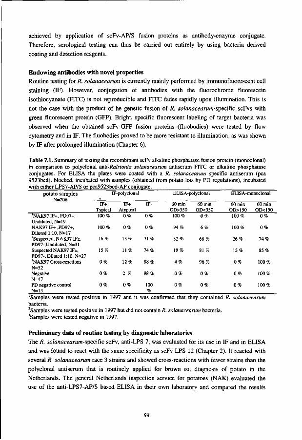
achieved by application of scFv-AP/S fusion proteins as antibody-enzyme conjugate.
Therefore, serological testing can thus be carried out entirely by using bacteria derived
coating and detection reagents.
Endowing antibodies with novel properties
Routine testing for R. solanacearum is currently mainly performed by immunofluorescent cell
staining (IF). However, conjugation of antibodies with the fluorochrome fluorescein
isothiocyanate (FITC) is not reproducible and FITC fades rapidly upon illumination. This is
not the case with the product of he genetic fusion of R. solanacearum-specific scFvs with
green fluorescent protein (GFP). Bright, specific fluorescent labeling of target bacteria was
observed when the obtained scFv-GFP fusion proteins (fluobodies) were tested by flow
cytometry and in IF. The fluobodies proved to be more resistant to illumination, as was shown
by IF after prolonged illumination (Chapter 6).
Table 7.1. Summary of testing the recombinant scFv alkaline phosphatase fusion protein (monoclonal) in comparison to polyclonal anti-Ralstonia solanacearum antiserum FITC or alkaline phosphatase conjugates. For ELISA the plates were coated with a R. solanacearum specific antiserum (pea 9523bcd), blocked, incubated with samples (obtained from potato lots by PD regulations), incubated with either LPS7-AP/S or pca9523bcd-AP conjugate.
potato samples N=206
'NAK97 IF+, PD97+, Undiluted, N=19 NAK97 IF+ ,PD97+, Diluted 1:10, N=17 2Suspected, NAK97 IF±, PD97-,Undiluted, N=31 Suspected NAK97 IF±, PD97-, Diluted 1:10, N=27 3NAK97 Cross-reactions N=52 Negative N=47 PD negative control N=13
9
IF+ Typical 100%
100%
16%
15%
0%
0%
0%
[F-polyclonal
IF+ Atypical
0%
0%
13%
11 %
12%
2 %
0%
IF-
0%
0%
71 %
74%
88%
98%
100 %
ELISA-
60 min OD>350 100%
94%
32%
19%
4%
0%
0%
polyclonal
60 min OD<350
0%
6%
68%
81 %
96%
0%
0%
ELISA-monoclonal
60 min OD>150 100%
100%
26%
15%
0%
0%
0%
60 min OD<150
0%
0%
74%
85%
100%
100%
100%
Samples were tested positive in 1997 and it was confirmed that they contained R. solanacearum bacteria. 2Samples were tested positive in 1997 but did not contain R. solanacearum bacteria. 'Samples were tested negative in 1997.
Preliminary data of routine testing by diagnostic laboratories
The R. solanacearum-spccific scFv, anti-LPS 7, was evaluated for its use in IF and in ELISA
and was found to react with the same specificity as scFv LPS 12 (Chapter 2). It reacted with
several R. solanacearum race 3 strains and showed cross-reactions with fewer strains than the
polyclonal antiserum that is routinely applied for brown rot diagnosis of potato in the
Netherlands. The general Netherlands inspection service for potatoes (NAK) evaluated the
use of the anti-LPS7-AP/S based ELISA in their own laboratory and compared the results
99
with those obtained with IF. The samples, obtained and already tested by the Dutch Plant Protection Service (PD) and NAK in 1997, were derived from various potato varieties such as Agria, Bildstar, Bintje Cardinal, Desiree, Diamant, Dore, Elkana, Felsina, van Gogh, Kanjer, Karnico, L. Rossetta, Monalisa, Spunta, Stefano and Symphony. The extraction of bacteria was performed according to the regulation of the PD and the results, summarized in Table 7.1, show that IF and ELISA gave comparable results for detection. Moreover, the ELISA that utilizes scFv-AP/S fusion proteins for detection is comparable with the polyclonal ELISA, but more importantly, the background is lower when the recombinant antibody is used for detection instead of the polyclonal alkaline phosphatase conjugate. No problems of increased background were observed regarding the sampling from the different potato varieties. The reduced number of cross-reactions that was observed for the recombinant antibodies in Chapter 2 also seems to hold for this field trial, as the number of positive samples found within the pool of suspected and cross-reacting samples was reduced. It can therefore be concluded that the recombinant scFv LPS7 is very useful under real testing conditions.
General conclusion and future perspectives This thesis shows that antibody phage display is a useful technique for the selection of specific recombinant antibodies against a variety of plant-pathogens. Thus far, recombinant antibodies have been selected only against viruses and bacteria. The results described are promising, and it is expected that the phage display technique can also be used successfully for raising specific MAbs against fungi and nematodes. Therefore, the perspective that was predicted after introduction of the hybridoma technique in 1975 [8], now becomes reality with application of the novel antibody phage display technique.
Although the synthetic naive Nissim library (108) was not compared directly to the naive Vaughan library (1010), the obtained results favor the application of the latter. The variability within the retrieved scFv-antibodies was higher and the yield of the scFvs was better. Moreover, the amount of scFv-AP/S fusion protein required for a strong signal in the ELISA was up to 50-fold lower, indicating that the recombinant antibodies retrieved from the Vaughan library were of higher affinity. In general, it can be stated that large (>1010) naive antibody libraries are a prerequisite for generating useful recombinant antibodies.
More than 50 different antigen-specific recombinant antibodies were selected without the use of experimental animals. However, screening the selected scFv-antibodies was a major problem since E. coli produced several of those, only with great difficulty. It remains disappointing that the choice of a scFv for further characterization was based more on its yield, than on its affinity and specificity for the antigen. Development of eukaryotic expression systems might be an option. Yeast, insect-cells, fungi and plants resemble more closely the mammalian cells than the prokaryotic bacterium E. coli with regard to protein expression, and improved yields of functional scFv-antibodies can be expected when eukaryotes are used.
100

There is an ongoing improvement of selection techniques from combinatorial antibody
phage display libraries such as selectively infective phage [1,9], expression of antibodies on
ribosomes [4] and selectively infected bacteria [10]. Meanwhile, combinatorial phage display
libraries are designed to give better folding, and consequently, better producing antibodies.
When these novel techniques find their way into plant pathology, it will undoubtedly result in
highly specific antibodies that can be used in diagnostic assays that are fast, reliable, robust
and cheap.
REFERENCES
1. Duenas, M. and Borrebaeck, C.A.K. (1994) Clonal selection and amplification of phage displayed antibodies by linking antigen recognition and phage replication. Bio/Technology 12: 999-1002.
2. George, R.A. and Converse, R.H. (1988) Methods for the enrichment of desired B-cell populations before anti-cauliflower mosaic virus hybridoma formation. Phytopathology 78: 1631-1636.
3. Griffiths, A.D., Williams, S.C., Hartley, O., Tomlinson, I.M., Waterhouse, P., Crosby, W.L., Kontermann, R.E., Jones, P.T., Low, N.M., Allison, T.J., Prospero, T.D., Hoogenboom, H.R., Nissim, A., Cox, J.P.L., Harrison, J.L., Zaccolo, M., Gherardi, E. and Winter, G. (1994) Isolation of high affinity antibodies directly from large synthetic repertoires. EMBO J. 13: 3245-3260.
4. Hanes, J. and Pliickthun, A. (1997) In vitro selection and evolution of functional proteins by ribosome display. Proc. Natl Acad. Sci. USA 94: 4937-4942.
5. Hoogenboom, H.R., Griffiths, A.D., Johnson, K.S., Chiswell, D.J., Hudson, P. and Winter, G. (1991) Multi-subunit proteins on the surface of filamentous phage: methodologies for displaying antibody (Fab) heavy and light chains. Nucleic Acids Res. 15: 4133-4137.
6. Hsu, H.T., Wang, Y.C., Lawson, R.H., Wang, M. and Gonsalves, D (1990) Splenocytes of mice with induced immunological tolerance to plant antigens for construction of hybridomas secreting tomato spotted wilt virus-specific antibodies. Phytopathology 80: 158-162.
7. Kerschbaumer, R.J., Hirschl, S., Schwager, C , Ibl, M. and Himmler, G. (1996) PDAP2: A vector for construction of alkaline phosphatase fusion-proteins. Immunotechnology 2: 145-150.
8. Kohler G. and Milstein C. (1975) Continuous cultures of fused cells secreting antibody of predefined specificity. Nature 256: 495-497.
9. Krebber, C , Spada, S., Desplancq, D. and Pliickthun, A. (1995) Co-selection of cognate anibody-antigen pairs by selectively infective phage. FEBS Lett. 377: 227-231.
10. Malmborg, A.C., Soderlind, E., Frost, L. and Borrebaeck C.A.K. (1998) Selective phage infection mediated by epitope expression on F pilus. J. Mol. Biol. 273: 544-551.
11. Matthew, W.D. and Sandrock, A.W. Jr. (1987) Cyclophosphamide treatment used to manipulate the immune response for the production of monoclonal antibodies. J. Immunol. Methods. 100: 73-82.
12. McCafferty, J., Griffiths, A.D., Winter, G. and Chiswell, D.J. (1990) Phage-antibodies: filamentous phage displaying antibody variable domains. Nature 348: 552-554.
13. Nissim, A., Hoogenboom, H.R., Tomlinson, I.M., Flynn, G., Midgley, C, Lane, D. and Winter, G. (1994) Antibody fragments from a single pot phage display library as immunochemical reagents. EMBO J. 13: 692-698.
14. Orlandi, O., Gussow, D.H., Jones, P.T. and Winter, G. (1989) Cloning immunoglobulin variable domains for the expression by the polymerase chain reaction. Proc. Natl. Acad. Sci. USA 86: 3833-3837.
101
15. Parks, D.R., Bryan, V.M., Oi, V.T. and Herzenberg, L.A. (1979) Antigen-specific identification and cloning of hybridomas with a fluorescence-activated cell sorter. Proc. Natl. Acad. Sci. USA 76: 1962-1966.
16. Robinson-Smith, A., Jones, P., Elphinstone, J.G. and Forde, S.M.D. (1995) Production of antibodies to Ralstonia solanacearum, the causative agent of bacterial wilt. Food Agric. Immunol. 7: 67-79.
17. Sastry, L., Alting-Mees, M., Huse, W.D., Short, J.M., Sorge, J.A., Hay, B.H., Janda, K.D., Benkovic, SJ. and Lerner, R.A. (1989) Cloning of the immunological repertoire in Escherichia coli for generation of monoclonal catalytic antibodies: Construction of a heavy chain variable region-specific cDNA library. Proc. Natl. Acad. Sci. USA 86: 5728-5732.
18. Vaughan, T.J., Williams, A.J., Pritchard, K., Osbourn, J.K., Pope, A.R., Earnshaw, J.C., McCafferty, J., Hodits, R.A., Wilton, J. and Johnson, K. (1996) Human antibodies with sub-nanomolar affinities isolated from a large non-immunized phage display library. Nature Biotechnology 14: 309-314.
102

Samenvatting
Achtergrond In de laatste decennia is het steeds duidelijker geworden dat het gebruik van chemische bestrijdingsmiddelen sterk verminderd moet worden. Veel van deze pesticiden zijn zeer belastend voor het milieu en vormen een gezondheidsrisico voor mens en dier. Het is dus zaak om milieuvriendelijke methoden te ontwikkelen om de plantenziekten in de hand te houden. In dit verband is het van belang dat het uitgangsmateriaal (knollen, bollen, zaad en ent-materiaal) vrij is van plantenpathogenen. Het optreden van ziektes zal dan in de regel achterwege blijven en de bestrijding van ziektes is niet of veel minder nodig. Voorts kan door het toetsen van grondmonsters informatie verkregen worden die te gebruiken is voor advies met betrekking tot gewasrotatie, cultivar selectie, toepassing van pesticiden, moment van oogsten en nabehandeling van het geoogste gewas.
Toetsmethoden Voor het opsporen van ziekteverwekkers (plantenpathogenen) zoals bijvoorbeeld virussen, bacterien, schimmels, insecten of parasitaire nematoden in zaaigoed, planten of grond, zijn betrouwbare toetsingsmethoden nodig. Er moet immers met zekerheid vastgesteld kunnen worden of plant- of grondmonsters besmet zijn of niet. Een methode die vaak wordt gebruikt, werkt met behulp van antilichamen uit het serum van dieren die opzettelijk zijn ingespoten met het betreffende plantenpathogeen. De dieren worden hier zelf niet ziek van, maar produceren wel grote hoeveelheden zogenaamde antilichamen. Deze antilichamen kunnen vervolgens in een serologische toets worden gebruikt (Fig. 1). Dergelijke serologische toetsen zijn niet alleen toepasbaar op grote aantallen monsters, ze zijn bovendien snel, robuust en goedkoop.
Specificiteit van ons afweersysteem Bepaalde bacterien, virussen en schimmels zijn in staat om mens en dier ziek te maken. Meestal wordt de patient na korte tijd weer beter. Dit komt omdat bepaalde cellen (witte bloedlichaampjes) van het afweersysteem op de ziekteverwekker reageren door onder andere antilichamen te maken tegen een scala aan structuren (epitopen) van deze lichaamsvreemde stoffen. Deze polyklonale antilichamen worden vervolgens afgegeven aan het bloed en kunnen de ziekteverwekker elders in het lichaam herkennen en deze vervolgens vernietigen. De lichaamscellen, die de antilichamen produceren, hebben een geheugenfunctie. Als je eenmaal de mazelen gehad hebt, krijg je gedurende de rest van je leven nooit meer mazelen. Dat andere ziekteverwekkers nog steeds kunnen toeslaan komt doordat de antilichamen, die gemaakt zijn tegen het mazelenvirus, bijvoorbeeld het griepvirus niet herkennen. Een antistof heeft dus een bepaalde specificiteit. Omdat ziekteverwekkers in talloze soorten en variaties in de natuur voorkomen beschikt het afweersysteem dan ook over een groot aantal verschillende antilichamen, elk met een andere specificiteit. Er wordt geschat dat het afweersysteem meer dan 1 miljard antilichamen kan maken, elk met een verschillende specificiteit!
103

o • •! •
Figuur 1. Een schematische weergave van een serologische toets. Extracten van gezonde planten (A) en zieke planten (B) worden gekoppeld aan het oppervlak van een vaste drager. Vervolgens worden antilichamen tegen de ziekteverwekker toegevoegd. Als de ziekteverwekker aanwezig is zal de antistof aan de ziekteverwekker binden (B). Deze binding is zo sterk dat na grondig wassen alle antilichamen die bij de gezonde plant werden toegevoegd (A) worden weggewassen (C) maar niet bij de zieke plant (D). Na het wassen wordt een chemische stof toegevoegd. Deze stof is kleurloos (O)maar wordt door het enzym alkalische fosfatase (AP) dat gekoppeld is aan de antistof omgezet in een kleurstof ( • ) . De zieke planten zijn dus detecteerbaar door een kleuromslag in de test.
Helaas zijn de polyklonale antilichamen die in het bloed aanwezig zijn vaak niet specifiek genoeg om toegepast te kunnen worden in een detectiesysteem. Ze zijn niet gericht tegen een bepaalde epitoop van de ziekteverwekker maar tegen een breed scala daarvan. Enkele van die structuren kunnen ook voorkomen bij andere, vaak onschadelijke, organismen die dikwijls aanwezig zijn in de directe omgeving van de plant. Het is dus zaak om alleen die antilichamen te produceren, die onderscheid kunnen maken tussen zieke en gezonde planten.
Productie van antilichamen in de reageerbuis. Technisch is het mogelijk om de geactiveerde antistof-producerende cellen van het afweersysteem te isoleren en ze te kloneren doormiddel van een celfusie met kankercellen. Hoewel er bij een dergelijke celfusie vele duizenden antistof-producerende cel-klonen
104

ontstaan, is dit aantal in veel gevallen toch niet toereikend om hieruit een kloon te selecteren
die antilichamen met de vereiste specificiteit produceert. In de praktijk blijkt namelijk vaak
dat het selecteren van een kloon die de gezochte specifieke antistof produceert vergelijkbaar is
met het zoeken van een naald in een hooiberg. De inefficiente celfusie en de eigenschap van
het afweersysteem, om juist tegen sommige epitopen sterk te reageren en tegen andere niet,
liggen hieraan ten grondslag.
Recombinant-DNA techniek: de uitkomst! Een uitkomst voor deze problemen werd geboden door de toepassing van moderne recombinant-DNA technieken. Deze technieken maakten het mogelijk om het genetische materiaal, dat de blauwdruk vormt voor de antilichamen, over te brengen van de antistof producerende cellen naar bacterien. Dit resulteerde uiteindelijk in zogenaamde antilichaam-bibliotheken die de blauwdrukken bevatten voor meer dan 10 miljard verschillende antilichamen. Deze blauwdrukken kunnen hier naar believen uitgehaald worden door de bacterien te infecteren met een bacterie-virus (bacteriofaag). Tijdens de vermeerdering van het virus in de bacterie neemt deze niet alleen een blauwdruk voor een antistof in zich op maar met behulp van de bacterie wordt deze antistof ook geproduceerd en vervolgens aan het oppervlak van het virus ingebouwd. Na een strenge selectie uit deze miljarden verschillende bacteriofagen op specificiteit kan dit uiteindelijk een monoklonaal antilichaam opleveren welke gebruikt kan worden om de gezonde van de zieke plant te onderscheiden. Een bijkomend voordeel is dat voor de selectie van deze monoklonale antilichamen geen proefdieren meer nodig zijn.
Toetsen op plantenpathogenen Het hoofdonderwerp van dit proefschrift is de productie en selectie van plantenpathogeen specifieke monoklonale antilichamen, die bruikbaar zijn in serologische toetsen. Daarbij werden met behulp van recombinant-DNA technologie monoklonale antilichamen geselecteerd die specifiek gericht zijn een tegen plantenpathogeen zoals de bacterie Ralstonia solanacearum (biovar 2, race 3), de veroorzaker van bruinrot bij de aardappel (Hoofdstuk 2) en het "beet necrotic yellow vein virus" (BNYVV), de veroorzaker van rhizomanie bij de suikerbiet (Hoofdstuk 3). Verder is er aandacht besteed aan de optimalisatie van productie en toetsing van de geselecteerde monoklonale antilichamen (Hoofdstuk 4). Deze optimalisatie werd vervolgens toegepast op de selectie van recombinante antilichamen, die gericht zijn tegen het tomatenbronsvlekkenvirus (TSWV, Hoofdstuk 5). Dat een recombinante antistof een handig stuk gereedschap vormt dat gemakkelijk gemanipuleerd kan worden werd aangetoond door middel van de productie als fusie-eiwit met het enzym alkalische fosfatase (Hoofdstuk 3 en Hoofdstuk 4) en als een fluorescerend eiwit (Hoofdstuk 6).
105

Curriculum vitae
Op 25 Juli 1963, zag Remko Albert Griep voor de eerste maal het licht in het pittoreske dorp Barsingerhorn. Na de lagere school doorlopen te hebben in Barsingerhorn, begon hij na de MAVO en de HAVO aan zijn voorbereiding op het wetenschappelijk onderwijs in Schagen (Rijks Scholen Gemeenschap). In 1983 behaalde hij zijn VWO diploma, en begon in hetzelfde jaar aan een studie Informatica aan de Technische Universiteit Delft. Helaas konden de levenloze computers hem niet lang boeien en begon hij in 1984 aan de studie Biologie aan de Rijks Universiteit te Utrecht. Het propaedeusediploma volgde een jaar later en hij besloot zich te gaan toeleggen op de afstudeerorientatie Chemische Biologie, met afstudeervakken bij de vakgroepen Klinische Immunologic (Prof. dr. R.E. Ballieux) begeleid door Pieter Koolwijk en Moleculaire Celbiologie (Prof. dr. H.O. Voorma) begeleid door Han van Heugten. Hij studeerde af in 1990 en begon als Assistent in Opleiding bij het Laboratorium voor Monoklonale Antistoffen op een vierjarig project dat gefinancierd werd door de programmacommissie landbouw en biotechnologie (PcLB). Het werk uit die periode betrof het maken van monoklonale antistoffen tegen diverse belangrijke plantenpathogenen door middel van recombinant-DNA technologie. Dit werk kreeg in 1994 een vervolg door financiering door de Europese Unie (EEG-AIR3) en de wetenschappelijk relevante resultaten staan beschreven in dit proefschrift.
107

Publications
R.A. Griep, C. Van Twisk, J.M. van der Wolf and A.Schots. Fluobodies: Green fluorescent single-chain Fv antibodies. (Submitted)
R.A. Griep, C. van Twisk, R. Kormelink, M. Prins, R.J. Kerschbaumer, R. Goldbach and A. Schots. Application of Phage Display in Selecting Tomato Spotted Wilt Virus-specific Single-chain Fvs for Sensitive Diagnosis in ELISA. (Submitted).
A.J.W.G. Visser, R.A. Griep, M. Hink, A. Van Hoek and A. Schots. Translational and rotational diffusion of green fluorescent protein alone and fused with a single-chain Fv protein. (Submitted).
R.A. Griep, R. Kerschbaumer, G. Himmler, C. van Twisk, K. Harper, A. Kaufman, J.M. van der Wolf, L. Torrance, and A. Schots. pSKAP/S: an expression vector for the production of single-chain Fv antibodies. Protein Expression and Purification, in press.
R.A. Griep, C. van Twisk and A. Schots. Selection of beet necrotic yellow vein virus specific single-chain antibodies from a semi-synthetic combinatorial antibody library. European Journal of Plant Pathology, in press.
R.A. Griep, C. van Twisk, J. van Beckhoven, J.M. van der Wolf, M.W. Prins, R. Kormelink and A. Schots. (1998) Selectie van specifieke monoklonale antilichamen met recombinant DNA-technologie. Gewasbescherming 29 (speciale editie): 26.
R.A. Griep, C. van Twisk, J. van Beckhoven, J.M. van der Wolf and A. Schots. (1998) Development of specific recombinant monoclonal antibodies against the lipopolysaccharide of Ralstonia solanaceamm race 3. Phytopathology 88:795-803.
P. Kastelein, J.M. van der Wolf, J.W.L. van Vuurde, R.A. Griep, A. Schots, en J.D. van Elsas. (1998) Detectie en ecologie van de bruinrot-bacterie. Recente resultaten van het onderzoek naar de detectie en ecologie van Ralstonia (Pseudomonas) solanaceamm (ras 3), de veroorzaker van bruinrot in aardappel. Gewasbescherming 29: 39-41.
R.A. Griep, C. van Twisk, J. van Beckhoven, J.M. van der Wolf and A. Schots. (1998) Selection of Pseudomonas solanaceamm race 3 specific monoclonal antibodies from combinatorial libraries. (ppl40-145). In: C. Allen, Ph. Prior & J. Elphinstone (eds.) Bacterial Wilt Disease, Molecular and ecological aspects. Springer Verlag, Berlin.
R.A. Griep, C. v Twisk and A. Schots. (1996) Abstract: Development of a diagnostic assay for the detection of beet necrotic yellow vein virus (BNYVV) in plants. GBF-Symposium: Antibody Technology and Applications in Health and Environment. Braunschweig
A. Schots, R.A. Griep and J. Bakker. (1995) Monoclonal Antibody technology, (pp 65-86) In: J.H. Skerrit & R. Appels (Eds.) New Diagnostics in Crop Sciences, CAB International, Wallingford, Oxon, UK.
R.A. Griep, C. van Twisk and A. schots. Abstract: Coliclonal antibodies for the detection of plantpathogens. Journal of Cellular Biochemistry, 1994, Supplement 18D, pag 213.
P. Koolwijk, J. Boot, R.A. Griep and B. Bast. Binding of the human complement subcomponent-Clq to hybrid Mouse monoclonal Antibodies. Molecular Immunology, 1991, Vol 28: pag 567-576.
109