development of f-fluoroethylcholine for cancer imaging...
TRANSCRIPT

Development of 18F-Fluoroethylcholine forCancer Imaging with PET: Synthesis,Biochemistry, and Prostate Cancer ImagingToshihiko Hara, MD, PhD1; Noboru Kosaka, MD1; and Hiroichi Kishi, MD, PhD2
1Department of Radiology, International Medical Center of Japan, Tokyo, Japan; and 2Department of Urology,International Medical Center of Japan, Tokyo, Japan
The effectiveness of 11C-choline PET in detecting various can-cers, including prostate cancer, is well established. This studywas aimed at developing an 18F-substituted choline analog,18F-fluoroethylcholine (FECh), as a tracer of cancer detection.Methods: No-carrier-added 18F-FECh was synthesized by2-step reactions: First, tetrabutylammonium (TBA) 18F-fluoridewas reacted with 1,2-bis(tosyloxy)ethane to yield 2-18F-fluoro-ethyl tosylate; and second, 2-18F-fluoroethyl tosylate was re-acted with N,N-dimethylethanolamine to yield 18F-FECh, whichwas then purified by chromatography. An automated apparatuswas constructed for preparation of the 18F-FECh injection solu-tion. In vitro experiments were performed to examine the uptakeof 18F-FECh in Ehrlich ascites tumor cells, and the metaboliteswere analyzed by solvent extraction followed by various kinds ofchromatography. Clinical studies of 18F-FECh PET were per-formed on patients with untreated primary prostate cancer asfollows: A dynamic 18F-FECh PET study was performed on 1patient and static PET studies were performed on 16 patients,and the data were compared with those of 11C-choline PET onthe same patients. Results: 18F-FECh was prepared in highyield and purity. The performance of the automated apparatuswas excellent. The in vitro experiment revealed that 18F-FEChwas incorporated into tumor cells by active transport, thenphosphorylated (yielding phosphoryl-18F-FECh) in the cells, andfinally integrated into phospholipids. The clinical PET studiesshowed marked uptake of 18F-FECh in prostate cancer. A dy-namic PET study on 1 patient revealed that the blood level of18F-FECh decreased rapidly (in 1 min), the prostate cancer levelbecame almost maximal in a short period (1.5 min) and it re-mained constant for a long time (60 min), and the urinary radio-activity became prominent after a short time lag (5 min). StaticPET studies conducted under bladder irrigation showed nodifference between 18F-FECh uptake and 11C-choline uptake inprostate cancer. However, 18F-FECh gave a slightly higher spa-tial resolution of the image, which was attributed to the shorterpositron range of 18F. Conclusion: The synthesis of 18F-FEChwas easy and reliable. 18F-FECh PET was very effective indetecting prostate cancer in patients. The chemical trap, con-sisting of active transport of 18F-FECh and formation of phos-
phoryl-18F-FECh, seemed to be involved in the uptake mecha-nism of 18F-FECh in tumors.
Key Words: 18F; choline; PET; prostate cancer
J Nucl Med 2002; 43:187–199
In most cancers a high content of phosphorylcholine hasbeen revealed by 31P nuclear magnetic resonance (NMR)studies, whereas in the corresponding normal tissues phos-phorylcholine is present at low levels, occasionally belowdetection (1,2). Phosphorylcholine, a product of the cholinekinase reaction, is the first intermediate in the stepwiseincorporation of choline, (CH3)3N�CH2CH2OH, into phos-pholipids by the Kennedy pathway. Katz-Brull and Degani(3) investigated choline transport in human breast cancercells in vitro by 31P, 13C, and 2H NMR and found thatcholine was incorporated into the tumor cells by a carrier-mediated mechanism and then it was converted into phos-phorylcholine within 1 h. Haeffner (4) investigated cholinetransport in Ehrlich ascites tumor cells using 3H-choline and14C-choline. When choline was incubated with tumor cellsat a low concentration, it was incorporated into the cells byan active-transport mechanism, then it was converted intophosphorylcholine also within 1 h, and finally it was inte-grated into phosphatidylcholine.
We previously developed 11C-choline as a PET tracer forcancer detection and have succeeded in visualizing braintumor (5), lung cancer (6), esophageal cancer (7), coloncancer (8), bladder cancer (8), prostate cancer (9), and manyother cancers (8). Motivated by this success, we attemptedto develop an 18F-labeled choline analog as a PET tracer,with an idea that 18F labeling would be superior to 11Clabeling because of the longer half-life and the shorterpositron range of 18F. We thought that 18F-fluoroethylcho-line (FECh) would be appropriate for this purpose. Thefollowing evidence supports our idea: Deves and Krupka(10) studied the binding affinity of the choline transportsystem for synthetic choline analogs, using red blood cells,and found that 2 methyl groups were essential, but the thirdmethyl group was replaceable with a longer alkyl group.
Received Mar. 1, 2001; revision accepted Jun. 14, 2001.For correspondence or reprints contact: Toshihiko Hara, MD, PhD, Depart-
ment of Radiology, International Medical Center of Japan, 1-21-1 Toyama,Shinjuku-ku, Tokyo 162, Japan.
E-mail: [email protected]
18F-FLUOROETHYLCHOLINE FOR PROSTATE CANCER IMAGING • Hara et al. 187
by on May 4, 2018. For personal use only. jnm.snmjournals.org Downloaded from
Clary et al. (11) studied the substrate specificity of cholinekinase for synthetic choline analogs, using yeast cholinekinase, and found that the 2 methyl groups and the hydroxyl-ethyl side chain were essential, but the third methyl groupwas replaceable with a longer alkyl group. We had alreadysynthesized 18F-FECh and studied its biodistribution in nor-mal and tumor-bearing rabbits; our results are reported in apreliminary form (12). In this article, we report the details ofthe synthesis, biochemistry, and clinical application of thiscompound.
MATERIALS AND METHODS
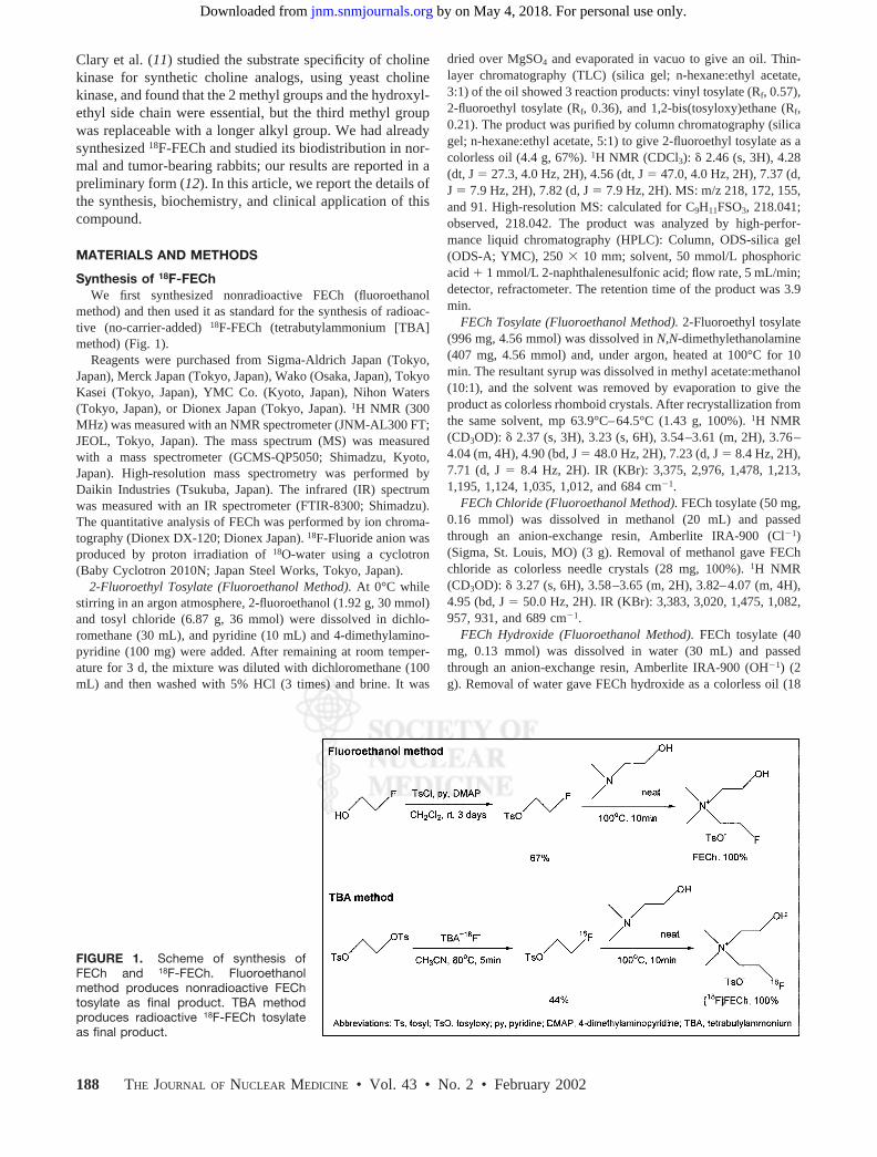
Synthesis of 18F-FEChWe first synthesized nonradioactive FECh (fluoroethanol
method) and then used it as standard for the synthesis of radioac-tive (no-carrier-added) 18F-FECh (tetrabutylammonium [TBA]method) (Fig. 1).
Reagents were purchased from Sigma-Aldrich Japan (Tokyo,Japan), Merck Japan (Tokyo, Japan), Wako (Osaka, Japan), TokyoKasei (Tokyo, Japan), YMC Co. (Kyoto, Japan), Nihon Waters(Tokyo, Japan), or Dionex Japan (Tokyo, Japan). 1H NMR (300MHz) was measured with an NMR spectrometer (JNM-AL300 FT;JEOL, Tokyo, Japan). The mass spectrum (MS) was measuredwith a mass spectrometer (GCMS-QP5050; Shimadzu, Kyoto,Japan). High-resolution mass spectrometry was performed byDaikin Industries (Tsukuba, Japan). The infrared (IR) spectrumwas measured with an IR spectrometer (FTIR-8300; Shimadzu).The quantitative analysis of FECh was performed by ion chroma-tography (Dionex DX-120; Dionex Japan). 18F-Fluoride anion wasproduced by proton irradiation of 18O-water using a cyclotron(Baby Cyclotron 2010N; Japan Steel Works, Tokyo, Japan).
2-Fluoroethyl Tosylate (Fluoroethanol Method). At 0°C whilestirring in an argon atmosphere, 2-fluoroethanol (1.92 g, 30 mmol)and tosyl chloride (6.87 g, 36 mmol) were dissolved in dichlo-romethane (30 mL), and pyridine (10 mL) and 4-dimethylamino-pyridine (100 mg) were added. After remaining at room temper-ature for 3 d, the mixture was diluted with dichloromethane (100mL) and then washed with 5% HCl (3 times) and brine. It was
dried over MgSO4 and evaporated in vacuo to give an oil. Thin-layer chromatography (TLC) (silica gel; n-hexane:ethyl acetate,3:1) of the oil showed 3 reaction products: vinyl tosylate (Rf, 0.57),2-fluoroethyl tosylate (Rf, 0.36), and 1,2-bis(tosyloxy)ethane (Rf,0.21). The product was purified by column chromatography (silicagel; n-hexane:ethyl acetate, 5:1) to give 2-fluoroethyl tosylate as acolorless oil (4.4 g, 67%). 1H NMR (CDCl3): � 2.46 (s, 3H), 4.28(dt, J � 27.3, 4.0 Hz, 2H), 4.56 (dt, J � 47.0, 4.0 Hz, 2H), 7.37 (d,J � 7.9 Hz, 2H), 7.82 (d, J � 7.9 Hz, 2H). MS: m/z 218, 172, 155,and 91. High-resolution MS: calculated for C9H11FSO3, 218.041;observed, 218.042. The product was analyzed by high-perfor-mance liquid chromatography (HPLC): Column, ODS-silica gel(ODS-A; YMC), 250 � 10 mm; solvent, 50 mmol/L phosphoricacid � 1 mmol/L 2-naphthalenesulfonic acid; flow rate, 5 mL/min;detector, refractometer. The retention time of the product was 3.9min.
FECh Tosylate (Fluoroethanol Method). 2-Fluoroethyl tosylate(996 mg, 4.56 mmol) was dissolved in N,N-dimethylethanolamine(407 mg, 4.56 mmol) and, under argon, heated at 100°C for 10min. The resultant syrup was dissolved in methyl acetate:methanol(10:1), and the solvent was removed by evaporation to give theproduct as colorless rhomboid crystals. After recrystallization fromthe same solvent, mp 63.9°C–64.5°C (1.43 g, 100%). 1H NMR(CD3OD): � 2.37 (s, 3H), 3.23 (s, 6H), 3.54–3.61 (m, 2H), 3.76–4.04 (m, 4H), 4.90 (bd, J � 48.0 Hz, 2H), 7.23 (d, J � 8.4 Hz, 2H),7.71 (d, J � 8.4 Hz, 2H). IR (KBr): 3,375, 2,976, 1,478, 1,213,1,195, 1,124, 1,035, 1,012, and 684 cm�1.
FECh Chloride (Fluoroethanol Method). FECh tosylate (50 mg,0.16 mmol) was dissolved in methanol (20 mL) and passedthrough an anion-exchange resin, Amberlite IRA-900 (Cl�1)(Sigma, St. Louis, MO) (3 g). Removal of methanol gave FEChchloride as colorless needle crystals (28 mg, 100%). 1H NMR(CD3OD): � 3.27 (s, 6H), 3.58–3.65 (m, 2H), 3.82–4.07 (m, 4H),4.95 (bd, J � 50.0 Hz, 2H). IR (KBr): 3,383, 3,020, 1,475, 1,082,957, 931, and 689 cm�1.
FECh Hydroxide (Fluoroethanol Method). FECh tosylate (40mg, 0.13 mmol) was dissolved in water (30 mL) and passedthrough an anion-exchange resin, Amberlite IRA-900 (OH�1) (2g). Removal of water gave FECh hydroxide as a colorless oil (18
FIGURE 1. Scheme of synthesis ofFECh and 18F-FECh. Fluoroethanolmethod produces nonradioactive FEChtosylate as final product. TBA methodproduces radioactive 18F-FECh tosylateas final product.
188 THE JOURNAL OF NUCLEAR MEDICINE • Vol. 43 • No. 2 • February 2002
by on May 4, 2018. For personal use only. jnm.snmjournals.org Downloaded from

mg, 92%). 1H NMR (CD3OD): � 3.26 (s, 6H), 3.55–3.69 (m, 2H),3.78–4.07 (m, 4H), 4.98 (bd, J � 50.0 Hz, 2H). IR (KBr): 3,444,1,474, 1,387, 1,350, 1,084, 1,052, and 957 cm�1. On the ODS-silica gel HPLC performed under the same condition as above, theretention time of FECh hydroxide was 4.6 min.
2-18F-Fluoroethyl Tosylate (TBA Method). No-carrier-added18F-fluoride (approximately 370 MBq), collected from an anion-exchange cartridge by elution with 2 mL 40 mmol/L TBA bicar-bonate in acetonitrile:water (4:1), was dried by evaporation at100°C and dried again with 2 mL dry acetonitrile. After additionof 1,2-bis(tosyloxy)ethane (20 mg) in dry acetonitrile (1 mL), themixture was heated at 80°C for 20 min. After the solvent wasevaporated at 80°C under reduced pressure, the dry residue wasanalyzed on TLC and HPLC. The Rf on TLC was identical withthat of 2-fluoroethyl tosylate from the fluoroethanol method. OnHPLC, the retention time of the product was identical with that of2-fluoroethyl tosylate. A small amount of radioactivity remained inthe column head.
18F-FECh Hydroxide (TBA Method). No-carrier-added 2-18F-fluoroethyl tosylate prepared as above was dried and then dis-solved in N,N-dimethylethanolamine (0.3 mL). The mixture washeated at 100°C for 5 min. After evaporation of N,N-dimethyleth-anolamine at 100°C under high vacuum, the dry residue wasanalyzed by HPLC. On the ODS-silica gel HPLC, a single radio-active peak, corresponding to 18F-FECh hydroxide, was found at4.6 min. There was no radioactive peak of 2-18F-fluoroethyl tosy-late. The radiochemical yield of 18F-FECh hydroxide comparedwith 18F-fluoride was 46.3% (decay corrected). The rest of theradioactivity was found in the reaction vessel and the column head.On this HPLC, a sharp mass peak of TBA (detected by a refrac-tometer) appeared far behind the radioactive peak of 18F-FEChhydroxide. There was no other mass peak that eluted closely to18F-FECh hydroxide.
Automated Synthesis of No-Carrier-Added18F-FECh Chloride
Design of Automated Apparatus. We constructed an automatedapparatus for the synthesis of 18F-FECh. The function of thisapparatus was as follows: (a) transfer of proton-irradiated 18O-water into a transit vessel and separation of 18F-fluoride ions usingan ion-exchange cartridge, (b) transfer of 18F-fluoride ions into areaction vessel, (c) 2-step chemical reactions (2-18F-fluoroethyltosylate synthesis followed by 18F-FECh synthesis) in the reactionvessel, (d) passage of the product through anion-exchange car-tridges to remove anionic by-products, (e) transfer of the effluentfrom cartridges to an HPLC apparatus, (f) HPLC, (g) passage ofthe effluent through anion-exchange cartridges to remove anionicmolecular species in the HPLC solvent (phosphoric acid and2-naphthalenesulfonic acid), (h) passage of the effluent from thecartridges through a cation-exchange cartridge to trap 18F-FECh,(i) washing of the cartridge with water, and (j) elution of 18F-FEChfrom the cartridge with saline. A close-up illustration of the reac-tion vessel is shown in Figure 2A, where the top of the vessel issealed by a rubber septum, and 3 side arms are fixed on the side ofthe vessel. All chemical reactions were performed in this reactionvessel. Delivery of reagents to the reaction vessel was achieved bymoving the upper and lower turntables; the upper table wasequipped with needle-and-syringe units, and the lower tablewas equipped with reagent reservoirs. After one of the reagentswas sucked from a reservoir into the corresponding needle-and-syringe unit, the upper and lower turntables moved vertically androtationally, and the reagent was injected then into the reactionvessel from the needle pierced through the rubber septum. Thepurpose of the side arms was (a) to receive 18F-fluoride ions intothe vessel; (b) to create negative, neutral, and positive pressureswithin the vessel; and (c) to send helium gas to the bottom of thevessel or transport the product of synthesis to the next HPLC unit.
FIGURE 2. (A) Automated apparatus for 18F-FECh synthesis. Reaction vessel is shown close up. Purification module is out ofview. (B) Preparative HPLC of 18F-FECh in automated apparatus for 18F-FECh synthesis. Column used was ODS-silica gel column,250 � 10 mm; solvent, 50 mmol/L phosphoric acid � 1 mmol/L 2-naphthalenesulfonic acid; flow rate, 5 mL/min. Radioactivity“halfway down the column” is reading of detector (detector 1) placed on side of column to watch approach of 18F-FECh.Radioactivity of effluent from column was monitored by another detector (detector 2).
18F-FLUOROETHYLCHOLINE FOR PROSTATE CANCER IMAGING • Hara et al. 189
by on May 4, 2018. For personal use only. jnm.snmjournals.org Downloaded from
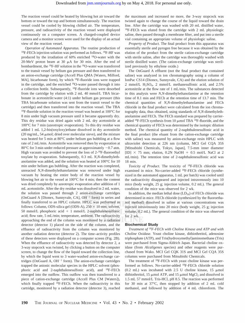
The reaction vessel could be heated by blowing hot air toward thebottom or toward the top and bottom simultaneously. The reactionvessel could be cooled by blowing chilled air. The temperature,pressure, and radioactivity of the reaction vessel were displayedcontinuously on a computer screen. A charged-coupled devicecamera and a monitor screen were used for the display of an actualview of the reaction vessel.
Operation of Automated Apparatus. The routine production of18F-FECh injection solution was performed as follows. 18F-HF wasproduced by the irradiation of 60% enriched 18O-water with the20-MeV proton beam at 30 �A for 30 min. After the end ofbombardment, the 18F-HF solution in the 18O-water was transferredto the transit vessel by helium gas pressure and then transferred toan anion-exchange cartridge (Accell Plus QMA [Waters, Milford,MA], bicarbonate form), by which 18F-fluoride ions were trappedin the cartridge, and the enriched 18O-water passed through it intoa collection bottle. Subsequently, 18F-fluoride ions were desorbedfrom the cartridge by elution with 2 mL 40 mmol/L TBA bicar-bonate in acetonitrile:water (4:1) under helium gas pressure (thisTBA bicarbonate solution was sent from the transit vessel to thecartridge) and then transferred into the reaction vessel. The TBA18F-fluoride solution in the reaction vessel was heated at 100°C for8 min under high vacuum pressure until it became apparently dry.This dry residue was dried again with 2 mL dry acetonitrile at100°C for 7 min (azeotropic evaporation). To this dry residue wasadded 1 mL 1,2-bis(tosyloxy)ethane dissolved in dry acetonitrile(20 mg/mL, 54 �mol; dried over molecular sieve), and the mixturewas heated for 5 min at 80°C under helium gas bubbling at a flowrate of 2 mL/min. Acetonitrile was removed then by evaporation at80°C for 3 min under reduced pressure at approximately �0.7 atm.High vacuum was avoided to prevent the loss of 2-18F-fluoroethyltosylate by evaporation. Subsequently, 0.3 mL N,N-dimethyleth-anolamine was added, and the solution was heated at 100°C for 10min under helium gas bubbling. After the reaction was completed,unreacted N,N-dimethylethanolamine was removed under highvacuum by heating the entire body of the reaction vessel byblowing hot air to the vessel at 100°C for 4 min; then the contentwas dried completely by azeotropic evaporation after addition of 1mL acetonitrile. After the dry residue was dissolved in 2 mL water,the solution was passed through 2 anion-exchange cartridges(OnGuard A [Dionex, Sunnyvale, CA], OH�1 form) in series andfinally transferred to an HPLC column. HPLC was performed asfollows: Column, ODS-silica gel (ODS-A), 250 � 10 mm; solvent,50 mmol/L phosphoric acid � 1 mmol/L 2-naphthalenesulfonicacid; flow rate, 5 mL/min; temperature, ambient. The radioactivityapproaching the end of the column was monitored by a radiationdetector (detector 1) placed on the side of the column, and theeffluence of radioactivity from the column was monitored byanother radiation detector (detector 2). The time–activity profilesof these detectors were displayed on a computer screen (Fig. 2B).When the effluence of radioactivity was detected by detector 2, a3-way stopcock was twisted, by clicking a button on the computerscreen, to change the flow of the liquid toward the collection line,by which the liquid went to 3 water-washed anion-exchange car-tridges (OnGuard A, OH�1 form). The anion-exchange cartridgestrapped the anionic molecular species in the HPLC solvent (phos-phoric acid and 2-naphthalenesulfonic acid), and 18F-FEChemerged into the outflow. This outflow was then transferred to apiece of cation-exchange cartridge (Accell Plus CM [Waters]),which finally trapped 18F-FECh. When the radioactivity in thiscartridge, monitored by a radiation detector (detector 3), reached
the maximum and increased no more, the 3-way stopcock wastwisted again to change the course of the liquid toward the drainline. After the cartridge was washed with 20 mL distilled water,18F-FECh was eluted from the cartridge with 2 mL physiologicsaline, then passed through a membrane filter, and put into a sterilevial containing an appropriate volume of physiologic saline.
Property of Product. The final product from this apparatus wasessentially sterile and pyrogen free because it was obtained by theelution of the product from the sterile cation-exchange cartridgewith sterile saline, after the cartridge was thoroughly washed withsterile distilled water. (The cation-exchange cartridge was steril-ized previously by ethylene oxide.)
The OnGuard A effluent (not the final product that containedsaline) was analyzed in ion chromatography using a column ofIonPac CS14 (Dionex, Sunnyvale, CA) and the elution solution of2 mmol/L H2SO4, 2 mmol/L methanesulfonic acid, and 2.5%acetonitrile at the flow rate of 1 mL/min. The substances detectedin this analysis were N,N-dimethylethanolamine at the retentiontime of 8.1 min and FECh at the retention time of 13.7 min. Thechemical quantities of N,N-dimethylethanolamine and FEChchloride in the final product were calculated from the ion chroma-tography data, thus obtained, using standards of N,N-dimethyleth-anolamine and FECh. The FECh standard was prepared by carrier-added 18F-FECh synthesis from 10 �mol TBA 18F-fluoride, and thechemical quantity of FECh was determined by the isotopic dilutionmethod. The chemical quantity of 2-naphthalenesulfonic acid inthe final product (the eluant from the cation-exchange cartridgewith saline) was measured by anion-exchange resin HPLC withultraviolet detection at 226 nm (column, MCI Gel CQA 35S[Mitsubishi Chemicals, Tokyo, Japan], 7.5-mm inner diameter[ID] � 75 mm; elution, 0.2N NaOH � 0.5 mol/L NaCl at 2mL/min). The retention time of 2-naphthalenesulfonic acid was18 min.
Toxicity of Product. The toxicity of 18F-FECh chloride wasexamined in mice. No-carrier-added 18F-FECh chloride (synthe-sized in the automated apparatus, 1 mL per batch) was cooled untilthe radioactivity disappeared and injected intravenously into 10mice (body weight, 25 g; injection volume, 0.2 mL). The generalcondition of the mice was observed for 2 wk.
In addition, the median lethal dose (LD50) of FECh chloride wasdetermined in mice. FECh chloride (synthesized by the fluoroetha-nol method) dissolved in saline at various concentrations wasinjected intravenously into 20 mice (body weight, 25 g; injectionvolume, 0.2 mL). The general condition of the mice was observedfor 2 wk.
Biochemical StudyTreatment of 18F-FECh with Choline Kinase and ATP and with
Choline Oxidase. Yeast choline kinase, dithiothreitol, adenosinetriphosphate (ATP), and Tris(hydroxymethyl)aminomethane (Tris)were purchased from Sigma-Aldrich Japan. Bacterial choline ox-idase (from Alcaligenes species) and other reagents were pur-chased from Wako. MCI Gel CQK 31S and MCI Gel CQA 35Scolumns were purchased from Mitsubishi Chemicals.
The treatment of 18F-FECh with yeast choline kinase was per-formed as follows. No-carrier-added 18F-FECh chloride solution(0.2 mL) was incubated with 2.5 U choline kinase, 15 �moldithiothreitol, 15 �mol ATP, and 15 �mol MgCl2 and dissolved in1.5 mL 57 mmol/L Tris-HCl, pH 8.5. The reaction was performedfor 30 min at 37°C, then stopped by addition of 2 mL coldmethanol, and followed by addition of 4 mL chloroform. The
190 THE JOURNAL OF NUCLEAR MEDICINE • Vol. 43 • No. 2 • February 2002
by on May 4, 2018. For personal use only. jnm.snmjournals.org Downloaded from
mixture was centrifuged to give an upper methanol-water layer,lower chloroform layer, and the proteins in the interface. Theupper methanol-water layer was separated, evaporated to dryness,and then dissolved in HPLC solvents. This sample was analyzedby HPLC using 2 kinds of ion-exchange columns (injection vol-ume, 10 �L each): (a) MCI Gel CQK 31S column (containingcarboxylmethyl group for cation exchange), 7.5-mm ID � 75 mm,eluted with 20 mmol/L sodium phosphate buffer, pH 6.5, at 1mL/min; and (b) MCI Gel CQA 35S column (containing quater-nary ammonium group for anion exchange), 7.5-mm ID � 75 mm,eluted with 10 mmol/L Tris-HCl buffer, pH 8.0, at 1 mL/min.One-milliliter fractions were collected, and the radioactivity ofeach fraction was measured in a well counter.
The treatment of 18F-FECh with bacterial choline oxidase wasperformed as follows. No-carrier-added 18F-FECh chloride solu-tion (0.2 mL) was incubated with 2.5 U choline oxidase, 15 �moldithiothreitol, and 15 �mol MgCl2 in 1.5 mL 57 mmol/L Tris-HCl,pH 8.5. The reaction was performed for 30 min at 37°C andstopped by addition of 2 mL cold methanol and 4 mL chloroform.The subsequent treatment was the same as the above.
Treatment of 18F-FECh with Choline Kinase and �-32P-ATP.�-32P-ATP (product of New England Nuclear, Boston, MA) waspurchased from Daiichi Pure Chemicals (Tokyo, Japan). A double-labeling experiment with 18F-FECh and �-32P-ATP was performedas follows. No-carrier-added 18F-FECh chloride solution (0.2 mL,370 MBq) containing approximately 0.005 �mol FECh was addedto the reaction solution containing 2.5 U yeast choline kinase, 10�mol dithiothreitol, 0.01 �mol �-32P-ATP (instead of 15 �molnonradioactive ATP), 0.01 �mol MgCl2, and 0.8 mL 62.5 mmol/LTris-HCl, pH 8.5. The mixture was incubated at 37°C for 60 min.After the reaction was stopped by cooling in ice, 1 �mol nonra-dioactive phosphoric acid was added to the mixture to lower thespecific activity of inorganic 32P-phosphate that might be formedfrom �-32P-ATP during the reaction. After addition of water,methanol, and chloroform, the methanol-water layer was sepa-rated, evaporated to dryness, and dissolved in 0.2 mL of the HPLCsolvent. After injection of 10 �L of the sample to the HPLCcolumn (MCI Gel CQA 35S), it was eluted with 10 mmol/LTris-HCl buffer, pH 8.0, at a flow rate of 1 mL/min. One-milliliterfractions were collected, and the radioactivity of 18F in eachfraction was measured in a well counter; then, after the decay of18F, the radioactivity of 32P was measured in a liquid scintillationcounter. Inorganic phosphate and ATP were not eluted from thecolumn into these fractions and only eluted by washing the columnwith 0.2N NaOH.
Metabolism of 18F-FECh in Tumor Cells. Ehrlich ascites tumorcells were obtained from Human Science Research Resource Bank(Osaka, Japan). Authentic phospholipid samples (phosphatidyl-choline, sphingomyelin, and lysophosphatidylcholine) and silicagel 60 plates were obtained from Sigma-Aldrich Japan. Hanks’balanced salt solution was obtained from Wako. An imagingplate-scanner-printer system (BAS-1800II) was obtained from FujiFilm (Tokyo, Japan).
The metabolism of 18F-FECh in tumor cells was measured asfollows. Ehrlich ascites tumor cells (approximately105 cells) wereimplanted intraperitoneally in Institute for Cancer Research (ICR)mice (Japan Clea, Tokyo, Japan), and the proliferated tumor cellswere collected 2–3 wk later. The tumor cells were washed twicewith 0.6% glucose-fortified Hanks’ balanced salt solution, pH 7.4,and suspended in the same solution to give a cell density ofapproximately 5 � 106 cells/mL; the volume density of the cells
was measured by hematocrit. No-carrier-added 18F-FECh chloridesolution (40 �L, 74 MBq) was added to the tumor-cell suspension(200 �L), and the mixture was incubated then at 37°C for 30 min.After the reaction, the cells were cooled in ice and washed 3 timesby addition of 5 mL unlabeled medium and centrifugation. Theprecipitated cells were mixed with 1.5 mL cold water, 2 mLmethanol, and 4 mL chloroform, successively. After centrifuga-tion, the upper methanol-water layer, lower chloroform layer, andprecipitate were separated. The radioactivity of each fraction wasmeasured in a well counter. The methanol-water layer was ana-lyzed further by HPLC: After the sample was evaporated todryness, it was dissolved in a small volume of HPLC solvents andpassed through HPLC MCI Gel CQK 31S and MCI Gel CQA 35Scolumns (injection volume, 10 �L) to obtain 1-mL fractions. Theradioactivity of each fraction was measured in a well counter. Inaddition to this experiment, the chloroform layer was analyzed,separately, in the following manner: After the chloroform layerwas washed with methanol-water and then concentrated, TLC wasperformed on the silica gel 60 thin-layer plates using the chloro-form, methanol, and 28% ammonia (65:35:5) and benzene, pyri-dine, and formic acid (50:40:10) solvents. The distribution ofradioactivity on the plates was measured using an imaging plate-scanner-printer system. As standards of TLC, 3 representativecholine-containing phospholipids (phosphatidylcholine, sphingo-myelin, and lysophosphatidylcholine) were used, and the locationof them was detected after staining with sulfuric acid.
Time Course of Uptake and Metabolism of 18F-FECh in TumorCells. The time course of 18F-FECh uptake and metabolism intumor cells was measured as follows. Ehrlich ascites tumor cells in50 �L 0.6% glucose-fortified Hanks’ solution (approximately 2 �106 cells/mL) were mixed with 10 �L no-carrier-added 18F-FEChchloride solution, and the mixture was incubated for 0, 5, 10, 20,30, 40, 50, and 60 min at 37°C. At the determined time ofincubation, each sample was diluted with 5 mL ice-cold unlabeledmedium and left in ice until the end of the incubation schedule.After washing the cells 3 times by centrifugation with glucose-fortified Hanks’ solution, the precipitated cells were treated suc-cessively with 1.5 mL cold water, 2 mL methanol, and 4 mLchloroform. After centrifugation, the upper methanol-water layer,lower chloroform layer, and precipitate were separated. The meth-anol-water layer was diluted then with 2 mL water and filteredthrough an anion-exchange resin cartridge (OnGuard A, OH�1).By this procedure, 18F-labeled anionic substances (i.e., phos-phoryl-18F-FECh) were trapped in this anion-exchange resin car-tridge and unreacted 18F-FECh passed through it. Subsequently,the radioactivity of each fraction was measured in a well counter.The total radioactivity in the cells and the chemical constitution ofthe total radioactivity is presented as the cell-to-medium ratio �(radioactivity concentration in cells)/(radioactivity concentrationin medium). The (volume of cells)/(volume of medium) was de-termined by the hematocrit, in which the tumor cell suspensionwas taken into a capillary tube and centrifuged, and the (volume ofpacked cells)/(total volume of liquid) was measured under a mi-croscope.
PET Study of Prostate Cancer Patients with18F-FECh Chloride
In a preliminary study (12), we found that the biodistribution of18F-FECh was almost the same as that of 11C-choline in normalrabbit and normal humans. The only difference was that 18F-FEChwas excreted rapidly into urine, whereas 11C-choline was excreted
18F-FLUOROETHYLCHOLINE FOR PROSTATE CANCER IMAGING • Hara et al. 191
by on May 4, 2018. For personal use only. jnm.snmjournals.org Downloaded from
slowly. We also found that the uptake of 18F-FECh in tumors ofrabbits (VX2 tumor) was very high, and it was comparable withthat of 11C-choline. Our clinical work of 18F-FECh PET began onpatients with prostate cancer because we had considerable expe-rience in studying these patients with 11C-choline PET (8,9). All ofthe following studies were approved by the institutional ethicalboard and were performed after receiving informed consent fromthe patients.
A single run of 18F-FECh PET, in a dynamic scan mode, wasperformed on 1 patient (80 y old) with untreated primary prostatecancer, after intravenous injection of 370 MBq 18F-FECh chloride,without bladder irrigation, to determine the most appropriate pro-tocol for the study of other patients. 18F-FECh PET and 11C-choline PET were performed on 16 untreated prostate cancerpatients, and the whole set of the studies was performed on 2consecutive days.
The 18F-FECh PET study was performed according to thefollowing protocol. Patients fasted overnight. A short intravenouscatheter was placed in the forearm for intravenous infusion. A3-way Foley catheter was placed in the bladder for irrigation, with1 tubing connected to warm saline and another connected to aurine collection bag. After completion of the transmission scan,18F-FECh (370 MBq) and furosemide (20 mg) were injected fromthe intravenous line successively, and saline (500 mL) was drippedfrom the same line until the end of the study. The bladder irrigationstarted shortly after the 18F-FECh injection and continued until theend of the study (total volume of saline, 4 L).
PET images were obtained using a PET camera (Headtome IV,6-mm spatial resolution; Shimadzu) equipped with 3 detector ringsto produce 5 slices at 13-mm intervals. When the patient, fixed onthe bed, underwent transmission or emission scanning, the bedposition was shifted 6 times upward from the level of the pelvis tothat of the liver, with a scan time of 3 min at a single bed position.The emission scan was obtained twice, starting at 30 and 60 min,respectively. PET images were reconstructed after attenuationcorrection. The horizontal images were displayed sequentially,with each horizontal level indicated in a planar image, on thecomputer screen. Usually, the horizontal images were displayedaccording to the standardized uptake value (SUV), where SUVwas defined as (regional radioactivity concentration)/(total injecteddose/body weight). Each pixel (4 � 4 � 6 mm in real size) waspainted a specified color that indicated a corresponding SUVvalue. Usually, red (the hottest color) indicated an SUV of �4.0.
The 11C-choline PET study was performed according to theprotocol reported previously (9).
RESULTS
Automated Synthesis of No-Carrier-Added18F-FECh Chloride
Operation of Automated Apparatus. In the automatedapparatus, 18F-FECh was eluted from HPLC, as shown inFigure 2B. Detector 1, placed on the side of the HPLCcolumn, showed the progression of radioactivity through thecolumn. Detector 2 showed the radioactivity of the effluent.
The total time required for the synthesis of 18F-FEChchloride was 65 min after the end of bombardment.
Property of Product. The radiochemical yield of 18F-FECh chloride was approximately 40%, with the decaycorrected. After proton bombardment (20 MeV, 30 �A) of
60% enriched 18O-water for 30 min, approximately 3.7 GBq18F-FECh chloride were obtained.
The ion chromatography analysis showed that the chem-ical quantities of ingredients in this preparation were N,N-dimethylethanolamine, 0.12 �mol (10.7 �g) per batch; andFECh chloride, 0.05 �mol (8.6 �g) per batch. The specificradioactivity of 18F-FECh chloride was calculated as 74GBq/�mol.
The anion-exchange resin HPLC analysis showed that2-naphthalenesulfonic acid was below detection (�0.1�mol per batch) in the final 18F-FECh preparation.
The stability of 18F-FECh chloride was examined byion-exchange HPLC. The result was as follows. If 18F-FEChchloride was left at room temperature at a high concentra-tion (1.85 GBq/mL) for 1 h, part of it decomposed to form18F-fluoroethylbetaine. This decomposition did not occur ifit was stored in a refrigerator at a low concentration (0.37GBq/mL) for hours.
Toxicity of Product. The 18F-FECh chloride preparationshowed no toxicity in 10 mice when it was injected intra-venously after decay of radioactivity: injection dose, onefifth of a single batch from the automated synthesis, 0.01�mol (1.7 �g) FECh per mouse (25 g).
The LD50 of FECh chloride (prepared by the fluoroetha-nol method) examined in 20 mice after intravenous injectionwas 0.13 g/kg.
Biochemical StudyFormation of Phosphoryl-18F-FECh by Choline Kinase
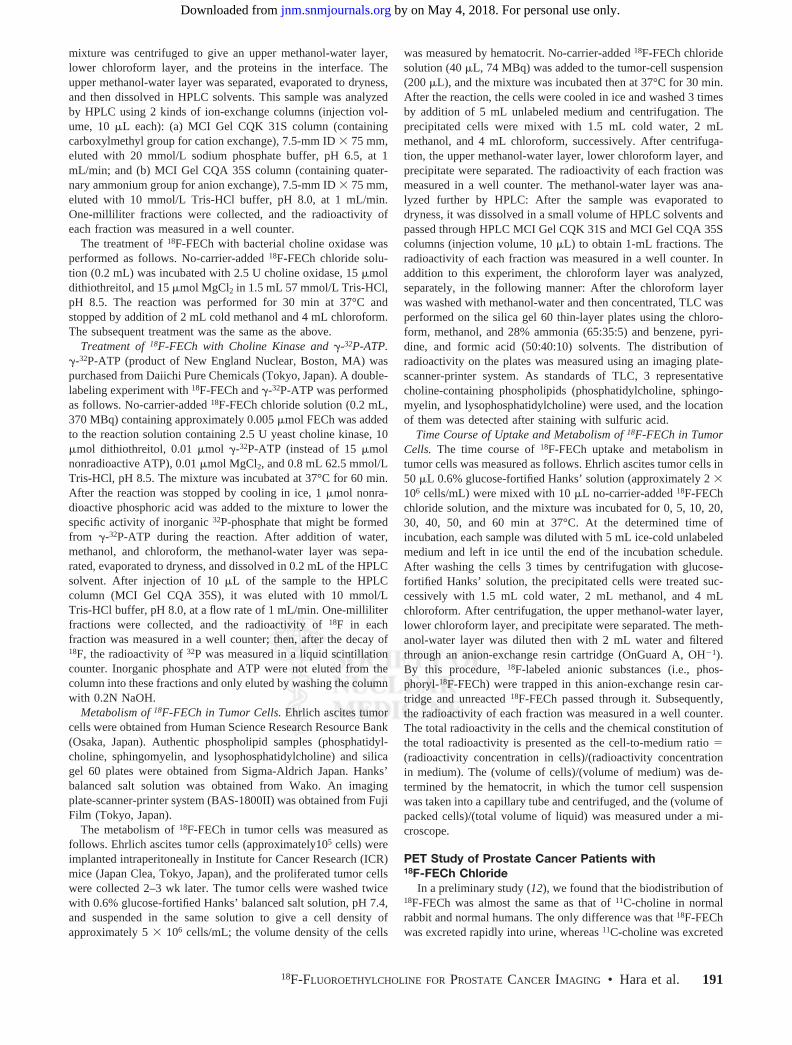
and ATP. No-carrier-added 18F-FECh was incubated withyeast choline kinase, ATP (15 �mol), MgCl2, and dithio-threitol at 37°C for 30 min. The reaction was stopped byaddition of methanol and chloroform, and the methanol-water layer was separated. After concentrating this layer,the sample was analyzed by ion-exchange HPLC usingcation-exchange resin (MCI Gel CQK 31S) and anion-exchange resin (MCI Gel CQA 35S). When HPLC wasperformed with unreacted 18F-FECh, the radioactivitymigrated slowly on the cation-exchange HPLC (retentiontime, 12 min) and migrated fast on the anion-exchangeHPLC (retention time, 3 min) (Fig. 3A). When HPLCwas performed after 18F-FECh was incubated with cho-line kinase and ATP, most radioactivity migrated fast onthe cation-exchange HPLC and migrated slowly on theanion-exchange HPLC, which was opposite to the behav-ior of unreacted 18F-FECh (Fig. 3A). When HPLC wasperformed after 18F-FECh was incubated with cholinekinase, but without ATP, this phenomenon did not occur.These observations seemed to indicate that 18F-FECh wasconverted into phosphoryl-18F-FECh by choline kinaseand ATP.
Formation of Fluoroethylbetaine by Choline Oxidase. Asimilar experiment was conducted using choline oxidase, inwhich choline kinase was replaced by choline oxidase, andATP was omitted. When HPLC was performed, most ra-dioactivity migrated fast on the cation-exchange HPLC and
192 THE JOURNAL OF NUCLEAR MEDICINE • Vol. 43 • No. 2 • February 2002
by on May 4, 2018. For personal use only. jnm.snmjournals.org Downloaded from

FIGURE 3. (A) HPLC after incubation of 18F-FECh with choline kinase and with choline oxidase. No-carrier-added 18F-FECh chloride wasincubated with yeast choline kinase and 15 �mol ATP or with bacterial choline oxidase. From incubated specimen, methanol-water–solublecomponent was separated and fractionated on cation- and anion-exchange HPLC. 18F radioactivity was measured in each fraction. (B) HPLCafter incubation of 18F-FECh with choline kinase and �-32P-ATP. No-carrier-added 18F-FECh chloride (approximately 0.005 �mol) was incubatedwith yeast choline kinase and 0.01 �mol �-32P-ATP. From incubated specimen, methanol-water–soluble component was separated andfractionated on anion-exchange HPLC. 18F and 32P radioactivities were measured in each fraction. kcpm � kilocounts per minute.
18F-FLUOROETHYLCHOLINE FOR PROSTATE CANCER IMAGING • Hara et al. 193
by on May 4, 2018. For personal use only. jnm.snmjournals.org Downloaded from
the anion-exchange HPLC (Fig. 3A). These findings pre-sented a great contrast to the result of the choline kinaseexperiment. This experiment seemed to indicate that 18F-FECh was converted into 18F-fluoroethylbetaine by cholineoxidase.
Formation of 32P-Phosphoryl-18F-FECh by Choline Ki-nase and �-32P-ATP. In the above experiment, it was shownthat 18F-FECh was converted into a new compound by thereaction with choline kinase and ATP and that the newcompound could be considered tentatively as phosphoryl-18F-FECh. In the following experiment, the chemical prop-erty of this new compound was studied more precisely.No-carrier-added 18F-FECh (containing approximately0.005 �mol FECh) was incubated with yeast choline kinase,�-32P-ATP (0.01 �mol), MgCl2, and dithiothreitol. (Theradioactivities of 18F-FECh and �-32P-ATP added were 12.3and 0.088 MBq, respectively.) After the reaction wasstopped, the methanol-water layer was concentrated andanalyzed on the HPLC using anion-exchange resin. Figure3B shows the result of the HPLC. When the radioactivity of18F was measured, 2 components were found: a large com-ponent that migrated fast and a small component that mi-grated slowly. When the radioactivity of 32P was measured,
no radioactivity was in the fast component but there wasdistinct radioactivity in the slow component, and the ratio of32P to 18F in the slow component was even in every fraction.It was evident from this observation that 32P and 18F weretagged by the same molecule. In other words, the newcompound produced by the reaction of 18F-FECh with cho-line kinase and �-32P-ATP was undoubtedly 32P-phos-phoryl-18F-FECh.
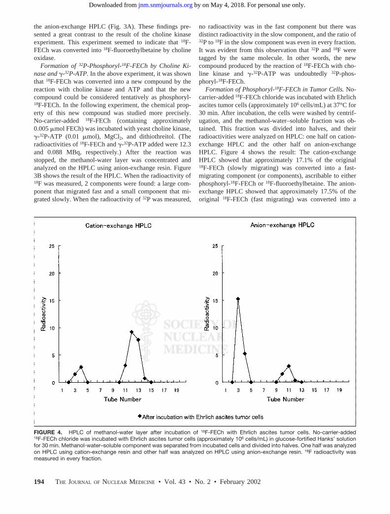
Formation of Phosphoryl-18F-FECh in Tumor Cells. No-carrier-added 18F-FECh chloride was incubated with Ehrlichascites tumor cells (approximately 106 cells/mL) at 37°C for30 min. After incubation, the cells were washed by centrif-ugation, and the methanol-water–soluble fraction was ob-tained. This fraction was divided into halves, and theirradioactivities were analyzed on HPLC: one half on cation-exchange HPLC and the other half on anion-exchangeHPLC. Figure 4 shows the result: The cation-exchangeHPLC showed that approximately 17.1% of the original18F-FECh (slowly migrating) was converted into a fast-migrating component (or components), ascribable to eitherphosphoryl-18F-FECh or 18F-fluoroethylbetaine. The anion-exchange HPLC showed that approximately 17.5% of theoriginal 18F-FECh (fast migrating) was converted into a
FIGURE 4. HPLC of methanol-water layer after incubation of 18F-FECh with Ehrlich ascites tumor cells. No-carrier-added18F-FECh chloride was incubated with Ehrlich ascites tumor cells (approximately 106 cells/mL) in glucose-fortified Hanks’ solutionfor 30 min. Methanol-water–soluble component was separated from incubated cells and divided into halves. One half was analyzedon HPLC using cation-exchange resin and other half was analyzed on HPLC using anion-exchange resin. 18F radioactivity wasmeasured in every fraction.
194 THE JOURNAL OF NUCLEAR MEDICINE • Vol. 43 • No. 2 • February 2002
by on May 4, 2018. For personal use only. jnm.snmjournals.org Downloaded from
slowly migrating component, ascribable only to phos-phoryl-18F-FECh. This observation indicated that the fast-migrating component in the cation-exchange HPLC wastotally ascribable to phosphoryl-18F-FECh. It also indicatedthat 18F-FECh was converted into phosphoryl-18F-FECh, butnot into 18F-fluoroethylbetaine, in this tumor type.
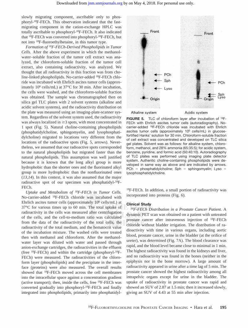
Formation of 18F-FECh-Derived Phospholipids in TumorCells. fter the above experiment in which the methanol-water–soluble fraction of the tumor cell extract was ana-lyzed, the chloroform-soluble fraction of the tumor cellextract, also containing radioactivity, was analyzed. Wethought that all radioactivity in this fraction was from cho-line-linked phospholipids. No-carrier-added 18F-FECh chlo-ride was incubated with Ehrlich ascites tumor cells (approx-imately 106 cells/mL) at 37°C for 30 min. After incubation,the cells were washed, and the chloroform-soluble fractionwas obtained. The sample was chromatographed then onsilica gel TLC plates with 2 solvent systems (alkaline andacidic solvent systems), and the radioactivity distribution onthe plate was measured using an imaging-plate-scanner sys-tem. Regardless of the solvent system used, the radioactivitywas always localized in �3 spots, with most concentrated in1 spot (Fig. 5). Natural choline-containing phospholipids(phosphatidylcholine, sphingomyelin, and lysophosphati-dylcholine) migrated to locations very different from thelocations of the radioactive spots (Fig. 5, arrows). Never-theless, we assumed that our radioactive spots correspondedto the natural phospholipids but migrated faster than thenatural phospholipids. This assumption was well justifiedbecause it is known that the long alkyl group is morehydrophobic than the shorter ones and the fluorinated alkylgroup is more hydrophobic than the nonfluorinated ones(13,14). In this context, it was also assumed that the majorradioactive spot of our specimen was phosphatidyl-18F-FECh.
Uptake and Metabolism of 18F-FECh in Tumor Cells.No-carrier-added 18F-FECh chloride was incubated withEhrlich ascites tumor cells (approximately 106 cells/mL) at37°C for various times up to 60 min. The total uptake ofradioactivity in the cells was measured after centrifugationof the cells, and the cell-to-medium ratio was calculatedfrom the data of the radioactivity of the total cells, theradioactivity of the total medium, and the hematocrit valueof the incubation mixture. The washed cells were treatedthen with methanol and chloroform. After the methanol-water layer was diluted with water and passed throughanion-exchange cartridges, the radioactivities in the effluent(free 18F-FECh) and within the cartridge (phosphoryl-18F-FECh) were measured. The radioactivities of the chloro-form layer (phospholipids) and the precipitate in the inter-face (proteins) were also measured. The overall resultsshowed that 18F-FECh moved across the cell membranesinto the intracellular space against a concentration gradient(active transport); then, inside the cells, free 18F-FECh wasconverted gradually into phosphoryl-18F-FECh and finallyintegrated into phospholipids, primarily into phosphatidyl-
18F-FECh. In addition, a small portion of radioactivity wasincorporated into proteins (Fig. 6).
Clinical Study18F-FECh Distribution in a Prostate Cancer Patient. A
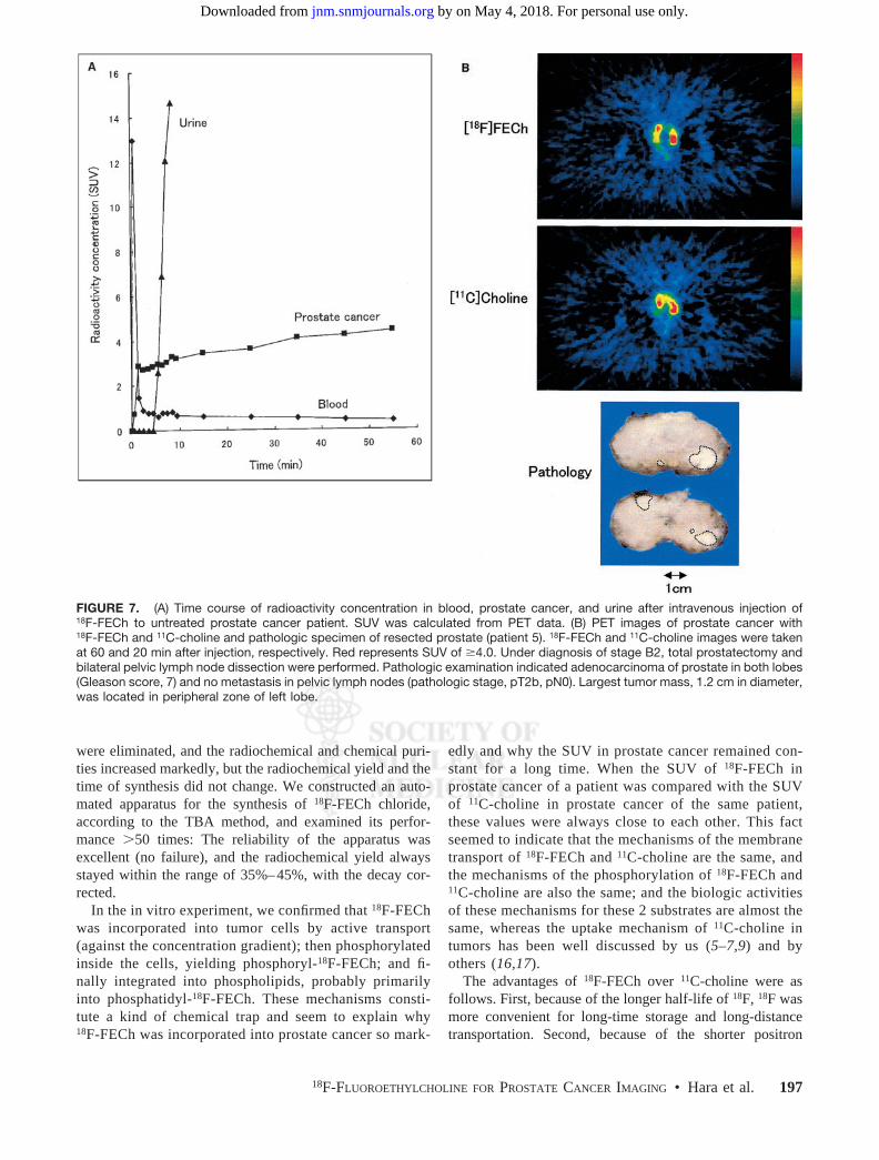
dynamic PET scan was obtained on a patient with untreatedprostate cancer after intravenous injection of 18F-FEChchloride without bladder irrigation. The distribution of ra-dioactivity with time in various organs, including aorticblood, prostate cancer, urine in the bladder (at the orifice ofureter), was determined (Fig. 7A). The blood clearance wasrapid, and the blood level became close to minimal in 1 min.The highest radioactivity was found in the kidneys and liver,and no radioactivity was found in the bones (neither in theepiphysis nor in the bone morrow). A large amount ofradioactivity appeared in urine after a time lag of 5 min. Theprostate cancer showed the highest radioactivity among allintrapelvic organs except for urine in the bladder. Theuptake of radioactivity in prostate cancer was rapid andshowed an SUV of 2.87 at 1.5 min; then it increased slowly,giving an SUV of 4.43 at 55 min after injection.
FIGURE 5. TLC of chloroform layer after incubation of 18F-FECh with Ehrlich ascites tumor cells (autoradiography). No-carrier-added 18F-FECh chloride was incubated with Ehrlichascites tumor cells (approximately 106 cells/mL) in glucose-fortified Hanks’ solution for 30 min. Chloroform-soluble fractionof cell extract was concentrated and developed on TLC silicagel plates. Solvent was as follows: for alkaline system, chloro-form, methanol, and 28% ammonia (65:35:5); for acidic system,benzene, pyridine, and formic acid (50:40:10). Autoradiographyof TLC plates was performed using imaging plate detectorsystem. Authentic choline-containing phospholipids were de-veloped in same way as above and are indicated by arrows.PCh � phosphatidylcholine; Sph � sphingomyelin; Lyso �lysophosphatidylcholine.
18F-FLUOROETHYLCHOLINE FOR PROSTATE CANCER IMAGING • Hara et al. 195
by on May 4, 2018. For personal use only. jnm.snmjournals.org Downloaded from

18F-FECh PET and 11C-Choline PET in Prostate CancerPatients. Static PET scans were obtained on 16 patients withuntreated primary prostate cancer. The radioactivity in urinewas eliminated by continuous bladder irrigation. The radio-activity of prostate cancer expressed by SUV was essen-tially unchanged between 30 and 60 min after injection.Radioactivity appeared sometimes in the intestinal fluid, forwhich the location and intensity changed with time. Thisartifact was easily discriminated from the true uptake ofprostate cancer by comparison of the 30- and 60-min images(our computer program facilitated this discrimination). Ta-ble 1 shows the data of the patients with untreated prostatecancer: number of positive (malignant) samples of biopsyfrom all samples examined (transrectal sonographicallyguided needle biopsy of the prostate), grade of tumor celldifferentiation in the biopsy samples (poorly, moderately, orwell differentiated), level of prostate-specific antigen (PSA)in the blood, and clinical stage (15) estimated from thefindings of biopsy, PET, and other modalities. Table 1 alsoshows the SUVs in the most radioactive area in the regionof the prostate, determined by PET with 18F-FECh and with11C-choline, where the 18F-FECh image was taken at 30 and60 min after injection and the 11C-choline image was takenat 5 and 20 min after injection, respectively. Of all 16patients examined, 1 patient (patient 5, Fig. 7) underwenttotal prostatectomy immediately after the PET study, and allothers were treated thereafter by hormonal therapy as animmediate measure.
With both 18F-FECh and 11C-choline, the prostate cancerwas always visualized as high-uptake areas surrounded bylow-uptake areas of normal prostate, where the high-uptakeand low-uptake areas of the PET image corresponded wellto the malignant and normal findings of the biopsy exami-
nation. With 18F-FECh, the tumor SUV (within an area of8 � 8 mm in real size) ranged from 1.97 to 6.47 (mean SD, 3.84 1.25) at 30 min and from 1.86 to 7.50 (4.02 1.46) at 60 min after injection, respectively. With 11C-choline, the tumor SUV ranged from 1.66 to 7.22 (4.03 1.38) at 5 min and from 1.86 to 7.58 (4.50 1.56) at 20min after injection, respectively. With 18F-FECh and 11C-choline, the SUVs stayed unchanged during the 2 scantimes. In addition, in the area of the cancer, the SUV of18F-FECh was almost the same as the SUV of 11C-choline.
In general, 18F-FECh PET gave slightly better images ofprostate cancer than 11C-choline PET in terms of spatialresolution. This observation is explainable by the shorterpositron range of 18F.
DISCUSSION
We reported, in a preliminary form, the synthesis of18F-FECh (2-fluoroethyl-dimethyl-2-oxyethylammonium)(12), in which we used Kryptofix 2.2.2. (Merck-Schuchardt,Hohenbrunn, Germany) as the synthetic catalyst. In thisstudy, we used TBA bicarbonate instead of Kryptofix 2.2.2.because we were concerned that Kryptofix 2.2.2. mightdeteriorate the purity of 18F-FECh. Our apprehensions werebased on the following facts: First, because the behavior ofKryptofix 2.2.2. in preparative HPLC was similar to that of18F-FECh (Kryptofix 2.2.2. migrated just behind 18F-FECh),it might spill over into the 18F-FECh peak (TBA migratedfar behind the 18F-FECh peak). Second, because Kryptofix2.2.2. contains 2 tertiary amine moieties, they might reactwith 2-18F-fluoroethyl tosylate to produce undesirable by-products. Using TBA bicarbonate, these apprehensions
FIGURE 6. Time course of uptake andmetabolism of 18F-FECh in Ehrlich ascitestumor cells. No-carrier-added 18F-FEChchloride was incubated with Ehrlich as-cites tumor cells (approximately 106 cells/mL) in glucose-fortified Hanks’ solutionfor various periods up to 60 min. Total 18Fuptake was measured after washing cellsby centrifugation, and cell-to-medium ra-tio was calculated after hematocrit deter-mination. After treatment of cells withmethanol and chloroform, radioactivitiesin free 18F-FECh, phosphoryl-18F-FECh,18F-labeled phospholipids (phosphatidyl-18F-FECh and so forth), and proteins weremeasured.
196 THE JOURNAL OF NUCLEAR MEDICINE • Vol. 43 • No. 2 • February 2002
by on May 4, 2018. For personal use only. jnm.snmjournals.org Downloaded from
were eliminated, and the radiochemical and chemical puri-ties increased markedly, but the radiochemical yield and thetime of synthesis did not change. We constructed an auto-mated apparatus for the synthesis of 18F-FECh chloride,according to the TBA method, and examined its perfor-mance �50 times: The reliability of the apparatus wasexcellent (no failure), and the radiochemical yield alwaysstayed within the range of 35%–45%, with the decay cor-rected.
In the in vitro experiment, we confirmed that 18F-FEChwas incorporated into tumor cells by active transport(against the concentration gradient); then phosphorylatedinside the cells, yielding phosphoryl-18F-FECh; and fi-nally integrated into phospholipids, probably primarilyinto phosphatidyl-18F-FECh. These mechanisms consti-tute a kind of chemical trap and seem to explain why18F-FECh was incorporated into prostate cancer so mark-
edly and why the SUV in prostate cancer remained con-stant for a long time. When the SUV of 18F-FECh inprostate cancer of a patient was compared with the SUVof 11C-choline in prostate cancer of the same patient,these values were always close to each other. This factseemed to indicate that the mechanisms of the membranetransport of 18F-FECh and 11C-choline are the same, andthe mechanisms of the phosphorylation of 18F-FECh and11C-choline are also the same; and the biologic activitiesof these mechanisms for these 2 substrates are almost thesame, whereas the uptake mechanism of 11C-choline intumors has been well discussed by us (5–7,9) and byothers (16,17).
The advantages of 18F-FECh over 11C-choline were asfollows. First, because of the longer half-life of 18F, 18F wasmore convenient for long-time storage and long-distancetransportation. Second, because of the shorter positron
FIGURE 7. (A) Time course of radioactivity concentration in blood, prostate cancer, and urine after intravenous injection of18F-FECh to untreated prostate cancer patient. SUV was calculated from PET data. (B) PET images of prostate cancer with18F-FECh and 11C-choline and pathologic specimen of resected prostate (patient 5). 18F-FECh and 11C-choline images were takenat 60 and 20 min after injection, respectively. Red represents SUV of �4.0. Under diagnosis of stage B2, total prostatectomy andbilateral pelvic lymph node dissection were performed. Pathologic examination indicated adenocarcinoma of prostate in both lobes(Gleason score, 7) and no metastasis in pelvic lymph nodes (pathologic stage, pT2b, pN0). Largest tumor mass, 1.2 cm in diameter,was located in peripheral zone of left lobe.
18F-FLUOROETHYLCHOLINE FOR PROSTATE CANCER IMAGING • Hara et al. 197
by on May 4, 2018. For personal use only. jnm.snmjournals.org Downloaded from

range of 18F, 18F gave a slightly higher quality of image withhigher spatial resolution.
The disadvantage of 18F-FECh was the rapid excretion ofradioactivity into urine (in contrast to 11C-choline), and itwas necessary to irrigate the bladder continuously using aurinary catheter to eliminate the bladder radioactivity. How-ever, this procedure was very uncomfortable for urologypatients.
DeGrado et al. (18) synthesized 18F-fluoromethylcholineand observed its biodistribution in mice with prostate cancerxenografts. They reported a high uptake of this compound intumors, and a high radioactivity in urine that was 10 timeshigher than that of 11C-choline at 30 min after injection.
Recently, DeGrado et al. (19) reported on their success inobtaining clear PET images of prostate cancer in patients inwhom 18F-fluoromethylcholine was used instead of 18F-FECh, the bladder irrigation was avoided, and the scanningwas conducted at 3–5 min after injection (before the emer-gence of radioactivity in urine). We also obtained a clearPET image of prostate cancer in 1 patient (Fig. 7A) using18F-FECh, without bladder irrigation, and conducting thescanning at 2–5 min after injection. We did not adopt thisprotocol in the rest of this study because we wanted toexamine the whole area of pelvis in the patients (our PETmachine covers only 6.5 cm longitudinally at 1 bed posi-tion).
CONCLUSION
It is established that 11C-choline PET is very effectivein detecting various cancers, including prostate cancer.
We developed a method to synthesize 18F-FECh as asubstitute for 11C-choline and constructed an automatedapparatus for the synthesis. In addition, we studied thetumor uptake of 18F-FECh in Ehrlich tumor cells in vitroand performed 18F-FECh PET on 16 patients with un-treated primary prostate cancer. Our method of synthesiswas easy and reliable, and the performance of our auto-mated apparatus was excellent. In the in vitro experiment,18F-FECh was incorporated into the tumor cells by activetransport, then phosphorylated within the cells, and fi-nally integrated into phospholipids, constituting a chem-ical trap mechanism in the tumor cells. In the clinicalstudy, 18F-FECh PET visualized prostate cancer of thepatients at the same uptake rate (SUV) as that observedby 11C-choline PET. However, 18F-FECh PET wasslightly superior to 11C-choline PET in the sharpness ofthe image. The only disadvantage of 18F-FECh PET com-pared with 11C-choline PET was the need to introduce aurinary catheter into the bladder for continuous with-drawal of urine during the time of PET scanning.
ACKNOWLEDGMENT
This work was supported in part by the Ministry ofEducation, Culture, Sports, Science and Technology of Ja-pan and the Japanese Smoking Research Foundation.
REFERENCES
1. Negendank W. Studies of human tumors by MRS: a review. NMR Biomed.1992;5:303–324.
TABLE 1Patients with Primary Prostate Cancer
Patientno.*
Age(y)
PSA(ng/mL)
Positivebiopsy Histology
Clinicalstage
SUV in most radioactive area18F-FECh 11C-Choline
30min
60min
5min
20min
1 67 4.6 1/6 Mod. diff. B1 2.84 3.25 3.43 4.752 71 5.5 3/6 Well-diff. B1 1.97 1.86 1.66 1.863 75 6.6 3/6 Well-diff. B1 2.78 2.83 2.85 2.754 63 7.7 2/6 Well-diff. B1 2.50 2.90 2.48 2.485 62 8.4 4/6 Mod. diff. B2 4.08 4.25 4.37 4.076 70 19.5 3/7 Well-diff. B1 5.39 5.14 4.79 5.437 78 19.6 3/7 Mod. diff. D2 4.25 4.36 3.10 4.908 77 36.0 7/7 Poor diff. D1 3.12 3.27 3.93 4.679 75 41.8 3/6 Poor diff. B1 2.71 3.30 3.28 3.28
10 68 45.7 5/6 Poor diff. D2 3.63 3.97 4.71 5.2111 72 45.9 3/6 Mod. diff. B1 3.43 2.55 4.56 4.5112 72 51.3 6/6 Poor diff. D2 5.51 6.46 7.22 7.5813 64 80.0 6/6 Mod. diff. C 3.42 3.35 3.88 4.0814 78 104.0 5/6 Poor diff. D2 4.41 4.66 3.17 3.5715 68 126.3 6/6 Mod. diff. D2 4.88 4.70 4.94 5.7716 82 242.6 4/4 Mod. diff. D2 6.47 7.50 6.04 7.11
*Patient 5 underwent total prostatectomy and all other patients received hormonal therapy after PET study.Mod. � moderately; diff. � differentiated; poor � poorly.
198 THE JOURNAL OF NUCLEAR MEDICINE • Vol. 43 • No. 2 • February 2002
by on May 4, 2018. For personal use only. jnm.snmjournals.org Downloaded from

2. De Certaines JD, Larsen VA, Podo F, et al. In vivo 31P MRS of experimentaltumors: a review. NMR Biomed. 1993;6:345–365.
3. Katz-Brull R, Degani H. Kinetics of choline transport and phosphorylation inhuman breast cancer cells: NMR application of the zero trans method. AnticancerRes. 1996;16:1375–1380.
4. Haeffner EW. Studies on choline permeation through the plasma membrane andits incorporation into phosphatidyl choline of Ehrlich-Lettre-ascites tumor cells invitro. Eur J Biochem. 1975;51:219–228.
5. Hara T, Kosaka N, Shinoura N, Kondo T. PET imaging of brain tumor with[methyl-11C]choline. J Nucl Med. 1997;38:842–847.
6. Hara T, Inagaki K, Kosaka N, Morita T. Sensitive detection of mediastinal lymphnode metastasis of lung cancer with 11C-choline PET. J Nucl Med. 2000;41:1507–1513.
7. Kobori O, Kirihara Y, Kosaka N, Hara T. Positron emission tomography ofesophageal carcinoma using 11C-choline and 18F-fluorodeoxyglucose. Cancer.1999;86:1638–1648.
8. Hara T, Kosaka N, Kondo T, Kishi H, Kobori O. Imaging of brain tumor, lungcancer, esophagus cancer, colon cancer, prostate cancer, and bladder cancer with[C-11]choline [abstract]. J Nucl Med. 1997;38(suppl):250P.
9. Hara T, Kosaka N, Kishi H. PET imaging of prostate cancer using carbon-11-choline. J Nucl Med. 1998;39:990–995.
10. Deves R, Krupka RM. The binding and translocation steps in transport as relatedto substrate structure: a study of the choline carrier of erythrocytes. BiochimBiophys Acta. 1979;557:469–485.
11. Clary GL, Tsai CF, Guynn RW. Substrate specificity of choline kinase. ArchBiochem Biophys. 1987;254:214–221.
12. Hara T, Yuasa M. Automated synthesis of fluorine-18 labeled choline analogue:2-fluoroethyl-dimethyl-2-oxyethylammonium [ abstract ]. J Nucl Med. 1997;38(suppl):44P.
13. Greiner J, Riess JG, Vierling P. Fluorinated surfactants intended for biomedicaluses. In: Filler R, Kobayashi Y, Yagupolskii LM, eds. Organofluorine Com-pounds in Medicinal Chemistry and Biomedical Applications. Amsterdam, TheNetherlands: Elsevier; 1993:339.
14. Santaella C, Vierling P, Riess JG, Gulik-Krzywicki T, Gulik A, Monasse B.Polymorphic phase behavior of perfluoroalkylated phosphatidylcholines. BiochimBiophys Acta. 1994;1190:25–39.
15. Van de Voorde. Pathology of prostatic carcinoma. In: Petrovich Z, Baert L, BradyLW, eds. Carcinoma of the Prostate: Innovations in Management. Berlin, Ger-many: Springer; 1996:27.
16. Pascali C, Bogni A, Iwata R, Cambre M, Bombardieri E. [11C]Methylation on aC18 Sep-Pak cartridge: a convenient way to produce [N-methyl-11C]choline.J Labelled Compd Radiopharm. 2000;43:195–203.
17. Kotzerke J, Prang J, Neumeier B, et al. Experience with carbon-11 cholinepositron emission tomography in prostate carcinoma. Eur J Nucl Med. 2000;27:1415–1419.
18. DeGrado TR, Coleman RE, Baldwin SW, Orr MD, Robertson CN, Price DT.[18F]Fluorocholine (FCH) as an oncologic PET tracer: evaluation in murineprostate cancer xenograft model [abstract]. J Nucl Med. 2000;41(suppl):231P.
19. DeGrado TR, Coleman RE, Wang S, et al. Synthesis and evaluation of 18F-labeled choline as an oncologic tracer for positron emission tomography: initialfindings in prostate cancer. Cancer Res. 2001;61:110–117.
18F-FLUOROETHYLCHOLINE FOR PROSTATE CANCER IMAGING • Hara et al. 199
by on May 4, 2018. For personal use only. jnm.snmjournals.org Downloaded from

2002;43:187-199.J Nucl Med. Toshihiko Hara, Noboru Kosaka and Hiroichi Kishi Biochemistry, and Prostate Cancer Imaging
F-Fluoroethylcholine for Cancer Imaging with PET: Synthesis,18Development of
http://jnm.snmjournals.org/content/43/2/187This article and updated information are available at:
http://jnm.snmjournals.org/site/subscriptions/online.xhtml
Information about subscriptions to JNM can be found at:
http://jnm.snmjournals.org/site/misc/permission.xhtmlInformation about reproducing figures, tables, or other portions of this article can be found online at:
(Print ISSN: 0161-5505, Online ISSN: 2159-662X)1850 Samuel Morse Drive, Reston, VA 20190.SNMMI | Society of Nuclear Medicine and Molecular Imaging
is published monthly.The Journal of Nuclear Medicine
© Copyright 2002 SNMMI; all rights reserved.
by on May 4, 2018. For personal use only. jnm.snmjournals.org Downloaded from