development of breast
DESCRIPTION
Development of breast. Learning Objectives: At the end of lecture the student should be able to: Define mammogenesis Describe the stages of development. Clinical correlations of developmental anatomy of mammary gland. - PowerPoint PPT PresentationTRANSCRIPT

Development of breast

• Learning Objectives:
• At the end of lecture the student should be able to:
• Define mammogenesis
• Describe the stages of development.
• Clinical correlations of developmental anatomy of mammary gland
• Mammogenesis is the term use to describe the development of the mammary gland.
• The mammary gland is one of a few tissues in mammals, which can repeatedly undergo growth, functional differentiation, and regression.

Development of Mammary Glands
• Are a modified and highly specialized type of apocrine sweat glands.
• Consist of parenchyma, which is formed from ducts, and connective tissue stroma.
• Parenchyma derives embryonically from surface ectoderm; stroma arises from surrounding mesenchyme.
• Mammary buds begin to develop during the sixth week as solid downgrowths of the epidermis into the underlying mesenchyme
• These changes occur in response to an inductive influence from the mesenchyme


• Mammary buds develop as downgrowths from thickened mammary crests, which are thickened strips of ectoderm extending from the axillary to the inguinal regions
• The mammary crests (ridges) appear during the fourth week but normally persist in humans only in the pectoral area, where the breasts develop

• Each primary bud gives rise to several secondary mammary buds that develop into lactiferous ducts and their branches
• Canalization of these buds is induced by placental sex hormones entering the fetal circulation. This process continues until late gestation, and by term, 15 to 19 lactiferous ducts are formed.
• The fibrous connective tissue and fat of the mammary gland develop from the surrounding mesenchyme.
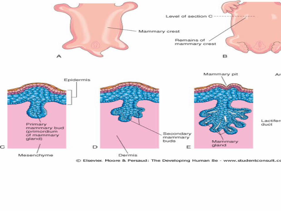

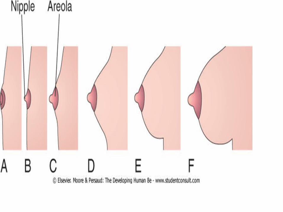
• Development of Nipples and Areola• During the late fetal period, the epidermis at the site of
origin of the mammary gland becomes depressed, forming a shallow mammary pit
• The nipples are poorly formed and depressed in newborn infants.
• Soon after birth, the nipples usually rise from the mammary pits because of proliferation of the surrounding connective tissue of the areola, the circular area of skin around the nipple.
• The smooth muscle fibers of the nipple and areola differentiate from surrounding mesenchymal cells.


• Postnatal Development
• The rudimentary mammary glands of newborn males and females are identical and are often enlarged.
• Some secretion, often called "witch's milk," may be produced caused by maternal hormones passing through the placental membrane into the fetal circulation.
• Newborns breasts contain lactiferous ducts but no alveoli. Before puberty, there is little branching of the ducts.
• In females, the breasts enlarge rapidly during puberty, mainly because of development of the mammary glands and the accumulation of the fibrous stroma and fat associated with them
• Full development occurs at approximately 19-20 years The lactiferous ducts of male breasts remain rudimentary throughout life.
Mammary glands Histogenesis
• Compound tubuloalveolar glands • Consist of 15 to 20 lobes radiating out from the
nipple and are • Separated from each other by adipose and
collagenous connective tissue. • Secrete milk, a fluid containing proteins, lipids,
and lactose as well as lymphocytes and monocytes, antibodies, minerals, and fat-soluble vitamins
• Provide the proper nourishment for the newborn.


• Develop in the same manner and are of the same structure in both sexes until puberty,
• At puberty changes in the hormonal secretions in females cause further development and structural changes within the glands.
• Secretions of estrogen and progesterone from the ovaries (and later from the placenta) and prolactin from the acidophils of the anterior pituitary gland initiate development of lobules and terminal ductules.
• Full development of the ductal portion of the breast requires glucocorticoids and further activation by somatotropin.
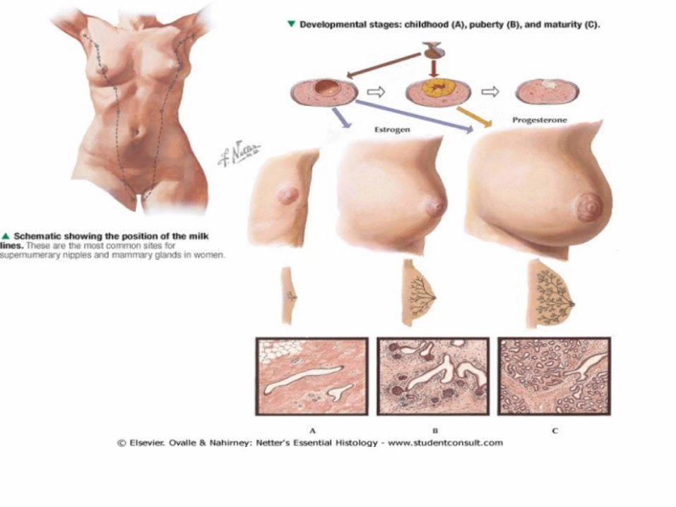

• Concomittant with these events is an increase in connective tissue and adipose tissue within the stroma, causing the gland to enlarge.
• Full development occurs at about 20 years of age
• Minor cyclic changes occur during each menstrual period,
• Major changes occur during pregnancy and in lactation.

• After age 40 or so, the secretory portions and some of the ducts and connective tissue elements of the breasts begin to atrophy, and they continue this process throughout menopause.
• Gynecomastia• The rudimentary lactiferous ducts in males normally undergo no
postnatal development. • Gynecomastia (Gr. gyne, woman + mastos, breast) refers to the
development of the rudimentary lactiferous ducts in the male mammary tissue.
• During midpuberty, approximately two thirds of boys develop varying degrees of hyperplasia of the breasts. This subareolar hyperplasia may persist for a few months to 2 years.
• A decreased ratio of testosterone to estradiol is found • 80% of males with Klinefelter syndrome (XXY) have
gynecomastia.•

• Absence of Nipples (Athelia) or Breasts (Amastia)
• Rare congenital anomalies may occur bilaterally or unilaterally.
• Result from failure of development or disappearance of the mammary crests.
• May also result from failure of mammary buds to form

• More common is hypoplasia of the breast, often found in association with gonadal agenesis and Turner syndrome .


Aplasia of Breast
• The breasts of a postpubertal female often differ in size. Marked differences are regarded as anomalies because both glands are exposed to the same hormones at puberty.
• In these cases, there is often associated rudimentary development of muscles of the thoracic wall, usually the pectoralis major
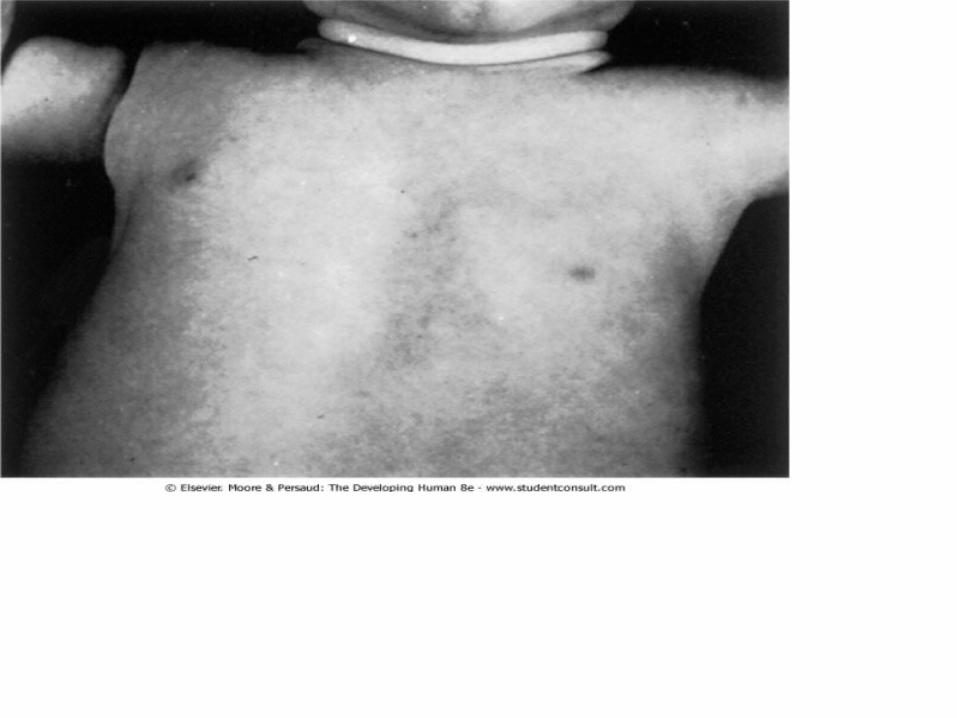

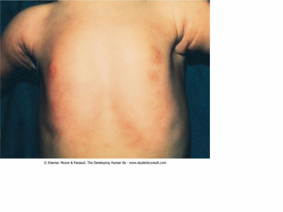
• Supernumerary Breasts and Nipples• An extra breast (polymastia) or nipple (polythelia)
occurs in approximately 1% of the female population as an inheritable condition.
• An extra breast or nipple usually develops just inferior to the normal breast.
• Supernumerary nipples are also relatively common in males; often they are mistaken for moles
• Less commonly, supernumerary breasts or nipples appear in the axillary or abdominal regions of females developing from extra mammary buds that develop along the mammary crests. They become more obvious in women when pregnancy occurs.

• Approximately one third of affected persons have two extra nipples or breasts.
• Supernumerary mammary tissue very rarely occurs in a location other than along the course of the mammary crests. It probably develops from tissue that was displaced from these crests.

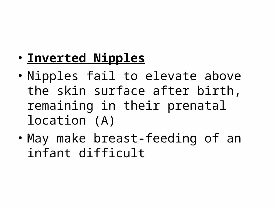
• Inverted Nipples
• Nipples fail to elevate above the skin surface after birth, remaining in their prenatal location (A)
• May make breast-feeding of an infant difficult