development of bioactive bread enriched with a seaweed
TRANSCRIPT

1
Development of bioactive bread enriched with a seaweed
peptide fraction with potential heart-health effects
Mr. Ciarán Pádraig Fitzgerald
A THESIS SUBMITTED IN FULFILMENT OF THE REQUIREMENTS FOR THE
DEGREE OF DOCTOR OF PHILOSOPHY
School of Pharmacy
University College London

2
THIS THESIS DESCRIBES RESEARCH CONDUCTED AT TEAGASC IRELAND IN
CONJUNCTION WITH THE SCHOOL OF PHARMACY, UNIVERSITY OF LONDON
BETWEEN OCTOBER 2009 AND JANUARY 2014 UNDER THE DIRECT
SUPERVISION OF DR MARIA HAYES AND EIMEAR GLALLAGHER WITH DR
DENIZ TASDEMIR AND DR JOSE PRIETO AS ACADEMIC SUPERVISORS. I
CERTIFY THAT THE RESEARCH DESCRIBED IS ORIGINAL AND THAT ANY
PARTS OF THE WORK THAT HAVE BEEN CONDUCTED BY COLLABORATION
ARE CLEARLY INDICATED. I ALSO CERTIFY THAT I HAVE WRITTEN THE TEXT
HEREIN AND HAVE CLEARLY INDICATED BY SUITABLE CITATION ANY PART
OF THIS DISSERTATION THAT HAS ALREADY APPEARED IN PUBLICATION.
Signature _______________________ Date___________________

3
ACKNOWLEDGEMENTS
First and foremost I would like to thank my supervisor Dr Maria Hayes for giving me the
opportunity to carry out this Ph.D and without whose constant guidance and encouragement
this work would have been impossible.
I would like to thank my supervisor Dr Eimear Gallagher for her advice and patience in
teaching me how to create and assess baked products for this project.
Thanks also to my academic supervisors Dr Deniz Tasdemir and subsequently Dr Jose Prieto
in London for their support.
I would like to thank Prof Maura Grealy of NUI Galway for her assistance and generosity in
allowing me access to her lab to carry out the Zebrafish larvae assays in this thesis.
I would like to thank Dr Rotimi Aluko of the University of Manitoba for his assistance in
carrying out the in vivo sections of this thesis.
I would like to thank the staff and students of the Nutraceutical building for their friendship
and “interesting” lunch time conversations during my time in Ashtown.
Thanks to all my friends in Castlegregory and Stradbally for their comradery and always
keeping me grounded during my visits home over the last three years.
Last but not least, a massive thank you to my parents Tom and Margaret, my grandmother
Bridge, my brother Chris and also Eveleen for their constant patience and support during my
studies over the years, I couldn’t have done it without you all.

4
TABLE OF CONTENTS
General aims..………………………………………………………………..….……...21
CHAPTER 1. General Introduction…………………………………..……………22
1.1 Aims .................................................................................................................23
1.2 Functional foods and health .............................................................................24
1.3 Bioactive compounds in marine macroalgae....................................................26
1.4 Red algae as an alternative source of protein ...................................................28
1.5 Epidemiology of hypertension and Cardiovascular disease (CVD).................29
1.6 Protease enzymes as targets for the prevention of hypertension......................31
1.6.1 Aspartic proteases .....................................................................................31
1.6.2 ACE-I inhibitors........................................................................................32
1.6.3 The enzyme renin and its mechanismof action in prevention of
hypertension.............................................................................................................34
1.6.4 Types of inhibitors in the RAAS...............................................................36
1.6.5 Development of renin inhibitors ...............................................................37
1.6.6 Renin inhibitory compounds and peptides from plant sources .................38
1.6.7 The RAAS: a potential therapeutic target in the treatment of diabetic
kidney disease and other disease treatments............................................................38
1.6.8 Platelet-activating factor acetylhydrolase (PAF-AH) and heart health.....40
1.6.9 Darapladib and the development of commercial PAF-AH inhibitors.......40
1.7 Bioactive peptides previously isolated from macroalgae.................................42
1.8 Effects of food processing on bioactive peptides .............................................45
1.9 Bioavailability of bioactive peptides and survival in the gastrointestinal tract
(GI tract). .....................................................................................................................47
1.10 Suitable food vehicles for delivery of bioactive peptides ................................49

5
1.10.1 Bread as a carrier for bioactive compounds..............................................52
1.11 Final remarks ....................................................................................................53
CHAPTER 2. Isolation and characterisation of bioactive pro-peptides with in vitro
renin inhibitory activities from the macroalga Palmaria palmata (Linnaeus) Weber and
Mohr ...…………………………………………………………………………57
2.1 Aims .................................................................................................................58
2.2 Introduction ......................................................................................................59
2.3 Materials and Methods .....................................................................................61
2.3.1 Materials and Reagents .............................................................................61
2.3.2 Extraction of Palmaria palmata (Linnaeus) Weber and Mohr protein.....61
2.3.3 Enzymatic Hydrolysis of the Palmaria palmata (Linnaeus) Weber and
Mohr Protein Extract.................................................................................63
2.3.4 Protein quantification and amino acid profiling .......................................63
2.3.5 Fractionation of the seaweed hydrolysate using RP-HPLC......................64
2.3.6 In vitro renin inhibitory assay ...................................................................64
2.3.7 Peptide identification by tandem mass spectrometry................................67
2.3.8 Database Search, confirmation of sequences, and de novo sequencing of
peptides.. ...................................................................................................68
2.3.9 Microwave-assisted solid phase peptide synthesis ...................................68
2.3.10 In silico prediction of peptide availability ................................................69
2.4 Results ..............................................................................................................70
2.4.1 Protein quantification ................................................................................70
2.4.2 RP-HPLC enrichment of the seaweed hydrolysate...................................72
2.4.3 In vitro inhibitory effects of P. palmaria papain hydrolysate on renin ....74

6
2.4.4 Identification of renin inhibitory peptides from P. palmata protein
hydrolysate ................................................................................................76
2.4.5 In silico analysis of the tridecopeptide IRLIIVLMPILMA ......................79
2.5 Discussion ........................................................................................................80
CHAPTER 3. Development of a seaweed derived platelet activating factor
acetylhydrolase (PAF-AH) inhibitory hydrolysate, synthesis of inhibitory peptides and
assessment of their toxicity using the Zebrafish larvae assay….………………………84
3.1 Aims .................................................................................................................85
3.2 Introduction ......................................................................................................87
3.3 Materials and Methods .....................................................................................89
3.3.1 Materials and Reagents .............................................................................89
3.3.2 Palmaria palmata protein extraction and hydrolysis with Papain............89
3.3.3 PAF-AH inhibitor screening assay............................................................89
3.3.4 Fractionation of Palmaria palmata hydrolysate using RP-HPLC ............92
3.3.5 Peptide identification by tandem mass spectrometry................................92
3.3.6 Database search, confirmation of sequences, and de novo sequencing ....92
3.3.7 Microwave-assisted solid phase peptide synthesis ...................................92
3.3.8 Zebrafish larvae assay...............................................................................92
3.4 Results ..............................................................................................................94
3.4.1 PAF-AH inhibitory activity of P. palmata hydrolysates and fractions.....94
3.4.2 PAF-AH inhibition of synthesised peptides..............................................96
3.4.3 Toxicity of P. palmata hydrolysate on Zebrafish larvae...........................98
3.5 Discussion ......................................................................................................101

7
CHAPTER 4. Identification of an active seaweed derived renin inhibitory dipeptide
and confirmation of a hypotensive effect in spontaneously hypertensive rats following
simulated gastrointestinal digestion……………………………………………….…..105
4.1 Aims ...............................................................................................................106
4.2 Introduction ....................................................................................................107
4.3 Materials and Methods ...................................................................................109
4.3.1 Materials..................................................................................................109
4.3.2 In silico analysis of the renin inhibitory peptide IRLIIVLMPILMA .....109
4.3.3 Simulated Gastric Digestion of the peptide IRLIIVLMPILMA .............109
4.3.4 Removal of polyethylene glycols from Palmaria palmata protein digested
samples using a titanium dioxide (TiO2) cleanup procedure. ................................112
4.3.5 Tandem mass spectrometry analysis of the hydrolysate .........................113
4.3.6 In vivo determination of the hypotensive effect of the P. palmata protein
hydrolysate and the tridecapeptide IRLIIVLMPILMA in spontaneously
hypertensive rats (SHRs).........................................................................113
4.3.7 Statistics ..................................................................................................114
4.4 Results ............................................................................................................115
4.4.1 In silico analysis of IRLIIVLMPILMA ..................................................115
4.4.2 ESI-Q-TOF analysis of the in vitro GI simulated digestion of
IRLIIVLMPILMA.................................................................................................115
4.4.3 Short term hypotensive effects of P. palmata protein hydrolysate and
IRLIIVLMPILMA in SHR .....................................................................117
4.5 Discussion ......................................................................................................119
CHAPTER 5. Increasing the health benefits of bread: Assessment of the physical
and sensory qualities of bread formulated using a renin inhibitory Palmaria palmata
protein hydrolysate.………………………………………………………………..….122
5.1 Aim.................................................................................................................123

8
5.2 Introduction ....................................................................................................124
5.3 Materials and Methods ...................................................................................126
5.3.1 Palmaria palmata protein extraction and hydrolysis with Papain..........126
5.3.2 Bread ingredients ....................................................................................126
5.3.3 Preparation of breads...............................................................................126
5.3.4 Bread evaluation – loaf volume, weight and loaf specific volume
calculations..............................................................................................128
5.3.5 Heavy metal and Iodine analysis of seaweed bread product...................129
5.3.6 Sensory analysis ......................................................................................129
5.3.7 Renin inhibitory assay.............................................................................129
5.3.8 Microscopy..............................................................................................129
5.3.9 Statistics ..................................................................................................131
5.4 Results ............................................................................................................132
5.4.1 Loaf volume and crust/crumb colour ......................................................132
5.4.2 Digital image analysis of bread crumb ...................................................133
5.4.3 Texture profile analysis (TPA) of bread crumb ......................................134
5.4.4 Brightfield light microscopy of bread samples .......................................136
5.4.5 Confocal scanning laser microscopy of bread samples...........................137
5.4.6 Heavy metal and Iodine analysis of the bread product ...........................140
5.4.7 Sensory analysis of breads ......................................................................140
5.4.8 Renin inhibitory activity of bread samples .............................................141
5.5 Discussion ......................................................................................................143
CHAPTER 6. General discussion and conclusion………………………………..148

9
TABLE OF FIGURES
Chapter 1
Figure 1.1: The Renin Angiotensin Aldosterone System................................................30
Figure 1.2: The structure of renin.. .................................................................................35
Figure 1.3: Sites of inhibition of the RAAS....................................................................36
Figure 1.4: Expansion of the necrotic core due to PAF-AH activity. .............................42
Figure 1.5: Intestinal transport mechanisms for peptides. ..............................................47
Chapter 2
Figure 2.1: Schematic representation of the bioassay guided isolation and
characterisation ...............................................................................................................62
Figure 2.2: Renin inhiition assay. ...................................................................................66
Figure 2.3: ESI-Q-TOF system for identifying renin inhibitory peptides. .....................67
Figure 2.4: RP-HPLC chromatogram of a papain hydrolysate of P. palmata protein....73
Figure 2.5: Renin inhibitory assay. .................................................................................75
Chapter 3
Figure 3.1: The PAF-AH inhibition assay. .....................................................................91
Figure 3.2: PAF-AH inhibitory activities of seven, chemically synthesized peptides
isolated from Palmaria palmata protein. ........................................................................97
Figure 3.3: Survival of zebrafish larvae over 48 hrs.......................................................99
Figure 3.4: Images of the larvae after 24 h exposure (A) and 48 h exposure (B).........100
Chapter 4
Figure 4.1: Schematic of simulated in vitro gastric digestion.......................................111
Figure 4.2: UPLC chromatograms of the hydrolysed analytes. ....................................116
Figure 4.3: Nano-ESI-MS spectra data of the hydrolysed peptide ...............................117
Figure 4.4: SBP of SHRs over 24hrs ............................................................................118
Chapter 5
Figure 5.1: Images of all four bread samples examined. ..............................................127
Figure 5.2: Texture profile analysis and moisture content............................................135
Figure 5.3: Light micrograph images of bread samples................................................137
Figure 5.4: Confocal micrographs of bread samples ....................................................139
Figure 5.5: Sensory analysis. ........................................................................................141
Figure 5.6: Renin inhibitory assay of breads. ...............................................................142

10
Annex
Figure 8.1: Purification of IRLIIVLMPILMA..............................................................184
Figure 8.2: Purification of NIGK.. ................................................................................185
Figure 8.3: Non linear regression of IRLIIVLMPILMA concentration versus percentage
inhibition. ......................................................................................................................186
Figure 8.4: Non linear regression of NIGK ..................................................................187
Figure 8.5: PAF-AH assay of HPLC fractions 10-25 ...................................................188

11
TABLE OF TABLES
Chapter 1
Table 1.1: Bioactive peptide containing products available commercially ....................51
Chapter 2
Table 2.1: Amino acid composition of protein extracted from Palmaria palmata .......71
Table 2.2: Peptides identified in RP-HPLC Fr-25 enriched from the P. palmata papain
hydrolysate. .....................................................................................................................78
Table 2.3: Cleavage of the peptide IRLIIVLMPILMA ..................................................79
Chapter 3
Table 3.1: Peptides identified by tandem mass...............................................................95
Chapter 4
Table 4.1: Concentrations of electrolytes used to create the simulated saliva fluid (SSF)
simulated gastric fluid (SGF) and simulated duodenal fluid (SDF). ............................110
Table 4.2: Cleavage points of the peptide IRLIIVLMPILMA .....................................115
Chapter 5
Table 5.1: The bread formulations used in this study ...................................................128
Table 5.2: Volume and colour analysis of the control ..................................................132
Table 5.3: Image analysis results ..................................................................................133
12
ABBREVIATIONS
∆ SBP – change in systolic blood
pressure
a* – redness/greenness
ACE-I – angiotensin converting enzyme
I
AChe – acetylcholinesterase
APP – amyloid precursor protein
ARA – aldosterone-receptor antagonists
ARB – angiotenisn receptor blocker
ATP – adenosine triphosphate
B – buckwheat bread
b* – brightness
BACE1 – β-secretase
BCA – bicinchoninic acid
BH – buckwheat bread containing 4%
P. palmata hydrolysate
BMI – body mass index
BPM – beats per minute
BSA – bovine serum albumin
C – control wheat bread
CH – control wheat bread containing
4% P. palmata hydrolysate
COPD – chronic obstructive pulmonary
disease
CVD – cardio-vascular disease
DBP – diastolic blood pressure
DDA – data dependent acquisition
DHA – docosahexaenoic acid
DMSO – dimethyl sulfoxide
DRI – direct renin inhibitor
DTNB – 5,5’ Dithio-bis (2-nitrobenzoic
acid)
EC – enzyme commission number
EDANS – 5-(2-
aminoethylamino)naphthalene-1-
sulfonic acid
EPA – eicosapentaenoic acid
ESI-Q-TOF MS – electro spray
ionization-quaternary-time of flight
mass spectrometry
FA – formic acid
FB – fluorescent brightener
FDA – food and drug administration
FG – fast green
FITC – fluorescein iso-thiocynate
Fr – fraction

13
gACE – germinal angiotensin
converting enzyme
GalNAc – N-Acetylgalactosamine
GI – gastro-intestinal
Glu-Fib – glufibrinopeptide
GRAS – generally recognized as safe
GSK – GlaxoSmithKline
HIV – human immunodeficiency virus
HMPB – 4-hydroxymethyl-3-
methoxyphenoxybutyric acid
HNA – 2″-Hydroxynicotianamine
HPLC – high performance liquid
chromatographyhrs – hours
IC50 –The half maximal inhibitory
concentration
IOP – intraocular pressure
L* – lightness
LAB – lactic acid bacteria
LDL – low density lipoprotein
LHRH – luteinizing hormone-releasing
hormone
LPC – lysophosphatidylcholine
Lp-PLA2 – lipoprotein-associated
phospholipase A2
m/z – mass/charge
MAFP – methyl arachidonyl
fluorophosphates
MALDI-TOF – Matrix-assisted laser
desorption/ionization-time of flight
Mpa – megapascal
MSG – monosodium glutamate
MWCO – molecular weight cut-off
MW-SPPS – microwave assisted solid
phase peptide synthesis
NCBI – National Center for
Biotechnology informationoxNEFA –
oxidized non-esterified fatty acids
PAF-AH – platelet activating factor
acetylhydrolase
PBS – phosphate buffered saline
PLGS- protein lynx global server
PUFAs – poly-unsaturated fatty acids
RAAS – Renin angiotensin aldosterone
system
RIP – type-2 ribosome inactivating
proteins
RP-HPLC – reverse phase- high
performance liquid chromatogrphy
rpm – revolutions per minute
sACE – somatic angiotensin converting
enzyme
SBP – systolic blood pressure

14
SDS-PAGE – sodium dodecyl sulfate-
polyacrylamide gel electrophoresis
SHR – spontaneous hypertensive rats
TAG – tumour associated glycoprotein
TFA – trifluoroacetic acid
TOF – time of flight
TPA – texture profile analysis
TRH – Thyrotropin Releasing Hormone
Tris –
tris(hydroxymethyl)aminomethane
UPLC – Ultra High Performance Liquid
Chromatography
UV – ultra violet
VLDL – very low density lipoprotein

15
GRAPHICAL ABSTRACT

16
LIST OF PUBLICATIONS WHICH RESULTED FROM THIS PH.D
THESIS
(1) Fitzgerald, C, Gallagher, E., O’Connor, P., Prieto, Soler-Vila, A., Grealy, M.
(2013), Development of a seaweed derived Platelet Activating Factor
Acetylhydrolase (PAF-AH) inhibitory hydrolysate, synthesis of inhibitory peptides
and assessment of their toxicity using the Zebrafish larvae assay. Peptides, In
press.
(2) Fitzgerald, C., Mora-Soler, L., Gallagher, E., O’Connor, P., Prieto, J., Soler-Vila,
A., Hayes, M. (2012), Isolation and characterisation of bioactive pro-peptides with
in vitro renin inhibitory activities from the macroalga Palmaria palmata. J.
Agricultural and Food Chemistry, 60, 30, 7421-7427.
(3) Fitzgerald, C., Gallagher, E., Tasdemir, D., Hayes, M. (2011), Heart health
peptides from macroalgae and their potential use in functional foods, Journal of
Agricultural food chemistry, 16, 6829-6836.
(4) Fitzgerald, C., Gallagher, E., Doran, L., Auty, M., Prieto, J., Hayes, M. (2014),
Increasing the health benefits of bread: Assessment of the physical and sensory
qualities of bread formulated using a renin inhibitory Palmaria palmata protein
hydrolysate. LWT - Food Science and Technology, 56, 2, 398-405.
(5) Fitzgerald, C., Aluko, R., Hossain, M., Rai, D., Hayes, M. (2014), Degradation of
a macroalgal derived tridecapeptide by gastrointestinal digestion liberates a
dipeptide which produces an antihypertensive effect in spontaneously hypertensive
rats. Journal of Agricultural food chemistry, In press.

17
CONFERENCE ATTENDED
(1) Presented a poster entitled “Isolation of heart healthy peptides derived from
Palmaria palmata and incorporation of these peptides in bread” at Euro
Food Chem XVI held in Gdansk in July 2011. This poster received the
young researcher award sponsored by CRC press for best poster
presentation.
(2) Presented a poster entitled “Isolation and characterisation of bioactive
peptides with in vitro renin inhibitory activities from the macroalga
Palmaria palmata” at the NutraMara 2012 conference held in Dublin from
the 25th to the 26th of April 2012. This poster received the best poster award
sponsored by United Fish Industries and Springer, New York.
(3) Presented a 15 minute talk entitled “Isolation and characterisation of
bioactive peptides with in vitro renin inhibitory activities from the macroalga
Palmaria palmata.” at the 10th Nordic Nutrition Conference in Reykjavik on
4th of June 2012.
(4) Gave an oral presentation entitled “Dulse seaweed proteins and peptides:
Potential as heart health ingredients and their delivery in bread products” at
the annual Walsh Fellowship seminar held in the RDS Dublin on the 22nd of
November 2012.
(5) Presented a poster entitled “Simulated digestion of a novel renin inhibitor
isolated from the macroalgae Palmaria palmata”at the 2nd International
Conference on Food Digestion (INFOGEST) which ran from the 6th -8th of
March 2013 at CIAL and ICMAT, Madrid, Spain.
(6) Gave an oral presentation of my PhD research entitled “Dulse seaweed
proteins and peptides: Potential as heart health ingredients and their delivery

18
in bread products” at the annual student research day at UCL, London on the
20th of September 2013.
(7) Presented a poster and oral presentation entitled “Development of a bioactive
bread enriched with seaweed protein and peptide fractions with potential
heart-health effects” at the NutraMara student event entitled “The World Is
Your Oyster: How You Eat It Is Up To You” on October 31st 2013 at
Teagasc Food Research Centre, Ashtown.

19
ABSTRACT
Cardiovascular disease (CVD) is currently a global epidemic and is now the leading
cause of mortality worldwide. The two major approaches for the prevention of CVD in
the developed world are public health based Policies and clinical based Strategies
focusing on high-risk individuals. Pharmaceutical companies have developed a range of
treatments to tackle the causes of CVD and these include the development of anti-
inflammatory, anti-hypertensive and anti-cholesterol drugs. However, unpleasant side-
effects often exist with prescription drug-taking. Sourcing of natural food components
from foods to provide protection against the development of CVDis a useful strategy to
help combat illness.
This thesis aims to utilise Irish macroalgae as a source of bioactive compounds which
can be delivered in the food vehicle Bread to confer a Heart health effect to the
consumer. Hypertension, one of the major risk factors associated with CVD may be
controlled by inhibiting enzymes of the reninangiotensin aldosterone System (RAAS).
Inhibition of the enzyme renin is an important strategy for the control of hypertension as
renin is the initial and rate limiting enzyme of the RAAS. Inhibition of the circulating
enzyme platelet activating factor acetylhydrolase (PAF-AH) is also important in the
control of atherosclerosis development. PAF-AH generates two pro-inflammatory
mediators lysophosphatidylcholine (LPC) and oxidized non-esterified fatty acids
(oxNEFAs). Both of these mediators are involved in promotion of atherosclerotic
plaque which may be lead to high blood pressure development.
Macroalgae are part of the regular diet of many Asian cultures and have a tradition of
being consumed in many coastal regions of the Western World. Regular consumption of
macroalgae is associated with a decline in the prevalence of breast cancer and diabetes

20
mellitus development. Species of macroalgae belonging to the group known as
Rhodophyta or the red macroalgae are known to contain levels as high as 47 % protein.
Several bioactive peptides, including ACE-Iinhibitory peptides were isolated previously
from macroalgae protein extracts and hydrolysates.
In this thesis the extraction and isolation of renin and PAF-AH inhibitory peptides from
the macroalgae Palmaria palmata was carried out. The effectiveness of the isolated
renin inhibitory tridecapeptide IRLIIVLMPILMA was further explored in terms of its
capacity to survive gastrointestinal (GI) digestion and its ability to lower blood pressure
in vivo in spontaneously hypertensive rats (SHRs). The Palmaria palmata protein
hydrolysate from which these peptides were identified was subsequently incorporated in
to bread and the effects of its addition were observed in terms of volume, colour, texture
profile, moisture, crumb structure, sensory attributes and renin inhibitory activity.

21
GENERAL AIMS
This project examines the feasibility of creating a bioactive bread product containing
renin and PAF-AH inhibitory peptides derived from the Red seaweed Palmaria palmata
(Linneaus) Weber and Mohr. The aims of this project were:
(1) To demonstrate that the red macroalgae Palmaria palmata is a valid source of
renin and platelet activating factor acetylhydrolase inhibitory peptides.
(2) To examine the functionality of the bioactive peptides and the protein
hydrolysates generated by studying their bioavailability, toxicity and bioactivity
in vivo through the use of simulated gastrointestinal digestion, zebrafish larvae
assays and in vivo analysis using spontaneously hypertensive rats (SHRs).
(3) To develop a bread product containing renin and PAF-AH inhibitory peptides
derived from the red macroalga Palmaria palmata and to assess if this bread
maintains its renin inhibitory activity following the baking process.

22
CHAPTER 1. GENERAL INTRODUCTION
Parts of this chapter were published as a review paper in the Journal of Agriculture and
Food Chemistry: Ciarán Fitzgerald, Eimear Gallagher, Deniz Tasdemir, and Maria
Hayes, Heart health peptides from macroalgae and their potential use in functional
foods, (2011), 59, 6829-683.

23
1.1 Aims
The aim of this introductory chapter is to highlight the potential of bioactive peptides
with health benefits derived from macroalgae and to discuss the feasibility of delivering
these peptides in baked goods and other food products. It also aims to give an overview
of the topics listed below:
(1) Functional Foods.
(2) Marine derived bioactive compounds and peptides.
(3) The epidemiology of cardiovascular disease (CVD).
(4) Protease inhibition and its role in CVD prevention.

24
1.2 Functional foods and health
Research has shown that diet, nutrition and health are intimately linked [1]. A functional
food may be defined as a food that imparts a health benefit to the consumer that goes
beyond basic human nutrition. The concept of functional foods as a means to protect the
health of consumers was developed at the beginning of the 1980s in Japan as a way to
reduce the high health costs of a population with long life expectancy projections [2].
According to the American Academy of Nutrition and Dietetics, all foods provide some
level of physiological function but the term functional foods is reserved for foods along
with fortified, enriched, or enhanced foods that have a potentially beneficial effect on
health when consumed as part of a varied diet, on a regular basis, at effective levels
based on significant standards of evidence [3]. A simple example of a functional food
readily available on the market at the moment would be bread enriched Omega-3 fatty
acids [4].
Peptides may be either protein derived molecules or exist endogenously within a system
and are the most diverse and most widely studied food derived biomolecules [3]. A
peptide is generally defined as a molecule 2-30 amino acids long linked by amide
bonds. Bioactive peptides are amino acid sequences of between 2-30 amino acids in
length that impart a positive, “hormone-like” response to the consumer with actual
health benefits in vivo. These peptides play an important role in living body systems by
directing and coordinating intra and inter-cellular communications and cellular
functions. Peptides have greater bioavailability than proteins or free amino acids and
peptides with low molecular weights are less allergenic than native proteins [1]. Indeed,
protein hydrolysates containing peptides are often used in hypoallergenic infant food
formulations for this reason [5]. Moreover, in the United States, there has been a 1300%
25
increase in the number of new peptide chemical entities entering clinical study since the
1970s.
One advantage of food-derived peptide candidates is that they are not anticipated to
have unforeseen side-effects as proteins and peptides have a long history of use and are
generally regarded as safe (GRAS)[6]. However, there are a number of problems
associated with the use of peptides including the short half-life of peptides and their
effective delivery to the target site [7]. Bioactive peptides are known to have a number
of therapeutic applications including their use as antimicrobial agents [8],
cytomodulatory [9] and immunomodulatory agents [10], derma-pharmaceutical
applications [11], antioxidant [12] and heart health activities [13]. Peptides derived from
dairy [14], soy [15] and terrestrial based plant materials [16] are also known to have
positive effects against diseases associated with the development of metabolic
syndrome; diabetes [17], hypertension and stroke [18] and obesity [19]. From 2011
onward the functional foods market is expected to reach U.S. $167 billion with a yearly
growth potential of 10% [20]. This predicted increased demand for functional foods
may be due to increased healthcare costs coupled with a steady increase in life
expectancy and a desire for an improved quality of life in advancing years.
This thesis focuses on the generation, isolation and characterisation of renin and platelet
activating factor acetylhydrolase (PAF-AH) inhibitory peptides from the red seaweed
Palmaria palmata (Linnaeus) Weber and Mohr. It describes chemical synthesis and
survival of a renin inhibitory peptide assessed using a simulated gastrointestinal
digestion model. Peptides identified in the seaweed hydrolysate were also tested for
their ability to inhibit the enzyme platelet activating factor acetylhydrolase (PAF-AH).
Spontaneously hypertensive rats were used to assess the effects of the renin peptide and
the hydrolysate on blood pressure in vivo. Finally, the peptide containing hydrolysate

26
was formulated into a bread product and renin activity was measured. Sensory analysis
on the final bread product using semi trained taste panels is also shown.
1.3 Bioactive compounds in marine macroalgae
Algae may be defined as oxygenic photosynthesisers other than that of terrestrial plants
[21]. Marine macroalgae, or seaweed, are plant-like algae that generally live attached to
rock or other hard substrata in coastal areas [21]. Marine organisms including seaweeds
and microalgae, as a result of their exigent, competitive, and aggressive surroundings
produce specific and active biomolecules and secondary metabolites [22]. These
secondary metabolites help to protect seaweeds from the harsh conditions in which they
exist, including extremes of salinity and temperature and ultra violet (UV) irradiation,
coupled with nutrient deficiencies [23]. Until recently, seaweeds or macroalgae were
primarily used as a source of functional and technological ingredients in the food
industry for use as emulsifying agents and to enhance viscosity and gelation in food
formulations, pharmaceuticals, and cosmetics [24]. In addition to their technological
properties, macroalgae exhibit original and interesting nutritional properties [25]. Edible
marine algae are regularly consumed among the East Asian populations of China,
Korea, and Japan, [26] and they are a rich source of polysaccharides, dietary fibre,
minerals, and proteins [25].
Seaweeds consumed in their whole form can have beneficial physiological effects, [27,
28] and many bioactive compounds including peptides, carbohydrates, and fats (more
importantly polyunsaturated fats PUFAs) sourced from macroalgae have been identified
[29, 30]. Seaweeds are viewed as “natural” by consumers, who favour natural rather
than chemically synthesised ingredients [31].
Edible macroalgae, including algae from the Protista orders, Phaeophyta (brown algae),
Chlorophyta (green algae), and Rhodophyta (red algae), have a long history of use in the

27
human diet [32]. The Japanese are the main consumers of macroalgae, eating 1.6 kg
(dry weight) per capita per year [32]. In Japanese and Korean cuisine, red algae
consumed include “Nori” (or “Kim”) and “Laver” from Porphyra species. Additionally,
the red alga Palmaria palmata, known as “Dulse”, has a long tradition of consumption
in coastal European and North American regions [33]. Brown kelp is also consumed in
Japan. For example, “hijiki” (Hijikia fusiformis (Harvey) Okomura), “Wakame”
(Undaria pinnatifida (Harvey) Suringar), and “Makonbu” (Laminaria japonica
Areschoug) and species of Laminaria are also eaten in China, where it is referred to as
“hai dai”. Furthermore, green algae from Ulva species are consumed as part of a
traditional Hawaiian cuisine which is known as “Limu palahalaha” [33]. From a
nutritional point of view, edible macroalgae are a low-calorie containing food, with a
high concentration of minerals, vitamins, and proteins that have low lipid content.
Macroalgae are an excellent source of vitamins A, C, D, and E along with the B
vitamins including riboflavin, niacin, pantothenic acid, and folic acid. They also contain
minerals including calcium (Ca), phosphorus (P), sodium (Na), and potassium (K) [25].
Macroalgae are rich in polysaccharides, notably cell wall, structural polysaccharides.
Most of these polysaccharides, which include the agars, carrageenans, ulvans, and
fucoidans, cannot be digested in the human gastrointestinal tract and therefore may be
regarded as a good source of dietary fibre and a potential source of prebiotics [34].
Prebiotics are selectively fermented ingredients that allow specific changes in both the
composition and/or activity of the gastrointestinal microflora that confers benefits upon
the host's well-being and health [34]. Regular consumption of macroalgae in the whole
form is associated with many positive health benefits. For example, a recent
epidemiological study of 362 women aged 30-65 years old suggested that daily
consumption of “gim” (Porphyra species), an edible macroalga traditionally eaten in
Korea, was inversely associated with the risk of breast cancer development [27]. Similar

28
research suggests that alga consumption may decrease the risk of diabetes mellitus in
Korean men [28].
1.4 Red algae as an alternative source of protein
Red algae, which belong to the phylum Rhodophyta, are an ancient group of eukaryotic
organisms that are mainly derived from the marine environment. They account for the
vast majority of seaweed species currently known and represent the dominant group in
terms of biodiversity in all seaweed floras of the world [35]. The red seaweed used in
this study, Palmaria palmata (Linneaus) Weber and Mohr is a reddish brown,
membranous or leathery macroalga with flattened fronds of between 50-300 (1000) mm
long, arising from a discoid base, usually with a small stipe expanding gradually to form
simple or dichotomously and palmately divided fronds, often with characteristic
marginal leaflets [36]. Traditionally, this seaweed was consumed as a food in the
Republic of Ireland, Scotland, Iceland, Norway and France. It has the non-scientific and
common names Dillisk, Dulse or Creathnach in Irish and is still consumed dried or
cooked. Palmaria palmata occurs in the North East Atlantic but has also been
successfully cultivated in Ireland and Germany and represents a suitable candidate
source of proteins for this reason. Depending on the season and geographical location,
the protein content of P. palmata can vary between nine and twenty-five percent
peaking during the winter months and lowest during the warmer months of summer
[32]. This fluctuation in protein is mostly caused by the amount of sunlight hours and
the need to create more pigments to up-regulate photosynthesis [37]. Moreover, P.
palmata protein contains all of the eight essential amino acids required by humans. The
essential amino acid content in P. palmata represents 26-50% of total protein content
which is similar to egg protein or proteins from legumes such as soya [38].

29
1.5 Epidemiology of hypertension and cardiovascular disease (CVD)
Cardiovascular disease claims more lives each year than the five next leading causes of
death combined, namely, cancer, influenza, chronic obstructive pulmonary disease
(COPD), accidents, and diabetes mellitus [39]. Currently in the United States, an
estimated 81 million adults (1 in 3) have one or more symptoms of CVD, and of this
figure, an estimated 74,500,000 people suffer from high blood pressure or hypertension
[40]. Hypertension or high blood pressure is one of the major, yet controllable, risk
factors in the development of CVD [41]. It is defined as systolic blood pressure (SBP)
above 140 mmHg and/or diastolic blood pressure (DBP) above 90 mmHg [42]. To
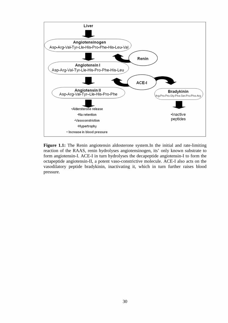
combat hypertension, various stages of the renin angiotensin aldosterone system
(RAAS) which is shown in Figure 1.1 can be positively affected. This system is
responsible for the control of blood pressure and fluid balance in humans. The RAAS
system involves the enzymes renin (Enzyme Commission number (E.C.) 3.4.23.15) and
angiotensin- I-converting enzyme (ACE-I) (E.C. 3.4.15.1) and is capable of stimulating
atherosclerosis by triggering basic reactions that ultimately lead to growth, instability,
and rupture of atherosclerotic plaques and facilitation of thrombosis [43]. Two ways of
inhibiting this system include firstly blocking the formation of angiotensin-II by the
enzyme ACE-I from angiotensin I and secondly by inhibiting the conversion of
angiotensinogen into angiotensin-I by the enzyme renin[43].
30
Figure 1.1: The Renin angiotensin aldosterone system.In the initial and rate-limitingreaction of the RAAS, renin hydrolyses angiotensinogen, its’ only known substrate toform angiotensin-I. ACE-I in turn hydrolyses the decapeptide angiotensin-I to form theoctapeptide angiotensin-II, a potent vaso-constrictive molecule. ACE-I also acts on thevasodilatory peptide bradykinin, inactivating it, which in turn further raises bloodpressure.

31
1.6 Protease enzymes as targets for the prevention of hypertension
Proteases are proteolytic enzymes that catalyse the hydrolysis of the peptide bond in
peptides and proteins [44]. Proteases are classified based on the key residue they use to
catalyze the hydrolysis of the peptide bond. Hydrolysis can occur either at the N- or C-
terminus of the peptide chain, catalysed by proteins referred to as exopeptidases, which
are divided into two groups; the aminopeptidases and the carboxypeptidases
respectively. Serine, cysteine and threonine proteases bind their substrates covalently,
whereas aspartic, metallo and glutamic proteases act through a general acid-base
mechanism [43]. Proteases and modified protease activities in disease states are targets
for multiple diseases and hence are potential bioactive peptide drug or functional food
targets. Human immunodeficiency virus protease (HIV-protease), high blood pressure
and thrombosis are examples of diseases where the disease related protease is the
therapeutic target [44, 45]. Recently, three new enzyme targets have been validated by
the Food Development Authority (FDA) approval of new enzyme inhibitor drugs. These
include mitogen-activated protein kinase, renin, and dipeptidyl peptidase IV [46].
1.6.1 Aspartic proteases
A minor protease class, aspartic proteases use the general acid-base hydrolysis
mechanism of action. Most proteases belong to either the A1 family of pepsin-like
proteases or the A2 retroviral family [47]. Aspartic proteases have been identified in
plants, fungi, mammals, viruses and protozoa [48]. In humans, members of the aspartic
protease class form part of the digestive system with non-specificity for peptide
substrates as is the case with pepsin (EC 3.4.23.2) and gastricin (EC 3.4.23.3).
However, in humans, it also includes proteases with a single substrate such as renin
with its substrate angiotensinogen, and β-secretase (EC 3.4.23.46) (BACE1) that
hydrolyses the amyloid precursor protein (APP) as its only substrate. Aspartic proteases

32
can be expressed in a single, cellular compartment as is the case with cathepsin D which
is expressed in the lysosomes [49]. Alternatively, it can be secreted with pepsin and
gastricin, or can be expressed in specific tissues that restrict their activities, such as
BACE1 in the brain [50].
1.6.2 ACE-I inhibitors
In mammals, two forms of ACE-I exist; one expressed in somatic tissue (sACE, 1306
residues), which has two active sites and the other ACE-I which is expressed in
germinal cells in the male testes (gACE, 732 residues) which has one active site. sACE
is a translated tandem duplication. This duplicated structure produces a protein with two
domains, the N-domain and the C-domain [51]. Despite having around 60% sequence
homology with each other, studies have highlighted the unique physiological roles of
the N- and C- domains of ACE-I [52]. The principle functional unit in each domain is
the M2-type zinc metallopeptidase motif, an His-Gluxx-His with a Glu positioned 23-24
residues further towards the C-terminus, these residues are ligands for the zinc cofactor
required for the peptidase catalytic activity [51]. Through its role in the RAAS, ACE-I
plays a key role in the regulation of blood pressure and electrolyte homeostasis [53]. It
carries out this function by hydrolysing peptides through the removal of a dipeptide
from the C-terminus, as is the case in the conversion of angiotensin I to angiotensin II,
or the degradation of bradykinin. It can also act as an endopeptidase, shown by cleavage
of peptides with amidated C-termini [54].
Inhibition of ACE-I is considered to be a useful therapeutic approach in the treatment of
hypertension [55]. The first ACE-I inhibitor was discovered when Ferreira (1964)
discovered a "bradykinin potentiating factor" in the venom of the snake Bothrops
jararaca [56]. Many studies have been attempted regarding the synthesis of ACE-I
inhibitors such as captopril, enalapril, alacepril and lisinopril, which are currently used
33
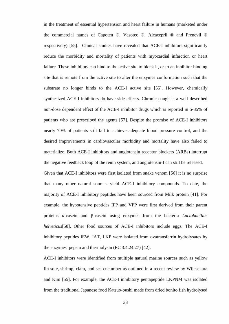
in the treatment of essential hypertension and heart failure in humans (marketed under
the commercial names of Capoten ®, Vasotec ®, Alcacepril ® and Prenevil ®
respectively) [55]. Clinical studies have revealed that ACE-I inhibitors significantly
reduce the morbidity and mortality of patients with myocardial infarction or heart
failure. These inhibitors can bind to the active site to block it, or to an inhibitor binding
site that is remote from the active site to alter the enzymes conformation such that the
substrate no longer binds to the ACE-I active site [55]. However, chemically
synthesized ACE-I inhibitors do have side effects. Chronic cough is a well described
non-dose dependent effect of the ACE-I inhibitor drugs which is reported in 5-35% of
patients who are prescribed the agents [57]. Despite the promise of ACE-I inhibitors
nearly 70% of patients still fail to achieve adequate blood pressure control, and the
desired improvements in cardiovascular morbidity and mortality have also failed to
materialize. Both ACE-I inhibitors and angiotensin receptor blockers (ARBs) interrupt
the negative feedback loop of the renin system, and angiotensin-I can still be released.
Given that ACE-I inhibitors were first isolated from snake venom [56] it is no surprise
that many other natural sources yield ACE-I inhibitory compounds. To date, the
majority of ACE-I inhibitory peptides have been sourced from Milk protein [41]. For
example, the hypotensive peptides IPP and VPP were first derived from their parent
proteins κ-casein and β-casein using enzymes from the bacteria Lactobacillus
helveticus[58]. Other food sources of ACE-I inhibitors include eggs. The ACE-I
inhibitory peptides IEW, IAT, LKP were isolated from ovatransferrin hydrolysates by
the enzymes pepsin and thermolysin (EC 3.4.24.27) [42].
ACE-I inhibitors were identified from multiple natural marine sources such as yellow
fin sole, shrimp, clam, and sea cucumber as outlined in a recent review by Wijesekara
and Kim [55]. For example, the ACE-I inhibitory pentapeptide LKPNM was isolated
from the traditional Japanese food Katsuo-bushi made from dried bonito fish hydrolysed
34
with thermolysin [59]. Suetsano et al. (2000) discovered four tetrapeptides (AITL,
TLTT, LPTG and TALL) with ACE-I inhibitory activity from a peptic digest of the
seaweed Undaria pinnatifida [29]. Indeed, over 556 ACE inhibitory peptides derived
from natural sources are reported in the peptide database BIOPEP
(http://www.uwm.edu.pl/biochemia/index.php/pl/biopep).
1.6.3 The enzyme renin and its mechanism of action in prevention of
hypertension
Inhibition of the renin enzyme was first reported in 1898 by Tigerstedt and Bergman
who observed that an extract from rabbit kidney was sufficient to increase blood
pressure in living rabbits. They called this vasoconstrictive substance renin[60]. Renin
has the EC number 3.4.23.15 and is a member of the aspartic protease family, which
also includes the enzymes pepsin, cathepsin, and chymosin [61]. Renin is a
monospecific enzyme that displays remarkable specificity for its only known substrate,
angiotensinogen [61] and indeed, renin is also known as angiotensinogenase. The most
important source of circulating renin is the granular cells of the juxtaglomerular
apparatus situated in the macula densa mechanism of the kidneys. Renin is produced in
response to three main stimuli; (1) Decrease in arterial blood pressure; (2) Decrease in
sodium chloride (NaCl) levels in the ultrafiltrate of the nephron in the kidneys and (3)
sympathetic nervous system activities which also control blood pressure levels. Renin
is produced through the activation of pro-renin, the enzymatic precursor of renin. Pro-
renin is inactive due to a 43 amino acid N-terminal pro-peptide that covers the active
site and blocks access of the active site to angiotensinogen. It is activated either through
proteolytic cleavage of the pro-peptide chain or by non-proteolytic activation in the
juxtaglomerular cells by the unfolding of the proteolytic propeptide, which is how the
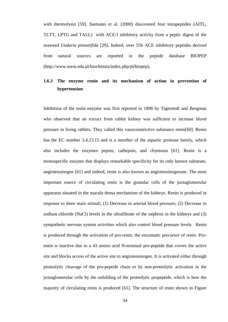
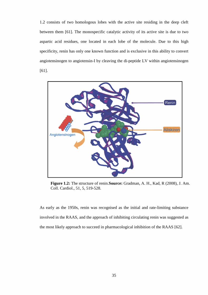
majority of circulating renin is produced [61]. The structure of renin shown in Figure
35
1.2 consists of two homologous lobes with the active site residing in the deep cleft
between them [61]. The monospecific catalytic activity of its active site is due to two
aspartic acid residues, one located in each lobe of the molecule. Due to this high
specificity, renin has only one known function and is exclusive in this ability to convert
angiotensinogen to angiotensin-I by cleaving the di-peptide LV within angiotensinogen
[61].
As early as the 1950s, renin was recognised as the initial and rate-limiting substance
involved in the RAAS, and the approach of inhibiting circulating renin was suggested as
the most likely approach to succeed in pharmacological inhibition of the RAAS [62].
Figure 1.2: The structure of renin.Source: Gradman, A. H., Kad, R (2008), J. Am.Coll. Cardiol., 51, 5, 519-528.

36
1.6.4 Types of inhibitors in the RAAS
Bioactive peptides and drugs which target the RAAS regulatory pathway can be
classified by which part of the RAAS they modify. Direct renin inhibitors (DRIs)
include β-andrenoceptor blockers which target renin, the ACE inhibitors, the AT1
blockers (ARBs), the aldosterone-receptor antagonists (ARAs) and combined ACE and
neutral endopeptidase inhibitors called vasopeptidase inhibitors [63]. The sites of where
enzymes may be inhibited within the RAAS are shown in Figure 1.3.
Figure 1.3: Sites of inhibition of the RAAS. Black arrows represent activation steps;Purple arrows denote enzyme actions. The sites of actions of RAAS inhibitors areindicated by numbers 1-8. Adapted from: Komers, R (2013) Clinical Science, 124, 553-566.
prorenin
renin
ATG Ang I
Ang II
ACE-I
AT1RNon-AT1R
MAS
Ang 1-7
Inactive fragments
Succinate glucose
GPR91
(P)RR BiologicalEffects
Aldosterone
Biological effects
7
1
2
3
4
5, 6, 8
1. Aliskiren2. HRP3. ACEI4. ARB
5. Vitamin D6. Urocortins
7. Putative GPR91 antagonist8. COX-2 inhibitors
prorenin
renin
ATG Ang I
Ang II
ACE-I
AT1RNon-AT1R
MAS
Ang 1-7
Inactive fragments
Succinate glucose
GPR91
(P)RR BiologicalEffects
Aldosterone
Biological effects
7
1
2
3
4
5, 6, 8
prorenin
renin
ATG Ang I
Ang II
ACE-I
AT1RNon-AT1R
MAS
Ang 1-7
Inactive fragments
Succinate glucose
GPR91
(P)RR BiologicalEffects
Aldosterone
Biological effects
7
1
2
3
4
5, 6, 8
1. Aliskiren2. HRP3. ACEI4. ARB
5. Vitamin D6. Urocortins
7. Putative GPR91 antagonist8. COX-2 inhibitors

37
1.6.5 Development of renin inhibitors
Pepstatin was the first synthesised renin inhibitor. However, it was initially described as
a pepsin inhibitor derived from various species of the bacterial genus Actinomyces in
1970 by Umezawa et al [64]. Pepstatin is a hexapeptide containing the unusual amino
acid statine [65]. Pepstatin was considered as a renin inhibitor due to its potency as an
inhibitor of aspartyl proteases. However, its activity on renin was found to be several
orders of magnitude lower in terms of potency compared to its pepsin inhibitory activity
[65]. The next generation of renin inhibitors came in the form of analogues of the 45
amino acid long pro-renin segment. This segment shields access to the catalytic site of
renin in its inactive form. Indeed, four peptides from this segment were described to
have potent in vitro renin inhibitory effects in the past but ultimately showed poor drug-
like properties [66]. Another strategy used to develop effective renin inhibitory drugs
looked at improving the specificity, stability and potency of inhibitors by developing
peptidometric inhibitors with improved structures [67]. This approach led to the
development of remikiren and zankiren. These drugs never proceeded past pre-clinical
trials due to their poor oral bioavailability resulting in a decreased hypotensive effect
[67]. Aliskiren was the first orally active non-peptide renin inhibitor on the market.
Developed by Novartis and marketed under the names Rasilez® and Tekturna®,
aliskiren inhibits renin by occupying the S3sp sub-pocket of renin’s active site which is
not exploited by peptide-like inhibitors[68].Figure 1.2 shows the inhibition of renin by
the pharmaceutically designed molecule aliskiren. The alkylether aromatic side chains
interact with the S3sp subpocket of the renin active site blocking access to
angiotensinogen [61].
38
1.6.6 Renin inhibitory compounds and peptides from plant sources
Renin inhibitory peptides were identified previously from plant sources including
soybean [69], hemp seed [70] and the plant Scutellaria baicalensis[71]. Previously, Li
and Aluko (2010) used the enzyme alcalase to hydrolyse pea protein and produced three
dipeptides: Isoleucine-Arginine, Lysine–Phenlyalanine, and Glutamine-Phenylalanine,
each with potent renin inhibitory activity [72]. In addition, Agomuoh et al. (2010)
identified renin inhibitors from the plant Nauclea latifolia. Studies were carried out
regarding the renin inhibitory activity of various foods [73]. Some studies have equally
looked at the inhibitory activities of some synthetic peptides [74-76]. Screening for
renin inhibition using synthetic analogues of plant compounds has also been studied
previously [77, 78].
1.6.7 The RAAS: a potential therapeutic target in the treatment of diabetic
kidney disease and other disease treatments
One of the risk factors associated with the development of CVD includes high blood
pressure, and this may be controlled by inhibition of a number of enzymes in the RAAS
and the human body that are known to increase blood pressure and atherosclerosis.
Angiotensin converting enzyme I (ACE-I; EC 3.4.15.1) is one such enzyme. ACE-I is a
zinc metalloprotease that plays an important role in RAAS and the control of blood
pressure and fluid regulation [51]. Inhibition of ACE-I is a well-established approach in
the treatment of hypertension. ACE-I removes a dipeptide from the C-terminus of
angiotensin I, converting it to angiotensin II, a potent vasoconstrictor. Chemically
synthesized ACE-I inhibitors including captopril (marketed as Capoten®), enalapril
(marketed as Vasotec®), alcacepril (marketed as Alaceril®), and lisinopril (marketed as
Prinivil®) are ordinarily prescribed for the treatment of high blood pressure [55]. ACE-I

39
is also involved in the degradation of bradykinin, which is a vasodilator [54]. ACE-I
also acts as an endopeptidase, shown by cleavage of peptides with amidated C-termini,
as seen in the cleavage of Histidine -Leucine in the RAAS [51]. Bradykinin-potentiating
peptides prevent the hypertensive effect of angiotensin II and potentiate the hypotensive
effect of the circulating vasodilatory peptide bradykinin by also inhibiting ACE-I (as
shown in Figure 1.1) [54].
As mentioned, renin inhibition may prevent the development of high blood pressure.
However, the blood pressure lowering effect of renin inhibition is not the sole gain of
the therapy. The RAAS is documented as playing a pivotal role in the prevention and
treatment of diabetic nephropathy and some other proteinuric kidney diseases [79].
RAAS inhibition can slow the progressive decrease in glomerular filtration rate, reduce
proteinuria and microalbuminuria, and reducing cardiovascular mortality and morbidity
in diabetic patient [79]. Use of renin inhibitors over ACE-I inhibitors in the treatment of
kidney diseases has a number of advantages. Directly inhibiting renin completely halts
the production of angiotensin peptides, whereas ACE-I inhibition merely reduces the
production. A phenomenon known as “ACE escape” or “aldosterone breakthrough”
occurs where angiotensin-II gets converted independent of the RAAS by circulating
chymases [80]. Therefore even with ACE-I inhibition many patients will still
experience renal failure, renin inhibition offers a more complete blockade of the RAAS
and thus may offset renal failure further [79].
Renin inhibitors may also be used in the treatment of glaucoma. Apart from cataracts,
glaucoma is the leading cause of sight loss world-wide [81]. Glaucoma is a condition
where optic neuropathy occurs due to intraocular pressure (IOP). Diet, ethnic and
genetic factors all play a role in the etiology of the condition, however the biggest cause
of IOP is Hypertension. An active RAAS has been described within the eye, and studies
40
now show that drugs such as renin inhibitors may relieve IOP and in turn offset the
development of glaucoma [81].
1.6.8 Platelet-activating factor acetylhydrolase (PAF-AH) and heart health
Another enzyme that is associated with the risk of cardiac events is the enzyme platelet-
activating factor acetylhydrolase also known as lipoprotein associated phospholipase A2
(Lp-PLA2)[82]. A proatherogenic role has been postulated for this enzyme as it
generates two key pro-inflammatory mediators [82]. This observation has led to
suggestions of a causative role for PAF-AH in the development of atherosclerosis [82].
PAF-acetylhydrolase is a circulating enzyme produced and secreted by inflammatory
cells centrally involved in atherosclerosis [83]. It is bound predominately to
apolipoprotein B-containing lipoproteins and is highly expressed in the necrotic core of
atherosclerotic lesions [83]. It generates two key pro-inflammatory mediators,
lysophosphatidylcholine (LPC) and oxidized non-esterified fatty acids (oxNEFAs).
Evidence exists for a regulatory role of these lipids in promoting atherosclerotic plaque
development that can ultimately lead to the formation of a necrotic core, a key
determinant in atherosclerotic plaque vulnerability, as illustrated in Figure 1.4 [82].
Mayer et al. [79] discovered a variety of compounds from chlorophyta, phaeophyta, and
rhodophyta that have inhibitory properties against bee-derived phospholipase A2 [84].
The discovery of natural PAF-AH inhibitors and their inclusion in the treatment of CVD
and incorporation into functional foods has high potential and these inhibitors are yet to
be fully exploited.
1.6.9 Darapladib and the development of commercial PAF-AH inhibitors
Chemically synthesised darapladib became the lead compound amongst a group of
substituted pyrimidones observed to have inhibitory activity towards PAF-AH in vitro.

41
Darapladib was shown previously to prevent necrotic core expansion, a key determinant
in atherosclerotic plaque vulnerability [83].However, a recently completed double
blinded trial by GSK using 15,828 patients with stable coronary heart disease
administered 160 mg of darapladib per day showed no significant reduction the risk of
the primary composite end point of cardiovascular death, myocardial infarction, or
stroke. It is possible though that the coronary risk among patients in this study may
already have been minimized by concurrent therapy [85]. An on-going GSK trial called
the stabilization of plaques using darapladib thrombolysis in myocardial infarction
(SOLID-TIMI 52) aims to determine the clinical benefit of direct inhibition of PAF-AH
activity with darapladib in patients after an acute coronary syndrome is expected to
finish in 2015 [86].Furthermore the anti-atherosclerosis activity of varespladib an
inhibitor of several sPLA2s is currently under investigation by the pharmaceutical
company Anthera [87].

42
Figure 1.4: Expansion of the necrotic core due to PAF-AH activity. In blood plasma,80% of PAF acetylhydrolase is bound to low-density lipoproteins (LDL). As LDL istransported into the intima from the lumen, it is oxidized; PAF-acetylhydrolase thengenerates two pro-inflammatory mediators, lysophosphatidylcholine (LPC) andoxidized nonesterified fatty acids (oxNEFAs). LPC increases plaque build-up as it is animportant chemo-attractant for macrophages and increases the expression of adhesionmolecules. oxNEFAs convert the attracted macrophages into foam cells, whichagglutinate in the lumen and constrict blood vessels, which may lead to high bloodpressure and potentially CVD.
1.7 Bioactive peptides previously isolated from macroalgae
In the sourcing of bioactive peptides from macroalgae, it is important to take into
account differences in protein content between species and also within species collected
at different locations and during different seasons [32]. Generally, the protein fraction of
brown macroalgae is low (3-15% of dry weight) compared with that of the green (10-
26% of dry weight) or red macroalgae (35-47% of dry weight) [32]. As mentioned
previously, the season in which the macroalga is harvested influences the protein
content. Glycoproteins known as lectins are also found in macroalgae. Lectins may be
divided into four main subgroups, namely, legume lectins, chitin binding lectins,
monocot mannose binding lectins, and type-2 ribosome inactivating proteins (RIP) [88].

43
The main characteristic of this class of protein is their ability to interact specifically
with carbohydrates and to combine with the glycol components of the cell surface [89].
Macroalgal lectins have been detected and isolated from several Rhodophyta [90].
Lectins have biotechnological applications in several scientific and medicinal fields of
research including biology, cytology, biochemistry, and medicine [88]. However,
lectins may present a problem if used for human food use as glycoproteins are known to
be responsible for allergic reactions and in some instances anaphylactic shock [91].
Macrolalgal and many other plant-derived lectins have been used for clinical blood
typing in medicinal biology. For example, lectins derived from the green alga Codium
fragile have been shown to recognize GalNAc, an antigen for blood group A [92]. They
also have a number of valuable bioactive properties. For example, lectins from
Perocladiella capillacea demonstrated analgesic and anti-inflammatory properties in
rodent model [88]. Holanda et al. showed that a lectin extracted from the red alga
Solieria filiformis had antibacterial activity against six pathogenic Gram-negative
species including Serratia marcescens, Salmonella typhi, Klebsiella pneumoniae,
Enterobacter aerogenes, Proteus sp., and Pseudomonas aeruginosa[90]. The lectin
known as amansin isolated from the red alga Amansia multifida is used as a mitogen,
which is a substance that encourages cell division, for human lymphocytes, and it
therefore has potential for use in anticancer therapies [86]. Phycobiliproteins found in
macroalgae are usually divided into three separate groups on the basis of their colour
and absorption properties, namely, phycoerythrin, phycocyanin, and allophycocyanin
[93]. They are components of the macromolecular light harvesting complex of
macroalgae called the phycobilisome [94]. In many red algae, phycoerythrin, the most
abundant phycobiliprotein found in the Rhodophyta, is the major soluble protein of the
cell [94]. The primary potential of these molecules seems to be as natural dyes, but an
increasing number of investigations have shown their health-promoting properties and

44
broad range of pharmaceutical applications [95]. Phycoerythrin is a powerful and highly
sensitive fluorescent reagent, which can serve as a label for antibodies, receptors, and
other biological molecules in a fluorescence- activated cell sorter, and
phycobiliproteins, in general, are used in immunolabelling experiments and
fluorescence microscopy and diagnostics [95]. These macromolecules also have potent
bioactivities such as antioxidant, antidiabetic, and anticancer properties. Bermejo et al.
demonstrated the antioxidant capabilities of the phycobiliprotein phycocyanin isolated
from a protein extract of the green microalga Spirulina platensis and suggested these
bioactive capabilities are attributed to the ability of the protein to chelate metal and to
scavenge free radicals [96]. Furthermore, purified C-phycoerythrin was shown to relieve
the symptoms of diabetic complications in rats through significant reductions in
oxidative stress and oxidized LDL-triggered atherogenesis [97]. Following
administration of 25 and 50 mg/kg per body weight per day over 28 days, C-
phycoerythrin decreased food intake, organ weight, serum concentration of glucose,
cholesterol, tumour associated glycoprotein (TAG), very low density lipoprotein
(VLDL)-cholesterol creatine, uric acid, and thiobarbituric acid reactive substances in the
rats, suggesting the possible therapeutic role in human diabetes of C-phycoerythrin [97].
Purified recombinant allophycocyanin was found to have a significant inhibitory effect
on S-180 carcinoma in mice with inhibition rates ranging from 7.9 to 61.9% with doses
ranging from 4.65 to 18.6 mg/(kg day) [97]. Other peptides include a hexapeptide from
Ulva species, which displays mitogenic properties, and agglutinin glycoprotein from
Soleria robusta and Eucheuma serra, which displays mitogenic, cytotoxic, and
anticancer properties [98].
Digestion of macroalgal proteins with proteolytic enzymes has led to the discovery of
many bioactive peptides. ACE-I inhibitory peptides were released from Undaria
pinnatifida proteins using enzymes including pepsin [29] and from the parent proteins

45
of Polysiphonia urceolata using the enzymes protamex, alcalase, and flavourzyme [92].
Peptide fractions isolated from the red macroalga Porphyra yezeonsis (“nori”) were
found to have a hypotensive effect when administered orally to SHR models [99].
1.8 Effects of food processing on bioactive peptides
Food processing provides an additional value to foods by improving food safety, shelf
life, palatability, nutritive, and functional values [100]. However, depending on the
processing technique employed, food processing may be detrimental to the survival of
bioactive peptides [100]. Changes in the molecular structure of an amino acid may lead
to changes in the bioactivity and absorption of the peptide of interest. Heat, the most
common and oldest form of food processing, modifies the food proteins to make them
more edible in terms of texture and flavour [101]. Heat can be used to enhance
functional properties of proteins. Denaturation of proteins for example improves the
water binding and emulsification properties. Heat also decreases protein solubility due
to aggregation and coagulation [101]. During heating, the lysine residues of proteins
may react with reducing carbohydrates in the same food system, resulting in the
maillard or non-enzymatic browning reaction [101]. This can reduce the nutritional
value of proteins as the bioavailability of lysine is reduced [101]. Heat may also destroy
the bioactivity of peptides. However, it has been shown that some ACE-I inhibitory
peptides can retain their activity when heated to temperatures of 70ºC and 100 °C for 20
min [102].
Other food production processes may facilitate the release of bioactive peptides from
their parent proteins. For example, during milk fermentation, lactic acid bacteria (LAB)
hydrolyse caseins into peptides and amino acids [100]. High hydrostatic pressure also
promotes the proteolysis and release of bioactive peptides. Quiros et al. showed that
proteolysis of ovalbumin with pepsin for the production of ACE-I inhibitory peptides

46
was accelerated under pressures of 200-400 MPa [103]. Another factor to consider
when bioactive peptides are incorporated into a food matrix is the often bitter taste of
hydrolysates, which is attributed to the formation of low molecular weight peptides
composed mainly of hydrophobic amino acids [100]. This can limit the use of some
bioactive peptides that possess proven bioactivities [100]. Strategies to de-bitter protein
hydrolysates include treatment with activated carbon, extraction with alcohol,
isoelectric precipitation and chromatographic separation. Masking the taste of these
peptides can be achieved through the use of ingredients including monosodium
glutamate (MSG) and also by enzymatic hydrolysis of the proteins/peptides [104]. The
latter method has limited capability due to the enzymatic hydrolysis of the active
peptides themselves, which may render the bioactive peptides inactive [100].
Microencapsulation is a technology that has solved many problems that limit the use of
bioactive peptides and additives in foods, as it can mask undesirable flavours and
reduce volatility, hygroscopicity, and reactivity [105]. Furthermore, microencapsulation
improves the stability of the products under adverse environmental conditions and
provides controlled liberation of the encapsulated material under pre-established
conditions [105]. For example, the oral delivery of tuna-derived antihypertensive
oligopeptides composed of hydrophilic peptides to consumers was achieved by
encapsulating them in liposomes [106]. Moreover, antimicrobial lysozymes were
previously encapsulated using zein protein derived from maize[107] to minimize
binding between antimicrobials and the food matrix [107]. In addition, casein
hydrolysates were previously encapsulated in maltodextrin to mask the off-flavour of
the hydrolysate, which facilitated its inclusion in a protein bar [105].

47
1.9 Bioavailability of bioactive peptides and survival in the gastrointestinal
tract (GI tract).
Following consumption, the gastrointestinal epithelium acts as both a physical and a
biochemical barrier to absorption of food derived bioactive peptides and drugs. The
physical barrier is represented by the impermeable gastrointestinal epithelium and the
biochemical barrier consists of enzymatic peptidases [108]. In order for a bioactive
peptide to be delivered to its active site it is necessary to fully understand these barriers.
The GI tract shown in Figure 1.5 has site specific absorption based on the peptide/drug
consumed and based on regional differences in pH, enzyme activity, thickness of
mucosa and residence time and surface area [108]. The pH of the GI tract varies from
1-7. The bioavailability of protein and peptides depends on their ability to cross the
intestinal mucosa and reach the systemic circulation.
Figure 1.5: Intestinal transport mechanisms for peptides.Adapted from:Renukuntla, J.,Dutt Vadlapudi, A., Patel, A., Boddu, S. H. S., and Mitra, A. K.International Journal ofPharmaceutics, 447, (2013), 75-93 [107].

48
Transport across the intestinal epithelium may be either active or passive and the
mechanisms of transport depend on the physicochemical properties of the peptide and
on the length of the peptide in question. Active transport of peptides involves movement
from low to high concentrations by transmembrane proteins and energy in the form of
ATP is used. Passive transport involves diffusion of drug molecules in the direction of
the concentration gradient [108]. Carrier mediated transport involves the movement of
molecules via transporters [108]. Detailed understanding of the structural features of a
bioactive peptide is needed to target these transports for efficient delivery of the peptide
to the target sites.
Bioactive peptides may have to interact with target sites at the luminal side of the
intestinal tract. Furthermore, they have to be absorbed in order to reach the target organ
[109]. In addition to being resistant to further enzymatic digestion by endogenous gut
enzymes, bioactive peptides must also show the ability to cross the intestinal epithelium
to reach target sites [110]. Previous studies have used Caco-2 cell monolayers to assess
the ability of bioactive peptides isolated from milk to cross the intestinal epithelium
[111]. While the exact route of transportation for bioactive peptides is not fully
understood some peptides cross via paracellular diffusion and transcytosis
[111].Different formulation strategies may be used to increase the oral bioavailability of
peptides and these include methods such as use of absorption enhancers [112], enzyme
inhibitors [113], hydrogels [114], liposomes [115], cyclodextrins [116] and
nanoparticles [117].
Factors such as peptide length also affect the bioavailability of the molecules. It was
once believed that dietary proteins were completely digested to amino acids after
ingestion, but, it is now evident, that about 30 - 50 % of dietary protein gets absorbed in
the form of small peptides [118]. Previously, it was proven that peptides in the range of
3 to 51 amino acid residues in length can be absorbed by the gastrointestinal tract and

49
may incur a physiological response [118]. However, increased length of the peptide
chain does diminish the amount of the molecule being absorbed. For example the tri-
peptide thyrotropin releasing hormone (TRH) has the same hormonal response when
administered orally as intravenously, whereas the decapeptide luteinizing hormone-
releasing hormone (LHRH) had half the maximal response when it was administered
orally than when administered intravenously [118]. Peptides consisting of two or three
amino acids are absorbed intact across the brush border membrane by the peptide
transporter PepT1 using a transmembrane electrochemical proton gradient as the driving
force and this transporter has broad specificity. In this instance they absorbed more
rapidly than free amino acids [119]. In terms of amino acid composition, peptides
containing hydrophobic residues such as proline and valine are more easily transported
across the GI tract [119].
1.10 Suitable food vehicles for delivery of bioactive peptides
There are many products already on the market, which utilise bioactive peptides as
functional components. For example, there are two products available that contain the
bioactive peptides IPP and VPP, namely, Calpis© and the Ameal-S drink developed in
Japan and Evolus developed by Valio Ltd. a Finnish company. Both products were
generated from fermented milk products and have ACE-I inhibitory actions and are
proven antihypertensive agents in spontaneously hypertensive rats (SHRs) and in
human clinical trials [120]. Furthermore, a thermolysin digest of dried bonito containing
antihypertensive peptides is used in a soup product in Japan and has claimed
antihypertensive effects [120, 121]. Table 1.1 shows commercially available products
that contain bioactive peptides with different health-promoting properties. However,
there is some controversy over the effect of lactotripeptides on blood pressure as recent
studies found no statistically significant differences between the groups that consumed

50
peptides and the placebo groups [17]. This highlights the importance of the bioactive
peptides being capable of reaching the bloodstream and careful selection of a test
population.

51
Table 1.1: Bioactive peptide containing products available commercially
Effect Product Pepitde(s) Company Source/FoodMatrix
Heart HealthHypotensive Ameal ® VPP,IPP Calpis MilkHypotensive Evolus® VPP, IPP Valio Ltd. MilkHypotensive Peptide soup LKPNM (katsuobushi oligopeptide) Nippon
Supplement, Inc.katsuobushi(dried bonito)
Hypotensive Casein DP ® FFVAPFPEVFGK Kanebo Ltd. MilkHypotensive C12 peptide ® FFVAPFPEVFGK DMV (De
MelkindustrieVeghel)International
Milk
Hypotensive Valtyron ® VT Senmi Ekisu Co.Ltd.
Sardine
Hypotensive Biozate ® Whey-derived peptides Davisco MilkCholesterollowering
CSPHP ® Enzymatically decomposed lecithin bound tohydrolysedisolated soy protein (ratio of 80:20)
Kyowa Hakko Soy
OtherEnergy Boost Cysteine peptide peptide
supplementMixture of whey protein derived peptidescomprising atleast 6.5 wt % cysteine
DMV International Milk
Satiety Glycomacropeptide Casein-derived whey peptide Davisco Milk
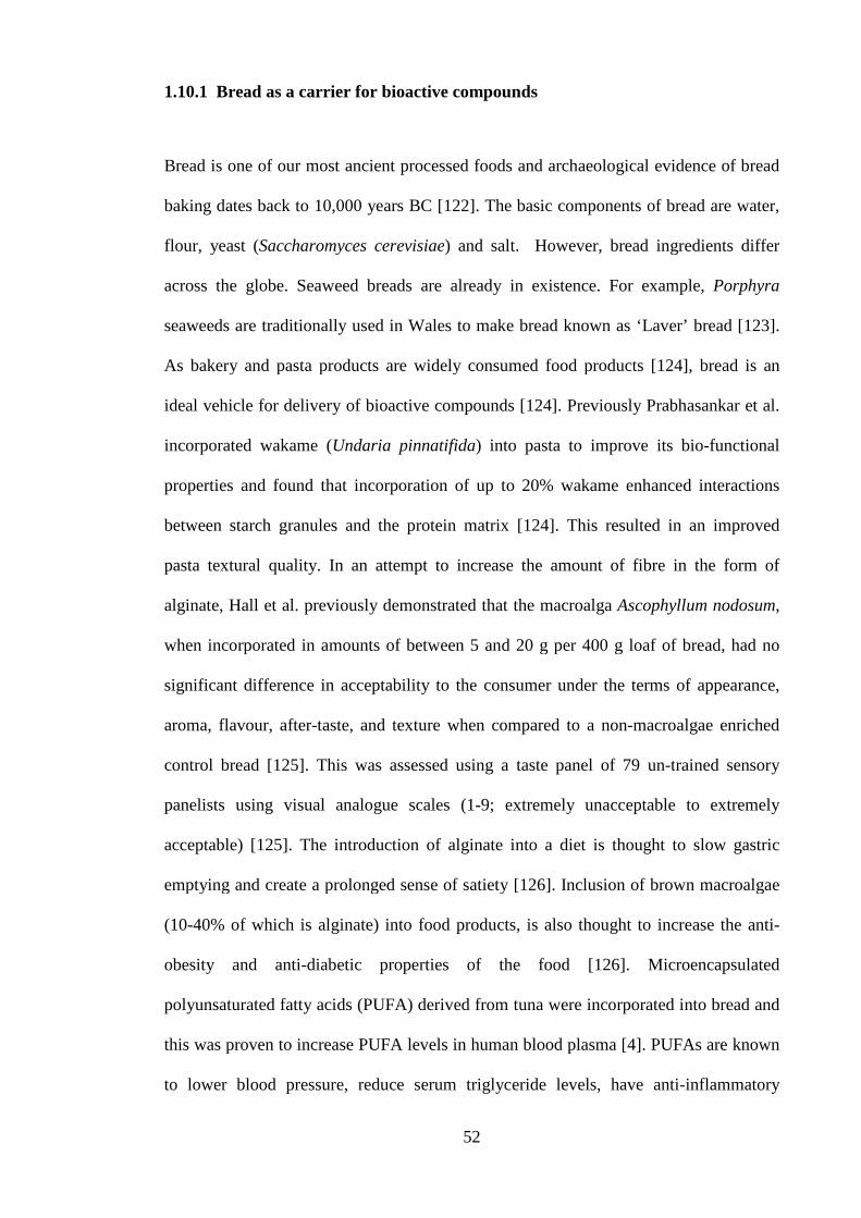
52
1.10.1 Bread as a carrier for bioactive compounds
Bread is one of our most ancient processed foods and archaeological evidence of bread
baking dates back to 10,000 years BC [122]. The basic components of bread are water,
flour, yeast (Saccharomyces cerevisiae) and salt. However, bread ingredients differ
across the globe. Seaweed breads are already in existence. For example, Porphyra
seaweeds are traditionally used in Wales to make bread known as ‘Laver’ bread [123].
As bakery and pasta products are widely consumed food products [124], bread is an
ideal vehicle for delivery of bioactive compounds [124]. Previously Prabhasankar et al.
incorporated wakame (Undaria pinnatifida) into pasta to improve its bio-functional
properties and found that incorporation of up to 20% wakame enhanced interactions
between starch granules and the protein matrix [124]. This resulted in an improved
pasta textural quality. In an attempt to increase the amount of fibre in the form of
alginate, Hall et al. previously demonstrated that the macroalga Ascophyllum nodosum,
when incorporated in amounts of between 5 and 20 g per 400 g loaf of bread, had no
significant difference in acceptability to the consumer under the terms of appearance,
aroma, flavour, after-taste, and texture when compared to a non-macroalgae enriched
control bread [125]. This was assessed using a taste panel of 79 un-trained sensory
panelists using visual analogue scales (1-9; extremely unacceptable to extremely
acceptable) [125]. The introduction of alginate into a diet is thought to slow gastric
emptying and create a prolonged sense of satiety [126]. Inclusion of brown macroalgae
(10-40% of which is alginate) into food products, is also thought to increase the anti-
obesity and anti-diabetic properties of the food [126]. Microencapsulated
polyunsaturated fatty acids (PUFA) derived from tuna were incorporated into bread and
this was proven to increase PUFA levels in human blood plasma [4]. PUFAs are known
to lower blood pressure, reduce serum triglyceride levels, have anti-inflammatory
53
activities, and decrease the risk of cardiac events [127]. Macroalgae and microalgae are
the nutritional source of PUFAs for many fish, hence, macroalgae are also a viable
source for these bioactive molecules in human nutrition [127]. Moreover, certain
components of marine algae appear to improve the rheological properties of flour dough
and the quality of the finished products made from it [90]. Oxidoreductase derived from
the algae Chondrus crispus when added to flour dough increased resistance to extension
and extensibility by at least 10% compared to similar dough that did not contain
oxidoreductase. It also increased the final volume of a finished baked product by 20%,
compared to that of a similarly baked product not containing oxidoreductase [128].
1.11 Final remarks
Every year around 4.3 million Europeans die from the effects of heart disease resulting
in €196 billion being spent per year on its treatment [129]. Coronary heart disease
remains the Western Worlds’ biggest killer. However these numbers are decreasing, this
is mainly due to scientific advances that have arrested and indeed reversed the growth in
mortality due to CVD [129]. High blood pressure is the highest risk factor associated
not only with cardiac related death but with mortality in general as it exacerbates
practically all human health issues [130]. Some of the main scientific advancements in
curtailing CVD include better diagnostic tools and new interventional procedures but
the most significant of these have been the emergence of families of drugs such as
ACE-I inhibitors, statins, beta blockers and ARBS [129]. Western society has a number
of rising statistics that will make the advancement of CVD treatment even more crucial.
One of these is the rapidly ageing population. By 2050, the number of people over the
age of 50 is expected to rise by 35% and the number of people over 80 is expected to
rise by 300% [129]. The recent rise in the incidence of obesity and with that, type II
diabetes, will also increase the significance of furthering CVD treatment research [129].

54
With these issues in mind new strategies to prevent heart health issues are constantly
sought. While inhibition of the RAAS is not a new concept in combating hypertension,
the approach of inhibiting it by debilitating its initial and rate limiting enzyme renin is
emerging again as a pharmaceutical strategy [131]. Recently, ACE-I inhibitory
compounds were isolated and characterised from natural food products and incorporated
into functional food products [120]. Very few natural renin inhibitory compounds are
present in the literature, but with the inherent advantages of renin inhibitors over ACE-I
inhibition [61] they are potentially a better candidate for inclusion in blood pressure
lowering functional foods. Similarly with PAF-AH inhibition, while this strategy is only
on the cusp of being realised as a viable treatment for atherosclerotic plaque, natural
PAF-AH inhibitors as functional food ingredients would be a feasible adjunct therapy
for at risk individuals.
In the past, the primary source of bioactive peptides was dairy products [41]. The
variety of macroalga species, the environments in which they are found, and their ease
of cultivation make macroalgae a relatively untapped source of new bioactive
compounds [2] and more efforts are needed to fully exploit their potential for use and
delivery to consumers in food products. Red seaweed such as Palmaria palmata have
for centuries been part of traditional diets of Atlantic coastal countries so it is already in
the public consciousness as being a nutritious food product. Its’ abundance around Irish
coasts combined with its rich protein content make it an ideal candidate for exploration
for bioactive peptides [32].
The idea of using bread as a vehicle for bioactive compound delivery is a natural one;
bread is one of the mostly widely consumed, processed foods globally and therefore can
be included in a candidates diet without augmenting it substantially. Previous methods
to introduce functional ingredients such as folate [132], Probiotics [133] and

55
microencapsulated marine lipids [134] have taken advantage of the ubiquitous
consumption of bread to deliver positive health results to the consumers.
The scientific rationale for the approach used in this work was to use protein material
isolated from the red seaweed Palmaria palmata to generate peptides and to screen
these peptides for their ability to inhibit two enzymes important in heart health and
blood circulation, namely renin and PAF-AH. This rationale was adopted as the red
seaweed Palmaria palmata is known to be rich in protein but was not examined
previously as a resource of bioactive peptides. Furthermore, renin and PAF-AH
inhibitory peptides were not previously identified from seaweeds and were only recently
identified from pea protein source. Within this work, a hydrolysate was generated and
peptides purified that exhibited both PAF-AH and renin inhibitory activities. These
peptides were synthesised to confirm the bioactivity. The ability of the hydrolysate
(from which these peptides were derived) to survive in the food vehicle bread was also
examined. This approach was used as the aim of the thesis was to develop a food
ingredient with heart and circulatory health benefits so the delivery of this ingredient in
bread was required. The thesis did not look at the possibility of using the chemically
synthesised peptides for pharmaceutical purposes and the work carried out reflects this.
This thesis strives to increase the understanding of how inhibition of various proteases
involved in cardiovascular health may be achieved through examining natural sources
of bioactive peptides. It expands on previous research carried out in the areas of food
proteomics and functional food. While there are many references to pharmaceutical
studies throughout this thesis, the research within focuses on food applications
according to the remit of the department of Food Biosciences at Teagasc Ashtown Food
Research Centre.

56
This thesis specifically aims to widen the scope of heart health peptide research to
include renin and PAF-AH inhibitory peptides derived from novel marine resources
specifically from the red seaweed Palmaria palmata (Linneaus) Weber and Mohr.
Furthermore, the feasibility of introducing bioactive hydrolysates and peptides isolated
from Palmaria palmata in to a baked product is explored along with the potential
bioavailability of seaweed derived hydrolysed proteins and peptides. Toxicity analysis
using a zebrafish larvae model are looked at along with the effects on the blood pressure
of spontaneously hypertensive rats (SHRs) following consumption of both the Palmaria
palmata protein hydrolysate and an identified renin inhibitory peptide.

57
CHAPTER 2. ISOLATION AND CHARACTERISATION OF
BIOACTIVE PRO-PEPTIDES WITH IN VITRORENIN
INHIBITORY ACTIVITIES FROM THE MACROALGA
PALMARIA PALMATA (LINNAEUS) WEBER AND MOHR
This chapter was published as a research article in the Journal of Agriculture and Food
Chemistry: Ciarán Fitzgerald, Leticia Mora-Soler, Eimear Gallagher, Paula
O’Connor, Jose Prieto, Anna Soler-Vila, and Maria Hayes “Isolation and
characterisation of bioactive pro-peptides with in vitro renin inhibitory activities from
the macroalga Palmaria palmata”, 2012, 60, 7421−7427.

58
2.1 Aims
The aim of this Chapter was to isolate and characterise renin inhibitory peptides fromthe
red macroalga Palmaria palmata which was assigned the reference number ISCG0022.
This species was collected and identified by a trained phycologist within the Irish
Seaweed Centre Galway (ISCG) and a voucher specimen of this seaweed is maintained
at the ISCG at NUI Galway under the code ISCG0122. The main objectives of this
chapter were to:
(1) Extract protein from Palmaria palmata (Linnaeus) Weber and Mohr and
hydrolyse it with the food grade enzyme papain.
(2) Use in vitro bioassay guided fractionation to identify renin inhibitory
hydrolysates and RP-HPLC fractions.
(3) Identify and confirm active peptides using purification techniques including
nano-electrospray ionization quadropole-time of flight mass spectrometry (ESI-
Q-TOF MS), de novo sequencing and solid phase peptide synthesis.

59
2.2 Introduction
Macroalgae represent a rich and sustainable source of nutritional protein [25]. The
Rhodophyta, or red seaweeds, in particular, can have a protein content of up to 47% of
their dry weight in certain species belonging to the taxon Porphyridiales. This is
considerably higher than the protein content obtained from the green macroalgae
(Chlorophyta). Chlorophyta can contain between 10 to 26% protein in terms of their dry
weight.The brown macroalgae (Phaeophyceae) contain between 3 to 15% protein of
their dry weight [32]. The red macroalga Palmaria palmata (Linnaeus) Weber and
Mohr, colloquially known as Dulse or Dillisk is already part of the diet of several
Atlantic coastal countries including Ireland, Wales and Iceland [33]. Previous seasonal
variation studies carried out on P. palmata sourced from the French coast found that it
had a protein content of between 9 and 25% of its dry weight [38]. The protein content
of Palmaria palmata is highest in the winter and spring months and decreases during
the summer months [38]. This increased protein content is thought to be caused by the
decrease of sunlight hours during winter and early spring months and the need of the
seaweed to create more pigments to up-regulate photosynthesis [37].
Pro-peptides can be defined as peptide sequences of between 2-30 amino acids in length
which form part of a protein sequence but that can be cleaved by enzymes to become
bioactive. The bioactivities associated with pro-peptides and peptides include
antimicrobial, antioxidant, angiotensin-I-converting Enzyme (ACE-I) inhibitory and
anti-inflammatory actions. ACE-I inhibitory peptides have been isolated from
numerous different natural food sources such as meat [135], vegetables [136] and
marine sources such as fish [59] and indeed macroalgae [29]. ACE-I (E.C. 3.4.15.1)
along with the enzyme renin (E.C 3.4.23.15 ) play an important role in the control and
regulation of blood pressure and salt water balance and, ACE-I inhibitors can prevent

60
the development of high blood pressure and have been identified from a number of food
sources previously. More recently, renin inhibitory compounds from plant sources
including peas [72], soybean [69], and the herb baicailin were identified [71].
Furthermore, structure−activity relationship modelling of renin inhibitory di-peptides
derived from these sources was carried out previously [137, 138]. However, Li and
Aluko (2011) are the only group to date to characterise and synthesize renin inhibitory
peptides from a plant protein source to date [72].
The objective of this chapter was to generate, enrich, isolate, and characterize renin
inhibitory peptides from P. palmata protein by hydrolysis of the protein with the food
grade enzyme papain. Reverse phase-high performance liquid chromatography (RP-
HPLC) was subsequently employed to enrich for peptides with renin inhibitory
activities. Further characterisation using electrospray ionization quadropole time-of-
flight mass spectrometry (ESI-Q-TOF MS) and de novo sequencing was used to
characterise peptides within the renin inhibitory RP-HPLC fractions. Renin inhibitory
peptides were synthesized using microwave assisted solid phase peptide synthesis
(MW-SPPS) and tested again for inhibitory activity using the renin inhibitory assay to
confirm bioactivity. The peptides identified in this study were not, to the best of the
authors’ knowledge, reported previously in peptide databases such as BIOPEP
(www.BIOPEP.com) or other peptide databases [139, 140]. However, di-peptides that
correspond to amino acids found within the identified bioactive peptide sequence were
reported previously in the literature as renin inhibitory, antioxidant, and ACE-I
inhibitory peptides [72, 141].

61
2.3 Materials and Methods
2.3.1 Materials and Reagents
P. palmata was collected at Spiddal, Co. Galway, Ireland, on the 4th of November
2010. Ammonium sulfate, papain from Carica papaya(Catalogue number 76216,
Activity Units 20.7 U/mg protein), and the specific renin inhibitor Z-Arg-Arg-Pro-Phe-
His-Sta-Ile-His-Lys-(Boc)-OMe, which was used as a positive control, were supplied by
Sigma Aldrich (Steinheim, Germany). The renin inhibitory screening assay kit was
supplied by Cambridge BioSciences (Cambridge, England, U.K.). Acetonitrile and
water were supplied by Romil Ltd., (Cambridge, England,U. K.). All other chemicals
used were of analytical grade.
2.3.2 Extraction of Palmaria palmata (Linnaeus) Weber and Mohr protein
The process used for bioassay guided fractionation of P. palmata protein hydrolysates
coupled with ESI-Q-TOF MS is summarized in Figure 2.1. Protein was extracted using
the method previously described by Galland-Irmouli et al. (1999) [38]. Briefly, 10 g of
dried P. palmata was suspended in 1 L of ultrapure water. Following ultra-sonication
for 1 h (Branson® 2510 Ultrasonic Bath, Branson Ultrasonics, Danbury, USA) the
seaweed solution was left to stir overnight on a magnetic stirrer plate (C-MAGHS
7KAMAG, IKA-Werke GmbH & Co. KG, Staufen, Germany) at 4 °C. The solution was
then centrifuged at 10, 000g for 1 h and the supernatant decanted. The pellet fraction
was suspended in 200 mL of ultrapure water and subjected to a second extraction
procedure as described above. Both supernatants were pooled together and subsequently
brought to 80% Ammonium sulfate saturation, stirred for an hour at 4 °C on a magnetic

62
stirrer plate (C-MAGHS 7KAMAG, IKA-Werke GmbH & Co. KG, Staufen, Germany),
and centrifuged at 20, 000g for 1 h to precipitate the protein fraction. The precipitates
were subsequently dialysed using 3.5 KDa MWCO dialysis tubing (Fisher Scientific,
New Hampshire State, USA) against ultrapure water at 4 °C overnight. The precipitates
were subsequently freeze-dried and stored at −80 °C until further use.
Figure 2.1: Schematic representation of the bioassay guided isolation andcharacterisation approach used in this chapter to isolate renin inhibitory peptides from aP. palmata protein hydrolysate with the enzyme papain.
63
2.3.3 Enzymatic Hydrolysis of the Palmaria palmata (Linnaeus) Weber and Mohr
Protein Extract
Papain hydrolysates (X 3) of the P. palmata protein were prepared using a New
Brunswick 1.5 L bioreactor (Cambridge, U.K.) with temperature and pH control. The
protein was dispersed in Romil HPLC grade water at a concentration of 0.015 g/mL at a
total volume of 1 L. The temperature was adjusted to 60 °C and the pH to 6.0. The pH
was regulated using 0.01 M NaOH. Once the appropriate conditions were achieved, the
enzyme papain was added at a concentration of 20.7 U/mg of protein to initiate
hydrolysis. Temperature and pH were kept constant for 24 h, and the hydrolysis of P.
palmata protein was carried out using the New Brunswick bioreactor (Cambridge,
U.K.) at 300 rpm. Following hydrolysis, the enzyme papain enzymes was deactivated
by heating the mixture at 95 C for 10 min in a water bath.
2.3.4 Protein quantification and amino acid profiling
The concentration of the P. palmata protein was quantified using the Quanti-Pro
bicinchoninic acid (BCA) assay kit according to the manufacturers’ instructions (Sigma,
Saint Louis, MO, USA). This method is commonly called the Lowry method and
detects protein in a reaction catalyzed by copper, a component of the Folin phenol
component of the assay kit. The chemical reaction detects peptide bonds and is also
sensitive to some amino acids such as Tyrosine and Tryptophan [142]. Unknown
protein content was quantified using a bovine serum albumin (BSA) standard curve.
The amino acid composition was then calculated by hydrolysing the protein in 6M HCl
at 110 ºC for 23 hours and analysing the amino acid profile using the free amino acid
method [143]. This method separates the amino acids by HPLC, each amino acid is

64
identified by their retention times and quantified by measuring area under the peak,
comparison with known standards yields the amount of sample.
2.3.5 Fractionation of the seaweed hydrolysate using RP-HPLC
The P. palmariapapain hydrolysates were further purified using the Varian Pro-Star
Reverse-phase high performance liquid chromatography (RP-HPLC) system coupled to
a photodiode array detector (SpectraLab Scientific Inc., Ontario, Canada). Prior to
analysis, samples were filtered using 0.22 μm filters (Millipore, Billerica,
Massachusetts, USA) and a volume of 1 mL at a concentration of 10 mg/mL was
injected onto a reverse phase Phenomenex (Torrance, California, USA) C18 column
with 5 μm particle size (100 μm × 21.2 mm). The column was equilibrated with
TFA/H2O (0.1% v/v) at a flow rate of 1.0 mL/min. Trifluoroacetic acid
(TFA)/acetonitrile (0.1% v/v) was used as the mobile phase and the concentration of the
eluting solvent was raised from 0% to 100% over 60 min. Chromatogram peaks were
detected at an absorbance of 214 nm. Fractions eluted were collected every minute. This
was repeated three times before corresponding fractions were pooled together.
Acetonitrile and TFA were then removed under nitrogen and the fractions subsequently
freeze-dried.
2.3.6 In vitro renin inhibitory assay
The renin inhibitory bioassay (Figure 2.2) utilises a renin-based synthetic peptide
substrate first synthesised by Wang et al. (1993) [144] which incorporates the
fluorophore 5-(2-aminoethylamino)-naphthalene-1-sulfonic acid (EDANS) at one end
and an EDAN-quenching molecule (Dabcyl) at the other end. After cleavage by renin,
the peptide-EDANS product is released yielding bright fluorescence which can be easily
analysed using excitation wavelengths of 335-345 nm and emission wavelengths of

65
485-510 nm. The assay was carried out in accordance with the manufacturers’
instructions. Briefly, prior to carrying out the assay, the renin assay buffer was diluted
by adding 3 mL of 50 mM Tris-HCl, pH 8.0 buffer to 27 mL of HPLC grade water
(Sigma Aldrich, Dublin) to give a final assay buffer concentration of 50 mM Tris-HCl
(pH 8.0) containing 100 mM NaCl. The renin protein solution was diluted 20-fold with
assay buffer before use, and the assay buffer was pre-heated to 37 °C in accordance
with the manufacturers’ instructions. Background wells were prepared by adding 20 μL
of substrate, 160 μL of assay buffer, and 10 μL of HPLC-grade water. The negative
control well was prepared by adding 20 μL of substrate, 150 μL of assay buffer, and 10
μL of HPLC grade water to wells. Inhibitor wells were prepared by adding 20 μL of
substrate, 150 μL of assay buffer, and 10 μL of sample together. The reaction was
initiated by adding 10 μL of renin to the control and sample wells. Fluorescence
intensity was recorded using an excitation wavelength of 340 nm and an emission
wavelength of 490 nm. All macroalgal extracts were assayed at a concentration of 1
mg/mL in triplicate. The known specific renin inhibitor, Z-R-R-P-F-H-Sta-I-H-K-
(Boc)-OMe, was used as a positive control. Z-R-R-P-F-H-Sta-I-H-K-(Boc)-OMe works
by mimicking the amino acid sequence around the cleavage site of angiotensinogen. It
is a proven in vitro renin inhibitor and has demonstrated prolonged renin inhibition in
marmosets [145]. Percentage inhibition was calculated using the following equation:
% Renin inhibition = ((100% initial activity (AF) – inhibitor (AF)) ÷ 100% initial
activity (AF)) ×100
where; AF is the average fluorescence and Initial activity was the absorbance measured
in the absence of an inhibitor or test agent. Data were compared using the Students t-test
and considered significantly different if P < 0.05. The inhibitory concentration that

66
reduced renin activity by 50% (IC50) was determined by nonlinear regression from a
plot of peptide concentration versus percentage inhibition.
N
CH3
CH3
NN
O
CONH-IHPFHLVIHT
NH
NH
SO3Na
NH
NH
SO3Na
Renin
VIHT
Val-Ile-His-Thr-EDANS
Dabcyl EDANS
Figure 2.2: Renin enzyme activity is measured when the renin cleaves the synthesizedsubstrate at renins active site (LV) and releases the fluorescent VIHT-EDANS cleavageproduct. Adapted from Wang, G. T. Chung, C. C. Holzman, T. F. Krafft, G. A.Analytical Biochemistry, 210, (1993), 351-359 [144].

67
2.3.7 Peptide identification by tandem mass spectrometry
Fractions and peptides were analysed using an electrospray ionization quadrupole time-
of-flight (ESI-Q-TOF) mass spectrometer coupled to a nano-ultra performance liquid
chromatography system (Waters Corporation, Milford, MA, USA) using positive
ionization mode as illustrated in Figure 2.3 The freeze-dried sample was dissolved in
Water/Acetonitrile (90:10, v/v) with 0.1% formic acid (FA) and filtered through a 0.22
μm syringe filter (Millipore). After filtering, 1 μL of the re-dissolved fraction was
loaded onto a Nanoacquity ™ ultra high performance liquid chromatography (UPLC)
column BEH130 C18 (100 μm × 100 mm, 1.7 μm particle size), preceded by a
Symmetry C18 (180 μm × 20 mm, 5 μm particle size) trapping column. Mobile phases
consisted of solvent A, which contained 0.1% FA in water, and solvent B, which
contained 0.1% FA in Acetonitrile. Trapping of the peptides was achieved using a
loading time of 3 min at a flow rate of 5 μL/min with 97% of solvent A and 3% of
solvent B and then elution onto the analytical column at 250 nL/min. Chromatographic
conditions consisted of 95% of solvent A and 5% of solvent B isocratically for 3 min,
followed by a linear gradient from 95 to 50% of solvent A over 48 min. Mass spectral
data were acquired in MSe mode with collision energy for a full mass scan of 6 V and a
collision energy ramp of 15−35 V. In the data dependent acquisition (DDA) mode, a 1 s
TOF MS scan from m/z 100 to m/z 1500 was performed. The Q-TOF was calibrated
externally using glufibrinopeptide (Glu-Fib) for the mass range m/z 100 to 1500.
Figure 2.3: ESI-Q-TOF system for identifying renin inhibitory peptides.

68
2.3.8 Database search, confirmation of sequences, and de novo sequencing of
peptides
Automated spectra processing and peak list generation was performed using the
software Protein Lynx Global Server (PLGS), v2.4 (Waters Corporation), the MaxEnt™
algorithms within PLGS work by collapsing multiple isotopes and charge states from
the raw MS data into monoisotopic peptide peaks. Database searches were performed
using Mascot interface 2.2 in combination with the Mascot Daemon interface 2.2.2
(Matrix Science, Inc., Massachusetts, USA) (http://www.matrixscience.com), against
the UniProt and NCBI non-redundant databases. Mascot searches were done with non-
enzymatic specificity and with a tolerance on the mass measurement of 100 ppm in the
MS mode and 0.6 Da for MS/MS ions. Oxidation of Methionine (Met) was used for
variable modification. Mascot uses a probabilistic scoring algorithm for protein
identification. Comparison between the sequences of proteins to determine the protein
origin of peptides was done using the UniProtKB/TrEMBL database. As there was no
prior knowledge of the amino acid sequence de novo sequencing was required to
identify the peptides. Peptides are realized through de novo sequencing by using the
mass difference between two fragment ions to calculate the mass of an amino acid
residue on the peptide backbone.
2.3.9 Microwave-assisted solid phase peptide synthesis
All synthesized peptides described in this thesis were produced following our
instruction by Paula O’Connor at Teagasc Food Research Centre Moorepark. The
bioactive peptides were synthesized using microwave-assisted solid phase peptide
synthesis (MW-SPPS) performed on a Liberty CEM microwave peptide synthesizer
(Mathews, North Carolina, USA). Peptides were synthesized on H-Ala-HMPB-

69
ChemMatrix and H-Ile- HMPB-ChemMatrix resins (PCAS Biomatrix Inc., Quebec,
Canada).Solid phase peptide synthesis was first described by Merrifield (1963) who was
later awarded the Nobel prize for this work in 1984 [146]. It involves the sequential
attachment of protected amino acid to an insoluble support resin. Synthetic peptides
were purified using RP-HPLC on a Semi Preparative Jupiter Proteo (4u, 90A) column
(Phenomenex, Cheshire, U.K.) developed in a gradient of 30−58% acetonitrile/0.1%
TFA over 35 min for the peptide IRLIIVLMPILMA. Fractions containing the desired
molecular mass were identified using MALDI-TOF mass spectrometry and were pooled
and lyophilized on a Genevac HT 4X (Genevac Ltd., Ipswitch, U.K.) lyophilizer. The
process of bioassay guided fractionation of P. palmata protein hydrolysates coupled
with ESI-Q-TOF MS is summarized in Figure 2.1.
2.3.10 In silico prediction of peptide availability
The most active peptide sequences were assessed for potential cleavage by
gastrointestinal tract (GI) enzymes using the program Expasy Peptide Cutter
(http://ca.expasy.org/cgi-bin/peptidecutter/peptidecutter.pl) [147]. The peptides were
evaluated against a number of enzymes that are found in the GI tract using by cleaving
the peptide in silico with enzymes including pepsin (pH > 1.3 and pH 2.0),
chymotrypsin, and trypsin.

70
2.4 Results
2.4.1 Protein quantification
Protein extracts were obtained using the method of Galland- Irmouli et al. (1999) [38].
The extraction procedure outlined in this study yielded protein levels of 10.01% (±0.24)
protein on a dry weight basis. This is in close correlation to data found by Galland-
Irmouli et al. (1999) in their seasonal variation study of the protein content of P.
palmata harvested in late October, where P. palmata harvested off the west coast of
France was found to have protein levels of approximately 10% of the total dry mass of
P. palmata[38]. All of the eight essential amino acids were found in this protein extract.
This compares favourably with protein extracted from other plant and animal protein
sources (Table 2.1).
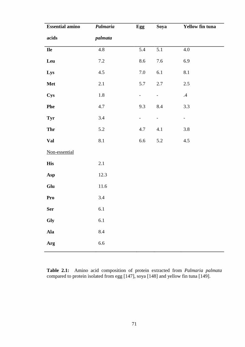
71
Essential amino
acids
Palmaria
palmata
Egg Soya Yellow fin tuna
Ile 4.8 5.4 5.1 4.0
Leu 7.2 8.6 7.6 6.9
Lys 4.5 7.0 6.1 8.1
Met 2.1 5.7 2.7 2.5
Cys 1.8 - - .4
Phe 4.7 9.3 8.4 3.3
Tyr 3.4 - - -
Thr 5.2 4.7 4.1 3.8
Val 8.1 6.6 5.2 4.5
Non-essential
His 2.1
Asp 12.3
Glu 11.6
Pro 3.4
Ser 6.1
Gly 6.1
Ala 8.4
Arg 6.6
Table 2.1: Amino acid composition of protein extracted from Palmaria palmatacompared to protein isolated from egg [147], soya [148] and yellow fin tuna [149].
72
2.4.2 RP-HPLC enrichment of the seaweed hydrolysate
To further isolate renin inhibitory peptides released during papain hydrolysis of P.
palmata protein, RP-HPLC was performed and resultant fractions collected every
minute were tested for their renin inhibitory activities. Figure 2.4 shows the RP-HPLC
chromatogram obtained for the papain hydrolysate of the P. palmata protein. The
fractions (Fr) observed between minute ten and minute twenty-five were collected and
assayed for renin inhibition. The fraction collected at minute 25 (Fr-25) gave the highest
renin inhibitory activity of all fractions tested and inhibited renin by 58.97% (±1.26) at a
concentration of 1 mg/mL compared to the positive control Z-Arg-Arg-Pro-Phe-His-
Sta-Ile-His-Lys-(Boc)-OMe which inhibited renin by 94.71% (±0.87) at a concentration
of 10 µM.

73
73
Figure 2.4: RP-HPLC chromatogram of a papain hydrolysate of P. palmata protein. Absorbance was plotted at 214 nm. The renin inhibitory resultsobtained from the RP-HPLC fractions collected between minute ten to twenty-five (Fr10−Fr25) of the P. palmata papain protein hydrolysate areshown. Samples were tested at a concentration of 1 mg/mL. Data is displayed as the percentage (%) inhibition values. The values obtained are mean ±SEM (n = 3).

74
2.4.3 In vitro inhibitory effects of P. palmaria papain hydrolysate on renin
A papain hydrolysate of the P. palmata protein extract was generated in order to release
renin inhibitory peptides from the parent proteins. When assayed for renin inhibition,
the papain hydrolysed protein extract inhibited renin by 41.89% (±3.22) compared to
the specific renin inhibitor, Z-AAPFH-Sta-IHK-(Boc)-OMe, which was used as the
positive control. This positive control was tested at two different concentrations: 1 μM
(half the maximal inhibitory concentration (IC50) and 10 μM, respectively. These
concentrations were selected due to previous studies where the half maximal inhibitory
concentration (IC50) value for Z-AAPFH-Sta-IHK-(Boc)-OMe was obtained and also
where this compound demonstrated >90% inhibition of renin at a concentration of 10
μM[145]. In this study, Z-AAPFH-Sta-IHK-(Boc)-OMe, assayed at 10 μM and 1 μM,
inhibited renin by 94.71% (±0.87) and 57.35% (±0.91), respectively, as shown in Figure
2.5.

75
Figure 2.5: The percentage renin inhibitory results obtained for the protein, the papain hydrolysed protein, and Fraction (Fr) Fr-25, when assayed at aconcentration of 1 mg/mL. Also shown is the chemically synthesised peptide IRLIIVLMPILMA assayed at the IC50 value of 3.344 mM (±0.31) and thepositive control Z-AAPFH-Sta-IHK-(Boc)-OMe which was assayed at 1 μM and 10 μM. Values are mean ± SEM (n = 3).

76
2.4.4 Identification of renin inhibitory peptides from P. palmata protein
hydrolysate
To identify the renin inhibitory peptides from Fr-25, ESI-QTOF MS was performed and
eleven peptides were elucidated using de novo sequencing (Table 2.2). All eleven
peptides identified had no homology to previously sequenced proteins from P. palmata.
However, only 35 proteins from P. palmata have been characterised to date and all
peptides identified from Fr-25, when aligned withBasic Local Alignment Search
Tool(BLAST), show 100% homology with sections of the proteins; Photosystem II
protein Y from Cyanidium caldarium; Galdieria sulphuraria, Photosystem I reaction
center subunit VII from Chlorella vulgaris; and Cytochrome b6-f subunit 7 from
Cyanidioschyzon merolae. These proteins are found in seaweed species closely related
to P. palmata, including Cyanidioschyzon merolae, Cyanidium caldarium, Galdieria
sulphuraria, and the microalga Chlorella vulgaris. For example, the protein
Cytochrome c oxidase (accession number D5I1R8) isolated from P. palmata has
80.50% homology with Cytochrome c oxidase (accession number Q9ZZQ9) isolated
from Cyanidioschyzon merolae. Furthermore, the protein Photosystem II D2 (accession
number Q52W49), identified in P. palmata previously, is 93.20% homologous, with the
same protein (accession number P28253) isolated from Cyanidium caldarium and
Galdieria sulphuraria (accession number Q9TM47). Finally, the protein Photosystem I
P700 chlorophyll A apoprotein (accession number Q8MAG9) isolated from P. palmata
is 85.30% homologous, with the same protein isolated from Chlorella vulgaris
(accession number P56341).
The chemically synthesized peptides listed in Table 2.2 were tested for renin inhibitory
activities as described. Of the eleven chemically synthesised peptides, the tridecapeptide

77
corresponding to the amino acid sequence IRLIIVLMPILMA had a renin inhibitory
IC50 value of 3.344 mM (±0.31).

78
78
Sequence Phylum Protein (assession number) Species sharing 100% homology Observed mass Calculated mass Charged state
D.IRLIIVLMPILMA.A Rhodophyta Photosystem II protein Y (O19893) Cyanidium caldarium,Galdieria sulphuraria 1494.93 499.29 (+3)
MNEIVALMI.I Rhodophyta Cytochrome b6-f complex (Q85FX8) Cyanidioschyzon merolae 1032.53 517.26 (+2)
P.ILMA.A Rhodophyta Photosystem II protein Y (O19893) Cyanidium caldarium,Galdieria sulphuraria 446.25 447.1235 (+1)
I.LMAASWAIY.N Rhodophyta Photosystem II protein Y (O19893) Cyanidium caldarium,Galdieria sulphuraria 1024.5 1025.48 (+1)
Q.ILPSILVPLV.G Rhodophyta Photosystem I reaction center subunit VII (P58214) Chlorella vulgaris 1062.7 532.32 (+2)
L.PSIL.V Rhodophyta Photosystem I reaction center subunit VII (P58214) Chlorella vulgaris 428.26 429.09 (+1)
I.LVPLVGLV.F Rhodophyta Photosystem I reaction center subunit VII (P58214) Chlorella vulgaris 808.54 809.4 (+1)
V.PLVGLVFPAI.A Rhodophyta Photosystem I reaction center subunit VII (P58214) Chlorella vulgaris 1024.63 1025.48 (+1)
L.VFPAIAM.A Rhodophyta Photosystem I reaction center subunit VII (P58214) Chlorella vulgaris 747.39 748.4 (+1)
V.FPAI.A Rhodophyta Photosystem I reaction center subunit VII (P58214) Chlorella vulgaris 446.25 447.12 (+1)
F.PAIA.M Rhodophyta Photosystem I reaction center subunit VII (P58214) Chlorella vulgaris 370.22 371.11 (+1)
Table 2.2: Peptides identified in RP-HPLC Fr-25 enriched from the P. palmatapapain hydrolysate. ., denotes the point of cleavage by the enzymepapain.

79
2.4.5 In silico analysis of the tridecapeptide IRLIIVLMPILMA
The tridecapeptide IRLIIVLMPILMA identified in this work is longer in length than
most previously identified heart health peptides, such as IPP and VPP, and may be
considered a pro-peptide. Therefore, this peptide was subjected to in silico analysis
using the online computer tool Expasy peptide cutter (http://ca.expasy.org/cgi-
bin/peptidecutter/peptidecutter.pl) [147] to determine potential cleavage peptides and
amino acids that may result from potential digestion of the peptide in vivo with enzymes
and acids. This tool is cost effective and provides a guide for researchers prior to in
vivo animal studies which can be expensive. The option of all enzymes and chemicals
from the list displayed in Expasy peptide cutter was chosen, and the results of cleavage
analysis are shown in Table 2.3.
Table 2.3: Cleavage of the peptide IRLIIVLMPILMA by proteolytic enzymes andacids using the programme Expasy Peptide Cutter
Enzyme Cleavages Position of cleavagesArg-C proteinase 1 2CNBr 2 8 12Chymotrypsin-low specificity (C-term to [FYWML], not before P) 4 3 7 11 12Clostripain 1 2Pepsin (pH1.3) 3 3 6 11Pepsin (pH>2) 3 3 6 11Proteinase K 9 1 3 4 5 6 7 10 11 13Thermolysin 9 2 3 4 5 6 9 10 11 12Trypsin 1 2

80
2.5 Discussion
The protein content obtained in this study were similar to those obtained in the
compositional study carried out previously by Galland-Irmouli et al. (1999). However,
seaweed collected in Ireland was found to contain more of the essential amino acids
isoleucine (4.8 g/ 100g of amino acid) and threonine (5.2 g/ 100g of amino acid)
compared to 4.6 and 0 g/100g of isoleucine and threonine for protein isolated from P.
palmata collected in France.Furthermore, the P. palmata hydrolysate was found to
contain higher levels of threonine and valine than the content of these amino acids
found in eggs [148] and soya [149]. In addition, the seaweed hydrolysate generated in
this work had higher levels of isoleucine, leucine, cysteine, phenylalanine, threonine
and valine than that found in the protein fraction of yellow fin tuna[150]. The Palmaria
palmata protein was also found to contain a substantial level of the essential amino acid
lysine (4.8 g/ 100g of amino acid). These results suggest that P. palmata protein is of
high nutritional value and may be a potential alternative source of proteins rich is
essential amino acids.
Inhibition of the renin angiotensin aldosterone system (RAAS) is a well-established
approach in the treatment of hypertension andthe generation of renin inhibitory
bioactive peptides from food protein sources has been reported previously[72]. In this
study, the enzyme papain was used to release bioactive peptides from P. palmata
protein. Papain was chosen to perform hydrolysis, as it has generally recognized as safe
(GRAS) status. Furthermore, previous literature detailed the peptide generating
capacity of papain, and it was used previously to generate peptides from algal protein
waste capable of reducing high blood pressure in the RAAS through ACE-I inhibition
[151]. Another reason why papain was chosen as a hydrolytic enzyme is that it has a
broad substrate specificity but also exhibits specific substrate preferences for bulky

81
hydrophobic or aromatic residues [152], which may correlate with renin inhibitory
peptides [72]. Inhibition of the enzyme ACE-I, with chemically synthesized ACE-I
inhibitors including captopril (Capoten ®), enalopril, alcacepril, and lisinopril is well-
known [55]. However, inhibition of renin is thought to have several health advantages
over ACE-I inhibition. Renin is the only known enzyme that converts angiotensinogen
to angiotensin I. In the phenomenon known as “ACE escape”, angiotensin II levels
increase in blood plasma due to other enzymes converting angiotensin I to angiotensin
II during ACE-I inhibition [80]. Renin inhibition in turn eliminates angiotensin I from
the bloodstream, preventing the formation of angiotensin II. Another advantage of renin
inhibition over ACE-I inhibition is that renin inhibitors do not affect kinin metabolism
and, hence, are not expected to cause the side effects associated with ACE-I inhibition
including dry cough or angioneurotic edema [131].
While ACE-I inhibitory peptides were isolated from the macroalgaUndaria
pinnatifidabefore [29], this study is, to the best of the authors knowledge, the first to
identify renin inhibitory activities for peptides isolated from P. palmata, or indeed any
macroalga. Papain hydrolysed P. palmata protein inhibited renin by 41.89% (±3.22). It
is well documented that hydrolysis with proteolytic enzymes such as papain is used
frequently as a method to liberate bioactive peptides from plant and other proteins [72,
153]. In this study, hydrolysis of P. palmata protein with papain and further
fractionation using RP-HPLC was found to enhance the renin inhibitory activity of the
protein. Fraction twenty-five (Fr-25) inhibited renin activity by 58.97% (±1.26) at a
concentration of 1 mg/mL compared to the chemically synthesized positive control, Z-
Arg-Arg-Pro-Phe-His-Sta-Ile- His-Lys-(Boc)-OMe, which inhibited renin by 50% at a
concentration of 1 μmol/mL (Figure 2.5).

82
In order to confirm the renininhibitory activity of Fr-25, the peptides within this
fraction were identified and characterised using ESI-Q-TOF mass spectrometry, and
elucidated using de novo sequencing. Eleven peptides (Table 2.1) were chosen for
chemical synthesis based on their amino acid sequences. Sequences with amino acids
containing bulky side chains at the C-terminus and hydrophobic amino acids including
isoleucine and arginine at the N-terminus were chosen for chemical synthesis, as
peptides containing these amino acid sequences were documented previously as
potential enzyme (ACE-I and renin) inhibitors [72, 154] In order to inhibit ACE-I and
renin, aromatic or branched side chain residues are preferred in the amino acid sequence
of the peptide. Aliphatic, basic and aromatic residues are preferred in the penultimate
positions and aromatic, proline and aliphatic residues are preferred in the ultimate
position for ACE-I inhibitory peptides [155]. In this study, chemical synthesis of the
peptides was necessary in order to confirm the observed renin inhibitory activities of
Fr-25. Table 2.1 displays the eleven peptides identified in Fr-25. It also shows the
parent macroalgal proteins that contain the amino acid sequences elucidated for the
identified peptides. These proteins were not characterised for P. palmata. The peptides
elucidated in this study do not share homology with any peptides discovered in P.
palmata, as to date there are only 35 proteins completely sequenced from P. palmata
[152]. However, the eleven peptides identified in Fr-25 were found to correspond to the
amino acid sequences of proteins found in seaweed species closely related to P.
palmata. P. palmata proteins are closely related to proteins sequenced from the seaweed
species Cyanidioschyzon merolae, Cyanidium caldarium, Galdieria sulphuraria, and
Chlorella vulgaris. Inherent protein sequence homologies exist between P. palmata and
these macroalgae species.
In this work, all chemically synthesized peptides were assayed for their abilities to
inhibit renin, and the tridecapeptide IRLIIVLMPILMA (Figure 2.5) inhibited reninby

83
50% at a concentration of 3.344 mM (±0.31). This compared favourably with the
positive control, which was tested at 10 μM and 1 μM, respectively, and also to
previously isolated peptidic renin inhibitors from plant pea sources that displayed IC50
values between 9.2 mM (± 0.18) and 22.66 mM (± 1.71) [72, 153].
Several published works explore the potential of in silico methods for screening
bioactive peptides [72, 154]. In this study, the propeptide IRLIIVLMPILMA identified
in Fr-25 and confirmed to inhibit reninin vitro using the Cayman renin inhibitory
bioassay method was subjected to in silico cleavage analysis using the computer
program Expasy peptide cutter. Enzymes found in the gastrointestinal (GI) tract were
chosen for cleavage analysis. Enzymes that may cleave the propeptide
IRLIIVLMPILMA potentially release di- and tri-peptides. For example, pepsin (pH 1.3
and pH > 2) is predicted to cleave this propeptide at positions 3, 6, and 11, releasing the
dipeptide IR and the tripeptide IIV and the di-peptide MA. It also may release the
pentapeptide LMPIL. In addition, the enzymes trypsin, thermolysin, and arg-C-
proteinase were predicted to release the dipeptide IR from IRLIIVLMPILMA (Table
2.3). Overall, the N-terminal amino acids of the propeptide remained largely uncleaved,
with only proteinase K capable of cleaving the peptide in the first position. These results
suggest that this propeptide and the di- and tripeptides released from it may cross the
lumen into the bloodstream and potentiate an antihypertensive effect. Indeed, certain
peptides including IR and IRL were previously documented as having renin,
antioxidant, and ACE-I inhibitory activities [72, 141]. The peptide IR has a documented
IC50 value of 9.2 mM for renin [72].Furthermore the peptide IRLIIVLMPILMA is quite
hydrophobic due to the isoleucine, leucine and valine residues. Hydrophobic peptides
were shown previously to be considerably bioavailable with the capabilities to cross the
gut mucosa and to enter the blood stream more efficiently [110].

84
CHAPTER 3. DEVELOPMENT OF A SEAWEED DERIVED
PLATELET ACTIVATING FACTOR ACETYLHYDROLASE
(PAF-AH) INHIBITORY HYDROLYSATE, SYNTHESIS OF
INHIBITORY PEPTIDES AND ASSESSMENT OF THEIR
TOXICITY USING THE ZEBRAFISH LARVAE ASSAY.
This chapter was published as a research article in the Journal Peptides: Ciarán
Fitzgerald, Eimear Gallagher, Paula O’Connor, José Prieto, Leticia Mora-Soler,
Maura Grealy, Maria Hayes, “Development of a seaweed derived platelet activating
factor acetylhydrolase (PAF-AH) inhibitory hydrolysate, synthesis of inhibitory peptides
and assessment of their toxicity using the zebrafish larvae assay”, (2013), 50, 119-124.
85
3.1 Aims
Dietary modifications for heart health continue to rise in popularity with consumers.
While nine in 10 (87%) persons with chronic high blood pressure and/or cholesterol
depend heavily on prescriptions to treat their condition, 50 % are also using diet to
manage their condition. Circulatory health ingredients are popular with consumers and
up and coming heart markets include stroke, atherosclerosis, minimizing blood plaque
and improving circulation. As well as targeting the enzyme Renin, this thesis examined
the use of the red seaweed as a source of PAF-AH inhibitory peptides. A PAF-AH
inhibitory peptide NIGK was generated from a hydrolysate of P palmata protein using
the enzyme papain and screened for its ability to inhibit PAF-AH. This work was
pursued as the vascular inflammatory role of platelet activating factor acetylhydrolase
(PAF-AH) is thought to be due to the formation of lysophosphatidyl choline and
oxidized non-esterified fatty acids. This enzyme is considered a promising therapeutic
target for the prevention of atherosclerosis and there is a need to expand the available
chemical templates of PAF-AH inhibitors. The prevention of atherosclerosis through
diet is an interesting area of scientific research and one which this thesis explores.
Within this chapter, PAF-AH inhibitory peptides were isolated and characterized from
the red macroalgae P. palmata. Furthermore, it examines the toxicity of a P. palmata
protein hydrolysate using a zebrafish larvae assay. The main objectives of this chapter
were to:
(1) Examine Palmaria palmata protein hydrolysate for peptides with the ability to
inhibit the action of the enzyme PAF-AH which is important in circulation
health
(2) Use in vitro bioassay guided fractionation to identify PAF-AH inhibitory
hydrolysates and RP-HPLC fractions.

86
(3) Identify and sequence PAF-AH inhibitory peptides using nano- ESI-Q-TOF
MS, de novo sequencing and solid phase peptide synthesis.
(4) Examine the toxicity of the P. palmata protein hydrolysate using a zebrafish
larvae assay as an experimental model

87
3.2 Introduction
Atherosclerosis is an inflammatory disease and the most common cause of stroke and
Cardiovascular disease worldwide [156] . Recent studies suggest that the enzyme
lipoprotein-associated phospholipase A2 (Lp-PLA2) or platelet activating factor
acetylydrolase (PAF-AH) plays an active role in atherosclerotic development and
progression [82, 157]. Indeed, in 2008, Wilensky and colleagues showed that selective
inhibition of PAF-AH prevented progression to advanced coronary atherosclerotic
lesions and confirmed the role of vascular inflammation, independent from
hypercholesterolemia, in the development of lesions implicated in the pathogenesis of
myocardial infarction and stroke [82].
As previously stated, PAF-AH is secreted from multiple inflammatory cells but is
highly expressed in the necrotic core of atherosclerotic lesions [83, 158]. PAF-AH is
predominately bound to apolipoprotein containing lipids and 80% of PAF-AH in blood
plasma is bound to low density lipoproteins (LDL) [83, 158]. LDL is oxidised following
its transportation from the lumen to the intima of the cell [83]. This facilitates
hydrolysis of LDL by the enzyme PAF-AH into two pro-inflammatory mediators;
lysophosphatidylcholine (LPC) and oxidised non-esterified fatty acids (oxNEFAs) [82,
159].
Growing evidence supports a pro-atherogenic role of PAF-AH in blood circulation.
Pharmaceutical companies including GlaxoSmithKline (GSK) have invested financially
in the development of synthetic compounds to combat atherosclerosis through PAF-AH
inhibition. For example, GSK are in phase three clinical trials with the substituted
pyrimidone darapladib, a synthetic, non-peptidic PAF-AH inhibitor [160]. However,
very few natural inhibitors of PAF-AH are known to date and a patent search regarding
PAF-AH inhibitors derived from plant and natural resources yielded only a few studies

88
regarding uncharacterized “crude” extracts. For example, Yu et al. identified PAF-AH
inhibitors from extracts generated from 224 terrestrial plants [160]. Methanolic extracts
were generated from these plants and were further partitioned with n-hexane,
chloroform and ethyl acetate [161]. Seven methanol extracts were found to inhibit PAF-
AH by 50% at a concentration of 100 mg/ml and two ethyl acetate extracts were found
to inhibit PAF-AH activity at the same concentration [161]. In another study, synthetic
analogs of cyclic enol-carbamates isolated from the bacteria Pseudomonas fluorescens
were identified as potent inhibitors of PAF-AH [162]. Drugs that influence lipoprotein
concentration were studied previously for their potential inhibitory effects on PAF-AH.
Drugs examined to date included niacin, statins and finofibrate [162].
In this chapter, the amino acid composition of Irish sourced P. palmata was determined.
Furthermore a PAF-AH inhibitory tetrapeptide with the amino acid sequence
Asparagine-Isoleucine-Glycine-Lysine (NIGK) was generated, enriched for and isolated
from the red seaweed P. palmata by hydrolysis of this seaweed protein with the food
grade enzyme papain. This enzyme was chosen as it was used successfully in previous
studies to release bioactive peptides from protein. Papain is also a food grade cysteine
protease with known proteolytic capabilities [153, 163]. The peptide NIGK was
chemically synthesized and its PAF-AH inhibitory activity confirmed using an in vitro
bioassay method. In addition, the toxicity of the seaweed hydrolysate was assessed
using the zebrafish larvae bioassay. Two-day-old zebrafish larvae were exposed to 1, 5,
and 10 mg/ml concentrations of the hydrolysate over a 48 hr period to examine any
toxic effects the hydrolysate may have in a living model.

89
3.3 Materials and Methods
3.3.1 Materials and Reagents
The PAF-AH inhibitor screening kit was supplied by the Cayman Chemical Company
(Ann Arbor, MI, USA). Acetonitrile and water were supplied by Romil Ltd.
(Cambridge, England, United Kingdom). Dimethyl sulfoxide (DMSO), Ammonium
sulphate and the specific platelet activating factor (PAF) inhibitor methyl arachidonyl
fluorophosphates (MAFP), which was used as a positive control, were supplied by
Sigma–Aldrich (Steinheim, Germany). H-Ala-HMPB-ChemMatrix and H-Ile-HMPB-
ChemMatrix resins were supplied by Produits Chimiques Auxiliaires et de
Syntheses(PCAS) Biomatrix Inc. (Quebec, Canada). All other chemicals used were of
analytical grade. Instant Ocean ® was acquired from Spectrum Brands (Ontario,
Canada)
3.3.2 Palmaria palmata protein extraction and hydrolysis with papain
Protein was extracted from P. palmata following the protocol of Galland-Irmouli et al.
[38] as described in section 2.2.2. The hydrolysis process used was the same as the
process used to isolate renin inhibitory peptides in section 2.2.3.
3.3.3 RP-HPLC analysis
RP-HPLC analysis was carried out according to the method described in Section 2.3.5.
The same RP-HPLC chromatogram was generated.
3.3.4 PAF-AH inhibitor screening assay
This assay was carried out using a PAF-AH inhibitor kit supplied by Cayman Chemical
Company in accordance with the manufacturers’ instructions. Briefly, prior to carrying
out the assay, the PAF-AH inhibitor assay buffer was diluted by adding 27 ml of HPLC

90
grade water to 3 ml of 0.1 M Tris–HCl (pH 7.2). The substrate, 2-thio PAF was
reconstituted in 12 ml of diluted assay buffer to achieve a concentration of 400 µM.
Background wells were prepared by adding 10 µl of assay buffer, 200 µl of 2-thio PAF
substrate and 10 µl of solvent (in this case DMSO/water 5:95, v/v) together in a well
(×3). Initial activity wells were prepared by adding 200 µl of the 2-thio PAF substrate
solution and 10 µl of the solvent DMSO/water (5:95, v/v) together in a well (X3).
Inhibitor wells were prepared by adding 200 µl of the 2-thio PAF substrate solution and
10 µl of the test fraction. The reaction was initiated by adding 10 µl of human plasma
PAF-AH to all wells except the background wells. The plate was then covered and
incubated for 20 min at 25 ºC. After incubation, 10 µl of 5, 5’ dithio-bis (2-
nitrobenzoicacid) (DTNB) was added to all wells to develop the reaction. All potential
inhibitors were assessed in triplicate. The PAF-AH inhibition assay uses 2-thio PAF as
a substrate for PAF-AH. The acetyl thioester bond is cleaved at the sn-2 position by
PAF-AH [164]. The sulphur bond of the DTNB molecule is then broken due to the
available ionic sulphur on the substrate leaving the 5-thio-2-nitrobenzoic acid which can
be detected at an absorbance of 414nm (as illustrated in Figure 3.1). Methyl arachidonyl
fluorophosphonate (MAFP), a known PAF-AH inhibitor was used as a positive control.
The inhibitory concentration that reduced PAF-AH activity by 50% (IC50) was
determined by nonlinear regression from a plot of peptide concentration versus
percentage inhibition.

91
Figure 3.1: The PAF-AH inhibition assay. PAF-AH enzyme activity is assessed bymeasuring the amount of 5-thio-2-Nitrobenzoic Acid produced after PAF-AH cleavesthe acetyl thioester bond at the sn-2 position of the 2-thio PAF substrate in the presenceof DTNB.

92
3.3.5 Fractionation of Palmaria palmata hydrolysate using RP-HPLC
The P. palmaria papain hydrolysate was enriched further as described in section 2.2.5.
3.3.6 Peptide identification by tandem mass spectrometry
Peptides were identified using the method described in section 2.2.7.
3.3.7 Database search, confirmation of sequences, and de novo sequencing
Sequencing and confirmation of peptide sequences were carried out by the same method
as described in 2.2.8.
3.3.8 Microwave-assisted solid phase peptide synthesis
Peptides were sequenced using the method described in section 2.2.9.
3.3.9 Zebrafish larvae assay
The protocols for the maintenance and care of zebrafish (Daniorerio) were carried out
according to guidelines published by Westerfield (1993) [165]. The fish were kept in a
room at 28 ºC with a light cycle of 14:10 light:dark and at a stocking density of 20 fish
per 20 L tank. On the evening before spawning, two male fish were placed in spawning
trays (20 cm × 9 cm) with a female separated from the males by a mesh insert. Upon
entering the light cycle the following morning, the fish were put together. Post
spawning, embryos were gathered from the base of the spawning trays and cultured in
filter-sterilized egg water (60 µg/ml “Instant Ocean” aquarium salts in distilled water) at
28 ºC. To assess the effects of the P. palmata protein hydrolysate on the zebrafish
larvae, 48 h old larvae were placed in a 12 well plate (ThermoFisher Scientific, MA,

93
USA). Ten zebrafish larvae were present in each well of this plate. The hydrolysate was
tested in triplicate (3 wells containing 10 zebrafish per well) at 1, 5, and 10 mg/ml
concentrations along with a control of distilled water. The heart rate and survival of the
larvae were then monitored over a 48 h period by visually counting the heart beats over
the course of a minute. Images were captured on a Nikon DXM1200C Digital camera at
a magnification of 4 and an exposure of 1/300 s using the program Nikon ACT-1C
(Nikon, Tokyo, Japan).

94
3.4 Results
3.4.1 PAF-AH inhibitory activity of P. palmata hydrolysates and fractions
The PAF-AH inhibitory peptides identified in this study were found to correspond to
peptide sequences found in proteins characterised from macroalgal species closely
related to P. palmata. The amino acid sequences of peptides identified in this study
using de novo sequencing are presented in Table 3.1. All peptides presented are
between four and 13 amino acids in length. When aligned using BLAST, these peptides
share 100% sequence homology with the protein Photosystem II protein Y from
Cyanidium caldarium and the protein Photosystem I reaction subunit VII from
Chlorella vulgaris. The peptide NIGK corresponded to the amino acid sequence 22–25
of the protein Photosystem II Protein Y found in the red algae C. caldarium and
Galdieria sulphuraria.
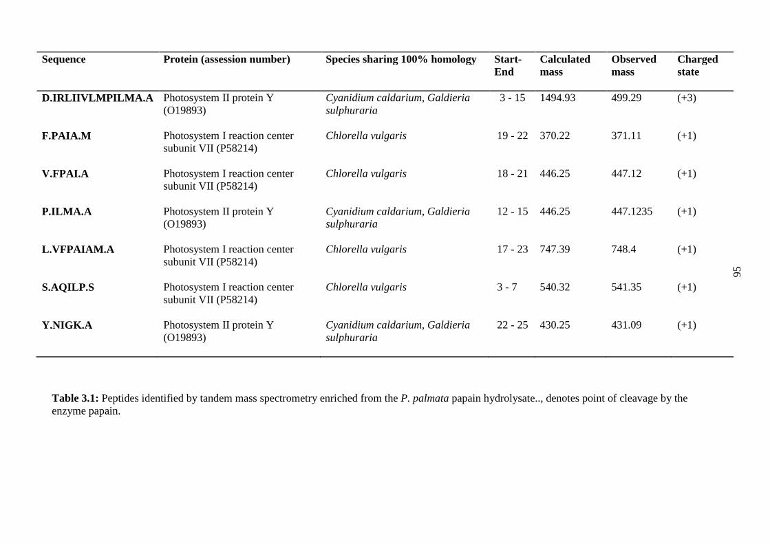
95
Sequence Protein (assession number) Species sharing 100% homology Start-End
Calculatedmass
Observedmass
Chargedstate
D.IRLIIVLMPILMA.A Photosystem II protein Y(O19893)
Cyanidium caldarium, Galdieriasulphuraria
3 - 15 1494.93 499.29 (+3)
F.PAIA.M Photosystem I reaction centersubunit VII (P58214)
Chlorella vulgaris 19 - 22 370.22 371.11 (+1)
V.FPAI.A Photosystem I reaction centersubunit VII (P58214)
Chlorella vulgaris 18 - 21 446.25 447.12 (+1)
P.ILMA.A Photosystem II protein Y(O19893)
Cyanidium caldarium, Galdieriasulphuraria
12 - 15 446.25 447.1235 (+1)
L.VFPAIAM.A Photosystem I reaction centersubunit VII (P58214)
Chlorella vulgaris 17 - 23 747.39 748.4 (+1)
S.AQILP.S Photosystem I reaction centersubunit VII (P58214)
Chlorella vulgaris 3 - 7 540.32 541.35 (+1)
Y.NIGK.A Photosystem II protein Y(O19893)
Cyanidium caldarium, Galdieriasulphuraria
22 - 25 430.25 431.09 (+1)
Table 3.1: Peptides identified by tandem mass spectrometry enriched from the P. palmata papain hydrolysate.., denotes point of cleavage by theenzyme papain.

96
3.4.2 PAF-AH inhibition of synthesised peptides
The PAF-AH inhibitory values are presented in Figure 3.2. All synthesised peptides and
the P. palmata protein hydrolysate were tested at 1 mg/ml while the known PAF-AH
inhibitor MAFP (thepositive control) was tested at a concentration of 259.35 ng/ml.
This concentration was chosen as previous studies reported this as the concetration at
which 90% of the PAF-AH enzyme was inhibited [166]. The peptide with the amino
acid sequence NIGK was the strongest peptidic inhibitor observed, inhibiting PAF-AH
by 50.74% (± 2.12) at a concentration of 1 mg/ml. The protein hydrolysate from which
these peptides were derived inhibited PAF-AH by 30.37% (± 1.97) at the same
concentration compared to the positive control MAFP which inhibited PAF-AH by
80.56% (±1.84) at a concentration of 259.35 ng/ml.

97
Figure 3.2: PAF-AH inhibitory activities of seven, chemically synthesized peptides isolated from Palmaria palmata protein; The P. palmatahydrolysate and as a control, the known PAF-AH inhibitor methyl arachidonyl fluorophosphates (MAFP). Values are mean ± SEM (n = 3).
98
3.4.3 Toxicity of P. palmata hydrolysate on zebrafish larvae
Figure 3.3.A shows the survival of the larvae over a 48 h period. A significant toxic
effect was observed when the larvae were subjected to a dose of 10 mg/ml of the P.
palmata hydrolysate. Less than 20% of the larvae survived after 24 h and none after 30
h of exposure. This effect was also apparent when the heart rate of the larvae was
monitored (Figure 3.3.B). The heart rate of the larvae dropped to 46 beats per minute
(BPM) compared to the control which had a heartbeat of 122.66 BPM after 24 h of
contact with the hydrolysate. When the larvae were exposed to 5 mg/ml of the
hydrolysate, no significant drop in percentage survival rates of the larvae or drop in
heart rates were observed after 24 h of exposure as shown in Figure 3.3.A and B.
However, after 48 h of contact with the hydrolysate at a concentration of 5 mg/ml,
survival rates of the larvae were down to 30% and the BPM of the surviving larvae was
113 BPM compared to the control BPM of 133.3. No adverse effects were seen in terms
of percentage survival or BPM of the larvae exposed to a concentration of 1 mg/ml of
the P. palmata hydrolysate. All larvae survived after 48 h exposure and the heart rate of
these larvae was not significantly different to that of the larvae used in the control wells.

99
0 10 20 30 40 500
50
100
Control
10 mg/ml
5 mg/ml
1mg/ml
Time (hrs)
Perc
enta
ge
surv
ival
Contr
ol
10m
g/ml
5m
g/ml
1mg/m
l0
50
100
150
***
Concentration of hydrolysate (mg/ml)
BP
M
Contro
l
10m
g/ml
5m
g/ml
1mg/m
l0
50
100
150
*
***
Concentration of hydrolysate (mg/ml)
BP
M
A
B C
Figure 3.3: Survival of zebrafish larvae over 48 h when exposed to the control and 10, 5 and 1 mg/ml of the P. palmata hydrolysate can be seen in A.Values are mean ± SEM (n = 3).B and C shows the heart rate in beats per minute (BPM) of the larvae when exposed to the control and 10, 5 and 1mg/ml of the hydrolysate after 24 h contact and 48 h contact, respectively. Values are mean ± SEM (n = 3). *P < 0.05, **P < 0.01 and ***P < 0.001compared with the control.

After 24 hr (Figure 3.4.A) of exposure to the hydrolysate at concentrations of 1 mg/ml
and 5 mg/ml, the larvae were found to have no significant morphological differences
when compared to the control larvae with the exception of a slightly swollen yolk sack
in larvae exposed to 5 mg/ml of the hydrolysate. The larva exposed to 10 mg/ml of the
hydrolysate (Figure 3.4.A) developed severe curvature of the spine and a deformed yolk
sack. After 48 h, (Fig 14.B) the larvae exposed to 1 mg/ml of the hydrolysate were still
morphologically identical to that of the control while the larvae exposed to 5 mg/ml
appeared to have swollen yolk sacks and also developed curved spines.
Figure 3.4: Images of the larvae acontrol and 10, 5 and 1 mg/ml of
A
B
5 mg/ml
100
fter 24 h exposure (A) and 48 h expthe hydrolysate.
10 mg/ml
Control1 mg/ml
Control
o
5 mg/ml
1 mg/ml
sure (B) to the

101
3.5 Discussion
RP-HPLC was used to further fractionate potential PAF-AH inhibitory peptides from
the P. palmata hydrolysate. Sixty fractions were generated and screened for PAF-AH
inhibitory activities [164]. Fraction twenty-five was further purified using ESI-Q-TOF
MS. All peptides identified and selected for chemical synthesis, with the exception of
NIGK, were hydrophobic in nature and had either an aromatic or a hydrophobic residue
at their N-terminus. For example, the peptide FPAI possessed the aromatic residue
phenylalanine while the hydrophobic residue proline was present in the peptide PAIA.
Previous studies have linked heart health bioactivity (ACE-I and renin enzyme
inhibition) with hydrophobic or aromatic amino acids at the N-terminus of the peptide
[72, 167]. The peptide NIGK was chosen for chemical synthesis and further bioactivity
assessment due to its water soluble nature as water soluble peptides are easily included
in food and pharmaceutical products and are suitable for large scale production. In
addition, the positive charge associated with both the arginine and lysine residues at the
N- and C-terminal ends of the peptide sequence are associated with the characteristics
of heart health peptides identified previously [83]. As discussed in Chapter two, a
problem that was encountered during this work was that only 35 P. palmata proteins
were fully characterised and accessible in the PubMed database
(http://www.ncbi.nlm.nih.gov). The peptides identified in this study and shown in Table
3.1 were found to correspond to peptide sequences found in proteins characterised from
macroalgal species closely related to P. palmata including C. caldarium, G. sulphuraria
and C. vulgaris. In the absence of an entirely sequenced proteome for P. palmata this
homology verifies the origins of these peptides along with e-values where available
using BLAST (http://blast.ncbi.nlm.nih.gov/Blast). The e-values obtained for the
peptides identified in this study ranged from 1.1 to 4.0 × 10⁻² when assessed using
102
BLAST. Furthermore, the sequences identified were not found in the peptide database
BIOPEP (http://www.uwm.edu.pl/biochemia/index.php/pl/biopep). However, the di-
peptides NI and GK that could potentially be generated from peptide NIGK were
identified in the sequences of other peptides found within BIOPEP. Figure 3.2 shows
the percentage PAF-AH inhibitory values of the seven peptides identified from the P.
palmata papain hydrolysate that were assayed for PAF-AH inhibition at a concentration
of 1 mg/ml. All synthesized peptides and the P. palmata protein hydrolysates were
tested at 1 mg/ml as this concentration was shown to be non-toxic when tested on
zebrafish larvae (Figures 3.3 and 3.4). Furthermore, where enzyme inhibition by
peptides generated from food sources was studied previously, concentrations of 1
mg/ml were used [72].
The concentration at which the tetrapeptide NIGK exhibited half the maximal inhibitory
concentration (IC50) was 2.32 mM (±2.12) compared to the positive control MAFP
which inhibited PAF-AH by 80.56% (± 1.84) at a concentration of 700 nM. The
positive control MAFP compound is a proven inhibitor of PAF-AH in endothelial cells
[166].
The PAF-AH inhibitory drug darapladib operates through inhibition of azetidinone
[168]. In this chapter, the mechanism of action of PAF-AH inhibition by the
tetrapeptide NIGK and whether it inhibits in a competitive or non-competitive manner
was not examined. The catalytic site of PAF-AH contains a Serine residue at position
273 [168]. Azetidinones target the serine residue in the active site of enzymes [168].
Inhibition of PAF-AH by the peptide NIGK may be due to salt bridge formation
between the Lysine residue at the C-terminus of NIGK and the Serine residue of the
active site of PAH-AH, which in turn, would incapacitate the active site [169]. The
inhibitory concentrations of NIGK and the P. palmata papain protein hydrolysate are

103
much higher than those reported for the crude plant extract PAF-AH inhibitory
activities reported by Yu et al., where extracts inhibited PAF-AH by 50% at a
concentration of 100 mg/ml [161]. However, these extracts were not purified and the
active components were not identified.
While seaweed and seaweed products have, for centuries, been consumed as part of a
normal diet in numerous countries across the globe, it was important to examine any
toxicological effects this particular hydrolysate may have in vivo. Sensory analysis on a
bread product containing this hydrolysate was carried out in a future Chapter (Chapter
5). These concerns were addressed by use of the zebrafish larvae assay. Zebrafish
embryos and larvae make for excellent models when screening compounds, where only
a small quantity of test material is available for use. Furthermore, zebrafish survive for
days in low volumes of water [170]. Results obtained in Figures 3.3 and 3.4
demonstrated that the zebrafish larvae exposed to 1 mg/ml of the P. palmata protein
hydrolysate were not adversely affected by this concentration of the PAF-AH inhibitory
hydrolysate. This assay was carried out in triplicate (3 × 10 zebrafish larvae). P.
palmata hydrolysate concentrations of 5 and 10 mg/ml however were toxic to the
zebrafish larvae assayed. Use of the zebrafish larvae assay as a model system goes back
to the early 1970s when George Streisinger selected zebrafish larvae to develop the first
Vertebrate assay enabling forward genetic screening [171, 172]. Zebrafish were mainly
used to study organ development. Indeed, zebrafish are seen as a relevant model for
human disease and pharmaceutical research [173, 174]. An advantage of the zebrafish
model is that they can be used in medium and large scale screening procedures as they
are small, robust and survive hostile conditions [175]. Other advantages for using
zebrafish larvae include; a clear embryo which permits unobstructed observation of
developmental morphology, the high rate of this development, high fecundity, and
develpment that is similar to that of mammals [176]. The main limitation with the use of

104
zebrafish larvae in assessing potential safety issues, as is the case in this study, is the
lack of extensive validation with diverse and comprehensive pharmacological
compound sets to adequately understand the advantages and limitations of the zebrafish
larve model [177]. Another drawback of the zebrafish larvae assays is that uptake of
compounds into the zebrafish larvae can vary and they should be measured for accurate
interpretation of results, thereby avoiding false negatives [177]. Furthermore, the
avoidance of a false negative result in terms of the toxicity of the peptide and
hydrolysate was overcome in Chapter four where the toxicity of the hydrolysates was
assessed by Eurofin Foods Ireland Ltd, (Finglas, Dublin 11, Ireland). Despite these
limitations, the zebrafish larvae model was chosen here as the author deduced that it
offered a novel method for partially predicting and assessing the safety liabilities of the
hydrolysate from which the tetrapeptide NIGK was derived. The effects of this
hydrolysate were also assessed in an animal rodent model in Chapter five of this thesis.
This chapter identified the amino acid sequence of a peptide isolated from a macroalga
that displayed in vitro PAF-AH inhibitory activity. The non-toxic effects displayed by
the hydrolysate at a concentration of 1 mg/ml coupled with its PAF-AH inhibitory
activity at this concentration, suggests it has a potential role as a functional food
ingredient for maintenance of heart health. As the peptide identified in this study,
NIGK, targets the enzyme PAF-AH, it could potentially be used with ACE-I or renin
inhibitors to prevent atherosclerosis and high blood pressure development; both risk
factors for the development of stroke and cardiovascular disease.
105
CHAPTER 4. IDENTIFICATION OF AN ACTIVE SEAWEED
DERIVED RENIN INHIBITORY DIPEPTIDE AND
CONFIRMATION OF A HYPOTENSIVE EFFECT IN
SPONTANEOUSLY HYPERTENSIVE RATS FOLLOWING
SIMULATED GASTROINTESTINAL DIGESTION.
This work is under review as a research article for publication in theJournal of
Agriculture and Food Chemistry: Ciarán Fitzgerald, Rotimi Aluko, Mohammad
Hossain, Dilip Rai, José Prieto and Maria Hayes. Identification of an active seaweed
derived renin inhibitory dipeptide and confirmation of a hypotensive effect in
spontaneously hypertensive rats following simulated gastrointestinal digestion.
Animal studies performed in section 4.3.6 were carried out by the staff of the
Department of Human Nutritional Sciences at the University of Manitoba closely
following the authors’ experimental design.

106
4.1 Aims
In order to exert a bioactive effect in vivo, oral delivery of the renin inhibitory peptide
identified in Chapter two and the hydrolysate are necessary. Oral delivery of bioactive
peptides and proteins face immense challenges due to the gastrointestinal environment.
After consumption, the gastrointestinal epithelium acts as a physical and biochemical
barrier for absorption which may result in low bioavailability. An ideal bioactive
peptide candidate should be able to reach the site of absorption intact. Alternatively, the
active component of the bioactive peptide should survive GI digestion and should be
able to exert a beneficial effect at the active site and a positive in vivo effect should be
observed.
The aims of this chapter were:
(1) To determine potential cleavage of the renin inhibitory peptide with the amino
acid sequence IRLIIVLPILMA using in silico analysis. This aim provides a
guide for future assessment in animal models.
(2) To assess if the renin inhibitory tridecapeptide could survive gastrointestinal
digestion and to determine the resulting dipeptides using mass spectral (MS)
analysis. This was assessed using an in vitro simulated gastrointestinal
digestion process and Nano-ESI-MS mass spectral (MS) analysis.
(3) To determine if the hydrolysate and the renin inhibitory tridecapeptide had any
hypotensive effect in an animal model. Spontaneously hypertensive rats (SHRs)
were chosen as a suitable model for this work. SHRs were used as the animal
group of choice previously for determination of the hypotensive effect of
bioactive peptides of both pharmaceutical and food origin. SHR rats are
recognized as a suitable animal model [178].

107
4.2 Introduction
To reach their proposed target, naturally occurring bioactive peptides intended for
inclusion in functional foods must first survive food processing conditions.
Furthermore, they must be capable of enduring gastrointestinal digestive enzymes.
Indeed, past studies have shown that ACE-I inhibitory peptides with inherent in vitro
activity exhibited poor activity in vivo due to inactivation during gut transit
[179].Conversely, previous studies have also shown that bioactive peptides that inhibit
the enzymes of the RAAS survived cleavage by digestive enzymes and increased
activity of the peptides post cleavage was reported [101, 119]. When investigating if a
compound with apparent in-vitro bioactivity can overcome the conditions of the GI
tract, simulated gastrointestinal digestion is a useful methodology that can be used prior
to animal experimentation in order to determine the in vivo effect of a compound
identified in vitro as bioactive [180]. There is great variety in the literature concerning
in vitro digestion studies. These varieties relate to the enzymes used, the conditions of
hydrolysis used and the type of analysis performed on the resultant products [181].
However, in general, most methods attempt to mimic the gastric phase through
digestion with pepsin at pH 2-3 and subsequent duodenal phase digestion using
pancreatic proteases such as trypsin and chymotrypsin at a pH of between 7 and 8 [181].
Spontaneously hypertensive rats (SHR) have been used as a model for the measurement
of hypertension since the late 1960’s [178]. SHR rats are not the only animal model
used to study the effects of hypertension in vivo. Other animal models used to study
hypertension include the Dahl salt-sensitive rats derived by Dahl from the Sprague-
Dawley stock. This breed was developed in order to have a suitable model for
monitoring hypertension caused by a high sodium chloride (NaCl) diet [182]. Other
animal models include renovascular hypertension rats [182]. However, due to their

108
broad use to date, far more data has been obtained with SHR and it is therefore easier to
compare results generated with SHRs to previous findings [178].Indeed, to date, two
studies have used the SHR rat model to verify the effect of naturally occurring renin
inhibitory peptides in vivo[74, 183]. In addition, our study wanted to assess the effects
of the peptide hydrolysate on hypertensive subjects.
In this work, the tridecapeptide IRLIIVLMPILMA identified in Chapter two was
subjected to a simulated gastrointestinal digestion procedure to assess if it could survive
GI transit and potentially be used to positively affect hypertension. In silico cleavage
analysis coupled with a simulated gastrointestinal digestion method were developed and
used to assess survival of this peptide in vivo using SHRs. The peptide hydrolysate from
which the renininhibitory peptide was derived was also observed in all models. The
effect on the blood pressure of the animals was observed over a 24 hour period.

109
4.3 Materials and Methods
4.3.1 Materials
The tridecapeptide IRLIIVLMPILMA was synthesized by GL Biochem Ltd.,
(Shanghai, China). All enzymes used for the simulated gastric digestion of the
tridecapeptide including; pepsin from porcine gastric mucosa, amylase, pancreatin and
porcine bile extract were obtained from Sigma Aldrich (Dublin, Ireland). All reagents
used in the formulation of the gastric fluids including, potassium chloride, monobasic
potassium phosphate, sodium bicarbonate, NaCl and magnesium chloride hexahydrate
were also sourced from Sigma Aldrich (Germany).
4.3.2 In silico analysis of the renin inhibitory peptide IRLIIVLMPILMA
The programme Expasy peptide cutter (http://web.expasy.org/peptide_cutter/) was used
to predict where the main proteases of the gastrointestinal (GI) tract could cleave the
renin inhibitory tridecapeptide IRLIIVLMPILMA.
The online tool ToxinPred (http://crdd.osdd.net/raghava/toxinpred/index.html) was used
to predict the toxicity of the peptide and its fragments after hydrolysis[147]. In addition,
the hydrolysate was assessed for its’ heavy metal and Iodine content by Eurofin Foods
Ireland (Finglas, Dublin 11, Ireland) as described in Chapter two.
4.3.3 Simulated Gastric Digestion of the peptide IRLIIVLMPILMA
The simulated gastric digestion method was carried out according to the procedures
developed by the European Cooperation in Science and Technology (COST) funded
group INFOGEST (http://www.cost-infogest.eu/). Simulation of the gastric digestion
process was carried out in three phases as shown in Figure 3.5. Initially, an oral phase

110
containing 1 g of the synthesised peptide was mixed with 1 ml of simulated salivary
fluid (SSF) (Table 4.1) and 150 U/ml of amylase. This mixture was left to stir in a
Water bath (Stuart Shaking Water Bath, Staffordshire, UK) at 37 ºC for 2 minutes. The
second phase of the simulation was the gastric phase. The oral phase was mixed with 2
ml of the prepared simulated gastric fluid (SGF) (Table 4.1) along with pepsin at a
concentration of 1000 U/ml of SGF and 0.0375. The pH was adjusted to pH 3 using 1M
hydrochloric acid (HCL) and left to stir in a Water bath (Stuart Shaking Water Bath,
Staffordshire, UK) at 37 ºC for two hours. The final stage of the digestion process was
the duodenal phase. For this phase, 4 ml of the simulated duodenal fluid (SDF) (Table
4.1) was added to the SGF along with pancreatin at a concentration of 100U/ml of
trypsin activity. Porcine bile extract was also added at a concentration of 2.4 mg/ml of
the total volume. The mixture was adjusted to pH 7 using 1 M NaOH and left to stir in a
water bath at 37 ºC for 2 hours before deactivation by heating at 90 ºC for 20 min and
subsequent freeze-dried.
Simulated Saliva Fluid (SSF) Simulated Gastric Fluid(SGF)
Simulated Duodenal Fluid(SDF)
Compound Concentration Compound Concentration Compound ConcentrationKCl 12.50 mM KCl 35.08 mM KCl 6.91 mMKH2PO4 14.02 mM KH2PO4 0.63 mM KH2PO4 0.56 mMNaHCO3 7.99 mM NaHCO3 25.98 mM NaHCO3 43.00 mMNaCl 4.10 mM NaCl 41.00 mM NaCl 32.85 mMMgCl2(H2O)6 0.29 mM MgCl2(H2O)6 0.59 mM MgCl2(H2O)6 0.32 mM
Table 4.1: Concentrations of electrolytes used to create the simulated saliva fluid (SSF)simulated gastric fluid (SGF) and simulated duodenal fluid (SDF).

111
Figure 4.1: Schematic of simulated in vitro gastric digestion. The simulation wascarried out in three stages; 1) Oral stage 2) Gastric stage 3) Duodenal stage. The 3Dimage of IRLIIVILMPILMA was generated using primary sequence information andthe programme Peplook.

112
4.3.4 Removal of polyethylene glycols from Palmaria palmata protein digested
samples using a titanium dioxide (TiO2) cleanup procedure.
Digested samples were cleaned-up for mass spectrometry analysis and determination of
the peptide/peptide fragments using Pierce™ Graphite spin Columns which were
supplied as part of the Pierce™ TiO2phospopeptide enrichment and clean-up kit[184].
The graphite spin columns were used according to the manufacturers’ instructions.
Briefly, the column was prepared by placing it in a 2 mL eppendorf tube followed by
centrifugation at 2000 × g for 1 minute to remove storage buffer. The column was then
rinsed twice with 100 µL of NH4OH and subsequently centrifuged at 2000 ×g. The
graphite was activated by adding 100 µL of acetonitrile followed by centrifugation at
2000 × g for 1 minute before adding 100 µL of 1 % trifluoroacetic acid (TFA) in water
(1:99 v/v) followed again by centrifugation of the column at 2000 × g for 1 minute.
After column preparation, 50 mg of the hydrolysate was added and allowed to bind to
the column for 10 minutes with periodical vortexing. The column was then centrifuged
at 1000 × g for 3 minutes and the flow through discarded. The column was subsequently
placed in a new tube and washed with 200 µL of 1% TFA followed by centrifugation at
2000 × g for 1 minute. The column was then placed in a new tube and spun at 2000 × g
for 1 minute following addition of 100 µL of 0.1% formic acid in a 50:50
acetonitrile:water (v/v) ratio. This step was repeated three times and the resulting
elution was freeze-dried. This method demonstrated that the TiO2microcolumn can
rapidly remove polyethylene glycol (PEG) contaminants from protein and
phosphorylated samples improving the quality of extracts for MS analysis [185].

113
4.3.5 Tandem mass spectrometry analysis of the hydrolysate
The enriched, digested fraction generated from the renin inhibitory peptide which was
identified in Chapter two from a Palmaria palmata protein papain hydrolysate was
further analysed using an electrospray ionization quadrupole time-of-flight (ESI-Q-
TOF) mass spectrometer coupled to a nano-ultra performance liquid chromatography
system (Waters Corporation, Milford, MA, USA) used in positive ionisation mode. The
sample was dissolved in water:acetonitrile80:20(v/v). The re-dissolved hydrolysate was
injected onto a nano-UPLC Acquity column BEH130 C18 (100 µm × 100 mm, 1.7 µm
particle size) preceded by a Symmetry C18 (180 µm × 200 mm, 5 µm particle size)
trapping column. Mobile phases consisted of solvent A, which contained 0.1% formic
acid (FA) in water, and solvent B, which contained 0.1% FA in acetonitrile. Trapping of
the peptide/peptide fragments was achieved using a loading time of 3 minutes at a flow
rate of 5 µL/min with 98% solvent A and 2% of solvent B and then elution onto the
analytical column at 300 nL/min. Chromatographic conditions consisted of 95 % of
solvent A and 5% of solvent B isocratically for 2 min, followed by a linear gradient
from 85 to 20% of solvent A over 63 min. Mass spectral data were acquired in MSe
mode with collision energy for a full mass scan of 6eV and a collision energy ramp of
15-35eV for a mass range m/z 155 to m/z 1600. The Q-TOF was calibrated externally
using MS/MS fragment ions of glu-fibrinopeptide (Glu-fib).
4.3.6 In vivo determination of the hypotensive effect of the P. palmata protein
hydrolysate and the tridecapeptide IRLIIVLMPILMA in spontaneously
hypertensive rats (SHRs).
Animal studies performed in section 4.3.6 were carried out by the staff of the
Department of Human Nutritional Sciences at the University of Manitoba closely

114
following the authors’ experimental design. Animal experiments were carried out in
accordance with the guidelines from the University of Manitoba Animal Protocol and
Management Review Committee. Adult (20-week old) Spontaneously hypertensive rats
(SHR), weighing between 360-400 g were kept under a 12 hour, day/night cycle at 21°C
and fed a standard chow diet and water ad libutum. The SHR were split into four groups
consisting of four rats in each group and administered the following treatment: (a) P.
palmata hydrolysate dissolved in phosphate buffer saline (PBS, pH 7.2) at 50 mg/kg
body weight (b) the synthesised tridecapeptide IRLIIVLMPILMA dissolved in PBS at
50 mg/kg body weight (c) the positive control Captopril ® dissolved in PBS at 10
mg/kg body weight and (d) PBS only. Each group received a 1 ml single dose of each
treatment via oral gavage. Before blood pressure was determined, rats were first
anesthetized in a chamber at 40 °C with 4% isofluorane for 4 min. The systolic blood
pressure (SBP) of SHR was measured at 0, 2, 4, 6, 8 and 24hrs by the tail cuff method
while the rats were in an unconscious state using the Mouse Rat Tail Cuff Blood
Pressure System (IITC Life Sciences, Woodland Hills, CA, USA). The change in SBP
mmHg (∆ SBP) was determined by subtracting the SBP at time n (where n is equal to 2,
4, 6, 8, 24hrs) from the SBP at time 0.
4.3.7 Statistics
The student’s t-test and ANOVA analysis were performed using GraphPad Prism
version 5.04 for Windows, GraphPad Software, (La Jolla, California, USA).

115
4.4 Results
4.4.1 In silico analysis of IRLIIVLMPILMA
Table 4.2 shows the resultant peptide fragments obtained from the peptide
IRLIIVLMPILMA following in silico analysis using the online tool Expasy Peptide
Cutter. The tridecapeptide was cleaved at position 3, 6, and 11 by pepsin at both pH 1.3
and pH > 2. The enzyme trypsin cleaves the peptide further at position 2 at the carboxy
end of the amino acid arginine releasing the Di-peptide, amino acid sequence IR.
Name of enzyme No. of cleavages Positions of cleavage sites
Pepsin (pH1.3) 3 3, 6, 11
Pepsin (pH>2) 3 3, 6, 11
Trypsin 1 2
Table 4.2: Cleavage points of the peptide IRLIIVLMPILMA by the main enzymes ofGI tract using the online tool Expasy Peptide Cutter.
4.4.2 ESI-Q-TOF analysis of the in vitro GI simulated digestion of
IRLIIVLMPILMA
Figures 4.2 and 4.3present the UPLC chromatograms and MS traces obtained following
MS analysis of the tridecapeptide following in vitro simulated gastrointestinal digestion
according to the INFOGEST procedure and the corresponding Nano-ESI-MS spectra
data. The base peak intensity chromatogram (Figure 4.2.a.) displays the peaks of all
hydrolysed analytes. The extracted chromatogram in Figure 4.2.b. displays doubly
charged [M+2H]2+ ions at m/z 748.43 representing the whole, un-cleaved tridecapeptide
IRLIVLMPILMA (theoretical mass = 1494.94). The corresponding Nano-ESI-MS
spectral data in Figure 4.3.a. shows both the protonated molecular ions for
IRLIIVLMPILMA at m/z 1495.8 and the doubly protonated at m/z 748.4. Figure 4.3.c.

116
displays the extracted chromatogram highlighting the peaks corresponding to the
[M+H]+ ions at m/z 1226.66 representing the cleaved fragment LIIVLMPILMA
(theoretical mass = 1225.75). Figure 4.3.b. displays the resultant Nano-ESI-MS spectra
data hydrolysed peptide fragment LIIVLMPILMA at m/z 1226.7. Figure 4.3.d. shows
the extracted ion chromatogram displaying [M+H]+ ions at m/z 288.25 the expected
observed mass of the peptide IR (theoretical mass = 287.20) now cleaved from the
parent peptide. The consequential Nano-ESI-MS spectral data in Figure 4.3.c. shows the
dipeptide IR at m/z 288.3.
Figure 4.2: a) Base peak intensity (BPI) chromatogram of the hydrolysed analytes. b)Extracted ion chromatograms (XIC) for [M+2H]2+ ions at m/z 748.43. c) [M+H]+ ions atm/z 1226.66. d) [M+H]+ ions at m/z 288.25.
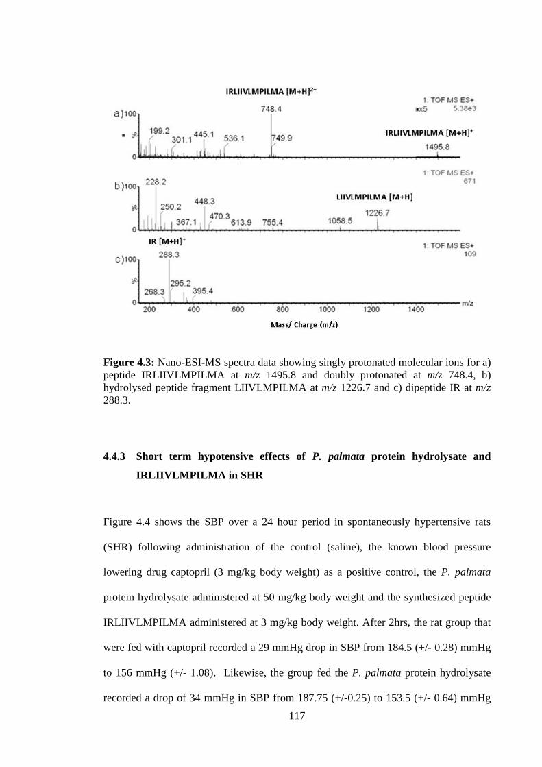
117
Figure 4.3: Nano-ESI-MS spectra data showing singly protonated molecular ions for a)peptide IRLIIVLMPILMA at m/z 1495.8 and doubly protonated at m/z 748.4, b)hydrolysed peptide fragment LIIVLMPILMA at m/z 1226.7 and c) dipeptide IR at m/z288.3.
4.4.3 Short term hypotensive effects of P. palmata protein hydrolysate and
IRLIIVLMPILMA in SHR
Figure 4.4 shows the SBP over a 24 hour period in spontaneously hypertensive rats
(SHR) following administration of the control (saline), the known blood pressure
lowering drug captopril (3 mg/kg body weight) as a positive control, the P. palmata
protein hydrolysate administered at 50 mg/kg body weight and the synthesized peptide
IRLIIVLMPILMA administered at 3 mg/kg body weight. After 2hrs, the rat group that
were fed with captopril recorded a 29 mmHg drop in SBP from 184.5 (+/- 0.28) mmHg
to 156 mmHg (+/- 1.08). Likewise, the group fed the P. palmata protein hydrolysate
recorded a drop of 34 mmHg in SBP from 187.75 (+/-0.25) to 153.5 (+/- 0.64) mmHg

118
SBP, while the group fed the tridecapeptide IRLIIVLMPLIMA presented a drop of 33
mmHg in blood pressure from 187.5 (+/-0.95) to 154.75 (+/-0.94) mmHg SBP. After
24hrs, the effect of all blood pressure lowering agents was a drop in mmHg SBP of 21,
16 and 17 mmHg respectively compared to the SBP recorded at 0hrs. ANOVA analysis
of the data shows that there is no statistical difference between the SBP of rats
administered the captopril, P. palmata protein hydrolysate or the peptide
IRLIIVLMPILMA over the 24hr period. However, all three are statistically different to
that of the saline control (P < 0.001) over the test period.
0 5 10 15 20140
160
180
200
IRLIIVLMPILMAHydrolysateCaptoprilSaline
Time (hrs)
SB
P(m
mH
g)
Figure 4.4: SBP of SHRs over 24hrs after oral administration of saline, captopril (10mg/kg body weight, P. palmata protein hydrolysate (50 mg/kg body weight) and thesynthesised peptide IRLIIVLMPILMA (50 mg/kg body weight). Values are mean ±SEM (n = 3).

119
4.5 Discussion
The feasibility of functional food or pharmaceutical application of bioactive peptides
depends on overcoming several challenges and one important aspect of use is oral
bioavailability. Bioactive peptide action is often hampered by proteolytic enzyme attack
in the gastrointestinal tract. In this work, the survival of a renin inhibitory peptide with
the amino acid sequence IRLIIVLMPILMA was assessed using a combination of in
silico analysis followed by a simulated gastrointestinal digestion procedure and
subsequent in vivo administration of the peptide and hydrolysate to SHRs.
The use of in silico methods to predict the cleavage sites of potential bioactive peptides
that are to be ingested is an important preliminary step when assessing their
bioavailability. If the active sequence of a bioactive peptide is predicted to be cleaved in
silico by the enzymes of the GI tract this would rule out further costly in vitro and
subsequent in vivo analysis. Previously, in silico methods were used to predict the
formation of ACE-I inhibitory peptides during gastrointestinal digestion [186]. The fact
that trypsin cleaved the peptide at position 2 (between the residues R-L) at the carboxy
end of the amino acid is particularly relevant from a renin inhibition point of view.
Previous work showed that this di-peptide inhibited renin, and ACE-I activities at
concentrations of 1 mg/ml when isolated from a pea protein hydrolysate [72].
In vitro digestion models are widely used to study the structural changes, digestibility,
and release of food components under simulated gastrointestinal conditions [187].
While not being as accurate as in vivo methods, these models are a less time consuming
and less costly precursor method [187]. Previous studies examined the ACE-I inhibitory
activity of pea and whey protein digests using in vitro digestion but chose to analyse the
resulting hydrolysates with SDS-PAGE and HPLC [136]. To confirm the results
obtained using in silico analysis, the bioactive tridecapeptide identified in this study was

120
subjected to the in vitro simulated gastric digestion outlined in Figure 4.1. In addition,
the sample was analysed using UPLC-MS in order to identify the breakdown products
and the dipeptide IR.
The MS data obtained and shown in Figures 4.2 and 4.3confirms the in silico cleavage
analysis results which predicted that the renin inhibitory di-peptide IR remains intact
during normal digestive processes. In this study, ESI-Q-TOF mass spectrometry was
used in tandem with UPLC due to the high sensitivity of peptide detection that ESI-Q-
TOF offers along with the rapid and high degree of separation offered by UPLC [188].
While in vitro and in silico methods are powerful, predictive tools, the likely mode of
action of a molecule within a living system cannot be fully mapped out due to the
myriad of complex reactions a molecule will encounter within an organism. Therefore,
animal models are necessary in functional food research as well as pharmaceutical
research [189]. Rats are a strong medical model for humans as almost all human genes
known to be associated with disease have orthologues in the rat genome [190]. The
results shown in Figure 4.4 present the short term changes in SBP observed over a 24
hour period in Spontaneously Hypertensive Rats (SHR) fed a diet of the seaweed
protein papain hydrolysate; the synthesized tridecapeptide IRLIIVLMPILMA; a
positive control (Captopril®); and a Saline solution (negative control). The ACE-I
inhibitor Captopril was used as an anti-hypertensive positive control as we were unable
to obtain a sample of the renin inhibitor aliskiren. Interestingly, both the hydrolysate
and the tridecapeptide IRLIIVLMPILMA displayed very similar trends in lowering the
SBP of the rats over a 24 hour period (Figure 4.4). Previous papers examined the blood
pressure lowering effects of a pea protein hydrolysate in SHR rats [183]. However, the
pea protein hydrolysates were administered at a concentration of 200mg/kg of body
weight and this dose resulted in a 19 mmHg drop in systolic blood pressure in SHRs.

121
Likewise, SHRs treated with an antihypertensive buckwheat hydrolysate recorded an
initial decrease in SBP of 27.0 ± 7.6 mmHg after 2 hrs but only when treated with 100
mg/kg body weight [191], twice the concentration of the P. palmata protein hydrolysate
and the tridecapeptide IRLIIVLMPLIMA used in this study. Further studies should be
carried out to assess the long-term effects of the hydrolysate and IRLIIVILMPILMA to
ensure that the decrease in SBP is sustained, and also to ensure that there are no
negative effects due to prolonged exposure. In silico analysis of the peptide using the
online tool ToxinPred also showed that the peptide was non-toxic, which indicates that
it may be suitable for use in functional foods and pharmaceutical agents. Furthermore,
some toxicological parameters were assessed in chapter 3 through the use of a zebrafish
larvae assay and heavy metal and Iodine content.
This Chapter demonstrated that the tridecapeptide IRLIIVLMPLIMA is a renin
inhibitory pro-peptide with the ability to impart a hypotensive effect in vivo confirmed
by monitoring changes in SBP in SHRs over a 24 hour period. The dipeptide IR is the
active sequence of the tridecapeptide IRLIIVLMPILMA and this was confirmed here
using the simulated digestion of the tridecapeptide and MS analysis of the breakdown
products. The renin inhibitory activity of the dipeptide IR was not assessed in this study
but was previously determined [183]. Both the peptide and the P. palmata protein
papain hydrolysate may have potential for use as functional ingredients in the growing
nutraceutical market for the prevention of hypertension.
122
CHAPTER 5. INCREASING THE HEALTH BENEFITS OF
BREAD: ASSESSMENT OF THE PHYSICAL AND SENSORY
QUALITIES OF BREAD FORMULATED USING A RENIN
INHIBITORY PALMARIA PALMATA PROTEIN
HYDROLYSATE.
This chapter was published as a research harticle in LWT-The Science of Food and
Technology The paper is entitled: Ciarán Fitzgerald, Eimear Gallagher, Linda Doran,
Mark Auty, José Prieto, Maria Hayes “Increasing the health benefits of bread:
Assessment of the physical and sensory qualities of bread formulated using a renin
inhibitory Palmaria palmata protein hydrolysate.” (2013), 56, 398-405.

123
5.1 Aim
For centuries, bread has been consumed as a staple of the human diet. The four aims of
this chapter were:
(1) To assess whether a seaweed protein bioactive hydrolysate identified as having
renin inhibitory properties could be incorporated into this common food
vehicle without adverse physical and sensory effects.
(2) To assess if the renin inhibitory protein hydrolysate generated from Palmaria
palmata could survive the bread baking process and if this product still
possessed renin inhibitory activities following the baking process.
(3) To determine any changes in the bread product as a result of addition of the
generated protein hydrolysate to the bread product and to provide a description
of these changes through the use of semi-trained consumer panelists.
(4) To confirm an increase in the protein and essential amino acid content of the
bread by the addition of the seaweed hydrolysate.

124
5.2 Introduction
Baked products are the most widely consumed foods in the world and therefore have
great potential as vehicles for bioactive ingredient delivery [124]. Indeed, many bread
companies routinely enrich their baked products with folic acid [192]. Increasing the
protein, nutritional and health benefits of bread is one strategy for improving consumer
nutrition and this is not a novel concept. The literature conveys how breads have been
improved by the addition of skim milk powder and soy proteins to breads[193].
Furthermore, numerous Omega-3 products have been launched in the United States,
with Omega-3 enriched bread being used as a delivery vehicle for eicosapentaenoic acid
(EPA) and docosahexaenoic acid (DHA). This is delivered in the form of
microencapsulated Tuna oil at levels of 10 mg of long chain n-3 polyunsaturated fatty
(PUFA) oils per slice [4]. Baked products can provide consumers with high quality
Omega-3 breads with higher contents of EPA and DHA than the average functional
food product found in the US. Furthermore, the Norwegian bakery company Bakers,
launched omega-3 fortified bread in 2009 for school children. Other common cereal
based bioactive products include biscuits enriched with Omega-3 fatty acids to impart
health benefits to the consumer [4].
Addition of alternative protein sources to bread increases the nutritional profile of
bread, but can also be used to enhance the potential health benefits. It is well
documented how wheat protein is deficient in some essential amino acids, in particular
lysine, which is the first limiting amino acid in wheat [149]. Lysine deficiency leads to
the poor utilization of protein and thus results in protein malnutrition [194]. Attempts to
improve the nutritional profile of breads are not new. For example, fish protein
concentrates were added to wheat bread previously to increase the lysine content of the
bread[195]. Indeed, previous work demonstrated that during the month of November, P.

125
palmata seaweed can contain levels of lysine of 5.9 g/100 g of total amino acids[38].
For centuries, red seaweed species were incorporated into traditional laver breads across
the United Kingdom (UK) and Ireland [123], however the health benefits of this
practice were not examined.
This work examined the textural and sensory attributes of a wheaten, yeast raised bread
formulated using the P. palmata protein hydrolysate described in Chapters one and two
and compared this potentially bioactive bread to control wheat breads. Four
formulations were examined; a wheat flour control bread, a control bread containing 4%
P. palmata protein hydrolysate, a buckwheat bread consisting of a blend of 70% wheat :
30% buckwheat, and a fourth formulation 70% wheat : 30% buckwheat containing 4%
P. palmata hydrolysate. Buckwheat was used as previous studies have described the
antihypertensive effects of this cereal [191, 196]. The volume, colour, texture profile,
moisture, crumb structure, sensory attributes and renin inhibitory activity of all four
bread formulations were assessed. Brightfield light microscopy and confocal scanning
laser microscopy images using a new triple labelling protocol were also obtained to
visualise the main ingredients of the breads.

126
5.3 Materials and Methods
5.3.1 Palmaria palmata protein extraction and hydrolysis with papain
Protein was extracted from P. palmata following the protocol of Galland-Irmouli et al.
[38] as described in Chapter one. The hydrolysis process used was the same as that
used to isolate renin and PAF-AH inhibitory peptides.
5.3.2 Bread ingredients
Standard Baker’s wheat flour (63 % water absorption, 13.5% protein) was supplied by
James Neills Ltd., Belfast, Northern Ireland. Buckwheat flour (13% protein) was
supplied by Ziegler and Co., Wunsiedel, Germany and dried Yeast was supplied by
Doves Farms Ltd., UK. The Dough improver was supplied by Fermex International Ltd,
Worcester, UK and salt was obtained from Ineos Enterprises, Cheshire, UK. Emulsified
bread fat was purchased from Irish Bakels Ltd. (Ireland).
5.3.3 Preparation of breads
The four bread formulations used in this study are presented in Table 5.1. Previous
sensory studies where seaweeds were used as food ingredients incorporated the brown
seaweed Ascophyllum nodosum into bread at concentrations of up to 4%[125]. The three
bread test formulations used in this work were:
1. C: the Control.
2. CH: the Control containing 4% of the P. palmata hydrolysate generated in
Chapter one.
3. B: a blend of 70% wheat: 30% Buckwheat.
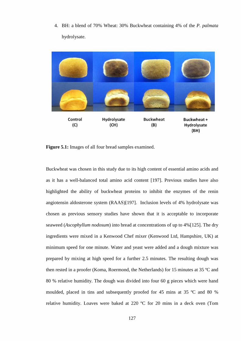
127
4. BH: a blend of 70% Wheat: 30% Buckwheat containing 4% of the P. palmata
hydrolysate.
Figure 5.1: Images of all four bread samples examined.
Buckwheat was chosen in this study due to its high content of essential amino acids and
as it has a well-balanced total amino acid content [197]. Previous studies have also
highlighted the ability of buckwheat proteins to inhibit the enzymes of the renin
angiotensin aldosterone system (RAAS)[197]. Inclusion levels of 4% hydrolysate was
chosen as previous sensory studies have shown that it is acceptable to incorporate
seaweed (Ascophyllum nodosum) into bread at concentrations of up to 4%[125]. The dry
ingredients were mixed in a Kenwood Chef mixer (Kenwood Ltd, Hampshire, UK) at
minimum speed for one minute. Water and yeast were added and a dough mixture was
prepared by mixing at high speed for a further 2.5 minutes. The resulting dough was
then rested in a proofer (Koma, Roermond, the Netherlands) for 15 minutes at 35 ºC and
80 % relative humidity. The dough was divided into four 60 g pieces which were hand
moulded, placed in tins and subsequently proofed for 45 mins at 35 ºC and 80 %
relative humidity. Loaves were baked at 220 ºC for 20 mins in a deck oven (Tom

128
Chandley Ltd., Manchester, UK) and subsequently cooled at room temperature for 2 hrs
and stored in polyethylene bags (two loaves per bag). Four loaves were produced for
each formulation. Two of these loaves were evaluated after 24hrs and the remaining two
after 120hrs storage at room temperature. Each bake was performed in triplicate.
Ingredient(g/100 g of flour)
Control (C) Control +Hydrolysate (CH)
Buckwheat (B) Buckwheat +Hydrolysate (BH)
Wheat Flour 100 100 70 70Buckwheat - - 30 30Fat 1 1 1 1Yeast 1.5 1.5 1.5 1.5Salt 2 2 2 2Improver 0.5 0.5 0.5 0.5Water 63 63 62 62Hydrolysate - 4 - 4
Table 5.1: The bread formulations used in this study; Ingredients are presented as g /100g of the wheat/buckwheat flour base
5.3.4 Bread evaluation – loaf volume, weight and loaf specific volume calculations
After 24 hrs, the loaf volume for all formulations was measured using a volume meter
(TexVol BVM-L30, Sweden). Loaf weight was recorded and the loaf specific volume
(ml/g) was calculated. Moisture was calculated following a procedure based on the
International Association of Cereal Chemists (ICC) method 110/1 [198]using a
Brabender moisture oven (Brabender, Duisberg, Germany). Crust and crumb colour
were measured using an Ultra-Scan Pro colour meter (Hunter Associates Laboratory
Inc., Reston, Virginia, USA) and results expressed using the Hunter crust colour L*
(lightness), a* (redness/greenness) and b* (yellowness/blueness). The crumb structure
of the centre slices of each bread formulation was evaluated using the C-Cell bread
Imaging System (Calibre Control International Ltd., UK). Crumb texture was assessed
by conducting a texture profile analysis (TPA) on 10 mm thick slices using a Texture
Analyser (TA-XT2i, Stable Micro Systems, Surrey, UK) equipped with a 10 mm

129
perspex probe and using the Texture Exponent Software. Moisture and TPA was
repeated after 120hrs on the two remaining loaves from each bread formulation. All
bread formulations were tested in triplicate.
5.3.5 Heavy metal and Iodine analysis of seaweed bread product
The total cadmium (Cd), lead (Pb), methyl-mercury ([CH3Hg]+), arsenic (As) and iodine
I2levels of the formulated bread were assessed by Eurofins Food Ireland Ltd., (Finglas,
Dublin 11, Ireland).
5.3.6 Sensory analysis
Sensory analysis was conducted on all four bread formulations 24hrs post-baking using
a sensory panel consisting of 12 semi-trained consumers. Using a hedonic scale,
panellists were asked to assess the breads under the characteristics of appearance,
texture, flavour and general acceptability by placing a mark on a 6 cm line (0 =
unacceptable, 6 = very acceptable)[199].
5.3.7 Renin inhibitory assay
This assay was carried out on bread samples (1 mg/ml) from each of the four
formulations according to the manufacturer instructions (Cayman chemical Company,
Michigan, USA) and as outlined previously in Chapter two.
5.3.8 Microscopy
For examination by brightfield light microscopy and confocal scanning laser
microscopy, bread samples were resin embedded, sectioned and stained as described
below. Samples of bread approximately 3 mm cubed in size were cut from the middle of

130
each loaf using a razor blade and placed in a fixative which consisted of 2.5% w/w
glutaraldehyde in 80% aqueous ethanol. After 2hrs, the fixative was replaced with 80%,
90%, 95% and 100% ethanol in sequential steps for 1hr each. Fixed bread cubes were
transferred to flat-ended Beem capsules containing LR White polymethacrylate resin
(Agar Scientific Ltd., Stansted, UK) and allowed to infiltrate at room temperature
overnight. The resin was replaced with fresh resin and capsules containing the fixed
bread cubes were cured at 60oC overnight in an oven. Semi-thin sections of 5 µm
thickness were then cut from the resin blocks on a Leica EM UC7 ultramicrotome
(Leica Microsystems, Mannheim, Germany) and transferred to light microscope slides
prior to imaging by conventional optical or confocal microscopy as described below.
Sections were cut from two replicate blocks.
5.3.8.1 Brightfield light microscopy
Resin sections were double stained with one drop of an aqueous solution of 0.1 %
Iodine in 0.2 % w/w potassium iodide to stain starch for 5s. The stain was then drained
off and one drop of 0.1 % w/w aqueous acid fuschin added. A coverslip was placed on
top and the sections were imaged using an Olympus BX51 light microscope (Mason
Technology, Dublin, Ireland) using brightfield illumination. Digital images (8 bit, TIFF,
2048 x 1024 pixels) were taken using a ProgRes CT3 camera (Jenoptik, Germany)
using 10x, 20x or 60x objectives.
5.3.8.2 Confocal scanning laser microscopy
Resin sections of the fixed bread cubes were triple labelled to show all major
ingredients. One drop of 0.1 % w/w ethanolic solution of fluorescein iso-thiocynate
(FITC) was added to the resin section to label starch. After 10 s, the FITC was drained
off and replaced with one drop of aqueous fluorescent brightener 28 (FB28) at a

131
concentration of 0.125 % (w/w) to label cellulosic material. Subsequently, one drop of
0.1 w/w aqueous fast green (FG) was added to label proteins. The sections were rinsed
gently with running water and a cover slip placed on top. Stained sections were imaged
using a Leica SP5 confocal scanning laser microscope (Leica Microsystems,
Mannheim, Germany) fitted with 20x 0.7 NA and 63x 1.4 NA oil immersion objectives.
Sequential images were acquired using triple-channel imaging: a 405 nm blue diode
laser was used to excite the fluorescent brightener; a 488nm argon laser was used for
excitation for FITC and a 633 nm helium-neon laser for rhodamine B excitation.
Emission signals for FB28, FITC and FG were sequentially collected using band pass
filters 450 – 490 nm; 510 – 550 nm and 650 – 700 nm, respectively. Digital 8-bit
images (1024 x 1024 pixels) were obtained for each separate excitation wavelength and
channels were combined and pseudo-coloured to show cellulosic material as blue, starch
as green and protein as red.
5.3.9 Statistics
The student’s t-test was performed using GraphPad Prism version 5.04 for Windows,
GraphPad Software, ( La Jolla, California, USA).
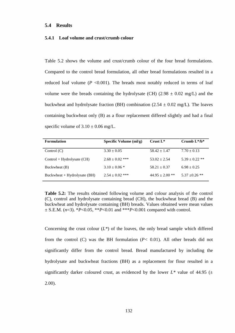
132
5.4 Results
5.4.1 Loaf volume and crust/crumb colour
Table 5.2 shows the volume and crust/crumb colour of the four bread formulations.
Compared to the control bread formulation, all other bread formulations resulted in a
reduced loaf volume (P <0.001). The breads most notably reduced in terms of loaf
volume were the breads containing the hydrolysate (CH) (2.98 ± 0.02 mg/L) and the
buckwheat and hydrolysate fraction (BH) combination (2.54 ± 0.02 mg/L). The loaves
containing buckwheat only (B) as a flour replacement differed slightly and had a final
specific volume of 3.10 ± 0.06 mg/L.
Formulation Specific Volume (ml/g) Crust L* Crumb L*/b*
Control (C) 3.30 ± 0.05 58.42 ± 1.47 7.70 ± 0.13
Control + Hydrolysate (CH) 2.68 ± 0.02 *** 53.02 ± 2.54 5.39 ± 0.22 **
Buckwheat (B) 3.10 ± 0.06 * 58.21 ± 0.37 6.98 ± 0.25
Buckwheat + Hydrolysate (BH) 2.54 ± 0.02 *** 44.95 ± 2.00 ** 5.37 ±0.26 **
Table 5.2: The results obtained following volume and colour analysis of the control(C), control and hydrolysate containing bread (CH), the buckwheat bread (B) and thebuckwheat and hydrolysate containing (BH) breads. Values obtained were mean values± S.E.M. (n=3). *P<0.05, **P<0.01 and ***P<0.001 compared with control.
Concerning the crust colour (L*) of the loaves, the only bread sample which differed
from the control (C) was the BH formulation (P< 0.01). All other breads did not
significantly differ from the control bread. Bread manufactured by including the
hydrolysate and buckwheat fractions (BH) as a replacement for flour resulted in a
significantly darker coloured crust, as evidenced by the lower L* value of 44.95 (±
2.00).

133
The crumb colour of bread loaves was altered by the addition of the P. palmata
hydrolysate (P< 0.01). Both the CH and BH bread loaves recorded similar L*/ b*
(lightness/yellowness ratio) values of 5.39 ± 0.22 and 5.37 ± 0.26 respectively, in
comparison to the control (7.70 ± 0.13).
5.4.2 Digital image analysis of bread crumb
The results of the crumb grain analysis of the bread formulations are presented in Table
5.3. The inclusion of buckwheat appeared to reduce the number of cells in the bread
slices significantly, as shown in bread formulation B (1462 ± 50) and bread formulation
BH (1496 ± 22) compared to the control formulation (C) (1760 ± 22).
The cell wall thickness and cell volume were not significantly affected by the inclusion
of the P. palmata hydrolysate (CH), buckwheat (B) or a combination of the two (BH),
indicating that the general coarseness of the slice texture remained unchanged.
Formulation Number of cells Cell Wall thickness Cell volume
Control (C) 1760 ± 22 0.430 ± 0.003 6.44 ± 0.19
Control + Hydrolysate (CH) 1640 ± 46 0.416 ± 0.006 6.59 ± 0.80
Buckwheat (B) 1462 ± 50** 0.443 ± 0.006 7.49 ± 0.44
Buckwheat + Hydrolysate (BH) 1496 ± 22** 0.426 ± 0.003 7.53 ± 0.61
Table 5.3: Image analysis results obtained for the control bread (C), control andhydrolysate bread (CH), buckwheat bread (B) and buckwheat and hydrolysatecontaining (BH) breads. Values are mean ± S.E.M. (n=3). *P<0.05, **P<0.01 and***P<0.001 compared with control.

134
5.4.3 Texture profile analysis (TPA) of bread crumb
The results for the crumb TPA and crumb moisture content are presented in Figure 5.2.
Compared to the control bread sample, the crumb texture of all three test breads was
found to be significantly firmer. For example, the BH bread was three times as firm
(828g ± 26g) as that of C (275g ± 10g). Wheat bread containing the hydrolysate (CH)
showed similar hardness properties to that of the buckwheat bread (B) (573g ± 25g) and
(506g ± 19g) respectively. Similarly, in terms of cohesiveness, the BH formulation
presented the least cohesive crumb. In addition, the CH and B bread formulations both
appeared to have higher cohesive values and these cohesive values were lower than
those of the control bread (Figure5.2.b). Likewise, the springiness properties of each
bread demonstrated a comparable trend to values obtained for cohesiveness with the
control formulation showing the highest result for this parameter (P<0.001). The
combined BH bread formulation had a lower springiness quality compared to the CH
and B formulations. The moisture content of the crumb in this bread was not
significantly affected by the addition of the hydrolysate or buckwheat.
The effect of storage time (120 hrs) on the bread qualities was also assessed and these
results areshown in Figure 5.2. As expected, crumb hardness increased for all four bread
formulations. The most significant increase, however, was observed with the BH bread.
The crumb hardness of the BH bread almost tripled from 828g (± 26g) to 2459g (±
315g) over the 120hrs period of storage. Crumb cohesiveness and springiness were also
greatly affected by storage time. The BH bread sample presented the most significant
changes over the testing period. Interestingly, the only formulation not to significantly
lose crumb moisture during the storage period was the buckwheat bread (B).

135
Figure 6: Texture profile analysis and moisture content. Crumb hardness (a), cohesiveness (b), springiness (c) andmoisture percentage (d), of the control (C), control + hydrolysate (CH), buckwheat (B) and buckwheat + hydrolysate(BH) breads. = 24hrs of storage. = 120hrs of storage. Values are mean ± S.E.M. (n=3). *P<0.05, **P<0.01and ***P<0.001 compared with control.

136
5.4.4 Brightfield light microscopy of bread samples
All microscopy carried out in this thesis was carried out by the staff of the National
Food Imaging Centre, at Teagasc Moorepark Food Research Centre. Light micrographs
of stained sections of the different bread formulations are shown in Figure 5.3. Starch
grains appeared as a dark blue colour, whilst protein was stained pink/red. Light
microscopy showed some differences between the four bread samples. In Figure 5.3.a
the control bread (C) showed partially gelatinised wheat starch granules of between 10 –
50 µm in diameter. Larger starch granules were elongated and irregularly shaped,
whilst smaller granules were more spherical and rounded. The pink stained protein
appeared as a semi-continuous network, often surrounding individual starch grains.
Figure 5.3.b, displays microscopic images of the bread containing 4% seaweed protein
hydrolysate (CH). This image shows similar starch granulation properties and a protein
network similar to that of the control. However, the starch granules appeared more
evenly distributed and less swollen. The buckwheat bread (B) in Figure 5.3.c also shows
a wheat starch and protein profile similar to the control bread sample. In addition, there
are large clusters of partially gelatinised, close-packed polyhedral starch granules of
between 5 – 10 µm in diameter found in the buckwheat bread (B). These starch clusters
are characteristic of buckwheat flour particles (results not shown). The BH bread shown
in Figure 5.3.d demonstrates features of both the hydrolysate (CH) and buckwheat bread
(B), with some large protein regions and close-packed polyhedral starch grains. These
properties are characteristic of buckwheat flour.

137
Figure 5.3: Light micrograph images of the control (a), control and hydrolysate (b),buckwheat(c) and buckwheat and hydrolysate (d) breads (x 20 obj). Starch grainsappeared as dark blue, whilst protein is shown as pink/red colour.
5.4.5 Confocal scanning laser microscopy of bread samples
Confocal microscopy of resin embedded sections of bread was carried out using a new
triple labelling protocol which effectively localised the main ingredients of the breads.
Representative images are shown in Figure 5.4. Plant cell wall material (blue), starch
(green) and protein (red) were imaged using fluorescent brightener (FB), fluorescein
iso-thiocynate (FITC) and fast green (FG) labelling, respectively. The location of starch
and protein correlated well with the light microscopy results, providing further
validation of the new confocal staining procedure. The control (C) sample showed
swollen starch granules with a semi-continuous protein network and cell wall fragments
randomly scattered throughout the resin section (Figure5.4.a). Figure 5.4.b displays
a b
c d

138
microscopic images of the bread containing 4 % of the seaweed protein hydrolysate
(CH), appeared similar to the control bread except that the starch granules were less
swollen and more evenly dispersed. Figure 5.4.c contained buckwheat starch (B) and
had a similar, swollen starch granule image to the control (C). In addition, this bread
had close-packed, clusters of polyhedral starch grains. Figure 5.4.d (BH) contained
similar clusters of polyhedral grains to those in the buckwheat bread formulation.
However, the wheat starch in this formulation appeared less swollen than in the
hydrolysate containing bread formulation.
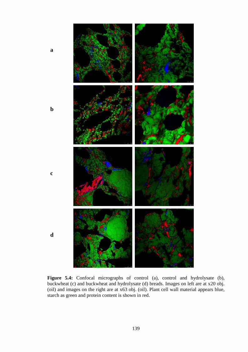
139
Figure 5.4: Confocal micrographs of control (a), control and hydrolysate (b),buckwheat (c) and buckwheat and hydrolysate (d) breads. Images on left are at x20 obj.(oil) and images on the right are at x63 obj. (oil). Plant cell wall material appears blue,starch as green and protein content is shown in red.
a
b
c
d

140
5.4.6 Heavy metal and Iodine analysis of the bread product
The level of cadmium, lead, arsenic and methyl-mercury detected in the bread product
was 0.024, 0.047, 0.032 and 0.002 µg/g of freeze dried bread respectively. The iodine
level was recorded as 1.19 µg per gram of bread tested.
5.4.7 Sensory analysis of breads
The appearance, texture, flavour and general acceptability scores are displayed in Figure
5.5. These results show the average score which the sensory panel assigned breads
regarding the sensory characteristics of appearance, texture flavour and general
acceptability as expressed by the panel member grading the bread from one to six. A
distinctive trend can be observed with all four breads in terms of their sensory attributes.
All three test breads scored lower than the wheat control (C) which scored just below
five out of six regarding the sensory attributes which included appearance, texture
flavour and general acceptability. The second favourite bread formulation was identified
as the buckwheat bread formulation (B). The test panel consistently scored this bread
four out of six for appearance, texture flavour and general acceptability. The third
favourite bread formulation was the CH bread formulation which scored three for
texture, flavour and general acceptability. It received a score of 3.58 (± 0.61) for
appearance. The BH bread was the least favourite bread and scored low for flavour
(1.54 ± 0.72) and general acceptability (1.56 ± 0.65). It scored similar results to the CH
bread in terms of texture. Many of the panel members commented that the BH bread
gave a strong, bitter after-taste.

141
Figure 5.5: Sensory analysis. A group of 12 panellists were asked to rate the control(C), control + hydrolysate (CH), buckwheat (B) and buckwheat + hydrolysate (BH)breads under the appearance, texture, flavour and overall acceptability. Control (C) = ♦. Control + hydrolysate (CH) = ■. Buckwheat (B) = ▲. Buckwheat + hydrolysate (BH) =x. Values are mean (n=12).
5.4.8 Renin inhibitory activity of bread samples
The percentage inhibition of all four breads is shown in Figure 5.6. All three test breads
demonstrated significantly higher renin inhibitory activity than that of the control bread
(P<0.01). The P. palmata papain protein hydrolysate bread (CH) inhibited renin by
11.21% (± 0.77) when tested at a concentration of 1mg/ml compared to the positive
control which was higher than the buckwheat bread formulation (B) which had a renin
inhibitory value of 9.546% (± 0.48). Not surprisingly, combining the buckwheat and
0
1
2
3
4
5
6
Appearance
Texture
Flavour
General acceptability

142
seaweed protein hydrolysate in bread formulation increased the renin inhibitory activity
to the bread to 14.92 % (± 1.88).
Figure 5.6: Renin inhibitory assay of the control (C), control + hydrolysate (CH),buckwheat (B) and buckwheat + hydrolysate (BH) breads. Values are mean ± S.E.M.(n=3). *P<0.05, **P<0.01 and ***P<0.001 compared with control.

143
5.5 Discussion
This chapter confirmed that a renin inhibitory hydrolysate generated from P. palmata
protein using the food grade enzyme papain retained bioactivity following the bread-
making process. Furthermore, the textural and sensory attributes of the formulated
breads containing the hydrolysate were found to be acceptable by a semi-trained,
sensory panel using the hedonic scale [199].
Loaf volume is important in terms of consumer acceptance of bread products as it is an
indicator of the density of the bread product and affects bread texture. The addition of
4% of a seaweed protein hydrolysate to wheat breads resulted in a decreased loaf
volume. This decrease may be due, in part, to the additional protein added to the bread
formulation which may compete with the wheat starch for free moisture, thereby
restricting the hydration and swelling of starch granules. (Figures 5.3.b and 5.4.b). The
starch grains shown in Figure 5.3.b and 5.4.b appeared less swollen and were evenly
distributed compared to the control (C) and buckwheat (B) breads. This indicates that
inhibition of the gelation process took place. A lower loaf volume is a common effect of
protein addition to bread[200]. In a previous study, Moroni, et al. (2011)[201]
demonstrated that the inclusion of buckwheat at 10% increased the loaf volume but the
addition of 20% buckwheat had no effect on breadloaf volume. The findings from this
work are in agreement with this observation as the addition of 30% buckwheat
decreased loaf volume significantly. The inclusion of coarse buckwheat flour may have
interfered with the elastic nature of the gluten network and may have contributed to a
decreased loaf volume. The combination of buckwheat and the P. palmata papain
protein hydrolysate (BH) resulted in greatly decreased loaf volume.
The crust and crumb colour of the bread darkened with the addition of the P. palmata
hydrolysate. This result was expected as the browning of bread is due to non-enzymatic,

144
Maillard reactions[202]. The lower volume of the BH bread may have contributed to the
darker crumb colour observed. Lower loaf volume, resulted in a lower surface area and
therefore a dense and darker crumb appearance. The colour of the loaf greatly affects
consumer perception of the quality of the bread, previous studies have suggested that
bread color had a relatively greater impact upon consumer choice than the price and
nutrition information combined [203].
Crumb structure of bread is an important factor in terms of the sensory aspects of bread,
and directly affects the texture and staling properties [202]. The addition of the renin
inhibitory hydrolysate had no effect on the number of cells, cell wall thickness or cell
volume of internal bread crumb. The addition of buckwheat decreased the number of
cells in the crumb in B bread (1462 ± 50) and the BH bread (1496 ± 22). Addition of
buckwheat had no effect on wall thickness or the cell volume of the bread (Table 5.3).
This result is positive in terms of the sensory acceptance of the bread product.
Addition of the hydrolysate which was identified as having a lysine content of 4.5 g per
100g of amino acid in chapter two improved the nutritional parameters of the bread
product. Furthermore, the iodine content of the bread was assessed. Fortification of
bread with iodine from marine sources including seaweed is a potentially attractive
option for bakeries and food scientists. Dulse bread is known to the consumer and, in
addition, Iodine is required by consumer groups including pregnant women [204]and
those suffering from thyroid-gland dysfunction (under active thyroid gland)[205]. The
level of iodine detected in the bread product was 1.19 µg per gram of bread tested. This
is within the amount recommended by the FDA. The iodine-replete healthy adult has
about 15–20 mg of Iodine, 70%–80% of which is contained in the thyroid[206].Median
urinary iodine concentrations of 100–199 µg/L in children and adults, 150–249 mg/L in
pregnant women and >100 µg/L in lactating women indicate iodine intakes are adequate

145
[207]. Iodine values lower than 100 µg/L in children and non-pregnant adults indicate
insufficient iodine intake, although iodine deficiency is not classified as severe until
urinary iodine levels are lower than 20 µg/L. Non-fortified bread ordinarily contains
0.39 µg iodine per gram of bread [208] therefore the bread containing P. palmata
protein hydrolysate may be seen as a good source of iodine. The levels of heavy metals
identified within the hydrolysate product were within the limits recommended [209] and
below the levels found in other cereal products [210].
Sensory analysis (Figure 5.5) correlated well with the texture profile analysis results
obtained (Figure 5.2), except where the panelists rated the texture of the CH and the BH
breads similarly. TPA results showed the BH bread to be almost twice as hard as the CH
bread. However, the crumb cohesive and springiness properties were similar, which
translated into a similar mouth feel and texture when chewed by the panelist. Previous
studies where sensory analysis was carried out on bread products that contained protein
from plant sources reported a similar trend in terms of texture [200, 211]. The flavour
characteristics of the two breads formulated using the renin inhibitory hydrolysate (CH
and BH) were rated significantly lower by the sensory panelists than the control bread
(C). Panelists described these breads as having a disagreeable and bitter flavour. A bitter
flavour is often attributed to peptide content [100].This bitterness is also associated with
Buckwheat flour, due to the high Tannin composition of this grain [212]. The use of
semi trained sensory panels and a hedonic scale is a common method of assessing the
sensory attributes of bread [213].
A number of different strategies could be employed to de-bitter protein hydrolysates.
These include masking the bitter taste with various flavour agents[104], further
enzymatic treatment of the protein[104]or, by the introduction of bitter inhibiting
compounds such as neodiosimine and cellotrioside into the food matrix[214, 215]. The

146
panel preference for bread appearance was observed in decreasing order of C bread >B
bread >CH bread>BH bread. Again, due to the added protein in the BH and CH breads,
it is thought that a greater number of maillard reactions occurred which, resulted in, a
darker bread appearance than the colour of the control bread (C) or buck wheat bread
(B) formulations.
The renin inhibition assay showed that the renininhibitory properties of the P. palmata
protein hydrolysate were retained during the baking process. Our previous study
showed at a concentration of 1 mg/ml the hydrolysate can inhibit reninin vitro by
41.89% (±3.22)[163]. Similar baking processes have recorded crumb temperatures of 98
ºC [216]and it has been shown that bioactive peptides, such as ACE-I inhibitors retained
their bioactivities following heating at 70 and 100 ºC for 20 minutes[102]. Starch is
gelatinised in wheat breads and proteins are denaturated during the baking process[217].
Denatured proteins release water while the gelatinisation of starch absorbs water [217].
From the light and confocal micrograph images obtained in this work of the CH bread,
this phenomenon of starch gelatinisation in tandem with protein denaturation is not
visible. Therefore, it can be said that the renin inhibitory activity obtained in a previous
study was not deleted due to the baking process, as the confocal micrograph images
obtained showed that the seaweed protein was not denatured during the baking process.
While the renin inhibitory effect of the buckwheat bread was not further investigated,
buckwheat is known to have a wide range of bioactive compounds[218], including
compounds that inhibit the RAS such as 2″-Hydroxynicotianamine (HNA) [196].
Seaweed enriched breads have already been developed with the potential to reduce
habitual energy intake. The bread, which was manufactured using Ascophyllum
nodosum could positively affect body mass index (BMI) and body composition[125]. In
this present study, the renin inhibitory bioactivity of a P. palmata papain hydrolysate

147
was maintained when bread was used as a food carrier and the baking process was not
found to inhibit renin inhibition. Furthermore, the bread developed using the seaweed
hydrolysate did not greatly affect the sensory attributes of the CH bread and this bread
could be developed further as a bioactive and consumer accepted product. Further work
will include assessment of the bioavailability of the renin inhibitory peptides within the
P. palmata hydrolysate bread in accepted cell models or in vivo. Also, the nutritional
composition of the breads will be further examined.

148
CHAPTER 6. GENERAL DISCUSSION AND CONCLUSION

149
6.1 General discussion and conclusion
To survive the harsh and ever-changing environment that is the ocean, macroalgae
produce distinctive biomolecules and secondary metabolites that have different
properties to those found in terrestrial plant life [23]. These attributes make macroalgae
an exciting and interesting source of bioactive compounds [2]. The overall aim of this
Ph.D thesis was to investigate the hypothesis that the Irish seaweed Palmaria
palmata(Linnaeus) Weber and Mohris a viable source of protein from which bioactive
peptides with renin and PAF-AH inhibitory activities could be generated using food
grade enzymes. These bioactivities could potentially have beneficial heart health effects,
once consumed and this thesis aimed to determine these beneficial effects if present.
One of the main aims of this work was to prove this hypothesis. Palmaria palmata is
well known and is distributed across most European, Atlantic coastal Shores. It is also
found on the coastline of Canada, Korea and Japan [219]. The broad distribution of this
seaweed species, coupled with the fact that aquaculture of this species has recently
occurred successfully made it an ideal choice of seaweed for this work.
The thesis also examined the potential survival of these bioactive peptides in the
functional food delivery vehicle bread and a baked bread product containing the heart
healthy hydrolysate was made. Survival of the renin inhibitory properties of the
hydrolysate was also confirmed following a simulated gastrointestinal digestion. Indeed,
this research aimed to prove that these peptides could provide a beneficial hypotensive
effect once consumed. This was also demonstrated through the use of an animal model
– spontaneously hypertensive rats (SHRs).
An important consideration in this study was the choice of seaweed species. Palmaria
palmata (Linnaeus) Weber and Mohr was chosen for a number of reasons. Firstly,
Palmaria palmata is known to have a high content of protein (as high as 25% according

150
to some studies) [32]. This makes P. palmata a more efficient starting material than
seaweed species known to have low protein content such as the brown seaweeds [32]
and increases the probability of recovering active peptides from its protein extract.
However the sustainability of the plant source is an important concern when considering
utilising it for industry. In Ireland, seaweed harvesting rights are traditionally held by
families and this makes industrial harvesting of seaweed a contentious issue.
Sustainability of the seaweed protein supply for bioactive peptide generation cannot
therefore be guaranteed. However, recent research carried out at NUI Galway and Bord
Iascaigh Mhara (BIM) in Ireland managed to successfully cultivate Palmaria palmata
through an aquaculture process [220].
From a biochemical point of view, a benefit of using seaweed derived protein from P.
palmata to generate bioactive peptides is that this protein source is rich in peptide
sequences that have both hydrophobic and bulky side chain containing amino acids
present [38]. These amino acids are often implicated in inhibiting enzymes of
importance in the control of blood pressure such as the ACE-I enzyme. Indeed, it has
been suggested that peptides containing bulky side chain amino acids and amino acids
including proline, isoleucine and leucine can inhibit enzymes such as ACE-I,
acetylcholinesterase (AChe) and others [119]. Finally, P. palmata has a long tradition of
use in bread products in countries including Ireland, Denmark and Wales [123].
Furthermore, it is consumed on a daily basis in Japan and under EU regulations P.
palmata is recognised as a sea vegetable. Therefore, for many consumers, the inclusion
of this Seaweed in a commercial bread product would not be seen as peculiar and
consumer acceptance of the bread product is likely.
The enzyme papain was used in this study in order to liberate bioactive peptides from
the parent seaweed proteins of Palmaria palmata. Papain was the enzyme of choice, as

151
a food grade enzyme was required for the hydrolysis process and papain has a broad
specificity for different substrates. The hydrolysate was intended for food use and
consumption [152]. In addition, papain was used previously to generate hypotensive
peptides from food matrices [151].
Effective and direct separation of the P. palmata protein hydrolysate was required in
this study to identify the bioactive peptides responsible for both renin and PAF-AH
inhibitory activities and indeed those peptides that survived gastrointestinal transit.
[221]. The approach used in this work is commonly termed bioassay guided
fractionation and was used previously to generate bioactive peptides for pharmaceutical
applications [222]. ESI-Q-TOF was used along with de novo sequencing and database
comparison in order to identify the peptides. This method is quicker and more accurate
than previously documented bioassay guided fractionation techniques such as RP-
HPLC. The renin and PAF-AH inhibitory peptides were compared to bioactive peptides
previously identified and found in databases such as BIOPEP and others [139] and were
chemically synthesized to ensure that the peptides identified were indeed the active
agents.
The findings of Chapters two and three support the hypothesis that the Irish seaweed P.
palmata is a valid source of heart health peptides with renin and PAF-AH inhibitory
activities that could reduce high blood pressure and artherosclerosis. We have reported
the discovery of the renin inhibitory tridecapeptide IRLIIVLMPILMA. This peptide
inhibited renin by 50% at a concentration of 3.344 mM in an in-vitro assay. This is the
first reported study that showed the generation of a renin inhibitory peptide from
Palmaria palmata. Furthermore, the peptide identified is, to the authors’ knowledge, the
most potent renin inhibitory peptide derived from a food or a plant source to date.
Furthermore we characterized and isolated a PAF-AH inhibitory peptide from the same

152
source. The peptide NIGK was identified and this peptide demonstrated substantial
PAF-AH inhibitory activity. NIGK inhibited PAF-AH and had an IC50 value of 2.32
mM. This is the first report of a PAF-AH inhibitory seaweed derived peptide to date.
We also looked at the toxicity of the hydrolysate generated which contained both the
PAF-AH and renin inhibitory peptides using zebrafish larvae assay. The toxic effects of
the hydrolysate, at three different concentrations, were examined by determining the
percentage survival of the zebrafish larvae and the heart rate of the zebrafish larvae over
a 48 hr period. Previous studies where zebrafish larvae were used to examine the toxic
effects of pharmaceutical drugs demonstrated a close correlation with results
subsequently obtained from trials carried out through use of human clinical trials [175].
The zebrafish larvae assay was used as it generates results rapidly while being a cost
efficient method which requires minimal economic resources [175]. In addition, Chapter
three is the first report where a potential functional food peptide was assessed using this
model. Following testing of the hydrolysate generated, at three different concentrations
of 1, 5, and 10 mg/ml, it was determined that the percentage survival of the zebrafish
larvae decreased along with the heart rate of the zebrafish when the hydrolysate was fed
at concentrations of either 5 or 10 mg/ml. However, when larvae were fed
concentrations of the hydrolysate up to 1 mg/ml over a 48hr period, no toxic effects
were determined in terms of percentage survival, heart rate or morphological anomalies
of the zebrafish larvae when compared to the control group. The main issue with the use
of zebrafish in assessing potential safety issues is the lack of extensive validation with
diverse and comprehensive pharmacological compound sets to adequately understand
the advantages and limitations of the zebrafish larvae assay [177].
The safety of the hydrolysate when incorporated in a bread product was also assessed in
terms of heavy metal and Iodine content; all of which were shown to be within advised

153
parameters set by the FDA and EFSA [194]. Macroalgae in the past have shown the
ability to bio-accumulate heavy metals such as lead, mercury, arsenic and cadmium
[223]. Almost all seaweeds are rich in Iodine [224], an important nutrient in metabolic
regulation [14]. However, consumption of large quantities of iodine can have adverse
effects such as decreasing thyroid hormone synthesis [225]. With this in mind, the bread
developed and enriched with P. palmata protein hydrolysate was assessed for iodine,
lead, mercury, arsenic and cadmium. Chapter four demonstrated that the levels of iodine
and heavy metals in the bread product are comparable to other common food stuffs. For
instance, the levels of lead, mercury, arsenic and cadmium are comparable to that found
in other wheaten breads [210] while still being within the EU regulatory guidelines
[209] and much lower than the main source of these toxins which is fish and shellfish
[210]. The level of iodine found in the bread containing the hydrolysate was 1.19 µg/g,
which is almost three times the level displayed in other non-fortified breads which
displayed levels of 0.39 µg/g in previous studies, yet not as high as the levels found in
marine fish which can contain 2.11 µg/g of the element [208]. Therefore the bread
containing the P. palmata hydrolysate can be seen as a good source of iodine which is
essential for thyroid metabolism while still being within FDA recommendations [204].
Bread was chosen as the delivery vehicle in this work as it is produced and consumed
globally, particularly in the Western World where CVD is the most prominent cause of
death [124]. This thesis aimed to develop a heart healthy bread that could be used as a
substitute for everyday bread and which could serve as a functional food for the
prevention of CVD. The addition of the P. palmata hydrolysate also boosted the
nutritional quality of the bread as it increased the Protein content of the bread by 4 %
and more specifically the lysine content. This is important as wheat bread is known to
be deficient in this amino acid [149]. Results from the in vitro renin inhibitory assay of
the four different bread formulations tested found that that the renin inhibition
154
previously observed in the P. palmata protein hydrolysate survived the baking process.
Microscopy analysis of the four breads developed in Chapter four support the
hypothesis that the bioactive, peptide-containing hydrolysate would not be denatured
during the bread baking process. Texture profile, colour and sensory analysis results
obtained suggested that the addition of the seaweed hydrolysate to the bread product did
not adversely affect the organoleptic characteristics of the bread product. Panelists
described the bread formulations with the seaweed hydrolysate as having a bitter taste, a
characteristic associated with peptide content [100] and also with buckwheat breads due
to high tannin composition of the flour [212].
The in vitro digestion study carried out in Chapter 5 was used to test the theory that the
tridecapeptide IRLIIVILMPILMA could also survive proteolysis by GI tract enzymes
and that the active dipeptide IR would be released from this peptide to ensure a
hypotensive effect. The in vitro model is a fast and inexpensive method and it does not
carry with it the ethical constraints associated with animal studies [181]. In the context
of this PhD thesis, it was used to justify the eventual use of spontaneously hypertensive
rats (SHRs) as an in vivo model for testing the hypotensive effect of the renin inhibitory
tridecapeptide IRLIIVILMPILMA and the P. palmata protein hydrolysate. The mass
spectral (MS) data generated after in vitro gastric digestion of the tridecapeptide
revealed that this peptide was cleaved in two, liberating the previously identified renin
inhibitory dipeptide IR [72]. This peptide was previously isolated from pea protein
hydrolysates where it displayed a renin inhibitory IC50 of 9.2 mM [72]. This pea protein
hydrolysate subsequently displayed a hypotensive effect in SHR rats [183].
While in silico and in vitro assay methods are useful tools that may be used to predict
the presence of a bioactive peptide in a protein source and its potential bioactivity, the
likely mode of action of a molecule within a living system cannot be determined

155
without the use of animal and human models. This is due to the myriad of complex
reactions a molecule will encounter within an organism, making animal research
necessary in determining health beneficial effects in pharmaceutical and functional food
research areas [189]. In this work, SHRs were used. This rat type is specifically bred to
be hypertensive and is a well-known animal model for determining bioactive agents
with potential for use in human clinical intervention trials. Consumption of both the
renin inhibitory peptide IRLIIVLMPILMA and the P. palmata protein hydrolysate by
SHR rats resulted in a substantial drop in blood pressure compared to the control.
This thesis demonstrated that the Irish seaweed P. palmata is a source of protein which
could be used to generate bioactive renin and PAF-AH inhibitory peptides through the
use of the food grade enzyme papain. Positive, hypotensive effects were demonstrated
both in vitro and in SHR animal models. Bread was also shown to be a suitable carrier
for these bioactive peptides and the hydrolysate from which the peptides were
generated. Future work in this area could include further pre-clinical trials to observe
the long term effect of the P. palmata protein hydrolysate and the peptide
IRLIIVLMPILMA followed by human clinical intervention studies. However, further
preclinical and clinical trials were outside the scope of this project for both economic
and resource reasons. Subsequent studies could assess if consumption of the seaweed
bread product would affect human blood pressure positively. In addition, further
augmentation of the bread formulation could improve the flavour and texture
characteristics of the end product.

156
REFERENCES
1. Danquah, M. and D. Agyei, Pharmaceutical applications of bioactive
peptides. OA Biotechnology, 2012. 1: p. 5-12.
2. Plaza, M., et al., Screening for bioactive compounds from algae. Journal
of pharmaceutical and biomedical analysis, 2010. 51(2): p. 450-455.
3. Crowe, K.M. and C. Francis, Position of the Academy of Nutrition and
Dietetics: Functional Foods. Journal of the Academy of Nutrition and
Dietetics, 2013. 113: p. 1096-1103.
4. Yep, Y.L., et al., Bread enriched with microencapsulated tuna oil
increases plasma docosahexaenoic acid and total omega-3 fatty acids in
humans. Asia Pacific journal of clinical nutrition, 2002. 11: p. 285-291.
5. Host, A. and S. Halken, Hypoallergenic formulas--when, to whom and
how long: after more than 15 years we know the right indication! Allergy,
2004. 59 : p. 45-52.
6. Agyei, D. and M.K. Danquah, Industrial-scale manufacturing of
pharmaceutical-grade bioactive peptides. Biotechnology Advances, 2011.
29: p. 272-277.
7. Saladin, P.M., B.D. Zhang, and J.M. Reichert, Current trends in the
clinical development of peptide therapeutics. IDrugs: The Investigational
Drugs Journal, 2009. 12: p. 779-784.
8. da Silva Malheiros, P., D.J. Daroit, and A. Brandelli, Food applications of
liposome-encapsulated antimicrobial peptides. Trends in Food Science &
Technology, 2010. 21: p. 284-292.
157
9. del Olmo, M., et al., Cytomodulation of interleukin-2 effect by L-2-
oxothiazolidine-4-carboxylate on human malignant melanoma. Cancer
Immunology, Immunotherapy, 2006. 55: p. 948-957.
10. HATA, I., S. HIGASHIYAMA, and H. OTANI, Identification of a
phosphopeptide in bovine αs1-casein digest as a factor influencing
proliferation and immunoglobulin production in lymphocyte cultures.
Journal of Dairy Research, 1998. 65: p. 569-578.
11. Lintner, K., Cosmetic or dermopharmaceutical use of peptides for
healing, hydrating and improving skin appearance during natural or
induced ageing (heliodermia, pollution). 2003, Google Patents.
12. Memarpoor-Yazdi, M., H. Mahaki, and H. Zare-Zardini, Antioxidant
activity of protein hydrolysates and purified peptides from Zizyphus jujuba
fruits. Journal of Functional Foods, 2012. 62-70.
13. Huang, W.-Y., S.T. Davidge, and J. Wu, Bioactive Natural Constituents
from Food Sources—Potential Use in Hypertension Prevention and
Treatment. Critical Reviews in Food Science and Nutrition, 2013. 53: p.
615-630.
14. Benkerroum, N., Antimicrobial peptides generated from milk proteins: a
survey and prospects for application in the food industry. A review.
International Journal of Dairy Technology, 2010. 63: p. 320-338.
15. Gibbs, B.F., et al., Production and characterization of bioactive peptides
from soy hydrolysate and soy-fermented food. Food Research
International, 2004. 37: p. 123-131.

158
16. Siow, H.-L. and C.-Y. Gan, Extraction of Antioxidative and
Antihypertensive Bioactive Peptides from Parkia speciosa Seeds. Food
Chemistry, 2013. 142: p. 3435-3442.
17. Ricci-Cabello, I., M. Olalla Herrera, and R. Artacho, Possible role of milk-
derived bioactive peptides in the treatment and prevention of metabolic
syndrome. Nutrition Reviews, 2012. 70: p. 241-255.
18. Pihlanto, A. and S. Mäkinen, Antihypertensive Properties of Plant Protein
Derived Peptides. Bioactive Peptides in Food Health and Disease, 2013:
2013. 45: p. 145-179.
19. Park, Y., et al., Thermal stability of yeast hydrolysate as a novel anti-
obesity material. Food Chemistry, 2012. 136: 316-321.
20. Granato, D., et al., Functional foods and nondairy probiotic food
development: trends, concepts, and products. Comprehensive Reviews in
Food Science and Food Safety, 2010. 9: p. 292-302.
21. Cavalier-Smith, T., Evolution and relationships of algae: major branches
of the tree of life. Systematics Association Special Volume, 2007. 75: p.
21-53.
22. Kim, S.-K. and I. Wijesekara, Development and biological activities of
marine-derived bioactive peptides: a review. Journal of Functional foods,
2010. 2: p. 1-9.
23. Plaza, M., A. Cifuentes, and E. Ibáñez, In the search of new functional
food ingredients from algae. Trends in Food Science & Technology, 2008.
19: p. 31-39.

159
24. Dhargalkar, V. and N. Pereira, Seaweed: promising plant of the
millennium. Science and Culture, 2005. 71: p. 60-66p.
25. Burtin, P., Nutritional value of seaweeds. EJEAFChe, 2004. 4: p. 498–503
26. Kolb, N., et al., Evaluation of marine algae wakame (Undaria pinnatifida)
and kombu (Laminaria digitata japonica) as food supplements. Food
Technology and Biotechnology, 2004. 42: p. 57-62.
27. Yang, Y.J., et al., A case–control study on seaweed consumption and the
risk of breast cancer. British Journal of Nutrition, 2010. 103: p. 1345-
1353..
28. Lee, H.J., et al., Algae consumption and risk of type 2 diabetes: Korean
National Health and Nutrition Examination Survey in 2005. Journal of
Nutritional Science and Vitaminology, 2010. 56: p. 13-18.
29. Suetsuna, K. and T. Nakano, Identification of an antihypertensive peptide
from peptic digest of wakame (Undaria pinnatifida). The Journal of
Nutritional Biochemistry, 2000. 11: p. 450-454.
30. El Gamal, A.A., Biological importance of marine algae. Saudi
Pharmaceutical Journal, 2010. 18: p. 1-25.
31. Dickson-Spillmann, M., M. Siegrist, and C. Keller, Attitudes toward
chemicals are associated with preference for natural food. Food Quality
and Preference, 2011. 22: p. 149-156.
32. Fleurence, J., Seaweed proteins: biochemical, nutritional aspects and
potential uses. Trends in Food Science & Technology, 1999. 10: p. 25-28.

160
33. Yuan, Y.V. and N.A. Walsh, Antioxidant and antiproliferative activities of
extracts from a variety of edible seaweeds. Food and Chemical
Toxicology, 2006. 44: p. 1144-1150.
34. O’Sullivan, L., et al., Prebiotics from marine macroalgae for human and
animal health applications. Marine Drugs, 2010. 8: p. 2038-2064.
35. Blouin, N.A., et al., Porphyra: a marine crop shaped by stress. Trends in
Plant Science, 2011. 16: p. 29-37.
36. Guiry, M.D.G., G.M. AlgaeBase. World-wide electronic publication,
National University of Ireland, Galway. 2013; Available from:
http://www.algaebase.org.
37. Sampath-Wiley, P., C.D. Neefus, and L.S. Jahnke, Seasonal effects of sun
exposure and emersion on intertidal seaweed physiology: Fluctuations in
antioxidant contents, photosynthetic pigments and photosynthetic
efficiency in the red alga Porphyra umbilicalis Kützing (Rhodophyta,
Bangiales). Journal of Experimental Marine Biology and Ecology, 2008.
361: p. 83-91.
38. Galland-Irmouli, A.V., et al., Nutritional value of proteins from edible
seaweed Palmaria palmata (dulse). Journal of Nutritional Biochemistry,
1999. 10: p. 353-359.
39. Callow, A.D., Cardiovascular disease 2005—the global picture. Vascular
Pharmacology, 2006. 45: p. 302-307.
40. Lloyd-Jones, D., et al., Heart disease and stroke statistics—2010 update A
report from the American Heart Association. Circulation, 2010. 121: p.
46-215.

161
41. Hartmann, R. and H. Meisel, Food-derived peptides with biological
activity: from research to food applications. Current Opinion in
Biotechnology, 2007. 18: p. 163-169.
42. Majumder, K. and J. Wu, Purification and characterisation of angiotensin
I converting enzyme (ACE) inhibitory peptides derived from enzymatic
hydrolysate of ovotransferrin. Food Chemistry, 2011. 126: p. 1614-1619.
43. Verdecchia, P., et al., The renin angiotensin system in the development of
cardiovascular disease: role of aliskiren in risk reduction. Vascular health
and risk management, 2008. 4: p. 971-981.
44. Turk, B., Targeting proteases: successes, failures and future prospects.
Nature Reviews Drug discovery, 2006. 5: p. 785-799.
45. Sivertsen, A., Development of novel rennin inhibitors and interaction of
antimicrobial and cytotoxic peptides with plasma proteins – A drug
discovery study, in Fakulty of Science and Technology Department of
Chemistry 2013, University of Tromso.
46. Robertson, J.G., Mechanistic basis of enzyme-targeted drugs.
Biochemistry, 2005. 44: p. 5561-5571.
47. Rawlings, N.D. and A.J. Barrett, Evolutionary families of peptidases.
Journal of Biochemistry, 1993. 290: p. 205-218.
48. Uren, A.G., et al., Identification of paracaspases and metacaspases: two
ancient families of caspase-like proteins, one of which plays a key role in
MALT lymphoma. Molecular Cell, 2000. 6: p. 961-967.

162
49. Benes, P., V. Vetvicka, and M. Fusek, Cathepsin D—many functions of
one aspartic protease. Critical Reviews in Oncology/Hematology, 2008.
68: p. 12-28.
50. Coombs, G.H., et al., Aspartic proteases of Plasmodium falciparum and
other parasitic protozoa as drug targets. Trends in parasitology, 2001. 17:
p. 532-537.
51. Coates, D., The angiotensin converting enzyme (ACE). The International
Journal of Biochemistry and Cell Biology, 2003. 35: p. 769-773.
52. Corradi, H.R., et al., Crystal structure of the N domain of human somatic
angiotensin I-converting enzyme provides a structural basis for domain-
specific inhibitor design. Journal of Molecular Biology, 2006. 357: p. 964-
974.
53. Shahidi, F. and Y. Zhong, Bioactive peptides. Journal of AOAC
International, 2008. 91: p. 914-931.
54. Menin, L., et al., High throughput screening of bradykinin-potentiating
peptides in Bothrops moojen snake venom using precursor ion mass
spectrometry. Toxicon, 2008. 51: p. 1288-1302.
55. Wijesekara, I. and S.-K. Kim, Angiotensin-I-converting enzyme (ACE)
inhibitors from marine resources: prospects in the pharmaceutical
industry. Marine Drugs, 2010. 8: p. 1080-1093.
56. Ferreira, S.H., D.C. Bartelt, and L.J. Greene, Isolation of bradykinin-
potentiating peptides from Bothrops jararaca venom. Biochemistry, 1970.
9: p. 2583-2593.

163
57. Dicpinigaitis, P.V., Angiotensin-converting enzyme inhibitor-induced
cough ACCP evidence-based clinical practice guidelines. CHEST Journal,
2006. 129: p. 169S-173S.
58. Hata, Y., et al., A placebo-controlled study of the effect of sour milk on
blood pressure in hypertensive subjects. The American Journal of Clinical
Nutrition, 1996. 64: p. 767-771.
59. Fujita, H. and M. Yoshikawa, LKPNM: a prodrug-type ACE-inhibitory
peptide derived from fish protein. Immunopharmacology, 1999. 44: p.
123-127.
60. Tigerstedt, R. and P. Bergman, Niere und Kreislauf1. Skandinavisches
Archiv für Physiologie, 1898. 8: p. 223-271.
61. Gradman, A.H. and R. Kad, Renin inhibition in hypertension. Journal of
the American College of Cardiology, 2008. 51: p. 519-528.
62. Skeggs, L.T., et al., The preparation, purification, and amino acid
sequence of a polypeptide renin substrate. The Journal of Experimental
Medicine, 1957. 106: p. 439-453.
63. Zaman, M.A., S. Oparil, and D.A. Calhoun, Drugs targeting the renin–
angiotensin–aldosterone system. Nature Reviews Drug Discovery, 2002.
1: p. 621-636.
64. Umezawa, H., et al., Pepstatin, a new pepsin inhibitor produced by
Actinomycetes. J Antibiot (Tokyo), 1970. 23: p. 259-62.
65. Udenigwe, C.C. and A. Mohan, Mechanisms of food protein-derived
antihypertensive peptides other than ACE inhibition. Journal of Functional
Foods, 2014. 8: p. 45-52.

164
66. Evin, G., et al., Synthesis of peptides related to the prosegment of mouse
submaxillary gland renin precursor: an approach to renin inhibitors.
Proceedings of the National Academy of Sciences, 1984. 81: p. 48-52.
67. Staessen, J.A., Y. Li, and T. Richart, Oral renin inhibitors. Lancet, 2006.
368: p. 1449-56.
68. Cohen, N.C., Structure-based drug design and the discovery of aliskiren
(Tekturna): perseverance and creativity to overcome a R&D pipeline
challenge. Chemistry, Biology, Drug Design, 2007. 70: p. 557-65.
69. Takahashi, S., et al., Isolation of human renin inhibitor from soybean:
soyasaponin I is the novel human renin inhibitor in soybean. Bioscience,
Biotechnology, and Biochemistry, 2008. 72: p. 3232-3236.
70. Girgih, A.T., et al., Kinetics of enzyme inhibition and antihypertensive
effects of hemp seed (Cannabis sativa L.) protein hydrolysates. Journal of
the American Oil Chemists' Society, 2011. 88: p. 1767-1774.
71. Deng, Y., et al., Inhibitory activities of baicalin against renin and
angiotensin-converting enzyme. Pharmaceutical Biology, 2012. 50: p.
401-406.
72. Li, H. and R.E. Aluko, Identification and inhibitory properties of
multifunctional peptides from pea protein hydrolysate. Journal of
Agricultural and Food Chemistry, 2010. 58: p. 11471-11476.
73. Beaulieu, P.L., et al., Practical Synthesis of BILA 2157 BS, a Potent and
Orally Active Renin Inhibitor: Use of an Enzyme-Catalyzed Hydrolysis for
the Preparation of Homochiral Succinic Acid Derivatives. Journal of
Organic Chemistry, 1999. 64: p. 6622-6634.

165
74. Hiwatashi, K., et al., Reduction of blood pressure by soybean saponins,
renin inhibitors from soybean, in spontaneously hypertensive rats.
Bioscience, Biotechnology, Biochemistry, 2010. 74: p. 2310-2312.
75. Takahashi, S., et al., Inhibition of human renin activity by saponins.
Biomedical Research, 2010. 31: p. 155-9.
76. Takahashi, S., et al., Renin inhibitory activity in rice and cereals. Jounal
of Biological Macromolecules, 2010. 10: p. 83-91.
77. Kokubu, T., et al., In vitro inhibition of human renin by statine-containing
tripeptide renin inhibitor (ES-1005). Journal of Cardiovascular
Pharmacology, 1987. 10: p. S88-890.
78. Cumin, F., et al., Inhibition of human renin by synthetic peptides derived
from its prosegment. Journal of Biological Chemistry, 1985. 260: p. 9154-
9157.
79. Komers, R., Renin inhibition in the treatment of diabetic kidney disease.
Clinical Science, 2013. 124: p. 553-566.
80. Ennezat, P.V., et al., Therapeutic implications of escape from angiotensin-
converting enzyme inhibition in patients with chronic heart failure.
Current Cardiology Reports, 2000. 2: p. 258-262.
81. Cullinane, A., et al., Renin-angiotensin system expression and secretory
function in cultured human ciliary body non-pigmented epithelium. British
Journal of Ophthalmology, 2002. 86: p. 676-683.
82. Wilensky, R.L., et al., Inhibition of lipoprotein-associated phospholipase
A2 reduces complex coronary atherosclerotic plaque development. Nature
Medicine, 2008. 14: p. 1059-1066.

166
83. Serruys, P.W., et al., Effects of the direct lipoprotein-associated
phospholipase A2 inhibitor darapladib on human coronary
atherosclerotic plaque. Circulation, 2008. 118: p. 1172-1182.
84. Mayer, A.M., et al. Phospholipase A2 inhibitors from marine algae. in
Fourteenth International Seaweed Symposium. 1993. Springer.
85. Investigators, S., Darapladib for Preventing Ischemic Events in Stable
Coronary Heart Disease. The New England Journal of Medicine, 2014.
86. O'Donoghue, M.L., et al., Study design and rationale for the Stabilization
of pLaques usIng Darapladib—Thrombolysis in Myocardial Infarction
(SOLID-TIMI 52) trial in patients after an acute coronary syndrome.
American Heart Journal, 2011. 162: p. 613-619.
87. Suckling, K.E., Phospholipase A2 inhibitors in the treatment of
atherosclerosis: a new approach moves forward in the clinic. Expert
Opinion on Investigational Drugs, 2009. 18: 1425-1430.
88. Silva, L.M.C.M., et al., Antinociceptive and anti-inflammatory activities
of lectin from marine red alga Pterocladiella capillacea. Biological and
Pharmaceutical Bulletin, 2010. 33: p. 830-835.
89. Wu, A.M., et al., Further characterization of the binding properties of a
GalNAc specific lectin from Codium fragile subspecies tomentosoides.
Glycobiology, 1997. 7: p. 1061-1066.
90. Holanda, M., et al., Differential activity of a lectin from Solieria filiformis
against human pathogenic bacteria. Brazilian Journal of Medical and
Biological Research, 2005. 38: p. 1769-1773.
167
91. David, T.J., Adverse reactions and intolerance to foods. British Medical
Bulletin, 2000. 56: p. 34-50.
92. He, H.-L., et al., High throughput and rapid screening of marine protein
hydrolysates enriched in peptides with angiotensin-I-converting enzyme
inhibitory activity by capillary electrophoresis. Bioresource Technology,
2007. 98: p. 3499-3505.
93. Viskari, P.J. and C.L. Colyer, Rapid extraction of phycobiliproteins from
cultured cyanobacteria samples. Analytical Biochemistry, 2003. 319: p.
263-271.
94. Spolaore, P., et al., Commercial applications of microalgae. Journal of
Bioscience and Bioengineering, 2006. 101: p. 87-96.
95. Bermejo, P., E. Piñero, and Á.M. Villar, Iron-chelating ability and
antioxidant properties of phycocyanin isolated from a protean extract of
Spirulina platensis. Food Chemistry, 2008. 110: p. 436-445.
96. Soni, B., N.P. Visavadiya, and D. Madamwar, Attenuation of diabetic
complications by C-phycoerythrin in rats: antioxidant activity of C-
phycoerythrin including copper-induced lipoprotein and serum oxidation.
British Journal of Nutrition, 2009. 102: p. 102.
97. Ge, B., et al., Scale-up of fermentation and purification of recombinant
allophycocyanin over-expressed in Escherichia coli. Process
Biochemistry, 2005. 40: p. 3190-3195.
98. Smit, A.J., Medicinal and pharmaceutical uses of seaweed natural
products: a review. Journal of Applied Phycology, 2004. 16: p. 245-262.
168
99. Suetsuna, K., Purification and identification of angiotensin I-converting
enzyme inhibitors from the red alga Porphyra yezoensis. Journal of
Marine Biotechnology, 1998. 6: p. 163-167.
100. Hernández-Ledesma, B., M. del Mar Contreras, and I. Recio,
Antihypertensive peptides: production, bioavailability and incorporation
into foods. Advances in Colloid and Interface Science, 2011. 165: p. 23-
35.
101. Korhonen, H., et al., Impact of processing on bioactive proteins and
peptides. Trends in Food Science and Technology, 1998. 9: p. 307-319.
102. Jang, A.-R., Jang, Ae-Ra, C.-R. Jo, Jo, Cheo-Run, and M.-H. Lee, Lee,
Moo-Ha, Storage stability of the synthetic angiotensin converting enzyme
(ACE) inhibitory peptides separated from beef sarcoplasmic protein
extracts at different pH, temperature, and gastric digestion. Food Science
and Biotechnology, 2007. 16: p. 572-575.
103. Quirós, A., et al., The use of high hydrostatic pressure to promote the
proteolysis and release of bioactive peptides from ovalbumin. Food
Chemistry, 2007. 104: p. 1734-1739.
104. Saha, B.C.H., K. , Debittering of protein hydrolysates. Biotechnology
Advances, 2001. 19: p. 355-370.
105. Rocha, G.A., et al., Microcapsules of a Casein Hydrolysate: Production,
Characterization, and Application in Protein Bars. Food Science and
Technology International, 2009. 15: p. 407-413.
106. Hwang, J.-S., Y.-L. Tsai, and K.-C. Hsu, The feasibility of
antihypertensive oligopeptides encapsulated in liposomes prepared with

169
phytosterols-β-sitosterol or stigmasterol. Food Research International,
2010. 43: p. 133-139.
107. Zhong, Q., et al., Sustained release of lysozyme from zein microcapsules
produced by a supercritical anti-solvent process. Food Chemistry, 2009.
115: p. 697-700.
108. Renukuntla, J., et al., Approaches for enhancing oral bioavailability of
peptides and proteins. International Journal of Pharmacology, 2013. 447:
p. 75-93.
109. Srinivas, S. and V. Prakash, Bioactive peptides from bovine milk α-casein:
isolation, characterization and multifunctional properties. International
Journal of Peptide Research and Therapeutics, 2010. 16: p. 7-15.
110. Fitzgerald, R.J. and B.A. Murray, Bioactive peptides and lactic
fermentations. International Journal of Dairy Technology, 2006. 59: p.
118-125.
111. Satake, M., et al., Transepithelial transport of the bioactive tripeptide,
Val-Pro-Pro, in human intestinal Caco-2 cell monolayers. Bioscience
Biotechnology Biochemistry, 2002. 66: p. 378-84.
112. Nakada, Y., et al., The effect of additives on the oral mucosal absorption
of human calcitonin in rats. Journal of Pharmacobiodynamics, 1988. 11:
p. 395-401.
113. Yamamoto, A., et al., Effects of various protease inhibitors on the
intestinal absorption and degradation of insulin in rats. Pharmaceutical
Research, 1994. 11: p. 1496-500.

170
114. Ichikawa, H. and N.A. Peppas, Novel complexation hydrogels for oral
peptide delivery: in vitro evaluation of their cytocompatibility and insulin-
transport enhancing effects using Caco-2 cell monolayers. Journal of
Biomedical Materiral Research, 2003. 67: p. 609-617.
115. Mohanraj, V.J., T.J. Barnes, and C.A. Prestidge, Silica nanoparticle
coated liposomes: a new type of hybrid nanocapsule for proteins.
International Journal of Pharmacology, 2010. 392: p. 285-93.
116. Shao, Z., et al., Cyclodextrins as mucosal absorption promoters of insulin.
II. Effects of beta-cyclodextrin derivatives on alpha-chymotryptic
degradation and enteral absorption of insulin in rats. Pharmaceutcal
Research, 1994. 11: p. 1174-9.
117. Singh, R. and J.W. Lillard Jr, Nanoparticle-based targeted drug delivery.
Experimental and Molecular Pathology, 2009. 86: p. 215-223.
118. Roberts, P.R., et al., Effect of chain length on absorption of biologically
active peptides from the gastrointestinal tract. Digestion, 1999. 60: p.
332-337.
119. Vermeirssen, V., J. Van Camp, and W. Verstraete, Bioavailability of
angiotensin I converting enzyme inhibitory peptides. British Journal of
Nutrition, 2004. 92: p. 357-366.
120. Arihara, K., Strategies for designing novel functional meat products. Meat
Science, 2006. 74: p. 219-229.
121. Nakamura, Y., et al., Antihypertensive effect of sour milk and peptides
isolated from it that are inhibitors to angiotensin I-converting enzyme.
Journal of Dairy Science, 1995. 78: p. 1253-1257.
171
122. Kislev, M., Emergence of wheat agriculture. Paleorient, 1984. 12: p. 61-
70.
123. Clifford, M.N., Miscellaneous phenols in foods and beverages–nature,
occurrence and dietary burden. Journal of the Science of Food and
Agriculture, 2000. 80: p. 1126-1137.
124. Kadam, S. and P. Prabhasankar, Marine foods as functional ingredients in
bakery and pasta products. Food Research International, 2010. 43: p.
1975-1980.
125. Hall, A., et al., Seaweed (Ascophyllum nodosum) enriched bread is
acceptable to consumers. Proceeding of the Nutrition society, 2010. 69: p.
352.
126. Dettmar, P.W., V. Strugala, and J. Craig Richardson, The key role
alginates play in health. Food Hydrocolloids, 2011. 25: p. 263-266.
127. Tierney, M.S., A.K. Croft, and M. Hayes, A review of antihypertensive
and antioxidant activities in macroalgae. Botanica Marina, 2010. 53: p.
387-408.
128. SØE, J., C. POULSEN, and P. HØSTRUP, A method of improving the
properties of a flour dough,a flour dough improving composition nd
improved food products. 1996, WO Patent 1,996,039,851.
129. Komajda, M., et al., Championing cardiovascular health innovation in
Europe. European Heart Journal, 2013. 34: p. 2630-2635.
130. Murray, C.J. and A.D. Lopez, Measuring the global burden of disease.
New England Journal of Medicine, 2013. 369: p. 448-457.

172
131. Gradman, A.H., et al., Aliskiren, a novel orally effective renin inhibitor,
provides dose-dependent antihypertensive efficacy and placebo-like
tolerability in hypertensive patients. Circulation, 2005. 111: p. 1012-1018.
132. Margo, G., et al., Prevention of folate deficiency by food fortification. VII.
The use of bread as a vehicle for folate supplementation. The American
Journal of Clinical Nutrition, 1975. 28: p. 761-763.
133. Lyte, M., Probiotics function mechanistically as delivery vehicles for
neuroactive compounds: microbial endocrinology in the design and use of
probiotics. Bioessays, 2011. 33: p. 574-581.
134. Jin, Y., et al., Microencapsulation of marine lipids as a vehicle for
functional food delivery. Marine Nutraceuticals and Functional Foods,
2007: 34: p. 115-155.
135. Muguruma, M., et al., Identification of pro-drug type ACE inhibitory
peptide sourced from porcine myosin B: Evaluation of its antihypertensive
effects in vivo. Food Chemistry, 2009. 114: p. 516-522.
136. Vermeirssen, V., et al., A quantitative in silico analysis calculates the
angiotensin I converting enzyme (ACE) inhibitory activity in pea and whey
protein digests. Biochimie, 2004. 86: p. 231-239.
137. Udenigwe, C.C., H. Li, and R.E. Aluko, Quantitative structure-activity
relationship modeling of renin-inhibiting dipeptides. Amino Acids, 2012.
42: p. 1379-86.
138. Ajibola, C.F., A.F. Eleyinmi, and R.E. Aluko, Kinetics of the inhibition of
renin and angiotensin i converting enzyme by polar and non-polar

173
polyphenolic extracts of Vernonia amygdalina and Gongronema
latifolium leaves. Plant Foods for Human Nutrition, 2011. 66: p. 320-7.
139. Minkiewicz, P., et al., BIOPEP database and other programs for
processing bioactive peptide sequences. Journal of AOAC International,
2008. 91: p. 965-980.
140. Update on activities at the Universal Protein Resource (UniProt) in 2013.
Nucleic Acids Research, 2013. 41: p. 43-47.
141. Pihlanto, A., Antioxidative peptides derived from milk proteins.
International Dairy Journal, 2006. 16: p. 1306-1314.
142. Barbarino, E. and S.O. Lourenço, An evaluation of methods for extraction
and quantification of protein from marine macro-and microalgae. Journal
of Applied Phycology, 2005. 17: p. 447-460.
143. Hill, R.L., Hydrolysis of proteins. Advances in Protein Chemistry, 1965.
20: p. 37-107.
144. Wang, G.T., et al., A Continuous Fluorescence Assay of Renin Activity.
Analytical Biochemistry, 1993. 210: p. 351-359.
145. Wood, J.M., et al., Effects of a specific and long-acting renin inhibitor in
the marmoset. Hypertension, 1985. 7: p. 797-803.
146. Merrifield, R.B., Solid phase peptide synthesis. I. The synthesis of a
tetrapeptide. Journal of the American Chemical Society, 1963. 85: p.
2149-2154.
147. Gasteiger, E., et al., Protein identification and analysis tools on the
ExPASy server, in The proteomics protocols handbook. 2005, Springer. p.
571-607.

174
148. Scherz, H. and F. Senser, Food composition and nutrition tables. 1994:
Medpharm GmbH Scientific Publishers.
149. Young, V., Soy protein in relation to human protein and amino acid
nutrition. Journal of the American Dietetic Association, 1991. 91: p. 828-
835.
150. Peng, S., et al., Amino Acid and Fatty Acid Composition of the Muscle
Tissue of Yellowfin Tuna (Thunnus albacares) and Bigeye Tuna (Thunnus
obesus). Journal of Food and Nutrition Research, 2013. 1: p. 42-45.
151. Sheih, I.C., T.J. Fang, and T.-K. Wu, Isolation and characterisation of a
novel angiotensin I-converting enzyme (ACE) inhibitory peptide from the
algae protein waste. Food Chemistry, 2009. 115: p. 279-284.
152. Kimmel, J.R. and E.L. Smith, Crystalline papain. I. Preparation,
specificity, and activation. Journal of Biological Chemistry, 1954. 207: p.
515-531.
153. Zhu, Z., N. Qiu, and J. Yi, Production and characterization of angiotensin
converting enzyme (ACE) inhibitory peptides from apricot (Prunus
armeniaca L.) kernel protein hydrolysate. European Food Research and
Technology, 2010. 231: p. 13-19.
154. Vercruysse, L., et al., Critical evaluation of the use of bioinformatics as a
theoretical tool to find high-potential sources of ACE inhibitory peptides.
Peptides, 2009. 30: p. 575-82.
155. Wilson, J., M. Hayes, and B. Carney, Angiotensin-I-converting enzyme
and prolyl endopeptidase inhibitory peptides from natural sources with a

175
focus on marine processing by-products. Food Chemistry, 2011. 129: p.
235-244.
156. Epstein, F.H. and R. Ross, Atherosclerosis—an inflammatory disease.
New England Journal of Medicine, 1999. 340: p. 115-126.
157. MacPhee, C., et al., Lipoprotein-associated phospholipase A2, platelet-
activating factor acetylhydrolase, generates two bioactive products during
the oxidation of low-density lipoprotein: use of a novel inhibitor. Journal
of Biochemistry, 1999. 338: p. 479-487.
158. Berman, J.P., M.E. Farkouh, and R.S. Rosenson, Emerging anti-
inflammatory drugs for atherosclerosis. Expert Opinion on Emerging
Drugs, 2013. 126: p. 1-13.
159. Rosenson, R.S. and E. Hurt-Camejo, Phospholipase A2 enzymes and the
risk of atherosclerosis. European Heart Journal, 2012. 33: p. 2899-2909.
160. Corson, M.A., Darapladib: an emerging therapy for atherosclerosis.
Therapeutic Advances in Cardiovascular Disease, 2010. 4: p. 241-248.
161. Yu, H., et al., Inhibitory Effects of Natural Plant Extracts on Lipoprotein-
Associated Phospholipase A2, Platelet-Activating Factor Acctylhydrolase.
Korean Journal of Pharmacognasy, 2003. 34:p. 7-13.
162. Pinto, I.L., H.F. Boyd, and D. Hickey, natural product derived inhibitors
of lipoprotein associated phospholipase a2, synthesis and activity of
analogues of sb-253514. Bioorganic and Medicinal Chemistry Letters,
2000. 10: p. 2015-2017.
163. Fitzgerald, C., et al., Isolation and Characterization of Bioactive Pro-
Peptides with in Vitro Renin Inhibitory Activities from the Macroalga

176
Palmaria palmata. Journal of Agricultural and Food Chemistry, 2012. 60:
p. 7421-7427.
164. Aarsman, A.J., F.W. Neys, and H. Van den Bosch, Catabolism of platelet-
activating factor and its acyl analog. Differentiation of the activities of
lysophospholipase and platelet-activating-factor acetylhydrolase.
European Journal of Biochemistry, 1991. 200: p. 187-93.
165. Westerfield, M., The zebrafish book. A guide for the laboratory use of
zebrafish Danio* (Brachydanio) rerio. 3rd ed. 1993, Eugene, Oregon:
University of Oregon Press.
166. Kell PJ, C.M., Crown KN, Wirsig K, , Inhibition ofplatelet-activating
factor (PAF) acetylhydrolase by methyl arachidonyl flu-orophosphates
potentiates PAF synthesis in thrombin-stimulated humancoronary artery
endothelial cells. Journal of Parmaceuticals and Experimental Thearpies,
2003. 307: p. 1163-1170.
167. Segura Campos, M.R., et al., Angiotensin I-Converting Enzyme Inhibitory
Peptides of Chia (Salvia hispanica) Produced by Enzymatic Hydrolysis.
International Journal of Food Science, 2013. 2013: p. 1-8.
168. Packard, C.J., Lipoprotein-associated phospholipase A2 as a biomarker of
coronary heart disease and a therapeutic target. Current Opinion in
Cardiology, 2009. 24: p. 358-63.
169. Kumar, S. and R. Nussinov, Close-range electrostatic interactions in
proteins. Chemistry and Biochemistry, 2002. 3: p. 604-617.
170. Rubinstein, A.L., Zebrafish assays for drug toxicity screening. 2006.

177
171. Streisinger, G., et al., Production of clones of homozygous diploid zebra
fish (Brachydanio rerio). Nature, 1981. 291: p. 293-296.
172. Grunwald, D.J. and J.S. Eisen, Headwaters of the zebrafish—emergence
of a new model vertebrate. Nature Reviews Genetics, 2002. 3: p. 717-724.
173. Driever, W., et al., A genetic screen for mutations affecting embryogenesis
in zebrafish. Development, 1996. 123: p. 37-46.
174. Aleström, P., J.L. Holter, and R. Nourizadeh-Lillabadi, Zebrafish in
functional genomics and aquatic biomedicine. Trends in Biotechnology,
2006. 24: p. 15-21.
175. Lee, S.H., et al., Teratogenic Potential of Antiepileptic Drugs in the
Zebrafish Model. Biomedical Research International, 2013. 2013: p. 1-6.
176. Lee, H.-y., et al., Alternative models in developmental toxicology. Systems
Biology in Reproductive Medicine, 2012. 58: p. 10-22.
177. Barros, T., et al., Zebrafish: an emerging technology for in vivo
pharmacological assessment to identify potential safety liabilities in early
drug discovery. British Journal of Pharmacology, 2008. 154: p. 1400-
1413.
178. Care, I.o.L.A.R.C.o. and U.o.S.H. Rats, Spontaneously hypertensive
(SHR) rats: Guidelines for breeding, care, and use. 1976: National
Academies.
179. Iroyukifujita, H., K. Eiichiyokoyama, and M. Yoshikawa, Classification
and antihypertensive activity of angiotensin I-converting enzyme
inhibitory peptides derived from food proteins. Journal of Food Science,
2000. 65: p. 564-569.

178
180. Ruiz, J.Á.G., M. Ramos, and I. Recio, Angiotensin converting enzyme-
inhibitory activity of peptides isolated from Manchego cheese. Stability
under simulated gastrointestinal digestion. International Dairy Journal,
2004. 14: p. 1075-1080.
181. Parrot, S., et al., In vitro study on digestion of peptides in Emmental
cheese: analytical evaluation and influence on angiotensin I converting
enzyme inhibitory peptides. Food/Nahrung, 2003. 47: p. 87-94.
182. Dornas, W.C. and M.E. Silva, Animal models for the study of arterial
hypertension. Journal of Biosciences, 2011. 36: p. 731-737.
183. Li, H., et al., Blood pressure lowering effect of a pea protein hydrolysate
in hypertensive rats and humans. Journal of Agricultural and Food
Chemistry, 2011. 59: p. 9854-60.
184. Larsen, M.R., et al., Improved detection of hydrophilic phosphopeptides
using graphite powder microcolumns and mass spectrometry evidence for
in vivo doubly phosphorylated dynamin I and dynamin III. Molecular and
Cellular Proteomics, 2004. 3: p. 456-465.
185. Zhao, C. and P.B. O’Connor, Removal of polyethylene glycols from
protein samples using titanium dioxide. Analytical Biochemistry, 2007.
365: p. 283.
186. Pripp, A.H., Initial proteolysis of milk proteins and its effect on formation
of ACE-inhibitory peptides during gastrointestinal proteolysis: a
bioinformatic, in silico, approach. European Food Research and
Technology, 2005. 221: p. 712-716.

179
187. Hur, S.J., et al., In vitro human digestion models for food applications.
Food Chemistry, 2011. 125: p. 1-12.
188. Yang, B., et al., Ultra-performance liquid chromatography coupled with
electrospray ionization/quadrupole-time-of-flight mass spectrometry for
rapid analysis of constituents of Suanzaoren decoction. Journal of
Seperation Science, 2011. 34: p. 3208-3215.
189. Press, N.A., Science, Medicine, and Animals. 1991: National Academies
Press.
190. Gibbs, R.A., et al., Genome sequence of the Brown Norway rat yields
insights into mammalian evolution. Nature, 2004. 428: p. 493-521.
191. Li, C.H., et al., Latent production of angiotensin I-converting enzyme
inhibitors from buckwheat protein. Journal of Peptide Science, 2002. 8: p.
267-74.
192. Crider, K.S., L.B. Bailey, and R.J. Berry, Folic acid food fortification—its
history, effect, concerns, and future directions. Nutrients, 2011. 3: p. 370-
384.
193. Mostafa, M., A. Hamed, and Y. Foda, Enrichment of wheat flour with dry
skim-milk and dry buttermilk and its effect on the baking quality. Egyptian
Journal of Food Science, 1980. 8: p. 33-39.
194. Pellett, P.L. and S. Ghosh, Lysine fortification: past, present, and future.
Food and Nutrition Bulletin-United Nations University-, 2004. 25: p. 107-
113.
195. Butt, M.S., et al., Effect of Flour Blending on Bread Characteristics.
Internet Journal of Food Safety 2011. 13: p. 142-149.
180
196. Higasa, S., et al., Distribution of a novel angiotensin I-converting enzyme
inhibitory substance (2 ″-hydroxynicotianamine) in the flour, plant parts,
and processed products of buckwheat. Food Chemistry, 2011. 125: p. 607-
613.
197. Tomotake, H., et al., High protein buckwheat flour suppresses
hypercholesterolemia in rats and gallstone formation in mice by
hypercholesterolemic diet and body fat in rats because of its low protein
digestibility. Nutrition, 2006. 22: p. 166-173.
198. (ICC)., I.A.f.C.C., Method 110/1: Determination of the Moisture Content
of Cereals and Cereal Products (Practical method). 1976.
199. Peryam, D.R. and F.J. Pilgrim, Hedonic scale method of measuring food
preferences. Food Technology, 1957. 11: p. 9-14.
200. Mubarak, A., Chemical, nutritional and sensory properties of bread
supplemented with lupin seed (Lupinus albus) products. Food/Nahrung,
2001. 45: p. 241-245.
201. Moroni, A.V., et al., Exploitation of buckwheat sourdough for the
production of wheat bread. European Food Research and Technology,
2012. 235: p. 659-668.
202. Mondal, A. and A.K. Datta, Bread baking – A review. Journal of Food
Engineering, 2008. 86: p. 465-474.
203. Peterson, R.A., Consumer Perceptions as a Function of Product Color,
Price and Nutrition Labeling. Advances in Consumer Research, 1977. 4:
p. 61-63.

181
204. Becker, D.V., et al., Iodine supplementation for pregnancy and lactation-
United States and Canada: Recommendations of the American Thyroid
Association. Thyroid, 2006. 16: p. 949-951.
205. Vanderpump, M., The epidemiology of thyroid disease. British medical
bulletin, 2010. 99: p. 39-51.
206. Organization, W.H., Assessment of iodine deficiency disorders and
monitoring their elimination. Book Assessment of Iodine Deficiency
Disorders and Monitoring their Elimination, pp. Pages (Editor, Ed.) Eds.
Department of Nutrition, World Health Organization, Geneva,
Switzerland, 2001.
207. Zimmermann, M.B., P.L. Jooste, and C.S. Pandav, Iodine-deficiency
disorders. The Lancet, 2008. 372: p. 1251-1262.
208. Haldimann, M., et al., Iodine content of food groups. Journal of Food
Composition and Analysis, 2005. 18: p. 461-471.
209. Olmedo, P., et al., Determination of toxic elements (mercury, cadmium,
lead, tin and arsenic) in fish and shellfish samples. Risk assessment for the
consumers. Environment International, 2013. 59: p. 63-72.
210. Llobet, J., et al., Concentrations of arsenic, cadmium, mercury, and lead
in common foods and estimated daily intake by children, adolescents,
adults, and seniors of Catalonia, Spain. Journal of Agricultural and Food
Chemistry, 2003. 51: p. 838-842.
211. Ivanovski, B., K. Seetharaman, and L.M. Duizer, Development of soy-
based bread with acceptable sensory properties. Journal of Food Science,
2012. 77: p. S71-6.
182
212. Torbica, A., M. Hadnađev, and T. Dapčević, Rheological, textural and
sensory properties of gluten-free bread formulations based on rice and
buckwheat flour. Food Hydrocolloids, 2010. 24: p. 626-632.
213. Alvarez-Jubete, L., et al., Baking properties and microstructure of
pseudocereal flours in gluten-free bread formulations. European Food
Research and Technology, 2010. 230: p. 437-445.
214. Gaudette, N.J. and G.J. Pickering, Modifying Bitterness in Functional
Food Systems. Critical Reviews in Food Science and Nutrition, 2012. 53:
p. 464-481.
215. Ley, J.P., Masking bitter taste by molecules. Chemosensory Perception,
2008. 1: p. 58-77.
216. Therdthai, N., W. Zhou, and T. Adamczak, Optimisation of the
temperature profile in bread baking. Journal of Food Engineering, 2002.
55: p. 41-48.
217. Thorvaldsson, K. and C. Skjöldebrand, Water diffusion in bread during
baking. LWT-Food Science and Technology, 1998. 31: p. 658-663.
218. Zhang, Z.-L., et al., Bioactive compounds in functional buckwheat food.
Food Research International, 2012. 49: p. 389-395.
219. Yuan, Y.V., D.E. Bone, and M.F. Carrington, Antioxidant activity of dulse
(Palmaria palmata) extract evaluated in vitro. Food Chemistry, 2005. 91:
p. 485-494.
220. Werner, A.D., M., Cultivating Palmaria palmata. Aquaculture Explained,
2011. 27.1-76

183
221. Weller, M.G., A unifying review of bioassay-guided fractionation, effect-
directed analysis and related techniques. Sensors, 2012. 12: p. 9181-9209.
222. Phillipson, J.D., Phytochemistry and medicinal plants. Phytochemistry,
2001. 56: p. 237-243.
223. Pinto, E., et al., HEAVY METAL–INDUCED OXIDATIVE STRESS IN
ALGAE1. Journal of Phycology, 2003. 39: p. 1008-1018.
224. MacArtain, P., et al., Nutritional value of edible seaweeds. Nutrition
Reviews, 2007. 65: p. 535-543.
225. Teas, J., et al., Variability of iodine content in common commercially
available edible seaweeds. Thyroid, 2004. 14: p. 836-841.
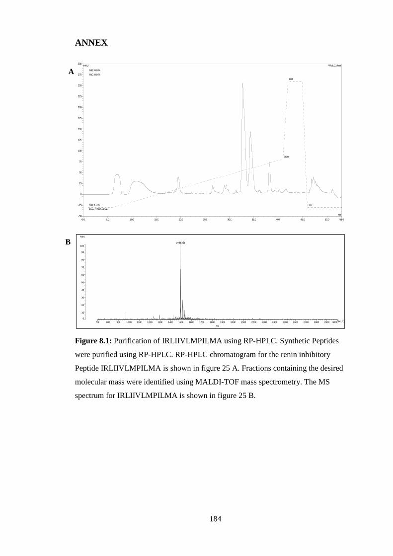
ANNEX
300mAU WVL:214 nm
0.0 5.0 10.0 15.0 20.0 25.0 30.0 35.0 40.0 45.0 50.0 53.0
-50
-25
0
25
50
75
100
125
150
175
200
225
250
275
min
Flow: 2.500 ml/min
%B: 1.0%
35.0
90.0
1.0
%C: 0.0%
%D: 0.0%
%Int.
A
184
Figure 8.1: Purification of IRLIIVLMPILMA using RP-HPLC. Synthetic Peptides
were purified using RP-HPLC. RP-HPLC chromatogram for the renin inhibitory
Peptide IRLIIVLMPILMA is shown in figure 25 A. Fractions containing the desired
molecular mass were identified using MALDI-TOF mass spectrometry. The MS
spectrum for IRLIIVLMPILMA is shown in figure 25 B.
0
10
20
30
40
50
60
70
80
90
100
700 800 900 1000 1100 1200 1300 1400 1500 1600 1700 1800 1900 2000 2100 2200 2300 2400 2500 2600 2700 2800 2900 3000
m/z
3[c].P15
1496.61B

800mAU WVL:214 nm
%C: 0.0 %
%D: 0.0 %A
0.0 5.0 10.0 15.0 20.0 25.0 30.0 35.0 40.0 45.0 50.0 53.0
-100
0
100
200
300
400
500
600
700
min
Flow: 2.500ml/min
%B: 5.0 %
25.0
90.0
5.0
%Int.
379.15B
185
Figure 8.2: Purification of NIGK. Synthetic Peptides were purified using RP-HPLC.
RP-HPLC chromatogram for the PAF-AH inhibitory Peptide NIGK is shown in figure
26 A. Fractions containing the desired molecular mass were identified using MALDI-
TOF mass spectrometry. The MS spectrum for NIGK is shown in figure 26 B.
0
10
20
30
40
50
60
70
80
90
100
100 120 140 160 180 200 220 240 260 280 300 320 340 360 380 400 420 440 460 480 500 520 540 560 580 600 620 640 660 680 700
m/z
1[c].G9
431.32

186
Figure 8.4: Non-linear regression of IRLIIVLMPILMA concentration versuspercentage inhibition.

187
Figure 8.4: Non-linear regression of NIGK concentration versus percentage inhibition.

188
Figure 8.5: PAF-AH assay of HPLC fractions 10-25 of the P. palmata papain proteinhydrolysate. Samples were tested at a concentration of 1 mg/mL. Data is displayed as %inhibition. Values are mean ± SEM (n = 3).