development of affibody molecules for radionuclide molecular
TRANSCRIPT
ACTAUNIVERSITATIS
UPSALIENSISUPPSALA
2016
Digital Comprehensive Summaries of Uppsala Dissertationsfrom the Faculty of Medicine 1237
Development of Affibodymolecules for radionuclidemolecular imaging and therapy ofcancer
HADIS HONARVAR
ISSN 1651-6206ISBN 978-91-554-9624-1urn:nbn:se:uu:diva-298740
Dissertation presented at Uppsala University to be publicly examined in Fåhraeus Hall, DagHammarskjölds väg 20, Uppsala, Saturday, 24 September 2016 at 09:30 for the degree ofDoctor of Philosophy (Faculty of Medicine). The examination will be conducted in English.Faculty examiner: Professor Raymond M. Reilly (Center for Pharmaceutical Oncology, LeslieDan Faculty of Pharmacy, University of Toronto, Canada).
AbstractHonarvar, H. 2016. Development of Affibody molecules for radionuclide molecularimaging and therapy of cancer. Digital Comprehensive Summaries of Uppsala Dissertationsfrom the Faculty of Medicine 1237. 71 pp. Uppsala: Acta Universitatis Upsaliensis.ISBN 978-91-554-9624-1.
Affibody molecules are a promising class of scaffold-based targeting proteins for radionuclide-based imaging and therapy of cancer. This thesis work is based on 5 original research articles(papers I-V), which focus on optimization of molecular design of HER2-binding Affibodyvariants for high contrast imaging of this predictive biomarker as well as development ofAffibody molecules suitable for radionuclide-based targeted therapies.
Papers I and II were dedicated to evaluation of the influence of the macrocyclic chelatorDOTA positioning at N-terminus, in the middle of helix-3 and at C terminus of a syntheticAffibody molecule, ZHER2:S1. These synthetic variants were labelled with different radionuclidesi.e. 111In and 68Ga to study also the effect of different labels on their biodistribution properties.
In paper III a 2-helix variant, Z342min, was developed using native ligation cyclization to cross-link helices one and two resulting in a stable 2-helix scaffold and characterized in vivo. Thisstudy was performed with the aim to obtain structure-properties relationship for developmentof smaller Affibody molecules.
Papers IV and V were devoted to development of therapeutic strategies. In paper IV, a seriesof peptide based chelators was investigated for labelling of Affibody molecules with 188Re toprovide low renal retention. In paper V, a pretargeting approach using peptide nucleic acid wasinvestigated. These studies were performed with the aim to overcome the high renal retentionof Affibody molecules when labelled with residualizing therapeutic radionuclides. Otherwise,the particle emitting radiometals could damage the kidneys more than the tumours.
The results obtained for anti-HER2 Affibody molecules summarized in this thesis might beof importance for the development of other scaffold protein based targeting agents.
Keywords: Affibody molecules, HER2, Molecular imaging, Radionuclide targeted therapy,Radionuclide molecular imaging, Labeling chemistry
Hadis Honarvar, Department of Immunology, Genetics and Pathology, Medical RadiationScience, Rudbecklaboratoriet, Uppsala University, SE-751 85 Uppsala, Sweden.
© Hadis Honarvar 2016
ISSN 1651-6206ISBN 978-91-554-9624-1urn:nbn:se:uu:diva-298740 (http://urn.kb.se/resolve?urn=urn:nbn:se:uu:diva-298740)

All we know of the truth is that the absolute truth, such as it is, is beyond our reach. Nicholas of Cusa
To the memory of my beloved parents

List of Papers
This thesis is based on the following papers, which are referred to in the text by their Roman numerals. I. Perols,A.,*Honarvar,H.,* Strand, J., Selvaraju,R.K., Orlova, A., Er-
iksson Karlström, A., Tolmachev, V. (2012). Influence of DOTA che-lator position on biodistribution and targeting properties of 111In-labeled synthetic anti-HER2 Affibody molecules. Bioconjugate Chem-istry. 23:1661-70.
II. Honarvar, H., Strand, J., Perols A, Orlova A, Selvaraju R.K., Eriksson Karlström, A., Tolmachev, V. (2014). Position for site-specific attach-ment of a DOTA chelator to synthetic Affibody molecules has a differ-ent influence on the targeting properties of 68Ga- compared to 111In-labeled conjugates. Molecular Imaging. 13:1-12.
III. Honarvar, H., Jokilaakso, N., Andersson, K., Malmberg, J., Rosik, D., Orlova, A., Eriksson Karlström, A., Tolmachev, V., Järver, P. (2013). Evaluation of backbone-cyclized HER2-binding 2-helix Affibody mol-ecule for In vivo molecular imaging. Nuclear Medicine and Biology. 40:378-86.
IV. Altai, M., Honarvar, H., Wållberg, H., Strand, J., Varasteh, Z., Ro-sestedt, M. Orlova, A., Dunås, F., Sandström, M., Löfblom, J., Tolmachev, V., Ståhl, S. (2014). Selection of an optimal cysteine-containing peptide-based chelator for labeling of affibody molecules with 188Re. European Journal of Medicinal Chemistry. 87:519-528.
V. Honarvar, H.,* Westerlund, K.,* Altai, M., Sandström, M., Orlova, A., Tolmachev, V., Eriksson Karlström, A. (2016). Feasibility of Af-fibody Molecule-Based PNA-Mediated Radionuclide Pretargeting of Malignant Tumors. Theranostics. 6:93-103.
* indicates equal contribution.
Reprints were made with permission from the respective publishers.
Additional papers (not included in this thesis): I Lindberg, H., Hofström, C., Altai, M., Honarvar, H., Wållberg, H.,
Orlova, A., Ståhl, S., Gräslund, T., Tolmachev, V. (2012). Evalua-tion of a HER2-targeting Affibody molecule combining an N-terminal HEHEHE-tag with a GGGC chelator for 99mTc-labellingat the C-terminus. Tumor Biology. 33:641-51.
II Hofström, C., Altai, M., Honarvar, H., Strand, J., Malmberg, J.,
Hosseinimehr, S.J., Orlova, A., Gräslund, T., Tolmachev, V. (2013). HAHAHA, HEHEHE, HIHIHI or HKHKHK: influence of histidine containing tags position and composition on biodistribution of [99mTc(CO)3]+-labelled affibody molecules. Journal of Medicinal Chemistry. 56: 4966-4974.
III Strand, J., Honarvar, H., Perols, A., Orlova, A., Selvaraju, R.K.,
Eriksson Karlström, A., Tolmachev, V. (2013). Influence of macro-cyclic chelators on targeting properties of 68Ga-labeled synthetic Af-fibody molecules. Comparison with 111In-labeled counterparts. PLoS ONE. 8:e70028.
IV Tolmachev, V., Varasteh, Z., Honarvar, H., Eriksson, O., Jonasson,
P. Frejd, F., Abrahmsen, L., Orlova, A. (2014). Imaging of PDGFRß in gliobalstoma xenografts using the Affibody molecule 111In-DOTA-ZPDGFRß:9591. J Nucl Med. 55:294-300.
V Rosik, D., Thibblin, A., Antoni, G., Honarvar, H., Strand, J., Selva-
raju, R.K., Altai, M., Orlova, A., Eriksson Karlström, A., Tolmachev, V. (2014). Triglutamyl spacer improves biodistribution of synthetic Affibody molecules radiofluorinated at the N-terminus via oxime formation with (18)F-4-fluorobenzaldehyde. Bioconjug Chem. 25:82-92.
VI Orlova, A., Malm, M., Andersson, K., Varasteh, Z., Honarvar, H.,
Strand, J., Selvaraju, R.K., Altai, M., Rosestedt, M., Tolmachev, V., Ståhl, S., Löfblom, J. (2014). Imaging of HER3-expressing xeno-grafts in mice using 99mTc(CO)3-HEHEHE-Z08698 Affibody mol-ecule. Eur J Nucl Med Mol Imaging. 41:1450-9.
VIII Altai, M., Wållberg, H., Honarvar, H., Strand, J., Orlova, A., Va-rasteh, Z., Sandström, M., Löfblom, J., Larsson, E., Strand, S.E., Lubberink, M., Ståhl, S., Tolmachev, V. (2014). 188Re-ZHER2:V2, a promising targeting agent against HER2-expressing tumors: in vitro and in vivo assessment. J Nucl Med. 55:1842-8.
IX Varasteh, Z., Rosenström, U., Velikyan, I., Mitran, B., Altai, M.,
Honarvar, H., Rosestedt, M., Lindeberg, G., Sörensen, J., Larhed, M., Tolmachev, V., Orlova A. (2014). The effect of mini-PEG-based spacer length on binding and pharmacokinetic properties of a 68Ga-labeled NOTA- conjugated antagonistic analog of bombesin. Mole-cules. 19:10455-72.
X Mitran, B., Altai, M., Hofström, C., Honarvar, H., Widström, C.H.,
Orlova, A., Tolmachev, V., Gräslund, T. (2015). Evaluation of 99mTc-ZIGF1R:4551-GGGC affibody molecule, a new probe for imaging of insulin-like growth factor type1 receptor expression. Amino Acids.47: 303-315.
XI Honarvar, H., Garousi, J., Gunneriusson, E., Höidén-Guthenberg, I.,
Altai, M., Widström, C., Tolmachev, V., Frejd, F.Y. (2015). Imaging of CAXI-expressing xenografts in vivo using 99mTc-HEHEHE-ZCAIX:1 Affibody molecule. Int J Oncol. 46: 513-520.
XII Strand, J., Nordeman, P., Honarvar, H., Altai, M., Larhed, M., Or-
lova, A., Tolmachev, V. (2015). Site-specific radioiodination of HER2-targeting affibody molecules using iodophenetyl-maleimide decreases renal uptake of radioactivity. Chemistry Open. 4: 174-82.
XIII Westerlund, K., Honarvar, H., Tolmachev, V., Eriksson Karlström,
A. (2015). Design, preparation and characterization of PNA-based hybridization probes for Affibody molecule-mediated pretargeting. Bioconjug Chem. 26:1724-36.
XIV Garousi, J., Lindbo, S., Nilvebrant, J., Åstrand, M., Buijs, J., Sand-
ström, M., Honarvar, H., Orlova, A., Tolmachev, V., Hober, S. (2015). ADAPT, a novel scaffold protein-based probe for radionu-clide imaging of molecular targets that are expressed in disseminated cancers. Cancer Res. 75:4364-71.
XVI Altai, M., Perols, A., Tsourma, M., Mitran, B., Honarvar, H., Robil-lard, M., Rossin, R., Ten Hoeve, W., Lubberink, M., Orlova, A., Er-iksson Karlstrom, A., Tolmachev, V. (2016). Feasibility of affibody-based bioorthogonal chemistry-mediated radionuclide pretargeting. J Nucl Med. 57:431-6.
XVII Lindbo, S., Garousi, J., Åstrand, M., Honarvar, H., Orlova, A.,
Hober, S., Tolmachev, V. (2016). Influence of histidine-containing tags on the biodistribution of ADAPT scaffold proteins. Bioconjugte Chemistry. 27:716-26.
XVIII Westerlund, K.,* Honarvar, H.,* Norrström, E., Strand, J., Mitran, B., Orlova, A., Erikson Karlström, A., Tolmachev, V. (2016). In-creasing the net negative charge by replacement of DOTA chelator with DOTAGA improves the biodistribution of radiolabeled second-generation synthetic Affibody molecules. Molecular Pharmaceutics. DOI: 10.1021/acs.molpharmaceut.6b00089.
* indicates equal contribution.
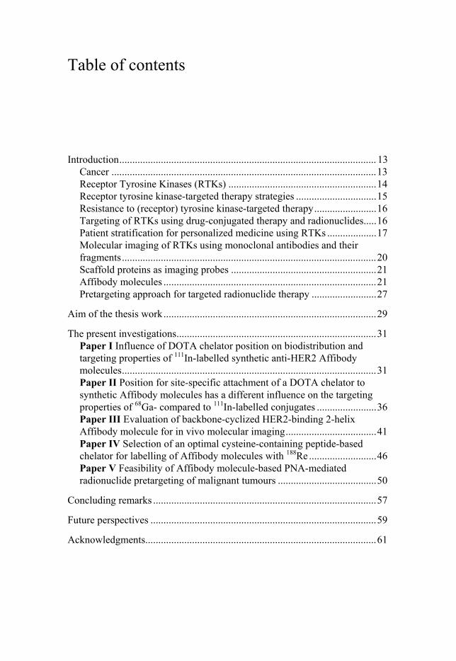
Table of contents
Introduction ................................................................................................... 13 Cancer ...................................................................................................... 13 Receptor Tyrosine Kinases (RTKs) ......................................................... 14 Receptor tyrosine kinase-targeted therapy strategies ............................... 15 Resistance to (receptor) tyrosine kinase-targeted therapy ........................ 16 Targeting of RTKs using drug-conjugated therapy and radionuclides ..... 16 Patient stratification for personalized medicine using RTKs ................... 17 Molecular imaging of RTKs using monoclonal antibodies and their fragments .................................................................................................. 20 Scaffold proteins as imaging probes ........................................................ 21 Affibody molecules .................................................................................. 21 Pretargeting approach for targeted radionuclide therapy ......................... 27
Aim of the thesis work .................................................................................. 29
The present investigations ............................................................................. 31 Paper I Influence of DOTA chelator position on biodistribution and targeting properties of 111In-labelled synthetic anti-HER2 Affibody molecules .................................................................................................. 31 Paper II Position for site-specific attachment of a DOTA chelator to synthetic Affibody molecules has a different influence on the targeting properties of 68Ga- compared to 111In-labelled conjugates ....................... 36 Paper III Evaluation of backbone-cyclized HER2-binding 2-helix Affibody molecule for in vivo molecular imaging ................................... 41 Paper IV Selection of an optimal cysteine-containing peptide-based chelator for labelling of Affibody molecules with 188Re .......................... 46 Paper V Feasibility of Affibody molecule-based PNA-mediated radionuclide pretargeting of malignant tumours ...................................... 50
Concluding remarks ...................................................................................... 57
Future perspectives ....................................................................................... 59
Acknowledgments......................................................................................... 61

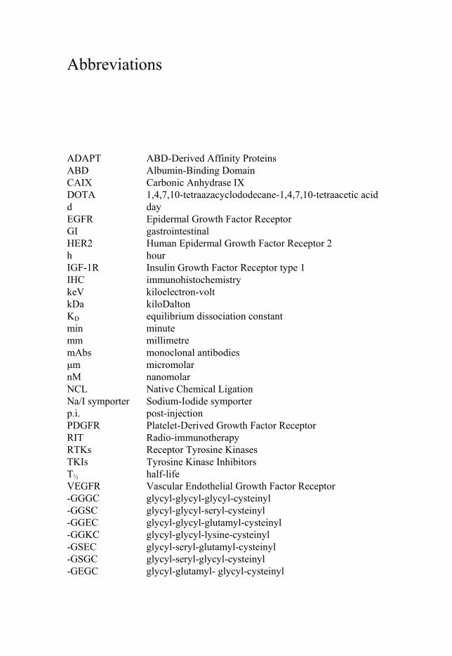
Abbreviations
ADAPT ABD-Derived Affinity Proteins ABD Albumin-Binding Domain CAIX Carbonic Anhydrase IX DOTA 1,4,7,10-tetraazacyclododecane-1,4,7,10-tetraacetic acid d day EGFR Epidermal Growth Factor Receptor GI gastrointestinal HER2 Human Epidermal Growth Factor Receptor 2 h hour IGF-1R Insulin Growth Factor Receptor type 1 IHC immunohistochemistry keV kiloelectron-volt kDa kiloDalton KD equilibrium dissociation constant min minute mm millimetre mAbs monoclonal antibodies μm micromolar nM nanomolar NCL Native Chemical Ligation Na/I symporter Sodium-Iodide symporter p.i. post-injection PDGFR Platelet-Derived Growth Factor Receptor RIT Radio-immunotherapy RTKs Receptor Tyrosine Kinases TKIs Tyrosine Kinase Inhibitors T½ half-life VEGFR Vascular Endothelial Growth Factor Receptor -GGGC glycyl-glycyl-glycyl-cysteinyl -GGSC glycyl-glycyl-seryl-cysteinyl -GGEC glycyl-glycyl-glutamyl-cysteinyl -GGKC glycyl-glycyl-lysine-cysteinyl -GSEC glycyl-seryl-glutamyl-cysteinyl -GSGC glycyl-seryl-glycyl-cysteinyl -GEGC glycyl-glutamyl- glycyl-cysteinyl

-GKGC glycyl-lysyl-glycyl-cysteinyl %ID/g percentage of injected dose per gram
13
Introduction
Cancer The worldwide incidence of cancer-related deaths is reported to be 100 to 350 per 100,000 populations per year (Lodish et al. 2008). Breast cancer and prostate cancer are the most frequently diagnosed forms of cancer in women and men, accounting for 29% and 26% of all new cancers, respectively (Siegel et al. 2015). Most cancers are caused by genetic damage due to life-time exposure to carcinogens and compounds encountered in the environ-ment. Such genetic damage (mutations) results in failure of the mechanisms that usually control the growth and proliferation of cells. Hanahan et al. (2011) has described this phenomenon in a logical framework as hallmarks of cancer that include evasion of growth suppressors, resistance to cell death, induction of angiogenesis, activation of invasion and metastasis, repro-gramming of energy metabolism and evasion of immune destruction. These characteristics are associated with malfunction in a number of cellular sig-nalling pathways.
The main treatment strategies for primary tumours are surgery and exter-nal-beam radiation therapy. In contrast, treatment of disseminated cancer requires a systemic approach and remains a challenge (Abraham et al. 2005). Currently, the most common systemic treatment is chemotherapy with a low selectivity for tumour tissue, which results in strong adverse effects. Taking into account the current cancer mortality statistics, the development of more efficient therapy strategies with fewer side effects is essential. Targeted ther-apy, which is based on molecular recognition of aberrantly expressed gene products in cancer cells, is one of the most promising approaches for treating disseminated cancer (Carlsson, 2008). One example of malignant transfor-mation of the cellular genome, which changes the molecular phenotype, is the aberrant expression or mutation of receptor tyrosine kinases (RTKs) (Hanahan et al. 2011).
14
Receptor Tyrosine Kinases (RTKs) RTKs comprise a superfamily of receptors subdivided into 20 classes. The most studied RTKs are class I (EGFR or HER (ErbB) family), class II (Insu-lin receptor family), and class V (VEGF receptors family) (Lemmon et al. 2010). In many cases, RTKs consist of an extracellular domain with a lig-and-binding site, a hydrophobic transmembrane domain, and an intracellular domain involved in signalling. It is known that ligand binding triggers RTK homo- or hetero-dimerization, followed by activation of the intracellular tyrosine kinases domain, which initiates downstream signalling cascades. Alternatively, preformed dimers undergo conformational changes after di-merization that triggers signalling.
Proto-oncogenes such as RTKs control cellular processes that are crucial for survival, proliferation, differentiation, motility, and apoptosis, and strong overexpression of these receptors in primary tumours and corresponding metastases make them interesting as putative targets for targeted therapy. Therapeutic targeting approaches are divided into two classes: targeting of the extracellular domain of the RTK (e.g., monoclonal antibodies trastuzumab, cetuximab) or their ligands (e.g., antibody bevacizumab) and intracellular inhibition (e.g., inhibition of signalling cascades). Thus, identi-fying RTKs-overexpression will assist oncologists in selecting an appropri-ate personalized therapy (Lemmon et al. 2010).
The HER (ErbB) family contains four structurally related receptors, HER1 (EGFR, ErbB1), HER2 (Neu, ErbB2), HER3 (ErbB3), and HER4 (ErbB4), that play an important role in cancer biology (Yarden, 2001). Sev-eral different ligands (i.e., NRG, EGF, TGFα, and HB-EGF), are known to bind to receptors of the HER family; some of these ligands bind to more than one receptor, whereas some are receptor specific. The only member of ErbB subfamily of receptor tyrosine kinases with no known ligand is human epi-dermal growth receptor type 2 (HER2), and this receptor does not require ligand binding for dimerization (Yarden, 2001). Dysregulated signalling of overexpressed HER2 receptors results in cell proliferation and suppression of apoptosis in cancer cells. Indeed, HER2 overexpression is associated with malignancy in a number of cancers, including breast, urinary bladder, ovari-an, colorectal and prostate cancers (Witton et al. 2003). Thus, HER2 is of high interest for clinical oncology.
15
Receptor tyrosine kinase-targeted therapy strategies
Immunotherapy The mechanism of action of monoclonal antibodies (mAbs) that inhibit the excessive function of RTKs (overexpressed in malignant tissues) includes (Weiner et al. 2010):
• Triggering of receptor internalization. • Inhibition of dimerization. • Blockage of the ligand-binding site.
In addition, mAbs can further trigger activation of antibody-dependent cellu-lar cytotoxicity (ADCC) and complement-dependent cytotoxicity (CDC). Such mAbs target the extracellular domain of aberrantly expressed RTKs in a selective way; this specific targeting spares normal cells and is accompa-nied by lower systemic toxicity in comparison to conventional therapies such as chemotherapy (Hudis, 2007).
Immunotherapy improves cancer patient survival. An example is the treatment of HER2-expressing tumours with the monoclonal antibody trastuzumab, which improves survival in breast and gastro-oesophageal can-cer patients (Weiner et al. 2010; Bang et al. 2010). Overexpression of HER2 is a predictive biomarker for these therapies. Other examples of anti-RTK mAbs introduced into clinical practice are pertuzumab (anti-HER2) (Harbeck et al. 2013), cetuximab and pantimumab (anti-EGFR) (Price et al. 2014 a), bevacisumab (anti-VEGF) (Keating, 2014) and ramucurimab (anti-VEGFR2) (Zhu et al. 2013).
Tyrosine Kinase Inhibitors (TKIs) Another strategy for treating RTK-overexpressing tumours involves target-ing of the intracellular domain of the receptor with tyrosine kinase inhibitors. TKIs are small molecules capable of penetrating the cell membrane and in-hibiting the catalytic activity of tyrosine kinases and, accordingly, signal transduction cascades. This often results in arrest of cell growth and cell death (Sharma et al. 2009). A growing number of TKIs have been approved by the Food and Drug Administration, and many others are currently in clin-ical or preclinical development. The first approved tyrosine kinase inhibitor was imatinib (inhibiting PDGFR, Kit, and Abl1-2). Currently, imatinib is used for treatment of chronic myeloid leukaemia and gastrointestinal stromal tumours (Demetri et al. 2002; Sawyers, 2003) with c-Kit mutation. Lapatinib (inhibiting EGFR and HER2) (Di Leo et al. 2008) is another TKI that is used for treating metastatic breast cancer.
16
Resistance to (receptor) tyrosine kinase-targeted therapy Although mAbs and TKIs are the most successful (receptor) tyrosine kinase-targeted therapies, treatment is efficient in only a fraction of patients. This is due to either de novo (Hazlehurst et al. 2003) resistance caused by certain genetic alterations or acquired resistance (Rosenzweig, 2012) following drug exposure, resulting in variability in therapeutic potency. Several proposed mechanisms are thought to mediate resistance to molecular targeted therapy. Although the biological basis of resistance to mAbs and TKIs is beyond the scope of this thesis, it is worth mentioning a few of the known mechanisms, which include alterations in the drug target, escape from cell cycle arrest, activation of alternative signalling pathways, changes in levels of target ex-pression and dysfunctional apoptosis (Holohan et al. 2013). For instance, secondary resistance to trastuzumab can develop due to increased IGF-1R or HER3 signalling (Pohlmann et al. 2009). Development of resistance is often accompanied by preserved overexpression of molecular targets.
Targeting of RTKs using drug-conjugated therapy and radionuclides When a patient develops resistance to drugs targeting RTKs, it is necessary to consider alternative treatment strategies. One approach is conjugation of cytotoxic payloads (e.g., drugs and toxins) or effector molecules to tumour-targeting mAbs or their fragments in an effort to enhance their effects and improve response duration and overall response rates (Wu et al. 2005).
A strong example of a drug-conjugated mAb is T-DM1, which is trastuzumab conjugated with emtansine (a highly potent anti-tubulin drug). The use of T-DM1 in clinical studies has resulted in significant improvement in PFS (progression-free survival) and OS (overall survival) with less fre-quent adverse effects than lapatinib plus capecitabine in patients with meta-static breast cancer (Verma et al. 2012; The MARIANNE clinical trial).
Another strategy is to arm tumour-targeting mAbs and their fragments with therapeutic radionuclides (Table 1) such as beta-emitters (e.g., 131I, 90Y, and 177Lu), alpha-emitters (e.g., 212Bi and 213Bi) or auger electron-emitters (e.g., 111In) to deliver cytotoxic radiation to targeted cells. Such radioim-munotherapy (RIT) enhances the effect of therapy by using an antibody with specificity for a tumour-associated antigen and irradiation to deliver a lethal absorbed dose to tumours (Boswell et al. 2007; Costantini et al. 2008). How-ever, to date, RIT has only shown a strong potential for treating radiosensi-tive hematologic malignancies, such as non-Hodgkin lymphoma and has not yet been successful for treating solid tumours (Pouget et al. 2011). Two ra-dioimmunoconjugates, 90Y-ibritumomab tiuxetan (Zevalin) and 131I-
17
tositumomab (Bexar), are FDA approved for treatment of B-cell non-Hodgkin lymphoma (Iagaru et al. 2010).
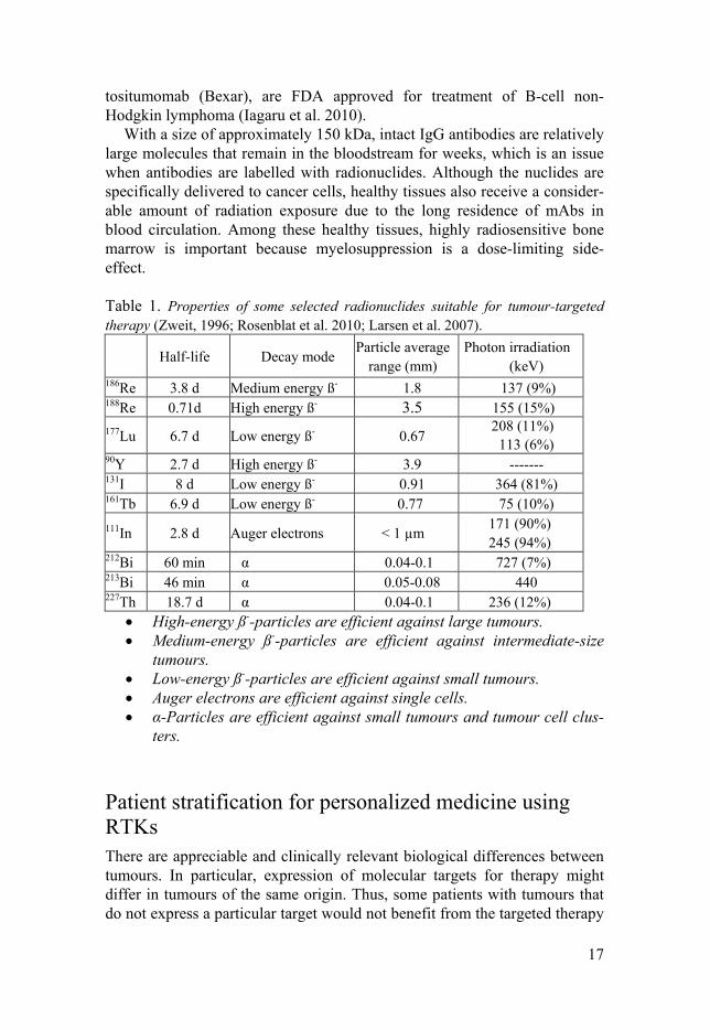
With a size of approximately 150 kDa, intact IgG antibodies are relatively large molecules that remain in the bloodstream for weeks, which is an issue when antibodies are labelled with radionuclides. Although the nuclides are specifically delivered to cancer cells, healthy tissues also receive a consider-able amount of radiation exposure due to the long residence of mAbs in blood circulation. Among these healthy tissues, highly radiosensitive bone marrow is important because myelosuppression is a dose-limiting side-effect. Table 1. Properties of some selected radionuclides suitable for tumour-targeted therapy (Zweit, 1996; Rosenblat et al. 2010; Larsen et al. 2007).
Half-life Decay mode Particle average
range (mm) Photon irradiation
(keV) 186Re 3.8 d Medium energy ß- 1.8 137 (9%) 188Re 0.71d High energy ß- 3.5 155 (15%) 177Lu 6.7 d Low energy ß- 0.67
208 (11%) 113 (6%)
90Y 2.7 d High energy ß- 3.9 ------- 131I 8 d Low energy ß- 0.91 364 (81%) 161Tb 6.9 d Low energy ß- 0.77 75 (10%) 111In 2.8 d Auger electrons < 1 µm 171 (90%)
245 (94%) 212Bi 60 min α 0.04-0.1 727 (7%) 213Bi 46 min α 0.05-0.08 440 227Th 18.7 d α 0.04-0.1 236 (12%)
• High-energy ß--particles are efficient against large tumours. • Medium-energy ß--particles are efficient against intermediate-size
tumours. • Low-energy ß--particles are efficient against small tumours. • Auger electrons are efficient against single cells. • α-Particles are efficient against small tumours and tumour cell clus-
ters.
Patient stratification for personalized medicine using RTKs There are appreciable and clinically relevant biological differences between tumours. In particular, expression of molecular targets for therapy might differ in tumours of the same origin. Thus, some patients with tumours that do not express a particular target would not benefit from the targeted therapy
18
but would still suffer from side effects (Chen et al. 2003). For instance, both trastuzumab and lapatinib are considered for treatment of HER2-expressing breast cancer. However, only 15-30% of breast cancer patients have HER2-expressing tumours. Thus, to predict a therapeutic outcome and reduce un-necessary toxicity, stratification of patients for a specific cancer-related bi-omarker is of importance (Yarden et al. 2001; ASCO guidelines).
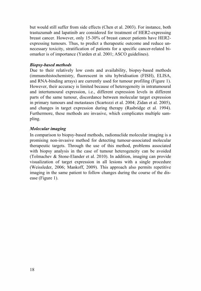
Biopsy-based methods Due to their relatively low costs and availability, biopsy-based methods (immunohistochemistry, fluorescent in situ hybridisation (FISH), ELISA, and RNA-binding arrays) are currently used for tumour profiling (Figure 1). However, their accuracy is limited because of heterogeneity in intratumoural and intertumoural expression, i.e., different expression levels in different parts of the same tumour, discordance between molecular target expression in primary tumours and metastases (Scartozzi et al. 2004; Zidan et al. 2005), and changes in target expression during therapy (Rasbridge et al. 1994). Furthermore, these methods are invasive, which complicates multiple sam-pling.
Molecular imaging In comparison to biopsy-based methods, radionuclide molecular imaging is a promising non-invasive method for detecting tumour-associated molecular therapeutic targets. Through the use of this method, problems associated with biopsy analysis in the case of tumour heterogeneity can be avoided (Tolmachev & Stone-Elander et al. 2010). In addition, imaging can provide visualization of target expression in all lesions with a single procedure (Weissleder, 2006; Mankoff, 2009). This approach also permits repetitive imaging in the same patient to follow changes during the course of the dis-ease (Figure 1).
19
Figure 1. IHC HER2 staining of a bone metastasis from a breast cancer patient indicating HER2-positive score 3+ (Left). Maximum Intensity Projection PET imag-es of a patient with wide-spread metastatic breast cancer using [68Ga]-ABY-025. Darker colours indicate higher uptake (Right). Arrows point at the kidneys. (The biopsy image was kindly provided by Prof. Jens Sörensen. The PET image was re-printed with permission from Theranostics.)
Although CT and MRI provide anatomical information, the molecular imag-ing approach generates information concerning the molecular and functional properties of a tumour (Tolmachev & Stone-Elander et al. 2010). Radionu-clide molecular imaging of therapeutic targets can be performed using PET (positron emission tomography) and scintigraphy-based modalities, i.e., pla-nar and SPECT (single-photon emission tomography). A planar and SPECT camera detects γ-quanta or high-energy x-rays emitted by a biodistributed radiotracer labelled with radionuclides such as 99mTc and 111In (Table 2). A PET camera detects pairs of annihilation photons emitted by a biodistributed radiotracer labelled with positron (β+)-emitting radionuclides such as 18F, 11C, or 68Ga (Table 2). Computer processing of SPECT and PET data enables reconstruction and visualization of the 3D distribution of the radionuclide in a living body. Fusion of radionuclide-based images with anatomical images produced by CT aids clinicians in precisely localizing and identifying the affected tissue. PET has advantages of higher sensitivity and spatial resolu-tion as well as better quantification accuracy compared to SPECT. Nonethe-less, SPECT facilities are more available and cost efficient than PET. It is expected that the availability of PET cameras will increase in the future due to their superior sensitivity (Rahmim et al. 2008).
20
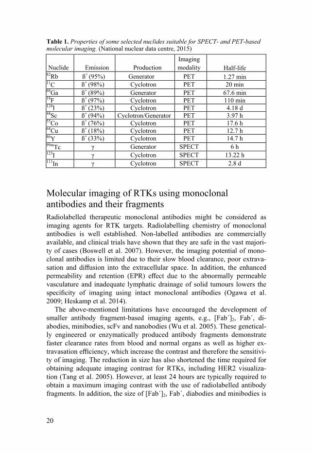
Table 1. Properties of some selected nuclides suitable for SPECT- and PET-based molecular imaging. (National nuclear data centre, 2015)
Nuclide Emission Production Imaging modality Half-life
82Rb ß+ (95%) Generator PET 1.27 min 11C ß+ (98%) Cyclotron PET 20 min 68Ga ß+ (89%) Generator PET 67.6 min 18F ß+ (97%) Cyclotron PET 110 min 124I ß+ (23%) Cyclotron PET 4.18 d 44Sc ß+ (94%) Cyclotron/Generator PET 3.97 h 55Co ß+ (76%) Cyclotron PET 17.6 h 64Cu ß+ (18%) Cyclotron PET 12.7 h 86Y ß+ (33%) Cyclotron PET 14.7 h 99mTc γ Generator SPECT 6 h 123I γ Cyclotron SPECT 13.22 h 111In γ Cyclotron SPECT 2.8 d
Molecular imaging of RTKs using monoclonal antibodies and their fragments Radiolabelled therapeutic monoclonal antibodies might be considered as imaging agents for RTK targets. Radiolabelling chemistry of monoclonal antibodies is well established. Non-labelled antibodies are commercially available, and clinical trials have shown that they are safe in the vast majori-ty of cases (Boswell et al. 2007). However, the imaging potential of mono-clonal antibodies is limited due to their slow blood clearance, poor extrava-sation and diffusion into the extracellular space. In addition, the enhanced permeability and retention (EPR) effect due to the abnormally permeable vasculature and inadequate lymphatic drainage of solid tumours lowers the specificity of imaging using intact monoclonal antibodies (Ogawa et al. 2009; Heskamp et al. 2014).
The above-mentioned limitations have encouraged the development of smaller antibody fragment-based imaging agents, e.g., [Fab´]2, Fab´, di-abodies, minibodies, scFv and nanobodies (Wu et al. 2005). These genetical-ly engineered or enzymatically produced antibody fragments demonstrate faster clearance rates from blood and normal organs as well as higher ex-travasation efficiency, which increase the contrast and therefore the sensitivi-ty of imaging. The reduction in size has also shortened the time required for obtaining adequate imaging contrast for RTKs, including HER2 visualiza-tion (Tang et al. 2005). However, at least 24 hours are typically required to obtain a maximum imaging contrast with the use of radiolabelled antibody fragments. In addition, the size of [Fab´]2, Fab´, diabodies and minibodies is
21
larger than the EPR cut-off (~45 kDa), which makes these molecules prone to non-specific tumour accumulation.
Scaffold proteins as imaging probes Scaffold proteins are based on a structured framework, which maintains a stable tertiary structure and a variable portion in which randomization of a number of surface-exposed residues produces a large library of binding mol-ecules (Nygren & Skerra, 2004). Several alternative non-immunoglobulin scaffold proteins, e.g., Affibody molecules and Designed Ankyrin Repeat Proteins (DARPins), have been developed using molecular display tech-niques (phage, ribosomal, yeast or bacterial surface display). Advantages of scaffold proteins over antibodies and their fragments include small size, high solubility, rapid folding, thermodynamic and chemical stability, a single-polypeptide chain format, and often the absence of disulphide bonds (Nuttall & Walsh 2008). Importantly, the small size (<45 kDa) of scaffold proteins, as well as nanobodies, overcomes the EPR effect and results in specific tu-mour accumulation (Wester et al. 2005). This is of particular importance in personalized medicine. Scaffold proteins have demonstrated appreciable potential as imaging probes (Miao et al. 2011). Among them, Affibody mol-ecules are the most studied class of scaffold proteins.
Affibody molecules
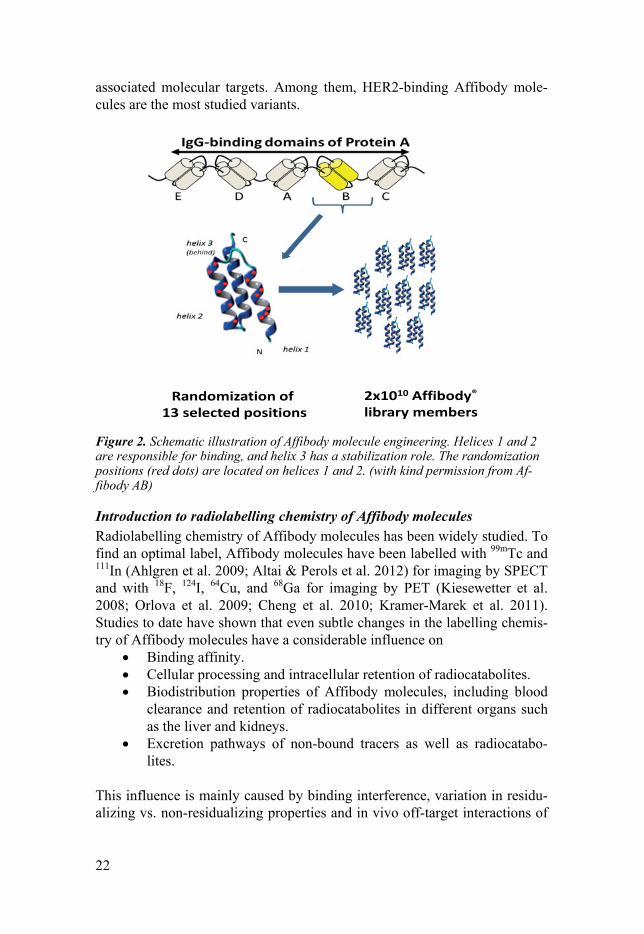
Background Affibody molecules are three-helix proteins derived from the immunoglobu-lin-binding domain of staphylococcal protein A (Figure 2). The protein con-sists of 58 amino acids with no internal disulphide bonds. Affibody mole-cules can be produced both recombinantly in E. coli and synthetically. A library is created by random substitution of amino acids on helices 1 and 2 of the scaffold, generating multiple variants for selection of high-affinity bind-ers (Nygren, 2008). The small size (6-7 kDa) and high affinity (in the low nanomolar or sub-nanomolar range) make them potential candidates for mo-lecular imaging (Löfblom et al. 2010). Affibody molecules targeting HER2 (human epidermal growth factor receptor type 2) (Tolmachev, 2008), EGFR (epidermal growth factor receptor) (Tolmachev & Rosik et al. 2010), HER3 (human epidermal growth factor receptor type 3) (Orlova et al. 2014), CAIX (hypoxia induced carbonic anhydrase IX) (Honarvar et al. 2015), PDGFRß (platelet derived growth factor receptor beta) (Tolmachev et al. 2014) and IGF-1R (insulin-like growth factor 1 receptor) (Tolmachev& Malmberg et al. 2012) have been successfully applied for imaging of these cancer-
22
associated molecular targets. Among them, HER2-binding Affibody mole-cules are the most studied variants.
Figure 2. Schematic illustration of Affibody molecule engineering. Helices 1 and 2 are responsible for binding, and helix 3 has a stabilization role. The randomization positions (red dots) are located on helices 1 and 2. (with kind permission from Af-fibody AB)
Introduction to radiolabelling chemistry of Affibody molecules Radiolabelling chemistry of Affibody molecules has been widely studied. To find an optimal label, Affibody molecules have been labelled with 99mTc and 111In (Ahlgren et al. 2009; Altai & Perols et al. 2012) for imaging by SPECT and with 18F, 124I, 64Cu, and 68Ga for imaging by PET (Kiesewetter et al. 2008; Orlova et al. 2009; Cheng et al. 2010; Kramer-Marek et al. 2011). Studies to date have shown that even subtle changes in the labelling chemis-try of Affibody molecules have a considerable influence on
• Binding affinity. • Cellular processing and intracellular retention of radiocatabolites. • Biodistribution properties of Affibody molecules, including blood
clearance and retention of radiocatabolites in different organs such as the liver and kidneys.
• Excretion pathways of non-bound tracers as well as radiocatabo-lites.
This influence is mainly caused by binding interference, variation in residu-alizing vs. non-residualizing properties and in vivo off-target interactions of
Randomization of13 selected positions
2x10¹⁰ Affibody®
library members
23
Affibody molecules (Orlova & Feldwisch et al. 2007; Tolmachev & Orlova 2012).
Until recently, conjugation of prosthetic groups and chelators to targeting proteins was mainly performed using amino-directed chemistry. Several reactive amino groups are present on Affibody molecules, such as the N-terminal amino group and the amino groups of lysine residues. Therefore, the use of unselective amino-directed chemistry should result in a heteroge-neous mixture of conjugates with different degrees of modification and dif-ferent label positions. The relatively small number of amino acid residues in the scaffold enables synthesis of Affibody molecules by Solid-Phase Peptide Synthesis (SPPS), enabling the conjugation of chelators to the scaffold in a site-specific way to produce a homogenous product. An alternative is incor-poration of a single cysteine and the use of thiol-directed chemistry.
The choice of the radionuclide determines which chelator or prosthetic group is utilized. For instance, radiohalogens can be directly covalently cou-pled to a protein or via prosthetic groups (Kiesewetter et al. 2008; Tolma-chev & Orlova, 2012), whereas labelling with radiometals requires incorpo-ration of a chelator (Engfeldt et al. 2007; Brechbiel, 2008). Stability of the chelated radiometal is fundamental to avoid the occurrence of free radioac-tivity in blood and non-target tissues, which minimizes the trans-chelation possibility of certain radiometals to iron-binding proteins (Fe3+) such as transferrin and consequently influences the rate of excretion from the body (Smith, 2005). The macrocyclic chelators 1,4,7,10-tetraazacyclododecane-1,4,7,10-tetraacetic acid (DOTA), 1,4,7-triazacyclononane-N,N′,N′′triacetic acid (NOTA), and 1-(1,3-carboxypropyl)-4,7 carboxymethyl-1,4,7 triazacy-clononane (NODAGA) form thermodynamically stable and kinetically inert ion complexes with many radiometals (Price et al. 2014 b). Although com-plex formation of macrocyclic chelators with radiometals requires elevated temperatures, this is not an issue when labelling robust peptides such as Af-fibody molecules that can tolerate high temperatures. Among macrocyclic chelators, the commercially available DOTA chelator provides stable com-plexation with several radiometals. Indeed, the DOTA chelator is currently widely used in the development of peptide-based radiopharmaceuticals (De Leon-Rodríguez et al. 2008).
High-contrast molecular imaging using Affibody molecules The accuracy of radionuclide molecular imaging depends on the specificity and sensitivity of the imaging probe. The specificity of the targeting agent is determined by the amount of off-target interactions of the probe, and the sensitivity depends on the imaging contrast. This is of particular importance in personalized medicine, in which maximal contrast is required to achieve high sensitivity of radionuclide molecular imaging (Eckelman et al. 2009).
Imaging of HER2-expressing tumours using Affibody molecules has demonstrated promising results both in preclinical and clinical studies (Tol-
24
machev et al. 2008; Baum et al. 2010; Sörensen et al. 2014 & 2016). The high affinity of HER2-binding Affibody molecules (low picomolar range) ensures high retention of the radiolabelled Affibody molecules in the tu-mour. In murine models, the small size and high affinity of Affibody mole-cules provides at least an order of magnitude higher contrast than antibodies a few hours after injection (Orlova et al. 2009; Malmberg et al. 2011). A high tumour-to-blood ratio is a precondition for high-contrast images (Ahlgren et al. 2010; Tolmachev et al. 2008).
Several factors affect the sensitivity and specificity of the images. Ac-cording to Schmidt et al. (2009) and Wittrup et al. (2012), the most im-portant factors are the size and affinity of the tracer. A small size provides rapid a extravasation rate and rapid clearance of unbound tracer. Additional-ly, proteins with a size less than 45 kDa are not subjected to the EPR effect (Heskamp et al. 2014). Non-specific accumulation of radiolabelled Affibody molecules in murine xenografts has been reported to be only 1-2 % of the specific accumulation (Orlova & Tolmachev et al. 2007). In the case of small scaffold Affibody molecules, a high affinity (single-digit nanomolar range for targets with high expression or sub-nanomolar range for targets with low expression) is a precondition for efficient tumour accumulation of radioactivity (Tolmachev & Tran et al. 2012).
According to Wittrup et al. (2012), the tumour extravasation rate is as im-portant as the rapid clearance and elimination from normal tissues. Schmidt et al. (2009) and Wittrup et al. (2012) showed that tracers of intermediate masses (25-60 kDa) display the lowest tumour uptake, whereas tracers with the smallest and largest molecular masses exhibit the highest tumour uptake. This feature could be explained by the relatively low extravasation rate and rapid clearance rate via kidney filtration of tracers of intermediate size. Thus, size reduction increases the extravasation rate and therefore tumour localiza-tion (Zahnd et al. 2010; Ahlgren et al. 2010).
The third helix of an anti-HER2 Affibody molecule, ABY-002, was re-moved to further improve the imaging contrast by enhanced tumour penetra-tion and more rapid clearance of unbound tracer from non-specific com-partments (Webster et al. 2009). Because the third helix stabilizes the scaf-fold, several mutations and an intramolecular homocysteine bridge were also introduced into the molecule. However, the size reduction of the 3-helix scaffold resulted in a considerable decrease in affinity of the 2-helix Af-fibody molecule. Ren et al. (2009) and Rosik et al. (2012) studied the prop-erties of the 2-helix variant labelled with 68Ga and 111In, and the results of these studies showed preserved specific accumulation of the radioconjugates in tumours and the capability of imaging xenografts shortly after injection. However, tumour uptake was significantly reduced compared with their pa-rental 3-helix variant (Tolmachev & Velikyan et al. 2010). This limited the application of the 2-helix variant to imaging of only a high level of target expression, with more targets being available to rebind (Rosik et al. 2012).
25
Further structural optimization is required to make the 2-helix variant gener-ally applicable for imaging.
Optimization of targeting properties of Affibody molecules for targeted radionuclide therapy Effective radiotherapy might be achieved when the maximum absorbed dose is delivered to the tumour cells and a minimal dose (within the tolerance threshold) to normal tissues to minimize toxicity (Pouget et al. 2011). The slow extravasation rate, low tumour penetration rate and prolonged circula-tion time of radiolabelled mAbs (150 kDa) limits the dose delivered to solid tumours due to the risk of bone marrow toxicity. Thus, engineered fragments could be a suitable alternative based on their faster blood clearance and con-sequently lower bone marrow exposure. However, their rapid clearance and relatively slow extravasation rate as well as retention in the liver and kidney must be considered in the overall toxicity profile (Kenanova et al. 2006).
An alternative to antibodies and their fragments are high-affinity small scaffold proteins. Among them, Affibody molecules with high affinity and rapid clearance from blood and non-target tissues may be considered as a potential candidate for targeted therapy (Löfblom et al. 2010). However, the Affibody molecule size below the kidney cut-off (60 kDa), results in their glomerular filtration. The efficient re-absorption of Affibody molecules in the proximal tubuli of the kidneys (Feldwisch et al. 2012) is a major problem when they are labelled with radiometals. After internalization by proximal tubular cells, Affibody molecules undergo proteolytic degradation in lyso-somal compartments, in which radiometal (e.g., 111In, 177Lu and 90Y) labels form hydrophilic, charged and bulky catabolites. These catabolites cannot diffuse through the lysosomal or cellular membrane and are trapped inside the tubular cells (Altai et al. 2013). Thus, the kidneys receive a much higher absorbed dose than the tumours. Such issues complicate the use of Affibody molecules for therapeutic purposes (Feldwisch et al. 2012).
Different approaches have been used to develop Affibody molecules with low renal uptake, as follows:
• Fusion of the albumin-binding-domain (ABD) to Affibody mole-cules resulted in several fold higher doses to the tumour than to the blood, kidneys, and liver. However, due to the prolonged blood cir-culation of the tracer, bone marrow toxicity could not be avoided (Tolmachev et al. 2007; Orlova et al. 2013).
• The use of non-residualizing radiohalogens reduced the high kid-ney retention of radiocatabolites while providing a reasonably high tumour uptake (Orlova et al. 2006) due to the lipophilic nature of the radiohalogen catabolites, which easily leak from cells. However, this restricts the selection of therapeutic radionuclides to halogens.
• Development of non-residualizing labels using peptide-based che-lators reduced the radioactivity concentration of 99mTc in excretory
26
organs such as the liver and kidneys (Wållberg et al. 2011, Feld-wisch et al. 2012).
Technetium-99m, with a half-life of 6 h, is a generator-produced radionu-clide that is commonly used in nuclear medicine applications. However, due to the absence of particle radiation, 99mTc is suitable only for imaging. Rhe-nium is a chemical analogue of technetium, and beta-emitting rhenium iso-topes, i.e., 186Re (T½=3.7 d) and 188Re (T½=17 h), are considered suitable for therapy. Thus, peptide-based chelators developed for labelling of Affibody molecules with 99mTc might also be used for 186/188Re labelling, facilitating the use of the same targeting probe for both imaging and therapy purposes.
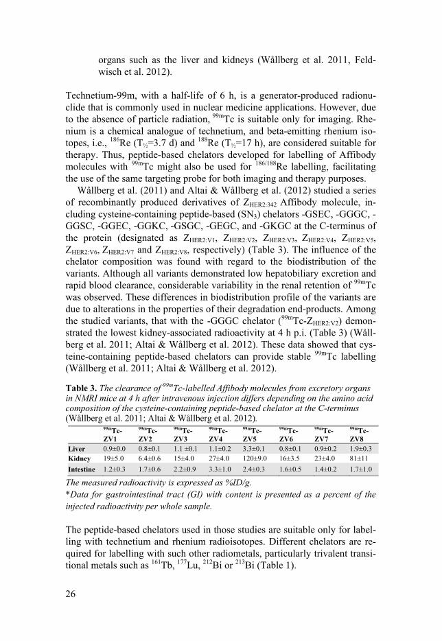
Wållberg et al. (2011) and Altai & Wållberg et al. (2012) studied a series of recombinantly produced derivatives of ZHER2:342 Affibody molecule, in-cluding cysteine-containing peptide-based (SN3) chelators -GSEC, -GGGC, -GGSC, -GGEC, -GGKC, -GSGC, -GEGC, and -GKGC at the C-terminus of the protein (designated as ZHER2:V1, ZHER2:V2, ZHER2:V3, ZHER2:V4, ZHER2:V5, ZHER2:V6, ZHER2:V7 and ZHER2:V8, respectively) (Table 3). The influence of the chelator composition was found with regard to the biodistribution of the variants. Although all variants demonstrated low hepatobiliary excretion and rapid blood clearance, considerable variability in the renal retention of 99mTc was observed. These differences in biodistribution profile of the variants are due to alterations in the properties of their degradation end-products. Among the studied variants, that with the -GGGC chelator (99mTc-ZHER2:V2) demon-strated the lowest kidney-associated radioactivity at 4 h p.i. (Table 3) (Wåll-berg et al. 2011; Altai & Wållberg et al. 2012). These data showed that cys-teine-containing peptide-based chelators can provide stable 99mTc labelling (Wållberg et al. 2011; Altai & Wållberg et al. 2012).
Table 3. The clearance of 99mTc-labelled Affibody molecules from excretory organs in NMRI mice at 4 h after intravenous injection differs depending on the amino acid composition of the cysteine-containing peptide-based chelator at the C-terminus (Wållberg et al. 2011; Altai & Wållberg et al. 2012).
The measured radioactivity is expressed as %ID/g. *Data for gastrointestinal tract (GI) with content is presented as a percent of the injected radioactivity per whole sample.
The peptide-based chelators used in those studies are suitable only for label-ling with technetium and rhenium radioisotopes. Different chelators are re-quired for labelling with such other radiometals, particularly trivalent transi-tional metals such as 161Tb, 177Lu, 212Bi or 213Bi (Table 1).
99mTc-ZV1
99mTc-ZV2
99mTc-ZV3
99mTc-ZV4
99mTc-ZV5
99mTc-ZV6
99mTc-ZV7
99mTc-ZV8
Liver 0.9±0.0 0.8±0.1 1.1 ±0.1 1.1±0.2 3.3±0.1 0.8±0.1 0.9±0.2 1.9±0.3 Kidney 19±5.0 6.4±0.6 15±4.0 27±4.0 120±9.0 16±3.5 23±4.0 81±11 Intestine 1.2±0.3 1.7±0.6 2.2±0.9 3.3±1.0 2.4±0.3 1.6±0.5 1.4±0.2 1.7±1.0
27
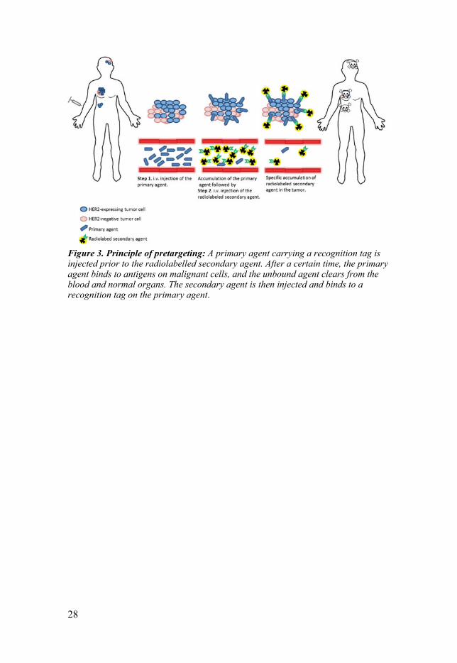
Pretargeting approach for targeted radionuclide therapy An alternative treatment approach to overcome the above-mentioned prob-lems associated with RIT is pretargeting (Goldenberg et al. 2006; Sharkey et al. 2005).
The pretargeting approach involves a two-step process. First, a tumour-antigen specific primary agent carrying a recognition tag is injected and al-lowed to localize in a tumour and clear from blood circulation. Thereafter, a radiolabelled secondary agent is injected, which accumulates radioactivity in the tumour by binding to the recognition tag on the primary agent. The sec-ond step is performed after a specific time at which the primary agent has localized in the tumour and the unbound agent has been eliminated from the blood (Figure 3). The smaller size of the secondary agent provides favoura-ble pharmacokinetics, with rapid distribution, low accumulation in healthy tissues and rapid blood clearance. Thus, the cytotoxic effect of the radionu-clides will be tumour specific, and healthy tissues will be exposed to a min-imal irradiation dose (Goldenberg et al. 2006; Sharkey et al. 2005).
The feasibility of the pretargeting approach has been validated using the (strept)avidin-biotin system (Hantowich et al. 1987). This system has certain disadvantages, such as the immunogenicity of (strept)avidin and the pres-ence of endogenous biotin, which competes with the radiolabelled biotin for binding sites (Rusckowski et al. 1997).
A number of alternative approaches have been developed to overcome these issues, including the following:
• The development of bispecific antibodies, with one of the arms be-ing specific to a small radiolabelled hapten (Reilly, 2006; Sharkey et al. 2005).
• The use of interactions of complementary oligonucleotides or their analogues (Kuijpers et al. 1993, Mardirossian et al. 1997, Liu et al. 2002).
• The use of bioorthogonal chemistry, e.g., reaction between trans-cyclooctene, TCO (recognition tag) and electron-deficient tetrazine (secondary agent) (Rossin et al. 2010; van de Watering et al. 2014).
Preclinical studies and early clinical trials have demonstrated more favoura-ble therapeutic indices for pretargeting compared with a directly radio-labelled antibody (Kraeber-Bodéré et al. 2012). Thus, it would be interesting to investigate whether application of the pretargeting approach can be used to reduce renal uptake in the case of Affibody-mediated radionuclide thera-py.
28
Figure 3. Principle of pretargeting: A primary agent carrying a recognition tag is injected prior to the radiolabelled secondary agent. After a certain time, the primary agent binds to antigens on malignant cells, and the unbound agent clears from the blood and normal organs. The secondary agent is then injected and binds to a recognition tag on the primary agent.
29
Aim of the thesis work
The general aim of this thesis was to develop Affibody-based targeting probes with optimal imaging contrast and therapeutic potential. To achieve this goal, structure-properties relationship studies concerning different as-pects of radiolabelling chemistry and the format of Affibody molecules and their effects on biodistribution and targeting properties are necessary.
The specific goals of this thesis work were as follows: • Evaluation of the influence of the DOTA chelator position at the N-
terminus, C-terminus or in the middle of helix 3 of Affibody molecules on biodistribution properties. This might be utilized for optimization of the targeting properties of Affibody molecules.
• Evaluation of the influence of the DOTA chelator position for Affibody molecules labelled with 68Ga, which is suitable for PET imaging. The 68Ga-labelled variants were compared with 111In-labelled variants to study the influence of different radionuclides on the biodistribution properties of the same variants.
• To evaluate whether cyclization using a native chemical ligation (NCL) approach provides 2-helix Affibody molecules with improved contrast in comparison to 2-helix Affibody molecules stabilized by a disulphide bond.
• Evaluation of the influence of a peptide-based chelating sequence on the biodistribution properties of Affibody molecules in an effort to develop a 188Re-labelled variant suitable for treatment of bulky non-operable tu-mours while sparing excretory organs from radiotoxicity.
• Development and evaluation of a PNA-mediated Affibody-based pretar-geting approach to overcome the high kidney retention issue when using residualizing radionuclides in an effort to provide a basis for developing Affibody molecules for treatment of cancer using residualizing radionu-clides such as 177Lu and 212Bi.
Although this thesis work focused on Affibody molecules targeting HER2, the structure-property relationships revealed might be used for developing Affibody molecules specific for other cancer-associated targets.
30
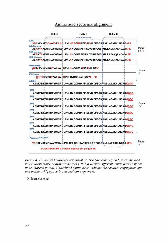
Figure 4. Amino acid sequence alignment of HER2-binding Affibody variants used in this thesis work; shown are helices I, II and III with different amino acid composi-tions (marked in red). Underlined amino acids indicate the chelator conjugation site and amino acid peptide-based chelator sequences.
* X: homocysteine
31
The present investigations
Paper I Influence of DOTA chelator position on biodistribution and targeting properties of 111In-labelled synthetic anti-HER2 Affibody molecules
Background and Aim This study was dedicated to optimization of the biodistribution of Affibody molecules labelled with 111In for SPECT imaging. Earlier studies have shown that changes in local charge and lipophilicity of labelled Affibody molecules might affect their biodistribution properties and, consequently, imaging contrast. These modifications were achieved by even subtle changes such as transferring the chelator moiety from the N-terminus to the C-terminus of Affibody molecules for labelling with 99mTc (Tolmachev & Hof-strom et al. 2010). The aim of this study was to evaluate the influence of DOTA chelator placement (at the N-terminus (A1), in the middle of helix three (K50) or at the C-terminus (K58)) on the biodistribution properties of a synthetic variant of the HER2-binding ZHER2:342 (Figure 4) Affibody mole-cule.
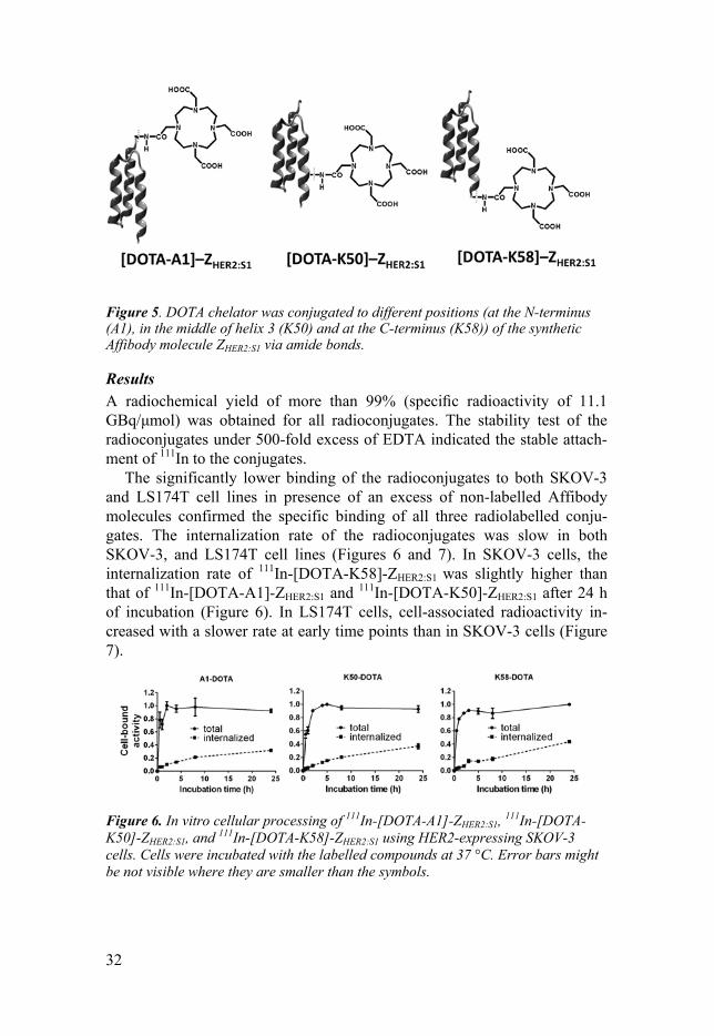
Methods The DOTA chelator was conjugated to the amino groups of lysines or N-terminus of the synthetic Affibody molecule ZHER2:S1 (Figure 5) and labelled with 111In. The labelling efficiencies of 111In-[DOTA-A1]-ZHER2:S1,
111In-[DOTA-K58]-ZHER2:S1 and 111In-[DOTA-K50]-ZHER2:S1 (Figure 4) were eval-uated. The binding specificity to HER2 and the cellular processing of 111In-labelled conjugates were studied using HER2-expressing cells (SKOV-3 with high HER2 expression and LS174T with moderate HER2 expression). The in vivo specificity of the radioconjugates was confirmed using female BALB/C nu/nu mice bearing HER2-negative Ramos xenografts. The biodis-tribution of the 111In-labelled conjugates was compared in female BALB/C nu/nu mice bearing LS174T colorectal cancer xenografts. Additionally, the biodistribution properties of 111In-[DOTA-K58]-ZHER2:S1 were studied using female BALB/C nu/nu mice bearing SKOV-3 ovarian cancer xenografts. For experimental details, see paper I.
32
Figure 5. DOTA chelator was conjugated to different positions (at the N-terminus (A1), in the middle of helix 3 (K50) and at the C-terminus (K58)) of the synthetic Affibody molecule ZHER2:S1 via amide bonds.
Results A radiochemical yield of more than 99% (specific radioactivity of 11.1 GBq/μmol) was obtained for all radioconjugates. The stability test of the radioconjugates under 500-fold excess of EDTA indicated the stable attach-ment of 111In to the conjugates.
The significantly lower binding of the radioconjugates to both SKOV-3 and LS174T cell lines in presence of an excess of non-labelled Affibody molecules confirmed the specific binding of all three radiolabelled conju-gates. The internalization rate of the radioconjugates was slow in both SKOV-3, and LS174T cell lines (Figures 6 and 7). In SKOV-3 cells, the internalization rate of 111In-[DOTA-K58]-ZHER2:S1 was slightly higher than that of 111In-[DOTA-A1]-ZHER2:S1 and 111In-[DOTA-K50]-ZHER2:S1 after 24 h of incubation (Figure 6). In LS174T cells, cell-associated radioactivity in-creased with a slower rate at early time points than in SKOV-3 cells (Figure 7).
Figure 6. In vitro cellular processing of 111In-[DOTA-A1]-ZHER2:S1, 111In-[DOTA-K50]-ZHER2:S1, and 111In-[DOTA-K58]-ZHER2:S1 using HER2-expressing SKOV-3 cells. Cells were incubated with the labelled compounds at 37 °C. Error bars might be not visible where they are smaller than the symbols.
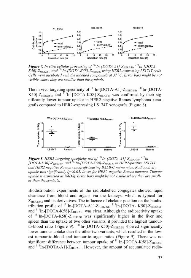
33
Figure 7. In vitro cellular processing of 111In-[DOTA-A1]-ZHER2:S1, 111In-[DOTA-K50]-ZHER2:S1, and 111In-[DOTA-K58]-ZHER2:S1 using HER2-expressing LS174T cells. Cells were incubated with the labelled compounds at 37 °C. Error bars might be not visible where they are smaller than the symbols.
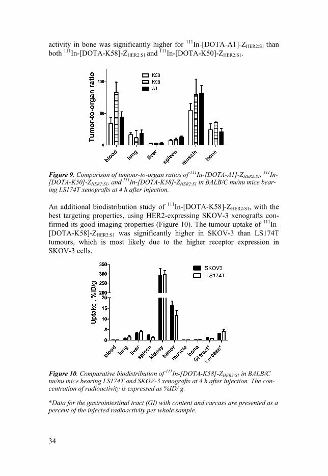
The in vivo targeting specificity of 111In-[DOTA-A1]-ZHER2:S1, 111In-[DOTA- K50]-ZHER2:S1, and 111In-[DOTA-K58]-ZHER2:S1 was confirmed by their sig-nificantly lower tumour uptake in HER2-negative Ramos lymphoma xeno-grafts compared to HER2-expressing LS174T xenografts (Figure 8).
Figure 8. HER2-targeting specificity test of 111In-[DOTA-A1]-ZHER2:S1, 111In-[DOTA-K50]-ZHER2:S1, and 111In-[DOTA-K58]-ZHER2:S1 in HER2-positive LS174T and HER2 negative Ramos xenograft-bearing BALB/C nu/nu mice. Radioactivity uptake was significantly (p<0.05) lower for HER2-negative Ramos tumours. Tumour uptake is expressed as %ID/g. Error bars might be not visible where they are small-er than the symbols.
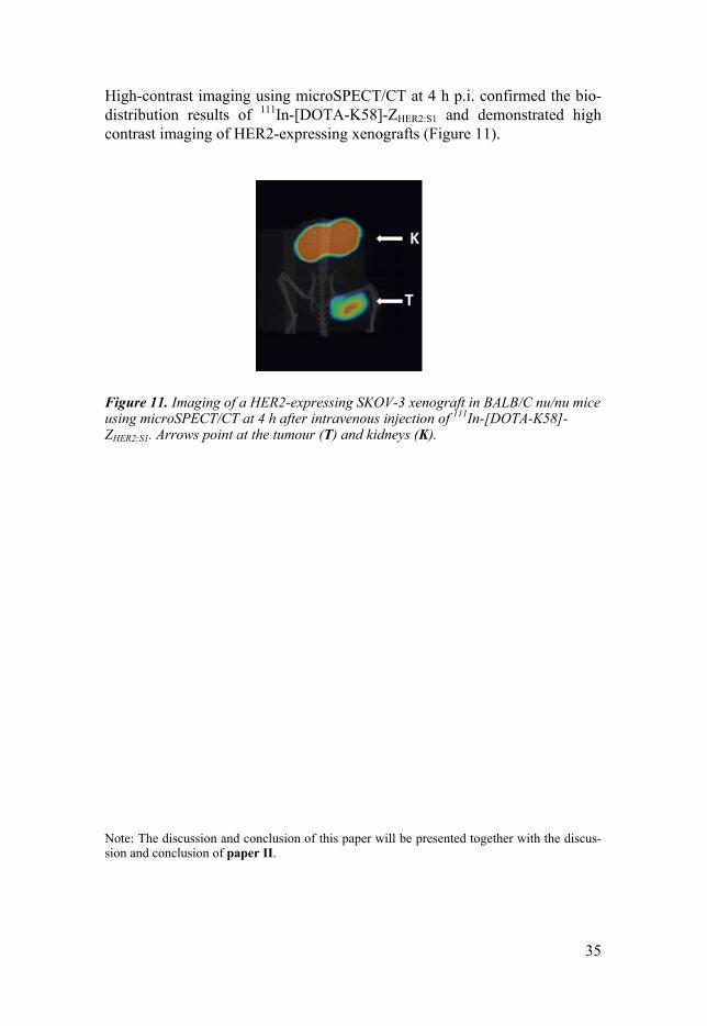
Biodistribution experiments of the radiolabelled conjugates showed rapid clearance from blood and organs via the kidneys, which is typical for ZHER2:342 and its derivatives. The influence of chelator position on the biodis-tribution profile of 111In-[DOTA-A1]-ZHER2:S1, 111In-[DOTA- K50]-ZHER2:S1, and 111In-[DOTA-K58]-ZHER2:S1 was clear. Although the radioactivity uptake of 111In-[DOTA-K58]-ZHER2:S1 was significantly higher in the liver and spleen than the uptake of two other variants, it provided the highest tumour-to-blood ratio (Figure 9). 111In-[DOTA-K50]-ZHER2:S1 showed significantly lower tumour uptake than the other two variants, which resulted in the low-est tumour-to-blood and tumour-to-organ ratios (Figure 9). There was no significant difference between tumour uptake of 111In-[DOTA-K58]-ZHER2:S1 and 111In-[DOTA-A1]-ZHER2:S1. However, the amount of accumulated radio-
34
activity in bone was significantly higher for 111In-[DOTA-A1]-ZHER2:S1 than both 111In-[DOTA-K58]-ZHER2:S1 and 111In-[DOTA-K50]-ZHER2:S1.
Figure 9. Comparison of tumour-to-organ ratios of 111In-[DOTA-A1]-ZHER2:S1, 111In-[DOTA-K50]-ZHER2:S1, and 111In-[DOTA-K58]-ZHER2:S1 in BALB/C nu/nu mice bear-ing LS174T xenografts at 4 h after injection.
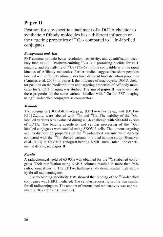
An additional biodistribution study of 111In-[DOTA-K58]-ZHER2:S1, with the best targeting properties, using HER2-expressing SKOV-3 xenografts con-firmed its good imaging properties (Figure 10). The tumour uptake of 111In-[DOTA-K58]-ZHER2:S1 was significantly higher in SKOV-3 than LS174T tumours, which is most likely due to the higher receptor expression in SKOV-3 cells.
Figure 10. Comparative biodistribution of 111In-[DOTA-K58]-ZHER2:S1 in BALB/C nu/nu mice bearing LS174T and SKOV-3 xenografts at 4 h after injection. The con-centration of radioactivity is expressed as %ID/ g.
*Data for the gastrointestinal tract (GI) with content and carcass are presented as a percent of the injected radioactivity per whole sample.
35
High-contrast imaging using microSPECT/CT at 4 h p.i. confirmed the bio-distribution results of 111In-[DOTA-K58]-ZHER2:S1 and demonstrated high contrast imaging of HER2-expressing xenografts (Figure 11).
Figure 11. Imaging of a HER2-expressing SKOV-3 xenograft in BALB/C nu/nu mice using microSPECT/CT at 4 h after intravenous injection of 111In-[DOTA-K58]-ZHER2:S1. Arrows point at the tumour (T) and kidneys (K).
Note: The discussion and conclusion of this paper will be presented together with the discus-sion and conclusion of paper II.
36
Paper II Position for site-specific attachment of a DOTA chelator to synthetic Affibody molecules has a different influence on the targeting properties of 68Ga- compared to 111In-labelled conjugates
Background and Aim PET cameras provide better resolution, sensitivity, and quantification accu-racy than SPECT. Positron-emitting 68Ga is a promising nuclide for PET imaging, and the half-life of 68Ga (T½=68 min) is compatible with the rapid kinetics of Affibody molecules. Earlier studies suggest that short peptides labelled with different radionuclides have different biodistribution properties (Antunes et al. 2007). In paper I, the influence of macrocyclic DOTA chela-tor position on the biodistribution and targeting properties of Affibody mole-cules for SPECT imaging was studied. The aim of paper II was to evaluate these properties in the same variants labelled with 68Ga for PET imaging using 111In-labelled conjugates as comparators.
Methods The conjugates [DOTA-K58]-ZHER2:S1, [DOTA-A1]-ZHER2:S1, and [DOTA-K50]-ZHER2:S1 were labelled with 111In and 68Ga. The stability of the 68Ga-labelled variants was evaluated during a 1-h challenge with 500-fold excess of EDTA. The binding specificity and cellular processing of the 68Ga-labelled conjugates were studied using SKOV-3 cells. The tumour-targeting and biodistribution properties of the 68Ga-labelled variants were directly compared with the 111In-labelled variants in a dual isotope study (Honarvar et al. 2013) in SKOV-3 xenograft-bearing NMRI nu/nu mice. For experi-mental details, see paper II.
Results A radiochemical yield of 65-95% was obtained for the 68Ga-labelled conju-gates. Their purification using NAP-5 columns resulted in more than 96% radiochemical purity. The EDTA-challenge study demonstrated high stabil-ity for all radioconjugates.
In vitro binding specificity tests showed that binding of the 68Ga-labelled conjugates was HER2 mediated. The cellular processing profile was similar for all radioconjugates. The amount of internalized radioactivity was approx-imately 10% after 2 h (Figure 12).
37
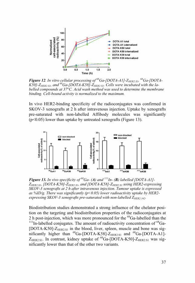
Figure 12. In vitro cellular processing of 68Ga-[DOTA-A1]-ZHER2:S1, 68Ga-[DOTA-K50]-ZHER2:S1, and 68Ga-[DOTA-K58]-ZHER2:S1. Cells were incubated with the la-belled compounds at 37°C. Acid wash method was used to determine the membrane binding. Cell-bound activity is normalized to the maximum.
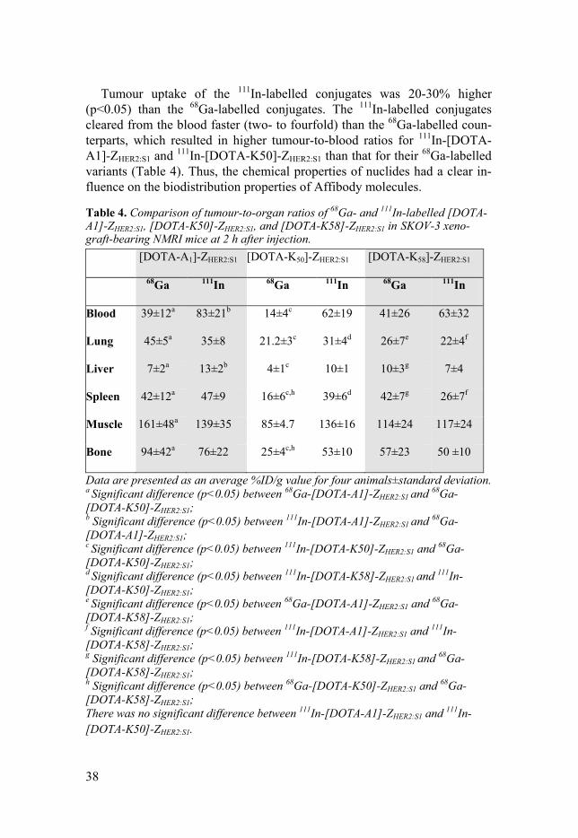
In vivo HER2-binding specificity of the radioconjugates was confirmed in SKOV-3 xenografts at 2 h after intravenous injection. Uptake by xenografts pre-saturated with non-labelled Affibody molecules was significantly (p<0.05) lower than uptake by untreated xenografts (Figure 13).
Figure 13. In vivo specificity of 68Ga- (A) and 111In- (B) labelled [DOTA-A1]-ZHER2:S1, [DOTA-K50]-ZHER2:S1, and [DOTA-K58]-ZHER2:S1 using HER2-expressing SKOV-3 xenografts at 2 h after intravenous injection. Tumour uptake is expressed as %ID/g. There was significantly (p<0.05) lower radioactivity uptake by HER2-expressing SKOV-3 xenografts pre-saturated with non-labelled ZHER2:342.
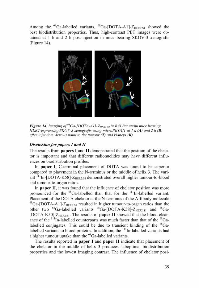
Biodistribution studies demonstrated a strong influence of the chelator posi-tion on the targeting and biodistribution properties of the radioconjugates at 2 h post-injection, which was more pronounced for the 68Ga-labelled than the 111In-labelled conjugates. The amount of radioactivity concentration of 68Ga-[DOTA-K50]-ZHER2:S1 in the blood, liver, spleen, muscle and bone was sig-nificantly higher than 68Ga-[DOTA-K58]-ZHER2:S1 and 68Ga-[DOTA-A1]-ZHER2:S1. In contrast, kidney uptake of 68Ga-[DOTA-K50]-ZHER2:S1 was sig-nificantly lower than that of the other two variants.
38
Tumour uptake of the 111In-labelled conjugates was 20-30% higher
(p<0.05) than the 68Ga-labelled conjugates. The 111In-labelled conjugates cleared from the blood faster (two- to fourfold) than the 68Ga-labelled coun-terparts, which resulted in higher tumour-to-blood ratios for 111In-[DOTA-A1]-ZHER2:S1 and 111In-[DOTA-K50]-ZHER2:S1 than that for their 68Ga-labelled variants (Table 4). Thus, the chemical properties of nuclides had a clear in-fluence on the biodistribution properties of Affibody molecules.
Table 4. Comparison of tumour-to-organ ratios of 68Ga- and 111In-labelled [DOTA-A1]-ZHER2:S1, [DOTA-K50]-ZHER2:S1, and [DOTA-K58]-ZHER2:S1 in SKOV-3 xeno-graft-bearing NMRI mice at 2 h after injection. [DOTA-A1]-ZHER2:S1
[DOTA-K50]-ZHER2:S1 [DOTA-K58]-ZHER2:S1
68Ga 111In 68Ga 111In 68Ga 111In
Blood 39±12a 83±21b 14±4c 62±19 41±26 63±32
Lung 45±5a 35±8 21.2±3c 31±4d 26±7e 22±4f
Liver 7±2a 13±2b 4±1c 10±1 10±3g 7±4
Spleen 42±12a 47±9 16±6c,h 39±6d 42±7g 26±7f
Muscle 161±48a 139±35 85±4.7 136±16 114±24 117±24
Bone 94±42a 76±22 25±4c,h 53±10 57±23 50 ±10
Data are presented as an average %ID/g value for four animals±standard deviation. a Significant difference (p<0.05) between 68Ga-[DOTA-A1]-ZHER2:S1 and 68Ga-[DOTA-K50]-ZHER2:S1; b Significant difference (p<0.05) between 111In-[DOTA-A1]-ZHER2:S1 and 68Ga-[DOTA-A1]-ZHER2:S1; c Significant difference (p<0.05) between 111In-[DOTA-K50]-ZHER2:S1 and 68Ga-[DOTA-K50]-ZHER2:S1; d Significant difference (p<0.05) between 111In-[DOTA-K58]-ZHER2:S1 and 111In-[DOTA-K50]-ZHER2:S1; e Significant difference (p<0.05) between 68Ga-[DOTA-A1]-ZHER2:S1 and 68Ga-[DOTA-K58]-ZHER2:S1; f Significant difference (p<0.05) between 111In-[DOTA-A1]-ZHER2:S1 and 111In-[DOTA-K58]-ZHER2:S1; g Significant difference (p<0.05) between 111In-[DOTA-K58]-ZHER2:S1 and 68Ga-[DOTA-K58]-ZHER2:S1; h Significant difference (p<0.05) between 68Ga-[DOTA-K50]-ZHER2:S1 and 68Ga-[DOTA-K58]-ZHER2:S1; There was no significant difference between 111In-[DOTA-A1]-ZHER2:S1 and 111In-[DOTA-K50]-ZHER2:S1.
39
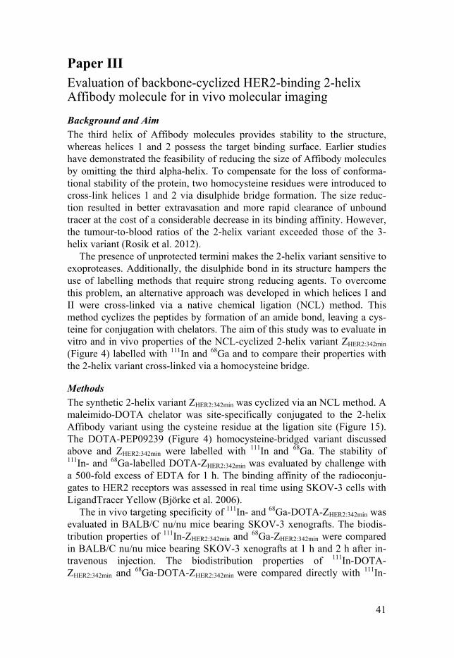
Among the 68Ga-labelled variants, 68Ga-[DOTA-A1]-ZHER2:S1 showed the best biodistribution properties. Thus, high-contrast PET images were ob-tained at 1 h and 2 h post-injection in mice bearing SKOV-3 xenografts (Figure 14).
Figure 14. Imaging of 68Ga-[DOTA-A1]-ZHER2:S1 in BALB/c nu/nu mice bearing HER2-expressing SKOV-3 xenografts using microPET/CT at 1 h (A) and 2 h (B) after injection. Arrows point to the tumour (T) and kidneys (K).
Discussion for papers I and II The results from papers I and II demonstrated that the position of the chela-tor is important and that different radionuclides may have different influ-ences on biodistribution profiles.
In paper I, C-terminal placement of DOTA was found to be superior compared to placement in the N-terminus or the middle of helix 3. The vari-ant 111In-[DOTA-K58]-ZHER2:S1 demonstrated overall higher tumour-to-blood and tumour-to-organ ratios.
In paper II, it was found that the influence of chelator position was more pronounced for the 68Ga-labelled than that for the 111In-labelled variant. Placement of the DOTA chelator at the N-terminus of the Affibody molecule 68Ga-[DOTA-A1]-ZHER2:S1 resulted in higher tumour-to-organ ratios than the other two 68Ga-labelled variants 68Ga-[DOTA-K58]-ZHER2:S1 and 68Ga-[DOTA-K50]-ZHER2:S1. The results of paper II showed that the blood clear-ance of the 111In-labelled counterparts was much faster than that of the 68Ga-labelled conjugates. This could be due to transient binding of the 68Ga-labelled variants to blood proteins. In addition, the 111In-labelled variants had a higher tumour uptake than the 68Ga-labelled variants.
The results reported in paper I and paper II indicate that placement of the chelator in the middle of helix 3 produces suboptimal biodistribution properties and the lowest imaging contrast. The influence of chelator posi-

40
tioning of the biodistribution properties of Affibody molecules could be due to a cooperative effect between the chelator and the surrounding amino acids of the targeting probe. Timing is essential, and the effect is more pronounced at later time points. The difference between the biodistribution properties of the 111In- and 68Ga-labelled variants, even when labelled with the same che-lator, could be due to the different charge distributions of their complex with the chelator. The structures of gallium and indium complexes of DOTA are quite different. Indium has a coordination number of eight and gallium a coordination number of six. In complex with monoamide DOTA, indium has the geometry of a slightly distorted square antiprism, whereas gallium has a cis-pseudo-octahedral geometry with one free carboxylate group. Thus, the local distribution of charge in indium and gallium complexes is different. This might influence off-target interactions of 68Ga-labelled conjugates and uptake by normal organs.
In conclusion, based on the results shown in papers I and II, dif-ferent positioning of the chelator may allow optimization of the bio-distribution and targeting properties of Affibody molecules.
The influence of the position of the chelator depends on the chemi-cal properties of the nuclides.
41
Paper III Evaluation of backbone-cyclized HER2-binding 2-helix Affibody molecule for in vivo molecular imaging
Background and Aim The third helix of Affibody molecules provides stability to the structure, whereas helices 1 and 2 possess the target binding surface. Earlier studies have demonstrated the feasibility of reducing the size of Affibody molecules by omitting the third alpha-helix. To compensate for the loss of conforma-tional stability of the protein, two homocysteine residues were introduced to cross-link helices 1 and 2 via disulphide bridge formation. The size reduc-tion resulted in better extravasation and more rapid clearance of unbound tracer at the cost of a considerable decrease in its binding affinity. However, the tumour-to-blood ratios of the 2-helix variant exceeded those of the 3-helix variant (Rosik et al. 2012).
The presence of unprotected termini makes the 2-helix variant sensitive to exoproteases. Additionally, the disulphide bond in its structure hampers the use of labelling methods that require strong reducing agents. To overcome this problem, an alternative approach was developed in which helices I and II were cross-linked via a native chemical ligation (NCL) method. This method cyclizes the peptides by formation of an amide bond, leaving a cys-teine for conjugation with chelators. The aim of this study was to evaluate in vitro and in vivo properties of the NCL-cyclized 2-helix variant ZHER2:342min (Figure 4) labelled with 111In and 68Ga and to compare their properties with the 2-helix variant cross-linked via a homocysteine bridge.
Methods The synthetic 2-helix variant ZHER2:342min was cyclized via an NCL method. A maleimido-DOTA chelator was site-specifically conjugated to the 2-helix Affibody variant using the cysteine residue at the ligation site (Figure 15). The DOTA-PEP09239 (Figure 4) homocysteine-bridged variant discussed above and ZHER2:342min were labelled with 111In and 68Ga. The stability of 111In- and 68Ga-labelled DOTA-ZHER2:342min was evaluated by challenge with a 500-fold excess of EDTA for 1 h. The binding affinity of the radioconju-gates to HER2 receptors was assessed in real time using SKOV-3 cells with LigandTracer Yellow (Björke et al. 2006).
The in vivo targeting specificity of 111In- and 68Ga-DOTA-ZHER2:342min was evaluated in BALB/C nu/nu mice bearing SKOV-3 xenografts. The biodis-tribution properties of 111In-ZHER2:342min and 68Ga-ZHER2:342min were compared in BALB/C nu/nu mice bearing SKOV-3 xenografts at 1 h and 2 h after in-travenous injection. The biodistribution properties of 111In-DOTA-ZHER2:342min and 68Ga-DOTA-ZHER2:342min were compared directly with 111In-

42
DOTA-PEP09239 and 68Ga-DOTA-PEP09239, which were cyclized using a di-homocysteine bridge, at 1 h p.i. For experimental details see, paper III.
Figure 15. Schematic drawing of the ZHER2:342min scaffold prior to cyclization. A) Red colour represents residues conferring HER2-binding specificity. B) Blue colour represents residues substituted to increase solubility. C) The substituted residues compared to the parental Z-domain. Red and blue show the binding residues and substitutions to increase solubility, respectively. Residues that were introduced to facilitate synthesis and enable subsequent cyclization are in green. Folded in C is the unmodified 3-helix Z-domain. D) Sequence of ZHER2:342min. Colouring is accord-ing to the descriptions above.
* indicates the DOTA-conjugation site. The amino acid numbering is according to the original 3-helix scaffold.
Results and discussion High labelling yields of more than 90% were obtained for all radioconju-gates. The binding kinetic analysis of the 2-helix Affibody molecules using LigandTracer showed a 1:2 interaction model indicating two binding site populations for binding to HER2-expressing SKOV-3 cells (Table 5). The interactions consisted of one strong, in the low nanomolar to subnanomolar range, and one weaker, in the high nanomolar range, interaction for all radi-oconjugates. The results indicated stronger binding affinities for the com-pounds cyclized using a homocysteine bridge than those cyclized using the NCL method. The binding of the 111In-labelled variants was stronger than their 68Ga-labelled counterparts for both ZHER2:342min and PEP09239. The results from LigandTracer also confirmed that the HER2-binding capacity of the radioconjugates was preserved after labelling (Table 5).
43
Table 5. Dissociation constants at equilibrium (KD) for the binding of radiolabelled 2-helix Affibody molecules to HER2-expressing SKOV-3 cells (measured using LigandTracer). 68Ga-DOTA-
ZHER2:342min 111In-DOTA- ZHER2:342min
68Ga- PEP09239
111In- PEP09239
KD1 (nM) 7.31 1.1 0.93 0.32 KD2 (nM) 429 107 15.2 64.1
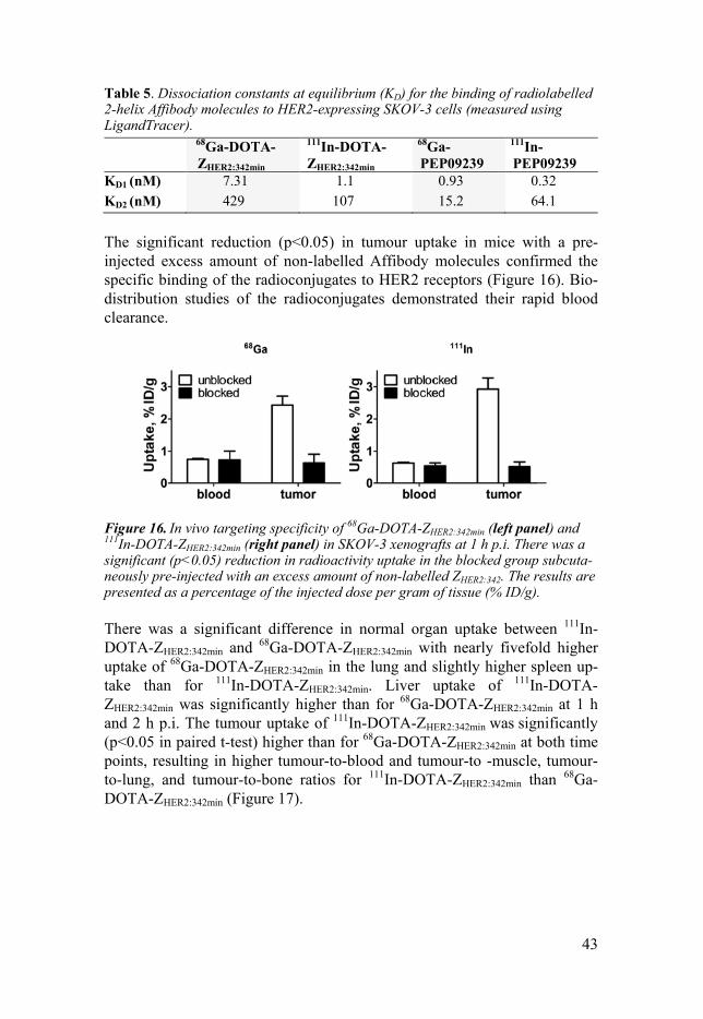
The significant reduction (p<0.05) in tumour uptake in mice with a pre-injected excess amount of non-labelled Affibody molecules confirmed the specific binding of the radioconjugates to HER2 receptors (Figure 16). Bio-distribution studies of the radioconjugates demonstrated their rapid blood clearance.
Figure 16. In vivo targeting specificity of 68Ga-DOTA-ZHER2:342min (left panel) and 111In-DOTA-ZHER2:342min (right panel) in SKOV-3 xenografts at 1 h p.i. There was a significant (p<0.05) reduction in radioactivity uptake in the blocked group subcuta-neously pre-injected with an excess amount of non-labelled ZHER2:342. The results are presented as a percentage of the injected dose per gram of tissue (% ID/g).
There was a significant difference in normal organ uptake between 111In-DOTA-ZHER2:342min and 68Ga-DOTA-ZHER2:342min with nearly fivefold higher uptake of 68Ga-DOTA-ZHER2:342min in the lung and slightly higher spleen up-take than for 111In-DOTA-ZHER2:342min. Liver uptake of 111In-DOTA-ZHER2:342min was significantly higher than for 68Ga-DOTA-ZHER2:342min at 1 h and 2 h p.i. The tumour uptake of 111In-DOTA-ZHER2:342min was significantly (p<0.05 in paired t-test) higher than for 68Ga-DOTA-ZHER2:342min at both time points, resulting in higher tumour-to-blood and tumour-to -muscle, tumour-to-lung, and tumour-to-bone ratios for 111In-DOTA-ZHER2:342min than 68Ga-DOTA-ZHER2:342min (Figure 17).
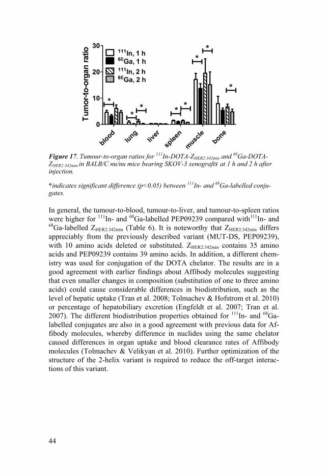
44
Figure 17. Tumour-to-organ ratios for 111In-DOTA-ZHER2:342min and 68Ga-DOTA-ZHER2:342min in BALB/C nu/nu mice bearing SKOV-3 xenografts at 1 h and 2 h after injection.
*indicates significant difference (p<0.05) between 111In- and 68Ga-labelled conju-gates.
In general, the tumour-to-blood, tumour-to-liver, and tumour-to-spleen ratios were higher for 111In- and 68Ga-labelled PEP09239 compared with111In- and 68Ga-labelled ZHER2:342min (Table 6). It is noteworthy that ZHER2:342min differs appreciably from the previously described variant (MUT-DS, PEP09239), with 10 amino acids deleted or substituted. ZHER2:342min contains 35 amino acids and PEP09239 contains 39 amino acids. In addition, a different chem-istry was used for conjugation of the DOTA chelator. The results are in a good agreement with earlier findings about Affibody molecules suggesting that even smaller changes in composition (substitution of one to three amino acids) could cause considerable differences in biodistribution, such as the level of hepatic uptake (Tran et al. 2008; Tolmachev & Hofstrom et al. 2010) or percentage of hepatobiliary excretion (Engfeldt et al. 2007; Tran et al. 2007). The different biodistribution properties obtained for 111In- and 68Ga-labelled conjugates are also in a good agreement with previous data for Af-fibody molecules, whereby difference in nuclides using the same chelator caused differences in organ uptake and blood clearance rates of Affibody molecules (Tolmachev & Velikyan et al. 2010). Further optimization of the structure of the 2-helix variant is required to reduce the off-target interac-tions of this variant.

45
Table 6. Direct comparison of the biodistribution properties of 111In-DOTA-ZHER2:342min, 111In-DOTA-PEP09239, 68Ga-DOTA-ZHER2:342min, and 68Ga-DOTA-PEP09239 in BALB/C nu/nu mice bearing SKOV-3 xenografts at 1 h after injection.
68Ga-DOTA-ZHER2:342min
68Ga-DOTA-PEP09239
111In-DOTA-ZHER2:342min
111In-DOTA-PEP09239
Blood 0.75±0.05c 0.59±0.20d 0.63±0.05 0.5±0.2 Lung 23±16a 1.0±0.2d 4.9±3.2b 0.7±0.2 Liver 8.1±1.0a,c 2.4±0.6d 9.8±0.6b 1.8±0.4 Spleen 2.5±0.5a 0.9±0.4 2.1±0.4b 0.6±0.2 Kidney 158±13a,c 280±39 179±14b 289±44 Tumour 2.4±0.5a,c 10.0±3.2d 2.9±0.7b 12±4 Muscle 0.18±0.05 0.16±0.02 0.17±0.05 0.15±0.04 Bone 0.45±0.07a 0.29±0.04d 0.38±0.04b 0.20±0.04 GI tract* 1.1±0.1 1.6±0.8 1.3±0.1 1.7±0.9
Data are presented as an average % ID/g. Radioactivity in the GI tract (with content) is expressed as % ID per whole sample. a Significant difference (p<0.05) between 68Ga-DOTA-Z342min and 68Ga-DOTA-PEP09239 at 1 h after injection; b Significant difference (p<0.05) between 111In-DOTA-Z342min and 111In-DOTA-PEP09239 at 1 h after injection;
c Significant difference (p<0.05) between 68Ga-DOTA-Z342min and 111In-DOTA- Z342min at 1 h after injection; d Significant difference (p<0.05) between 68Ga-DOTA-PEP09239 and 111In-DOTA-PEP09239 at 1 h after injection.
In conclusion, the use of NCL provides 2-helix Affibody mole-cules capable of targeting HER2-expressing tumours in vivo with a nanomolar binding affinity. The cross-linking method along with other aspects of the molecu-lar design influences the biodistribution properties of Affibody mol-ecules.
46
Paper IV Selection of an optimal cysteine-containing peptide-based chelator for labelling of Affibody molecules with 188Re
Background and Aim Affibody molecules are efficiently reabsorbed in the kidneys. In the case of radiometal labels, radionuclides are retained in the kidneys, which prevent radionuclide therapy using radiometal-labelled Affibody molecules. Earlier studies have demonstrated that the use of the cysteine-containing chelator -GGGC for labelling of Affibody molecules with technetium-99m provided low renal retention of 99mTc (Wållberg et al. 2011). Technetium and rhenium have similar chemical properties. In this study, we replaced 99mTc, which is suitable for SPECT imaging, with 188Re, which is suitable for therapy. To find an optimal peptide-based chelator for the labelling of an anti-HER2 Affibody molecule with 188Re, we evaluated a series of cysteine-containing C-terminal peptide-based chelators. The hypothesis tested was that an opti-mal cysteine-containing peptide-based chelator would provide higher uptake by tumour than by the kidneys.
Methods The tested variants are presented in Figure 4. All the variants were site-specifically labelled with 188Re using freeze-dried labelling kits. In vitro HER2-binding specificity and the cellular retention of the 188Re-labelled variants were evaluated using SKOV-3 cells. The affinity of the variants to HER2 receptors was measured using LigandTracer Yellow at 4°C.
The biodistribution properties of all the 188Re-labelled Affibody mole-cules containing -GGXC or -GXGC peptide chelators (where X is G, S, E or K) were studied using NMRI mice and compared with the parental Affibody molecule ZHER2:2395 (Figure 4), which has -KVDC as a peptide chelator. The tumour targeting properties of the best variant 188Re-ZHER2:V2 and parental 188Re-ZHER2:2395 were compared in Balb/C nu/nu mice bearing SKOV-3 xen-ografts. For experimental details, see paper IV.
Results and discussion A radiochemical yield of more than 95% was obtained for all the 188Re-labelled variants, except for 188Re-ZHER2:2395 (89.3 ± 1.2).
The in vitro HER2-binding specificity of the radioconjugates was shown by significant (p<5×10⁻⁴) reduction in uptake by SKOV-3 cells pre-saturated with excess amount of unlabelled protein. The binding kinetic evaluation of the radiolabelled variants using LigandTracer indicated a single interaction with HER2 receptors with an affinity in the low picomolar range (Table 7). An apparent influence of the chelator composition on the cellular retention of the 188Re-labelled conjugates was observed. After 24 h of incubation, the
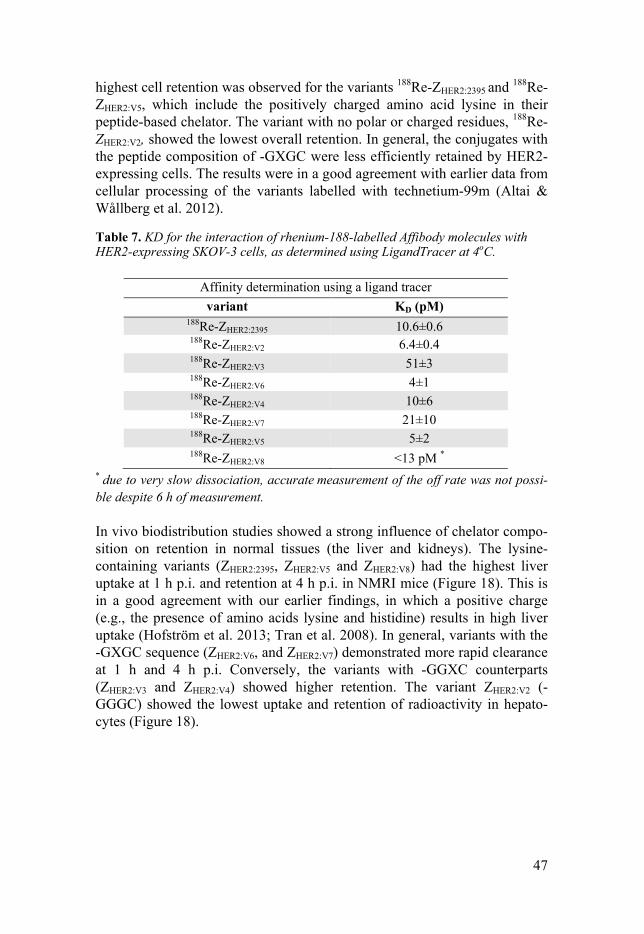
47
highest cell retention was observed for the variants 188Re-ZHER2:2395 and 188Re-ZHER2:V5, which include the positively charged amino acid lysine in their peptide-based chelator. The variant with no polar or charged residues, 188Re-ZHER2:V2, showed the lowest overall retention. In general, the conjugates with the peptide composition of -GXGC were less efficiently retained by HER2-expressing cells. The results were in a good agreement with earlier data from cellular processing of the variants labelled with technetium-99m (Altai & Wållberg et al. 2012).
Table 7. KD for the interaction of rhenium-188-labelled Affibody molecules with HER2-expressing SKOV-3 cells, as determined using LigandTracer at 4oC.
* due to very slow dissociation, accurate measurement of the off rate was not possi-ble despite 6 h of measurement.
In vivo biodistribution studies showed a strong influence of chelator compo-sition on retention in normal tissues (the liver and kidneys). The lysine-containing variants (ZHER2:2395, ZHER2:V5 and ZHER2:V8) had the highest liver uptake at 1 h p.i. and retention at 4 h p.i. in NMRI mice (Figure 18). This is in a good agreement with our earlier findings, in which a positive charge (e.g., the presence of amino acids lysine and histidine) results in high liver uptake (Hofström et al. 2013; Tran et al. 2008). In general, variants with the -GXGC sequence (ZHER2:V6, and ZHER2:V7) demonstrated more rapid clearance at 1 h and 4 h p.i. Conversely, the variants with -GGXC counterparts (ZHER2:V3 and ZHER2:V4) showed higher retention. The variant ZHER2:V2 (-GGGC) showed the lowest uptake and retention of radioactivity in hepato-cytes (Figure 18).
Affinity determination using a ligand tracer variant KD (pM)
188Re-ZHER2:2395 10.6±0.6 188Re-ZHER2:V2 6.4±0.4 188Re-ZHER2:V3 51±3 188Re-ZHER2:V6 4±1 188Re-ZHER2:V4 10±6 188Re-ZHER2:V7 21±10 188Re-ZHER2:V5 5±2 188Re-ZHER2:V8 <13 pM *
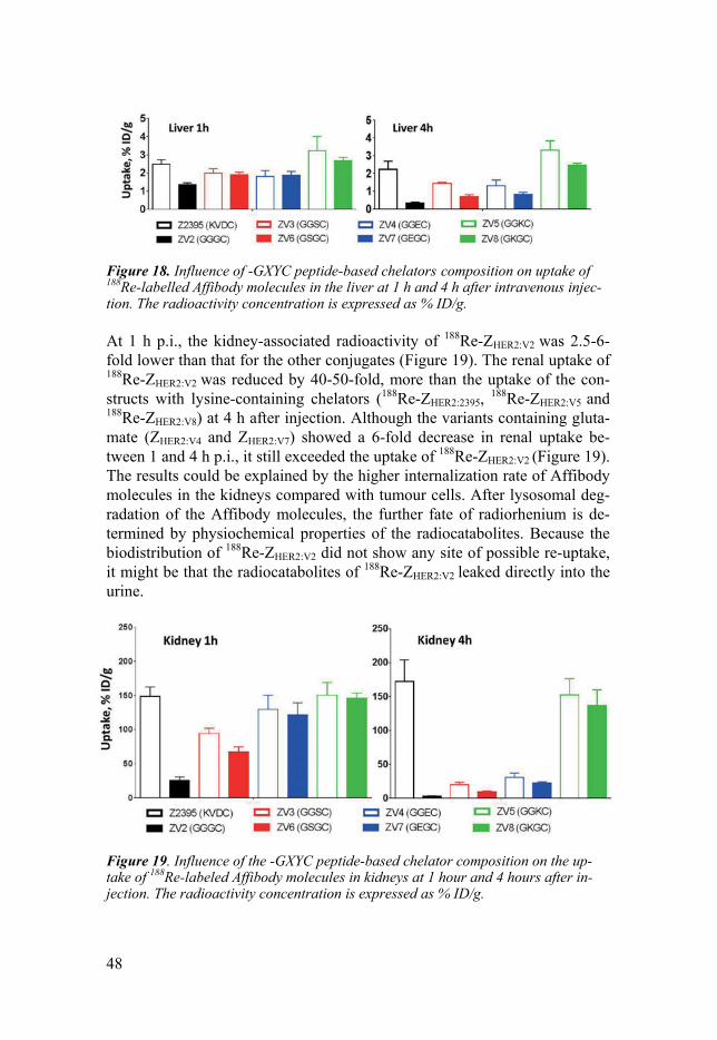
48
Figure 18. Influence of -GXYC peptide-based chelators composition on uptake of 188Re-labelled Affibody molecules in the liver at 1 h and 4 h after intravenous injec-tion. The radioactivity concentration is expressed as % ID/g.
At 1 h p.i., the kidney-associated radioactivity of 188Re-ZHER2:V2 was 2.5-6-fold lower than that for the other conjugates (Figure 19). The renal uptake of 188Re-ZHER2:V2 was reduced by 40-50-fold, more than the uptake of the con-structs with lysine-containing chelators (188Re-ZHER2:2395, 188Re-ZHER2:V5 and 188Re-ZHER2:V8) at 4 h after injection. Although the variants containing gluta-mate (ZHER2:V4 and ZHER2:V7) showed a 6-fold decrease in renal uptake be-tween 1 and 4 h p.i., it still exceeded the uptake of 188Re-ZHER2:V2 (Figure 19). The results could be explained by the higher internalization rate of Affibody molecules in the kidneys compared with tumour cells. After lysosomal deg-radation of the Affibody molecules, the further fate of radiorhenium is de-termined by physiochemical properties of the radiocatabolites. Because the biodistribution of 188Re-ZHER2:V2 did not show any site of possible re-uptake, it might be that the radiocatabolites of 188Re-ZHER2:V2 leaked directly into the urine.
Figure 19. Influence of the -GXYC peptide-based chelator composition on the up-take of 188Re-labeled Affibody molecules in kidneys at 1 hour and 4 hours after in-jection. The radioactivity concentration is expressed as % ID/g.
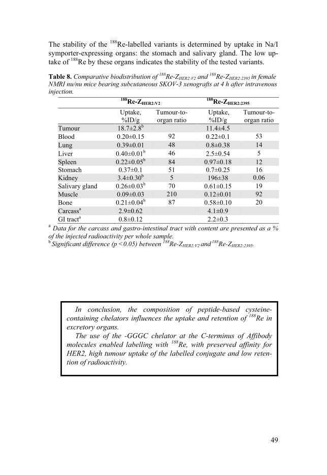
49
The stability of the 188Re-labelled variants is determined by uptake in Na/I symporter-expressing organs: the stomach and salivary gland. The low up-take of 188Re by these organs indicates the stability of the tested variants.
Table 8. Comparative biodistribution of 188Re-ZHER2:V2 and 188Re-ZHER2:2395 in female NMRI nu/nu mice bearing subcutaneous SKOV-3 xenografts at 4 h after intravenous injection.
188Re-ZHER2:V2 188Re-ZHER2:2395
Uptake, %ID/g
Tumour-to- organ ratio
Uptake, %ID/g
Tumour-to-organ ratio
Tumour 18.7±2.8b 11.4±4.5 Blood 0.20±0.15 92 0.22±0.1 53 Lung 0.39±0.01 48 0.8±0.38 14 Liver 0.40±0.01b 46 2.5±0.54 5 Spleen 0.22±0.05b 84 0.97±0.18 12 Stomach 0.37±0.1 51 0.7±0.25 16 Kidney 3.4±0.30b 5 196±38 0.06 Salivary gland 0.26±0.03b 70 0.61±0.15 19 Muscle 0.09±0.03 210 0.12±0.01 92 Bone 0.21±0.04b 87 0.58±0.10 20 Carcassa 2.9±0.62 4.1±0.9 GI tracta 0.8±0.12 2.2±0.3
a Data for the carcass and gastro-intestinal tract with content are presented as a % of the injected radioactivity per whole sample. b Significant difference (p <0.05) between 188Re-ZHER2:V2 and
188Re-ZHER2:2395.
In conclusion, the composition of peptide-based cysteine-containing chelators influences the uptake and retention of 188Re in excretory organs.
The use of the -GGGC chelator at the C-terminus of Affibody molecules enabled labelling with 188Re, with preserved affinity for HER2, high tumour uptake of the labelled conjugate and low reten-tion of radioactivity.
50
Paper V Feasibility of Affibody molecule-based PNA-mediated radionuclide pretargeting of malignant tumours
Background and Aim The high renal retention of the residualizing radiometal-labelled Affibody molecules prevents their use for therapeutic purposes. We hypothesised that a pretargeting approach using Affibody molecules may enable higher accu-mulation of radiometals in tumours than in the kidneys. In this study, we designed and evaluated an Affibody-based peptide nucleic acid (PNA)-mediated pretargeting system consisting of an Affibody-PNA chimera, ZHER2:342-SR-HP1 (Figure 4), containing a 15-mer HP1 PNA recognition tag and a complementary HP2 hybridization probe that permits labelling with both 125I and 111In (Figure 20).
Figure 20. A) Structure of DNA (left) and PNA (right). B) Schematic illustration of two-step Affibody molecule-based PNA-mediated pretargeting in vivo. First, the primary probe (ZHER2:342-SR-HP1) is injected. The primary agent ZHER2:342-SR-HP1 accumulates specifically in HER2-expressing tumours, and the unbound agents is cleared from the body. Then, after a certain time that is selected for optimizing the tumour-to-normal tissue ratio of ZHER2:342-SR-HP1, the radiolabelled secondary probe HP2 is injected. HP2 is complementary to HP1 and designed to hybridize to the ZHER2:342-SR-HP1 chimera.
Methods HER2-binding ZHER2:342-SR-HP1 was produced by a semi-synthetic approach using sortase A. To enable evaluation of the biodistribution properties of ZHER2:342-SR-HP1, a DOTA chelator was conjugated to HP1 (Figure 21). The PNA-based HP1-probe and a complementary HP2-probe were prepared by
51
solid-phase synthesis. The HP2-probe carries a DOTA chelator and a tyrosine that allows radiolabelling with both radioiodine and radiometals (Figure 21).
Figure 21. Illustration of the sequences of the hybridization probes, proteins, and conjugates. The one-letter code (upper-case) represents amino acids, and the lower-case (a, c, g and t) letters represent the PNA monomers. The macrocyclic DOTA and [AEEA] denote the chelator 1,4,7,10-tetraacacyclododecane-1,4,7,19-tetraacetic acid and the spacer unit [2-(2-aminoethoxy) ethoxy]-acetic acid, respectively. The parental sequence of the ZHER2:342 Affibody molecule is marked by bullets.
The in vitro specificity and cellular processing of 111In-ZHER2:342-SR-HP1 and 111In-/125I-HP2 were studied using HER2-expressing BT474 (breast cancer cell line) and SKOV-3 cells. The binding kinetics of 111In-ZHER2:342-SR-HP1 to HER2 receptors and the binding affinity between 111In-/125I-HP2 and 111In-ZHER2:342-SR-HP1 were evaluated using LigandTracer Yellow.
In vivo, the specificity and biodistribution properties of 111In-ZHER2:342-SR-HP1 and 111In-/125I-HP2 were examined using HER2-positive SKOV-3 or HER2-negative Ramos xenograft-bearing BALB/C nu/nu mice. For experi-mental details, see paper V.
Results and discussion In vitro, the binding of 111In-ZHER2:342-SR-HP1 to BT474 and SKOV-3 cancer cells was mediated by HER2 (Figure 22 A and B). The affinity of 111In-ZHER2:342-SR-HP1 for HER2-expressing SKOV-3 cells was 6±2 pM.
The complementary PNA probe 111In/125I-HP2 showed specific binding (PNA-PNA hybridization mediated) to ZHER2:342-SR-HP1 pretreated cells, with an affinity of 200-400 pM. In vitro cell specificity tests confirmed 111In/125I-HP2 binding was only mediated by ZHER2:342-SR-HP1 (Figure 22 C-F).
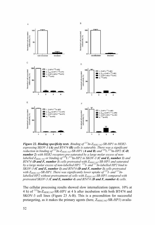
52
Figure 22. Binding specificity tests. Binding of 111In-ZHER2:342-SR-HP1 to HER2-expressing SKOV-3 (A) and BT474 (B) cells is saturable. There was a significant reduction in binding of 111In-ZHER2:342-SR-HP1 (A and B) and 125I /111In-HP2 (C-D, number 2) with HER2 receptors pre-saturated by a large molar excess of non-labelled ZHER2:342 or binding of 125I /111In-HP2 in SKOV-3 (C and E, number 3) and BT474 (D and F, number 3) cells pretreated with ZHER2:342-SR-HP1 and saturated by a large molar excess of non-labelled HP2. 125I- and 111In-labelled HP2 bind to SKOV-3 (C and E, number 1) and BT474 (D and F, number 1) cells pretreated with ZHER2:342-SR-HP1. There was significantly lower uptake of 125I- and 111In-labelled HP2 without pretreatment of cells with ZHER2:342-SR-HP1 compared with pretreated SKOV-3 (C and E, number 4) and BT474 (D and F, number 4) cells.
The cellular processing results showed slow internalization (approx. 10% at 4 h) of 111In-ZHER2:342-SR-HP1 at 4 h after incubation with both BT474 and SKOV-3 cell lines (Figure 23 A-B). This is a precondition for successful pretargeting, as it makes the primary agents (here, ZHER2:342-SR-HP1) availa-
53
ble on the tumour cell surface at the time of injection of the secondary agent (here, 111In/125I-HP2). 111In-HP2 provided better retention of radioactivity (81.2±0.8% and 78.9±0.3% for BT474 and SKOV-3 cells) than 125I-HP2 (40.2±0.4% and 49±1% for BT474 and SKOV3 cells) at 24 h (Figure 23 C-F). This could be due to the residualizing properties of Indium-111.
Figure 23. Cellular processing of 111In-ZHER2:342-SR-HP1 (A and B), 111In-HP2 (C and D), and 125I-HP2 (E and F) by HER2-expressing SKOV-3 (A, C, and E) and BT474 (B, D and F) cells after interrupted incubation with labelled compounds. For 125I- and 111In-labelled HP2, cells were pretreated with non-labelled ZHER2:342-SR-HP1. The total cell-associated radioactivity is shown by circles. In the case of radi-ometals, an acid wash was used to discriminate between membrane-bound (squares) and internalized (triangles) radioactivity. In all assays, incubation was performed at 37oC. Error bars might be not visible where they are smaller than the symbols.
In vivo, pre-saturation of SKOV-3 xenografts with non-labelled anti-HER2 Affibody molecules or the use of Ramos (HER2-negative) xenografts result-ed in significantly lower tumour uptake of 111In-ZHER2:342-SR-HP1 (Figure
54
24), indicating HER2-specific accumulation of 111In-ZHER2:342-SR-HP1. The accumulation of the complementary PNA probe 111In/125I-HP2 in SKOV-3 xenografts was detected only in the presence of ZHER2:342-SR-HP1, which was injected 4 h earlier. Without ZHER2:342-SR-HP1 pre-injection, tumour accumulation of 111In/125I-HP2 was negligible and at the same level as in the muscles. The results demonstrated that binding of the complementary probe HP2 to cells is dependent on the presence of ZHER2:342-SR-HP1 bound to HER2. Uptake of 111In-HP2 by SKOV-3 xenografts pre-injected with ZHER2:342-SR-HP1 was 19±2 %ID/g and approximately 2-fold higher than in the kidneys (12±2 %ID/g) at 1 h after injection.
Figure 24. In vivo HER2-targeting of 111In-ZHER2:342-SR-HP1 at 5 h p.i. All animals were injected with 5 μg 111In-ZHER2:342-SR-HP1. A group with HER2-positive SKOV-3 xenografts was co-injected with an excess amount (500 μg) of non-labelled ZHER2:342 Affibody molecule (grey bars). The HER2-negative Ramos xenografts were used as a negative control (black bars). Data are presented as an average % ID/g with a standard deviation for 5 mice.
The biodistribution results demonstrated rapid blood clearance of 111In-ZHER2:342-SR-HP1 at 4 h p.i. in NMRI mice (0.16±0.02% ID/g) and in tu-mour-bearing BALB/C nu/nu mice (0.14±0.05% ID/g) at 5 h after injection. The results showed that the blood clearance of the ZHER2:342-SR-HP1 chimera was as rapid as the clearance of non-conjugated ZHER2:342. The hybridization probes 111In-/125I-HP2 showed rapid blood clearance and equal tumour up-take at 1 h after injection when the mice were preinjected 4 h earlier with ZHER2:342-SR-HP1. The kidney uptake of 111In-HP2 was 2-fold higher than for 125I-HP2, which might be due to the difference in radiometabolite reten-tion. However, the considerably higher concentration of radioactivity for 125I-HP2 in other organs resulted in significantly higher tumour-to -blood, -lung, -liver, -bone, and -muscle ratios for 111In-HP2 (Table 9).
55
Table 9. Comparison of tumour-to-organ ratios for 125I-HP2 and 111In-HP2 (1 µg) in BALB/C nu/nu mice bearing SKOV-3 xenografts at 1 h p.i. A dose of 100 µg of ZHER2:342-SR-HP1 was pre-injected at 4 h prior to the injection of HP2.
111In-HP2 125I-HP2 Blood 54±19 9±3*
Lung 23±6 6±2*
Liver 62±9 19±5*
Spleen 72±17.08 19±6*
Kidney 1.8±0.3 3.3±0.9*
Muscle 123±38 30±17*
Bones 118±63 53±36*
* There was a significant (p<0.05) difference in tumour-to-organ ratios of 111In-HP2 and 125I-HP2 in SKOV-3 xenografts at 1 h p.i.
The imaging experiment demonstrated much higher renal uptake than tu-mour uptake after injection of 111In-DOTA-ZHER2:K58. (Figure 25 A). The high-contrast visualization of SKOV-3 xenografts (pre-injected with ZHER2:342-SR-HP1) with 111In-HP2 confirmed the ex vivo results showing higher radioactivity accumulation in tumours than in any other organ (Figure 25 B). 111In-HP2 was cleared from all organs, tissues and tumour xenografts without pre-injection of ZHER2:342-SR-HP1 (Figure 25 C).

56
Figure 25. Gamma-camera imaging of HER2-expressing SKOV-3 xenografts using radiolabelled 111In-HP2 and 111In-DOTA-ZHER2:K58 at 1 h p.i. All animals were in-jected with 650 kBq of a radiolabelled compound. A single image of all animals was acquired simultaneously. (A), Animals were injected with 2 μg 111In-DOTA-ZHER2:K58. (B) Animals were pre-injected with 100 μg ZHER2:342-SR-HP1 at 4 h prior to injection of 111In-HP2 (1 μg). (C), 111In-HP2 was injected without pre-injection of ZHER2:342-SR-HP1.
In conclusion, ZHER2:342-SR-HP1 HER2-specifically accumu-lates in xenografts. Accumulation of 111In/125I-HP2 in tumours depends on pre-injection of ZHER2:342-SR-HP1.
The use of Affibody-based PNA-mediated pretargeting provides a higher radiometal concentration in tumours than in the kidneys, which may permit the use of such therapeutic radiometals as the low-energy beta-emitter 177Lu or alpha-emitters 212Bi, 213Bi, and 227Th for Affibody-mediated therapy.
57
Concluding remarks
This thesis work represents a part of the preclinical development of HER2-targeting Affibody molecules aimed for radionuclide-based molecular imag-ing and therapy of cancer. The work began with optimization of the radio-labelling chemistry of a synthetic Affibody molecule followed by evaluation of the in vitro and in vivo properties of a truncated variant, ZHER2:342min. The aim of these studies was to improve the biodistribution properties and imag-ing sensitivity of radiolabelled Affibody tracers. The last two papers were devoted to the development of Affibody molecules suitable for therapeutic purposes. A special focus was placed on the design and development of a PNA-mediated pretargeting approach using Affibody molecules.
The major conclusions from the work presented in this thesis are as follows: • It is evident that changing the chelator position (the N-terminus, mid-
dle of helix 3 or C-terminus) and substituting the radionuclide can have an important impact on the imaging properties of Affibody mol-ecules.
• Positioning of a DOTA chelator at the C-terminus for 111In-labelled and at the N-terminus for 68Ga-labelled synthetic HER2-targeting Af-fibody molecules provided the best tumour-to-organ ratios.
• Placement of a DOTA chelator in the middle of helix-3 resulted in sub-optimal biodistribution properties using both 68Ga and 111In la-bels.
• DOTA chelator-conjugated Affibody molecules can be used as multi-functional tracers for labelling with 111In and 68Ga, suitable for both SPECT and PET applications. However, it should be taken into ac-count that differences in radionuclide chemical nature can influence the biodistribution properties of Affibody molecules differently. Thus, careful optimization of the tracer is required before its use in the clinic.
• The use of NCL allows for conformational stabilization of 2-helix Affibody molecules and provides binding of the stabilized protein to HER2 with nanomolar affinity and targeting of HER2-expressing tu-mours in vivo.
• The cross-linking method along with other aspects of molecular de-sign influences the biodistribution properties of Affibody molecules.
58
• There is a strong influence of the radionuclide chosen on the target-ing properties of 2-helix Affibody variants.
• Cellular processing of 188Re-labelled variants is similar to their 99mTc-labelled counterparts.
• Retention of radiocatabolites after internalization of 188Re-labelled Affibody molecules by cancer cells or in excretory organs is longer in the case of GGXC-containing chelators compared with GXGC-variants.
• Substitution of only one or two amino acids in the chelating sequence can alter the excretion pathway of radiorhenium-labelled Affibody molecules from hepatobiliary to renal excretion. Thus, modification of the chelating moiety of the Affibody molecules is an effective way to modify their in vivo biodistribution features.
• 188Re-ZHER2:V2, which provided low retention of radioactivity in the kidneys, is a promising candidate for development as a therapeutic tumour-targeting agent for the treatment of bulky HER2-expressing tumours.
• The high affinity and low internalization rate of HER2-binding ZHER2:342-SR-HP1 makes it a potential primary agent for pretargeting approaches.
• The hybridization probe HP2 can be labelled with both 111In and 125I without decreasing its binding properties to ZHER2:342-SR-HP1.
• The Affibody molecule-based PNA-mediated pretargeting approach overcomes the high renal radioactivity retention of radiometals, facil-itating the possibility of using low-energy beta- or alpha-emitting ra-diometals suitable for treating micrometastases.
The results of this thesis work may be applied to Affibody molecules bind-ing to other targets (e.g., CAIX, HER3, IGF-1R, PDGFRß) aimed for inves-tigation of radionuclide molecular imaging and therapy purposes and also to other HER2-targeting scaffold protein-based probes, such as ADAPTs or DARPins.
59
Future perspectives
Affibody molecules are a very promising tracer format for radionuclide- molecular imaging of a number of cancer biomarkers, particularly HER2 overexpression. The robust nature of Affibody molecules along with their high affinity and binding specificity to the target protein provides a great flexibility with regard to modification of the protein sequence and the choice of radionuclide. This thesis has demonstrated in detail that the in vitro and in vivo properties of Affibody molecules depend on their conjugation and radi-olabelling chemistry. The feasibility of a promising pretargeting approach using Affibody molecules has also been shown in this thesis. The results from papers I and II have demonstrated the direct influence of the chelator position and type of radionuclide on the biodistribution profile of a HER2-binding Affibody molecule. This necessitates future evaluation of the influence of these modifications on Affibody molecules binding to other targets, such as EGFR, IGF-1R, PDGFRß, HER3, and CAIX. The results from paper III indicate that the cyclization method and the radionuclide type have profound effects on the in vitro (i.e., binding kinetics) and in vivo (i.e., off-target interactions) properties of HER2-binding 2-helix scaffolds. The reduction in affinity and off-target interactions of the 2-helix variant Z342min might be improved by further affinity maturation and sequence modifica-tions of the variant. In addition, evaluation of the effects of size reduction of other Affibody molecules binding to EGFR, IGF-1R, PDGFRß, HER3, and CAIX might be of interest in the future. Recently, we studied the properties of a series of CAIX-binding Affibody molecules, (HE)3-ZCAIX:1, (HE)3-ZCAIX:2, (HE)3-ZCAIX:3, and (HE)3-ZCAIX:4, labelled with 99mTc and 125I, which are suitable for imaging of CAIX-expressing tumours (Honarvar et al. 2015; Garousi et al. 2016). Over-expression of CAIX is associated with tumour cell hypoxia in a variety of human tumours, which contributes to resistance to chemotherapy and radio-therapy and increases the potential for metastasis and poor cancer prognosis (Brizel et al. 1999; Hockel et al. 1996). Thus, identification of the hypoxic regions within tumours is of importance for personalized medicine. Through these studies, we have demonstrated the feasibility of in vivo imaging of CAIX-expressing tumours using radiolabelled Affibody molecules. Howev-er, the renal retention of 99mTc-labelled variants was as high, as is typical for
60
other Affibody molecules labelled with residualizing radiometals. To be able to image primary renal cell carcinoma tumours using Affibody molecules, a low kidney uptake is required. For this reason, introduction of a GGGC pep-tide-based chelator, studied in paper IV, may reduce the kidney uptake of CAIX-binding Affibody molecules when labelled with 99mTc. In paper V, we showed the feasibility of the Affibody-based PNA-mediated pretargeting approach. Although the kidney uptake of PNA was very low and most probably can be reduced even more by co-injection of lysine or gelofusine, engineering of PNA using different sequence compositions might result in a variant with even lower kidney uptake. The Affibody-based PNA-mediated pretargeting approach can also be applied to CAIX-binding Af-fibody molecules, which exhibits limited expression in normal organs. The pretargeting approach was studied using 111In, which indicated the feasi-bility of high-contrast imaging of HER2-expressing tumours using SPECT. It would be of interest to examine this feasibility with 68Ga labelling, which is suitable for PET imaging, providing higher resolution than SPECT. This is of particular importance for detecting the presence of receptors for personal-ized medicine.
The pretargeting approach was developed for treatment of cancer using re-sidualizing radionuclides. Thus, it is important to evaluate it using such radi-onuclides as the beta-emitter 177Lu for treatment of micrometastases and the alpha-emitters 212Bi/213Bi or 227Th for treatment of single cells. I hope that the initial studies presented in this thesis work will contribute to the development of Affibody-based molecular imaging and therapy of cancer in the future.

61
Acknowledgments
Thinking back; it is crystal clear that without constant support of my family, friends, colleagues, supervisor and co-supervisors this accomplishment wouldn’t have been possible. I would like to express my very great appreciation to the following: Vladimir, thank you very much for giving me the opportunity to enter the world of research and thank you for sharing the ocean of your knowledge and your infinite experience with me! I really appreciate all your support! Anna, thank you for all cell and in vivo studies that you have taught me. You are a great teacher! I am also truly thankful for all the dinner parties and activities that you always arranged in the best way. Karl, advices given by you have always been valuable to me. Also not to forget, your sense of humor was always a boost of energy for me! Thank you for your support! Amelie, thank you very much for the great collaboration that we have had together! It was a pleasure, indeed, to work with you! Mohamed (Mamal ), to write an acknowledgment to you was really diffi-cult for me! When I wrote all my appreciations to you it exceeded a page (after ten times reducing it), and I still wasn’t satisfied. You know why? Because acknowledging a teacher, colleague and most importantly great friend like you is far from any word. Thank you so much for everything Mamal! Joanna, my badminton buddy! I really miss to hear ¨…’n abs pass? Pump it pass? ... pass?¨ I have been really lazy in training, since you left. You have always been a symbol of a strong person for me and I learned a lot from you during those 4 years. Thank you for all the joy we had together beautiful Dr2. Joanna! ;-) Javad, thank you very much for a peaceful and fruitful collaboration! And thank you for all the chocolates and cookies that you shared with me. They were gasoline to the engine of my brain. Maria, without you and your cute sense of humor I may have not been able to pass the days with no results in the lab. You were always supportive in both beautiful and ugly days of my life and wanted the best for me. It means a lot to me and I am really thankful and wish all the best for you too! Bogdan, the most positive person I have ever met in my life! From what I know, your future is going to be just brilliant Dr. Bogdan. It has been a

62
great joy to be your colleague, indeed! Thank you very much for all your support and a wonderful friendship! Diana, my lovely and caring friend! You have always brought happy tears to my eyes and it means a lot to me. I am really grateful for your endless kind-ness and support! I am glad for having you, Christer and your lovely kids in my life! Jonas and Magdalena, the lovely couple! Thank you very much for a great friendship and for all happiness that you have always given me! I wish you a blissful life together! Hanna Björkelund, my fancy chitchat buddy. It was always such a great feeling to know you were sitting next door to me. Having a fika or lunch with you was a great boost of energy for me. Thank you! Jennie Andersson, thank you for all your kind support and help! Nazila, thank you for everything my gorgeous friend! I miss you! Karin, thank you for your support and all the crazy things we have done together. I want you to know that our friendship means a lot to me! Suheir, den snälla personen som alltid får mig att skratta. Tack så mycket för alla mörka och ljusa dagarna som du har varit med mig och stöttat mig! Maria Teresa, min vackra och snälla vän. Tack så mycket för din underbara vänskap och ditt stöd. Du och din mamma Elizabeth är underbara och jag är lycklig att ni finns i mitt liv! Monica B, Elin L, Malin S, and Ellinor A, mina salsa stjärnor! Ni är un-derbara människor som öppnade era hjärtan och släppte in mig första gången vi träffades. Tack så mycket för allt vänner! Louise, thank you for all your help and support in everything! You are just great! Inger och Bertil, tusen tack för ert stöd och inspiration! Ni är underbara mäniskor som gav mig allt krävdes för att känna mig som hemma i Sverige! Kicki (Christina), thank you for always having an eye on everything to be perfect and thank you for planning all those fun activities and fika s. My project student Martin, thank you for your help and magic iodinations! All-BMS crew, Marika, Bo, Anja, Jos, John, Andris, Sina and all others for the fun after works and great discussions during lunch. People in KTH: Kristina (for being such a great collaborator! Working with you was just of my pleasure. You are the best!), Hanna (for all the fun activ-ities that we have always had together!), Daniel, Anna P and all others. All people in PPP, Ram (thank you for providing me with beautiful PET/SPECT images. They will shine in my thesis forever.), Veronica, Ser-gio, Ola, and Olof. Former and current staff at BMS and IGP administration, Christle Richter Frohm, Heléne Lyngå, Christina Magnusson, Ulla Steimer, Camilla

63
Sandgren, Helene Norlin and all others. Thank you for your various forms of support during my PhD! Jag skulle vilja rikta ett speciellt tack till ljuset i mitt liv, Mattias. Tack så mycket för att du alltid fyller mitt liv med så mycket kärlek och stöd och tack så mycket för att jag får ta del av ditt stora hjärta!! Jane man duuu! My beloved parents, although you do not have a physical presence any-more, you are with me in my everyday life and I know you will read this. So, I would like to express my biggest heart-felt gratitude to you for the pure love and care you gave me! If I am in this position today is only and only because of you. Thank you for everything you taught me!! I hope I have become who you wished. My beloved siblings, the most precious people in my life! You have always made my dark days, bright and bright days, brighter. Many thanks for always being a constant source of love, concern, support and strength for me! The feeling that you are always there for me makes me super strong and loved. I love you so much! Not the last, a lot of thanks to all other friends, that I have not mentioned in this acknowledgment, in Sweden, Iran, Australia, Canada, Turkey and all other places worldwide, who have helped me through these difficult years in different ways. I greatly value your friendship and I deeply appreciate your belief in me.
64
References
Altai, M., Perols, A., Eriksson Karlström, A., Sandström M., Boschetti, F., Orlova, A., Tolmachev, V. (2012). Preclinical evaluation of anti-HER2 Affibody mole-cules site-specifically labeled with 111In using a maleimido derivative of NODAGA. Nucl Med Biol. 39; 518-529.
Altai, M., Wållberg, H., Orlova, A., Rosestedt, M., Hosseinimehr, S.J., Tolmachev, V., Ståhl, S. (2012). Order of amino acids in C-terminal cysteine-containing peptide-based chelators influences cellular processing and biodistribution of 99mTc-labeled recombinant Affibody molecules. Amino acids. 42:1975-85.
Altai, M., Varasteh, Z., Andersson, K., Eek, A., Boerman, O., Orlova, A. (2013). In vivo and in vitro studies on renal uptake of radiolabeled affibody molecules for imaging of HER2 expression in tumors. Cancer Biotherapy and Radiopharma-ceuticals. 28:187-95.
Antunes, P., Ginj, M., Zhang, H., Waser, B., Baum, R.P., Reubi J.C., Maecke, H. (2007). Are radiogallium-labelled DOTA-conjugated somatostatin analogues superior to those labeled with other radiometals? Eur J Nucl Med Mol Imaging. 34:982-93.
Ahlgren, S., Wållberg, H., Tran, T. A., Widström, C., Hjertman, M., Abrahmsén, L., Berndorff, D., Dinkelborg, LM., Cyr, J. E., Feldwisch, J., Orlova, A., Tol-machev, V. (2009). Targeting of HER2-expressing tumors with a site-specifically 99mTc-labeled recombinant Affibody molecule, ZHER2:2395, with C-terminally engineered cysteine. J Nucl Med. 50:11-2.
Ahlgren, S., and Tolmachev, V. (2010). Radionuclide molecular imaging using Affibody molecules. Curr Pharm Biotechnol. 11:581-9.
Abraham, J., Allegra Carmen, J., Gulley, J. (2005). Handbook of Clinical Oncolo-gy: Lip-3. pincott Williams & Wilkins.
Baum, R.P., Prasad, V., Müller, D., Schuchardt, C., Orlova, A., Wennborg, A., Tolmachev, V., Feldwisch, J. (2010). Molecular imaging of HER2-expressing malignant tumors in breast cancer patients using synthetic 111In- or 68Ga-labeled affibody molecules. J Nucl Med. 51:892-7.
Björke, H., Andersson, K. (2006). Automated, high-resolution cellular retention and uptake studies in vitro. Appl Radiat Isot. 64:901-5.
Boswell, C.A., Brechbiel, M.W. (2007). Development of radio-immunotherapeutic and diagnostic antibodies: an inside-out view. Nucl Med Biol. 34:757-78.
Bang, Y.J., Van Cutsem, E., Feyereislova, A., Chung, H. C., Shen, L., Sawaki, A., Lordick , F., Ohtsu, A., Omuro, Y., Satoh, T., Aprile, G., Kulikov, E., Hill, J., Lehle, M., Rüschoff, J., Kang, Y.K., and ToGA Trial Investigators. (2010). Trastuzumab in combination with chemotherapy versus chemotherapy alone for treatment of HER2-positive advanced gastric or gastro-esophageal junction can-cer (ToGA): a phase 3, open-label, randomised controlled trial. Lancet. 376: 687-97.
65
Brizel, D.M., Dodge, R.K., Clough, R.W., Dewhirst, M.W. (1999). Oxygenation of head and neck cancer: changes during radiotherapy and impact on treatment outcome. Radiother Oncol. 53:113-117.
Brechbiel M.W. (2008). Bifunctional Chelates for Metal Nuclides. Q J Nucl Med Mol Imaging. 52: 166-173.
Costantini, D.L., Bateman, K., McLarty, K., Vallis, K.A., Reilly, R.M. (2008). Trastuzumab-resistant breast cancer cells remain sensitive to the auger electron–emitting radiotherapeutic agent 111In-NLS-trastuzumab and are radiosensitized by Methotrexate. J Nucl Med. 49:1498-1505.
Carlsson, J. (2008). EGFR-family expression and implications for targeted radionu-clide therapy. Stigbrand, T., Carlsson, J., Adams, G., editors. In: Targeted radi-onuclide tumour therapy: biological aspects. Springer. 25-58. Chen, X., Leung, S.Y., Yuen, S.T., Chu, K.M., Ji, J., Li, R., Chan, A.S.Y., Law, S., Troyanskaya, O.G., Wong. J., So, S., Botstein, D., Brown, P.O. (2003). Variation in gene ex-pression patterns in human gastric cancers. Mol Biol Cell.14:3208-3215.
Cheng, Z., De Jesus, O.P., Kramer, D.J., De, A., Webster, J.M., Gheysens, O., Levi, J., Namavari, M., Wang, S., Park, J.M., Zhang, R., Liu, H., Lee, B., Syud, F.A., Gambhir, S.S. (2010). 64Cu-Labeled Affibody Molecules for Imaging of HER2 Expressing. Tumors. Mol Imaging Biol. 12:316-24.
De Leon-Rodríguez, L.M., and Kovacs, Z. (2008). The synthesis and chelation chemistry of DOTA-peptide conjugates. Bioconjugate Chem. 19:391-402.
Demetri GD, von Mehren, M., Blanke, C.D., Van den Abbeele, A.D., Eisenberg, B., Roberts, P.J., Heinrich, M.C., Tuveson, D.A., Singer, S., Janicek, M., Fletcher, J.A., Silverman, S.G., Silberman, S.L., Capdeville, R., Kiese, B., Peng, B., Di-mitrijevic, S., Druker, B.J., Corless, C., Fletcher, C.D.M., Joensuu, H. (2002). Efficacy and safety of imatinib mesylate in advanced gastrointestinal stromal tumors. N Engl J Med. 347:472-80.
Di Leo, A., Gomez, H.L., Aziz, Z., Zvirbule, Z., Bines, J., Arbushites, M.C., Guer-rera, S.F., Koehler, M., Oliva, C., Stein, S.H., Williams, L.S., Dering, J., Finn, R.S., Press, M.F. (2008). Phase III, double-blind, randomized study comparing lapatinib plus paclitaxel with placebo plus paclitaxel as first-line treatment for metastatic breast cancer. J Clin Oncol. 26:5544-52.
Eckelman, W.C., Kilbourn, M.R., Mathis, C.A. (2009). Specific to nonspecific binding in radiopharmaceutical studies: it's not so simple as it seems! Nucl Med Biol. 36:235-7.
Engfeldt, T., Tran, T., Orlova, A., Widström, C., Feldwisch, J., Abrahmsén, L., Wennborg, A., Eriksson Karlström, A., Tolmachev, V. (2007). 99mTc chelator engineering to improve tumour targeting properties of a HER2-specific Af-fibody molecule. Eur J Nucl Med Mol Imaging. 34:1843-53.
Feldwisch, J., and Tolmachev, V. (2012). Engineering of affibody molecules for therapy and diagnostics. Therapeutic Proteins: Methods and Protocols, Meth-ods in Molecular Biology, vol. 899.Springer. DOI 10.1007/978-1-61779-921-17.
Goldenberg, D.M., Sharkey, R.M., Paganelli, G., Barbet, J., Chatal, J.F. (2006). Antibody pretargeting advances cancer radioimmunodetection and radioim-munotherapy. J Clin Oncol. 24:823-34.
Garousi , J., Honarvar, H., Andersson, K.G., Mitran, B., Orlova, A., Buijs, J., Löfblom, J., Frejd, F.Y., and Tolmachev, V. (2016). Comparative evaluation of affibody molecules for radionuclide imaging of in vivo expression of carbonic anhydrase IX. Manuscript under preparation.
Hantowich, D.J., Virzi, F., Rusckowski, M. (1987). Investigations of avidin and biotin for imaging applications. J Nucl Med. 28:1294-302.
66
Holohan, C., Van Schaeybroeck, S., Longley, D.B., Johnston, P.G. (2013). Cancer drug resistance: an evolving paradigm. Nat Rev Cancer. 13:714-26.
Hockel, M., Schlenger, K., Aral, B., Mitze, M., Schaffer, U., Vaupel, P. (1996). Association between tumor hypoxia and malignant progression in advanced cancer of the uterine cervix. Cancer Res. 56:4509-4515.
Hanahan, D., and Weinberg, R.A. (2011). Hallmarks of Cancer: The Next Genera-tion. J. Cell. 144:646-674.
Hudis, C.A. (2007). Trastuzumab-mechanism of action and use in clinical practice. N Engl J Med. 357:39-51.
Heskamp, S., van Laarhoven, H.W., van der Graaf, W.T., Oyen, W.J., Boerman, O.C. (2014). Radionuclide imaging of drug delivery for patient selection in tar-geted therapy. Expert Opin Drug Deliv. 11:175-85.
Harbeck, N., Beckmann, M.W., Rody, A., Schneeweiss, A., Müller, V., Fehm, T., Marschner, N., Gluz, O., Schrader, I., Heinrich, G., Untch, M., Jackisch, C. (2013). HER2 dimerization inhibitor pertuzumab-mode of action and clinical data in breast cancer. Breast Care (Basel). 8:49-55.
Honarvar, H., Garousi, J., Gunneriusson, E., Höidén-Guthenberg, I., Altai, M., Widström, C., Tolmachev, V., Frejd, FY. (2015). Imaging of CAIX-expressing xenografts in vivo using 99mTc-HEHEHE-ZCAIX:1 affibody molecule. Int J Oncol. 46:513-20.
Honarvar, H., Jokilaakso, N., Andersson, K., Malmberg, J., Rosik, D., Orlo-va, A., Eriksson Karlström, A., Tolmachev, V., Järver, P. (2013). Evaluation of backbone-cyclized HER2-binding 2-helix affibody molecule for in vivo molecu-lar imaging. Nuclear Medicine and Biology. 40:378-86.
Hofström, C., Altai, M., Honarvar, H., Strand, J., Malmberg, J., Hosseinimehr, S.J., Orlova, A., Gräslund, T., Tolmachev, V. (2013). HAHAHA, HEHEHE, HIHIHI or HKHKHK: influence of histidine containing tags position and composition on biodistribution of [99mTc(CO)3]+-labelled affibody molecules. Journal of Medicinal Chemistry. 56: 4966-4974.
Hazlehurst, L.A., Landowski, T.H., Dalton, W.S. (2003). Role of the tumor micro-environment in mediating de novo resistance to drugs and physiological media-tors of cell death. Oncogene. 22:7396-7402.
Iagaru, A., Mittra, E.S., Ganjoo, K., Knox, S.J., Goris, M.L. (2010). 131I-Tositumomab (Bexxar®) vs.90Y-Ibritumomab (Zevalin®) Therapy of Low-Grade Refractory/ Relapsed Non-Hodgkin Lymphoma. Mol Imaging Biol. 12:198-203.
Keating, G.M. (2014). Bevacizumab: A Review of Its Use in Advanced Cancer. Drugs. 74:1891-1925.
Kuijpers, W.H., Bos, E.S., Kaspersen, F.M., Veeneman, G.H., van Boeckel, C.A. (1993). Specific recognition of antibody-oligonucleotide conjugates by radio-labeled antisense nucleotides: a novel approach for two-step radioimmunothera-py of cancer. Bioconjug Chem. 4:94-102.
Kramer-Marek, G., Shenoy, N., Seidel, J., Griffiths, G.L., Choyke, P., Capala, J. (2011). 68Ga-DOTA-Affibody molecule for in vivo assessment of HER2/neu expression with PET. Eur J Nucl Med Mol Imaging. 38:1967-1976.
Kenanova, V., and Wu, A.M. (2006). Tailoring antibodies for radionuclide delivery. Expert Opin. Drug Deliv. 3:53-70.
Kraeber-Bodéré, F., Salaun, P.Y., Ansquer, C., Drui, D., Mirallié, E., Faivre-Chauvet, A., Barbet, J., Goldenberg, D.M., Chatal, J.F. (2012). Pretargeted radi-oimmunotherapy (pRAIT) in medullary thyroid cancer (MTC). Tumour Biol. 33:601-6.
67
Kiesewetter, D.O., Krämer-Marek, G., Ma, Y., Capala, J. (2008). Radiolabeling of HER2 specific Affibody molecule with F-18. J Fluor Chem. 129:799-805.
Löfblom, J., Feldwisch, J., Tolmachev, V., Carlsson, J., Ståhla, S., Frejd, F.Y. (2010). Affibody molecules: Engineered proteins for therapeutic, diagnostic and biotechnological applications. FEBS Letters. 584: 2670-2680.
Lodish, H., Berk A., S., Kaiser, C.A, Krieger, M. Scott, M.P., Bretscher, A., Ploegh, H., Matsudaira, P. (2008). Molecular cell biology sixth edition. W. H. Freeman and Company Copyright. Molecular Cell Biology Lodish 6th. Chapter 25, can-cer.
Lemmon, M.A., Schlessinger, J. (2010). Cell signaling by receptor tyrosine kinases. Cell. 141:1117-34.
Liu, G., Mangera, K., Liu, N., Gupta, S., Rusckowski, M., Hnatowich, D.J. (2002). Tumor pretargeting in mice using (99m)Tc-labeled morpholino, a DNA analog. J Nucl Med. 43:384-91.
Larsen, RH., Borrebaek, J., Dahle, J., Melhus, K.B., Krogh, C., Valan, M.H., and Bruland, O.S. (2007). Preparation of TH227-Labeled Radio-immunoconjugates, Assessment of Serum Stability and Antigen Binding Ability. Cancer Biotherapy & Radiopharmaceuticals. 22: 3.
Mardirossian, G., Lei, K., Rusckowski, M., Chang, F., Qu, T., Egholm, M., Hna-towich, D.J. (1997). In vivo hybridization of technetium-99m-labeled peptide nucleic acid (PNA). J Nucl Med. 38:907-13.
Malmberg, J., Sandström, M., Wester, K., Tolmachev, V., Orlova, A. (2011). Comparative biodistribution of imaging agents for in vivo molecular profiling of disseminated prostate cancer in mice bearing prostate cancer xenografts: focus on 111In- and 125I-labeled anti-HER2 humanized monoclonal trastuzumab and ABY-025 affibody. Nucl Med Biol. 38:1093-102.
Mankoff, D.A. (2009). Molecular imaging to select cancer therapy and evaluate treatment response. Q. J. Nucl. Med. Mol. Imaging. 53:181-92.
Miao, Z., Levi, J., Cheng, Z. (2011). Protein scaffold-based molecular probes for cancer molecular imaging. Amino Acids. 41:1037-47.
Nygren, P. Å., Skerra, A. (2004). Binding proteins from alternative scaffolds. J Immunol Method, 290:3-28.
Nuttall, S.D., Walsh, R.B. (2008). Display scaffolds: protein engineering for novel therapeutics. Curr Opin Pharmaco. 8:609-15.
Nygren, P.Å. (2008). Alternative binding proteins: affibody binding proteins devel-oped from a small three-helix bundle scaffold. The FEBS journal. 275:2668-76.
National nuclear data center Brookhaven. (2015). http://www.nndc.bnl.gov/ Ogawa, M., Regino, C.A.S., Seidel, J., Green, MV., Xi, W., Williams, M., Kosaka,
N., Choyke, P.L., Kobayashi, H. (2009). Dual-modality molecular imaging us-ing antibodies labeled with activatable fluorescence and a radionuclide for specific and quantitative targeted cancer detection. Bioconjug Chem. 20:2177-84.
Orlova, A., Nilsson, F.Y., Wikman, M., Widström, C., Ståhl, S., Carlsson, J., Tol-machev, V. (2006). Comparative in vivo evaluation of technetium and iodine labels on an anti-HER2 affibody for single-photon imaging of HER2 expression in tumors. J Nucl Med. 47:512-9.
Orlova, A., Tolmachev ,V., Pehrson, R., Lindborg, M., Tran, T., Sandström, M., Nilsson, F.Y., Wennborg, A., Abrahmsén, L., Feldwisch, J. (2007). Synthetic affibody molecules: a novel class of affinity ligands for molecular imaging of HER2-expressing malignant tumors. Cancer Res. 67:2178-86.
68
Orlova, A., Feldwisch, J., Abrahmsén, L., Tolmachev, V. (2007). Update: affibody molecules for molecular imaging and therapy for cancer. Cancer Biother Radio-pharm. 22:573-84.
Orlova, A., Wållberg. H., Stone-Elander, S., Tolmachev, V. (2009). On the selec-tion of a tracer for PET imaging of HER2-expressing tumors: direct comparison of a 124I-labeled affibody molecule and trastuzumab in a murine xenograft mod-el. J Nucl Med. 50:417-25.
Orlova, A., Andreas, Jonsson., Daniel, Rosik., Lundqvist, H., Lindborg, M., Abrahmsen, L., Ekblad, C., Frejd, F.Y., Tolmachev, F.Y. (2013). Site-specific radiometal labeling and improved biodistribution using ABY-027, a novel HER2-targeting Affibody molecule–albumin-binding domain fusion protein. J Nucl Med. 54:6961-968.
Orlova, A., Malm, M., Rosestedt, M., Varasteh, Z., Andersson, K., Selvaraju, R.K., Altai, M., Honarvar , H., Strand, J., Ståhl, S., Tolmachev, V., Löfblom, J. (2014). Imaging of HER3-expressing xenografts in mice using a 99mTc(CO)3-HEHEHE-ZHER3:08699 affibody molecule. Eur J Nucl Med Mol Imaging. 41:1450-1459.
Price, T.J., Peeters, M., Kim, T.W., Li, J., Cascinu, S., Ruff, P., Suresh, A.S., Thomas, A., Tjulandin, S., Zhang, K., Murugappan, S., Sidhu, R. (2014) a. Pani-tumumab versus cetuximab in patients with chemotherapy-refractory wild-type KRAS exon 2 metastatic colorectal cancer (ASPECCT): a randomised, multi-centre, open-label, non-inferiority phase 3 study. Lancet Oncology. 569-79.
Price, E.W., and Orvig, C. (2014) b. Matching chelators to radiometals for radio-pharmaceuticals. Chem. Soc. Rev. 43:260-90.
Pohlmann, P.R., Mayer, I.A., Mernaugh, R. (2009). Resistance to Trastuzumab in Breast Cancer. Clin Cancer Res.15:7479-7491.
Pouget, J.P., Navarro-Teulon, I., Bardiès, M., Chouin, N., Cartron, G., Pèlegrin, A., Azria, D. (2011). Clinical radioimmunotherapy: the role of radiobiology. Nat Rev Clin Oncol. 8:720-34.
Reilly, R. M. (2006). Radioimmunotherapy of solid tumors: The Promise of Pretar-geting Strategies Using Bispecific Antibodies and Radiolabeled Haptens. J Nucl Med. 47:196-199.
Rasbridge, S.A., Gillett, C.E., Seymour, A.M., Patel, K., Richards, M. A., Rubens, R. D., Millis R.R. (1994). The effects of chemotherapy on morphology, cellular proliferation, apoptosis and oncoprotein expression in primary breast carcinoma. Br J Cancer. 70:335-41.
Rossin R., Verkerk, P.R., van den Bosch, S.M., Vulders, R.C., Verel, I., Lub, J., Robillard, M.S. (2010). In vivo chemistry for pretargeted tumor imaging in live mice. Angew Chem Int Ed Engl. 49:3375-8.
Rosik, D., Orlova, A., Malmberg, J., Altai, M., Varasteh, Z., Sandström, M., Karl-ström, A.E., Tolmachev, V. (2012). Direct comparison of 111In-labelled two-helix and three-helix Affibody molecules for in vivo molecular imaging. Eur J Nucl Med Mol Imaging. 39:693-702.
Ren, G., Zhang, R., Liu, Z., Webster, J.M., Miao, Z., Gambhir, S.S., Syud, F.A., Cheng, Z. (2009). A 2-helix small protein labeled with 68Ga for PET imaging of HER2 expression. J Nucl Med. 50:1492-9.
Rosenzweig S.A. (2012). Acquired resistance to drugs targeting receptor tyrosine kinases. Biochem Pharmacol. 83:1041-8.
Rusckowski, M., Fogarasi, M., Fritz, B., Hantowich, D.J. (1997). Effect of endoge-nous biotin on the applications of streptavidin and biotin in mice. Nucl Med and Biol. 24:263-268.
69
Rahmim, A., and Zaidi, H. (2008). PET versus SPECT: strengths, limitations and challenges. Nucl Med Commun. 29:193-207.
Rosenblat, T.L. and Jurcic, J.G. (2010). Radioimmunotherapy of Acute Myeloid Leukemia (Chapter 7). In: Monoclonal antibody and peptide-targeted radiother-apy of cancer. Edited by Reilly, R.M. John Wiley & Sons, Inc. 219-240.
Siegel, R.L, Kimberly, D., Rebecca L. Jemal A. (2015). Cancer Statistics. American Cancer Society. 65:5-29.
Sharkey, R.M., Karacay, H., Cardillo, T.M., Chang, C.H., McBride, W.J., Rossi, E.A., Horak, I.D., Goldenberg, D.M. (2005). Improving the delivery of radionu-clides for imaging and therapy of cancer using pretargeting methods. Clin Can-cer Res. 11:7109-7121.
Scartozzi, M., Bearzi, I., Berardi, R., Mandolesi, A., Fabris, G., Cascinu, S. (2004). Epidermal growth factor receptor (EGFR) status in primary colorectal tumors does not correlate with EGFR expression in related metastatic sites: implications for treatment with EGFR-targeted monoclonal antibodies. J Clin Oncol. 22:4772-78.
Sharma, P.S., Sharma, R., Tyagi, T. (2009). Receptor tyrosine kinase inhibitors as potent weapons in war against cancers. Curr Pharm Des. 15:758-76.
Sawyers, C.L. (2003). Opportunities and challenges in the development of kinase inhibitor therapy for cancer. Genes Dev. 17:2998-3010.
Schmidt, M.M., and Wittrup, K.D. (2009). A modeling analysis of the effects of molecular size and binding affinity on tumor targeting. Mol Cancer Ther. 8:2861-71.
Smith, T.A. (2005). Human serum transferrin cobalt complex: stability and cellular uptake of cobalt. Bioorg Med Chem. 13:4576-9.
Sörensen, J., Sandberg, D., Sandström, M., Wennborg, A., Feldwisch, J., Tolma-chev, V., Åström, G., Lubberink, M., Garske-Román, U., Carlsson, J., Lindman, H. (2014). First-in-human molecular imaging of HER2 expression in breast can-cer metastases using the 111In-ABY-025 Affibody Molecule. J Nucl Med. 55:730-5.
Sörensen, J., Velikyan, I., Sandberg, D., Wennborg, A., Feldwisch, J., Tolmachev, V., Orlova, A., Nilsson, G., Sandström, M., Lubberink, M., Olofsson, H., Carls-son, J., Lindman, H. (2016). Measuring HER2-Receptor Expression In Metastat-ic Breast Cancer Using [68Ga]ABY-025 Affibody PET/CT. Theranostics. 6:262-271.
Tolmachev, V., Orlova, A., Pehrson, R., Galli, J., Baastrup, B., Andersson, K., Sandström, M., Rosik, D., Carlsson, J., Lundqvist, H., Wennborg, A., Nilsson, F.Y. (2007). Radionuclide therapy of HER2-positive microxeno-grafts using a 177Lu-labeled HER2-specific Affibody molecule. Cancer Res. 67:2773-82.
Tolmachev, V. (2008). Imaging of HER-2 overexpression in tumors for guiding therapy. Curr Pharm Des. 14:2999-3019.
Tolmachev, V., Stone-Elander, S., Orlova, A. (2010). Radiolabelled receptor-tyrosine-kinase targeting drugs for patient stratification and monitoring of thera-py response: prospects and pitfalls. Lancet Oncology. 11:992-1000.
Tolmachev, V., Velikyan, I., Sandström, M., Orlova, A. (2010). A HER2- binding Affibody molecule labelled with 68Ga for PET imaging: direct in vivo compari-son with the 111In-labelled analogue. Eur J Nucl Med Mol Imaging. 37:1356–67.
70
Tolmachev, V., Rosik, D., Wållberg, H., Sjöberg, A., Sandström, M., Hansson, M., Wennborg, A., Orlova, A. (2010). Imaging of EGFR expression in murine xen-ografts using site-specifically labelled anti-EGFR 111In-DOTA-ZEGFR:2377 Af-fibody molecule: aspect of the injected tracer amount. Eur J Nucl Med Mol Im-aging. 37:613-22.
Tolmachev, V., Hofstrom, C., Malmberg, J., Ahlgren, S., Hosseinimehr, S.J., Sand-ström, M., Abrahmsén, L., Orlova, A., Gräslund, T. (2010). HEHEHE-tagged affibody molecule may be purified by IMAC, is conveniently labeled with [99mTc(CO)3](+), and shows improved biodistribution with reduced hepatic radi-oactivity accumulation. Bioconjug Chem. 21:2013-22.
Tolmachev, V., Malmberg, J., Hofström, C., Abrahmsén, L., Bergman, T., Sjöberg, A., Sandström, M., Gräslund, T., Orlova, A. (2012). Imaging of insulin like growth factor type 1 receptor in prostate cancer xenografts using the affibody molecule 111In-DOTA-ZIGF1R:4551. J Nucl Med. 53:90-7.
Tolmachev, V., Tran, T., Rosik, D., Sjöberg, A., Abrahmsén, L., Orlova, A. (2012) Tumor targeting using affibody molecules: interplay of affinity, target expres-sion level, and binding site composition. J Nucl Med. 53:953-60.
Tolmachev, V., and Orlova, A. (2012). Influence of Labelling Methods on Biodis-tribution and Imaging Properties of Radiolabelled Peptides for Visualisation of Molecular Therapeutic Targets. Current Medicinal Chemistry. 17:21.
Tolmachev, V., Varasteh, Z., Honarvar, H., Hosseinimehr, S.J., Eriksson, O., Jonas-son, P., Frejd, F.Y., Abrahmsen, L., Orlova, A. (2014). Imaging of platelet-derived growth factor receptor β expression in glioblastoma xenografts using af-fibody molecule 111In-DOTA-Z09591. J Nucl Med. 55:294-300.
Tang, Y., Scollard, D., Chen, P., Wang, J., Holloway, C., Reilly, R.M. (2005). Im-aging of HER2/neu expression in BT474 human breast cancer xenografts in athymic mice using [99mTc]-HYNIC-trastuzumab (Herceptin) Fab fragments. Nucl Med Commun. 26:427-32.
The MARIANNE clinical trial (NCT01120184). Tran, T., Engfeldt, T., Orlova, A., Sandström, M., Feldwisch, J., Abrahmsén, L.,
Wennborg A., Tolmachev, V., Eriksson Karlström, A. (2007). 99mTc-maEEE-Z(HER2:342), an Affibody molecule-based tracer for the detection of HER2 ex-pression in malignant tumors. Bioconjug Chem. 18:1956-64.
Tran, T.A., Ekblad, T., Orlova, A., Sandström, M., Feldwisch, J., Wennborg, A., Abrahmsén, L., Tolmachev, V., Eriksson Karlström, A. (2008). Effects of lysine-containing mercaptoacetyl-based chelators on the biodistribution of 99mTc-labeled anti-HER2 Affibody molecules. Bioconjug Chem. 12:2568-76.
Verma, S., Miles, D., Gianni, L., Krop, I.E., Welslau, M., Baselga, J., Pegram, M., Oh, D.Y., Diéras, V., Guardino, E., Fang, L., Lu, M.W., Olsen, S., Blackwell, K. (2012). Trastuzumab emtansine for HER2-positive advanced breast cancer. N Engl J Med. 367:1783-91.
Van de Watering, F.C., Rijpkema, M., Robillard, M., Oyen, W.J., Boerman, O.C. (2014). Pretargeted imaging and radioimmunotherapy of cancer using antibodies and bioorthogonal chemistry. Front Med (Lausanne). 1:44.
Witton, C. J., Reeves, J. R., Going, J. J., Cooke, T. G., Bartlett, J. M. (2003). Ex-pression of the HER1-4 family of receptor tyrosine kinases in breast cancer. J Pathol. 200: 290-297.
Weissleder, R. (2006). Molecular imaging in cancer. Science. 312:1168-71. Wu, A.M., Senter, P.D. (2005). Arming antibodies: prospects and challenges for
immunoconjugates. Nat Biotechnol. 23:1137-46.
71
Wållberg, H., Orlova, A., Altai, M., Hosseinimehr, S.J., Widström, C., Malmberg, J., Ståhl, S., Tolmachev, V. (2011). Molecular design and optimization of 99mTc-labeled recombinant Affibody molecules improves their biodistribution and im-aging properties. J Nucl Med. 52:461-469.
Weiner, L.M., Surana, R., Wang, S. (2010). Monoclonal antibodies: versatile plat-forms for cancer immunotherapy. Nat Rev Immnol.10:317-27.
Wester, H.J., and Kessler, H. (2005). Molecular Targeting with Peptides or Pep-tidePolymer Conjugates: Just a Question of Size? The J of Nucl Med. 46:12.
Webster, J.M., Zhang, R., Gambhir, S.S., Cheng, Z., Syud, F.A. (2009). Engineered two-helix small proteins for molecular recognition. Chem biochem.10:1293-6.
Wittrup, K.D., Thurber, G.M., Schmidt, M.M., Rhoden, J.J. (2012). Practical theo-retic guidance for the design of tumor-targeting agents. Methods Enzymol. 503:255-68.
www.asco.org/guidelines Yarden, Y. (2001). Biology of HER2 and its importance in breast cancer. Oncology.
2:1-13. Zweit J. (1996). Radionuclides and carrier molecules for therapy Phys. Med. Biol.
41:1905-1914. Zahnd, C., Kawe, M., Stumpp, M.T., de Pasquale, C., Tamaskovic, R., Nagy-
Davidescu, G. Dreier, B., Schibli, R., Binz, H.K., Waibel, R. Plückthun, A. (2010). Efficient tumor targeting with high-affinity designed ankyrin repeat pro-teins: effects of affinity and molecular size. Cancer Res. 70:1595-605.
Zhu, A.X., Finn, R.S., Mulcahy, M., Gurtler, J., Sun, W., Schwartz, J.D., Dalal, R.P., Joshi, A., Hozak, R.R., Xu, Y., Ancukiewicz, M., Jain, R.K., Nugent, F.W., Duda, D.G., Stuart, K. (2013). A phase II and biomarker study of ramu-cirumab, a human monoclonal antibody targeting the VEGF receptor-2, as first-line monotherapy in patients with advanced hepatocellular cancer. Clin Cancer Res. 19:6614-23.
Zidan, J., Dashkovsky, I., Stayerman, C., Basher, W., Cozacov C., Hadary, A. (2005). Comparison of HER-2 overexpression in primary breast cancer and metastatic sites and its effect on biological targeting therapy of metastatic dis-ease. Br J Cancer. 93:552-56.
Acta Universitatis UpsaliensisDigital Comprehensive Summaries of Uppsala Dissertationsfrom the Faculty of Medicine 1237
Editor: The Dean of the Faculty of Medicine
A doctoral dissertation from the Faculty of Medicine, UppsalaUniversity, is usually a summary of a number of papers. A fewcopies of the complete dissertation are kept at major Swedishresearch libraries, while the summary alone is distributedinternationally through the series Digital ComprehensiveSummaries of Uppsala Dissertations from the Faculty ofMedicine. (Prior to January, 2005, the series was publishedunder the title “Comprehensive Summaries of UppsalaDissertations from the Faculty of Medicine”.)
Distribution: publications.uu.seurn:nbn:se:uu:diva-298740
ACTAUNIVERSITATIS
UPSALIENSISUPPSALA
2016