development of a spontaneous liver disease … of a spontaneous liver disease resembling autoimmune...
TRANSCRIPT

Development of a Spontaneous Liver DiseaseResembling Autoimmune Hepatitis in Mice LackingTyro3, Axl and Mer Receptor Tyrosine KinasesNan Qi1,2, Peipei Liu1, Yue Zhang1, Hui Wu3, Yongmei Chen1, Daishu Han1*
1 Department of Cell Biology, Institute of Basic Medical Sciences, Chinese Academy of Medical Sciences, School of Basic Medicine, Peking Union Medical College, Beijing,
China, 2 Department of Biochemistry and Molecular Biology, School of Basic Medicine, Anhui Medical University, Hefei, China, 3 Department of Pathology, Navy General
Hospital, Beijing, China
Abstract
Autoimmune hepatitis (AIH) is a severe type of chronic liver disease. The lack of appropriate animal models has resulted in alimited understanding regarding the etiology of AIH. Here, we demonstrated that mice deficient in Tyro3, Axl and Mer (TAM)receptor tyrosine kinases (RTKs) developed persistent inflammatory liver damage resembling AIH. Tyro32/2Axl2/2Mer2/2
triple mutant (TAM2/2) mice exhibited chronic hepatitis, manifested by progressive appearance of interface hepatitis,immune cell infiltrations and elevated inflammatory cytokine levels in the liver. Accordingly, increased levels oftransaminases were observed. Moreover, characteristic autoantibodies and high levels of plasma immunoglobulin G for AIHwere detected as TAM2/2 mice aged. Finally, we provided evidence that the liver damage in TAM2/2 mice mainly resultfrom bone marrow-derived cells and could be rescued by transplantation of WT bone marrow cells. Results suggest thatTAM RTKs play an important role in maintaining immune tolerance of the liver.
Citation: Qi N, Liu P, Zhang Y, Wu H, Chen Y, et al. (2013) Development of a Spontaneous Liver Disease Resembling Autoimmune Hepatitis in Mice Lacking Tyro3,Axl and Mer Receptor Tyrosine Kinases. PLoS ONE 8(6): e66604. doi:10.1371/journal.pone.0066604
Editor: David L. Boone, University of Chicago, United States of America
Received January 14, 2013; Accepted May 7, 2013; Published June 17, 2013
Copyright: � 2013 Qi et al. This is an open-access article distributed under the terms of the Creative Commons Attribution License, which permits unrestricteduse, distribution, and reproduction in any medium, provided the original author and source are credited.
Funding: This work was supported by the Special Funds for Major State Basic Research Project of China (Grant No. 2007CB947504), the National Supporting Plansof China (Grant No. 2008BAI54B04) and the National Natural Science Foundation of China (Grant No. 30971459). The funders had no role in study design, datacollection and analysis, decision to publish, or preparation of the manuscript.
Competing Interests: The authors have declared that no competing interests exist.
* E-mail: [email protected]
Introduction
Although the liver is an immunoprivileged site, autoimmune
liver diseases, such as autoimmune hepatitis (AIH), develop
globally in diverse ethnic groups [1]. AIH is a progressive
inflammatory liver disorder that is characterized histologically by
interface hepatitis and serologically by high levels of transaminases
and the presence of non-organ specific autoantibodies. Two AIH
types have been defined according to the autoantibody profile:
type 1 (AIH-1) is positive for anti-nuclear antibody (ANA) and/or
smooth muscle antibody (SMA), and type 2 (AIH-2) is positive for
anti-liver kidney microsomal type 1 (anti-LKM1) antibody [2].
Children and adults of any age may develop AIH [3,4]. AIH,
which is often diagnosed late in its progression, usually results in
severe consequences for the patients. Unfortunately, the etiology of
the disease and the mechanisms leading to liver damage in AIH
are poorly understood. One reason for this limited understanding
regarding the etiology of AIH is the absence of reliable animal
models. Several mouse models of AIH have been described
[5,6,7,8]. However, none of these models show persistent
autoimmune liver damage [9]. Studies on immune reactivity
against the liver have indicated that self-reactive lymphocytes
alone are not sufficient for disease induction without additional
inflammatory signals, namely ‘‘liver tolerance’’ [10]. However,
upregulation of costimulatory factors in the liver during pathogen-
induced inflammation can break this tolerance [11]. Activation of
toll-like receptors (TLRs) is engaged to break the immuno-tolerant
status of the liver and convert autoreactivity into overt AIH [12].
TLRs belong to a family of pattern recognition receptors that
recognize pathogen-associated molecular patterns (PAMPs) from
microbes to induce production of pro-inflammatory cytokines for
the initial host defense against pathogens [13]. Endogenous
components derived from dying host cells, termed damage-
associated molecular patterns (DAMPs), can also activate TLRs
[14]. Various TLRs are expressed and regulate innate immune
responses in the liver [15,16]. TLR signaling must be tightly
controlled for the homeostasis of immune responses, because
unrestrained TLR activation generates a chronic inflammatory
milieu that can lead to the development of autoimmune diseases
[17,18]. Several mechanisms for the negative regulation of TLR
signaling have been identified [19,20].
TAM subfamily of receptor tyrosine kinases (RTKs) contains
three members: Tyro3, Axl and Mer [21]. Two highly similar
vitamin K-dependent proteins, product of growth arrest-specific
gene 6 (Gas6) and Protein S (ProS, a blood anticoagulant cofactor),
are the common ligands of TAM RTKs [22]. Gene knockout
studies have provided directly insights into the physiologic
functions of TAM RTKs. TAM RTK triple mutant (TAM2/2)
mice displayed multiple defects in the immune, neuro, reproduc-
tive and hematopoietic systems [23,24,25,26,27]. One of the most
prominent functions of TAM RTKs is the negative regulation of
innate immune response via inhibition of TLR signaling [28,29].
PLOS ONE | www.plosone.org 1 June 2013 | Volume 8 | Issue 6 | e66604

In the present study, we demonstrate that TAM RTKs are
required for the immune tolerance of the liver, and loss of these
receptors results in progressive inflammatory liver damage
resembling AIH. The finding fits the previous reports that TAM
RTKs inhibit inflammatory response.
Materials and Methods
AnimalsTAM RTK mutant mice were kindly provided by Dr. Qingxian
Lu (Salk Institute for Biological Studies, La Jolla, CA), and were
progenies of the original colony with a genetic background of 50%
129/SV 650% C57BL/6. Wild-type (WT) control mice were the
littermates of the mutant mice. The mice were inbred in pathogen-
free conditions, and had free access to food and water. Animal
study protocol was reviewed and approved by the Institutional
Animal Care and Use Committee of Peking Union Medical
College Hospital, China [the permit number: SCXK (Jing) 2007-
0001]. Female mice were used in this study. All efforts were made
to minimize suffering.
AntibodiesRat anti-Axl (MAB854), anti-Mer (MAB591 and anti-Tyro3
(MAB759) antibodies were purchased from R&D Systems
(Minneapolis, MN). Goat anti-Gas6 (sc-1936), anti-protein S (sc-
27027), rabbit anti-NF-kB P65 (sc-372) and anti-phospho-NF-kB
P65 (sc-33020) antibodies were purchased from Santa Cruz
Biotechnology (Santa Cruz, CA). Rabbit anti-F4/80 (ab6640)
antibody was purchased from Abcam (Cambridge, MA). Anti-
ED1 (MCA341R) and anti-ED2 (MCA342R) antibodies were
purchased from Serotec (Kidlington, UK). Rabbit anti-IRF3 (no.
4302) and anti-phospho-IRF3 (no. 4947) antibodies were pur-
chased from Cell Signaling Technology (Beverly, MA). FITC-
conjugated anti-CD4 (557307), anti-CD8 (553034) and anti-B220
(553091) antibodies were purchased from BD bioscience (San Jose,
CA).
Transaminase activity assayPeripheral blood was collected from the tail vena of mice.
Activities of serum alanine aminotransferase (ALT) and aspartate
aminotransferase (AST) were measured using ALT and AST
enzymatic assay kits (Rongsheng Co., Shanghai, China) following
the supplier9s protocol.
Histological analysis and immunohistochemistryFor histological analysis, the livers were removed from mice and
embedded in paraffin. A series of sections (4 mm thick) were cut,
and stained with hematoxylin and eosin (H&E).
Immunohistochemistry was performed based on previous
description [30]. Briefly, the frozen liver sections (8 mm thick)
were incubated with the primary antibodies F4/80, ED1 or ED2
for 60 min at room temperature. The sections were then incubated
with the appropriate biotinylated secondary antibodies (Zhong-
shan Biotechnology Co., Beijing, China) for 60 min, followed by
incubation with streptavidin-peroxidase complex for 30 min.
Peroxidase binding sites were demonstrated by diaminobenzidine
method.
Immunofluorescence stainingIndirect IF staining was used to detect serum autoantibodies.
Hepa1-6 cells cultured on Lab-Tek chamber slides (Nunc,
Naperville, IL) were fixed with cold methanol at -20uC for 3
min. After blocking with 10% normal goat serum in PBS at room
temperature for 1 h, the cells were incubated with sera obtained
from TAM2/2 and WT mice at 37uC for 1 h, followed by
incubation with fluorescent isothiocyanate (FITC)-conjugated goat
anti-mouse immunoglobulin G (IgG) (Zhongshan) for 30 min.
Direct IF staining was performed to examine lymphocytes in the
liver. The frozen liver sections were incubated with FITC-
conjugated antibodies against B220, CD4 or CD8 for 90 min at
room temperature. After rinsing with PBS, the sections were
mounted with Canada balsam (Sigma, St. Louis, MO) for
observation under a fluorescence microscope (IX71, Olympus).
Isolation and analysis of lymphocytes from the liverLymphocytes were isolated from the liver based on previous
description [31]. Briefly, mice were anesthetized with 10 ml 16PBS (pH 7.0) from the portal vein. The liver was cut into ,1 mm3 pieces using a surgical scissor, then were treated with
0.5 mg/ml collagenase type 1 (Sigma) at room temperature for 20
min with gentle pipetting. The suspensions were filtered through
80-mm copper meshes. The cells were separated in 70% percoll
solution (Sigma) according to manufacturer9s instructions. After
washing twice with PBS, the cells were labeled with FITC-
conjugated antibodies against CD4, CD8, or B220, and subse-
quently analyzed using FACSCanto flow cytometer (BD Biosci-
ences, San Jose, CA).
Autoantibody detectionThe liver was lysed by freezing and grinding. Cytokine
concentration in the supernatant was measured using ELISA kits
(eBioscience, San Diego, CA). Serum autoantibodies against actin
and total plasma IgG were determined using Quanta LiteTM
ELISA (INOVA Diagnostic, Inc, San Diego, CA), in accordance
with manufacturer9s instructions. The titers of serum autoanti-
bodies against SMA were detected using microscope slides (Beijing
EIAab Science Co. Beijing, China) according to manufacturer9s
instructions.
Real-time RT-PCRTotal RNA was isolated from the liver and cells using TRIzol
reagent (Invitrogen, Carlsbad, CA) according to the manufactur-
er9s instructions. RNA (1 mg) was reverse-transcribed into cDNA
using Moloney murine leukemia virus reverse transcriptase and
random hexamer primers (Promega, Madison, WI). PCR was
performed using Power SYBR Green PCR master mix kit
(Applied Biosystems, Foster City, CA) in an ABI PRISM 7300
real-time cycler (Applied Biosystems). The relative mRNA levels of
target genes were normalized to b-actin. The primers for PCR are
listed in Table 1.
Western blottingTissues or cells were lysed using RIPA lysis buffer (50 mM Tris-
HCl, 150 mM NaCl, 0.1% sodium dodecyl sulfate (SDS), 0.5%
sodium deoxycholate 1% Triton X-100, 2 mM EDTA, 1 mM
dithiothreitol (DTT), pH 7.4). Supernatants were separated by
SDS-PAGE and subsequently electrotransferred onto polyvinyli-
dene difluoride membrane (Millipore, Bedford, MA). The
membranes were incubated with primary antibodies at 4uCovernight, followed by incubation with the appropriate peroxi-
dase-conjugated second antibodies at room temperature for 1 h.
Antigen- antibody complexes were visualized using an enhanced
chemiluminescence detection kit (Zhongshan).
TAM RTKs in Immune Liver Damage of Mice
PLOS ONE | www.plosone.org 2 June 2013 | Volume 8 | Issue 6 | e66604

Isolation of mouse parenchymal and nonparenchymalliver cells
Primary parenchymal hepatocytes were isolated as described
previously [32]. Briefly, the livers from 10-week-old mice were
perfused and digested using 0.05% collagenase IV (Sigma) for 10
min. The liver cell suspension was separated by natural
sedimentation once and centrifugation twice at 30 g for 5 min.
The parenchymal hepatocytes were recovered from the cell pellets.
The fraction of cells in supernatant contained mainly nonpar-
enchymal liver cells including Kupffer cells and sinusoidal
endothelial cells, which were separated via density centrifugation
on Percoll gradients at 800 g for 15 min based on procedures
described previously [33].
Bone marrow transplantationBone marrow cells were collected from donor mice at age 2
months. WT and TAM2/2 recipients at 6 months age were
lethally irradiated with a single dose of 8.5 Gy 60Co and injected
with bone marrow cells (56106 cells each mouse) from donors
through tail vein. At 6 months after transplantation, the engrafted
mice were analyzed.
Statistical analysesData are presented as mean 6 SEM for n given samples.
Student’s t-test was used to determine statistical significance for all
comparisons. Calculations were performed with SPSS version 11.0
statistical software (SPSS Inc., Chicago, IL). P,0.05 was
considered significant.
Results
Persistent liver damage in TAM2/2 miceBased on histological analysis, a phenotype of chronic hepatitis
was found in TAM RTK triple mutant (Tyro32/2Axl2/2Mer2/2,
TAM2/2) mice. Severe portal inflammation with piecemeal
necrosis and cellular infiltrations into parenchymal regions were
frequently observed as TAM2/2 mice aged (Figure 1A). Accord-
ingly, increased ALT and AST activities were detected in the sera of
TAM2/2 mice (Figure 1B). Although a great variance in ATL and
AST levels was observed in different mice, all 14 female TAM2/2
mice at age 12 months exhibited significant high serum ALT and
AST activities compared to age-matched WT controls (Figure 1B).
Mean values of ALT and AST activities reached 781 and 653 U/
liter serum respectively, which were approximately 15-fold that of
WT controls. The liver damage occurred progressively as mice
aged. At age 2 months, we detected only basal ALT and AST
activities in WT and TAM2/2 mice. However, ALT and AST
levels were significantly increased at age 6 months and sharply
elevated at 12 months in TAM2/2 mice (Figure 1C).
The liver damage exhibited a gene dosage effect in TAM RTK
mutant series. The increased ALT and AST levels were not
observed in mouse single mutants for individual TAM RTKs
(Tyro32/2, Axl2/2, and Mer2/2) and double mutants for
Tyro32/2/Axl2/2 and Tyro32/2/Mer2/2. However, double
mutant Axl2/2/Mer2/2 mice had significantly increased ALT
and AST levels compared to WT controls. The most severe liver
damage appeared in TAM2/2 mice (Figure 1D).
Immune cell infiltrations in the liverHistological analysis revealed an inflammatory phenotype
characterized by immune cell infiltrations in the liver parenchymal
regions of TAM2/2 mice (Figure 2A). This phenotype was more
severe as mice aged. No apparent cell infiltration was observed in
the liver of 2-month-old mice (M2). However, evident immune cell
infiltrations were found in the liver of TAM2/2 mice at age 6
months (M6). The colonies of infiltrated cells grew larger as mice
aged. At age 12 months (M12), infiltrated cell colonies were
confluent, and most clusters of hepatocytes were disrupted. In
contrast, no cellular infiltration was found in WT controls at any
age from 1 to 12 months (data not shown).
The infiltrated cells contain macrophages and lymphocytes. The
lymphocytes were confirmed by immunofluorescence staining,
which consisted predominantly of CD4+ and CD8+ T cells, and
relative few B cells (Figure 2B, upper panels). By contrast, much
less lymphocytes were observed in the liver of age-matched WT
mice (Figure 2B, lower panels). The three types of lymphocytes in
the liver of TAM2/2 mice were quantitatively analyzed using flow
cytometry (Figure 2C). The percentage of CD4+ cells was about 3-
fold more than CD8+ cells. The macrophages in the parenchyma
of TAM2/2 liver were determined by immunohistochemistry for
F4/80. Abundant macrophages were detected in the TAM2/2
liver, which consisted of ED1+ circulating macrophages and ED2+
resident macrophages (Figure 2D, upper panels). In controls, only
few F4/80+ and ED2+ cells were observed in the liver
parenchymal regions of WT mice (Figure 2D, lower panels).
Increased plasma IgG level and generation ofautoantibodies in TAM2/2 mice
Given that TAM2/2 mice exhibited phenotypes with several
autoimmune diseases [25], we speculated that they may develop
chronic AIH. Therefore, we examined characteristic serologic
hallmarks of AIH, circulating autoantibodies and IgG level.
Immunofluorescence staining on mouse hepatoma (Hepa1-6) cells
showed the presence of ANA in the sera from TAM2/2 mice at
age 12 months (Figure 3A). All sera obtained from 10 TAM2/2
Table 1. Primers used for real-time RT-PCRs.
Primer pairs (59-39)
Target genes Forward Reverse
IFN-a GACCTCCACCAGCAGCTCAA ACCCCCACCTGCTGCAT
IFN-b GACGTGGGAGATGTCCTCAAC GGTACCTTTGCACCCTCCAGTA
TNF-a CATCTTCTCAAAATTCGAGTGACAA TGGGAGTAGACAAGGTACAACCC
IL-1b CAACCAACAAGTGATATTCTCCATG GATCCACACTCTCCAGCTGCA
IL-6 GAGGATACCACTCCCAACAGACC AAGTGCATCATCGTTGTTCATACA
b-actin GAAATCGTGCGTGACATCAAAG TGTAGTTTCATGGATGCCACAG
doi:10.1371/journal.pone.0066604.t001
TAM RTKs in Immune Liver Damage of Mice
PLOS ONE | www.plosone.org 3 June 2013 | Volume 8 | Issue 6 | e66604

TAM RTKs in Immune Liver Damage of Mice
PLOS ONE | www.plosone.org 4 June 2013 | Volume 8 | Issue 6 | e66604

female mice positively stained nuclei of Hepa1-6 cells at 1:800
times dilution (Figure 3B). The positive staining was even detected
using 1,600 times diluted sera from three of these mice. In
contrast, no ANA in the sera from WT mice was detected at
.1:20 dilution. The anti-actin autoantibody component of SMA
was detected by ELISA in 7 of the 10 sera from TAM2/2 mice
(Figure 3C). Titration analysis showed that SMA titers were more
than 1:20 in 8 of the 10 sera from TAM2/2 mice (Figure 3D).
Plasma total IgG level in TAM2/2 mice at age 12 months was
about 4-fold greater than that in WT mice (Figure 3E). These data
indicate that TAM2/2 mice generate characteristic autoantibod-
ies and high level of IgG.
Expression of TAM RTKs and their ligands in the liverTo understand the function of TAM RTKs in the liver, we
examined the expression of TAM RTKs and their ligands (ProS
and Gas6). All TAM RTKs and their ligands were detected in the
liver of WT mice (Figure 4A). In contrast, TAM RTKs were
absent in the liver of TAM2/2 mice, confirming that TAM alleles
were null. To clarify the cell type-specific expression of TAM
RTKs and ligands, we performed Western blotting of the isolated
liver parenchymal cells (PCs) and nonparenchymal cells: Kupffer
cells (KCs) and sinusoidal endothelial cells (SECs). As shown in
Figure 4B, Tyro3 was only detected in KCs. However, Axl was
abundantly expressed at a similar level in the three types of the
liver cells. Mer was detected in KCs and SECs, but not in PCs.
Gas6 and Protein S were prominently expressed in PCs, and
relative weakly in KCs and SECs (Figure 4B). Immunohistochem-
istry confirmed that Tyro3 and Mer were localized in spindle-
shaped sinusoidal cells, whereas Axl was evidently detected in
some PCs (Figure 4C). Gas6 and Protein S were observed in all
types of the liver cells based on immunohistochemical staining
(Figure 4C).
Upregulation of inflammatory cytokines in the liver ofTAM2/2 mice
Given that TLR-mediated inflammatory cytokine production is
involved in the development of autoimmune liver diseases [12]
and that TAM RTKs are inhibitors of inflammatory responses
[28], we examined the expression of inflammatory cytokines in the
liver of TAM2/2 mice. We found that, the inflammatory
cytokines including interleukin (IL)-1b, IL-6, tumor necrosis factor
alpha (TNF-a) and type 1 interferons (IFN-a and IFN-b) were
dramatically upregulated in the liver of TAM2/2 mice (Figure
5A). These cytokines can be induced by the activation of nuclear
factor (NF)-kB and interferon regulatory factor 3 (IRF3), leading
to the induction of numerous inflammatory cytokines. According-
ly, NF-kB and IRF3 are activated in the liver of TAM2/2 mice as
evident phosphorylation of NF-kB and IRF3 was observed (Figure
5B).
Bone marrow-derived cells are critical in the liver damageTo determine the contribution of infiltrated cells and resident
liver cells to the liver damage in TAM2/2 mice, we performed
bone marrow transplantation assays. At 6 months after transplan-
tation, WT bone marrow cells transferred into irradiated TAM2/2
(WTRTAM2/2) mice resulted in dramatically down-regulation of
inflammatory cytokine expression in the liver (Figure 6A). The liver
damage was reduced in the engrafted mice, as serum ALT and AST
activities in TAM2/2 recipients were significantly decreased (Figure
6B). By contrast, the transplantation of TAM2/2 bone marrows
into WT (TAM2/2RWT) mice resulted in significant augmenta-
tion in inflammatory cytokine production in the liver and serum
ALT/AST activities at 6 months after transplantation (Figure 6C,
D). Notably, histological analysis revealed that cellular infiltrations
were observed in the liver of TAM2/2RWT mice, but not in the
liver of WTRTAM2/2 mice (data not shown). To confirm the
origin of circulating cells, the genotype of peripheral leukocytes was
examined by PCR. Amplification of mutant and wild-type Axl
showed that the circulating leukocytes in engrafted mice were from
donor mice (Figure 6E). The results suggest that bone marrow-
derived cells lacking TAM RTKs are critical in the liver damage.
Discussion
TAM RTKs are potent negative regulators of innate immunity
and required for the optimal phagocytic clearance of apoptotic
cells [29,34,35]. Mice lacking function of TAM RTKs display a
profound dysregulation of the immune responses [25]. Here, we
demonstrate that TAM2/2 mice develop progressively immune
liver damage. This is the first report showing the liver inflamma-
tory disease in the TAM2/2 mice.
The lack of a reliable animal model impedes the understanding
the etiology of AIH. Several attempts have been made to develop
animal models that can reflect the persistent liver damage
occurring in human AIH. However, most models have only been
successful in causing a transient form of liver damage [36,37]. Our
present study shows that TAM2/2 mice develop progressively
liver inflammation and a persistent liver damage. Moreover, we
demonstrated that TAM2/2 mice generate increased plasma IgG
level, non-organ specific autoantibodies including ANA and SMA.
These phenotypes are the characteristics of chronic AIH [37].
Notably, ANA and SMA in TAM2/2 mice could be secondary to
an antigen driven response by the accumulation of apoptotic and
necrotic cells in different tissues, because TAM receptor mutation
impairs phagocytic clearance of apoptotic cells by macrophages
[35].
The liver is constantly exposed to the microbial products from
the enteric microflora through the portal circulation to the liver in
physiological condition [38]. However, no obvious inflammation
occurs under what would be pathologic condition in other tissues,
due to ‘‘ liver tolerance ’’ [10]. The liver tolerance may arise from
different mechanisms [39]. Immune damage of the liver requires
the priming of liver-specific T cells, which then migrate into the
liver to induce autoimmunity [40,41]. Our present study showed
that the immune cells including macrophages and T lymphocytes
evidently infiltrate into the liver of TAM2/2 mice. Notably, more
CD4+ vs CD8+ T cells were found, which correspond to previous
observation in AIH [42]. A recent study indicated that activated
liver-specific T cells alone were not sufficient to trigger the liver
damage, and TLR3-driven inflammatory cytokine release was
Figure 1. Liver damage in TAM2/2 mice. (A) Representative images of H&E stained paraffin-embedded liver sections of 12-month-old WT andTAM2/2 female mice. Scale bar = 10 mm. (B) Serum ALT and AST levels in 12-month-old mice. Each circle represents unit of ALT or AST in sera of anindividual mouse. (C) Serum ALT and AST levels in mice at the indicated ages. Note that the livers were progressively damaged as TAM2/2 mice aged.(D) Serum ALT and AST levels in 12-month-old mice mutant singly, doubly, and triply for TAM RTKs. T, A, and M represent Tyro3, Axl and Mer,respectively. Data are mean 6 SEM, n$10. *P,0.05, **P,0.01.doi:10.1371/journal.pone.0066604.g001
TAM RTKs in Immune Liver Damage of Mice
PLOS ONE | www.plosone.org 5 June 2013 | Volume 8 | Issue 6 | e66604

Figure 2. Histologic analysis of cellular infiltrations in the liver of TAM2/2 mice. (A) H&E staining of paraffin-embedded liver sections ofTAM2/2 mice at the indicated ages. M1, M6 and M12 represent the liver sections of 1, 6 and 12 month-old mice respectively. Note a progression ofcellular infiltrations in the liver as TAM2/2 mice aged. Arrows indicate focal infiltrates. (B) Characterization of infiltrated lymphocytes.Immunofluorescence was performed using FITC-conjugated antibodies against lymphocyte markers. Note that the infiltrated lymphocytespredominantly consist of CD4+ and CD8+ T cells. (C) Quantitatively analysis of lymphocytes. The cells were labeled with FITC-conjugated antibodiesagainst CD4, CD8 and B220, and subsequently subject to flow cytometry. (D) Identification of macrophages. Immunohistochemistry was used for theidentification of total macrophages (F4/80+), circulating macrophages (ED1+) and resident macrophages (ED2+). Note that the macrophages consist ofresident and infiltrated cells in the liver of TAM2/2 mice, whereas only resident macrophages were observed in WT controls. Frozen liver sections of10-month-old mice were used for the immune staining. Images are representatives of at least 5 mice. Scale bar = 20 mm.doi:10.1371/journal.pone.0066604.g002
TAM RTKs in Immune Liver Damage of Mice
PLOS ONE | www.plosone.org 6 June 2013 | Volume 8 | Issue 6 | e66604

Figure 3. Autoantibodies diagnostic of AIH in sera from TAM2/2 mice. (A) Immunofluorescence on Hepa1-6 cells for detection of ANAs. Theimages are representative staining patterns using 6200 diluted sera of 12-month-old WT and TAM2/2 mice (n = 5 mice each genotype). Scale bar= 40 mm. (B) The sera were diluted in series. Each circle indicates the maximum dilution of sera from individual mice, in which ANAs can be detectable.(C) Measurement of anti-actin autoantibody component of SMA using ELISA. Each circle indicates unit of anti-actin autoantibodies in sera ofindividual mice. (D) Titer of SMA. The microscope slides (Rat stomach) were stained using sera at the indicated dilutions. (E) Plasma IgG. The plasmawere collected from 12-month-old mice. IgG levels were measured using ELISA.doi:10.1371/journal.pone.0066604.g003
TAM RTKs in Immune Liver Damage of Mice
PLOS ONE | www.plosone.org 7 June 2013 | Volume 8 | Issue 6 | e66604

required to induce autoimmune liver disease [43]. The immune
cell infiltrations and elevated inflammatory factors in the liver of
TAM2/2 mice should be responsible for the liver damage. This
speculation is also supported by the previous reports that IFN-
a,IFN-b and TNF-a are involved in the pathogenesis of
autoimmune diseases and that elevated TNF-a plays an essential
role in the progression of liver injury [44,45,46]. Moreover, we
observed augmentations in the expression of cell adhesion proteins
including VCAM-1, ICAM-1 and E-selectin in the liver of
TAM2/2 mice (data not shown), which should be responsible
for the immune cell infiltrations.
The induction of inflammatory cytokines could result from
activation of NF-kB and IRF3. We showed that NF-kB and IRF3
are activated in the liver of TAM2/2 mice. Upon pathogen
stimulation, wild-type Kupffer cells and TAM2/2 peritoneal
macrophages produce high levels of inflammatory cytokines
[33,47,48]. Hepatic stellate cells (HSCs) express cell adhesion
molecules (VCAM-1, ICAM-1 and E-selectin) in response to
invading pathogens [49]. In addition, hepatic dendritic cells (DCs)
and NK cells synthesize inflammatory factors and chemokines
[50,51]. Notably, these hepatic cells are less responsive to
pathogen stimulation compared to those sentinel cells such as
circulating DCs and macrophages [15]. Therefore, the upregula-
tion of these inflammatory cytokines in the liver of TAM2/2 mice
should be mainly attributable to the infiltrated immune cells. This
speculation is supported by the literatures showing that TAM
receptors inhibit peripheral DC and macrophage activation [25].
TAM RTKs were prominently expressed in non-parenchymal
cells except that Axl was also detected in parenchymal hepato-
cytes. Recent findings revealed that TAM RTKs are potent
inhibitors of TLR-triggered innate immune response in different
types of cells [28,52,53]. Further, all hepatic cells express ProS and
Gas6. It should be worthwhile to investigate whether ProS/Gas6-
Figure 4. Expression of TAM RTKs, Gas6 and Protein S. (A) Western blot analyses of the liver lysates for the examination of TAM RTKs, Gas6and Protein S. (B) Expression of TAM RTKs, Gas6 and Protein S in isolated liver cells: parenchymal cells (PCs), Kupffer cells(KCs) and sinusoidalendothelial cells (SECs). The primary cells were subjected to Western blotting. (C) Immunohistochemistry for the detection of TAM RTKs, Gas6 andProtein S. Arrowheads indicate PCs, and arrows indicate spindle-shaped sinusoidal cells corresponding to KCs and SECs. In negative controls (islets),sections were incubated with primary antibodies pre-incubated with an excess of blocking peptide. The livers of 15-week-old WT mice were used forthe protein analyses. The images are representatives of at least three experiments. Scale bar = 20 mm.doi:10.1371/journal.pone.0066604.g004
TAM RTKs in Immune Liver Damage of Mice
PLOS ONE | www.plosone.org 8 June 2013 | Volume 8 | Issue 6 | e66604

TAM signaling inhibits innate immune responses in the liver, thus
contribute to the maintenance of the liver tolerance.
Although some resident liver cells express TAM RTKs, loss of
these receptors in the resident liver cells is not sufficient for the
liver damage. However, bone marrow-derived cells play a critical
role in the liver damage. The inflammatory cytokine expression
and the liver damage in WTRTAM2/2 mice were significantly
reduced compared to TAM2/2 mice, and cellular infiltrations
were not observed in the liver of these engrafted mice. By contrast,
we observed the immune cell infiltrations, upregulation of
inflammatory cytokines in the liver of TAM2/2RWT mice and
the liver damage. These results suggest that the infiltrated cells
lacking TAM RTKs are responsible for the cellular infiltrations,
elevated inflammatory cytokines and liver damage. We speculate
that loss of TAM RTKs in the immune cells promotes cellular
infiltrations into the liver, and the infiltrated cells produced the
elevated inflammatory cytokines, thus results in the liver damage.
In conclusion, the present study reveals that TAM RTKs play a
critical role in maintaining the liver tolerance. Disruption of TAM
function converts spontaneous inflammatory responses leading to
chronic inflammatory liver damage. Data provide insights into the
mechanisms underlying the liver tolerance.
Figure 5. Activation of TLR signaling in the liver of TAM2/2 mice. (A) Expression of inflammatory cytokines. The mRNAs were analyzed byreal-time RT-PCR (left panel) and the protein levels were determined by ELISA assay (right panel). Total RNAs were extracted from the liver andanalyzed for the cytokine expression using RT-PCR, and cytokine levels in liver lysates were measured using ELISA. Data are mean 6 SEM of threeindependent experiments. **P,0.01. (B) Analysis on the phosphorylation of NF-kB and IRF3. The liver lysates were used for immunoblots probed forphosphor-P65 (p-P65) and phosphor-IRF3 (p-IRF3).doi:10.1371/journal.pone.0066604.g005
TAM RTKs in Immune Liver Damage of Mice
PLOS ONE | www.plosone.org 9 June 2013 | Volume 8 | Issue 6 | e66604
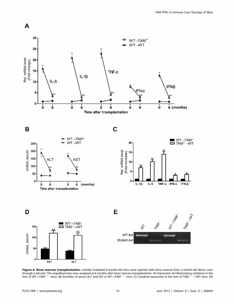
Figure 6. Bone marrow transplantation. Lethally irradiated 6-month-old mice were injected with bone marrow from 2-month-old donor micethrough a tail vein. The engrafted mice were analyzed at 6 months after bone marrow transplantation. (A) Expression of inflammatory cytokines in theliver of WTRTAM2/2 mice. (B) Activities of serum ALT and AST in WTRTAM2/2 mice. (C) Cytokine expression in the liver of TAM2/2RWT mice. (D)
TAM RTKs in Immune Liver Damage of Mice
PLOS ONE | www.plosone.org 10 June 2013 | Volume 8 | Issue 6 | e66604

Acknowledgments
We sincerely thank Dr. Qingxian Lu for providing TAM mutant mice.
Author Contributions
Conceived and designed the experiments: DH. Performed the experiments:
NQ PL YZ HW YC. Analyzed the data: NQ YZ DH. Contributed
reagents/materials/analysis tools: YC. Wrote the paper: NQ YZ DH.
References
1. Vergani D, Longhi MS, Bogdanos DP, Ma Y, Mieli-Vergani G (2009)
Autoimmune hepatitis. Semin Immunopathol 31: 421–435.
2. Zachou K, Rigopoulou E, Dalekos GN (2004) Autoantibodies and autoantigens
in autoimmune hepatitis: important tools in clinical practice and to studypathogenesis of the disease. J Autoimmune Dis 1: 2.
3. Mieli-Vergani G, Heller S, Jara P, Vergani D, Chang MH, et al. (2009)
Autoimmune hepatitis. J Pediatr Gastroenterol Nutr 49: 158–164.
4. Czaja AJ (2009) Autoimmune liver disease. Curr Opin Gastroenterol 25: 215–222.
5. Lapierre P, Djilali-Saiah I, Vitozzi S, Alvarez F (2004) A murine model of type 2
autoimmune hepatitis: Xenoimmunization with human antigens. Hepatology39: 1066–1074.
6. Kuriki J, Murakami H, Kakumu S, Sakamoto N, Yokochi T, et al. (1983)
Experimental autoimmune hepatitis in mice after immunization with syngeneicliver proteins together with the polysaccharide of Klebsiella pneumoniae.
Gastroenterology 84: 596–603.
7. Holdener M, Hintermann E, Bayer M, Rhode A, Rodrigo E, et al. (2008)
Breaking tolerance to the natural human liver autoantigen cytochrome P450
2D6 by virus infection. J Exp Med 205: 1409–1422.
8. Amoh Y, Yasumizu R, Yamamoto Y, Toki J, Nishio N, et al. (1997) The
appearance of unusual phenotypic cells (CD4+ Mac-1+ class II+) in the liver of
(NZW x BXSB)F1 mice is possibly an animal model for autoimmune hepatitis.Immunobiology 197: 31–43.
9. Christen U, Hintermann E, Jaeckel E (2009) New animal models forautoimmune hepatitis. Semin Liver Dis 29: 262–272.
10. Crispe IN (2003) Hepatic T cells and liver tolerance. Nat Rev Immunol 3: 51–62.
11. Sacher T, Knolle P, Nichterlein T, Arnold B, Hammerling GJ, et al. (2002)CpG-ODN-induced inflammation is sufficient to cause T-cell-mediated auto-
aggression against hepatocytes. Eur J Immunol 32: 3628–3637.
12. Seki E, Brenner DA (2008) Toll-like receptors and adaptor molecules in liverdisease: update. Hepatology 48: 322–335.
13. Akira S, Uematsu S, Takeuchi O (2006) Pathogen recognition and innateimmunity. Cell 124: 783–801.
14. Chen CJ, Kono H, Golenbock D, Reed G, Akira S, et al. (2007) Identification of
a key pathway required for the sterile inflammatory response triggered by dyingcells. Nat Med 13: 851–856.
15. Schwabe RF, Seki E, Brenner DA (2006) Toll-like receptor signaling in the liver.
Gastroenterology 130: 1886–1900.
16. Szabo G, Dolganiuc A, Mandrekar P (2006) Pattern recognition receptors: a
contemporary view on liver diseases. Hepatology 44: 287–298.
17. Pandey S, Agrawal DK (2006) Immunobiology of Toll-like receptors: emergingtrends. Immunol Cell Biol 84: 333–341.
18. Lang KS, Recher M, Junt T, Navarini AA, Harris NL, et al. (2005) Toll-likereceptor engagement converts T-cell autoreactivity into overt autoimmune
disease. Nat Med 11: 138–145.
19. Wang J, Hu Y, Deng WW, Sun B (2009) Negative regulation of Toll-likereceptor signaling pathway. Microbes Infect 11: 321–327.
20. Marshak-Rothstein A (2006) Toll-like receptors in systemic autoimmune disease.
Nat Rev Immunol 6: 823–835.
21. Hafizi S, Dahlback B (2006) Signalling and functional diversity within the Axl
subfamily of receptor tyrosine kinases. Cytokine Growth Factor Rev 17: 295–304.
22. Hafizi S, Dahlback B (2006) Gas6 and protein S. Vitamin K-dependent ligands
for the Axl receptor tyrosine kinase subfamily. FEBS J 273: 5231–5244.
23. Lemke G, Lu Q (2003) Macrophage regulation by Tyro 3 family receptors. Curr
Opin Immunol 15: 31–36.
24. Lu Q, Gore M, Zhang Q, Camenisch T, Boast S, et al. (1999) Tyro-3 familyreceptors are essential regulators of mammalian spermatogenesis. Nature 398:
723–728.
25. Lu Q, Lemke G (2001) Homeostatic regulation of the immune system byreceptor tyrosine kinases of the Tyro 3 family. Science 293: 306–311.
26. Tang H, Chen S, Wang H, Wu H, Lu Q, et al. (2009) TAM receptors and theregulation of erythropoiesis in mice. Haematologica 94: 326–334.
27. Zheng Y, Wang Q, Xiao B, Lu Q, Wang Y, et al. (2012) Involvement of receptor
tyrosine kinase Tyro3 in amyloidogenic APP processing and beta-amyloiddeposition in Alzheimer’s disease models. PLoS One 7: e39035.
28. Rothlin CV, Ghosh S, Zuniga EI, Oldstone MB, Lemke G (2007) TAM
receptors are pleiotropic inhibitors of the innate immune response. Cell 131:1124–1136.
29. Lemke G, Rothlin CV (2008) Immunobiology of the TAM receptors. Nat Rev
Immunol 8: 327–336.
30. Wang H, Chen Y, Ge Y, Ma P, Ma Q, et al. (2005) Immunoexpression of Tyro3 family receptors—Tyro 3, Axl, and Mer—and their ligand Gas6 in postnatal
developing mouse testis. J Histochem Cytochem 53: 1355–1364.31. Dong ZJ, Wei HM, Sun R, Tian ZG, Gao B (2004) Isolation of murine hepatic
lymphocytes using mechanical dissection for phenotypic and functional analysis
of NK1.1+ cells. World J Gastroenterol 10: 1928–1933.32. Shen W, Kamendulis LM, Ray SD, Corcoran GB (1991) Acetaminophen-
induced cytotoxicity in cultured mouse hepatocytes: correlation of nuclear Ca2+accumulation and early DNA fragmentation with cell death. Toxicol Appl
Pharmacol 111: 242–254.33. Wu J, Lu M, Meng Z, Trippler M, Broering R, et al. (2007) Toll-like receptor-
mediated control of HBV replication by nonparenchymal liver cells in mice.
Hepatology 46: 1769–1778.34. Qingxian L, Qiutang L, Qingjun L (2010) Regulation of phagocytosis by TAM
receptors and their ligands. Front Biol 5: 227–237.35. Lemke G, Burstyn-Cohen T (2010) TAM receptors and the clearance of
apoptotic cells. Ann N Y Acad Sci 1209: 23–29.
36. Christen U, Holdener M, Hintermann E (2007) Animal models for autoimmunehepatitis. Autoimmun Rev 6: 306–311.
37. Liu Y, Meyer C, Xu C, Weng H, Hellerbrand C, et al. (2013) Animal models ofchronic liver diseases. Am J Physiol Gastrointest Liver Physiol 304: G449–468.
38. Gao B, Jeong WI, Tian Z (2008) Liver: An organ with predominant innate
immunity. Hepatology 47: 729–736.39. Tiegs G, Lohse AW (2010) Immune tolerance: what is unique about the liver. J
Autoimmun 34: 1–6.40. Manns MP, Vogel A (2006) Autoimmune hepatitis, from mechanisms to
therapy. Hepatology 43: S132–144.41. Uchida Y, Ke B, Freitas MC, Yagita H, Akiba H, et al. (2010) T-cell
immunoglobulin mucin-3 determines severity of liver ischemia/reperfusion injury
in mice in a TLR4-dependent manner. Gastroenterology 139: 2195–2206.42. Ichiki Y, Aoki CA, Bowlus CL, Shimoda S, Ishibashi H, et al. (2005) T cell
immunity in autoimmune hepatitis. Autoimmun Rev 4: 315–321.43. Lang KS, Georgiev P, Recher M, Navarini AA, Bergthaler A, et al. (2006)
Immunoprivileged status of the liver is controlled by Toll-like receptor 3
signaling. J Clin Invest 116: 2456–2463.44. Kunz M, Ibrahim SM (2009) Cytokines and cytokine profiles in human
autoimmune diseases and animal models of autoimmunity. Mediators Inflamm2009: 979258.
45. Czaja MJ, Flanders KC, Biempica L, Klein C, Zern MA, et al. (1989)Expression of tumor necrosis factor-alpha and transforming growth factor-beta 1
in acute liver injury. Growth Factors 1: 219–226.
46. Iimuro Y, Gallucci RM, Luster MI, Kono H, Thurman RG (1997) Antibodies totumor necrosis factor alfa attenuate hepatic necrosis and inflammation caused by
chronic exposure to ethanol in the rat. Hepatology 26: 1530–1537.47. Seki E, Tsutsui H, Nakano H, Tsuji N, Hoshino K, et al. (2001)
Lipopolysaccharide-induced IL-18 secretion from murine Kupffer cells
independently of myeloid differentiation factor 88 that is critically involved ininduction of production of IL-12 and IL-1beta. J Immunol 166: 2651–2657.
48. Deng T, Zhang Y, Chen Q, Yan K, Han D (2012) Toll-like receptor-mediatedinhibition of Gas6 and ProS expression facilitates inflammatory cytokine
production in mouse macrophages. Immunology 135: 40–50.49. Paik YH, Schwabe RF, Bataller R, Russo MP, Jobin C, et al. (2003) Toll-like
receptor 4 mediates inflammatory signaling by bacterial lipopolysaccharide in
human hepatic stellate cells. Hepatology 37: 1043–1055.50. Shu SA, Lian ZX, Chuang YH, Yang GX, Moritoki Y, et al. (2007) The role of
CD11c(+) hepatic dendritic cells in the induction of innate immune responses.Clin Exp Immunol 149: 335–343.
51. Sawaki J, Tsutsui H, Hayashi N, Yasuda K, Akira S, et al. (2007) Type 1
cytokine/chemokine production by mouse NK cells following activation of theirTLR/MyD88-mediated pathways. Int Immunol 19: 311–320.
52. Shang T, Zhang X, Wang T, Sun B, Deng T, et al. (2011) Toll-like receptor-initiated testicular innate immune responses in mouse Leydig cells. Endocrinol-
ogy 152: 2827–2836.
53. Sun B, Qi N, Shang T, Wu H, Deng T, et al. (2010) Sertoli cell-initiatedtesticular innate immune response through toll-like receptor-3 activation is
negatively regulated by Tyro3, Axl, and mer receptors. Endocrinology 151:2886–2897.
Serum AST and ALT activities in the TAM2/2RWT mice. Total RNA was extracted from the liver, and the mRNAs of cytokines were determined by real-time RT-PCR. Serum AST and ALT activities were measured by enzymatic assay kits. (E) The expression of Axl in the circulating leukocytes in engraftedmice. Data are mean 6 SEM (n = 10). **P,0.01.doi:10.1371/journal.pone.0066604.g006
TAM RTKs in Immune Liver Damage of Mice
PLOS ONE | www.plosone.org 11 June 2013 | Volume 8 | Issue 6 | e66604