development of a human extracellular matrix for ... · development of a human extracellular matrix...
TRANSCRIPT
Development of a Human Extracellular Matrix for ApplicationsRelated with Stem Cells and Tissue Engineering
Carmen Escobedo-Lucea & Angel Ayuso-Sacido & Chen Xiong & Sonia Prado-López &
Manuel Sanchez del Pino & Dario Melguizo & Carmen Bellver-Estellés &
Susana Gonzalez-Granero & M. Luz Valero & Rubén Moreno & Deborah J. Burks &
Miodrag Stojkovic
Published online: 28 June 2011# The Author(s) 2011. This article is published with open access at Springerlink.com
Introduction
The recent progress in stem cell biology has created newapproaches for their study as well as their application to thetreatment of human diseases [1–3]. The success of stem-cell based technologies in the clinical setting [4, 5] hasemphasized the need to improve the standards of quality forall phases of cell therapy, particularly the development ofculture methods that circumvent products of animal originsince these might provoke infections or immune rejectionfollowing transplantation in patients. Indeed, since Martinet al. (2005) [6] demonstrated that hESC cultured with
animal or serum products retained non-human sialic acidwhich was immunogenic when transplanted in humans, theestablishment of animal-free conditions to support themaintenance and differentiation of human stem cells hasbeen a major goal of the field of regenerative medicine [7].Thus, chemically-defined culture systems that are devoid ofnon-human substances will greatly facilitate the use of stemcells in regenerative strategies.
The concept of a niche is crucial for the organization ofstem cells. A niche is consider as a subset of tissue cells andextracellular substrates (matrix and soluble factors) that cansupport stem cells and control their self-renewal in vivo [8].
Deborah J. Burks and Miodrag Stojkovic contributed equally to themanuscript.
Electronic supplementary material The online version of this article(doi:10.1007/s12015-011-9270-6) contains supplementary material,which is available to authorized users.
C. Escobedo-Lucea (*) : S. Gonzalez-GraneroComparative Neurobiology Unit, Instituto Cavanilles,University of Valencia- RETICS,46980 Valencia, Spaine-mail: [email protected]
C. Escobedo-Lucea :A. Ayuso-Sacido : C. Xiong :S. Prado-López :D. Melguizo :C. Bellver-Estellés : R. Moreno :D. J. Burks :M. StojkovicRegenerative Medicine Program,Centro de Investigación Principe Felipe,46012 Valencia, Spain
M. S. del Pino :M. L. ValeroProteomics Service, Centro de Investigación Principe Felipe,46012 Valencia, Spain
D. J. BurksCIBER of Diabetes and Metabolic Diseases,Instituto de Salud Carlos III,Valencia, Spain
M. StojkovicSPEBO Medical,16000 Leskovac, Serbia
M. StojkovicDepartment of Human Genetics, Faculty of Medicine,University of Kragujevac,Kragujevac, Serbia
Present Address:C. XiongDepartment of Microbiology,Hubei University of Medicine,30 South Remin Rd. Shiyan,Hubei 442000, China
Present Address:S. Prado-LópezRemedi-National Centre for Biomedical Engineering Science,National University Of Ireland,Galway, Ireland
Stem Cell Rev and Rep (2012) 8:170–183DOI 10.1007/s12015-011-9270-6

Extracellular matrices help to structure niches spatially andmodulate the concentration of adhesive and signallingmolecules locally. The ECM is a molecular complex thatcontains collagens and other glycoproteins, hyaluronic acid,proteoglycans, glycosaminoglycans (GAGs), and elastins.Additionally, the ECM harbours growth factors or cyto-kines to protect against degradation [9]. ECM componentsare responsible for adhesion during the majority of cellinteractions and are implicated in the maintenance ofembryonic induction during development as well as stemcell differentiation in vitro [10]. Thus, local changes inECM can dramatically modulate the proliferation andmigration of stem cells and may participate in thespecification of lineages.
hESC have provided invaluable tools for gaining insightinto the developmental origins of human tissues. However, torealize the full biological and clinical potential of hESC,certain problems related with the routine culture of these cellsmust be solved. Mouse embryonic fibroblasts (MEFs) andmurine derivatives such as Matrigel are widely used in themaintenance and differentiation of hESC. Recently, consider-able effort has been dedicated to the elimination of animal-derived reagents from the culture of hESC and in parallel, tothe control of cell growth parameters by avoiding humanfeeder cells. For example, in 2006 Ludwig et al. reported theuse of conditioned media and high doses of FGF to maintainthe undifferentiated state in hESC cultured on plastic, but someabnormalities were detected at passage 20 under theseconditions [11, 12]. The use of human feeders complicatesthe growth and molecular analysis of both pluripotency anddifferentiation since experimental data may reflect thecombined effects of hESC and feeder cells in the culture.Given these considerations, the use of an ECM of human orsynthetic origin would provide many advantages. Indeed,there have been attempts [13, 14] to produce such a tool butthe results have not been satisfactory because the productswere unable to maintain hESC in the undifferentiated stateover time. Matrix proteins have been used as coating for invitro cultures of human stem cells but they have usually beenapplied as undefined protein mixtures [11] of animal origin[15] with undefined media [13], serum [16] or a syntheticmixture [17, 18]. However, most of these human biologicalreagents are expensive to manufacture and thus, are cost-prohibitive for many laboratories.
Decellularization procedures have been used traditionallyto isolate ECM from cells in culture, tissues or organs [19].The goal of decellularization protocols is to efficientlyremove cellular and nuclear material while minimizing anyadverse effect on the composition, biological activity andmechanical and structural integrity of the remainingECM [20, 21]. Decellularized human scaffolds havefacilitated the remodelling of various tissues in bothanimal models and humans [4, 22]. However, any
biochemical procedure employed to remove cells mayalso alter the native three-dimensional architecture of theECM and thus, a balance must be achieved betweenchemical and physical treatments during the decellulariza-tion process [4, 5, 23].
Given the potential importance of hESC in translationalresearch and regenerative medicine, the aim of the presentstudy was to develop a simple, efficient protocol for theproduction of a human ECM that is both safe andeconomical. Here we report that hypotonic lysis of humanforeskin fibroblasts (HFF) generates a human ECM thatretains protein components which are essential for attach-ment and cell-cell interaction. This hffECM was capable ofmaintaining the pluripotency of hESC and supporting theirdifferentiation when used with the appropriate medium.Therefore, our results reveal hffECM as a novel tool whichmay facilitate the clinical application of hESC-basedtechnologies.
Results
Optimisation of Conditions for the Generationof a Functional ECM from Human Foreskin Fibroblasts
To obtain a human ECM which would support themaintenance of undifferentiated stem cells, we initiallydesigned a protocol using RIPA buffer to extract ECMfrom cultured HFF [24]. However, to optimise thismatrix for clinical applications, we contemplated the useof a buffer to induce osmotic shock, thereby circum-venting the use of detergents to lyse attached fibroblasts.Thus, HFF (passage 11–18) were grown to confluenceand then inactivated with mitomycin C. Subsequently,these cultures were lysed with a Tris-Buffer whichtriggered osmotic shock. Incubation with this buffercaused the cells to swell and detach from the plate. Thisprocess was facilitated by six washes with PBS to removebroken cells and debris (see Fig. 1 for the details).
Structural Characterization of hffECM
To assess whether ECM proteins were retained after the celllysis procedure, plates were examined by various microscopytechniques. Control plates of intact HFF (Fig. 2 a1–a4) andexperimental cell-free plates (Fig. 2 a5–a8) were fixed andprocessed for ultrastructural analysis using transmissionelectron microscopy (TEM). The optimal length of cellculture for the obtention of hffECM was determined byassessing HFF which were cultured for either two, nine or12 days prior to lysis (Fig. 2a). Following treatment withlysis buffer, intact fibroblasts were not observed on any ofthe plates as verified by DAPI staining of nuclei (Fig. 2
Stem Cell Rev and Rep (2012) 8:170–183 171

b2,4,6). The structure and distribution of the hffECM werepreserved based on microscopic analysis (Fig. 2 a5). ThehffECM obtained from HFF cultured for only 2 days had alow content of structural proteins (Fig. 2 a-6), as evidencedby the loose lattice of fibers, suggesting that two days ofculture is insufficient for preparation of a quality hffECM.After 12 days of culture, protein fibers displayed signs ofdegradation (Fig. 2 a-8). Thus, the quantity and architectureof the ECM from HFF cultured for 7–9 days (see Fig. 2 a-7)suggested that this amount of time might provide the bestquality cell-free matrix. The presence of characteristic ECMadhesion proteins before and after the treatment wasconfirmed by immunostaining (Fig. 2b) using antibodiesagainst fibronectin (Fig. 2 b1,2), collagen (Fig. 2 b3,4) andlaminin (Fig. 2 b5,6). Collectively, the results of thesestudies demonstrate that the hffECM retains a structuresimilar to the ECM of intact HFF, suggesting that the lysisconditions do not damage significantly the organization ofthe matrix.
Detergent-Free Cell Lysis Preserves the Structureand Composition of the hffECM
To characterize the composition of the matrix obtained fromthe human fibroblasts, we eluted the hffECM from cultureplates and performed a proteomic study. A total of 220
proteins were identified with a false discovery rates (FDRs)below 5% (Table 1, Supplementary information). Toevaluate the origin of the proteins, the enrichment of thecellular compartment terms of gene ontology was analyzedusing Babelomics [25, 26] (Supplementary Fig. 1). At level4, only the terms extracellular matrix (GO:0031012) andcell surface components (GO:0009986) were significantlyenriched (p<0.01). The function of the proteins included inthese terms are highly related as indicated by the networkcreated by STRING analysis (Fig. 3a). These resultsdemonstrate that many of the proteins responsible for theinteraction between the cell surface and the ECM networkare present in the hffECM. Although laminin was detectedin the intact hffECM by immunocytochemistry, we werenot able to identify it by mass spectrometry in samples ofeluted hffECM. The most probable explanation for thisdiscrepancy is the low recovery of laminin from the culturedishes due to its low solubility once assembled in the ECM.
To obtain further information regarding the functionalaspects of the hffECM proteins, the enrichment of KyotoEncyclopedia of Genes and Genomes (KEGG) pathwayswas also analyzed. Of the ten pathways significantlyenriched (p<0.01, Supplementary information), three arerelated with focal adhesions and the cytoskeleton and playimportant roles in cell proliferation, differentiation, andsurvival. A part of the focal adhesion pathway is the
Fig. 1 Experimental design for the obtention, analysis and applica-tions of hffECM. Human foreskin fibroblasts (passage 11 to 18) werecultured during 7–9 days before inactivation with mitomycin C. Atthis stage, cultures were treated overnight at 4°C with lysis buffer toremove cells and the resulting hffECM was subjected to one of thefollowing procedures: (a-1) Assessment of hECM protein integrityand characterization. Electron microscopy analysis was performed toevaluate the integrity and composition of hECM after the treatmentand to establish the optimal period for obtention (see Fig. 2a).
Immunocytochemistry (ICQ) was used to confirm visually thepresence of characteristic ECM components (see Fig. 2b). (a-2)Identification of ECM proteins by proteomic analysis (Fig. 3). (a-3)Application of hffECM to biological processes. To test the functionalcapacity of the hffECM, we designed and performed differentexperiments related with pluripotency (Fig. 4a-e) and differentiationof hESC towards the three germ layers (Fig. 5 and SupplementaryFig. 1a-f) as well as the migration of adults cells (Fig. 6 a-c andSupplementary videos 1–3)
172 Stem Cell Rev and Rep (2012) 8:170–183

extracellular matrix-receptor interaction pathway, whichmediates the interactions with integrins and proteoglycans.
As indicated in Fig. 3b, most of the extracellular partners ofthe pathway were detected in hffECM. Collectively, the
Fig. 2 Evaluation of the structural integrity of the hffECM. aUltrastructural analysis of the efficiency of extraction protocol usingtransmission electron microscopy. Plates of lysed cultures and theirrespective controls with intact foreskin fibroblasts were fixed andprocessed for transmission electron microscopy ultrastructural analy-sis. To determine the optimal length of culture for production ofhffECM, HFFs were maintained for the indicated times and thensubjected to hypotonic lysis. (a1, a5) Semi-thin sections wereprepared from control and lysed cultures and were stained withtoluidine blue. (a2-a4) Representative TEM images of intact HFFcontrol cultures at the indicated time-points. (a6-a8) Representative
TEM images of lysed culture plates at the indicated time-points. Scalebars, 100μm (a-1 and a-5), 2 μm (a-3 and a-4), 1 μm (a-2,a-7 and a-8).b Immunocytochemical analysis of structural proteins in hffECM.Control cultures of intact HFF or lysed plates were immunostained withantibodies against fibronectin (b-1, b-2), collagen type I (b-3, b-4) andlaminin (b-5, b-6). Note that fibronectin, collagen type I and lamininwere retained on plates after the treatment with lysis buffer. Cellnuclei as detected by DAPI were absent in lysed cultures. Allimages were captured at 63X magnification using a Leica confocalmicroscope, with the appropriate lasers to excite the secondaryantibodies (Alexa, 488 nm) and 405 nm for DAPI
Stem Cell Rev and Rep (2012) 8:170–183 173

Fig. 3 Identification of proteincomponents of the hffECM.After removal of HFF by lysis,the remaining hffECM waseluted from culture plates andsubjected to proteomic analysis.a STRING analysis of theproteins contained in the cellularcomponent terms of geneontology that are significantlyenriched in the hffECMsamples. The two principalclusters correspond mainly toproteins of the extracellularmatrix (blue) or cellular surfacecomponents (green). Note thatthe components form tightnetworks with several inter-cluster connections. b Proteincomponents of the human ECM-receptor interaction pathway ofthe KEGG database. Theproteins of the hffECM identi-fied by mass spectrometry are inindicated in red. Laminin, inblue, was detected by immnu-nocytochemical analysis of thehffECM. Most of the extracel-lular matrix partners of thepathway have been identified
174 Stem Cell Rev and Rep (2012) 8:170–183

results of the proteomics analysis demonstrate that theprocedure we have developed for preparation of hffECMretains most of the key components necessary for cellattachment and regulation of proliferation and differentia-tion. Regarding the other enriched pathways, some mayreflect contaminants due to non-specific binding (e.g.,ribosomal proteins and histones) whereas others may havebeen identified because they are anchored to the plasmamembrane, cytoskeleton, and/or ECM. Interestingly, someglycolytic pathways were enriched (pathways hsa00620and hsa00010). Although their origin is not clear, it isnoteworthy that they are components of the anaerobicglycolysis pathway and one might speculate that they maybe located at the extracellular space to provide energylocally to the ATP-dependent processes that may occur inthis vicinity.
hffECM Maintains Pluripotency of hESC
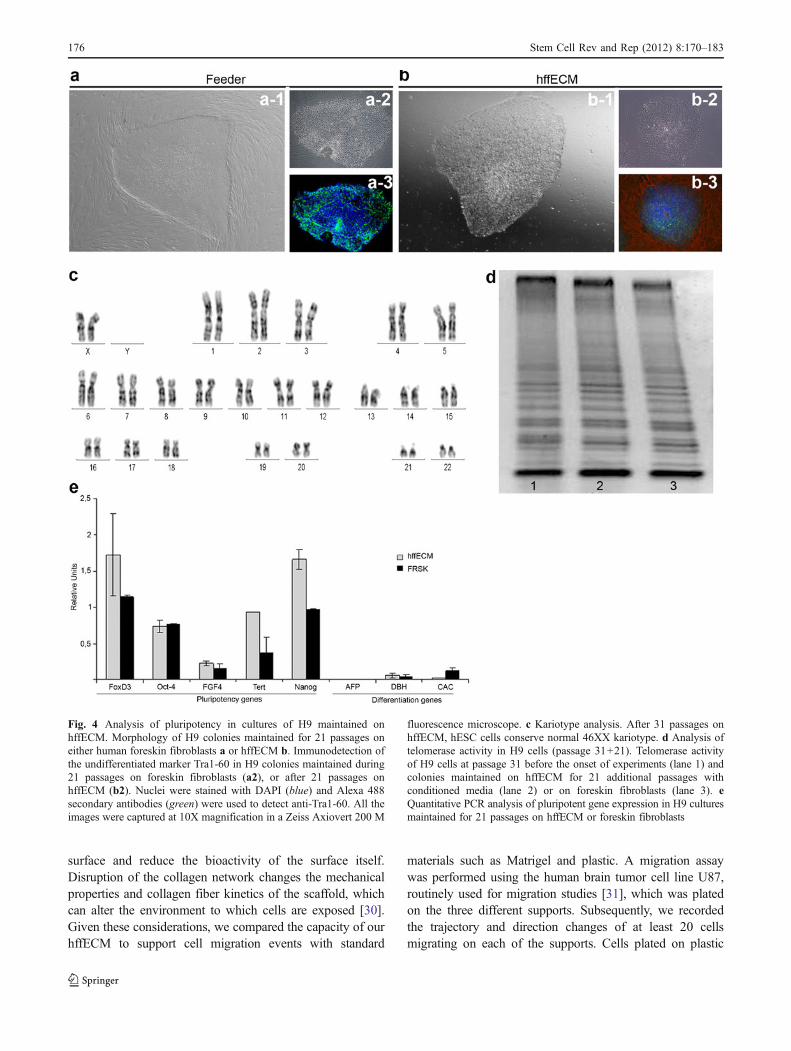
Colonies of H9 (passage 31), maintained on HFF, were cutmechanically and distributed equally into 4 experimentalgroups. One group was again plated on HFF andmaintained during the assay using traditional conditionsfor hESC culture. The other 3 groups of hESC colonies,were seeded onto hffECM-coated plates, using eitherTESR1 feeder-conditioned medium, feeder-conditionedmedium 1, or the traditional KSR medium + bFGF as acontrol. Subsequently, cultures were passaged mecanicallyevery 5–6 days. Colonies seeded on human hffECM andmaintained with KSR or conditioned medium 1 began todisplay a morphology consistent with spontaneous differ-entiation after 2 to 5 passages. In contrast, colonies platedon hffECM with TERS1 conditioned media retained themorphology of undifferentiated colonies with definedborders and a compact sphere, similar to hESC maintainedon feeders (Fig. 4a, b). To ensure that colonies were indeedgrowing on the hffECM, cultures were stained with anti-fibronectin (Fig. 4b-3). Detection of this ECM componentunder and around the colonies suggests that the cells wereplated on hffECM and not the plastic of the culture plate.DAPI was used to counter-stain the nuclei of cultured cells.In the image a-3 of Fig. 4, blue nuclei were observed inhESC colonies and feeder cells. However, in culturesmaintained on hffECM, DAPI staining was detectedexclusively in the colonies but not around them, reinforcingthe observation that intact human feeder cells wereeliminated during preparation of the hffECM. (For moredetails, see Supplementary Fig. 2)
After 21 passages on hffECM, the H9 coloniesdisplayed a normal female kariotype of 46 XX(Fig. 4c). Telomerase activity was similar between H9cells at the onset of the experiment (passage 31) and thosemaintained for 21 additional passages (total of 52
passages) on either hffECM or HFF (Fig. 4d). Addition-ally, the hESC maintained on hffECM continued toexpress markers of pluripotency at levels equivalent orsuperior to those detected in hESC grown on feeder layers(Fig. 4e). These data suggest that hffECM supports H9colonies in the undifferentiated state. Similar observationswere made with the H1 cell line.
hffECM Supports Differentiation of hESC
To facilitate clinical applications, ECM that meets therequirements of an animal-free product should alsosupport differentiation of hESC. To test spontaneousdifferentiation, H9 colonies were allowed to overgrowon hffECM under conditions of bFGF deprivation for atleast 2 weeks. Subsequent analysis by immunostainingrevealed that the cultures had differentiated to ectoderm(Tuj1 positive cells) and mesoderm (cardiac actin)(Supplementary Fig. 3 a,b).
To determine the compatibility of the hffECM with thedifferentiation of hESC by chemically-defined protocols,H9 colonies were cultured on this matrix under conditionsknown to generate human adipocytes [27]. After 15 days inthis differentiation medium (Fig. 5a), the cultures wereanalysed for lipid accumulation and markers of adiposetissue. We detected the presence of many Oil red O-stainedlipid droplets in the cytoplasm of the cells on hffECM(Fig. 5b). Detailed ultrastructure analysis of differentiatedcells using TEM revealed that their morphology wascompatible with mature adipocytes (Fig. 5c), suggestingthe functional capacity of this matrix to promote directeddifferentiation. To confirm that these cells were indeedadipocytes, the expression of adipose-specific genes wasexamined by RT-PCR using human adipose tissue as apositive control. Consistent with the presence of lipiddroplets, aP2 and PPARγ, transcriptional factors which arerequired for adipogenesis, were up-regulated significantlyafter 3 days in differentiation medium, demonstrating that thehffECM promotes differentiation of hESC under definedconditions. Consistent with this, when H9 grown on hffECMwere subjected to a published protocol for the step-wiseinduction of endoderm [28], markers of definitive endodermwere observed after 3 weeks (Supplementary Fig. 3 c-f).Additionally, an earlier version of our hffECM was usedsuccessfully for differentiating H9 to endothelial precursorsand mature endothelial cells [29].
hffECM Supports Cell Migration in 3-D
The removal of adhesion proteins and GAGs from the ECMwould be expected to slow cell migration on the coated
Stem Cell Rev and Rep (2012) 8:170–183 175
surface and reduce the bioactivity of the surface itself.Disruption of the collagen network changes the mechanicalproperties and collagen fiber kinetics of the scaffold, whichcan alter the environment to which cells are exposed [30].Given these considerations, we compared the capacity of ourhffECM to support cell migration events with standard
materials such as Matrigel and plastic. A migration assaywas performed using the human brain tumor cell line U87,routinely used for migration studies [31], which was platedon the three different supports. Subsequently, we recordedthe trajectory and direction changes of at least 20 cellsmigrating on each of the supports. Cells plated on plastic
Fig. 4 Analysis of pluripotency in cultures of H9 maintained onhffECM. Morphology of H9 colonies maintained for 21 passages oneither human foreskin fibroblasts a or hffECM b. Immunodetection ofthe undifferentiated marker Tra1-60 in H9 colonies maintained during21 passages on foreskin fibroblasts (a2), or after 21 passages onhffECM (b2). Nuclei were stained with DAPI (blue) and Alexa 488secondary antibodies (green) were used to detect anti-Tra1-60. All theimages were captured at 10X magnification in a Zeiss Axiovert 200 M
fluorescence microscope. c Kariotype analysis. After 31 passages onhffECM, hESC cells conserve normal 46XX kariotype. d Analysis oftelomerase activity in H9 cells (passage 31+21). Telomerase activityof H9 cells at passage 31 before the onset of experiments (lane 1) andcolonies maintained on hffECM for 21 additional passages withconditioned media (lane 2) or on foreskin fibroblasts (lane 3). eQuantitative PCR analysis of pluripotent gene expression in H9 culturesmaintained for 21 passages on hffECM or foreskin fibroblasts
176 Stem Cell Rev and Rep (2012) 8:170–183

(Fig. 6a) changed continuously their trajectory and directionas they moved around the center of the well. In contrast, thecells maintained on either Matrigel or hffECM migratedlonger distances inside the well. Additionally, the movementof cells on these supports was less haphazard and directionchanges were reduced drastically. Regarding the speed ofmigration, cells grown on hffECM moved significantly fasterwhile no differences were observed between Matrigel andplastic (Fig. 6b). Representative migration patterns of theU87 cells cultured on the various supports can be viewed inthe videos 1–3 of Supplementary Information.
Discussion
The development of a standardized, animal-free culturematrix is an essential tool for both elucidating themechanisms that control human stem cell biology and for
facilitating their application to cell-based clinical therapies[32]. Here we report an efficient method for the obtentionand preservation of a functional ECM from human foreskinfibroblasts. Our method has several key advantages overother strategies for obtaining ECM. First of all, it is asimple, low-cost biochemical method based on hypotoniclysis that can be reproduced easily in cell biology labs.Additionally, in contrast to previous chemical proceduresthat disrupt some of the ECM protein structures [29, 33],the hypotonic shock used in our method preserves thearchitecture and bioactivity of the ECM scaffold. Indeed,our hffECM was capable of maintaining hESC in theundifferentiated state without inducing chromosomal aber-rations during a significant amount of time (21 passages).One observation regarding the hffECM that we initiallyinterpreted as a limitation was the slight reduction in thenumber of colonies when hESC were continuously pas-saged on hffECM as compared with those grown on HFF.
Fig. 5 Evaluation of hffECM as a support for directed differentiationof hESC. Phase contrast images of hESC plated on hffECM andsubjected to a chemically-defined method for differentiation toadipocytes. a 12 days in differentiation media, b Oil red O stainingof H9 cultures on day 11 of differentiation protocol. Scale bars:100 μm, c Transmission electron microscopy characterization ofadipocytes obtained from H9 after 15 days of the differentiation
protocol. The cells display small nuclei, with sparse organelles andlarge lipid droplets distributed along the cytoplasm. Scale bar: 5 μm. dAnalysis of adipocyte differentiation markers using RT-PCR. At theindicated time points, mRNA was prepared for analysis by PCR.Products were visualized by gel electrophoresis. The expression ofaP2 and PPAR gamma was quantified and normalized to 18 s mRNA
Stem Cell Rev and Rep (2012) 8:170–183 177

However, when we analysed markers of pluripotency, theexpression of certain pluripotent genes such as Nanog andFoxD3 was enhanced in the colonies maintained onhffECM relative to the colonies on HFF, suggesting that,although the attachment of hESC may be lower on hffECMthan on feeders, cells display a higher grade of pluripotencywhen grown on this support.
Proteomic studies confirmed that the majority of proteinswhich comprise the ECM [34, 35] were present in hffECMafter the decellularization induced by osmotic shock ofHFF, demonstrating that the procedure prevents enzymaticdegradation of the ECM. To date, proteomics has not beenapplied to define the ECM required for the maintenance anddifferentiation of hESC. The attachment and growth of alltypes of cells is dependent on the interaction of integrinsexpressed on the cell surface with the ECM. The subset ofintegrins expressed in undifferentiated hESC is modifiedduring differentiation, implicating an important role forintegrins in maintaining pluripotency and growth [36]. Ourproteomics data reveal that the majority of the ECMpartners which interact with integrins are retained in thehffECM (Fig. 3b), providing at least one molecularexplanation for the capacity of this matrix to supportgrowth and pluripotency of hESC during an extended
period of time (21 passages). Recent studies have demon-strated that both integrin signaling and E-Cadherin signal-ing are required for the survival and proliferation of hESC[13]; enzymatic dissociation of hESC colonies inducesapoptosis by disrupting these critical cell-ECM interactions.Collectively, our results demonstrate that hffECM retainsthe composition and integrity of ECM molecules whichmodulate self-renewal and survival of hESC. Thus, the useof mass spectrometry techniques to precisely define theprotein composition of the hffECM may enable thesynthesis of a functional support on a large-scale andprovides basic information to tailor the composition toparticular cell types.
When hESC on hffECM were subjected to differenti-ation using chemically-defined conditions, we observedsatisfactory results. Using an established protocol [27],the efficiency of differentiation to adipocytes was compa-rable to colonies maintained initially on feeder layers.Combinations of growth factors and ECM can mimic invitro the cell-cell interactions that occur during develop-ment, but data concerning the functional or spatialsimilarities with real embryo development are lacking[15]. Thus, hffECM provides an excellent opportunity todifferentiate hESC towards the three embryonic germ
Fig. 6 Analysis of random migration of human tumour cells onhffECM, Matrigel and plastic. a Plots representing the differenttrajectories followed by 20 randomly chosen cells growing on
hffECM, Matrigel and plastic. b Histogram depicting the averagespeed of cells growing on the three different surfaces. * P<0.00007;** P<0.0008
178 Stem Cell Rev and Rep (2012) 8:170–183
layers on a complex and basement-rich support where the 'basal lamina in vivo.
During embryo development or tumour progression, cellsusually migrate within a 3D extracellular matrix environment.The majority of in vitro assays have been performed using 2Dmodels. Computational studies have predicted that somecrucial migration parameters such as matrix stiffness and thedistribution of integrin forces at cell matrix-interface aredramatically different in 2D versus 3D environment [37].With the aim of extrapolating in vitro experiments to in vivoprocesses, Zaman et al., 2006 [38] compared cell migrationparameters in 2D [39] with Matrigel and observed cleardifferences between these supports. When we compared themigration of U87 cells on Matrigel versus hffECM as 3Dsupports, we observed similar patterns, although the speed ofmigration was superior on hffECM.
In summary, we have developed a simple method thatgenerates a biologically active human ECM underconditions which are compatible with clinical-gradestudies. Our hffECM supports the maintenance anddifferentiation of hESC and based on our data, can beapplied to other types of stem cells which require aphysical support.
Materials and Methods
Culture and Maintenance of hESC
The present study was approved by the Commission forControl of Transplants of the Carlos III Health Instituteand complies with the Spanish regulations for hESCresearch. The human embryonic stem cell lines H9 andH1 were purchased from Wicell (Wisconsin, USA) andcultured as previously described [16]. H9 and H1colonies were routinely passaged mechanically andmaintained on human foreskin fibroblasts (HFF) at 5%CO2 and 37°C until the onset of the experimentsdescribed below.
Culture of Human Fibroblasts and Preparationof Conditioned Media
Human foreskin fibroblasts (ATCC Catalog No. CRL-2429,Passage 11–18) were grown in Dulbecco’s modified Eaglemedium (DMEM) with high glucose, supplemented with10% human serum (HS), 1% L-glutamine, and 1%nonessential amino acid (NEAA). The HFF were splitusing Triple Select every 5–7 days (all the above reagentsare from GIBCO, Invitrogen). When the cells reachedconfluence, they were inactivated by 10μg/ml Mitomycin C(Fluka, Catalog No. 69824) for 3 h, then washed threetimes with PBS, digested, and 6×10⁶ cells were seeded in
T75 flask coated with 0.1% gelatin (Sigma, Catalog No.G1890). To generate conditioned media, cells were culturedat 37°C, 5% CO2 in one of the following: Medium 1 orTESR medium. Medium 1 contained 80% Knockout-Dulbecco’s modified Eagle’s medium (KO-DMEM), 20%KNOCKOUT serum replacement (Knockout SR), 1%L-glutamine, 1% NEAA, 100μM 2-Mercaptoethanol (Sig-ma, Catalog No.M7522). Medium was collected dailyduring 1 week of culture. Before use as the culture mediumfor hESC, 4 ng/ml human basic fibroblast growth factor(bFGF) (Invitrogen, Catalog 13256–029) was added.Conditioned medium was stored at −80°C for up to6 months with no loss of biological activity.
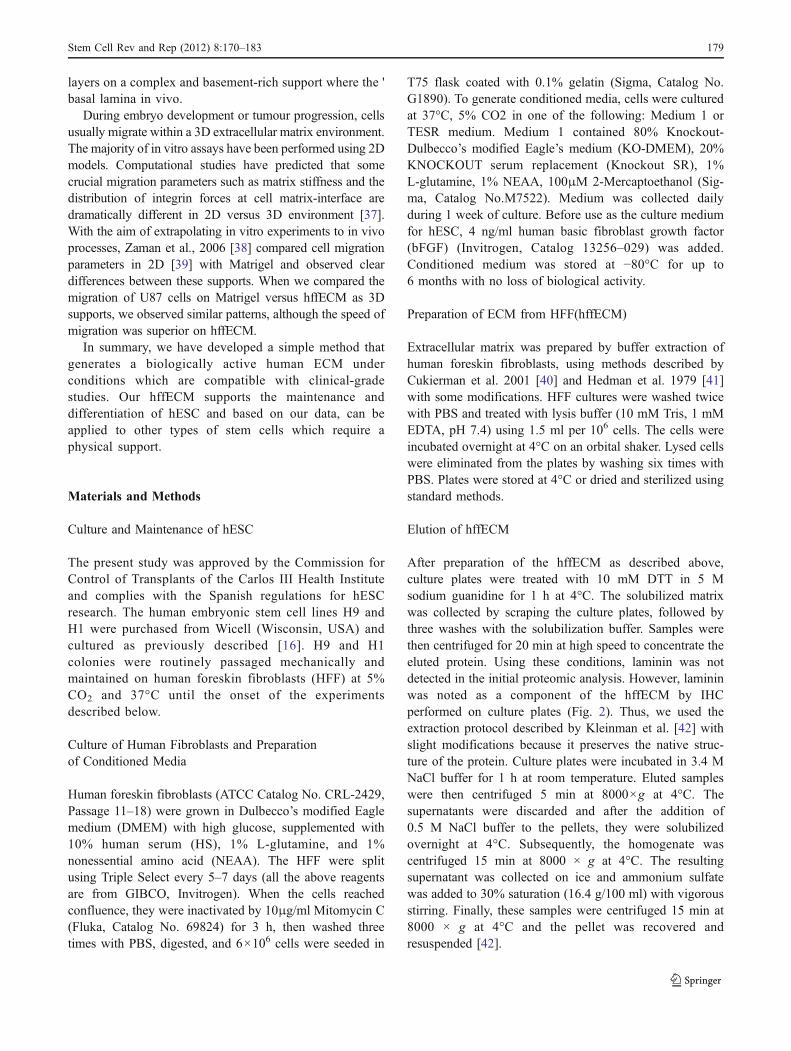
Preparation of ECM from HFF(hffECM)
Extracellular matrix was prepared by buffer extraction ofhuman foreskin fibroblasts, using methods described byCukierman et al. 2001 [40] and Hedman et al. 1979 [41]with some modifications. HFF cultures were washed twicewith PBS and treated with lysis buffer (10 mM Tris, 1 mMEDTA, pH 7.4) using 1.5 ml per 106 cells. The cells wereincubated overnight at 4°C on an orbital shaker. Lysed cellswere eliminated from the plates by washing six times withPBS. Plates were stored at 4°C or dried and sterilized usingstandard methods.
Elution of hffECM
After preparation of the hffECM as described above,culture plates were treated with 10 mM DTT in 5 Msodium guanidine for 1 h at 4°C. The solubilized matrixwas collected by scraping the culture plates, followed bythree washes with the solubilization buffer. Samples werethen centrifuged for 20 min at high speed to concentrate theeluted protein. Using these conditions, laminin was notdetected in the initial proteomic analysis. However, lamininwas noted as a component of the hffECM by IHCperformed on culture plates (Fig. 2). Thus, we used theextraction protocol described by Kleinman et al. [42] withslight modifications because it preserves the native struc-ture of the protein. Culture plates were incubated in 3.4 MNaCl buffer for 1 h at room temperature. Eluted sampleswere then centrifuged 5 min at 8000×g at 4°C. Thesupernatants were discarded and after the addition of0.5 M NaCl buffer to the pellets, they were solubilizedovernight at 4°C. Subsequently, the homogenate wascentrifuged 15 min at 8000 × g at 4°C. The resultingsupernatant was collected on ice and ammonium sulfatewas added to 30% saturation (16.4 g/100 ml) with vigorousstirring. Finally, these samples were centrifuged 15 min at8000 × g at 4°C and the pellet was recovered andresuspended [42].
Stem Cell Rev and Rep (2012) 8:170–183 179
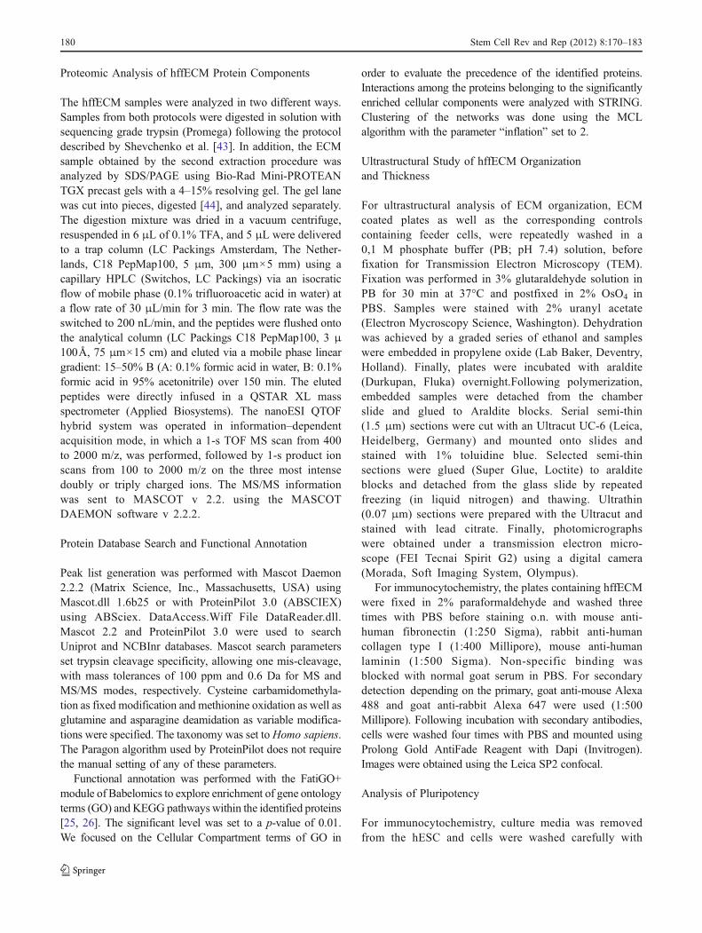
Proteomic Analysis of hffECM Protein Components
The hffECM samples were analyzed in two different ways.Samples from both protocols were digested in solution withsequencing grade trypsin (Promega) following the protocoldescribed by Shevchenko et al. [43]. In addition, the ECMsample obtained by the second extraction procedure wasanalyzed by SDS/PAGE using Bio-Rad Mini-PROTEANTGX precast gels with a 4–15% resolving gel. The gel lanewas cut into pieces, digested [44], and analyzed separately.The digestion mixture was dried in a vacuum centrifuge,resuspended in 6 μL of 0.1% TFA, and 5 μL were deliveredto a trap column (LC Packings Amsterdam, The Nether-lands, C18 PepMap100, 5 μm, 300 μm×5 mm) using acapillary HPLC (Switchos, LC Packings) via an isocraticflow of mobile phase (0.1% trifluoroacetic acid in water) ata flow rate of 30 μL/min for 3 min. The flow rate was theswitched to 200 nL/min, and the peptides were flushed ontothe analytical column (LC Packings C18 PepMap100, 3 μ100Å, 75 μm×15 cm) and eluted via a mobile phase lineargradient: 15–50% B (A: 0.1% formic acid in water, B: 0.1%formic acid in 95% acetonitrile) over 150 min. The elutedpeptides were directly infused in a QSTAR XL massspectrometer (Applied Biosystems). The nanoESI QTOFhybrid system was operated in information–dependentacquisition mode, in which a 1-s TOF MS scan from 400to 2000 m/z, was performed, followed by 1-s product ionscans from 100 to 2000 m/z on the three most intensedoubly or triply charged ions. The MS/MS informationwas sent to MASCOT v 2.2. using the MASCOTDAEMON software v 2.2.2.
Protein Database Search and Functional Annotation
Peak list generation was performed with Mascot Daemon2.2.2 (Matrix Science, Inc., Massachusetts, USA) usingMascot.dll 1.6b25 or with ProteinPilot 3.0 (ABSCIEX)using ABSciex. DataAccess.Wiff File DataReader.dll.Mascot 2.2 and ProteinPilot 3.0 were used to searchUniprot and NCBInr databases. Mascot search parametersset trypsin cleavage specificity, allowing one mis-cleavage,with mass tolerances of 100 ppm and 0.6 Da for MS andMS/MS modes, respectively. Cysteine carbamidomethyla-tion as fixed modification and methionine oxidation as well asglutamine and asparagine deamidation as variable modifica-tions were specified. The taxonomy was set toHomo sapiens.The Paragon algorithm used by ProteinPilot does not requirethe manual setting of any of these parameters.
Functional annotation was performed with the FatiGO+module of Babelomics to explore enrichment of gene ontologyterms (GO) and KEGG pathways within the identified proteins[25, 26]. The significant level was set to a p-value of 0.01.We focused on the Cellular Compartment terms of GO in
order to evaluate the precedence of the identified proteins.Interactions among the proteins belonging to the significantlyenriched cellular components were analyzed with STRING.Clustering of the networks was done using the MCLalgorithm with the parameter “inflation” set to 2.
Ultrastructural Study of hffECM Organizationand Thickness
For ultrastructural analysis of ECM organization, ECMcoated plates as well as the corresponding controlscontaining feeder cells, were repeatedly washed in a0,1 M phosphate buffer (PB; pH 7.4) solution, beforefixation for Transmission Electron Microscopy (TEM).Fixation was performed in 3% glutaraldehyde solution inPB for 30 min at 37°C and postfixed in 2% OsO4 inPBS. Samples were stained with 2% uranyl acetate(Electron Mycroscopy Science, Washington). Dehydrationwas achieved by a graded series of ethanol and sampleswere embedded in propylene oxide (Lab Baker, Deventry,Holland). Finally, plates were incubated with araldite(Durkupan, Fluka) overnight.Following polymerization,embedded samples were detached from the chamberslide and glued to Araldite blocks. Serial semi-thin(1.5 μm) sections were cut with an Ultracut UC-6 (Leica,Heidelberg, Germany) and mounted onto slides andstained with 1% toluidine blue. Selected semi-thinsections were glued (Super Glue, Loctite) to aralditeblocks and detached from the glass slide by repeatedfreezing (in liquid nitrogen) and thawing. Ultrathin(0.07 μm) sections were prepared with the Ultracut andstained with lead citrate. Finally, photomicrographswere obtained under a transmission electron micro-scope (FEI Tecnai Spirit G2) using a digital camera(Morada, Soft Imaging System, Olympus).
For immunocytochemistry, the plates containing hffECMwere fixed in 2% paraformaldehyde and washed threetimes with PBS before staining o.n. with mouse anti-human fibronectin (1:250 Sigma), rabbit anti-humancollagen type I (1:400 Millipore), mouse anti-humanlaminin (1:500 Sigma). Non-specific binding wasblocked with normal goat serum in PBS. For secondarydetection depending on the primary, goat anti-mouse Alexa488 and goat anti-rabbit Alexa 647 were used (1:500Millipore). Following incubation with secondary antibodies,cells were washed four times with PBS and mounted usingProlong Gold AntiFade Reagent with Dapi (Invitrogen).Images were obtained using the Leica SP2 confocal.
Analysis of Pluripotency
For immunocytochemistry, culture media was removedfrom the hESC and cells were washed carefully with
180 Stem Cell Rev and Rep (2012) 8:170–183

PBS. Cells were fixed 30 min with 4% paraformalde-hyde. Subsequently, cells were washed three times withPBS and permeabilized with 0.5% Triton X-100(Sigma- Aldrich) at room temperature. Non-specificbinding was blocked with normal goat serum in PBS.Primary antibodies (SSEA-3/4, SSEA-1, TRA- 1–60,TRA-1- 81) of the ES Cell Characterizat ionKit (1:50 Millipore) were diluted according to themanufacturer’s instructions and incubated overnight at4°C. For detection with secondary antibodies, IgG1Alexa488 was used for SSEA-4, and IgM Alexa488 forTra1-60 and Tra1-80. After 1 h of incubation at roomtemperature, samples were washed four times with PBSand mounted using Prolong Gold Anti-Fade Reagentwith Dapi (Invitrogen). Images were obtained at a 10Xmagnification using the Zeiss Axiovert 200 M.
Chromosomal Analysis
Cytogenetic analysis was conducted at passages 17 and 21after transfer of hESC colonies to hffECM-coated plates.Dividing cells were arrested at metaphase by the addition of50 μg/ml of colchicine for 2 h and then harvested usingstandard cytogenetic techniques. Harvested metaphasechromosomes were stained. Giemsa/Trypsin/Leishman(GTL) banding techniques were employed for karyotypeanalysis. An average of 30 banded metaphase spreads werekaryotyped and analyzed for chromosomal rearrangements.GTL- banded metaphase spreads were captured andarranged using Cytovision software from Applied Imaging(Santa Carla, CA, USA).
RNA Preparation and RT-PCR
Total RNA was prepared from cells using RNeasy mini kit(Quiagen, Gilden; no. 74104). To eliminate contaminatinggenomic DNA, the initial RNA pellet was incubated withdeoxyribonuclease (DNase) I (2 to 4 U/μL; Qiagen,Carlsbad; no.79254) for 15 min at room temperature inthe buffer supplied by the manufacturer. RT–PCR andprimer sequences were as described in the SupplementalTable S2. They were designed using Primer3 software andsynthesized by Sigma–Aldrich. For each experiment,controls were performed in which reverse transcriptasewas omitted from the cDNA reaction mixture and templateDNA was omitted from the PCR mixture. For quantitativereal-time PCR (QRT-PCR), 5 μg RNA was converted intocDNA, and a series of diluted samples were used for40-cycle PCR in Light Cycler 480 SYBR Green I Master(Kit no. 04707516001) in an Lightcycler 480 (RocheDiagnostics,Mannheim) instrument. Reactions (20 μL total)contained 1 μL cDNA, 10 μM each primer, and 4 μMprobe and were run using the default Lightcycler 480
program. To generate a standard curve for comparison ofmRNA levels in different samples, multiple dilutions of thecontrol cDNA sample, spanning at least 3 orders ofmagnitude, were prepared. The equation describing theplot of threshold cycle, Ct, versus log concentration wasused to determine relative amounts of mRNA in experi-mental samples. Using the optimized conditions andthreshold values, individual samples were analyzed intriplicate using the probe of interest and an internal controlexpected to be unchanged between samples. Three differentinternal controls were used: glyceraldehyde-3-phosphate-dehydrogenase (Gapdh), β-2 microglobulin, and β-actin.From the Ct values, the relative transcript concentration wascalculated and normalized to that of the internal control.The maximum expression data point was adjusted to 100.Data are shown for samples normalized to Gapdh, butresults were comparable when analysis was performedusing either β-2 microglobulin alone or a combination of all3 controls.
Telomerase Activity Assay
Telomerase activity was assayed by telomere repeatamplification protocol using Trapeze Kit (Chemicon) andaccording to the manufacturer’s protocol.
Analysis of hESC Differentiation
Spontaneous in vitro differentiation was evaluated byallowing hESC to overgrow on hffECM in growth mediawithout basic fibroblast growth factor bFGF (Invitrogen). Toinduce adipocyte differentiation, the hESC were seeded intriplicate on ECM-coated wells of a 6-well plate at a density ofaround 30 hESC colonies/well (about 300–500 cells percolony) in CM. Cultures were then maintained continuouslyin Differentiation medium 1 (DM1): 80% Knock-out Dulbec-co’s modified Eagle’s medium DMEM/F12, 20% Knock-outserum replacement, 1 mmol/L L-glutamine, 100 μM2-Mercaptoethanol, 1% MEM non-essential amino acids.The medium was changed every other day. Culture sampleswere collected at day 0, day 3, day 6, day 9, day12, and day15and analyzed by Oil Red O staining and RT-PCR.
Analysis of Cell Migration
The human brain tumour cell line U-87 MG was grownin Ham’s F-12 supplemented with 10% FBS. For theassay, 5×10³ per well were seeded onto three replicate96-well plates coated with Matrigel, hffECM or plasticrespectively, and maintained for at least 3 h in a incubatorbefore the experiment. During the migration experiment(8 h), plates were placed in a pre-equilibrated In-CellAnalyzer 1000 Chamber and imaged every 10 min using
Stem Cell Rev and Rep (2012) 8:170–183 181
10X objective with 2000 exposure. Images were analyzedwith In-Cell Investigator software using the cell trackinganalysis tool to create plots of X-Y position and timevariables. The particles with the same identificationnumber were connected by a line representing thecontinuous track for a cell (GE Healthcare applicationNote 28-9327-11 AA). For statistical comparison of theparametric data obtained, we used the t-test analysis ofvariance for two groups.
Acknowledgements We are grateful to Dr. JoseManuel Garcia-Verdugofor his assistance with electron microscopy and to Enrique Navarro andMªTeresa Minguez of the University of Valencia for technical support with theconfocal microscope. Additionally, we thank Dr. Clara Rodríguezand Sandra Pinto for their assistance with the migration assays.Carmen Escobedo-Lucea is financed by post-doctoral program SaraBorrell from Institute of health Carlos III, Ministry of Health,Spain. This study was supported with financial support from theRegenerative Medicine Program of the Valencian Community, Prometeo2009/011, TerCel, CIBERDEM (ISCIII), the Spanish Ministry of Scienceand Innovation (SAF2008-00011), EMER07 (ISCIII) and LIVES, aninitiative of the FP7 program. The Proteomics core facility of the CIPF isa member of ProteoRed-ISCIII
Open Access This article is distributed under the terms of theCreative Commons Attribution Noncommercial License which per-mits any noncommercial use, distribution, and reproduction in anymedium, provided the original author(s) and source are credited.
References
1. Thomson, J. A., Itskovitz-Eldor, J., Shapiro, S. S., et al. (1998).Embryonic stem cell lines derived from human blastocysts.Science, 282, 1145–1147.
2. Takahashi, K., & Yamanaka, S. (2006). Induction of pluripotentstem cells from mouse embryonic and adult fibroblast cultures bydefined factors. Cell, 126, 663–676.
3. Zuk, P. A., Zhu, M., Mizuno, H., et al. (2001). Multilineage cellsfrom human adipose tissue: implications for cell-based therapies.Tissue Engineering, 7, 211–228.
4. Macchiarini, P., Jungebluth, P., Go, T., et al. (2008). Clinicaltransplantation of a tissue-engineered airway. Lancet, 372, 2023–2030.
5. Atala, A., Bauer, S. B., Soker, S., Yoo, J. J., & Retik, A. B.(2006). Tissue-engineered autologous bladders for patients need-ing cystoplasty. Lancet, 367, 1241–1246.
6. Martin, M. J., Muotri, A., Gage, F., & Varki, A. (2005). Humanembryonic stem cells express an immunogenic nonhuman sialicacid. Natural Medicines, 11, 228–232.
7. Rodin, S., Domogatskaya, A., Strom, S., et al. Long-term self-renewal of human pluripotent stem cells on human recombinantlaminin-511. Nature Biotechnology, 28, 611–615.
8. Spradling, A., Drummond-Barbosa, D., & Kai, T. (2001). Stemcells find their niche. Nature, 414, 98–104.
9. Kagami, S., Kondo, S., Loster, K., et al. (1998). Collagen type Imodulates the platelet-derived growth factor (PDGF) regulation ofthe growth and expression of beta1 integrins by rat mesangialcells. Biochemical and Biophysical Research Communications,252, 728–732.
10. Derda, R., Li, L., Orner, B. P., Lewis, R. L., Thomson, J. A., &Kiessling, L. L. (2007). Defined substrates for human embryonic
stem cell growth identified from surface arrays. ACS ChemicalBiology, 2, 347–355.
11. Ludwig, T. E., Bergendahl, V., Levenstein, M. E., Yu, J.,Probasco, M. D., & Thomson, J. A. (2006). Feeder-independentculture of human embryonic stem cells. Nature Methods, 3, 637–646.
12. Ludwig, T. E., Levenstein, M. E., Jones, J. M., et al. (2006).Derivation of human embryonic stem cells in defined conditions.Nature Biotechnology, 24, 185–187.
13. Xu, C., Inokuma, M. S., Denham, J., et al. (2001). Feeder-freegrowth of undifferentiated human embryonic stem cells. NatureBiotechnology, 19, 971–974.
14. Amit, M., Shariki, C., Margulets, V., & Itskovitz-Eldor, J. (2004).Feeder layer- and serum-free culture of human embryonic stemcells. Biology of Reproduction, 70, 837–845.
15. Klimanskaya, I., Chung, Y., Meisner, L., Johnson, J., West, M. D.,& Lanza, R. (2005). Human embryonic stem cells derived withoutfeeder cells. Lancet, 365, 1636–1641.
16. Stojkovic, P., Lako, M., Przyborski, S., et al. (2005). Human-serummatrix supports undifferentiated growth of human embryonic stemcells. Stem Cells, 23, 895–902.
17. Klim, J. R., Li, L., Wrighton, P. J., Piekarczyk, M. S., &Kiessling, L. L. A defined glycosaminoglycan-binding substratumfor human pluripotent stem cells. Nature Methods, 7, 989–994.
18. Mei, Y., Saha, K., Bogatyrev, S. R., et al. Combinatorialdevelopment of biomaterials for clonal growth of humanpluripotent stem cells. Nature Materials, 9, 768–778.
19. Hodde, J. P., Record, R. D., Tullius, R. S., & Badylak, S. F.(2002). Retention of endothelial cell adherence to porcine-derivedextracellular matrix after disinfection and sterilization. TissueEngineering, 8, 225–234.
20. Hodde, J. P., Badylak, S. F., Brightman, A. O., & Voytik-Harbin, S. L.(1996). Glycosaminoglycan content of small intestinal submucosa: abioscaffold for tissue replacement. Tissue Engineering, 2, 209–217.
21. Hodde, J. P., Record, R. D., Liang, H. A., & Badylak, S. F.(2001). Vascular endothelial growth factor in porcine-derivedextracellular matrix. Endothelium, 8, 11–24.
22. Badylak, S. F., Freytes, D. O., & Gilbert, T. W. (2009).Extracellular matrix as a biological scaffold material: structureand function. Acta Biomaterialia, 5, 1–13.
23. Ott, H. C., Matthiesen, T. S., Goh, S. K., et al. (2008). Perfusion-decellularized matrix: using nature’s platform to engineer abioartificial heart. Natural Medicines, 14, 213–221.
24. Escobedo-Lucea, C., & Stojkovic, M. (2010). Growth of humanembryonic stem cells using derivates of human fibroblasts.Methods Mol Biol, 584, 55–69.
25. Al-Shahrour, F., Minguez, P., Tarraga, J., et al. (2006). BABE-LOMICS: a systems biology perspective in the functionalannotation of genome-scale experiments. Nucleic Acids Research,34, W472–W476.
26. Al-Shahrour, F., Minguez, P., Tarraga, J., et al. (2007). FatiGO +: afunctional profiling tool for genomic data. Integration of functionalannotation, regulatory motifs and interaction data with microarrayexperiments. Nucleic Acids Research, 35, W91–W96.
27. Xiong, C., Xie, C. Q., Zhang, L., et al. (2005). Derivation ofadipocytes from human embryonic stem cells. Stem Cells andDevelopment, 14, 671–675.
28. D’Amour, K. A., Agulnick, A. D., Eliazer, S., Kelly, O. G.,Kroon, E., & Baetge, E. E. (2005). Efficient differentiation ofhuman embryonic stem cells to definitive endoderm. NatureBiotechnology, 23, 1534–1541.
29. Prado-Lopez, S., Conesa, A., Arminan, A., et al. Hypoxiapromotes efficient differentiation of human embryonic stem cellsto functional endothelium. Stem Cells, 28, 407–418.
30. Gilbert, T. W., Sellaro, T. L., & Badylak, S. F. (2006). Decellulariza-tion of tissues and organs. Biomaterials, 27, 3675–3683.
182 Stem Cell Rev and Rep (2012) 8:170–183
31. Catacuzzeno, L., Aiello, F., Fioretti, B., et al. Serum-activated Kand Cl currents underlay U87-MG glioblastoma cell migration.Journal of Cellular Physiology 2010.
32. Villa-Diaz, L. G., Nandivada, H., Ding, J., et al. Syntheticpolymer coatings for long-term growth of human embryonic stemcells. Nature Biotechnology, 28, 581–583.
33. Grauss, R. W., Hazekamp, M. G., Oppenhuizen, F., vanMunsteren, C. J., Gittenberger-de Groot, A. C., & DeRuiter, M.C. (2005). Histological evaluation of decellularised porcine aorticvalves: matrix changes due to different decellularisation methods.European Journal of Cardiothoracic Surgery, 27, 566–571.
34. Tong, W. Y., Liang, Y. M., Tam, V., et al. (2010). Biochemicalcharacterization of the cell-biomaterial interface by quantitativeproteomics. Molecular & Cellular Proteomics, 9, 2089–2098.
35. Xu, Y., Zhu, X., Hahm, H. S., et al. Revealing a core signalingregulatory mechanism for pluripotent stem cell survival and self-renewal by small molecules. Proceedings of the National Academyof Sciences of the United States of America, 107, 8129–8134.
36. Rowland, T. J., Miller, L. M., Blaschke, A. J., et al. (2010). Rolesof integrins in human induced pluripotent stem cell growth onMatrigel and vitronectin. Stem Cells and Development, 19, 1231–1240.
37. Zaman, M. H., Kamm, R. D., Matsudaira, P., & Lauffenburger,D. A. (2005). Computational model for cell migration inthree-dimensional matrices. Biophysical Journal, 89, 1389–1397.
38. Zaman, M. H., Trapani, L. M., Sieminski, A. L., et al. (2006).Migration of tumor cells in 3D matrices is governed by matrixstiffness along with cell-matrix adhesion and proteolysis. Proceed-ings of the National Academy of Sciences of the United States ofAmerica, 103, 10889–10894.
39. Palecek, S. P., Loftus, J. C., Ginsberg, M. H., Lauffenburger, D.A., & Horwitz, A. F. (1997). Integrin-ligand binding propertiesgovern cell migration speed through cell-substratum adhesiveness.Nature, 385, 537–540.
40. Cukierman, E., Pankov, R., Stevens, D. R., & Yamada, K. M.(2001). Taking cell-matrix adhesions to the third dimension.Science, 294, 1708–1712.
41. Hedman, K., Kurkinen, M., Alitalo, K., Vaheri, A., Johansson, S.,& Hook, M. (1979). Isolation of the pericellular matrix of humanfibroblast cultures. The Journal of Cell Biology, 81, 83–91.
42. Kleinman, H. K. (2001). Preparation of basement membranecomponents from EHS tumors. Current Protocol in Cell BiologyChapter 10:Unit 10 2.
43. Shevchenko, A., Jensen, O. N., Podtelejnikov, A. V., et al. (1996).Linking genome and proteome by mass spectrometry: large-scaleidentification of yeast proteins from two dimensional gels.Proceedings of the National Academy of Sciences of the UnitedStates of America, 93, 14440–14445.
44. Shevchenko, A., Wilm, M., Vorm, O., & Mann, M. (1996). Massspectrometric sequencing of proteins silver-stained polyacrylamidegels. Analytical Chemistry, 68, 850–858.
Stem Cell Rev and Rep (2012) 8:170–183 183