development including mitosis and meiosis biology 155 krilowicz spring 2010
Post on 21-Dec-2015
214 views
TRANSCRIPT
Development Including Mitosis and Meiosis
Biology 155
Krilowicz
Spring 2010
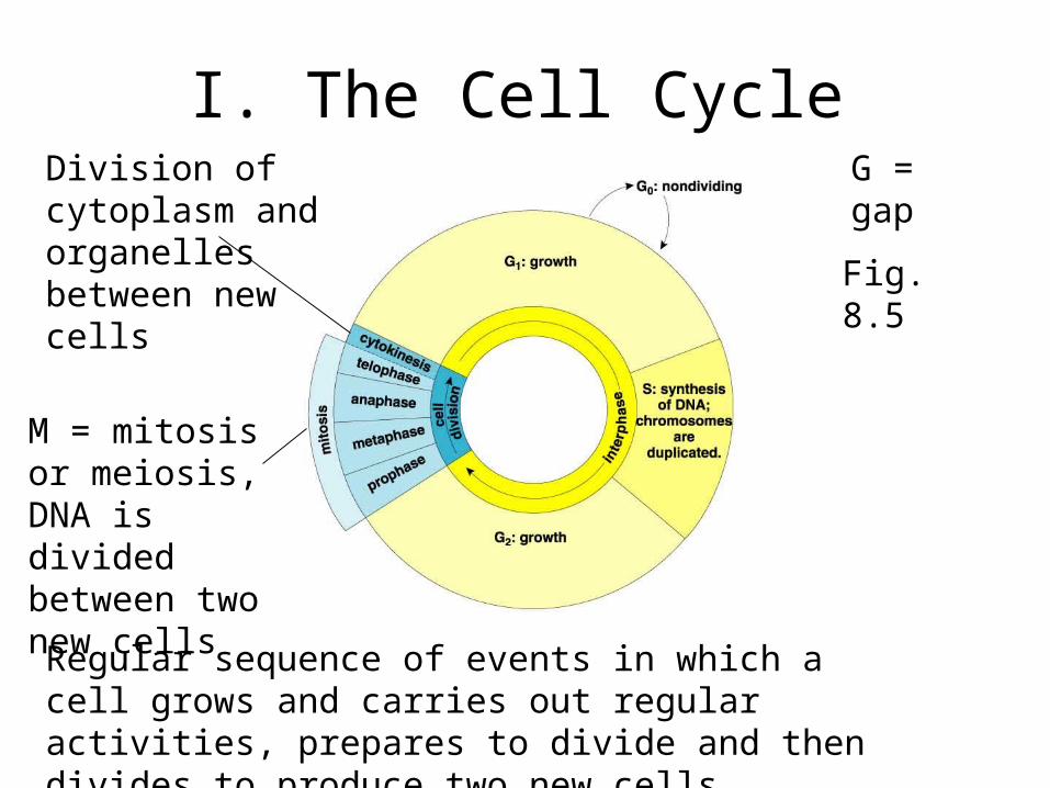
I. The Cell Cycle
Regular sequence of events in which a cell grows and carries out regular activities, prepares to divide and then divides to produce two new cells
G = gap
M = mitosis or meiosis, DNA is divided between two new cells
Division of cytoplasm and organelles between new cells
Fig. 8.5
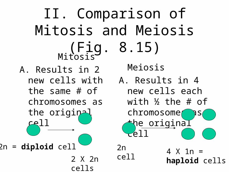
II. Comparison of Mitosis and Meiosis (Fig. 8.15)
Mitosis
A. Results in 2 new cells with the same # of chromosomes as the original cell
Meiosis
A. Results in 4 new cells each with ½ the # of chromosomes as the original cell
2n = diploid cell
2 X 2n cells
2n cell 4 X 1n = haploid cells
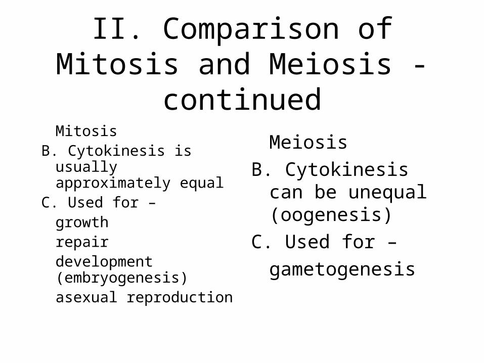
II. Comparison of Mitosis and Meiosis - continued
MitosisB. Cytokinesis is usually
approximately equalC. Used for –
growth repairdevelopment (embryogenesis)asexual reproduction
Meiosis
B. Cytokinesis can be unequal (oogenesis)
C. Used for –
gametogenesis
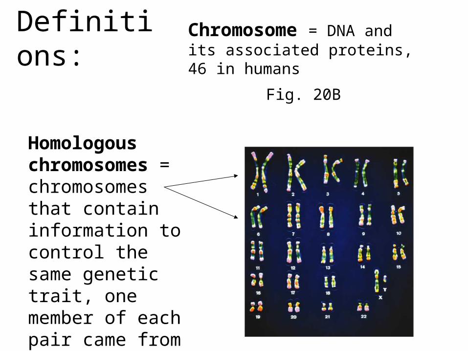
Definitions: Chromosome = DNA and its associated proteins, 46 in humans
Homologous chromosomes = chromosomes that contain information to control the same genetic trait, one member of each pair came from mom and one came from dad
Fig. 20B
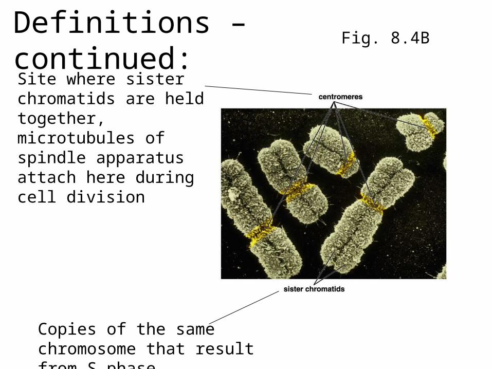
Definitions – continued:
Copies of the same chromosome that result from S phase
Site where sister chromatids are held together, microtubules of spindle apparatus attach here during cell division
Fig. 8.4B
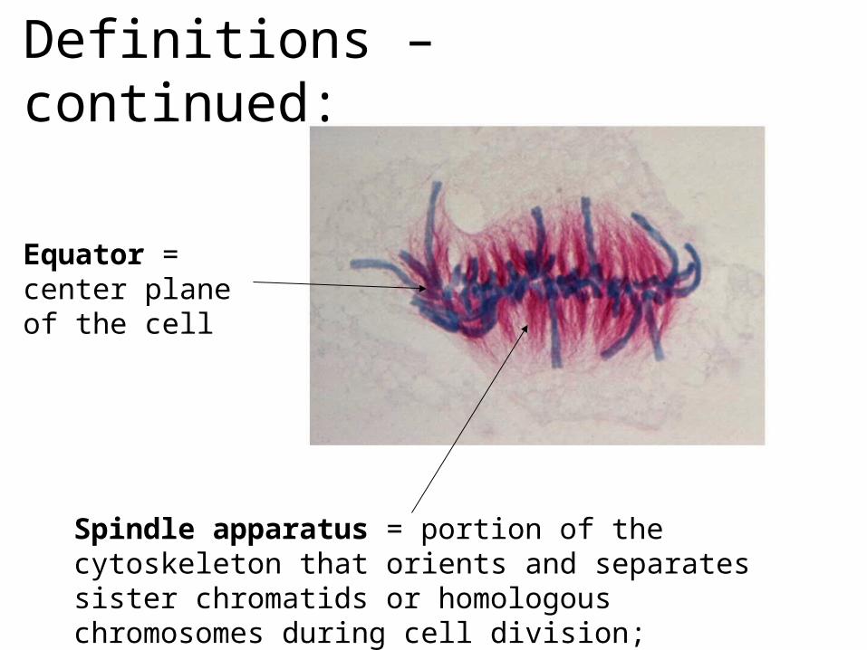
Definitions – continued:
Equator = center plane of the cell
Spindle apparatus = portion of the cytoskeleton that orients and separates sister chromatids or homologous chromosomes during cell division; contains poles and microtubules
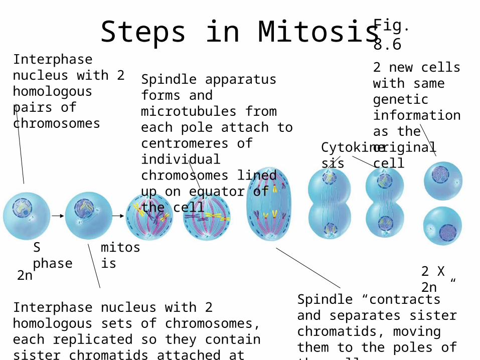
Steps in MitosisInterphase nucleus with 2 homologous pairs of chromosomes
S phase
Interphase nucleus with 2 homologous sets of chromosomes, each replicated so they contain sister chromatids attached at centromere
mitosis
Spindle apparatus forms and microtubules from each pole attach to centromeres of individual chromosomes lined up on equator of the cell
Spindle “contracts” and separates sister chromatids, moving them to the poles of the cell
Cytokinesis
2 new cells with same genetic information as the original cell
2n 2 X 2n
Fig. 8.6

Steps in MeiosisNucleus at the end of interphase with 4 replicated chromosomes
Meiosis division 1
Synapsis = physical joining of homologous chromosomes as they move to the equator of the cell, microtubules from one pole of the spindle apparatus attach to the centromere of one member of each homologous pair
Spindle “contracts” and separates homologous chromosomes
cytokinesis
2n 2 X 1n, but with sister chromatids
4 X 1n
division 2 cytokinesis
Individual chromosomes move to equator
Spindle “contracts” and separates sister chromatids
4 new cells with ½ the genetic information of the original cell
Fig. 8.14
Steps in Development
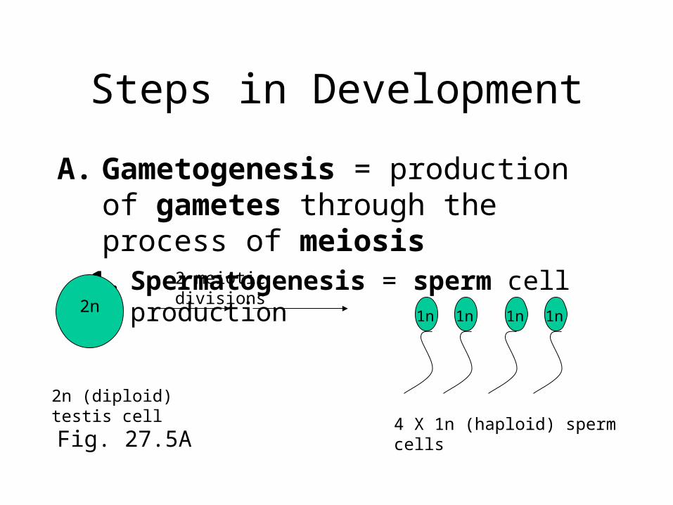
A. Gametogenesis = production of gametes through the process of meiosis
1. Spermatogenesis = sperm cell production
2n1n 1n 1n 1n
2 meiotic divisions
2n (diploid) testis cell
4 X 1n (haploid) sperm cellsFig. 27.5A
A. Gametogenesis - continued
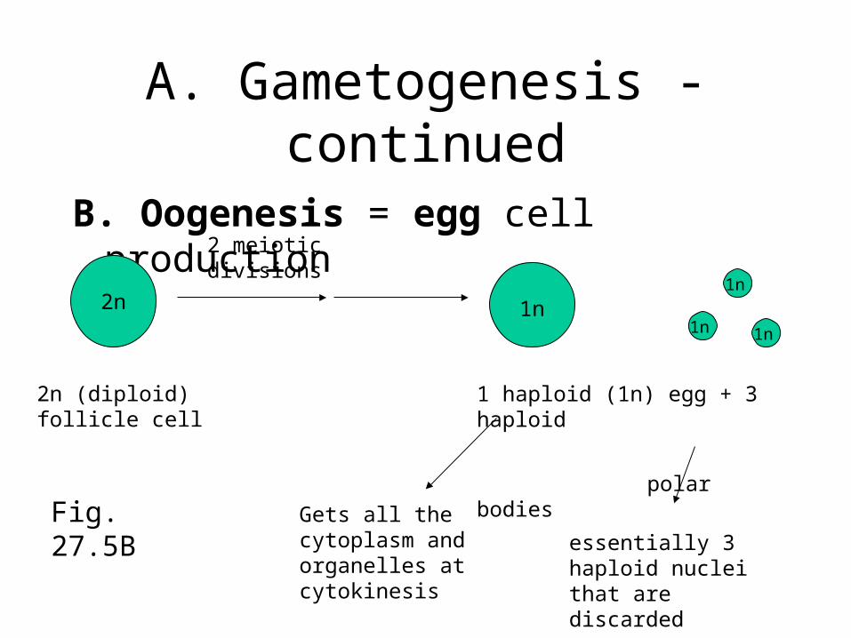
B. Oogenesis = egg cell production
2n 1n1n
1n 1n
2 meiotic divisions
2n (diploid) follicle cell 1 haploid (1n) egg + 3 haploid
polar bodies
Gets all the cytoplasm and organelles at cytokinesis
essentially 3 haploid nuclei that are discarded
Fig. 27.5B
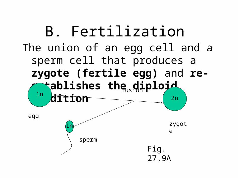
B. Fertilization The union of an egg cell and a sperm cell that
produces a zygote (fertile egg) and re-establishes the diploid condition
1n
1n
2nfusion
egg
sperm
zygote
Fig. 27.9A
C. Embryogenesis
embryo formation – consists of cleavage + gastrulation + organ formation
All three steps will make use of mitosis to produce new cells
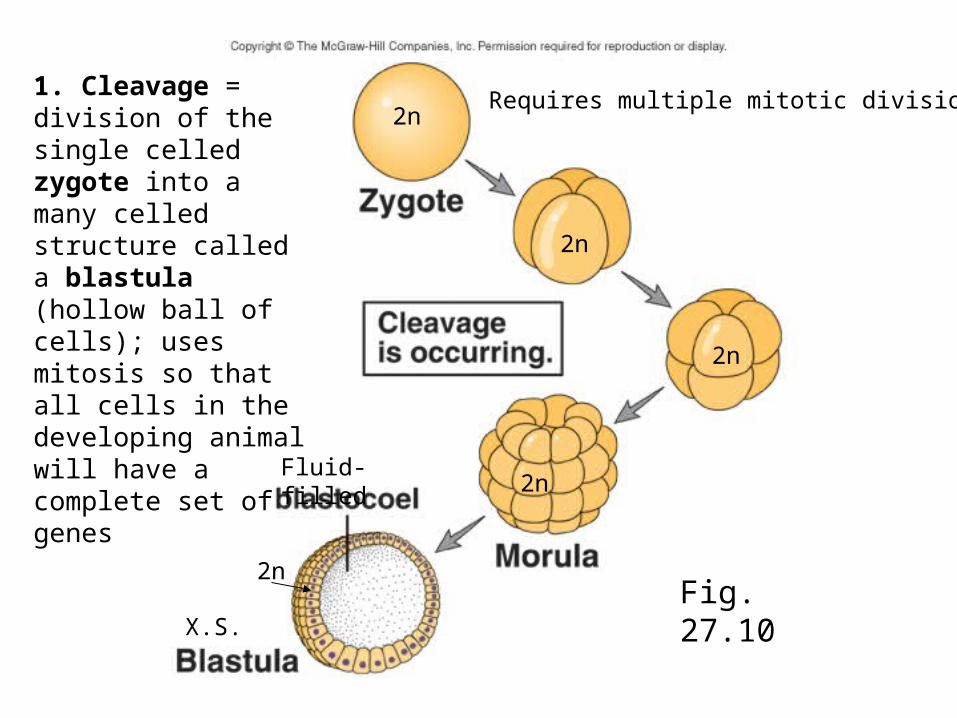
1. Cleavage = division of the single celled zygote into a many celled structure called a blastula (hollow ball of cells); uses mitosis so that all cells in the developing animal will have a complete set of genes
Requires multiple mitotic divisions2n
2n
2n
2n
2n
Fluid-filled
X.S.Fig. 27.10
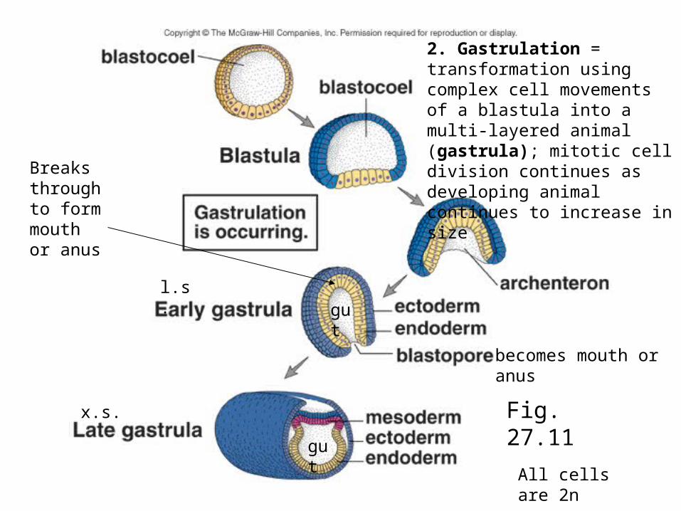
2. Gastrulation = transformation using complex cell movements of a blastula into a multi-layered animal (gastrula); mitotic cell division continues as developing animal continues to increase in size
All cells are 2n
l.s.
x.s.
becomes mouth or anus
Breaks through to form mouth or anus
gut
gut
Fig. 27.11
3. Organ Formation
Differentiation and specialization of embryonic tissues (endoderm, ectoderm and mesoderm) into adult tissues (epithelial, connective, muscle and nervous tissues) and organs
Final Steps in Development
D. Growth = increase in size; differentiation usually continues
E. Maturation = attainment of adult body form capable of reproduction
metamorphosis as an example of a dramatic maturation process