determinationt of endotoxin in injectable antibioticpreparations by
TRANSCRIPT

Vol. 23, No. 1JOURNAL OF CLINICAL MICROBIOLOGY, Jan. 1986. p. 11-160095-1137/186/010011-06$02.00/0Copyright ©O 1986, American Society for Microbiology
Determinationt of Endotoxin in Injectable Antibiotic Preparations bythe Chromogenic Assay Method Using a Limulus Reagent
(Tachypleus Hemocyte Lysate) and a Chromogenic SubstrateSHIGEO YANO,* YAStJKO HOTTA, AND SAKIKO TAKAHASHI
Department of Antibiotics, National Institiute of Health, Kainiosaki, Shlinagawi a-kia, Tokyo 141, Japan
Received 11 March 1985/Accepted 28 September 1985
The effects of 50 antibiotics on the detection and determination of bacterial endotoxins by the chromogenicmethod using a Limulus reagent (Tachypleus hemocyte lysate) and a chromogenic substrate of p-nitroanilinederivatives were tested, and the antibiotic concentration for 50% inhibition of the chromogenic reaction in thepresence of 0.5 ng of endotoxin (Escherichia coli 0111 :B4) per ml was estimated. All the antibiotic preparationswere depyrogenized by ultrafiltration treatment before they were subjected to the test. The reaction was
conducted in the presence of a high concentration (0.5 M) of Tris buffer to constantly maintain the pH of thereaction mixture, and liberated p-nitroaniline was determined by high-pressure liquid chromatography.Several aminoglycosides (amikacin, bekanamycin, kanamycin, and streptomycin sulfate), bleomycin hydro-chloride, and fosfomycin disodium showed no inhibition of the reaction up to 20 mg/ml. However, otherantibiotics, including penicillins, cephalosporins, macrolides, and tetracyclines, inhibited the reaction concen-
tration dependently. Polymyxin B sulfate was the most potent inhibitor, with less than 8 ,ug/ml for 50%inhibition. It was concluded that the chromogenic method can be applied to the detection and determination ofendotoxin in most of the antibiotic preparations. An application of this method to carbenicillin disodiumpreparations was exemplified.
Because of high sensitivity of rabbits to pyrogen (4). allinjectable antibiotic preparations have been subjected to thepyrogen test by rabbits specified in the pharmacopeias (7,23). Although the pyrogen test is troublesome and expen-sive, it has been shown to have adequate reliability except ina rare case (19) in which a trace amount of pytogen contam-ination could not be detected by the test. The most activepyrogen is a bacterial endotoxin, lipopolysaccharide (10)from gram-negative bacteria, Which has high molecularweights. In recent years, the gelation method (1, 11, 16) andthe chromogenic method (3, 6), which use the Limululisamoebocyte lysate (LAL) of a horseshoe crab to detect invitro trace amounts of bacterial endotoxin, have becomeavailable. Recently it was reported that a polysaccharide,(1-3)-3-D-glucan (8, 12), activates the LAL reagent asstrongly as does endotoxin, but it is not so common asendotoxinl, and its contamination in antibiotic preparations isunlikely. The gelation method, the so-called LAL test, issimple and semiquantitative (22). On the other hand, thechrormogenic method is more accurate in determiningendotoxin activity by measuring the concentration of p-nitroaniline (PNA) which is enzymatically liberated from thechromrogenic substrate; this enzymatic hydrolysis proceedsin proportion to the amount of endotoxin present.
In the present study, we tested the effect of 50 antibioticpreparations on the chromogenic reaction. The results willbe of importance when the chromogenic method is applied tothe determination of endotoxin contaminating the prepara-tions.
Before the test, we determined the optimal pH for enzy-matic reaction and the buffer concentration that wouldprevent a pH change by antibiotics. Furthermore. someantibiotics interfere with the spectrophotometric determina-tion of the liberated PNA and we therefore used a high-pres-
* Corresponding author.
sute liquid chromatography (HPLC) niethod to isolate andquantitate the liberated PNA from the reaction mixtures. Inaddition, we used ultrafiltration treatment (18) of all theantibiotic preparations before the test to remove traceamounts of possibly contaminated endoioxin.
MATERIALS AND METHODS
Glassware and water. All glass pipettes and test tubes weredepyrogenized at 250°C for 2 h. The water used in this studywas the sterile water for injection recommended by theJapanese Pharmacopeia (7).
Antibiotics. Commercial, injectable antibiotic preparationscertified suitable according to the Japanese Minimum Re-quirements for Antibiotics (Ministry of Health and Welfare)were used. Antibiotic preparations in dry form were recon-stituted with water. All the antibiotic solutions were of thehighest concentrations and received ultrafiltration treatment.Antibiotic concentration was represented by antibacterialpotency.
Endotoxin. Commercially available endotoxin (1 ,ug) fromEscherichia coli O111:B4, obtained from Seikagaku KogyoCo. Ltd., Tokyo, Japan. was reconstituted with 10 ml ofsterile physiological saline (0.9% [wt/vol]) at room tempera-ture with vigorous, intermittent mixing for 5 min with avortex mixer and sonicating for 3 min and diluted to therequired concentrations, providing control endotoxin solu-tions. Each solution was sonicated again to prevent adsorp-tion of endotoxin on a test tube before use. Endotoxinactivity of the control solutions was almost constant for 6 hat room temperature.Reagent for the chromogenic method. Pyrodick, the am-
poule kit of the chromogenic reagent composed of hemocytelysate from the Japanese horseshoe crab, Tachvpleiustriidentatius, that has the same biochemical properties asLAL (15) and a chromogenic oligopeptide substate (tert-butoxycarbonyl-Leu-Gly-Arg-p-nitroanilide) was obtained
11

12 YANO ET AL.
1.2Ec
ciDCn
0U).40
09
0.6c
0.3 1
0.050 6040
Incubation Time (min)FIG. 1. Time course of the chromogenic reaction in the presence
of 0.5 rig of endotoxin (E. coli O111:B4) per ml with Tris buffer (pH8.0) of various concentrations at 37°C. Symbols: *, 0.05 M; 0, 0.25M; *, 0.5 M; A, 0.75 M; A, 1.0 M.
from Seikagaku Kogyo Co. Ltd. The same lot of reagent wasused,in a serial inhibition test.
Tris buffer. Tris hydrochloride solutions of various con-centrations containing 10 mM 0gCl2 were made with adjust-ment to the required pH values at 22°C with 4 N HCI. Eachbuffer was stored in sealed ampoules after ultrafiltration.Endotoxin activity in buffers without ultrafiltration treat-ment could not be abolished completely even at 120°C for 6h.Measurement of endotoxin. Ultrafiltration treatment was
done by using a Diaflo-cell (Amicon Far East Ltd., Tokyo,Japan) of the stirring type (model 12) and a Diaflo-membrane(PM 10; Amicon) with a fraction molecular weight of 10,000and a diameter of 25 mm; the surface of the membrane waswashed with water after it was soaked in 0.1 N NaOHovernight. After the cell was assembled, 10 ml of water wascharged and filtered under the pressure of nitrogen gas (3kg/cm2). This procedure was repeated until no alkali in thefiltrate was detected by a pH test paper. Then 1 ml of thecontrol endotoxin solution (2 nglml) was charged and fil-tered; the filtrate should contain less than 0.05 ng ofendotoxin per ml. After this was confirmed, Tris buffers andantibiotic solutions were ultrafiltered; 2 ml of the firstfraction of the filtrate was discarded, and the followingfractions were collected; test solutions were made by mixingcontrol endotoxin solutions with these antibiotic solutions.The contents ini each Pyrodick ampoule was reconstituted
with 0.1 ml of Tris buffer in an ice-water bath, and then 0.1ml of either a test solution or a control endotoxin solutionwas added to each ampoule. The ampoules were swirledgently by a vortex mixer and placed in a water bath at 37°Cfor the required period and cooled rapidly again in theice-water bath. A 1-ml sample of aqueous acetic acid (0.6 M)was added to each mixture to stop the reaction. Whenturbidity or a precipitate was present, the acetic solution waspassed through a Millipore membrane filter (HA; 0.45-,umpore size). In the inhibition test, twofold, serial dilutions(onefold to eightfold dilution) of the antibiotic solution of thehighest concentration were made, as test solutions, bydiluting the antibiotic solution with control endotoxin solu-tions under conditions such that each test solution contained
1 ng of endotoxin per ml, and five experiments for four serialtest solutions and the control endotoxin solution (1 ng/ml)were always performed in parallel.
Liberated PNA in the acetic solution was assayed byHPLC with a Waters Associates model 6000A solvent deliv-ery system and Lambda Max 480 LC spectrophotometer.The analysis was performed on a Nucleosil SC18 column (4by 150 mm; Macherey-Nagel Co.).One of the three eluent systems (A, water-methanol [2:1];
B, water-acetonitrile [1:1]; C, water-dioxane [4:1]) was ap-plied (see Table 1 for the choice of system). A 5-,u1 sampleof the acetic solution was injected, and elution in 1 'ml/minwas monitored at 405 nm. The inhibition by antibiotics or theamount of endotoxin was determined from the ratio of thepeak height of the PNA in the acetic solution to that in thecontrol PNA solution. The absorbancy of the PNA in theacetic solution was measured at 405 nm in 1-cm cuvettes(MT4; Carl Zeiss Co.) with a Carl Zeiss PQII spectropho-tometer.Determination of endotoxin in injectable carbenicillin
disodium preparatioiis. Six carbenicillin disodium prepara-tions (1 g per vial) of three lots from the same batch wereused as test samples which were positive in the rabbit test bythe procedure of the Japanese Minimum Requirements forAntibiotics (100 mg/ml per kg of body weight). Three sam-ples from two lots which were negative in the test were usedas controls. These preparations were made by the samemanufacturer. Each sample was reconstituted with 5 ml ofwater. These solutions were further diluted 10-fold and wereused as the test solutions; they contained about 17.5 mg/ml,close to twice the antibiotic concentration for 50% inhibition(IC50; 8 mg/ml). The liberated PNA was assayed by HPLC,and the amounts of endotoxin were estimated from a cali-bration curve (0 to 1 ng/ml) of the same antibiotic concen-tration as in the test solutions; the antibiotic solution, afterultrafiltration treatment, was used for the calibration curve.
RESULTSThe time course of the chromogenic reaction in the
presence of 0.5 ng of endotoxin per ml and various concen-trations of Tris buffer (pH 8.0) is shown in Fig. 1. From theseexperimental data and other preliminary data indicating that
(U)
cr
7.0 8.0 9.0p H
FIG. 2. Effect of Tris buffer pH on the enzymatic activity in thechromogenic reaction at 37°C for 50 tnin in the presence of 0.5 ng ofendotoxin (E. coli O111:B4) per ml. Tris buffers (pH 6.2 to 9.0) wereadjusted to a 0.5-M concentration.
Ar- a I
J. CLIN. MICROBIOL.
1

ENDOTOXIN DETERMINATION BY CHROMOGENIC ASSAY
1000-0
1-
4-.C
0E
z
0~
4.,
80
60
40
20
00 0.5 1 1.5 2 2.5
Endotoxin ( ng/ ml)FIG. 3. Calibration curves of endotoxin (E. coli O111-B4) con-
centration in the chromogenic reaction at 37°C for 50 min with orwithout carbenicillin disodium with 0.5 M Tris buffer (pH 8.1).Concentration of carbenicillin disodium: 0, 0 mg/ml; 0,5 mg/ml; A,10 mg/ml. The percentage of the liberated PNA obtained by theHPLC method was the same as that obtained by the spectrophoto-metric method.
0.5 M and greater concentrations of Tris buffer were re-quired to maintain pH constantly in our inhibition test, 0.5 MTris buffer and an incubation time of 50 min were chosen forour experiments.The effect of various pHs of 0.5 M Tris buffer on the
enzymatic reaction is shown in Fig. 2, giving the optimal pHas 8.2. The pH value of 8.1 + 0.1 was chosen for 0.5 M Trisbuffer in the following chromogenic reaction.
The linear relationship between amounts of endotoxin andliberated PNA was determined in the presence or absence oftwo different concentrations (5 and 10 mg/ml) of carbenicillindisodium (Fig. 3). The calibration curves for a control andthe experimental groups had a good linearity below 1.5 ng ofendotoxin per ml; however, after more than 70 to 80% of thePNA was liberated, the two curves (0 and 5 mg/ml) tended tolevel off. Also, carbenicillin disodium inhibited thechromogenic reaction concentration dependently (Fig. 3).Because the three calibration curves gave linearity withendotoxin concentrations between 0.5 and 1.5 ng/ml, 0.5 ngof endotoxin per ml was used for the subsequent inhibitiontest.
Typical examples of results of the inhibition test withcloxacillin sodium, kanamycin sulfate, doxycycline hyclate,and cefotaxime sodium, each from one of the four mainantibiotic groups, are shown in Fig. 4. Two inhibition curvesobtained by two different analytical methods (the HPLC andspectrophotometric methods) were similar in cloxacillin so-dium and kanamycin sulfate, indicating either method can beused for the assay. On the other hand, a yellow substance,doxycycline hyclate of the tetracyclines, and other coloredantibiotics, such as aclarubicin hydrochloride, should beapplied to the HPLC method, owing to their intense spec-trophotometric disturbance. The same result for tetracyclinewas reported previously (H. Usami and K. Akiyama, Abstr.Annu. Meet. Jpn. Pharm. Soc. 1982, 102, p. 654). However,cefotaxime sodium, which is a colorless antibiotic as arecloxacillin sodium and kanamycin sulfate, gave differentinhibition curves in the two assay methods; the false-inhibition curve in the spectrophotometric method could,possibly, have been due to color development by decompo-sition of the cefotaxime sodium itself during the chro-mogenic reaction. Color development which was not neces-sarily proportional to antibiotic concentration was observedin most of the cephalosporins tested and less frequently in
Cloxacillin Kanamycin Doxycycli ne CefotaximeI+ 0.1 sodium sulfate hyclate sodium
0.0- 0 1WIq
-0.1
100
50 -
Ia0
0.625 1.25 2.5 5 2.5 5 10 20 0.250.5 1 2 2.5 5 10 20
Antibiotic Concentration (mg/mi)FIG. 4. Effects of antibiotics on the chromogenic reaction in the presence of 0.5 ng of endotoxin (E. coli O111:B4) per ml at 370C for 50
min with 0.5 M Tris buffer (pH 8.1). Symbols: 0 and 0, percentage of relative activity obtained by the HPLC and spectrophotometric method,
respectively; 0, pH changes of 0.5 M Tris buffer (pH 8.1).
0
-
4-
.)
Cu
a)
4-
(a
VOL. 23, 1986 13
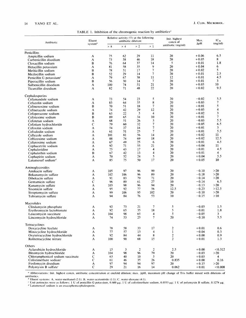
TABLE 1. Inhibition of the chromogenic reaction by antibiotics'
Relative activity (9/) at the following Init. highest... ~~~~~Eluent antibiotic dilution: Max. IC5,,Antibiotic system" aniitcdlto:concn of p(M/l
x 8 x 4 x 2 x 1 antibiotic (mg/ml) ApH (mgmi)
PenicillinsAmpicillin sodiumCarbenicillin disodiumCloxacillin sodiumHetacillin potassiumMethicillin sodiumMeziocillin sodiumPenicillin G potassium"Piperacillin sodiumSulbenicillin disodiumTicarcillin disodium
CephalosporinsCefamandole sodiumCefazolin sodiumCefmenoxime sodiumCefmetazole sodiumCefoperazone sodiumCefotaxime sodiumCefotetan sodiumCefotiam hydrochlorideCefoxitin sodiumCefsulodin sodiumCeftezole sodiumCeftizoxime sodiumCefuroxime sodiumCephacetrile sodiumCephaloridineCephalothin sodiumCephapirin sodiumLatamoxef sodium"
AminoglycosidesAmikacin sulfateBekanamycin sulfateDibekacin sulfateGentamicin sulfateKanamycin sulfateSisomicin sulfateStreptomycin sulfateTobramycin sulfate
MacrolidesClindamycin phosphateErythromycin lactobionateLeucomycin succinateLincomycin hydrochloride
TetracyclinesDoxycycline hyclateMinocycline hydrochlorideOxytetracycline hydrochlorideRolitetracycline nitrate
OthersAclarubicin hydrochlorideBleomycin hydrochlorideChloramphenicol sodium succinateColistimethate sodium'Fosfomycin disodiumPolymyxin B sulfate'
A 75 62 29 11 20A 73 58 46 18 20B 76 64 37 14 5A 81 58 35 9 20B 78 51 17 7 20B 52 29 14 7 20A 79 67 38 11 12B 56 30 14 7 20A 100 74 51 21 20A 82 71 48 22 20
A 73 54 23 5 20A 83 64 35 8 20B 70 51 18 7 20A 74 42 29 12 20B 63 22 5 4 20B 89 65 34 10 20A 68 51 26 5 20C 79 60 32 7 20A 54 37 14 7 20A 61 51 25 7 20A 101 81 56 14 20A 88 74 69 18 20A 67 49 25 6 20A 92 71 55 21 20A 75 43 17 4 20B 67 42 13 4 20A 70 52 24 5 20A 85 75 50 17 20
A 105 97 96 99 20A 102 106 96 89 20A 91 83 79 71 20C 96 89 61 27 10A 103 98 96 94 20A 95 92 77 56 12.5A 99 106 95 102 20A 94 84 76 53 10
A 92 73 21 7A 92 65 35 18A 104 98 65 4A 74 53 25 7
A 78 58 33 17A 77 57 13 4A 92 84 44 8A 10( 90 68 15
555
20
212
A 15 3 2 2 2.5A 104 98 96 79 20C 63 40 10 5 20C 61 46 35 26 0.835A 97 94 94 97 20C 29 21 16 14 0.062
+0.06+ 0.05+ 0.01+0.04+ 0.02+ 0.01+ 0.01+ 0.01+ 0.03+ 0.02
-0.02+ 0.03+ 0.01+ 0.03+ 0.02+ 0.01-0.03-0.05+ 0.01+ 0.01+ 0.02+ 0.02+ 0.01+0.04-0.01+ 0.01+ 0.04+ 0.05
-0.10-0.18-0.14-0.14-0.13-0.23-0.10-0.15
-0.03-0.01-0.05-0.10
+ 0.01-0.04+ 0.00+ 0.01
+ 0.00-0.03+ 0.03+ 0.00+ 0.15+ 0.01
6.581.86S2.54.53
109.5
5.57S4375.56.535.5
1112.54.5
114.545.510
>20>20>20
6.5>20>12.5>20>10
1.51.835.5
0.60.30.91.3
<0.312>20
40.16
>20<0.008
" Abbreviations: Init. highest concn, antibiotic concentration at onefold dilution; max. ApH, maximum pH change of Tris buffer mixed with dilutions ofantibiotics.
' Eluent systems: A. water-methanol (2:1); B, water-acetonitrile (1:1); C, water-dioxane (4:1).Unit potencies were as follows: 1 U of penicillin G potassium. 0.600 jig; 1 U of colistimethate sodium, 0.0333 ,ug: 1 U of polymyxin B sulfate, 0.1274 ,ug.
d Latamoxcef sodium is an oxacephemcephalosporin.
J. CLIN. MICROBIOL.14 YANO ET AL.
ENDOTOXIN DETERMINATION BY CHROMOGENIC ASSAY
the penicillins. The aminoglycosides tested showed no coloreffect at all.Because the effect of pH changes on enzymatic activity,
within + 0.15 pH unit of the Tris buffer (pH 8.1), wasnegligible (Fig. 2), cloxacillin disodium, doxycycline hycl-ate, and cefotaxime sodium were found to give concentra-tion-dependent and pH-independent inhibition.A good correlation between the extent of inhibition and
the logarithm of antibiotic concentration was obtained in anumber of the antibiotics tested (Fig. 4).The results of the inhibition tests with various antibiotics
are summarized and the antibiotic concentration giving theIC50 is estimated in Table 1; the percentage of relativeactivity and the IC50 were variable to some extent, depend-ing on the lot of antibiotic preparations. The inhibition maybe clf.rsified as folows. (i) The antibiotic was noninhibitorywhen the IC50 was more than 20 mg/ml. (ii) The antibioticwas moderately inhibitory when the IC50 was in the range of5 to 20 mg/ml. (iii) The antibiotic was strongly inhibitorywhen the IC50 was less than 5 mg/ml. The two ,B-lactamantibiotic groups tested showed similar inhibition; the aver-ages and the standard deviations of the IC50s of the penicil-lins and the cephalosporins were 5.7 + 2.7 and 6.4 ± 2.8mg/ml, respectively. The aminoglycosides tested could bedivided clearly into noninhibitory and moderately inhibitorygroups. In particular, the sulfates of amikacin, bekanamycin,kanamycin, and streptomycin were noninhibitory up to 20mg/ml. In sisomicin sulfate, some effect of the p1I changecould be considered. Clindamycin phosphate inhibitednearly four times as strongly as lincomycin hydrochloride inwhich only a chloride atom of clindamycin was replaced bya hydroxy group. Polymyxin B sulfate was found to be thestrongest inhibitor among the antibiotics tested. The an-thracycline antibiotic, aclarubicin hydrochloride, showed anunlexpectedly strong inhibition. Strong inhibition was alsoobserved when precipitates were formed in the chromogenicreaction (e.g., clindamycin phosphate, leucomycin suc-cinate, and minocycline hydrochloride).The results of the determination of endotoxin contents in
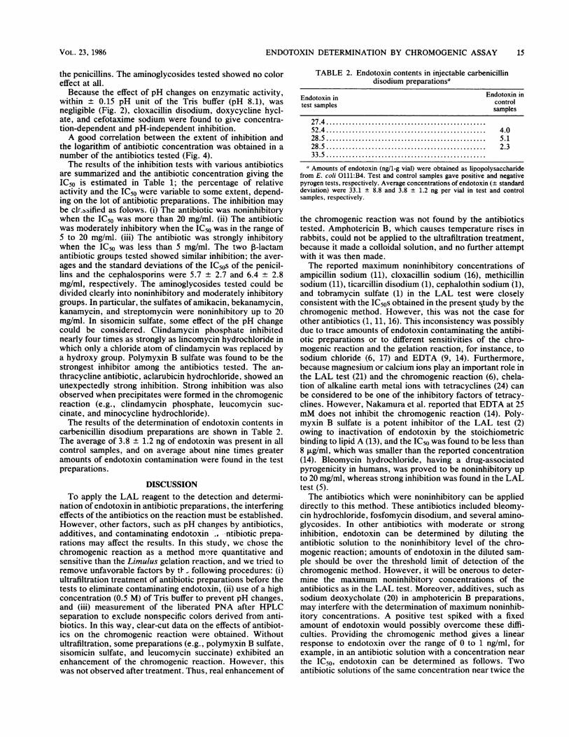
carbenicillin disodium preparations are shown in Table 2.The average of 3.8 ± 1.2 ng of endotoxin was present in allcontrol samples, and on average about nine times greateramounts of endotoxin contamination were found in the testpreparations.
DISCUSSIONTo apply the LAL reagent to the detection and determi-
nation of endotoxin in antibiotic preparations, the interferingeffects of the antibiotics on the reaction must be established.However, other factors, such as pH changes by antibiotics,additives, and contaminating endotoxin _. --ntibiotic prepa-rations may affect the results. In this study, we chose thechromogenic reaction as a method mo?re quantitative andsensitive than the Limulus gelation reaction, and we tried toremove unfavorable factors by ti following procedures: (i)ultrafiltration treatment of antibiotic preparations before thetests to eliminate contaminating endotoxin, (ii) use of a highconcentration (0.5 M) of Tris buffer to prevent pH changes,and (iii) measurement of the liberated PNA after HPLCseparation to exclude nonspecific colors derived from anti-biotics. In this way, clear-cut data on the effects of antibiot-ics on the chromogenic reaction were obtained. Withoutultrafiltration, some preparations (e.g., polymyxin B sulfate,sisomicin sulfate, and leucomycin succinate) exhibited anenhancement of the chromogenic reaction. However, thiswas not observed after treatment. Thus, real enhancement of
TABLE 2. Endotoxin contents in injectable carbenicillindisodium preparationsa
Endotoxin in Endotoxin intest samples controlsamples
27.4.52.4 ...................................... 4.028.5 ...................................... 5.128.5 ...................................... 2.333.5.
a Amounts of endotoxin (ng/1-g vial) were obtained as lipopolysaccharidefrom E. coli 0111:B4. Test and control samples gave positive and negativepyrogen tests, respectively. Average concentrations of endotoxin (+ standarddeviation) were 33.1 + 8.8 and 3.8 + 1.2 ng per vial in test and controlsamples, respectively.
the chromogenic reaction was not found by the antibioticstested. Amphotericin B, which causes temperature rises inrabbits, could not be applied to the ultrafiltration treatment,because it made a colloidal solution, and no further attemptwith it was then made.The reported maximum noninhibitory concentrations of
ampicillin sodium (11), cloxacillin sodium (16), methicillinsodium (11), ticarcillin disodium (1), cephalothin sodium (1),and tobramycin sulfate (1) in the LAL test were closelyconsistent with the IC50s obtained in the present s,tudy by thechromogenic method. However, this was not the case forother antibiotics (1, 11, 16). This inconsistency was possiblydue to trace amounts of endotoxin contaminating the antibi-otic preparations or to different sensitivities of the chro-mogenic reaction and the gelation reaction, for instance, tosodium chloride (6, 17) and EDTA (9, 14). Furthermore,because magnesium or calcium ions play an important role inthe LAL test (21) and the chromogenic reaction (6), chela-tion of alkaline earth metal ions with tetracyclines (24) canbe considered to be one of the inhibitory factors of tetracy-clines. However, Nakamura et al. reported that EDTA at 25mM does not inhibit the chromogenic reaction (14). Poly-myxin B sulfate is a potent inhibitor of the LAL test (2)owing to inactivation of endotoxin by the stoichiometricbindirig to lipid A (13), and the IC50 was found to be less than8 ,ug/ml, which was smaller than the reported concentration(14). Bleomycin hydrochloride, having a drug-associatedpyrogenicity in humans, was proved to be noninhibitory upto 20 mg/ml, whereas strong inhibition was found in the LALtest (5).The antibiotics which were noninhibitory can be applied
directly to this method. These antibiotics included bleomy-cin hydrochloride, fosfomycin disodium, and several amino-glycosides. In other antibiotics with moderate or stronginhibition, endotoxin can be determined by diluting theantibiotic solution to the noninhibitory level of the chro-mogenic reaction; amounts of endotoxin in the diluted sam-ple should be over the threshold limit of detection of thechromogenic method. However, it will be onerous to deter-mine the maximum noninhibitory concentrations of theantibiotics as in the LAL test. Moreover, additives, such assodium deoxycholate (20) in amphotericin B preparations,may interfere with the determination of maximum noninhib-itory concentrations. A positive test spiked with a fixedamount of endotoxin would possibly overcome these diffi-culties. Providing the chromogenic method gives a linearresponse to endotoxin over the range of 0 to 1 ng/ml, forexample, in an antibiotic solution with a concentration nearthe IC50, endotoxin can be determined as follows. Twoantibiotic solutions of the same concentration near twice the
VOL. 23, 1986 15

16 YANO ET AL.
IC50 are made, one of which is spiked in advance withendotoxin, containing 1 ng/ml. The spiked and unspikedantibiotic solutions are applied to the chromogenic method,giving a and b for PNA concentration, respectively. Theamount of contaminating endotoxin in the antibiotic solutionis calculated from the formula bl(a-b) ng/ml. Thus, thechromogenic method is found to be more applicable than thegelation method to the determination of endotoxin amountsin antibiotic preparations.
ACKNOWLEDGMENTWe thank S. Okamoto for many constructive comments and
suggestions during the course of this work.
LITERATURE CITED1. Case, M. J., S. S. Ryther, and T. J. Novitsky. 1983. Detection of
endotoxin in antibiotic solutions with Limulus amoebocytelysate. Antimicrob. Agents Chemother. 23:649-652.
2. Cooperstock, M. S. 1974. Inactivation of endotoxin by poly-myxin B. Antimicrob. Agents Chemother. 6:422-425.
3. Fujita, Y., and C. Nakahara. 1982. Preparation and applicationof a new endotoxin kit, Pyrodick, using a chromogenic sub-strate. Prog. Clin. Biol. Res. 93:173-182.
4. Greisman, S. E., and R. B. Hornick. 1969. Comparativepyrogenic reactivity of rabbit and man to bacterial endotoxin.Proc. Soc. Exp. Biol. Med. 131:1154-1158.
5. Gutteridge, J. M. C., D. J. Shute, and J. W. Lightbown. 1980.Chromatographic separation of endotoxin and bleomycin forpyrogenicity testing. J. Biol. Stand. 8:15-21.
6. Harada-Suzuki, T., T. Morita, S. Iwanaga, S. Nakamura, and M.Niwa. 1982. Further studies on the chromogenic substrate assaymethod for bacterial endotoxins using horseshoe crab(Tachypleus tridentatus) hemocyte lysate. J. Biochem.92:793-800.
7. Japanese Pharmacopeia. 1981. Pyrogen test, p. 771-772. In Thepharmacopoeia of Japan, 10th ed., English version. Ministry ofHealth and Welfare, Tokyo.
8. Kakinuma, A., H. Asano, and Y. Sugino. 1981. Gelation ofLimulus amoebocyte lysate by antitumor (1-3)-j3-D-glucan.Biochem. Biophys. Res. Commun. 101:434-439.
9. Kobayashi, M., and M. Yamamoto. 1975. Studies on the gelationreaction of Limulus lysate (Pre-gel). IV. Effect of alkalin earthmetals and chelating agents on the gelation reaction of Limuluslysate. Yakugaku Zasshi 95:959-965.
10. Luederitz, 0. 1970. Recent results on the biochemistry of thecell wall lipopolysaccharides of Salmonella bacteria. Angew.
Chem. Int. Ed. Engl. 9:649-663.11. McCullough, K. Z., and S. A. Scolnick. 1976. Effect of semisyn-
thetic penicillins on the limulus lysate test. Antimicrob. AgentsChemother. 9:856-858.
12. Morita, T., S. Tanaka, T. Nakamura, and S. Iwanaga. 1981. Anew (1-3)-P-D-glucan-mediated coagulation pathway found inLimulus amebocytes. FEBS Lett. 129:318-321.
13. Morrison, D. C., and D. M. Jacobs. 1976. Binding of polymyxinB to the lipid A portion of bacterial lipopolysaccharides. Immu-nochemistry 13:813-818.
14. Nakamura, S., T. Morita, T. Harada-Suzuki, S. Iwanaga, K.Takahashi, and M. Niwa. 1982. A clotting enzyme associatedwith the hemolymph coagulation system of horseshoe crab(Tachypleus tridentatus): its purification and characterization.J. Biochem. 92:781-792.
15. Nakamura, S., T. Takagi, S. Iwanaga, M. Niwa, and K.Takahashi. 1976. A clottable protein (coagulogen) or horseshoecrab hemocytes: structural change of its polypeptide chainduring gel formation. J. Biochem. 80:649-652.
16. Newsome, P. M. 1977. Penicillins and the Limnilus amoebocytelysate test for endotoxin. J. Pharm. Pharmacol. 29:704-706.
17. Scheef, R. R., D. M. Kenney, and D. Shepro. 1979. Effect ofsodium chloride on Limulus amebocyte: inhibition of endotoxinactivation of procoagulate. Thromb. Haemostasis 41:329-336.
18. Sweadner, K. J., M. Forte, and L. L. Nelson. 1977. Filtrationremoval of endotoxin (pyrogens) in solution in different states ofaggregation. AppI. Environ. Microbiol. 34:382-385.
19. Takahashi, S., S. Yano, Y. Nagaoka, K. Kawamura, and S.Minami. 1983. A highly sensitive pyrogen test for antibiotics. I:Detection of trace amounts of endotoxin in injectable sodiumampicillin preparations. J. Pharm. Sci. 72:739-742.
20. Tarmina, D. F., K. C. Milner, E. Ribi, and J. A. Rudbach. 1968.Modification of selected host-reactive properties of endotoxinby treatment with sodium deoxycholate. J. Bacteriol.96:1611-1616.
21. Tsuji, K., and K. A. Steindler. 1983. Use of magnesium toincrease sensitivity of Limulu.s amoebocyte lysate for detectionof endotoxin. Appl. Environ. Microbiol. 45:1342-1350.
22. U.S. Pharmacopeia. 1985. Bacterial endotoxin test, p.1165-1167. In United States pharmacopeia, 21st ed. U.S.Pharmacopeial Convention. Inc. Mack Publishing Company,Easton, Pa.
23. U.S. Pharmacopeia. 1985. Pyrogen test, p. 1181-1182. In UnitedStates pharmacopeia, 21st ed. U.S. Pharmacopeial Convention,Inc. Mack Publishing Company, Easton, Pa.
24. Weinberg, E. D. 1957. The mutual effects of antimicrobialcompounds and metallic cations. Bacteriol. Rev. 21:46-68.
J. CLIN. MICROBIOL.