determination in regenerating tissue of dugesias dorotocephala
TRANSCRIPT
/ . Embryo/, exp. Morph. Vol. 33, 1, pp. 85-93, 1975 8 5
Printed in Great Britain
Determination in regenerating tissues of Dugesiadorotocephala: the influence of nerve cord grafts
By PAMELA J. SPERRY1 AND KRYSTYNA D. ANSEVIN2
From Department of Biology, Rice University, Houston
SUMMARYLateral fragments which contained no nerve cord were isolated from the postpharyngeal
body section of Dugesia dorotocephala and fused with nerve cord grafts soon after isolationand at daily intervals through 8 days of regeneration. Fragments fused soon after isolationformed 'headless' regenerates but had normal body proportions. If the lateral cordlessfragment was allowed to regenerate for 1 day or longer before fusion with the nerve cordfragment, the head always developed and the body proportions were normal. Therefore,head structures become determined in the lateral fragment within the first 24 h of regeneration;during this time these tissues can also respond to the head-inhibiting influence of the nervecord. The competence to form particular structures of the postcerebral body regions mustemerge after head-forming competence is lost, that is about 24h after isolation; however,it persists at least through the first 8 days of regeneration. Normal body proportions can beinduced by nerve cord grafts throughout the first 8 days of regeneration.
Lateral fragments fused at any time after isolation with another fragment containing nonerve cord developed head structures but failed to differentiate tissues of the postcerebralregions. This confirms that the nerve cord is responsible for inhibition of head structuresand induction of differentiation of body regions and normal body proportions.
INTRODUCTION
In a recent study (Sperry, Ansevin & Tittel, 1973), it was demonstrated thatin Dugesia dorotocephala, tissues other than the main nerve cord have aninherent potential to build head structures only. The nerve cord has an in-hibitory as well as an inductive function in regeneration: it inhibits transforma-tion of all available tissues into head structures and induces (directly orindirectly) the differentiation of tissues of the different body regions andthe formation of normal body proportions.
Earlier studies (Ansevin, 1969) showed that in the whole postpharyngeal bodysection (which contains two nerve cords) of the species Dugesia tigrina, headstructures are determined within several hours after isolation, but the posteriorregions of the body are not determined for several days. Also, in prepharyngealbody sections of Bdellocephala brunnea, the head becomes determined withinseveral hours after isolation (Teshirogi, 1963).
1 Author's address: Department of Cell Biology, Baylor College of Medicine, Houston,Texas 77025, USA.
2 Author's address: Department of Biology, Rice University, Houston, Texas 77001, USA.
86 P. J. SPERRY AND K. D. ANSEVIN
Control
Host lateral cordless fragmentHost lateral cordless fragment
Nerve cord graft Cordless graft
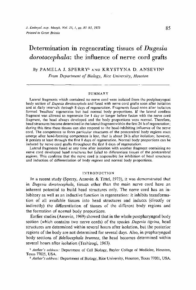
Fig. 1. Procedures for isolation of host and graft fragments and for grafting of anerve cord fragment or a cordless fragment (control) to a regenerating lateralcordless fragment. The anterior and posterior ends of the host cordless fragmenthave fused to form a rounded regenerate.
In the present investigation we have sought to define the period of time inregeneration during which the inhibitory and inductive actions of the nervecord are performed. It was assumed that for particular body tissues there is aperiod of competence of limited duration during which they can be directly orindirectly induced by the nerve cord to form particular body regions andstructures, or inhibited from forming some other structures. The goal of thisstudy was to define the periods of specific competences and thus also specifywhen determinations of particular body structures occur.
The technique of grafting was selected for the present study. Nerve cord wasgrafted to an isolated postpharyngeal lateral cordless fragment which had beenallowed to regenerate for varying periods of time. It was assumed that if, duringthe 'cordless' period of regeneration, the tissues of the lateral section lose thecompetence to be induced (or inhibited) to form a specific structure, subsequentgrafting of the nerve cord would fail to induce (or inhibit) this particular organor body part. Conversely, if the grafting of the nerve cord was performed duringthe stage of this particular competence, induction (or inhibition, as the casemight be) of that structure would occur.
METHODS AND MATERIALS
The procedures for maintaining our colony of Dugesia dorotocephala and formicrosurgical operations were described previously (Sperry et al. 1973).
A lateral cordless fragment was isolated, allowed to regenerate for specified

Nerve cord grafts in regenerating Dugesia 87
Muslincloth
Fragments
Fig. 2. A 'Schotte table,' used in these experiments to immobilize planarianfragments while fusion occurred.
periods of time, then a fragment containing nerve cord was grafted to theregenerating lateral fragment. The isolation and grafting procedures are shownin Fig. 1. For isolation of the 'host' lateral cordless fragment, the entire post-pharyngeal section was removed from a whole worm and cut into halves; fromeach half, the lateral fragment was cut away. The lateral fragments were placedin small culture dishes containing 1-5 ml of the solution of Shapira, Coleman &Castellani (1966) and incubated in the dark at 18 °C for 1-2 h, or for one througheight days. At these daily intervals, the 'graft' fragment was prepared fromanother intact worm: a half-postpharyngeal segment containing a single nervecord was isolated and the median area was removed.
The grafting procedure, based on that of Brondsted (1939), is as follows(Fig. 1): From a regenerating 'host' lateral fragment a small slice of tissue wascut away from its lateral side in order to create a wounded surface. The cordlessfragment and 'graft' fragment containing nerve cord were placed on a 'Schottetable' (Fig. 2), usually with the ventral surfaces facing upwards, pressed togetherat the wounded edges, and incubated in the dark at 10 °C for 12-18 h. At thistime excess tissue lateral to the nerve cord was removed so that from thatmoment on the 'graft' contained primarily nerve cord. The fused fragmentswere transferred to a culture dish and allowed to regenerate in the dark at18 °C. Observations were recorded and medium changed every 2 days for30-40 days.
For controls a lateral fragment containing no nerve cord was grafted to aregenerating lateral cordless fragment (Fig. 1). The figure shows that control andexperimental fragments were isolated from the same worm; in practice, how-ever, in most cases separate worms were used for the control series.
In these experiments, a 'Schotte table' (Br0ndsted, 1939; Fig. 2) was pre-pared by stretching a piece of muslin cloth across a plastic ring which was2-3 cm in diameter and 0-4 cm in height; a second ring of slightly smallerdiameter interlocked with the first ring to hold the cloth taut across the 'table'.The ' Schotte table' was placed in a small Petri dish and sufficient saline solutionwas added so that the cloth was well moistened. Maintenance of the fragmentson 'Schotte tables' and at low temperatures for the first hours after the opera-tion were necessary for the fragments to remain immobilized long enough forfusion to take place.
P. J. SPERRY AND K. D. ANSEVIN
7,

Nerve cord grafts in regenerating Dugesia 89
RESULTS
A representative graft of the host lateral cordless fragment and the nervecord fragment is shown in Fig. 3 at two days after fusion. The anterior andposterior ends of the host lateral fragment had fused prior to grafting to producea rounded regenerate.
Altogether 63 successful grafts were obtained in the experimental series and14 in the control group. Efforts were made to obtain a significant number ofsuccessful grafts at 1-2 h after isolation ('0 day') and a t ' 1 day' after isolation;thereafter, emphasis was placed on the even days of regeneration.
The following results are shown in Table 1.(A) In all cases in which the lateral cordless fragment was fused with the
nerve cord fragment within 24 h after isolation ('0 day'), 'headless' regeneratesdeveloped (types 1-3, Table 1; Fig. 4). Neither the host cordless fragment northe nerve cord fragment differentiated head structures, but normal body pro-portions were established.
FIGURES 3-8
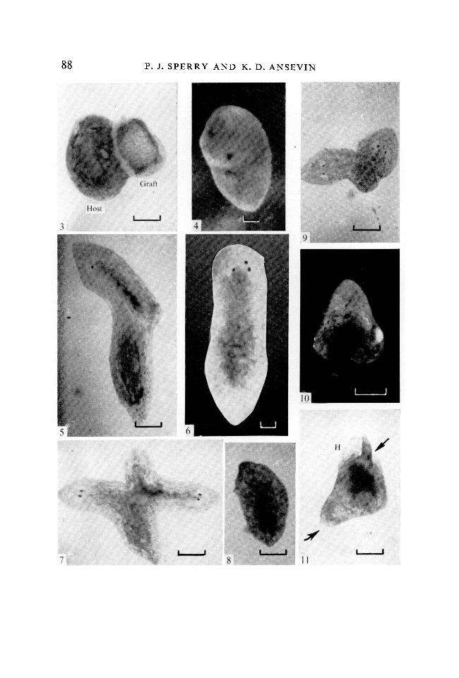
Types of regenerates formed as a result of a nerve cord fragment grafted to a lateralcordless fragment (scale line = 0-4 mm).Fig. 3. Representative graft of a nerve cord fragment to a host cordless fragment,shown 2 days after grafting, x 25.Fig. 4. A 'headless' regenerate (type 1), 68 days after grafting. The nerve cordfragment was grafted to the cordless fragment at '0 days' after isolation. (Fixedspecimen, x 50.)Fig. 5. Regenerate of normal body proportions with normal head structures(type 4), 14 days after grafting. The cordless fragment has formed the anteriorregions and the nerve cord graft has developed tissues of the posterior region.x25.
Fig. 6. Regenerate of normal body proportions with atypical head structures(type 5), 56 days after grafting, x 50.Fig. 7. A 'doublet' type regenerate (type 6), 40 days after grafting. Individual onthe left developed from the nerve cord graft fragment, x 25.Fig. 8. Regenerate of normal body proportions with atypical head structures(type 11), 40 days after grafting. Tissues on the right half of the body developedfrom the host cordless fragment and tissues on the left half are of nerve cordgraft origin, x 25.
FIGURES 9-11
Types of regenerates formed as a result of a cordless fragment grafted to a regener-ating lateral cordless fragment.Fig. 9. Regenerate in which host and graft fragments have developed as 'head-hump' regenerates of opposing antero-posterior polarity (type 14), 29 days aftergrafting. x25.Fig. 10. 'A head-hump' regenerate (type 16), 14 days after grafting. x25.Fig. .11. A 'head-hump' regenerate having one large but otherwise normal head('H') and two incomplete heads (arrows), type 18, 16 days after grafting, x 25.

Tab
le 1
. T
ypes
of
rege
nera
tes
form
ed
whe
n a
nerv
e co
rd fr
agm
ent
or a
cor
dles
s fra
gmen
t w
as g
raft
ed t
o a
cord
less
lat
eral
frag
men
t at
var
ious
sta
ges
of r
egen
erat
ion
Eac
h bl
ock
repr
esen
ts o
ne r
egen
erat
e. S
hade
d re
gion
s re
pres
ent
tissu
es w
hich
dev
elop
ed f
rom
the
ner
ve c
ord
graf
t.
Typ
es o
f re
gene
rate
s
10
11
12N
umbe
r of
day
sla
tera
l fr
agm
ent
had
rege
nera
ted
whe
n gr
aft
•atta
ched
Oda
y
1 da
y
2 da
ys
3 da
ys'
4 da
ys
5 da
ys
6 da
ys
7 da
ys
8 da
ys
Tot
alnu
mbe
r
13 17 10 1 15 2 6 5 8
II
III
IIIII
I ••
•
II III
III II
II
Gra
ft o
f co
rdle
ss f
ragm
ent
14
15
16
17
18
I I
II
II II
I I I
IIC/3
Nerve cord grafts in regenerating Dugesia 91
(B) When the lateral cordless fragment was allowed to regenerate for oneday or longer before fusion with a nerve cord fragment, the resulting regeneratesformed head structures and normal body proportions (types 4-13, Table 1).
(1) The most common type of regenerate, found in all groups except' 3 day',was one in which the cordless fragment formed the anterior region while thegraft gave rise to the posterior region (types 4 and 5, Table 1; Figs. 5 and 6).Some of these regenerates formed atypical head structures: three eyes appearedbut only one auricle developed (type 5, Table 1, Fig. 6).
(2) 'Doublet' type regenerates were found among the '1-4 day' group(types 6-13, Table 1). A few of these were bipolar: the host and graft fragmentsdeveloped anterior and posterior regions of opposing polarity (types 6 and 7,Table 1; Fig. 7). Some regenerates formed two anterior regions but had onetail (types 8 and 9, Table 1). Others fused to form a single head and tail inwhich the nerve cord graft developed tissues of one half of the body and thecordless fragment formed tissues of the other half (types 10 and 11, Table 1;Fig. 8). Some of these also developed atypical head structures (type 11, Fig. 8).
(3) Only in the '1 day' group a few regenerates were formed in which thecordless fragment was almost completely absorbed by the nerve cord fragment(type 12, Table 1) or the nerve cord graft almost fully incorporated into thecordless fragment (type 13).
(C) The control series gave uniform, distinct results. Lateral cordless frag-ments which at any time after isolation were fused with a fragment which had nonerve cord formed 'head-hump' regenerates: an oversized but otherwise normalhead regenerated, but normal body proportions failed to develop and the post-cerebral region formed only a 'hump' of undifferentiated tissues (types 14-18,Table 1). A few cases appeared in the control '1 day' group where host andgraft lateral fragments each formed 'head-hump' regenerates but of opposingpolarity (type 14, Table 1; Fig. 9) and in a few regenerates the host fragmentdeveloped as 'head-hump' but the graft formed only a 'hump' (type 15,Table 1). More often the fragments fused to develop a single 'head-hump'regenerate (types 16 and 17, Table 1; Fig. 10), a type found in all control groups.Head structures were incomplete in some cases: only one eye and one auricleformed (type 17). A few regenerates developed multiple head structures,resembling 'Janus-head' forms, with a single 'hump' (type 18, Table 1; Fig. 11).A regenerate of this type had one oversized but otherwise normal head('H', Fig. 11) and two atypical heads, each of which had two eyes but lackedauricles (arrows, Fig. 11).
DISCUSSION
The present study shows that induction of normal body proportions inDugesia dorotocephala can occur and is effective at any time through eight daysof regeneration, since nerve cord grafts fused with the lateral pieces as late asafter eight days of regeneration still prevented the 'head-hump syndrome'.Head differentiation could be inhibited only during the first 24 h of regeneration

92 P. J. SPERRY AND K. D. ANSEVIN
of a lateral postpharyngeal fragment. This we consider to be evidence that thecompetence of tissues to become determined as head structures lasts for lessthan the first 24 h of regeneration and that head determination is always ac-complished by 24 h or sooner. Thus, the 'non-head' competence (to form otherorgans of the body) must emerge close to the end of the first 24 h. However,it persists for at least the next 7 days since determination of specific organs andbody parts can be induced by nerve cord grafts in lateral pieces that wereregenerating in isolation for as long as eight days.
These results confirm the observations of Ansevin (1969) and Teshirogi (1963)which showed that head structures were determined in isolated body sectionswithin several hours after isolation, but that more posterior regions were deter-mined later in regeneration. The results also further confirm the gradual natureof determination of body tissues under the influence of the nerve cord (Sperryet al 1973).
The present study has also further confirmed that the inhibition of headstructures and the determination of the body tissues is specifically a function ofthe nerve cord. A fragment containing only branch nerves when fused with acordless fragment failed to inhibit head formation even if it was grafted soonafter isolation of the host fragment. In some cases where multiple head structuresdeveloped, head formation appeared to be stimulated under the influence ofthe cordless graft. Also, branch nerves did not induce development of tissuesof more posterior regions: postcerebral regions remained hump-like.
No attempt was made in the present study to control the antero-posteriororientation of the nerve cord graft with respect to that of the host cordlessfragment. By chance one would expect a 50:50 relationship between the numberof homopolar and heteropolar grafts and thus regenerates. However, only 4out of 63 regenerates in the experimental series were bipolar (types 6 and 7) andthese occurred only in the '2-4 day' groups. In all other cases the form of theregenerate indicated that the polarity of the nerve cord graft coincided with thepolarity of the host cordless fragment. The present results suggest that thepolarity of the final regenerate may be under the control of the differentiatingtissues of the host lateral cordless fragment and that the polarity becomesdetermined within the first four days after isolation. However, other possibleexplanations such as sampling error or greater survival of homopolar graftshave not been eliminated. The question of establishment of polarity should bepursued further before definite conclusions can be drawn.
The nature of the processes by which planarian tissues become determinedto form particular structures is unknown. The earlier and present findingssuggest that the information coded and released in each cell of the body (otherthan nerve) is to form head structures. Unless this message is altered in anotherdirection the information is expressed as head structures. The nerve cord playsa decisive role in altering the head-forming potential and inducing the develop-ment of other body tissues.
Nerve cord grafts in regenerating Dugesia 93
The authors express sincere thanks to Mrs Elga Lewis for diligent care of the worm colony.This work was submitted in partial fulfillment of the requirements for the Ph.D. degree
(P.J.S.).
REFERENCES
ANSEVIN, K. D. (1969). The influence of a head graft on regeneration of the isolated post-pharyngeal body section of Dugesia tigrina. J. exp. Zool. Ill, 235-248.
BRONDSTED, H. V. (1939). Regeneration in planarians investigated with a new transplanttechnique. K. danske Vidensk. Selsk. Bioll. Medd. 15, 1-39.
SHAPIRA, J., COLEMAN, J. O. & CASTELLANI, P. C. (1966). Differential nucleic acid metabolismof planarian segments. Proc. Soc. exp. Biol. Med. 122, 1266-1269.
SPERRY, P. J., ANSEVIN, K. D. & TITTEL, F. K. (1973). The inductive role of the nerve cordin regeneration of isolated postpharyngeal body sections of Dugesia dorotocephala.J. exp. Zool. 186, 159-174.
TESHIROGI, W. (1963). Transplantation experiments of two short pieces of a freshwaterplanarian, Bdellocephala brunnea. Jap. J. Zool. 14, 21-48.
{Received 23 April 1974)