determinants of ventricular defibrillation in adults -...
TRANSCRIPT

Determinants of Ventricular Defibrillation in AdultsJOSEPH A. GASCHO, M.D., RICHARD S. CRAMPTON, M.D., MICHAEL L. CHERWEK, M.D.,JAMES N. SIPES, M.D., FRANK P. HUNTER, B.E.E., AND WILLIAM M. O'BRIEN, M.D.
SUMMARY Conventional defibrillators which stored no more than 400 J and used damped sine wave pulsesdefibrillated 240 of 253 (95%) episodes of ventricular fibrillation (VF) in 94 prospectively assessedresuscitations in 88 adults. Shocks of 80-240 J (under 3 J/kg) delivered to the chest wall defibrillated moreoften than higher energy levels. Defibrillation rate did not correlate with weight. Defibrillation was determinedby the diagnosis and setting in which VF occurred. Patients with acute myocardial infarction (AMI) andprimary VF or with coronary disease and no AMI defibrillated more easily than patients with AMI and secon-dary VF or with no coronary disease. VF in a terminal patient (agonal VF) defibrillated less often than VF inother clinical situations. Age, weight, delivered energy, duration of pulse wave, and duration of VF had little, ifany, influence on rate of defibrillation. These data fail to support the use of more expensive, high-outputdefibrillators sold by 11 of 14 American manufacturers.
ALTHOUGH ELECTRIC SHOCK has defibrillatedhuman hearts since 1947,1 the threshold energy orcurrent for human ventricular defibrillation is con-troversial.2 A retrospective analysis of 178 patientsshowed an apparent inverse relation betweendefibrillation rate and weight.3 4 These investigatorsalso suggested that contemporary devices which storeno more than 400 J can defibrillate only half the pa-tients who weigh more than 80 kg. In contrast,prospective studies indicated no relation betweenweight and defibrillation rate.5- The recommendedenergy to defibrillate the ventricles ranges from200-400 J.6, 9-17
If heavy patients do fail to defibrillate after shocksfrom devices now in clinical use for nearly 2 decades,then more powerful devices should be manufactured.However, if no relation exists between weight and out-come of countershock, the admonition to use the"megawatt" defibrillator'8 may not only injure theheart,'9 but also preclude successful resuscitation.2We prospectively evaluated 94 resuscitative events
in 88 patients shocked for ventricular fibrillation (VF).We examined the relations among defibrillation,weight, delivered energy, pulse wave, duration of VF,clinical diagnosis, and setting in which VF occurred.
MethodsFrom September 1, 1975, resident physicians,
nurses in the coronary unit and cardiac ambulancetechnicians, instructed to attempt ventricular
From the Divisions of Cardiology, Biomedical Engineering andBiometrics, and the Emergency Medical Service, University ofVirginia Medical Center, Charlottesville, Virginia.
Supported in part by the Charles A. Frueauff Foundation, NewYork, New York, the Robert Wood Johnson Foundation Emergen-cy Medical Communications Network, project 1420, and theThomas Jefferson Emergency Medical Services System, DHEWproject 03-H-00, 366-02.
Presented at the 51st Scientific Sessions of the American HeartAssociation, November 15, 1978, Dallas, Texas.
Dr. Gascho's present address: Division of Cardiology, Universityof Iowa Hospitals and Clinics, Iowa City, Iowa 52242.
Address for reprints: Richard S. Crampton, M.D., 158 MedicalCenter, University of Virginia, Charlottesville, Virginia 22908.
Received July 28, 1978; revision accepted February 6, 1979.Circulation 60, No. 2, 1979.
defibrillation with 200 J or less, participated in 94resuscitative events in 88 patients in VF. Ventriculartachycardia and flutter were specifically excluded. Thestored energy of initial shocks was 200 J or less in 78of 94 (83%), and for all shocks, stored energy was 200J or less in 220 of 414 (53%). All but one of the shockswere delivered via conventional electrodes 8.0-8.8 cmin diameter, placed on the chest wall at the right uppersternal edge at the second interspace and over the car-diac apex at the left fifth interspace at themidclavicular and anterior axillary lines. During elec-tive cardioversion of atrial fibrillation with electrodesplaced in an anteroposterior orientation, one patientacquired VF and defibrillated with one shock. Elec-trode paste or saline-soaked gauze pads were usuallyplaced between the electrodes and skin.The clinical diagnosis, setting in which VF oc-
curred, age and weight of each patient were noted. Thepatients were grouped into four categories: 1) acutemyocardial infarction (AMI) and primary VF; 2)AMI and secondary VF; 3) coronary disease withoutAMI with either primary VF or secondary VF; and 4)all other patients. Myocardial infarction wasdiagnosed by typical serial, evolving Q, ST and Tchanges, elevation of cardiac enzymes, or freshnecrosis at necropsy. Coronary disease without infarc-tion was diagnosed by a history of ischemic chest pain,normal cardiac enyzmes, or absence of recent necrosisat necropsy. Primary and secondary VF were defined,respectively, as VF without or with left ventricularfailure. The latter diagnosis was made by detection ofa third sound gallop. VF which appeared secondarilyin patients with terminal illness or after suddencollapse without basic life support was designatedagonal VF. All other VF was defined as nonagonal.The number of episodes of VF and the number of
shocks for each episode and for all episodes of VFwere recorded for each resuscitative event. Eachresuscitative event included all episodes of VF that oc-curred within 2 hours of each other. If two episodes ofVF occurred more than 6 hours apart in the same per-son, they constituted, for analytic purposes, separateevents in separate patients. Thus, three patientsentered the study twice and one four times for VFepisodes 9, 12 and 15 hours and 1 and 5 weeks apart.
231
by guest on June 12, 2018http://circ.ahajournals.org/
Dow
nloaded from

VOL 60, No 2, AUGUST 1979
The outcome of each shock and of eachresuscitative event was noted. A shock was consideredsuccessful if VF was converted to any other rhythm.3 5The outcome of the resuscitative event was classifiedinto four categories: 1) failure of the shock at the lastattempt; 2) defibrillation by the last shock but deathlater in the resuscitative event; 3) survival of theresuscitative event but death before discharge; and 4)discharge from hospital alive with resumption of ac-tive life.
Twenty-six standard devices (Physio-Control Series70 and Lifepaks 33, 2 and 4; Hewlett-Packard 7802Band D and 780-02A; Cardiac Recorders IPCO-Pantridge 280; Zenith-Travenol Monopulse) delivered414 DC shocks. Four hundred eleven of 414 shocksused damped sine waves 5 or 12 msec in duration.Three shocks used the 8-msec pulse of the Zenith-Travenol device. The stored energy was read from themeter or dial of the device. The energy delivered bydischarging each device through a 50 Q test load(Dempsey tester) was measured at least three times ateach of five metered stored levels of energy of 50, 100,200, 300 and 400 J. The recorded energy delivered injoules at the selected stored energy was considered theenergy delivered to the patient.3 The stored anddelivered energy and the delivered energy per unitweight were recorded for each shock.
Because the success or failure of the initial shockmight influence selection of the energy level later inthe resuscitative event, and because deterioratingmetabolic conditions might deleteriously affect theoutcome of the VF episode, the outcome of all shocks,the outcome of the first shock of each VF episode, andthe outcome of the first shock of the resuscitativeevent were each noted. Success in terminating VF onthe first shock and success regardless of the number ofshocks required was tabulated. To remove the bias ofenergy level chosen after the outcome of the firstshock, an analysis of variance was used to examine thefirst shock of the first VF episode in the 74 patients inwhom the duration of VF was known.
All patients received standard basic and advancedcardiac life support.20 All with witnessed onset of VFreceived basic life support in less than 30 seconds if ashock failed.Whenever possible the duration of VF before the
first shock of the resuscitative event was noted. Itcould most often be determined accurately inmonitored patients. The duration of VF in patientswho collapsed outside the hospital was defined as timefrom collapse to first shock.
All data are presented as the mean ± SD. Thestatistical techniques used were Fisher's exact test(FET), chi-square test, analysis of variance and dis-criminant function (do).
Results
Ninety-four patients received 414 cross-chest DCshocks for 253 episodes of VF. Sixty-nine (73%) weremen and 25 were women; mean age was 60.5 ± 15.6years. They weighed from 40-225 kg (mean72.7 ± 20.6 kg); 12 (13%) weighed more than 90 kg.Two hundred forty of 253 (95%) episodes of VF con-verted to another rhythm, 190 (75%) on the first shock(fig. 1). Eleven (12%) patients were defibrillated out-side the hospital. All other resuscitative events beganor ended in the following areas: coronary care unit32, general hospital - 25, emergency department -14, other critical care units - 11, progressive cor-onary care unit - three, and cardiac catheterizationlaboratory - one. Events which began in onegeographical area and ended in another were assignedto both areas if ventricular defibrillation wasattempted in each area. Thus, there were 97 locationsfor the 94 resuscitative events.
Relation of Weight to Defibrillation
Figure 2 relates the frequency of defibrillation of the253 episodes of VF to weight. The success ofcountershock did not decline as weight increased.Twelve patients weighed more than 90 kg. Forty-five
FIGURE 1. Cumulative rate of defibrilla-tion of 253 episodes of ventricular fibrilla-tion. Mean defibrillating energy was196 ± 85 J, or 2.7 ± 1.4 J/kg. Three-fourths defibrillated with a mean first shock180 ± 74 J, or 2.5 ± 1.2 J/kg.
CIRCULATION232
by guest on June 12, 2018http://circ.ahajournals.org/
Dow
nloaded from

VENTRICULAR DEFIBRILLATION IN ADULTS/Gascho et al.
100
75 -
50 F-
25 -
.100-22540- 60- 70- 80- 90-59.9 69.9 79.9 89.9 99.9
Weight (Kilograms)FIGURE 2. Ventricular defibrillation rate vs weight.Weight did not determine rate of defibrillation.
of 46 (98%) episodes of VF defibrillated with 194 76delivered J or 1.8 .6 J/kg in these heavy people.
Relation of Delivered Energy to Defibrillation
The mean delivered defibrillating energy was
196 ± 85 J (2.7 1.4 J/kg) for all 240 episodesdefibrillated. The 190 episodes ended on the first shockdefibrillated with only 180 ± 74 J (2.5 ± 1.2 J/kg). In201 of the 253 episodes, defibrillation was attemptedwith shocks delivering 200 J or less. This energy levelsucceeded in 181 (90%) and in 160 (80%) with the firstattempt.
Figure 3 relates the outcome of the shock to thedelivered energy per kilogram. For all shocks (fig. 3A)there was a significant decline in defibrillation asdelivered energy increased above 3 J/kg (X2 = 49,df = 5, p < 0.05). Shocks which delivered under 2J/kg were not significantly less effective than shockswhich delivered 2-2.9 J/kg (x2 = 2.6, df = 1, p= 0.1 1).
Figure 4 relates the outcome of the shock to thetotal delivered energy. For all shocks for all episodesof VF, three groups of energy levels at or below 240 Jappeared equally effective (fig. 4A). In contrast,shocks delivering over 240 J failed more often(x2 = 163, df = 3, p < 0.0005).The lower success rate of shocks of higher energy
might result from many unsuccessful shocks at fulldevice output late in resuscitation in a minority ofpatients. To remove this possible bias, we examinedthe frequency of defibrillation by the first shock foreach episode of VF and of the first shock of the firstepisode of VF for each patient, to delivered energy perunit weight (figs. 3B and C) and energy (figs. 4B andC). Although shocks that delivered 2-2.9 J/kgappeared more successful than either lower or higherenergy levels, no significant trend was detected (figs.3B and C). There was no apparent optimal energylevel (figs. 4B and C).
Relation of Duration of VF to Defibrillation
The relation of duration of VF to defibrillation wasexamined in the 74 patients in whom the duration ofVF before the first shock of the resuscitative event wasknown. The mean duration ofVF was 175.6 ± 323 sec(range 2-1800 sec). The four episodes of VF which didnot defibrillate lasted 153 ± 97 sec and the 70defibrillated episodes lasted 176 ± 328 sec. Figure 5relates defibrillation to duration of VF in three
AALL SHOCKSALL EPISODES
2.0- 3.0- 4.0- 5.0- 6.0-1.9 2.9 3.9 4.9 5.9
BFIRST SHOCKSALL EPISODES
C
FIRST SHOCKSFIRST EPISODES
38
- 2.0- 3.0- 4.0 - 2.0- 3.0-1.9 2.9 3.9 1.9 2.9 4.9
Delivered Energy in Joules per KilogramFIGURE 3. Ventricular defibrillation rate vs delivered energy per unit weight. A) As delivered energy perunit weight exceeded 2.9 J/kg in 414 shocks in 253 ventricularfibrillation (VF) episodes, defibrillation rate
declined significantly. In first shocks of253 episodes (B) and in first shocks of94 first VF episodes (C), the
energy range of 2-2.9 J/kg seemed superior, but was not significantly so.
=0
._M
0i
.-
0
a
a-
100 r-
75 F rE'7-U
0
.0-00a
a-
50 F-
25 -
233
5634
by guest on June 12, 2018http://circ.ahajournals.org/
Dow
nloaded from

VOL 60, No 2, AUGUST 1979
BFIRST SHOCKSALL EPISODES
2,
-80 81- 161- 241- -80 81- 161- 241-160 240 160 240
cFIRST SHOCKSFIRST EPISODES
-80 81- 161- 241-160 240
Delivered Energy in JoulesFIGURE 4. Ventricular defibrillation rate vs total delivered energy. A) When total delivered energy ex-
ceeded 240 J in 414 shocks in 253 ventricular fibrillation (VVF) episodes, defibrillation rate fell significantly.B) No optimal energy level appeared in first shocks of 253 VF episodes. C) In first shocks of 94 first VFepisodes, no optimal energy level appeared.
100
36
1
11fO1 19 19
CL
0-30 31-120 >120
Duration ofVentricular Fibrillation
SecondsFIGURE 5. Ventricular defibrillation rate vs duration ofventricular fibrillation (VF) before first shock of firstepisode. The duration of VF did not determine rate ofdefibrillation.
LEFT HOSPITAL~~ALIVE
19.SURVIVED RESUS-CITATIVE EVENT,
......- DIED IN HOSPITAL.*..............
DEFIBRILLATED ON
LAST SHOCK, DIED
10 DURING RESUS-
19 CITATIVE EVENT
............... ....
NOT DEFIBRILLATEDON LAST SHOCK
AGONALVENTRICULARFIBRILLATION
I IS31
JON-AGONALVENTRICULARFIBRILLATION
N=19 N=75FIGURE 6. Outcome of resuscitative event: agonal vs non-
agonal ventricular fibrillation (VVF). The type of VF deter-mined the outcome of the resuscitative event. None withagonal VF lived to return home.
100 r AALL SHOCKSALL EPISODES
la
Uao.
.a
C10
75 I-
50 I-
25 M-
234 CIRCULATION
by guest on June 12, 2018http://circ.ahajournals.org/
Dow
nloaded from

VENTRICULAR DEFIBRILLATION IN ADULTS/Gascho et al.
DEFIBRILLATIONOF EPISODEBY FIRSTSHOCK
DEFIBRILLATIONOF EPISODEIRRESPECTIVEOF NUMBEROF SHOCKS
p< .0005 p< .00012 = 14.5 Fisher's
Agonal 2 Non-agonal
FIGURE 7. Ventricular defibrillation rate: agonal vs non-agonal ventricular fibrillation (VF). The type of VF deter-mined rate of defibrillation. Nonagonal VF defibrillatedsignificantly more often by a first shock or regardless of thenumber of shocks.
categories of time. The duration of VF did not appearto be a determinant of defibrillation. Of eight patientsin VF for 10-30 minutes before the first shock, alldefibrillated, six on the first shock.
DEFIBRILLATION OF DEFIBRILLATION OFEPISODE BY FIRST SHOCK EPISODE IRRESPECTIVE
OF NUMBER OF SHOCKS
100
tion of Type of VF to Defibrillation
ie setting in which VF occurred was related torillation (fig. 6). Nineteen of the 94 (20%) patients55 episodes of agonal VF. None left the hospitalIn contrast, 29 of 75 (39%) of patients with non-
al VF returned home. Ten of the 19 patients) with agonal VF did not defibrillate with'the lastk. In contrast, only three of 75 patients (4%) withigonal VF failed to defibrillate with the last shock.znts with nonagonal VF both defibrillated moreiently, and responded more frequently to the firstk (fig. 7). When first shocks delivered 200 J or137 of 164 (84%) episodes of nonagonal VF
)rillated more frequently than did 24 of 37 (65%)al episodes (p < 0.03). There was no difference inof defibrillation between patients with agonal andgonal VF when duration of VF was 30 seconds orHowever, patients with agonal VF defibrillatedoften at the first shock and also less oftenrdless of the number of shocks (fig. 8).
tion of Diagnosis to Defibrillation
ible 1 relates defibrillation to diagnosis. Eighty-resuscitative events took place in 82 medical
nts. Four patients had recently had a majoration: insertion of prosthetic cardiac valves inneurologic surgery in one and plastic surgery inTwo patients had injured chests from automobileLents.tients with AMI and primary VF and with cor-y disease and no AMI usually defibrillated at theshock and never failed to defibrillate. Three
.nts with AMI and secondary VF did notrillate. When attempted, under 200 J defibrillatedf 38 (97%) VF episodes with 33 (87%) defibrillatedone shock.tients without coronary disease defibrillatedy if VF was nonagonal. Nine of 12 such in-
FIGURE 8. Ventricular defibrillation rate:agonal vs nonagonal ventricularfibrillation(VF) <30- vs >30-second duration of VF.Type and duration ofVF together influencedrate ofdefibrillation in some groups. AgonalVF (hatched left-hand bars) for over 30seconds defibrillated significantly less oftenthan nonagonal VF (open right-hand bars)by afirst shock or regardless of the numberof shocks.
Duration of Ventricular Fibrillation (Seconds)
100
-o046-
.0
a
0Ub-0
0L.
75
50
25
la0
ah.S0U6Sa-
75
so
25
0-30 OVER 0-30 OVERSECONDS 30 SECONDS SECONDS 30 SECONDS
N.S. FISHER'S p <.03 N.S. FISHER'S p< .003
235
0% -% 0% -..l-
by guest on June 12, 2018http://circ.ahajournals.org/
Dow
nloaded from
VOL 60, No 2, AUGUST 1979
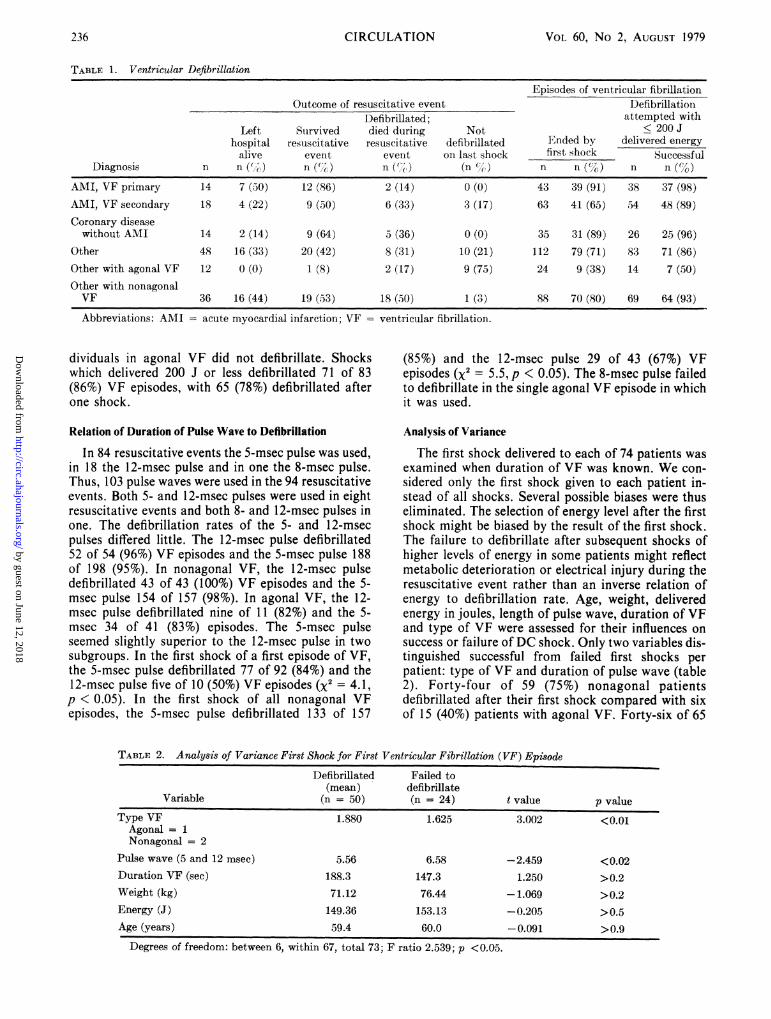
TABLE 1. Ventricular Defibrillation
Episodes of ventricular fibrillationOutcome of resuscitative event Defibrillation
Defibrillated; attempted withLeft Survived died during Not < 200 J
hospital resuscitative resuscitative defibrillated Ended by delivered energyalive event event on last shock first shock Successful
Diagnosis n n (') n (%) n (') (n () n n (%) n n (%)
AMI, VF primary 14 7 (50) 12 (86) 2 (14) 0 (0) 43 39 (91) 38 37 (98)AMI, VF secondary 18 4 (22) 9 (50) 6 (33) 3 (17) 63 41 (65) 54 48 (89)Coronary disease
without AMI 14 2 (14) 9 (64) 5 (36) 0 (0) 35 31 (89) 26 25 (96)Other 48 16 (33) 20 (42) 8 (31) 10 (21) 112 79 (71) 83 71 (86)Other with agonal VF 12 0 (0) 1 (8) 2 (17) 9 (75) 24 9 (38) 14 7 (50)Other with nonagonalVF 36 16 (44) 19 (53) 18 (50) 1 (3) 88 70 (80) 69 64 (93)
Abbreviations: AMI - acute myocardial infarction; VF = ventricular fibrillation.
dividuals in agonal VF did not defibrillate. Shockswhich delivered 200 J or less defibrillated 71 of 83(86%) VF episodes, with 65 (78%) defibrillated afterone shock.
Relation of Duration of Pulse Wave to Defibrillation
In 84 resuscitative events the 5-msec pulse was used,in 18 the 12-msec pulse and in one the 8-msec pulse.Thus, 103 pulse waves were used in the 94 resuscitativeevents. Both 5- and 12-msec pulses were used in eightresuscitative events and both 8- and 12-msec pulses inone. The defibrillation rates of the 5- and 12-msecpulses differed little. The 12-msec pulse defibrillated52 of 54 (96%) VF episodes and the 5-msec pulse 188of 198 (95%). In nonagonal VF, the 12-msec pulsedefibrillated 43 of 43 (100%) VF episodes and the 5-msec pulse 154 of 157 (98%). In agonal VF, the 12-msec pulse defibrillated nine of 11 (82%) and the 5-msec 34 of 41 (83%) episodes. The 5-msec pulseseemed slightly superior to the 12-msec pulse in twosubgroups. In the first shock of a first episode of VF,the 5-msec pulse defibrillated 77 of 92 (84%) and the12-msec pulse five of 10 (50%) VF episodes (x2 = 4.1,p < 0.05). In the first shock of all nonagonal VFepisodes, the 5-msec pulse defibrillated 133 of 157
(85%) and the 12-msec pulse 29 of 43 (67%) VFepisodes (X2 = 5.5, p < 0.05). The 8-msec pulse failedto defibrillate in the single agonal VF episode in whichit was used.
Analysis of Variance
The first shock delivered to each of 74 patients wasexamined when duration of VF was known. We con-sidered only the first shock given to each patient in-stead of all shocks. Several possible biases were thuseliminated. The selection of energy level after the firstshock might be biased by the result of the first shock.The failure to defibrillate after subsequent shocks ofhigher levels of energy in some patients might reflectmetabolic deterioration or electrical injury during theresuscitative event rather than an inverse relation ofenergy to defibrillation rate. Age, weight, deliveredenergy in joules, length of pulse wave, duration of VFand type of VF were assessed for their influences onsuccess or failure of DC shock. Only two variables dis-tinguished successful from failed first shocks perpatient: type of VF and duration of pulse wave (table2). Forty-four of 59 (75%) nonagonal patientsdefibrillated after their first shock compared with sixof 15 (40%) patients with agonal VF. Forty-six of 65
TABLE 2. Analysis of Variance First Shock for First Ventricular Fibrillation (VF) Episode
Defibrillated Failed to(mean) defibrillate
Variable (n = 50) (n = 24) t value p value
Type VF 1.880 1.625 3.002 <0.01Agonal = 1Nonagonal = 2
Pulse wave (5 and 12 msec) 5.56 6.58 -2.459 <0.02Duration VF (sec) 188.3 147.3 1.250 >0.2Weight (kg) 71.12 76.44 -1.069 >0.2Energy (J) 149.36 153.13 -0.205 > 0.5Age (years) 59.4 60.0 -0.091 >0.9
Degrees of freedom: between 6, within 67, total 73; F ratio 2.539; p <0.05.
236 CIRCULATION
by guest on June 12, 2018http://circ.ahajournals.org/
Dow
nloaded from

VENTRICULAR DEFIBRILLATION IN ADULTS/Gascho et al.
TABLE 3. Ventricular Defibrillation Patients Never Defibrillated
Highest energyCase Age not defibrillating Shocks VFno. (years) Diagnosis (J) (J/kg) (n) type
34 82 Septic shock 323 7.5 8 Agonal56 43 Chronic lymphocytic leukemia, 236 4.2 6 Agonal*
septic shock59 65 Lymphoma, pneumonia 260 4.1 4 Agonal60 63 Chronic obstructive pulmonary 319 6.3 3 Agonalt
disease, acute renal failure63 62 Subarachnoid hemorrhage 319 4.9 5 Agonal*77 42 Neurofibromatosis, 4 days 319 3.5 12 Nonagonal
after operationMean- SD 60=14 296 - 35 5.1 - 1.4 6.3 - 3.0
*VF preceded by electromechanical uncoupling.tVF preceded by asystole.Abbreviation: VF = ventricular fibrillation.
(71 %) pulse waves 5 msec long defibrillated, comparedwith four of nine (44%) pulse waves 12 msec long.
Patients Failing Defibrillation
Table 3 portrays clinical data of six individualsnever defibrillated. Five had agonal VF. In three, VFappeared only after onset of electromechanical un-coupling or asystole.
Table 4 depicts the clinical data of seven patientswho defibrillated early during the resuscitative effort,but who did not defibrillate during the last episode ofVF. Five had agonal VF. In three, VF appeared onlyafter resuscitation for electromechanical uncouplingor asystole. Three patients (cases 55, 83 and 93)received only one shock during the last episode of VF
because the responsible physicians felt that repetitivedefibrillation would neither prevent death nor restoresatisfactory quality of life. Three patients with non-agonal VF did not defibrillate with the last attemptedshock. Case 77, a 42-year-old man with disseminatedneurofibromatosis and its cardiomyopathy who died 4days after excision of brachial lesions, neverdefibrillated, despite 13 shocks (table 3). Case 72, a72-year-old man recovering from acute renal tubularnecrosis and pneumonitis from gram negative rods,had mild metabolic acidosis. After sudden onset ofatrial fibrillation with ventricular rate 200 beats/minand hypotension, he received 0.375 mg of digoxin i.v.Cardiopulmonary arrest due to asystole occurred.With basic life support and pharmacologic interven-tion,20 the asystole converted to VF, and the first
TABLE 4. Ventricular Defibrillation-Patients Defibrillated Early Who Failed to Defibrillate in the Last Episode of VentricularFibrillation (VF)
FinalEnergy episodeLowest defibrillating Highest not failed
Case Age earlier defibrillating shocks VFno. (years) Diagnosis (J) (J/kg) (J) (J/kg) n Type3 70 Stroke, acute myocardial
infarction, secondary VF 152 3.1 332 6.9 5 Agonal36 54 Myesthenia gravis, necrotizing
pneumonia 225 3.8 255 3.8 4 Agonal55 50 After prosthetic mitral and aortic
valve implant, pacemaker 167 2.8 342 5.7 1 Agonal72 72 Renal failure, acidemia,
digoxin treatment 155 2.6 319 5.4 11 Nonagonalt83 62 Cardiogenic shock, acute
myocardial infarction 185 2.4 95 1.2 1 Nonagonal*84 61 Acute myocardial infarction,
secondary VF 95 1.5 372 5.9 10 Agonal*93 91 Pneumonia, saddle embolus 155 2.3 319 4.6 1 Agonal
Mean SD 66= 13 162 - 36 2.6 - 0.7 291 - 86 4.8 - 1.7 4.7 4.0
*VF preceded by electromechanical uncoupling.tVF preceded by asystole.
237
by guest on June 12, 2018http://circ.ahajournals.org/
Dow
nloaded from

VOL 60, No 2, AUGUST 1979
shock, which delivered 155 J, defibrillated. When VFrecurred, 11 subsequent shocks, each delivering 319 J,failed to defibrillate (table 4). Case 83, a 62-year-oldman with AMI and left ventricular failure, suddenlydeveloped complete atrioventricular block with sub-sequent electromechanical uncoupling. After basic lifesupport and pharmacologic intervention,20 VFappeared, and each of four VF episodes terminatedwith a single 185-J shock. A fifth VF episode did notrespond to a single shock, and resuscitation was aban-doned (table 4).
DiscussionWe found no relation of weight of the patient to
defibrillation rate (fig. 2). Others have also failed tocorrelate weight and defibrillation rate.5 8, 21 In fourprospective studies,5 defibrillation rate did notdecrease with increase of weight. One retrospective in-vestigation21 revealed no difference in energy deliveredto defibrillated patients and those not defibrillated.
These reports5-8 21 and our results differ from aretrospective study which showed an apparentdecrease in defibrillation as weight increased.3 4 Thereason for this difference is unclear. In the retrospec-tive analysis suggesting that defibrillation ratedecreased with increase of weight, 40% of theattempted defibrillations in 111 patients occurred im-mediately after cardiac surgery.2 22 The diagnoses inthe remaining 60% are unknown. None of the patientsin the largest prospectively assessed group weredefibrillated immediately after cardiac surgery.5 6'8Only four patients in our study were postoperative,two cardiac and two noncardiac. It seems possible thatsurgical complications, metabolic derangement, oracute cardiac dysfunction might render defibrillationin the postoperative state difficult or impossible.Our data indicated no relation in the energy spec-
trum we used between defibrillation and deliveredenergy, whether expressed as joules or joules perkilogram. A trend appeared for a higher success ratewith 2.0-2.9 J/kg than with a higher or lower energylevels per unit weight (fig. 3). Moreover, when allshocks were analyzed, defibrillation rate decreased asenergy level rose above 3.0 J/kg (fig. 3A). This mayhave reflected multiple high-energy shocks given to aminority of patients failing earlier shocks. Never-theless, injurious effects of higher levels of electricalenergy cannot be excluded as a reason for our failureto defibrillate these patients.
In most of our patients' episodes of VF (201 of 253,80%), defibrillation was attempted with 200 J or less.One hundred eighty-one (90%) defibrillated at that lowenergy (table 1), and 160 of 201 (80%) defibrillated atthe first shock. A higher percentage might havedefibrillated at this energy level had the resuscitatorsnot increased energy after only one failed shock at 200J or less. In only four of 20 episodes failing defibrilla-tion at that energy was a second shock delivering 200 Jor less attempted. Others have reported high successrates with energy of 74-165 J,8 Several investigators
defibrillate,23 but only when "multiple shocks with lessenergy"24 are used in contrast to "a single shock ofslightly higher energy."23 Our data support none ofthese contentions.The lack of relation among defibrillation, weight,
energy, or energy per unit weight was not noted byother investigators.25 Ventricular defibrillationthresholds were determined for many animal speciesfrom the rabbit to the horse by lowering energy orcurrent in 10% decrements from the level of an ini-tially successful shock until the shock failed.25 A closerelation between weight and energy (E = 0.73 wt1'52;E = energy in joules, wt = animal weight inkilograms), and an even closer relation betweenweight and current (I = 1.87 wt088; I = current inamperes, wt = animal weight in kilograms), wasclaimed. If this equation were applied to man, onewould predict that devices storing only 400 J anddelivering only 300-340 J could not defibrillate heavypatients. Our data, which show high success rates evenwith shocks delivering 200 J or less, suggest that theseanimal data do not apply to defibrillation of man. Infact, the 100-kg calf in VF needed four to five timesmore energy to defibrillate, 862 J or 8.6 J/kg,26 thandid our patients in this weight range. Pigs27 andponies2' required even more energy, 958 J or 8.7 J/kg,and 1602 J or 10.6 J/kg, respectively, to defibrillate.Since animals whose weight was identical to ourheavier patients (91-225 kg) needed four to eighttimes as much energy to defibrillate, the extrapolationof energy levels to defibrillate calves, pigs and poniesto humans seems unwarranted, if not dangerous.The limitations of a similar formula (E = 1.88
wtl''5; E = energy in J, wt = patient weight in kg) forenergy requirements for human ventricular defibril-lation3 have been delineated.2 The data base for thehuman equation consisted of only 13 human defibril-lations (seven adult and six pediatric). In 10, defibril-lation failed at one energy level but occurred at a
higher energy level. This higher level was deemed thedefibrillation threshold energy.3 In three infants alsoincluded in the derivation of this formula, the initialshock was apparently successful.The human formula is questionable for several
reasons. First, it is derived from retrospective data ofonly 10 patients. It is truly incorrect to include thethree infants, since no failing shocks of lower energydefined threshold. Second, it is uncertain that theenergy level determined was truly the lowest capableof defibrillating. There may have been large increasesin energy between the initial failing and finalsuccessful shock without investigation of intermediateenergy levels. Any intermediate value might havesucceeded. Third, frequently a shock of the same
energy as the initial failing shock may defibrillate,perhaps because the first shock lowers impedance,allowing more current to flow with the secondSO .2, 28, 29shock.2'2,2
Our data do not support the assertion that only halfof patients weighing more than 82 kg can bedefibrillated with conventional defibrillating devices
have acknowledged that "low-energy shocks" may
238 CIRCULATION
storing no more than 400 J;3 nor do they support the
by guest on June 12, 2018http://circ.ahajournals.org/
Dow
nloaded from

VENTRICULAR DEFIBRILLATION IN ADULTS/Gascho et al.
suggestion that more powerful defibrillators bedeveloped.-', 1
It is generally accepted that there is an inverse rela-tion between the duration of VF and rate of defibrilla-tion. Our data do not suggest that duration ofVF is animportant determinant of defibrillation (fig. 8). Infact, several patients in VF for 10-30 minutes outsideand inside the hospital defibrillated, perhaps reflectingour community's aggressive basic life support.
Analysis of variance indicaLted that the 5-msec pulseon the first shock was a determinant of ventriculardefibrillation. However, thie number of 12-msecshocks in this category was small. The 5- and 12-msecpulses were equally effective in most of our otherlarger samples. In those subgroups where the 5-msecpulse seemed superior at the first shock, no differencein defibrillation rate was seen for all shocks, regardlessof the number. Moreover, the 12-msec pulse has beenvery effective in first shocks of a large number ofpatients with VF from an acute coronary event.5' 6, 8We confirmed5' 6, 8 that patients with AMI and
primary VF or with corona:ry artery disease withoutAMI defibrillated easily an(d at low levels of energy.Moreover, we demonstratecl that patients with non-agonal VF without coronary disease also defibrillatedeasily. On the other hand, patients with secondary VFand AMI, and patients with agonal VF without cor-onary disease defibrillated with more difficulty (table1). The diagnosis and type of VF thus predictdefibrillation better than weight or energy. Althoughthese parameters have not been formally assessed inthe past, patients with VF secondary to metabolic ab-normalities, terminal malignancy or AMI com-plicated by cardiogenic shock or heart failure wouldbe more difficult to defibrillate than patients withprimary VF or no AMI.
Other reports indicate that the diagnosis is an im-portant determinant.3-6, 8 A high success rate has beendocumented in primary VF due to acute coronaryevents.5' , 8 In 40% of patients in the large retrospec-tive study,3 defibrillation was attempted im-mediately after cardiac surgery. The lower rate ofdefibrillation noted in this study may relate to surgicalcomplications, a long operation, anesthetic agents,other drugs, metabolic derangement, or other acutepostoperative problems.A more powerful defibrillator might defibrillate a
few more patients with agonal VF, but it is unlikelythat defibrillation of such patients would significantlyprolong life, let alone maintain or improve its quality.Of the 19 patients with agonal VF, nine defibrillated atlast shock. Only two survived the resuscitative event,and then died later in the hospitalization (fig. 6).The heterogeneous VF population, the location of
such patients at onset of VF outside hospital, in a cor-onary care or other critical care unit, or in the generalhospital, the skill of the resuscitator in performingbasic life support, in placing intravenous lines, in ad-ministering medications, and in properly placingdefibrillator electrodes, and the use of electrode pastebefore attempting defibrillaition, are all variables thatmake analysis of the determinants of ventricular
defibrillation in man difficult. For instance, thedecreased defibrillation rate in the retrospectiveanalysis of some heavy patients may be attributed togreater difficulty in rendering basic life support or in-serting intravenous lines.The toxic effects of electric shocks are not fully
known. Several patients received as many as 140shocks of 180-220 J without apparent cardiacdamage.30' 31 Although some workers failed to detectserious cardiac damage after experimental shocks inthe dog,32 it nevertheless seems likely that high energyinjures the heart.19' 33 36 In an animal model, ven-tricular arrhythmias occur more frequently as elec-trical energy increases.37 Although little morphologicdamage was detected when dogs were shocked withcurrent or energy similar to humans receiving 400J,32 38 a radionuclide technique distinguished injury ofthe canine heart from a single 400-J shock from theapparently harmless 200-J shock.34 Studies using therabbit heart36 and cultured cardiac cells from the chickembryo35 indicate that serious damage can occur atenergy levels comparable to full device output ofdefibrillators storing no more than 400 J.Our data confirm other reports5-8 and indicate that
contemporary defibrillators storing no more than 400J defibrillate 95% of all VF episodes and 99% ofepisodes of VF occurring in viable patients. Since highenergy may damage the heart, there is no evidence tosupport manufacture and distribution of more power-ful defibrillators; yet 11 of 14 American manufac-turers now sell higher-output devices. It seems morelogical to produce lighter, more portable and less ex-pensive devices which deliver only 200 J for use in cer-tain locations6 such as police, fire and rescue vehicles,office buildings, factories, large apartment blocks,large transportation terminals and carriers, stadiums,auditoriums, and especially homes of high-risk cor-onary patients.39
References1. Beck CS, Pritchard WH, Fiel HS: Ventricular fibrillation of
long duration abolished by electric shock. JAMA 135: 985,1947
2. Lown B, Crampton RS, DeSilva RA, Gascho JA: The energyfor ventricular defibrillation - too little or too much? N Engl JMed 298: 1252, 1978
3. Tacker WA Jr, Galioto FM Jr, Guiliani E, Geddes LA,McNamara DG: Energy dosage for human transchest electricalventricular defibrillation. N Engl J Med 290: 214, 1974
4. Collins RE, Guiliani ER, Tacker WA Jr, Geddes LA:Transthoracic ventricular defibrillation: success and bodyweight. (abstr) Med Instrum 12: 53, 1978
5. Pantridge JF, Adgey AAJ, Webb SW, Anderson J: Electricalrequirements for ventricular defibrillation. Br Med J 2: 313,1975
6. Pantridge JF, Adgey AAJ, Geddes JS, Webb SW: The AcuteCoronary Attack. New York, Grune and Stratton, 1975, p 71
7. Anderson GJ, Suelzer J: The efficacy of trapezoidal wave formsfor ventricular defibrillation. Chest 70: 298, 1976
8. Campbell NPS, Webb SW, Adgey AAJ, Pantridge JF:Transthoracic ventricular defibrillation in adults. Br Med J 2:1379, 1977
9. Rubin L, Gross H, Arbeit S, Hutcheon D: Treatment of HeartDisease in the Adult. London, Henry Kimpton, 1972, p 433
10. Silber E, Katz L: Heart Disease. New York, Macmillan, 1975,p 1206
11. Huszar RJ: Emergency Cardiac Care. Bowie, Maryland,
239
by guest on June 12, 2018http://circ.ahajournals.org/
Dow
nloaded from

VOL 60, No 2, AUGUST 1979
Robert J. Brady, 1974, p 16212. Gilston A, Resnekov L: Cardio-Respiratory Resuscitation.
Philadelphia, FA Davis, 1971, p 6913. Bellet S: Clinical Disorders of the Heart Beat. Philadelphia,
Lea and Febiger, 1971, p 122214. Fleming JS, Braimbridge MV: Lecture Notes on Cardiology.
Oxford, Blackwell Scientific, 1974, p 10315. Zipes DP, McIntosh H: Cardiac Arrhythmias. In Cardiac and
Vascular Diseases, edited by Conn H, Horwitz 0. Philadelphia,Lea and Febiger, 1971, p 352
16. Oram S: Clinical Heart Disease. Philadelphia, FA Davis, 1971,p 670
17. McCall HM: Cardiopulmonary resuscitation. In The Heart,edited by Hurst JW, Logue RB, Schlant RC, Wenger NK. NewYork, McGraw-Hill, 1974, p 596
18. Geddes LA, Bourland JD, Coulter TW, Cantrell G, Moore AG,Vasku J, Cabler P, Tacker WA: A megawatt defibrillator fortranschest defibrillation of heavy subjects. Med Biol Eng 11:747, 1973
19. Lepeschkin E, Jones JL, Rush S, Jones RE: Analysis of cardiacdamage following electric cardiac defibrillation. WestLafayette, Indiana, Proc Cardiac Defibrillation Conference,Engineering Station Document #00147, Purdue University,1975, p 85
20. American Heart Association and National Academy ofSciences - National Research Council: Standards for car-diopulmonary resuscitation (CPR) and emergency cardiac care(ECC). JAMA 227 (suppl): 833, 1974
21. Kerber RE, Sarnat W: Clinical studies on defibrillation dose:effects of body weight and heart weight. (abstr) Med Instrum12: 551, 1978
22. Tacker WA, Cole JS, Geddes LA: Clinical efficacy of a trun-cated exponential decay defibrillator. J Electrocardiol 9: 273,1976
23. Geddes LA: Addressing the issue of dose levels for defibrilla-tion. Med Instrum 12: 11, 1978
24. Nicholas WC, Hodgson JL, Kline DE, Griel LC Jr,Rosenberger JL: Performance characteristics of three commer-cial defibrillators in transthoracic ventricular defibrillation ofsheep, dogs and calves. Med Instrum 12: 12, 1978
25. Geddes LA, Tacker WA, Rosborough JP, Moore AG, CablerPS: Electrical dose for ventricular defibrillation of large andsmall animals using precordial electrodes. J Clin Invest 53: 310,
197426. Gold JH, Schuder JC, Stoeckle H, Grandberg TA, Hamdani
SJ, Rychlewski JM: Transthoracic ventricular defibrillation inthe 100 kg calf with unidirectional rectangular pulses. Circula-tion 56: 745, 1977
27. Geddes LA, Tacker WA, Rosborough JP, Cabler P, ChapmanR, Rivera R: The increased efficacy of high energy defibrilla-tion. Med Biol Eng 14: 330, 1976
28. Wiggers CJ: The physiologic basis for cardiac resuscitationfrom ventricular fibrillation - method for serial defibrillation.Am Heart J 20: 413, 1940
29. MacKay RS, Leeds SE: Physiological effects of condenser dis-charges with application to tissue stimulation and ventriculardefibrillation. J Appl Physiol 6: 67, 1953
30. Kong TQ, Proudfit WL: Repeated direct current countershockwithout myocardial injury. JAMA 187: 60, 1964
31. Marriott HJL, Sandier Al: Multiple countershocks. N Engl JMed 270: 1019, 1964
32. Tacker WA Jr, Davis JS, Lie JT, Titus JL, Geddes LA: Car-diac damage produced by transchest damped sine wave shocks.Med Instrum 12: 27, 1978
33. Warner ED, Dahl C, Ewy GA: Myocardial injury fromtransthoracic defibrillation countershock. Arch Pathol 99: 55,1975
34. DiCola VD, Freedman GS, Downing SE, Zaret BL: Myocar-dial uptake of technetium-99m stannous pyrophosphate follow-ing direct current transthoracic countershock. Circulation 54:980, 1976
35. Jones JL, Lepeschkin E, Jones RE, Rush S: Response ofcultured myocardial cells to countershock - type electric fieldstimulation. Am J Physiol: Heart Circ Physiol 4: 214, 1978
36. Koning G, Veefkind AH, Dreschler WA: Biochemical andfunctional characterization of cardiac damage after experimen-tal direct defibrillation. (abstr) Med Instrum 12: 55, 1978
37. Lown B, Kleiger R, Williams J: Cardioversion and digitalisdrugs: changed threshold to electric shock in digitalizedanimals. Circ Res 17: 519, 1965
38. Van Fleet JF, Tacker WA Jr, Cechner PE, Bright RM, GreeneJA, Raffe MR, Geddes LA: Effect of shock strength on survivaland acute cardiac damage induced by open-chest defibrillationof dogs. (abstr) Med Instrum 12: 55, 1978
39. Friedberg CK: Symposium: myocardial infarction (part I). Cir-culation 45: 179, 1972
240 CIRCULATION
by guest on June 12, 2018http://circ.ahajournals.org/
Dow
nloaded from

J A Gascho, R S Crampton, M L Cherwek, J N Sipes, F P Hunter and W M O'BrienDeterminants of ventricular defibrillation in adults.
Print ISSN: 0009-7322. Online ISSN: 1524-4539 Copyright © 1979 American Heart Association, Inc. All rights reserved.
is published by the American Heart Association, 7272 Greenville Avenue, Dallas, TX 75231Circulation doi: 10.1161/01.CIR.60.2.231
1979;60:231-240Circulation.
http://circ.ahajournals.org/content/60/2/231the World Wide Web at:
The online version of this article, along with updated information and services, is located on
http://circ.ahajournals.org//subscriptions/
is online at: Circulation Information about subscribing to Subscriptions:
http://www.lww.com/reprints Information about reprints can be found online at: Reprints:
document. Permissions and Rights Question and Answer information about this process is available in the
located, click Request Permissions in the middle column of the Web page under Services. FurtherEditorial Office. Once the online version of the published article for which permission is being requested is
can be obtained via RightsLink, a service of the Copyright Clearance Center, not theCirculationpublished in Requests for permissions to reproduce figures, tables, or portions of articles originallyPermissions:
by guest on June 12, 2018http://circ.ahajournals.org/
Dow
nloaded from