determinants of skin sensitisation potential
TRANSCRIPT

DETERMINANTS OF SKIN SENSITISATION POTENTIAL 377
Copyright © 2007 John Wiley & Sons, Ltd. J. Appl. Toxicol. 2008; 28: 377–387
DOI: 10.1002/jat
JOURNAL OF APPLIED TOXICOLOGYJ. Appl. Toxicol. 2008; 28: 377–387Published online 17 August 2007 in Wiley InterScience(www.interscience.wiley.com) DOI: 10.1002/jat.1289
Determinants of skin sensitisation potential
David W. Roberts1* and Aynur O. Aptula2
1 School of Pharmacy and Chemistry, Liverpool John Moores University, Byrom Street, Liverpool L3 3AF, England2 SEAC, Unilever Colworth, Sharnbrook, Bedford MK44 1LQ, England
Received 10 May 2007; Revised 1 June 2007; Accepted 6 June 2007
ABSTRACT: Skin sensitisation is an important toxicological endpoint. The possibility that chemicals used in the
workplace and in consumer products might cause skin sensitisation is a major concern for individuals, for employers and
for marketing. In European REACH (Registration, Evaluation, and Authorisation of Chemicals) legislation, the sensitising
potential should therefore be assessed for chemicals below the 10 ton threshold. Development of methods for prediction
of skin sensitisation potential without animal testing has been an active research area for some time, but has received fur-
ther impetus with the advent of REACH and the EU Cosmetics Directive (EU 2003). This paper addresses the issue of
non-animal based prediction of sensitisation by a mechanistic approach. It is known that the sequence of molecular,
biomolecular and cellular events between exposure to a skin sensitiser and development of the sensitised state involves
several stages, in particular penetration through the stratum corneum, covalent binding to carrier protein, migration of
Langerhans cells, presentation of the antigen to naïve T-cells. In this paper each of these stages is considered with respect
to the extent to which it is dependent on the chemical properties of the sensitiser. The evidence suggests that, although
penetration of the stratum corneum, stimulation of migration and maturation of Langerhans cells, and antigen recogni-
tion are important events in the induction of sensitisation, except in certain specific circumstances they can be taken
for granted. They are not important factors in determining whether a compound will be a sensitiser or not, nor are they
important factors in determining how potent one sensitiser will be relative to another. The ability to bind covalently to
carrier protein is the major structure-dependent determinant of skin sensitisation potential. A chemistry-based predic-
tion strategy is proposed involving reaction mechanistic domain assignment, reactivity and hydrophobicity determination,
and application of quantitative mechanistic modelling (QMM) or read-across. Copyright © 2007 John Wiley & Sons, Ltd.
KEY WORDS: skin sensitiser; penetration; protein binding; migration; antigen recognition
* Correspondence to: David W. Roberts, School of Pharmacy and Chemis-
try, Liverpool John Moores University, Byrom Street, Liverpool L3 3AF,
England.
E-mail: [email protected]
Ryan et al., 2005) which can be summarised briefly as
follows.
The sensitising chemical reacts with skin protein in
the epidermis so as to make it antigenic. The antigenic
protein is processed by Langerhans cells in the epidermis
and these Langerhans cells are consequently stimulated to
migrate to a lymph node where they present the antigen
to naïve T-cells. T-cells with receptors able to specifically
recognise the antigen are stimulated to proliferate and
circulate throughout the body. Sensitisation has now been
induced, i.e. the subject is now sensitised. These events
take place during the induction stage of a sensitisation
test, and are collectively referred to as the induction phase
or afferent phase.
On subsequent exposure to the same sensitiser, or a
second sensitiser cross-reactive with the first, reaction with
protein and processing of the resulting antigenic protein
by Langerhans cells again occurs, after which the antigen
presented by the Langerhans cells is recognised by the
circulating T-cells, triggering a cascade of biochemical
and cellular processes that produce the clinical sensitisa-
tion response. These events take place at the challenge
stage of a sensitisation test and are collectively referred
to as the elicitation phase or efferent phase.
Introduction
Skin sensitisation is an important toxicological endpoint.
The possibility that chemicals used in the workplace
and in consumer products might cause skin sensitisa-
tion is a major concern for individuals, for employers
and for marketing. In European REACH (Registration,
Evaluation, and Authorisation of Chemicals) legislation
(EU, 2006), the sensitising potential should therefore
be assessed for chemicals below the 10 ton threshold
(Annex V). According to an ECB assessment of addi-
tional testing needs under REACH, the highest number
of tests is required for this endpoint (EU, 2003).
Skin sensitisation is a T-cell mediated immune
response. Research dating back more than 7 decades has
established a very strong, although still incomplete,
mechanistic understanding of the chemical and biological
basis of skin sensitisation (Lepoittevin et al., 1997;
Roberts et al., 2007a, 2007b; Rustenmeyer et al., 2006;

378 D. W. ROBERTS AND A. O. APTULA
Copyright © 2007 John Wiley & Sons, Ltd. J. Appl. Toxicol. 2008; 28: 377–387
DOI: 10.1002/jat
Guinea-pig testing was widely used for the identifica-
tion of skin sensitising chemicals up to the mid 1990s.
These tests involve an induction stage in which the
animals are treated with the test compound, followed by
a challenge stage where the animals are again treated
with the test compound and examined to determine
whether a sensitisation response has occurred (Maurer
et al., 1994). More recently the murine local lymph node
assay (LLNA) has tended increasingly to supplant
guinea-pig testing. In the LLNA mice are treated with the
test compound applied topically, and the stimulation
of proliferative responses by draining lymph node cells
is measured and compared with the response in control
animals. A test/control ratio (referred to as the stimulation
index, SI) of 3 or more is taken as an indication of
sensitisation, and potency is quantified as EC3, (EC for
Effect Concentration), the test concentration which gives
a stimulation index of 3 (Kimber et al., 2002). This
method has been evaluated extensively and validated
internationally, and is now embraced widely as a guide-
line method for skin sensitisation hazard identification
(Basketter et al., 2002).
Development of methods for prediction of skin
sensitisation potential without animal testing has been
an active research area for some time, but has received
further impetus with the advent of REACH and the EU
Cosmetics Directive (EU, 2003).
The sequence of molecular, biomolecular and cellular
events between exposure to an allergen and development
of the sensitised state involves several stages, including
penetration through the stratum corneum, covalent binding
to carrier protein, migration of Langerhans cells, presenta-
tion of the antigen to naïve T-cells (Kimber and Dearman,
2003; Jowsey et al., 2006). In attempting to predict
sensitisation potential from chemical data, it is necessary
to consider which of these stages are dependent on the
chemical properties of the allergen, and which are not.
It is convenient, following the precedent of Jowsey
et al. (2006), to consider the above stages of the skin
sensitisation process in terms of an analogy with a set
of hurdles which a chemical must negotiate successfully
in order to cause skin sensitisation. A dose of chemical
applied to the skin in a sensitisation test or a human
exposure situation consists of many millions of molecules
(for dinitrochlorobenzene at its LLNA EC3 a single dose,
25 μl at 0.6% by weight, consists of about 4.5 million
million million molecules). Some of these will fail at
one hurdle, some at another, but some will negotiate all
hurdles and these will be the ones which contribute to
sensitisation. Of the molecules of the chemical that come
into contact with the skin, only those which negotiate all
the hurdles can succeed in contributing to sensitisation.
The more molecules which fail at these hurdles, the less
potent the sensitiser will be.
It is therefore appropriate to consider the nature of
these cutaneous hurdles and to consider the chemical
properties which determine how well or how badly the
molecules can negotiate them. This is the purpose of the
present paper.
Penetration of the Stratum Corneum
It seems logical to assume that this first hurdle should
be an important one. It seems an obvious argument that
if a compound cannot penetrate the stratum corneum
(SC) then it should not be able to sensitise and that,
other things being equal, a compound with greater
ability than another to penetrate the SC will be the
stronger sensitiser of the two. However, there are several
pieces of evidence against SC penetration being a deter-
mining factor for sensitisation potential, and very little
evidence in favour.
Role of Partition Coefficient inStructure-Sensitisation Correlations
As pointed out by Basketter (1998), quantitative structure
activity relationships for skin sensitisers always tend
to include a hydrophobicity parameter (usually log P,
P being the octanol–water partition coefficient), as well
as a reactivity parameter (typically log k, or an equivalent
calculated parameter, k being the rate constant for reac-
tion with a model nucelophile). Several of these are
reviewed by Barratt et al. (1997), and a more recent
example is reported by Aptula et al. (2006). At first sight
it may seem reasonable to assume that the hydropho-
bicity term models the penetration of the sensitiser
through the stratum corneum and the reactivity term
models covalent binding to the carrier protein in the
epidermis. This assumption is often made (e.g. de Silva
et al., 1996; Ashby et al., 1995; Miller et al., 2005), and
the model which appears to be implicitly assumed may
be summarised:
Sensitiser → Penetrated sensitiser Rate constant ∝ P
Penetrated → Carrier proteinRate constant ∝ k
sensitiser binding
Thus, it is implied, overall rate of carrier protein binding
∝ Pk
However, it is easily demonstrated that this model
does not lead to dependence on both reactivity and
hydrophobicity.
If kp and kr are the rate constants for penetration and
reaction with carrier protein respectively, and D is the
concentration of sensitiser applied at the skin surface,
then the extent of carrier protein binding PB after time
t is given by:
PB = D{1 + [krexp(−kpt) − kpexp(−krt)]/(kp − kr)} (1)

DETERMINANTS OF SKIN SENSITISATION POTENTIAL 379
Copyright © 2007 John Wiley & Sons, Ltd. J. Appl. Toxicol. 2008; 28: 377–387
DOI: 10.1002/jat
This is the integrated kinetic expression for two
consecutive first order or pseudo-first order processes
(Frost and Pearson, 1961). Since both penetration rates
and reaction rates for sensitisers can range over several
orders of magnitude, in the vast majority of cases either
kp >> kr or kp << kr will apply. For the situation where
penetration is faster than reaction, i.e. kp >> kr, the
expression is simplified to:
PB = D[1 − exp(−krt)] (2)
i.e. the extent of protein binding is dependent on the
reaction rate but not on the penetration rate. For the
situation where the reaction is faster than penetration, i.e.
kp << kr, the expression is simplified to:
PB = D[1 − exp(−kpt)] (3)
i.e. the extent of protein binding is dependent on the
penetration rate but not on the reaction rate.
Thus, if carrier protein binding is simply a consecutive
process of penetration followed by reaction, structure-
activity relationships based on both reactivity and
hydrophobicity would not be expected.
The foregoing is a mathematically formalised presen-
tation of the principle of the rate determining step: the
overall rate of a process consisting of a sequence of steps
is equal to the rate of the slowest step.
In fact the RAI (relative alkylation index) model relat-
ing sensitisation potency to a combination of reactivity
and hydrophobicity was originally derived (Roberts and
Williams, 1982) without taking penetration into account.
The role of the hydrophobicity term was to model the
partitioning of the sensitiser into aqueous biological fluid
(leading to elimination) in competition with reaction with
lipid-bound nucleophiles (leading to sensitisation).
In summary, the observation that skin sensitisation
potency depends both on reactivity and on hydrophobicity
is not evidence for dependence of potency on SC pen-
etration ability. It is, however, evidence that the reaction
with carrier protein occurs in a hydrophobic environment.
If it is accepted that the epidermal reaction of the
sensitiser with carrier protein (contributing to sensitisation)
occurs in competition with an inversely log P-dependent
epidermal process (leading to loss of sensitiser), then it
remains conceivable that SC penetration ability may con-
tribute to potency, if it is slower than the epidermal pro-
cesses, by affecting the bioavailability of the sensitiser.
At this point it is appropriate to define rigorously what
we mean by bioavailability in the context of the skin
sensitisation process. As we define it, bioavailability is
the proportion of the sensitiser applied to the outer skin
which reaches the epidermis in the timescale of the skin
sensitisation assay, thereby becoming available to con-
tribute to sensitisation, in competition with other pro-
cesses. As we will now discuss, the evidence suggests that,
except in certain specific circumstances, bioavailability is
a constant proportion of the dose applied to the skin.
Quantitative Mechanistic Models forSkin Sensitisation
Many quantitative models correlating sensitisation poten-
tial with chemical parameters have been reported, which
do not require SC penetration to be invoked (Roberts and
Williams, 1982; Roberts et al., 1983; Roberts, 1987, 1995;
Roberts and Basketter, 1990 and 1997; Basketter et al.,
1992; Franot et al., 1994; Mekenyan et al., 1997; Aptula
et al., 2006). Most of these are based on a combination
of a reactivity parameter with a hydrophobicity para-
meter. It is conceivable that the hydrophobicity parameter
in these correlations could be serving a dual purpose,
contributing firstly to modelling the competition between
carrier protein binding and loss of sensitiser from the
epidermis by epidermal partitioning, and secondly to
modelling the bioavailability of the sensitiser, i.e. the
extent to which it penetrates through the SC. However,
ability to penetrate is a function of more than hydro-
phobicity alone. It can be expressed in terms of the
permeability coefficient Kp, which can be related to
log P and molecular weight (MW) (Potts and Guy, 1992):
log Kp = 0.71 log P − 0.0061MW − 2.72 (4)
It follows that if the log P term in a correlation of
sensitisation potency is modelling both epidermal parti-
tioning and bioavailability, then the correlation should
be improved by adding molecular weight as an extra
parameter. Furthermore, the regression coefficient for the
molecular weight parameter should be negative. To test
this concept, we re-analysed the QMM reported by
Aptula et al. (2006) for the ‘Schiff base domain’, i.e.
compounds which are in principle capable of sensitising
via reaction of aliphatic aldehyde or keto groups with
protein nucleophiles. The reported correlation between
LLNA pEC3 values and a combination of a reactivity
parameter (Σσ*, the sum of Taft substituent constants of
the groups bonded to the reactive carbonyl group) with
log P is:
pEC3 = 1.12(± 0.07)Σσ* + 0.42(± 0.04) log P −0.62(± 0.13) (5)
n = 16, R2 = 0.952, R 2adj = 0.945, s = 0.12, F = 129.6
Incorporation of MW gives the regression equation:
pEC3 = 1.09(± 0.07)Σσ* + 0.37(± 0.04) log P +0.002(± 0.001)MW − 0.80(± 0.14) (6)
n = 16, R2 = 0.964, R 2adj = 0.956, s = 0.11, F = 108.4
The MW parameter has only a marginal effect on the
regression, and its coefficient is positive. If MW was
helping to model SC penetration, the coefficient should
be negative. Thus we conclude that for this data set, with
log P values ranging from −1.66 to 4.05, SC penetration
is not a determining factor for sensitisation potency.
380 D. W. ROBERTS AND A. O. APTULA
Copyright © 2007 John Wiley & Sons, Ltd. J. Appl. Toxicol. 2008; 28: 377–387
DOI: 10.1002/jat
Vehicle Effects
For different vehicles large differences between vehicle/
SC partition coefficients would be expected and should
lead to large differences in the rate of SC penetration as
the vehicle is varied. However, changing the vehicle has
relatively small effects on sensitisation potential — there
appear to be no cases of a compound being classed as a
strong sensitiser when tested in one vehicle and as a
weak sensitiser when tested in another. The small vehicle
effects which are observed, corresponding to variation
in observed EC3 (this being the dose (expressed as
percent concentration by weight) giving stimulation
index, SI = 3) values by little more than an order of
magnitude, and in many cases much less (Basketter et al.,
2001; Wright et al., 2001), are more consistent with
limited variation in bulk properties of the solutions —
e.g. density, viscosity.
Timescale of Events following TopicalApplication
When 2,4-dinitrochlorobenzene (DNCB) is applied topi-
cally under LLNA conditions, significant activity of
cytokines in the epidermis results within 15 min (Enk and
Katz, 1992). Similarly Cumberbatch et al. (2005) re-
ported significant cytokine activity within 30 min after
topical application of DNCB or trimellitic anhydride
(TMA). In the case of DNCB, significant migration of
Langerhans cells from the epidermis to the draining
lymph node occurs within 4 h (Cumberbatch et al.,
2005). For these two compounds to reach the epidermis
and stimulate cytokine release and Langerhans cell migra-
tion in such a short timescale does not support the
concept of SC penetration being a major hurdle. It may
be noted that while DNCB has a calculated log P value
of 2.14, close to the value of 2 which is reported to be
optimum for penetration (Smith and Hotchkiss, 2001),
TMA is a strong acid (pKa, 2.5), predominantly ionised
at skin pH. The TMA anion has a calculated log P value
of −2.5, and would be considered too hydrophilic to pen-
etrate readily.
Strong sensitisers are known with log P values (P
being the octanol/water partition coefficient) which are
usually considered too low or too high for effective SC
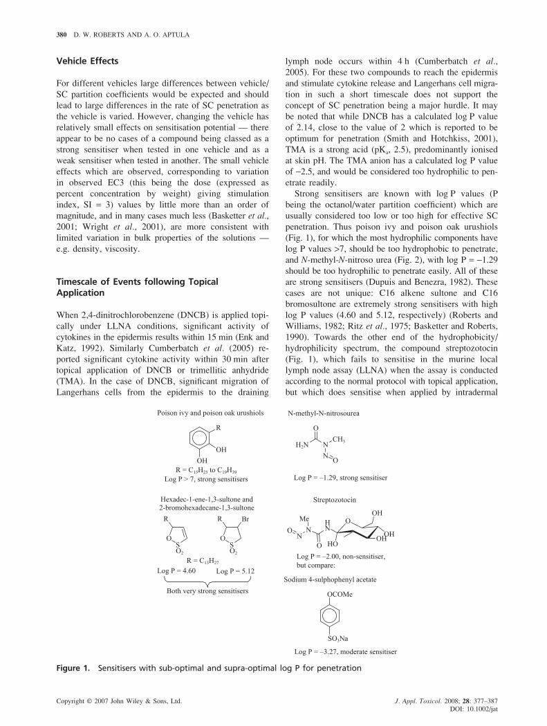
penetration. Thus poison ivy and poison oak urushiols
(Fig. 1), for which the most hydrophilic components have
log P values >7, should be too hydrophobic to penetrate,
and N-methyl-N-nitroso urea (Fig. 2), with log P = −1.29
should be too hydrophilic to penetrate easily. All of these
are strong sensitisers (Dupuis and Benezra, 1982). These
cases are not unique: C16 alkene sultone and C16
bromosultone are extremely strong sensitisers with high
log P values (4.60 and 5.12, respectively) (Roberts and
Williams, 1982; Ritz et al., 1975; Basketter and Roberts,
1990). Towards the other end of the hydrophobicity/
hydrophilicity spectrum, the compound streptozotocin
(Fig. 1), which fails to sensitise in the murine local
lymph node assay (LLNA) when the assay is conducted
according to the normal protocol with topical application,
but which does sensitise when applied by intradermal
Figure 1. Sensitisers with sub-optimal and supra-optimal log P for penetration

DETERMINANTS OF SKIN SENSITISATION POTENTIAL 381
Copyright © 2007 John Wiley & Sons, Ltd. J. Appl. Toxicol. 2008; 28: 377–387
DOI: 10.1002/jat
and hence fails to sensitise via topical application in
the LLNA, as a result of its high reactivity to SC
nucleophiles. A similar special case has been observed
with a series of aryl benzoates, which are activated esters
and act as acyl transfer electrophiles. These compounds
are also reactive to hard nucleophiles, although less so
than streptozotocin, and their sensitisation potential, in a
guinea-pig assay, has been found to be correlated with
hydrophobicity but not with reactivity (Barratt et al.,
1997). The rationale is that increasing reactivity towards
carrier protein is cancelled out by increasing loss of
sensitiser due to reaction in the SC.
At this point it is worth pointing out that sensitisation
potential in the LLNA, but not in other tests, does seem
to be to some extent dependent on ability to penetrate,
for compounds with high log P values. Thus for alkyl
alkanesulphonates with carbon numbers above C12
sensitisation potential is inversely correlated with log P
(Roberts and Basketter, 2000). However, in guinea-pig
assays the dependence on log P is positive (Roberts and
Basketter, 1997). We consider this as an indication
of underprediction by the LLNA for compounds of this
type. We are not aware of any LLNA data on very
hydrophobic compounds known to be strong sensitisers
in man, such as C12–C16 alkene sultones and urushiols
(Dupuis and Benezra, 1982).
Thus we conclude that SC penetration is not a signifi-
cant hurdle in the skin sensitisation process, but is a
potential limiting factor which only becomes important
in specific circumstances — (a) compounds with high log
P values; (b) compounds reactive to hard nucleophiles
in the SC (streptozotocin, activated esters).
Why is SC penetration usually unimportant in deter-
mining sensitisation potential? The simplest interpreta-
tions are either:
– usually all of the compound penetrates the SC in the
timescale of the test, or . . .
– a more or less fixed proportion, which could be small,
reaches the epidermis rapidly, possibly by-passing the
SC via ‘shunt pathways’ (Smith and Hotchkiss, 2001)
such as hair follicles, sweat glands etc., while the re-
mainder fails to penetrate significantly during the test
timescale.
The first of these interpretations may appear contrary to
what would be expected based on the Potts and Guy
equation for percutaneous penetration (Potts and Guy,
1992). However, the Potts and Guy equation is derived
from experimental data on penetration through all the
layers of the skin, involving passage through lipid mem-
brane barriers. If the SC is considered in terms of the
frequently used ‘bricks and mortar’ representation (Smith
and Hotchkiss, 2001), the bricks representing dead cells
(hydrophilic) and the mortar representing the lamellar
phase lipid separating them (Fig. 2a), then it seems
Figure 2. Building materials representations of thestratum corneumIn (a) and (b) the shaded zones (mortar) representslamellar phase lipid and bricks represent dead cells.In (c) the straight lines (greased sheeting) representlamellar phase lipid and irregular shapes (gravel) repre-sent dead cells
injection, has been quoted as an example of a compound
whose inability to penetrate due to its hydrophilicity
(Clog P = −2.00) prevents it from sensitising (Ashby
et al., 1995). However, there are compounds which are
even more hydrophilic than streptozotocin which never-
theless are able to sensitise in the LLNA. An example
is sodium 4-sulphophenyl acetate (Fig. 1), whose log P
value, calculated by the Leo and Hansch method as used
in ClogP software, is −3.27, which is clearly positive in
the LLNA, giving SI values of 3.8, 3.9 and 10.1 at 5%,
10% and 25%, respectively (Ashby et al., 1995),
corresponding to at least a moderate sensitiser classifica-
tion. This positive result with sodium 4-sulphophenyl
acetate makes it difficult to sustain the argument that
streptozotocin’s failure to sensitise in the LLNA is due to
its being too hydrophilic to penetrate. Our interpretation
is that streptozotocin, being not only hydrophilic but also
a very hard electrophile, reacts with hard nucleophilic
groups of hydrophilic proteins such as esterases, which
have a reactive hard nucleophilic hydroxyl group as part
of a serine unit (McMurry and Begley, 2005), during its
passage through the SC. Esterases are known to be
present in the stratum corneum (Menon et al., 1986).
Thus streptozotocin is a special case: it fails to penetrate,

382 D. W. ROBERTS AND A. O. APTULA
Copyright © 2007 John Wiley & Sons, Ltd. J. Appl. Toxicol. 2008; 28: 377–387
DOI: 10.1002/jat
reasonable to assume that the Potts and Guy equation,
should be applicable to SC penetration, since the lamel-
lar phase lipid in the SC will have similar barrier prop-
erties to lipid membranes. However, the ‘bricks and
mortar’ analogy, implying a continuous lamellar phase
barrier, should not be taken too literally, and in particu-
lar should not be represented as a regular recently built
wall. In our view a more realistic analogy would be
a brick wall which has been neglected, so that it has
become irregular and in parts the mortar is missing
(Fig. 2b). An alternative analogy would be a bed of
coarse gravel (representing dead cells) in which dis-
continuous layers of greased sheeting (representing the
lamellar phase lipid) have been interspersed (Fig. 2c). In
Fig. 2 (b and c) the lamellar phase lipid serves to bind
the dead cells together in a matrix, but it does not con-
stitute a continuous barrier to the passage of liquids
through the SC. However, it does provide a phase into
which hydrophobic compounds can partition, thereby
retarding their passage through the SC. This has been
proposed as the mechanistic basis for the negative
correlation of sensitisation potential with log P for hydro-
phobic alkyl alkanesulphonates in the LLNA (Roberts
and Basketter, 2000).
Figure 2 could also be relevant to the second possible
interpretation for the low relevance of penetration to skin
sensitisation, i.e. that a fixed, possibly small, proportion
of the applied sensitiser reaches the epidermis very
rapidly. If most of the SC corresponds to Fig. 2a, but
some proportion corresponds more to Figs 2b and/or 2c
these latter could provide shunt pathways. It is conceiv-
able that the proportion of 2b/2c-like SC would be
greater in damaged, irritated or inflamed skin. A similar
argument has been made for psoriatic and eczematous
skin: these conditions are associated with a rapidly pro-
duced SC which may be structurally flawed with impaired
barrier function (Kligman, 1983).
Migration of Langerhans Cells
For sensitisation to occur, Langerhans cells (LC) which
have acquired and processed sensitiser-modified carrier
protein have to migrate from the epidermis to the drain-
ing lymph node. Much progress has been made in recent
years in understanding the rather complex details of this
process, and has been well described by Corsini and Galli
(2000), Kimber et al. (2000) and Cumberbatch et al.
(2005). The LC have to detach themselves from the
matrix of surrounding keratinocytes and travel along
the basement membrane between the epidermis and the
dermis, eventually penetrating through this membrane to
reach the lymph node. In the course of this journey they
develop into mature dendritic cells (DC), losing the
ability to process sensitiser-modified carrier protein but
acquire the ability to present the corresponding antigen to
T-cells. In LLNA studies, significant LC migration to the
draining lymph node is observed within 4 h of topical
application of DNCB, and continues for up to 72 h
(Cumberbatch et al., 2005).
These events are stimulated and controlled by several
cytokines (these are glycoproteins released in the epider-
mis, which act on the LC via binding to their receptors).
Notable among these cytokines are tumour necrosis
factor α (TNF-α), interleukin (IL) 14 (IL-14) and IL-18
(Cumberbatch et al., 1997, 2001). If the necessary
cytokines are unavailable at the necessary concentrations,
then the response of LCs to sensitisers is impaired
(Kimber et al., 1998, 2000). An important stimulus for
the production, or increased production, of such cytokines
is dermal trauma (Kimber and Cumberbatch, 1992;
Kimber et al., 2001) such as irritation. Many sensitisers,
for example 2,4-dinitrochlorobenzene (DNCB), are to
some extent irritant and may thereby be able to stimulate
production of the cytokines necessary for sensitisation.
The ability of irritant trauma to facilitate sensitisation
is well demonstrated by LLNA studies with DNCB
(Cumberbatch et al., 1993). At reduced concentrations,
DNCB alone gives only a minimal sensitisation response,
but when the irritant sodium lauryl sulphate (SLS) is
administered at the same time, the sensitisation response
to DNCB is increased. At higher DNCB concentrations,
at which the irritancy of DNCB is presumably sufficient
to stimulate cytokine production, the response is not
modified by SLS. This finding has been interpreted as
suggesting that a certain level of skin irritation is required
for the optimal acquisition of skin sensitisation, and that
insufficiently irritant chemicals, in the absence of any
other source of irritation, may not show their full sen-
sitisation potency (Kimber and Dearman, 2003; Jowsey
et al., 2006).
However, many very strong sensitisers, such as long
chain alkene sultones and alkyl alkanesulphonates, are
very mild, i.e. non-irritant, to the extent that they can be
tested even at 100%. Furthermore, there appear to be no
known cases where a compound which should otherwise
sensitise fails to do so because it fails to stimulate the
migration and maturation of LC. Possibly the stimulus
can be produced by other processes which do not lead to
clinically observable irritation. It may be that the acqui-
sition by the LC membrane of a sufficient concentration
of sensitiser-modified carrier protein is enough to stimu-
late the required cytokine production. In any event, it
seems clear that LC migration can be taken for granted,
and does not constitute a hurdle in the sensitisation
process.
It is, however, interesting to note that the sensitiser
trimellitic anhydride (TMA), which is also a respiratory
allergen, produces a different pattern of LC and cytokine
activity in LLNA studies when compared with DNCB.
Whereas topical application of DNCB leads to rapid pro-
duction of, inter alia, the cytokine IL-1β, TMA leads not

DETERMINANTS OF SKIN SENSITISATION POTENTIAL 383
Copyright © 2007 John Wiley & Sons, Ltd. J. Appl. Toxicol. 2008; 28: 377–387
DOI: 10.1002/jat
to IL-1β but to rapid production of IL-10, which appears
to be able to down-regulate production of IL-1β. Migra-
tion of LC to the lymph node occurs more slowly than
with DNCB, and the T-cells which are stimulated to
proliferate are of different types (Cumberbatch et al.,
2005). DNCB and TMA differ in their reaction chemis-
try and their physical chemistry (Roberts et al., 2007a,
2007b): DNCB is an SNAr electrophile, whereas TMA
is an acyl transfer electrophile; DNCB is relatively hydro-
phobic, enabling it to partition well into membranes,
whereas TMA, in its ionised form which will predomi-
nate at epidermal pH, is hydrophilic and less able to
partition into membranes. It remains to be determined
whether the different responses seen to DNCB and TMA
represent a more general pattern of different LC/cytokine
response depending on the reaction mechanistic domain
of the sensitiser.
Antigen Recognition
The nature of the antigen which is presented by the
matured Langerhans cells to naïve T-cells in the lymph
node clearly depends on the nature of the sensitising
compound: this is the basis of the specificity of the
sensitisation. It seems reasonable therefore to consider
that the ability of the antigen to stimulate proliferation
of T-cell clones will depend on the nature of the antigen.
It follows from this argument that the sensitisation poten-
tial of a compound should depend, inter alia, on its in-
trinsic antigenicity, i.e. how well the antigen derived from
it is able to be recognised by T-cell receptors. This, it
seems reasonable to suppose, should depend on the extent
to which the sensitiser, after reaction with protein, pro-
duces groups which can bind strongly to T-cell receptors
through complementary regions of polarity and mole-
cular shape, as illustrated in Fig. 3.
Although this argument appears at first sight highly
plausible, there is no convincing evidence to support it,
and substantial evidence against it.
An early indication that intrinsic antigenicity might
be less dependent on the structure of the sensitiser than
previously assumed came from work with simple alkyl
alkanesulphonates, R1SO3R2, published in 1988 (Roberts
et al., 1988). These compounds are SN2 electrophiles, and
transfer the R2 group to nucleophiles. If a compound of
this type, with R2 = methyl, reacts with a protein, that
protein will become modified simply by covalent binding
of methyl groups. Methyl groups are small, regularly
shaped, and non-polar, so they would not be expected
to bind strongly to T-cell receptors. Nevertheless, the
compounds R1SO3R2 (total carbon number 13 or higher)
were found to be very strong sensitisers in guinea-pig
tests. Subsequently, strong sensitisation has also been
observed in the LLNA — methyl dodecansulphonate has
an EC3 value of 0.39 (Gerberick et al., 2005). Another
finding from the guinea-pig work was that there was
strong cross-reactivity between compounds R1SO3R2 with
R2 = methyl and the compounds with R2 = higher alkyl.
It appears therefore that antigen recognition occurs read-
ily with alkyl transfer agents, and does not vary as the
alkyl group is changed.
Further evidence comes from LLNA data for a wider
range of SN2 electrophiles, covering not just alkyl group
transfer agents but also compounds which transfer polar
groups to the nucleophile. Figure 4 shows a graph in
which the stimulation index values are plotted against
RAI values, the latter being calculated from the test con-
centrations (dose term), rate constants for reaction with
butylamine, and log P values (Roberts et al., 2007a). The
good fit to the curve indicates that there is no need
to invoke differences in degree of antigen recognition
to explain the data.
Even more compelling evidence comes from a recently
published quantitative mechanistic model (QMM) for
the ‘Schiff base domain’, i.e. compounds which are in
Figure 3. Antigen recognition. This figure is available incolour online at www.interscience.wiley.com/journal/jat Figure 4. RAI plot for H-polar SN2 electrophiles
384 D. W. ROBERTS AND A. O. APTULA
Copyright © 2007 John Wiley & Sons, Ltd. J. Appl. Toxicol. 2008; 28: 377–387
DOI: 10.1002/jat
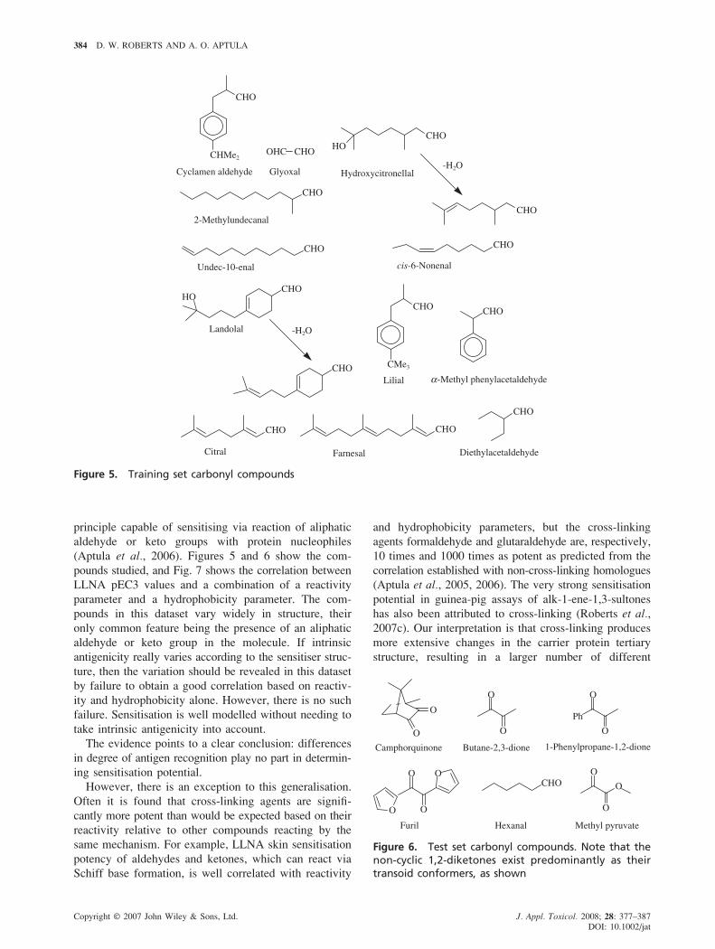
Figure 5. Training set carbonyl compounds
principle capable of sensitising via reaction of aliphatic
aldehyde or keto groups with protein nucleophiles
(Aptula et al., 2006). Figures 5 and 6 show the com-
pounds studied, and Fig. 7 shows the correlation between
LLNA pEC3 values and a combination of a reactivity
parameter and a hydrophobicity parameter. The com-
pounds in this dataset vary widely in structure, their
only common feature being the presence of an aliphatic
aldehyde or keto group in the molecule. If intrinsic
antigenicity really varies according to the sensitiser struc-
ture, then the variation should be revealed in this dataset
by failure to obtain a good correlation based on reactiv-
ity and hydrophobicity alone. However, there is no such
failure. Sensitisation is well modelled without needing to
take intrinsic antigenicity into account.
The evidence points to a clear conclusion: differences
in degree of antigen recognition play no part in determin-
ing sensitisation potential.
However, there is an exception to this generalisation.
Often it is found that cross-linking agents are signifi-
cantly more potent than would be expected based on their
reactivity relative to other compounds reacting by the
same mechanism. For example, LLNA skin sensitisation
potency of aldehydes and ketones, which can react via
Schiff base formation, is well correlated with reactivity
and hydrophobicity parameters, but the cross-linking
agents formaldehyde and glutaraldehyde are, respectively,
10 times and 1000 times as potent as predicted from the
correlation established with non-cross-linking homologues
(Aptula et al., 2005, 2006). The very strong sensitisation
potential in guinea-pig assays of alk-1-ene-1,3-sultones
has also been attributed to cross-linking (Roberts et al.,
2007c). Our interpretation is that cross-linking produces
more extensive changes in the carrier protein tertiary
structure, resulting in a larger number of different
Figure 6. Test set carbonyl compounds. Note that thenon-cyclic 1,2-diketones exist predominantly as theirtransoid conformers, as shown

DETERMINANTS OF SKIN SENSITISATION POTENTIAL 385
Copyright © 2007 John Wiley & Sons, Ltd. J. Appl. Toxicol. 2008; 28: 377–387
DOI: 10.1002/jat
elicitation of the allergic response occur in and require
the skin sensitisers to reach the viable epidermis. However,
further considerations suggest caution about applying this
rule at least in the area of workplace chemicals. Because
this rule relies on the Potts-Guy equation, it applies to
only intact skin. However, cuts, dermatitis, and other skin
injuries are quite common in the workplace environment.
For the same reason, this rule is unlikely to apply to
workers wearing occlusive gloves. Additionally . . . in
recent studies of DEREK for Windows’s performance
using the BgVV . . . database . . . it was shown that there
are not any meaningful limiting values of skin permea-
tion parameters that will have an impact on the induction
of skin sensitisation.’
Approaching the issue from a different perspective,
Fedorowicz et al. arrive at a similar practical interpreta-
tion to our own, which is that there is an important
corollary to our conclusion that SC penetration is not
a significant hurdle in the sensitisation process. When
trying to predict sensitisation hazard, to assume that SC
penetration is a sensitisation-determining factor is to risk
underestimation of the sensitisation potency.
There is a similar corollary to our argument regarding
migration and maturation of Langerhans cells. This is
reinforced when we consider the implications of the facts
that people are not usually exposed to one chemical at
a time, and that cytokine production can be stimulated
by a different, not necessarily allergenic, chemical.
Consider a hypothetical compound which meets all the
criteria to be a sensitiser except that it cannot produce
a stimulus for cytokine production. Then all that would
be needed to release its latent sensitisation potential
would be simultaneous exposure to a compound which
can produce a danger signal (e.g. a surfactant). Thus, to
assume that, because a particular compound is unable
to produce a danger signal, it cannot sensitise, could lead
to underestimation of the risk of sensitisation.
Conclusions
Penetration of the stratum corneum, stimulation of migra-
tion and maturation of Langerhans cells and antigen
recognition are important events in the induction of
sensitisation, but they can be taken for granted. They are
not important factors in determining whether a compound
will be a sensitiser or not, nor are they important factors
in determining how potent one sensitiser will be relative
to another.
This leaves covalent binding to carrier protein as
the key factor which determines sensitisation potential.
For non-animal prediction of skin sensitisation potential,
this is the process which needs to be modelled and
the area where research effort should be focused. This is
not a trivial problem but much progress has been made,
Figure 7. pEC3 observed vs pEC3 calculated for SchiffBase electrophiles. pEC3 calc is obtained from theQMM equation 5 for Schiff base electrophiles XCOY:pEC3 = 1.12 Σσ* + 0.42 log P − 0.62, where Σσ* is thesum of the Taft substituent constants for the groups Xand Y
antigenic determinants and consequently proliferation
of a larger number of T-cell clones. We are not aware of
any definitive information as to whether skin sensitisation
should be considered a monoclonal or a polyclonal phe-
nomenon, but we are encouraged to assume the latter on
the basis of what has been written about T-cell response
to pathogens: ‘. . . a pathogen, even the smallest virus,
offers not one but dozens of shapes (antigens) to be re-
cognised by different lymphocytes . . . As a result of all
this . . . about 1 in 10 000 to 1 in 1 000 000 lymphocytes
will recognise some part of any one pathogen. With
a population of 10 000 000 000 lymphocytes, we are in
the range where there might be as many as a million
lymphocytes per pathogen, even before they start to
proliferate’ (Playfair, 2004).
Practical Implications for Hazard and RiskAssessment
One of the conclusions from our analysis is that skin
penetration ability does not normally constitute a signifi-
cant hurdle in the skin sensitisation process. At this point
it is useful to quote from a QSAR paper by Fedorowicz
et al. (2005). They are discussing the rule within
the DEREK expert system that for compounds with
log Kp < −5 (Kp = permeability coefficient) the probab-
ility of skin sensitisation of the chemical is significantly
lowered as the penetration of such chemicals across
human skin is negligible, and thus, they are unable to
exert their toxic effects:
‘At first, the presence of this rule in DEREK for Windows
seems to be justified, because both skin sensitisation and

386 D. W. ROBERTS AND A. O. APTULA
Copyright © 2007 John Wiley & Sons, Ltd. J. Appl. Toxicol. 2008; 28: 377–387
DOI: 10.1002/jat
building on the pioneering work reported by Landsteiner
and Jacobs (1936). Compounds can be classified into a
limited number of reaction mechanistic domains (Aptula
and Roberts, 2006), and within these domains QMMs can
be derived, based on the RAI model, relating sensitisation
potential to a combination of electrophilic reactivity and
hydrophobicity. Applying these mechanistic principles,
the following strategy can be used (Roberts et al.,
2007b).
Presented with a new compound:
1. The first step is to classify it into its reaction mecha-
nistic domain. One domain is the ‘unreactive’ domain,
populated by predicted non-sensitisers. For several
mechanistic domains there are corresponding pro-
electrophilic sub-domains. For example many sensi-
tisers, such as hydroquinone and 3-alkyl/alkenyl
catechols (active components of poison ivy) are
thought to act as pro-Michael acceptors. Domain
classification may often be possible by inspection of
structure, but inevitably in some cases a confident
prediction may not be possible. In such situations,
experimental work will be needed to determine the
reaction chemistry, in particular to determine if the
compound is electrophilic or pro-electrophilic and
the nature of the reactions.
2. Having assigned the compound to its reaction mecha-
nistic applicability domain, the next step is to quantify
its reactivity/hydrophobicity relative to known sensi-
tisers in the same mechanistic applicability domain.
These properties may sometimes be confidently pre-
dictable from structure, using physical organic chemis-
try approaches such as linear free energy relationships
based on substituent constants or on molecular orbital
parameters. In other cases it will be necessary to per-
form physical organic chemistry measurements, such
as determination of reaction kinetics and measurement
of partition coefficients.
Having assigned the compound to its reaction mechan-
istic applicability domain and quantified its reactivity/
hydrophobicity relative to known sensitisers in the same
domain, QMM or mechanistic read-across can be used to
predict the sensitisation potential.
Although this strategy has only recently been presented
as above, we have in practice been applying it for many
years in the context of interpreting sensitisation data on
raw materials, for example in considering whether a posi-
tive sensitisation test result should be taken as indicating
that the chemical is a sensitiser or whether an impurity is
responsible, and in the latter case identifying the sensitis-
ing impurity and setting specifications.
Of course the above strategy can only be applied with
confidence for substances whose chemistry is sufficiently
understood. There are still some general areas where
further work is needed to better understand the chemical
basis of sensitisation by certain structural classes of com-
pounds. These include: aromatic compounds containing
more than one hydroxyl and/or amino group, hydroper-
oxides and compounds which can readily give rise to
them by autoxidation, epoxides and their autoxidation
precursors.
Our ultimate vision for skin sensitisation prediction is
that the animal testing laboratory should be replaced by
the physical organic chemistry laboratory. Particularly
bearing in mind that many compounds are easily predict-
able without experimentation, the experimental studies to
generate the chemical data required should be no more
costly or time consuming than the animal tests that have
hitherto been used.
References
Aptula AO, Patlewicz GY, Roberts DW. 2005. Skin sensitisation:Reaction mechanistic applicability domains for structure-activityrelationships. Chem. Res. Toxicol. 18: 1420–1426.
Aptula AO, Roberts DW. 2006. Mechanistic applicability domains fornon-animal based toxicological endpoints. General principles, andapplication to reactive toxicity. Chem. Res. Toxicol. 19: 1097–1105.
Aptula AO, Roberts DW, Patlewicz G. 2006. Mechanistic applicabilitydomains for non-animal based toxicological endpoints. QSAR analy-sis of the Schiff Base applicability domain for skin sensitization.Chem. Res. Toxicol. 19: 1228–1233.
Ashby J, Basketter DA, Paton D, Kimber I. 1995. Structure activityrelationships in skin sensitization using the murine local lymph nodeassay. Toxicology 103: 177–194.
Barratt MD, Basketter DA, Roberts DW. 1997. Structure-activity rela-tionships for contact hypersensitivity. In Allergic Contact Dermatitis.
The Molecular Basis, Lepoittevin J-P, Basketter DA, Goossens A,Karlberg A-T (eds). Springer: Heidelberg; 129–154.
Basketter DA. 1998. Chemistry of contact allergens and irritants. Am.
J. Contact. Derm. 9: 119–124.Basketter DA, Evans P, Fielder RJ, Gerberick GF, Dearman RJ, Kimber
I. 2002. Local lymph node assay — validation, conduct and use inpractice. Food Chem. Toxicol. 40: 593–598.
Basketter DA, Gerberick GF, Kimber I. 2001. Skin sensitization,vehicle effects and the local lymph node assay. Food Chem. Toxicol.
39: 621–627.Basketter DA, Roberts DW. 1990. Structure/activity relationships in
contact allergy. Int. J. Cosmet. Sci. 12: 81–90.Basketter DA, Roberts DW, Cronin M, Scholes EW. 1992. The value
of the local lymph node assay in quantitative structure–activity inves-tigations. Contact Dermatitis 27: 137–142.
Corsini E, Galli CL. 2000. Epidermal cytokines in experimental contactdermatitis. Toxicology 142: 203–211.
Cumberbatch M, Clelland K, Dearman RJ, Kimber I. 2005. Impact ofcutaneous IL-10 on resident epidermal Langerhans’ cells and thedevelopment of polarized immune responses. J. Immunol. 33: 47– 62.
Cumberbatch M, Dearman RJ, Antonopoulos C, Groves RW, KimberI. 2001. Interleukin (IL)-18 induces Langerhans cell migration by atumour necrosis factor-alpha- and IL-1beta-dependent mechanism.Immunology 102: 323–330.
Cumberbatch M, Dearman RJ, Kimber I. 1997. Langerhans cells requiresignals from both tumour necrosis factor-alpha and interleukin-1 betafor migration. Immunology 92: 388–395.
Cumberbatch M, Scott RC, Basketter DA, Scholes EW, Hilton J,Dearman RJ, Kimber I. 1993. Influence of sodium lauryl sulphate on2,4-dinitrochlorobenzene-induced lymph node activation. Toxicology
77: 181–191.Dupuis G, Benezra C. 1982. Allergic Contact Dermatitis to Simple
Chemicals: A Molecular Approach. Dekker: New York.EU 2003. Directive 2003/15/EC of the European Parliament and the
Council of 27 February 2003 amending Council Directive 76/768/
DETERMINANTS OF SKIN SENSITISATION POTENTIAL 387
Copyright © 2007 John Wiley & Sons, Ltd. J. Appl. Toxicol. 2008; 28: 377–387
DOI: 10.1002/jat
EEC on the approximations of laws of the Member States relating tocosmetic products. Official J. Eur. Union L66: 26–35.
EU 2006. Council of the European Union. 2006. Common positionadopted by the Council with a view to the adoption of a Regulationof the European Parliament and of the Council concerning theRegistration, Evaluation, Authorisation and Restriction of Chemicals(REACH), establishing a European Chemicals Agency, amendingDirective 1999/45/EC of the European Parliament and of the Counciland repealing Council Regulation (EEC) No 793/93 and CommissionRegulation (EC) No 1488/94 as well as Council Directive 76/769/EEC and Commission Directives 91/155/EEC, 93/67/EEC, 93/105/EC and 2000/21/EC. European Council document 7524/06 of 12 June2006. http://europa.eu.int/comm/enterprise/reach/index_en.htm
Enk AH, Katz SI. 1992. Early molecular events in the inductionphase of contact sensitivity. Proc. Natl Acad. Sci. USA 89: 1398–1402.
Fedorowicz A, Singh H, Soderheim S, Demchuk E. 2005. Structure-activity models for contact sensitization. Chem. Res. Toxicol. 18:954–969.
Franot C, Roberts DW, Basketter DA, Benezra C, Lepoittevin J-P.1994. Structure-activity relationships for contact allergenic potentialof γ,γ-dimethyl-γ-butyrolactone derivatives. 2. Quantitative structure-skin sensitisation relationships for α-substituted-α-methyl-γ,γ-dimethyl-γ-butyrolactones. Chem. Res. Toxicol. 7: 307–312.
Frost AA, Pearson RG. 1961. Kinetics and Mechanism, 2nd edn. Wiley:New York.
Gerberick GF, Ryan CA, Kern PS, Schlatter H, Dearman RJ,Kimber I, Patlewicz GY, Basketter DA. 2005. Compilation ofhistorical local lymph node data for evaluation of skin sensitizationalternative methods. Dermatitis 16: 157–202.
Jowsey IR, Basketter DA, Westmoreland C, Kimber I. 2006. A futureapproach to measuring relative skin sensitising potency: a proposal.J. Appl. Toxicol. 26: 341–350.
Kimber I, Cumberbatch M. 1992. Dendritic cells and cutaneous immuneresponses to chemical allergens. Toxicol. Appl. Pharmacol. 117: 137–146.
Kimber I, Cumberbatch M, Dearman RJ, Bhushan M, Griffiths CEM.2000. Cytokines and chemokines in the initiation and regulationof epidermal Langerhans cell mobilization. Br. J. Dermatol. 142:401–412.
Kimber I, Dearman R. 2003. What makes a chemical an allergen? Ann.
Allergy Asthma Immunol. 90(Suppl): 28–31.Kimber I, Dearman RJ, Basketter DA, Ryan CA, Gerberick GF. 2002.
The local lymph node assay: past, present and future. Contact
Dermatitis 47: 315–328.Kimber I, Dearman RJ, Cumberbatch M, Huby RJ. 1998. Langerhans
cells and chemical allergy. Curr. Opin. Immunol. 10: 614–619.Kimber I, Pichowski JS, Betts CJ, Cumberbatch M, Basketter DA,
Dearman RJ. 2001. Alternative approaches to the identification andcharacterization of chemical allergens. Toxicol. In Vitro 15: 307–312.
Kligman AM. 1983. Skin permeability: dermatological aspects oftransdermal; delivery. Am. Heart J. 108: 200–206.
Landsteiner K, Jacobs J. 1936. Studies on the sensitization of animalswith simple chemical compounds. J. Exp. Med. 64: 643–655.
Lepoittevin J-P, Basketter DA, Goossens A, Karlberg A-T (eds).1997. Allergic Contact Dermatitis. The Molecular Basis. Springer:Heidelberg.
McMurry JE, Begley TP. 2005. The Organic Chemistry of Biological
Pathways. Roberts: Englewood, Colorado, USA.Maurer T, Arthur A, Bentley P. 1994. Guinea-pig contact sensitization
assays. Toxicology 93: 47–54.Mekenyan O, Roberts DW, Karcher W. 1997. Molecular orbital para-
meters as predictors of skin sensitization potential of halo- andpseudohalobenzenes acting as SNAr electrophiles. Chem. Res. Toxicol.
10: 994–1000.Menon GK, Grayson S, Elias PM. 1986. Cytochemical and biochemi-
cal localization of lipase and sphingomyelinase activity in mamma-lian epidermis. J. Invest. Derm. 86: 591–597.
Miller MD, Yourtee DM, Glaros AG, Chappelow CC, Eick JD, HolderAJ. 2005. Quantum mechanical structure-activity relationship analy-ses for skin sensitization. J. Chem. Inf. Model. 45: 924–929.
Playfair J. 2004. Living with Germs. In Sickness and in Health. OxfordUniversity Press: Oxford, UK; 97– 98.
Potts RO, Guy RH. 1992. Predicting skin permeability. Pharm. Res. 9:663–669.
Ritz HL, Connor DS, Sauter ED. 1975. Contact sensitization of guineapigs with unsaturated and halogenated sultones. Contact Dermatitis
1: 349–358.Roberts DW. 1987. Structure–activity relationships for skin sensitisation
potential of diacrylates and dimethacrylates. Contact Dermatitis 17:281–289.
Roberts DW. 1995. Linear free energy relationships for reactions ofelectrophilic halo- and pseudohalobenzenes, and their application inprediction of skin sensitisation potential for SNAr electrophiles. Chem.
Res. Toxicol. 8: 545–551.Roberts DW, Aptula AO, Patlewicz G. 2007a. Electrophilic chemistry
related to skin sensitization. Reaction mechanistic applicability do-main classification for a published data set of 106 chemicals testedin the mouse local lymph node assay. Chem. Res. Toxicol. 20: 44–60.
Roberts DW, Basketter DA. 1990. A quantitative structure–activity/doserelationship for contact allergenic potential of alkyl group transferagents. Contact Dermatitis 23: 331–5.
Roberts DW, Basketter DA. 1997. Further evaluation of the quantitativestructure-activity relationship for skin-sensitizing alkyl transfer agents.Contact Dermatitis 37: 107–112.
Roberts DW, Basketter DA. 2000. Quantitative structure-activityrelationships: sulfonate esters in the local lymph node assay. Contact
Dermatitis 42: 154–161.Roberts DW, Goodwin BF, Basketter D. 1988. Methyl groups as
antigenic determinants in skin sensitisation. Contact Dermatitis 18:219–225.
Roberts DW, Goodwin BFJ, Williams DL, Jones K, Johnson AW,Alderson CJE. 1983. Correlations between skin sensitisation poten-tial and chemical reactivity for p–nitrobenzyl compounds. Food
Chem. Toxicol. 21: 811–813.Roberts DW, Patlewicz G, Kern PS, Gerberick GF, Kimber I, Dearman
RJ, Ryan CA, Basketter DA, Aptula AO. 2007b. Mechanistic appli-cability domain classification of a local lymph node assay dataset forskin sensitization. Chem. Res. Toxicol. 20: 1019–1030.
Roberts DW, Williams DL. 1982. The derivation of quantitative corre-lations between skin sensitisation and physico-chemical parametersfor alkylating agents and their application to experimental data forsultones. J. Theor. Biol. 99: 807–825.
Roberts DW, Williams DL, Bethell D. 2007c. Electrophilic reactions ofskin sensitizing sultones. Chem. Res. Toxicol. 20: 61–71.
Rustemeyer T, van Hoogstraten IMW, von Blomberg BME, Scheper R.2006. Mechanisms in allergic contact dermatitis. In Contact Derma-
titis, 4th edn, Frosch PJ, Menné T, Lepoittevin J-P (eds). Springer:Heidelberg; 11–44.
Ryan CA, Gerberick GF, Gildea LA, Hulette BC, Betts CJ,Cumberbatch M, Dearman RJ, Kimber I. 2005. Interactions ofcontact allergens with dendritic cells: opportunities and challengesfor the development of novel approaches to hazard assessment.Toxicol. Sci. 88: 4–11.
De Silva O, Basketter DA, Barratt MD, Corsini E, Cronin MTD,Das PK, Degwert J, Enk A, Garrigue JL, Hauser C, Kimber I,Lepoittevin J-P, Peguet J, Ponec M. 1996. Alternative methodsfor skin sensitisation testing. The report and recommendations ofECVAM Workshop 19. ATLA 24: 683–705.
Smith CK, Hotchkiss SAM. 2001. Allergic Contact Dermatitis: Chemi-
cal and Metabolic Mechanisms. Taylor and Francis: London.Wright ZM, Basketter DA, Blaikie L, Cooper KJ, Warbrick EV,
Dearman RJ, Kimber I. 2001. Vehicle effects on skin sensitizingpotency of four chemicals: assessment using the local lymph nodeassay. Int. J. Cosmet. Sci. 23: 75–83.