detection of mycobacterium tuberculosis resistance ... · pdf filedetection of mycobacterium...
TRANSCRIPT

ORIGINAL ARTICLE
Year : 2010 | Volume : 28 | Issue : 3 | Page : 211-216
Indian Journal of Medical Microbiology
Detection of Mycobacterium tuberculosis resistance mutations to rifampin and
isoniazid by real-time PCR
A Hristea1, D Otelea
1, S Paraschiv
1, A Macri
2, C Baicus
3, O Moldovan
2, M Tinischi
1, V Arama
1,
A Streinu-Cercel1
1 'Prof. Dr. Matei Bals' National Institute for Infectious Diseases, Str. Calistrat Grozovici, Nr. 1,
Sector 2, 021105 Bucharest, Romania 2 'Marius Nasta' National Institute of Pneumology, Sos. Viilor Nr 90, Sector 5, 050512
Bucharest, Romania 3 Department of Internal Medicine, Colentina Clinical Hospital, Sos. Stefan Cel Mare, 19-21,
Sector 2, 020125 Bucharest, Romania
Date of Submission 28-Jul-2009
Date of Acceptance 05-Apr-2010
Date of Web Publication 17-Jul-2010
Correspondence Address:
A Hristea
'Prof. Dr. Matei Bals' National Institute for Infectious Diseases, Str. Calistrat Grozovici, Nr. 1,
Sector 2, 021105 Bucharest Romania
DOI: 10.4103/0255-0857.66474 PMID: 20644308
Abstract
Objective: The objective of our study was to evaluate the use of a real-time polymerase chain
reaction (PCR)-based technique for the prediction of phenotypic resistance of Mycobacterium
tuberculosis. Materials and Methods: We tested 67 M tuberculosis strains (26 drug resistant
and 41 drug susceptible) using a method recommended for the LightCycler platform. The
susceptibility testing was performed by the absolute concentration method. For rifampin
resistance, two regions of the rpoB gene were targeted, while for identification of isoniazid
resistance, we searched for mutations in katG and inhA genes. Results: The sensitivity and
specificity of this method for rapid detection of mutations for isoniazid resistance were 96%
(95% CI: 88% to 100%) and 95% (95% CI: 89% to 100%), respectively. For detection of
rifampin resistance, the sensitivity and specificity were 92% (95% CI: 81% to 100%) and 74%
(95% CI: 61% to 87%), respectively. The main isoniazid resistance mechanism identified in our
isolates is related to changes in the katG gene that encodes catalase. We found that for rifampin
resistance the concordance between the predicted and observed phenotype was less than
satisfactory. Conclusions: Using this method, the best accuracy for genotyping compared with
phenotypic resistance testing was obtained for detecting isoniazid resistance mutations. Although
real-time PCR assay may be a valuable diagnostic tool, it is not yet completely satisfactory for
detection of drug resistance mutations in M tuberculosis.

Keywords: Mycobacterium tuberculosis, real-time PCR, resistance
Introduction
Tuberculosis (TB) remains a major global health problem despite the availability of effective
antituberculosis therapy for over 50 years. The World Health Organization (WHO) estimates that
approximately one-third of the global community is infected with Mycobacterium tuberculosis.
According to WHO data, with regard to infection rates, Romania is among the top five countries
from the European region, with high notification rates both for new and relapse cases of TB;
more than 25000 new and relapse cases are recorded every year. [1],[2]
Since the early 1990s, an alarming trend and a growing source of public health concern has been
the emergence of resistance to multiple drugs. Multidrug resistance (MDR) is defined as resist-
ance to at least isoniazid (INH) and rifampicin (RMP). Although it remains unclear whether the
drug-resistant strains are less transmissible than the susceptible strains, [3]
infection-control pre-
cautions need to be maintained, since patients with drug-resistant TB are likely to remain infec-
tious for long periods. Thus the public health consequences of drug-resistant tuberculosis might
be more serious than those of drug-susceptible disease.
The prevalence of M tuberculosis (MTB) drug resistance in Romania was recently evaluated by a
national survey performed between 2003-2004. This showed that 3.6% of the strains isolated
from newly diagnosed patients and 8.6% from relapse cases were resistant to one antitubercu-
losis drug (INH). [2]
Moreover, the results of this study indicate that MDR was observed in 2.9%
of the MTB strains from newly diagnosed patients and in 11% of those isolated from relapse ca-
ses. Taking into account that in Romania more than 25000 TB cases (new cases and relapses) are
reported each year, we can estimate that more than 1100 patients are infected with MDR-TB
strains.
The need to limit the transmission of drug-resistant strains and to reduce the time between
diagnosis and effective therapy requires rapid identification of resistance. Classical phenotypic
determination of resistance may take up to 10 weeks after referral of a sample to the laboratory.
Nucleic acid amplification assays can greatly shorten the detection time. Due to this major ad-
vantage, in the last few years, a lot of effort has been invested in designing performance pro-
tocols for genotyping MTB strains. Real-time PCR came to be the main approach because of its
special features: high sensitivity and specificity as well as speed, with no need for any post-PCR
sample manipulation. The results from fundamental research (such as the sequencing of the com-
plete MTB genome) were used to design specific primers and probes that would allow the iden-
tification of gene mutations associated with drug resistance in MTB.
It is known that RMP interferes with RNA synthesis by binding to bacterial RNA polymerase.
Resistance to RMP is conferred by mutations resulting in at least eight amino acids substitutions
in the rpoB subunit of RNA polymerase. Mutations in a limited region of rpoB have been found
in >95% of RMP-resistant clinical isolates of MTB and has been shown to result in high-level
resistance (MIC >32 μg/mL) to RMP and cross-resistance to all rifamycins.[4]
INH acts by inhibiting an oxygen-sensitive pathway in the mycolic acid biosynthesis of the cell
wall. At least four genes have been described to be involved in resistance to isoniazid: the katG
gene, which encodes a catalase; the inhA gene, whose product is a target for INH; and the oxyR
gene and the neighboring aphC gene, as well as their intergenic region. [5]
Several real-time
PCR-based methods targeting these specific genomic regions have been described. [6],[7],[8],[9],[10],[11],[12],[13]
The purpose of the present study was to evaluate the LightCycler
instrument in the detection of these mutations associated with resistant MTB strains isolated
from Romanian patients.
Materials and Methods
Strains and resistance testing
Forty-one susceptible and twenty-six resistant clinical isolates of MTB (23 resistant to both INH
and RMP, 1 mono-RMP resistant, and 2 resistant to INH only) from 62 different patients were
studied. The susceptibility testing was performed by the absolute concentration method (Meis-
sner). [14]
This method is based on the comparison between the growth of mycobacteria on drug-
free medium with that of growth on drug-containing media (antituberculosis drugs incorporated
in the medium at different concentrations) 21 days after inoculation with a standardized inocu-
lum. Two critical concentrations were used for every tested drug: 0.2 μg/mL and 1 μg/mL for
INH and 20 μg/mL and 40 μg/mL for RMP. According to this method, resistance to a drug is
defined by the growth of more than 20 colonies on drug-containing media (INH 1 μg/mL, RMP
40 μg/mL).
Extraction of mycobacterial DNA
We extracted MTB DNA by the thermal lysis procedure in the presence of Chelex 100 (Amer-
sham Pharmacia Biotech, Uppsala, Sweden). Briefly, we obtained one loopful of bacteria scra-
ped from Lφwenstein-Jensen solid medium and suspended it in 100 μL sterile water; the same
volume of Chelex 10% suspension was added and the mixture was incubated for 45 minutes at
45°C and 5 minutes at 100°C. The samples were centrifuged at 12000 g for 5 minutes and the
supernatant was used in the subsequent steps of the experiment.
Real-time PCR using the LightCycler
The MTB drug-resistance genotyping was performed by adapting a previously described proto-
col. [6]
The method published by Torres et al. was designed as a single-tube method capable of
detecting RMP and INH resistance mutations; one set of primers and two fluorescently labeled
hybridization probes were used for each targeted region. One set of primers and two sets of pro-
bes (rpoB1 and rpoB2) that targeted the rpoB gene were used for detection of RMP resistance
and one set of primers and probes each for the katG and inhA genes in order to test for INH re-
sistance. All primers and probes were synthesized by TIB Molbiol (DNA Synthesis Service;
Roche Diagnostics, Berlin, Germany). The real-time PCR was followed by melting curve ana-
lysis, both performed on the LightCycler instrument (Roche Diagnostics, Mannheim, Germany).
We used the same PCR conditions (components concentration, cycling, and melting programs)
as previously described, but we added 10 more cycles of amplification to the 35 recommended. [7]
We included into each experimental run one negative control (the DNA template was replaced
with PCR-grade water) and one positive control (the DNA template was isolated from M
tuberculosis H37Rv, a strain susceptible to both INH and RMP).
DNA sequencing

Direct PCR sequencing was performed with the commercial BigDye terminator DNA
sequencing kit (Applied Biosystems, CA, USA), according to the manufacturer's recommend-
dations. Briefly, the extracted DNA was amplified with the same primers used in the real-time
PCR. The thermal cycling was performed on a GeneAmp System 9700 (Applied Biosystems,
CA, USA) thermal cycler. The resulting PCR product was purified using MicroCon YM-100
concentrators (spin columns) and sequenced bidirectionally using the BigDye terminator che-
mistry . The capillary electrophoresis was performed on an ABI Prism 3100-Avant genetic
analyzer (Applied Biosystems, CA, USA) and the raw analysis of the sequences was made by
Sequencing Analysis Software, version 3.7. Finally, the sequences were aligned with BioEdit
software (version 5.0.6) ( www.mbio.ncsu.edu/BioEdit/bioedit.html ) in order to generate an
assembled full range sequence, which was then compared with the MTB H37Rv sequence
(GenBank Accession No. NC000962).
Statistical analysis
In order to determine the cutoff of T m changes to predict mutations associated with resistance,
we generated ROC curves. The area under the ROC curves was then determined, and the cutoff
points were identified to maximize test sensitivity (and thus decrease the false negative rate). To
further enhance sensitivity we also assessed in parallel the tests detecting the presence of either
rpoB1 or rpoB2 for RMP resistance and the presence of either katG or inhA gene for INH resis-
tance. SPSS 10.0 software (SPSS, Inc., Chicago, IL, USA) was used for the database construc-
tion and ROC curves, and CAT maker 1.1 (Centre for Evidence-Based Medicine, Oxford, GB,
2004) to calculate the attributes of the diagnostic tests studied.
Results
During the real-time PCR experiments, the amplification of the DNA template was monitored by
continuously measuring the fluorescence level. For samples as well as for the positive control,
the fluorescent signals started to rise at a number of cycles, ranging between 20-35. The T m s for
the probes annealed to the PCR product were generated by running the melting analysis program
(ramping from 50°C to 85°C with 0.1°C per second) and calculated using the LightCycler soft-
ware. While running different sets of samples along with the positive control, we observed that
the melting temperature (T m ) for the MTB H37Rv was variable, ranging between 70.08°C and
71.26°C. Therefore, for each experimental run we analyzed the changes in T m for the PCR pro-
ducts derived from our collection of resistant and susceptible MTB clinical isolates as compared
with the T m of the H37Rv tested in the same run rather than using directly the observed T m s.
We also noticed that the T m values for the H37Rv strain as well as for other wild-type (wt) field
strains tested were lower than expected (70.08°C for H37 as compared with 72.8°C in the origi-
nal communication). For the resistant strains, in one case (sample 2312), we obtained a lower T m
value than the expected T m; the δ TM was however consistent with AGC > ACC mutation in
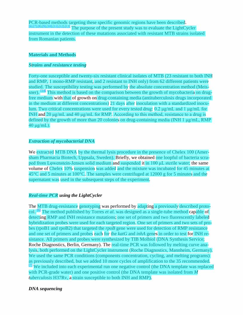
position 315. In [Figure 1] we have represented the melting profiles for the positive control
(H37Rv) and the other seven samples when we analyzed the katG PCR products. It can be seen
that sample 2312 has a melting profile different from that of H37Rv; this is in agreement with
the phenotypic results, which scored this sample as resistant to INH. The other isolates had the
same melting profile as the positive control and were also found to be susceptible to INH by the
phenotypic analysis.
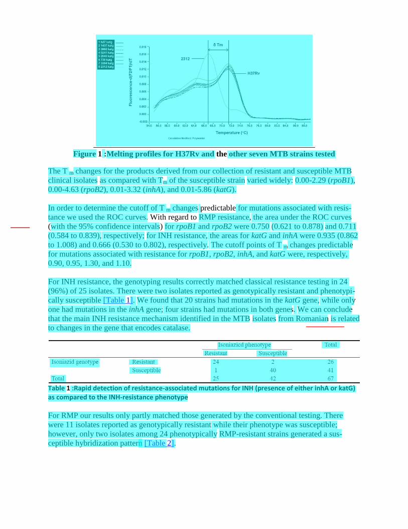
Figure 1 :Melting profiles for H37Rv and the other seven MTB strains tested
The T m changes for the products derived from our collection of resistant and susceptible MTB
clinical isolates as compared with Tm of the susceptible strain varied widely: 0.00-2.29 (rpoB1),
0.00-4.63 (rpoB2), 0.01-3.32 (inhA), and 0.01-5.86 (katG).
In order to determine the cutoff of T m changes predictable for mutations associated with resis-
tance we used the ROC curves. With regard to RMP resistance, the area under the ROC curves
(with the 95% confidence intervals) for rpoB1 and rpoB2 were 0.750 (0.621 to 0.878) and 0.711
(0.584 to 0.839), respectively; for INH resistance, the areas for katG and inhA were 0.935 (0.862
to 1.008) and 0.666 (0.530 to 0.802), respectively. The cutoff points of T m changes predictable
for mutations associated with resistance for rpoB1, rpoB2, inhA, and katG were, respectively,
0.90, 0.95, 1.30, and 1.10.
For INH resistance, the genotyping results correctly matched classical resistance testing in 24
(96%) of 25 isolates. There were two isolates reported as genotypically resistant and phenotypi-
cally susceptible [Table 1]. We found that 20 strains had mutations in the katG gene, while only
one had mutations in the inhA gene; four strains had mutations in both genes. We can conclude
that the main INH resistance mechanism identified in the MTB isolates from Romanian is related
to changes in the gene that encodes catalase.
Table 1 :Rapid detection of resistance-associated mutations for INH (presence of either inhA or katG) as compared to the INH-resistance phenotype
For RMP our results only partly matched those generated by the conventional testing. There
were 11 isolates reported as genotypically resistant while their phenotype was susceptible;
however, only two isolates among 24 phenotypically RMP-resistant strains generated a sus-
ceptible hybridization pattern [Table 2].

Table 2 :Rapid detection of resistance-associated mutations for RMP (presence of either rpoB1 or rpoB2) as compared to the RMP-resistance phenotype
The sensitivity and specificity of the rapid detection of mutations for INH (presence of either
inhA or katG) were 96% (95% CI: 88 to 100) and 95% (95% CI: 89 to 100), respectively, with a
positive likelihood ratio (LR+) of 20 and a negative likelihood ratio (LR−) of 0.04. For RMP
(presence of rpoB1 or rpoB2), the sensitivity and specificity were 92% (95% CI: 81 to 100%)
and 74% (95% CI: 61 to 87%), respectively, with LR+ of 3.58 and LR− of 0.10.
Sequencing was performed in order to obtain more information about nucleotide changes in the
examined genes so that differences between the results generated with the two resistance tech-
niques could be explained. Therefore, five MTB strains were partially sequenced in the inhA and
katG regions and the other six strains in the rpoB gene. Unfortunately, not all the samples with
discordant results between genotyping and phenotyping were available for the sequencing expe-
riment. For comparison, we included also samples found sensitive or resistant to INH and RMP
by both tests. Nucleotide sequence analysis (three genes targeted) of the strains with discordant
results revealed a number of differences from the sequence of H37Rv [Figure 2]. When
translated into amino acids, no peptide sequence changes were observed.
Figure 2 :Nucleotide alignment (rpoB gene) of H37Rv along with four MTB isolates tested. Samples 2165 and 331 had concordant results by phenotypic and genotypic analysis (2165 susceptible to RMP, 331 resistant to RMP), while 2282 and 3550 had discordant results: sample 2282 phenotypically resis-tant and genotypically susceptible, sample 3550 phenotypically susceptible and genotypically resistant
Discussion
There are a variety of methods to determine the susceptibility of MTB to antituberculosis drugs,
but none of them is perfect. [15]
The objective of our study was to evaluate to what extent differences in sequence between
circulating strains might hamper the use of real-time PCR-based techniques for the prediction of
phenotypic resistance of MTB strains. We used a technique recommended for the LightCycler
platform to analyze 67 sequences from 26 drug-resistant and 41 drug-susceptible MTB strains.
Our results suggest that this platform can be used, but there are some limitations. The least
important is related to reproducibility. While testing the resistance to INH and RMP with real-
time PCR we found that the T m for the sensitive as well as for the resistant strains varied from
one experiment to another due to factors that could not be identified. However, the differences
between the T m s of the resistant and sensitive strains were consistently observed and could be
reliably associated with predicted resistance.

Although genotypic assays are very useful for the rapid detection of drug resistance, there are
some limitations. First, not all MTB-DR isolates have mutations in the so-called hot spots of the
genes associated with resistance. For instance, about 20%-30% of the INH-resistant strains do
not have mutations in katG, inhA, kasA, or aphC genes. [4]
For that reason, it is very difficult to
design a test that could identify all the possible mutations that confer resistance to anti-MTB
drugs. This was the case with the MTB isolates from Romanian patients. Here, only two of the
main genes involved in conferring resistance to INH were targeted by PCR. We found that for
the Romanian strains, targeting katG was adequate to detect INH resistance. In the analysis of
data from other studies, geographical differences in the frequencies of specific mutations are also
apparent: the katG gene was mutated at codon 315 in 64% of INH-resistant strains from South
Africa and central and western Africa but in only 26% of Singaporean isolates. [16],[17]
Further-
more, even the commercial tests for genotyping MTB drug resistance have been reported to have
some limitations. A recent study evaluated the results of the two commercially available line
probe assays and showed that while the accuracy for RMP resistance was very good, the
sensitivity for INH was variable. [17]
We found that for RMP resistance, the concordance between the predicted and observed phenol-
type was less than satisfactory. This is not entirely unexpected, because a single mutation, alth-
ough implicated in resistance, might not be enough to generate a resistant phenotype. Two ex-
planations can account for these observations. The most important is the presence of mutations
within the rpoB locus that are not associated with resistance but nevertheless influence the ann-
ealing properties of the probes. This is most likely why a significant number of strains were clas-
sified as resistant to RMP by genetic analysis and sensitive by phenotypic testing.
Furthermore, when performed sequencing, we observed changes at the nucleotide level that did
not affect the amino acid sequence, but could alter the sensitivity of the genotypic test: an extra
one or two mismatches could influence the T m value, which could affect the interpretation of the
sensitivity based on hybridization. This was observed for rpoB, with a high number of false posi-
tive results (26%). A much smaller number of false negative results has been observed in strains
tested for resistance to INH (4%) and RMP (8%); in this case no mutations were found in the
target sequence of the tested genes. The molecular determinants of the resistant phenotype are
expected to be found elsewhere.
On the other hand, it should be kept in mind that isolates that are susceptible according to mole-
cular assays that target specific mutations may contain other unknown mechanisms of resistance,
and these mechanisms will be missed by these techniques.
A much smaller number of strains were reported sensitive by the hybridization analysis and re-
sistant by the phenotypic analysis. The explanation for this is that a small albeit significant num-
ber of strains have determinants of resistance outside the area targeted by the assays we used. A
similar phenomenon has been reported by others. [18],[19],[20]
Another possibility is that changes
have occurred in genes whose products participate in antibiotic permeation or metabolism. [18]
In addition, the results of the absolute concentration method used in the phenotypic test are less
reliable compared with the proportion method (the most preferred choice). Errors in the suscep-
tibility testing may be related to any of the following: cultures older than 21-30 days, incorrect
size of inoculum, incorrect dilution, or errors in incorporation of antibiotics in culture media. [14]
This technique should be further evaluated since the circulating strains in different geographical

regions might behave differently when genotypically tested.
This real-time PCR assay could be useful when investigation of drug-resistant TB is mandatory,
for example, in cases with a history of one or more previous treatment(s) with several failing,
discontinued regimen, or in the situation of exposure to a known source of drug-resistant TB.
Although real-time PCR assays may be a valuable diagnostic tool, they are not yet completely
satisfactory for MTB drug-resistance detection. Phenotype-based assays will continue to have a
place in the clinical mycobacteriology laboratory.
Conclusion
Thus, based on our experience, the real-time PCR assay could be used in clinical practice, albiet
with caution, in cases with risk factors for resistance. The results can be used for guiding the
initiation of therapy, but the treatment should be adjusted correspondingly as soon as the phenol-
typic testing results are available. The best accuracy for genotyping compared with phenotypic
resistance testing was obtained for detecting INH resistance mutations targeting the katG gene.
References
1. Aziz MA, Laszlo A, Raviglione M, Rieder H, Espinal M, Wright A. Guidelines for
surveillance of drug resistance in tuberculosis. 2nd ed. World Health Organization 2003.
Available from: http://whqlibdoc.who.int/publications/2003/9241546336.pdf. [Accessed on
2009 July 26].
2. Stoicescu IP, Homorodean D, Chiotan D, Moldovan O, Diculencu D, Popa C, et al.
Romanian anti-TB drugs resistance surveillance 2003-2004. Pneumologia 2008;57:131-
7. [PUBMED]
3. Soolingen D van, Kremer K, Hermans PWM. Molecular epidemiology: Breakthrough
achievements and future prospects. Tuberculosis 2007. In: Palomino JC, Leao SC, Ritacco
V, editors. From basic science to patient care. 2007. p. 315-40. Available from:
http://www.tuberculosistextbook.com . [Accessed on 2009 July 27].
4. Cho SN, Brennan PJ. Tuberculosis: Diagnostics. Tuberculosis (Edinb) 2007;87:S14-
7. [PUBMED] [FULLTEXT]
5. Ramaswamy SV, Reich R, Dou SJ, Jasperse L, Pan X, Wanger A, et al. Single nucleotide
polymorphisms in genes associated with isoniazid resistance in Mycobacterium
tuberculosis. Antimicrob Agents Chemother 2003;47:1241-
50. [PUBMED] [FULLTEXT]
6. Marin M, Garcia de Viedma D, Ruiz-Serrano MJ, Bouza E. Rapid direct detection of
multiple rifampin and isoniazid resistance mutations in Mycobacterium tuberculosis in
respiratory samples by real-time PCR. Antimicrob Agents Chemother 2004;48:4293-
300.
7. Torres MJ, Criado A, Palomares JC, Aznar J. Use of real-time PCR and fluorimetry for
rapid detection of rifampin and isoniazid resistance-associated mutations in Mycobacterium
tuberculosis. J Clin Microbiol 2000;38:3194-9. [PUBMED] [FULLTEXT]
8. Garcia de Viedma D, del Sol Diaz Infantes M, Lasala F, Chaves F, Alcala L, Bouza E. New
real-time PCR able to detect in a single tube multiple rifampin resistance mutations and
high-level isoniazid resistance mutations in Mycobacterium tuberculosis. J Clin Microbiol
2002;40:988-95.
9. Kocagoz T, Saribas Z, Alp A. Rapid determination of rifampin resistance in clinical isolates
of Mycobacterium tuberculosis by real-time PCR. J Clin Microbiol 2005;43:6015-
9. [PUBMED] [FULLTEXT]
10. Espasa M, Gonzalez-Martin J, Alcaide F, Aragon LM, Lonca J, Manterola JM, et al. Direct
detection in clinical samples of multiple gene mutations causing resistance of
Mycobacterium tuberculosis to isoanizid and rifampicin using fluorogenic probes. J
Antimicrob Chemother 2005;55:860-5.
11. Wada T, Maeda S, Tamaru A, Imai S, Hase A, Kobayashi K. Dual-probe assay for rapid
detection of drug-resistant Mycobacterium tuberculosis by real-time PCR. J Clin Microbiol
2004;42:5277-85. [PUBMED] [FULLTEXT]
12. Ruiz M, Torres MJ, Llanos AC, Arroyo A, Palomares JC, Aznar J. Direct detection of
rifampin- and isoanizid-resistant Mycobacterium tuberculosis in auramine-rhodamine-
positive sputum specimens by real-time PCR. J Clin Microbiol 2004;42:1585-
9. [PUBMED] [FULLTEXT]
13. Hillemann D, Weizenegger M, Kubica T, Richter E, Niemann S. Use of the genotype
MTBDR assay for rapid detection of rifampin and isoanizid resistance in Mycobacterium
tuberculosis complex isolates. J Clin Microbiol 2005;43:3699-
703. [PUBMED] [FULLTEXT]
14. Kim SJ. Drug-susceptibility testing in tuberculosis: Methods and reliability of results. Eur
Respir J 2005;25:564-9. [PUBMED] [FULLTEXT]
15. Haas WH, Schilke K, Brand J, Amthor B, Weyer K, Fourie PB, et al. Molecular analysis of
katG gene mutations in strains of Mycobacterium tuberculosis complex from Africa.
Antimicrob Agents Chemother 1997;41:1601-3. [PUBMED] [FULLTEXT]
16. Lee AS, Lim IH, Tang LL, Telenti A, Wong SY. Contribution of kasA analysis to detection
of isoniazid-resistant Mycobacterium tuberculosis in Singapore. Antimicrob Agents
Chemother 1999;43:2087-9. [PUBMED] [FULLTEXT]
17. Ling DI, Zwerling AA, Pai M. GenoType MTBDR assays for diagnosis of multidrug-
resistant tuberculosis: A meta-analysis. Eur Respir J 2008;32:1165-
74. [PUBMED] [FULLTEXT]
18. Kapur V, Li LL, Iordanescu S, Hamrick MR, Wanger A, Kreiswirth BN, et al.
Characterization by automated DNA sequencing of mutations in the gene (rpoB) encoding
the RNA polymerase beta subunit in rifampin-resistant Mycobacterium tuberculosis strains
from New York City and Texas. J Clin Microbiol 1994;32:1095-
8. [PUBMED] [FULLTEXT]
19. Kim BJ, Kim SY, Park BH, Lyu MA, Park IK, Bai GH, et al. Mutations in the rpoB gene of
Mycobacterium tuberculosis that interfere with PCR-single-strand conformation
polymorphism analysis for rifampin susceptibility testing. J Clin Microbiol 1997;35:492-
4. [PUBMED] [FULLTEXT]
20. Telenti A, Imboden P, Marchesi F, Lowrie D, Cole S, Colston MJ, et al. Detection of
rifampicin-resistance mutations in Mycobacterium tuberculosis. Lancet 1993;341:647-
50. [PUBMED] [FULLTEXT]

CLINICAL MICROBIOLOGY REVIEWS,0893-8512/01/$04.00�0 DOI: 10.1128/CMR.14.4.836–871.2001
Oct. 2001, p. 836–871 Vol. 14, No. 4
Copyright © 2001, American Society for Microbiology. All Rights Reserved.
Molecular Detection of Antimicrobial ResistanceAD C. FLUIT,* MAARTEN R. VISSER, AND FRANZ-JOSEF SCHMITZ
Eijkman-Winkler Institute, University Medical Center Utrecht,Utrecht, The Netherlands
INTRODUCTION .......................................................................................................................................................837MOLECULAR TECHNIQUES USED IN CLINICAL MICROBIOLOGY.........................................................837ANTIBIOTIC RESISTANCE IN MYCOBACTERIUM TUBERCULOSIS ............................................................840
Introduction .............................................................................................................................................................840Rifampin Resistance...............................................................................................................................................840Isoniazid Resistance ...............................................................................................................................................842Multidrug Resistance .............................................................................................................................................842New Developments ..................................................................................................................................................843Conclusion ...............................................................................................................................................................843
RESISTANCE TO �-LACTAM ANTIBIOTICS .....................................................................................................843Mechanisms of Resistance.....................................................................................................................................843Methicillin-Resistant Staphylococci .....................................................................................................................843Penicillin-Resistant Pneumococci .........................................................................................................................846Common �-Lactamases..........................................................................................................................................846Extended-Spectrum �-Lactamases .......................................................................................................................848Metallo-�-Lactamases............................................................................................................................................848Conclusion ...............................................................................................................................................................848
RESISTANCE TO AMINOGLYCOSIDES..............................................................................................................848Mechanisms of Resistance.....................................................................................................................................848Staphylococci ...........................................................................................................................................................849Enterococci and Streptococci ................................................................................................................................849Gram-Negative Bacteria.........................................................................................................................................850Conclusion ...............................................................................................................................................................851
RESISTANCE TO FLUOROQUINOLONES .........................................................................................................851Mechanisms of Resistance.....................................................................................................................................851Detection of Resistance ..........................................................................................................................................852Conclusion ...............................................................................................................................................................853
RESISTANCE TO MLS ANTIBIOTICS .................................................................................................................853Mechanisms of Resistance.....................................................................................................................................853Staphylococci ...........................................................................................................................................................854Pneumococci ............................................................................................................................................................854Streptococci..............................................................................................................................................................855Enterococci...............................................................................................................................................................855Helicobacter pylori ....................................................................................................................................................855Other Species ..........................................................................................................................................................856Conclusion ...............................................................................................................................................................856
RESISTANCE TO GLYCOPEPTIDES....................................................................................................................856Mechanisms of Resistance.....................................................................................................................................856Detection of Resistance ..........................................................................................................................................857Conclusion ...............................................................................................................................................................858
RESISTANCE TO TETRACYCLINES ....................................................................................................................858Mechanisms of Resistance.....................................................................................................................................858Detection of Resistance ..........................................................................................................................................858Conclusion ...............................................................................................................................................................860
RESISTANCE TO TRIMETHOPRIM.....................................................................................................................860RESISTANCE TO CHLORAMPHENICOL ...........................................................................................................860RESISTANCE TO MUPIROCIN .............................................................................................................................861MULTIDRUG RESISTANCE ...................................................................................................................................861CONCLUDING REMARKS......................................................................................................................................862REFERENCES ............................................................................................................................................................862
* Corresponding author. Mailing address: Eijkman-Winkler Insti-tute, University Medical Center Utrecht, Room G04.614, P.O. Box85500, 3508 GA Utrecht, The Netherlands. Phone: 31 30 2507630. Fax:31 30 2541770. E-mail: [email protected].
836
on April 24, 2014 by guest
http://cmr.asm
.org/D
ownloaded from

sistance determinants. These techniques include the DNA am-plification techniques of strand displacement amplification(377, 378) and ligase chain reaction (16, 19, 25, 398, 405) andthe RNA amplification techniques of Q� replication (156) andself-sustained sequence replication or nucleic acid-based se-quence amplification (55, 148). This latter method can be mod-ified to amplify DNA (99).
The variety of molecular techniques used for diagnostic ap-plications demonstrate that no universal technique exists whichis optimal for detection of nucleic acids. The choice of a par-ticular technique is also dependent on the information re-quired or the targets under consideration, but some techniquesare more favored than others. New techniques continue to bedeveloped that involve a new approach to amplification, hy-bridization, formats, and labels (158).
ANTIBIOTIC RESISTANCE INMYCOBACTERIUM TUBERCULOSIS
Introduction
In the wake of the human immunodeficiency virus epidemicand the breakdown of medical services in several Eastern Eu-ropean countries, the incidence of tuberculosis is rising rapidly.Of note, the treatment of tuberculosis is threatened by theemergence of multidrug-resistant strains of Mycobacterium tu-berculosis. M. tuberculosis is usually treated with only a limitednumber of antimicrobial agents, the most important ones beingrifampin, isoniazid, streptomycin, and ethambutol. Resistanceto rifampin is conferred by mutations resulting in at least eightamino acid substitutions in the RpoB subunit of RNA poly-merase (335). Isoniazid acts by inhibiting an oxygen-sensitivepathway in the mycolic acid biosynthesis of the cell wall. Atleast four genes have been described to be involved in resis-tance to isoniazid: the katG gene, which encodes a catalase; theinhA gene, which is the target for isoniazid; and the oxyR geneand neighboring aphC gene and their intergenic region (739).Streptomycin resistance has been associated with mutations inthe rrs gene encoding 16S rRNA and the rspL gene encodingthe S12 ribosomal protein (69, 196, 208). Ethambutol resis-tance is associated with an altered EmbB protein (2, 322), aprotein involved in the synthesis of the cell wall componentarabinogalactan.
Because the organism is slow growing, traditional diagnosisis time-consuming. Traditional phenotypic determination of
resistance may take up to 10 weeks after referral of a sample tothe laboratory, but both commercial and in-house amplifica-tion assays can greatly improve the detection time. Therefore,it is not surprising that within the past 8 years a multitude ofdifferent resistance assays based on molecular techniques werespecifically developed for M. tuberculosis. However, many lab-oratories have had trouble with the technical rigor imposed bythese assays (222). For review of mycobacterial resistance, seereference 128.
Rifampin Resistance
One of the first assays for the detection of rifampin resis-tance using PCR-SSCP was published by Telenti et al. (336). Ina second paper the assay was more extensively evaluated bothin a manual format with radioactively labeled amplificationproducts and with 5�-fluorescein-labeled primers for detectionon an automated DNA sequencer (337). Evaluation of theresults showed that all 17 of the then known mutations in therpoB gene leading to resistance could be detected. Equallyimportant, the assays could be applied to minimally growncultures in Bactec 12B medium with a growth index of �100 oron sputa with at least 10 organisms per field at a magnificationof �250. This clearly established the potential of PCR-SSCP asa powerful technique for the early detection of antimicrobialdrug resistance in M. tuberculosis. The application of rifampinresistance detection by PCR-SSCP to cerebrospinal fluid spec-imens from patients with tuberculosis of the central nervoussystem also yielded excellent results (289).
PCR-SSCP requires careful control over electrophoresisconditions, which is difficult to achieve in many laboratories.This recognition led to a comparison of PCR-SSCP and dide-oxy fingerprinting (84). Dideoxy fingerprinting is in fact anextension of SSCP. After PCR amplification of the gene frag-ment of interest, a second PCR is performed with a radioac-tively labeled primer. A dideoxynucleotide is added, whichleads to chain termination similar to that obtained in dideoxysequencing. The products are then analyzed in a similar man-ner to that in SSCP. Because more fragments are generated,differences between the susceptible and resistance types aremore easily obtained in accordance with conventional suscep-tibility testing and PCR-SSCP. A drawback of this method is itsuse of a radioactive label. However, by using fluorescent labels,this assay can probably be adapted for use with an automatedsequencer.
However, Kim et al. (150) observed that PCR-SSCP re-ported some isolates as resistant whereas their phenotype wassusceptible, but in these isolates the part of the rpoB gene thatwas amplified contained a silent mutation and a deletion of twoamino acids. Apparently, these mutations do not affect thesusceptibility to rifampin. These authors therefore concludedthat sequencing probably could rule out false-positive results.
Direct testing of a clinical specimen for resistance to ri-fampin by PCR without prior species determination is believedto be difficult because of the high levels of homology reportedbetween different mycobacterial species, but Whelen et al.(392) devised a rpoB-based seminested amplification which wasspecific for M. tuberculosis. The assay correctly identified 21 of24 culture-positive specimens, 13 of which were acid-fast smearnegative in a panel of 51 clinical specimens. Three specimens
FIG. 5. Principle of oligonucleotide array sequencing. Alignmentof the overlapping probes reconstructs the complement of the originaltarget (see the text for details).
840 FLUIT ET AL. CLIN. MICROBIOL. REV.
on April 24, 2014 by guest
http://cmr.asm
.org/D
ownloaded from

sistance determinants. These techniques include the DNA am-plification techniques of strand displacement amplification(377, 378) and ligase chain reaction (16, 19, 25, 398, 405) andthe RNA amplification techniques of Q� replication (156) andself-sustained sequence replication or nucleic acid-based se-quence amplification (55, 148). This latter method can be mod-ified to amplify DNA (99).
The variety of molecular techniques used for diagnostic ap-plications demonstrate that no universal technique exists whichis optimal for detection of nucleic acids. The choice of a par-ticular technique is also dependent on the information re-quired or the targets under consideration, but some techniquesare more favored than others. New techniques continue to bedeveloped that involve a new approach to amplification, hy-bridization, formats, and labels (158).
ANTIBIOTIC RESISTANCE INMYCOBACTERIUM TUBERCULOSIS
Introduction
In the wake of the human immunodeficiency virus epidemicand the breakdown of medical services in several Eastern Eu-ropean countries, the incidence of tuberculosis is rising rapidly.Of note, the treatment of tuberculosis is threatened by theemergence of multidrug-resistant strains of Mycobacterium tu-berculosis. M. tuberculosis is usually treated with only a limitednumber of antimicrobial agents, the most important ones beingrifampin, isoniazid, streptomycin, and ethambutol. Resistanceto rifampin is conferred by mutations resulting in at least eightamino acid substitutions in the RpoB subunit of RNA poly-merase (335). Isoniazid acts by inhibiting an oxygen-sensitivepathway in the mycolic acid biosynthesis of the cell wall. Atleast four genes have been described to be involved in resis-tance to isoniazid: the katG gene, which encodes a catalase; theinhA gene, which is the target for isoniazid; and the oxyR geneand neighboring aphC gene and their intergenic region (739).Streptomycin resistance has been associated with mutations inthe rrs gene encoding 16S rRNA and the rspL gene encodingthe S12 ribosomal protein (69, 196, 208). Ethambutol resis-tance is associated with an altered EmbB protein (2, 322), aprotein involved in the synthesis of the cell wall componentarabinogalactan.
Because the organism is slow growing, traditional diagnosisis time-consuming. Traditional phenotypic determination of
resistance may take up to 10 weeks after referral of a sample tothe laboratory, but both commercial and in-house amplifica-tion assays can greatly improve the detection time. Therefore,it is not surprising that within the past 8 years a multitude ofdifferent resistance assays based on molecular techniques werespecifically developed for M. tuberculosis. However, many lab-oratories have had trouble with the technical rigor imposed bythese assays (222). For review of mycobacterial resistance, seereference 128.
Rifampin Resistance
One of the first assays for the detection of rifampin resis-tance using PCR-SSCP was published by Telenti et al. (336). Ina second paper the assay was more extensively evaluated bothin a manual format with radioactively labeled amplificationproducts and with 5�-fluorescein-labeled primers for detectionon an automated DNA sequencer (337). Evaluation of theresults showed that all 17 of the then known mutations in therpoB gene leading to resistance could be detected. Equallyimportant, the assays could be applied to minimally growncultures in Bactec 12B medium with a growth index of �100 oron sputa with at least 10 organisms per field at a magnificationof �250. This clearly established the potential of PCR-SSCP asa powerful technique for the early detection of antimicrobialdrug resistance in M. tuberculosis. The application of rifampinresistance detection by PCR-SSCP to cerebrospinal fluid spec-imens from patients with tuberculosis of the central nervoussystem also yielded excellent results (289).
PCR-SSCP requires careful control over electrophoresisconditions, which is difficult to achieve in many laboratories.This recognition led to a comparison of PCR-SSCP and dide-oxy fingerprinting (84). Dideoxy fingerprinting is in fact anextension of SSCP. After PCR amplification of the gene frag-ment of interest, a second PCR is performed with a radioac-tively labeled primer. A dideoxynucleotide is added, whichleads to chain termination similar to that obtained in dideoxysequencing. The products are then analyzed in a similar man-ner to that in SSCP. Because more fragments are generated,differences between the susceptible and resistance types aremore easily obtained in accordance with conventional suscep-tibility testing and PCR-SSCP. A drawback of this method is itsuse of a radioactive label. However, by using fluorescent labels,this assay can probably be adapted for use with an automatedsequencer.
However, Kim et al. (150) observed that PCR-SSCP re-ported some isolates as resistant whereas their phenotype wassusceptible, but in these isolates the part of the rpoB gene thatwas amplified contained a silent mutation and a deletion of twoamino acids. Apparently, these mutations do not affect thesusceptibility to rifampin. These authors therefore concludedthat sequencing probably could rule out false-positive results.
Direct testing of a clinical specimen for resistance to ri-fampin by PCR without prior species determination is believedto be difficult because of the high levels of homology reportedbetween different mycobacterial species, but Whelen et al.(392) devised a rpoB-based seminested amplification which wasspecific for M. tuberculosis. The assay correctly identified 21 of24 culture-positive specimens, 13 of which were acid-fast smearnegative in a panel of 51 clinical specimens. Three specimens
FIG. 5. Principle of oligonucleotide array sequencing. Alignmentof the overlapping probes reconstructs the complement of the originaltarget (see the text for details).
840 FLUIT ET AL. CLIN. MICROBIOL. REV. on A
pril 24, 2014 by guesthttp://cm
r.asm.org/
Dow
nloaded from

SERIES ‘‘CONTROVERSIAL ISSUES IN TUBERCULOSIS’’Edited by A. Torres and J. CamineroNumber 4 in this Series
Drug-susceptibility testing in tuberculosis:
methods and reliability of resultsS.J. Kim
ABSTRACT: The demand for reliable drug-susceptibility testing (DST) increases with the
expansion of antituberculosis drug-resistance surveillance, and with the need for an appropriate
treatment of multidrug-resistant tuberculosis, whose incidence gradually increases in many parts
of the world. However, the reliability of DST results obtained through widely used methods does
not meet acceptable levels, except for DST to isoniazid and rifampicin.
In general, susceptibility results are highly predictable, while resistance results show low
predictive values when the resistance prevalence is ,10%. Poor reliability stems from a weak
correlation with clinical response and a low reproducibility due to the poor standardisation of the
complex and fragile test procedures. Therefore, in vitro criteria of resistance for susceptibility
testing should be carefully determined with representative clinical samples of Mycobacterium
tuberculosis isolated from patients never treated with any antituberculosis drug, and from
patients having failed treatment with a regimen containing the tested drug; DST should then be
carefully standardised to obtain reproducible results.
The critical concentration of some drugs is close to the minimal inhibitory concentration for wild
susceptible strains and, thus, drug-susceptibility testing is prone to yield poorly reproducible
results. These issues call for physicians’ attention when using the results from drug-susceptibility
testing for case management.
KEYWORDS: Drug resistance, drug-susceptibility testing, tuberculosis
In many countries, the wide use of thestandard short-course regimen has led to anincreasing incidence of multidrug-resistant
(MDR) tuberculosis (TB), defined as resistance toat least isoniazid (INH) and rifampicin (RFP) [1–3]. Significant high rates of MDR-TB wereobserved in some parts of the world, not onlyamong previously treated TB patients, due topoor case management, but also among newcases due to transmission in the community. Thesituation has turned into a pressing demand fordrug-susceptibility testing (DST) in order toaccomplish drug-resistance surveillance (DRS),and also to develop efficient regimens forappropriate treatment of individual cases.
As a result of inappropriate and/or inadequatetreatment, drug resistance emerges by selectivemultiplication of resistant mutants within the
lesions, in spite of the presence of growth-inhibitory concentrations of a drug. The frequencyof drug-resistant mutants and their resistancelevels vary depending on the drug and themutated genes and sites, whose phenotypicexpressions include the following: alterations ofthe binding site of drug-target molecules; loss ofenzymes activating drug molecules; permeabilitychanges to the drug, including efflux; and produc-tion of drug-inactivating enzymes, such as b-lactamase. There are a variety of methods todetermine the susceptibility of Mycobacteriumtuberculosis to antituberculosis drugs, but none ofthem is perfect, and their results do not satisfyclinicians for the effective treatment of TB patients.
Most of the currently used DST methods sufferfrom low predictability associated with clinicalirrelevance of the results and from unacceptable
AFFILIATIONS
International Union Against
Tuberculosis and Lung Disease,
Paris, France.
CORRESPONDENCE
S.J. Kim
International Union Against
Tuberculosis and Lung Disease
101–703 Unjeongmaul
Guseongup
Yonginsi
Kyeonggido 449-560
South Korea
Fax: 82 313044301
E-mail: [email protected]
Received:
September 25 2004
Accepted:
October 06 2004
European Respiratory Journal
Print ISSN 0903-1936
Online ISSN 1399-3003
Previous articles in this series: No. 1: Cardona P-J, Ruiz-Manzano J. On the nature of Mycobacterium tuberculosis-latent bacilli. Eur Respir J 2004; 24:
1044–1051. No. 2: Rieder H. Annual risk of infection with Mycobacterium tuberculosis. Eur Respir J 2005; 25: 181–185. No. 3: Mitchison DA. Drug resistance in
tuberculosis. Eur Respir J 2005; 25: 376–379.
564 VOLUME 25 NUMBER 3 EUROPEAN RESPIRATORY JOURNAL
Eur Respir J 2005; 25: 564–569
DOI: 10.1183/09031936.05.00111304
Copyright�ERS Journals Ltd 2005

ANTIMICROBIAL AGENTS AND CHEMOTHERAPY,0066-4804/99/$04.0010
Aug. 1999, p. 2087–2089 Vol. 43, No. 8
Copyright © 1999, American Society for Microbiology. All Rights Reserved.
Contribution of kasA Analysis to Detection of Isoniazid-Resistant Mycobacterium tuberculosis in Singapore
ANN S. G. LEE,1* IRENE H. K. LIM,2 LYNN L. H. TANG,2 AMALIO TELENTI,3 AND SIN YEW WONG2
Department of Clinical Research, Ministry of Health, Singapore General Hospital, Singapore 169608,1 andCommunicable Disease Centre, Tan Tock Seng Hospital, Singapore 308433,2 Singapore, and
Division of Infectious Diseases, Centre Hospitalier Universitaire Vaudois,1011 Lausanne, Switzerland3
Received 12 November 1998/Returned for modification 19 March 1999/Accepted 27 May 1999
Genotypic analysis of resistance to isoniazid (INH) in Mycobacterium tuberculosis is complex due to thevarious genes potentially involved. Mutations in ketoacyl acyl carrier protein synthase (encoded by kasA) werepresent in 16 of 160 (10%) INH-resistant isolates (R121K [n 5 1], G269S [n 5 3], G312S [n 5 11], G387D [n 51]). However, G312S was also present in 6 of 32 (19%) susceptible strains. kasA analysis contributed marginallyto the performance of INH genotypic testing in Singapore. The significance of kasA polymorphisms in INHresistance should be carefully established.
Several genes and genomic regions of Mycobacterium tuber-culosis participate in the development of resistance to isoniazid(INH), a frontline antituberculous drug. Mutations in the cata-lase-peroxidase gene (katG) diminish activation of INH, andstructural or promoter mutations of enoyl acyl carrier proteinreductase (encoded by inhA) modify the interaction of thisdrug target with INH. Mutations in the oxyR-ahpC intergenicregion represent a surrogate marker for katG lesions (1, 3–9,12–18).
Analysis of these regions does not, however, allow identifi-cation of all INH-resistant M. tuberculosis strains. The recentdescription of a novel target, ketoacyl acyl carrier protein syn-thase (encoded by kasA), involved in elongation of fatty acidsintermediate in the biosynthetic pathway of mycolic acids,opened the possibility for identifying additional INH-resistantorganisms (10). The aim of this study was to assess the contri-bution of kasA analysis to the investigation of INH resistancein a large collection of M. tuberculosis isolates from Singapore.
All drug-resistant isolates in Singapore are sent to the Cen-tral Tuberculosis Laboratory, Department of Pathology, Sin-gapore General Hospital. Consecutive INH-resistant M. tuber-culosis isolates collected from August 1994 to December 1996(n 5 160) and 32 susceptible controls were included in thestudy. Drug susceptibility testing was done by the BACTEC460 radiometric method (Becton Dickinson, Towson, Md.),and the isoniazid concentration was 0.1 mg/ml.
Genotypic analysis by PCR amplification and sequencingtargeted the codon 315 region of katG (codons 292 to 387) (5)and the promoter regions of inhA and ahpC (18). The entirekasA gene (GenBank accession no. Z70692) was investigatedby amplifying three overlapping fragments with the oligonu-cleotide primers shown in Table 1.
Among INH-resistant strains, targeted analysis of katG iden-tified mutations W300stop (n 5 1), S302R (n 5 1), S315T (n 536), S315N (n 5 5), and L336R (n 5 2) and katG deletions innine strains. To confirm that these deletions were not artifac-tual, PCR of the katG gene with primers to other regions of thegene was done (5). Mutation of katG at codon 315 was ob-served in 41 of 160 (26%) INH-resistant isolates.
Analysis of the inhA promoter identified the following nu-cleotide substitutions flanking the presumed ribosome bindingsite: 215 C3T (n 5 43) and 28 T3A (n 5 1) (numerationaccording to Ramaswamy and Musser [14]). A novel A3Tsubstitution (n 5 1) located 92 nucleotides 59 of the ribosomebinding site was also identified.
Analysis of the oxyR-ahpC intergenic region identified sub-stitutions at positions 246 (G3A [n 5 1]), 230 (C3T [n 52]), 212 (C3T [n 5 2]), and 26 (G3A [n 5 1]) relative to themRNA start site (14). Mutations in the 59-terminal region ofthe ahpC gene product were observed at P2S (n 5 1), associ-ated with deletion of katG, and T5I (n 5 1). Nucleotide sub-stitutions in the defective oxyR gene were observed at nucleo-
TABLE 1. Oligonucleotide primer sequences used to amplify the kasA gene targeta
Primer Description Sequence Nucleotides
kasA1S First fragment, sense 59CGTTCAGGCGAGGCTTGAGG 30633–30652kasA1AS First fragment, antisense 59CCGGTCTGGATCGACCTCCG 30983–30964kasA2S Second fragment, sense 59GGACAGCTATGGGAGTCCGC 30936–30955kasA2AS Second fragment, antisense 59ACCCAGCAATCGGGCCAACG 31463–31444kasA3S Third fragment, sense 59GCACGCCAAAGCCCGTGGCG 31418–31437kasA3AS Third fragment, antisense 59GGGCCTCGCGACCCGCGATG 31940–31921
a The M. tuberculosis sequence used to design the primers was obtained from GenBank (accession no. Z70692).
* Corresponding author. Mailing address: Department of ClinicalResearch, Block 6, Level 6, Singapore General Hospital, Outram Road,Singapore 169608, Singapore. Phone: 65-3213730. Fax: 65-2257796.E-mail: [email protected].
2087

tides 18 (G3A [n 5 2]), 27 (G3T [n 5 1]), and 28 (C3A [n 51]). All oxyR mutations were observed in the presence of othermutations established to be associated with resistance, e.g.,katG S315T or an inhA promoter mutation. While an associ-ation of ahpC coding region mutations with INH resistanceremains plausible, the functional role of oxyR mutations re-mains doubtful.
Overall, the targeted strategy identified katG mutations in54 of 160 strains (34%), inhA mutations in 45 strains (28%),and oxyR-ahpC mutations in 12 strains (7.5%) (Table 2). Twen-ty-three of 160 INH-resistant strains (14%) carried more thanone mutation. No alterations were identified in susceptiblestrains, with the exception of one isolate having a point muta-tion in the defective oxyR gene (nucleotide 18).
Analysis of kasA identified a number of polymorphic sitesboth in resistant and in susceptible isolates (Tables 2 and 3).Sixteen resistant isolates presented mutations (R121K [n 5 1],G269S [n 5 3], G312S [n 5 11], G387D [n 5 1]); however,most (13 of 16) presented mutations associated with resistancein other genes. A particular polymorphism, G312S, was alsopresent in 6 of 32 (19%) susceptible strains.
The present study raises two relevant points for discussion ofthe implementation of genotypic strategies for detection ofdrug resistance in M. tuberculosis. First, it demonstrates thattargeted approaches that limit the number of genetic regionsanalyzed may not be universally applicable. The strategy im-plemented in Singapore (analysis of the codon 315 region andthe promoter regions of inhA and oxyR-ahpC) detected muta-tions in 100 of 160 (62.5%) resistant strains, while it provedsuccessful in Spain (detection of 87% of resistant strains).Additional mutations could be present in katG regions notincluded in the analysis or in the structural inhA gene or couldcorrespond to unidentified mechanisms of resistance (2, 11,14).
Geographical differences in the frequencies of specific mu-tations are also apparent in analysis of data from other studies:the katG gene was mutated at codon 315 in 64% of INH-resistant strains from South Africa and central and westernAfrica (4) but in only 26% of Singaporean isolates; mutationsin the regulatory region of the inhA gene have been reportedin 6.5 to 21.6% of INH-resistant isolates (7, 12–15); and oxyR-
ahpC intergenic region substitutions have been reported in24.2 to 32.9% of INH-resistant isolates (7, 17). Interestingly,investigation of the same set of isolates for rpoB mutationsassociated with rifampin resistance demonstrated the sameprevalence and distribution of specific mutations as are presentin other geographical regions (data not shown).
In the case of INH, discrepant results between studies likelyreflect different geographical prevalences of specific genotypes.Certainly, the possibility of a limited number of epidemicstrains contributing to these differences needs to be assessed.These geographical differences in the prevalences of specificpolymorphisms were underscored by our previous report onthe katG R463L substitution in Singaporean isolates, wherethis substitution constitutes a frequent natural polymorphismunrelated to INH resistance (8). Therefore, information re-garding the frequencies and types of mutations or deletionswhich have been documented in one country or geographicalregion may not be applicable elsewhere.
Due to the limited performance of the chosen targeted ap-proach to INH resistance, we investigated the contribution ofkasA analysis to the overall performance of targeted genotypicdetection of INH resistance. Mdluli et al. (10) identified kasApolymorphisms in 4 of 28 (14.3%) INH-resistant isolates(codons 66, 269, 312, and 413) but not among 43 INH-suscep-tible strains. While kasA polymorphisms (codons 121, 269, 312,and 387) were identified in 10% of INH resistant isolates in thepresent study, the most frequent substitution (G312S) was alsoshown to be a frequent polymorphism (19%) among suscepti-ble strains. In this study, mutation of kasA did not represent afrequent event associated with INH resistance, and analysis ofthis target contributed minimally to the diagnostic strategy.
We acknowledge the Central Tuberculosis Laboratory, Departmentof Pathology, Singapore General Hospital, for providing isolates.
We acknowledge the National Medical Research Council of Singa-pore for funding this project.
REFERENCES
1. Altamirano, M., J. Marostenmaki, A. Wong, M. FitzGerald, W. A. Black, andJ. A. Smith. 1994. Mutations in the catalase-peroxidase gene from isoniazid-resistant Mycobacterium tuberculosis. J. Infect. Dis. 169:1162–1165.
2. Chen, P., and W. R. Bishai. 1997. Novel selection for isoniazid resistancegenes identifies nicotinamide-binding proteins of M. tuberculosis—implica-tions for isoniazid resistance, abstr. B-36. In Abstracts of the AmericanSociety for Microbiology Conference on Tuberculosis: Past, Present andFuture. American Society for Microbiology, Washington, D.C.
3. Deretic, V., E. Pagan-Ramos, Y. Zhang, S. Dhandayuthapani, and L. E. Via.1996. The extreme sensitivity of Mycobacterium tuberculosis to the front-lineantituberculosis drug isoniazid. Nat. Biotechnol. 14:1557–1561.
4. Haas, W. H., K. Schilke, J. Brand, B. Amthor, K. Weyer, P. B. Fourie, G.Bretzel, V. Sticht-Groh, and H. J. Bremer. 1997. Molecular analysis of katG
TABLE 2. Genetic characterizations of 160 INH-resistant isolatesand 32 INH-susceptible isolates from Singapore
Phenotype(n)a
No. (%) withindicatedgenotypes
Genotypeb
katG inhA ahpC-oxyR kasA
INH-R (160) 36 (23) Mut —c — —6 (4) Del — — —
31 (19) — Mut — —4 (3) — — Mut —3 (2) — — — Mut2 (1) Mut/Del Mut — —4 (3) Mut/Del — Mut —5 (3) Mut/Del — — Mut4 (3) — Mut Mut —7 (4) — Mut — Mut1 (0.5) Mut Mut — Mut
57 (36) — — — —
INH-S (32) 25 (78) — — — —1 (3) — — Mut —6 (19) — — — Mut
a INH-R, INH resistant; INH-S, INH susceptible.b Mut, mutation; Del, deletion.c —, no detectable mutation.
TABLE 3. Genetic polymorphisms of the kasA gene in INH-resistant and -susceptible clinical isolates of M. tuberculosis
Phenotype (n) CodonAmino
acidchange
Mutation
No. (%) ofisolates with
indicatedmutation
INH resistant (160) 121a Arg3Lys AGG3AAG 1 (0.6)269 Gly3Ser GGT3AGT 3 (2)312b Gly3Ser GGC3AGC 11 (7)387a Gly3Asp GGC3GAC 1 (0.6)
None None 144 (90)
INH susceptible (32) 312 Gly3Ser GGC3AGC 6 (19)None None 26 (81)
a Novel mutation.b Polymorphism also present in susceptible isolates.
2088 NOTES ANTIMICROB. AGENTS CHEMOTHER.