detection of biomolecules and bioconjugates by monitoring
TRANSCRIPT

Detection of biomolecules and bioconjugates
by monitoring rotated grating-coupled surface
plasmon resonance
Anikó Szalai1, Emese Tóth
1, Anikó Somogyi
1, András Szenes
1, Balázs Bánhelyi
2, Edit Csapó
3,
Imre Dékány3, Tibor Csendes
2, Mária Csete
1*
1Department of Optics and Quantum Electronics, University of Szeged,
H-6720, Dóm tér 9, Szeged, Hungary
2Institute of Informatics, University of Szeged, H-6720, Árpád tér 2, Szeged, Hungary
3MTA-SZTE Supramolecular and Nanostructured Materials Research Group, Department of
Medical Chemistry, Faculty of Medicine, University of Szeged, H-6720, Dóm tér 8, Szeged,
Hungary
ABSTRACT: Plasmonic biosensing chips were prepared by fabricating wavelength-scaled
dielectric-metal interfacial gratings on thin polycarbonate films covered bimetal layers via two-
beam interference laser lithography. Lysozyme (LYZ) biomolecules and gold nanoparticle
(AuNP-LYZ) bioconjugates with 1:5 mass ratio were seeded onto the biochip surfaces. Surface
plasmon resonance spectroscopy was performed before and after biomolecule seeding in a
modified Kretschmann-arrangement by varying the azimuthal and polar angles to optimize the

conditions for rotated grating-coupling. The shift of secondary and primary resonance peaks
originating from rotated grating-coupling phenomenon was monitored to detect the biomolecule
and bioconjugate adherence. Numerical calculations were performed to reproduce the measured
reflectance spectra and the resonance peak shifts caused by different biocoverings. Comparison
of measurements and calculations proved that monitoring the narrower secondary peaks under
optimal rotated-grating coupling condition makes it possible to achieve enhanced sensitivity in
biodetection. The sensitivity is further increased in case of bioconjugates due to coupled
localized resonances on Au NPs. The enlarged resonance peak shift is resulted by the two-fold
antisymmetric long-range plamonic modes propagating at the edge of the valleys and hills, which
originate from Bragg scattered surface plasmon polaritons. Optimal configuration of ideal chips
supporting rotated grating-coupled long-range plasmonic modes are proposed for biosensing.
KEYWORDS: biosensor, surface plasmon resonance, rotated grating coupling, gold-lysozyme
bioconjugate, long-range surface plasmon polaritons, enhanced sensitivity
1 Introduction: Surface plasmon polaritons (SPPs) propagating either on flat or on patterned surfaces have
intriguing near- and far-field properties. In the primary literature of SPPs there are several papers
describing the effects, which are exerted on SPR by different periodic surface corrugations [1].
The surface profile, namely the period and the modulation amplitude together, determines the
coupling efficiency of light into SPPs, which is maximal, when the filling-factor is 50% [2].
Detailed theoretical studies performed to analyze SPP excitation efficiency on periodic slit and
groove arrays on metallic surfaces have shown that the SPP excitation efficiency is the highest
close to the Rayleigh condition [3]. Characteristic differences between optical responses

originating from short-range (SRSPP) and long-range (LRSPP) plasmonic modes as well as
localized modes involved into grating coupling phenomena were also uncovered [4, 5].
Presence of gratings results in opening of band-gaps in those parameter regions, where Bragg
scattering occurs [6-10]. Caused by possible absence of extrema at the minigaps, it was
concluded that to uncover plasmonic gaps a more reliable method is to plot the extrema as a
function of frequency with a fixed angle of incidence [6, 7]. The physical origin of PBG was
ascribed to the coexistence of two modes, which possess different field and charge distributions
[8]. Band gap opening on the dispersion characteristics can originate from strong-coupling
between localized and propagating modes as well. Besides the overlapping frequencies
corresponding to resonances, necessary condition of strong localized-propagating mode-coupling
is the appropriate symmetry of interacting modes [9]. Coupling between symmetric and
antisymmetric modes on nanowires and cavity resonances manifests itself in a huge anticrossing,
which provides a general design-scheme for three-dimensional Bragg structures design [10].
However, significantly smaller amount of theoretical and experimental studies has been
published about the effect of periodic structures' azimuthal orientation [11-21]. These studies
resulted in development of several novel biosensing methodologies [22-28]. Azimuthal
orientation-dependent grating-coupling of evanescent and radiative modes was considered on
sinusoidal gratings [13]. It was proven that the overlapping bands of modes supported by
structured multilayers manifests themselves in a gap at the Brillouin zone boundary [11, 12, 14].
In most of conical grating studies the light-to- SPP coupling is realized by the grating directly
[15, 20-22, 27 28]. It was shown that two SPPs possessing the same wave vector, but different
propagation directions can be excited in azimuthal orientations corresponding to grating-coupled
surface plasmon resonance (GCSPR) [15, 20, 21, 28]. In our previous studies we have presented

another phenomenon, the rotated grating coupling surface plasmon resonance (RGC-SPR)
occurring, when SPP excitation is performed on a periodically structured multilayer, which is
aligned in a conical mounting onto a prism inside a modified Kretschmann setup [16-19, 23-26].
Two SPPs with different wave vector are excitable inside a narrow azimuthal orientation region,
which result in double minima on the polar angle dependent reflectance [19].
Periodic metallic surfaces are important elements in design of future plasmonic devices, such
as SPP generators, couplers and reflectors [29]. Dielectric ridges on metal films at proper
azimuthal orientation act as Bragg mirrors due to the large reflectance inside the gap [30].
In recent nanophotonics there are tremendous efforts to overcome the limitations of
conventional biosensors via SPR phenomena. Traditional SPR spectroscopy (SPRS) methods are
based on the sensitivity of propagating SPPs' resonance characteristics to the dielectric
environment [31]. Metal gratings were primarily applied as in-coupling elements in waveguide
based SPR optical biosensors [32-34]. Important application area of plasmon Bragg-gratings is
biosensing due to the enhanced sensitivity of grating-coupled modes [35, 36]. High sensitivity
intra-cavity biosensing has been realized via cavity modes arising inside plasmonic band gaps on
selectively loaded gratings and via defects in waveguides [37, 38].
Application grating-coupled SPR biosensors relying on multiple diffracted orders monitoring
at different azimuthal orientations is limited by the rotation sensitivity of gratings in conical
mounting [22]. However, it was demonstrated, that enhanced sensitivity is achievable by
monitoring the reflectance peaks originating from two identical SPPs in GCSPR configuration
[27]. The advantages of long-range modes, manifesting themselves in narrower peaks, higher
and spatially broader E-field enhancement, were also considered [21, 28, 39]. In our previous
experimental studies, we have shown that rotation to appropriate azimuthal orientation region

capable of resulting in double resonance peaks also makes it possible to enhance sensitivity in
RGC-SPR based biosensing [23-26].
Novel class of localized surface plasmon resonance (LSPRS) sensing methods relies on the
sensitivity of LSPR modes to dielectric cover layers on plasmonic nano-objects [40, 41, 42].
At the same time noble metal particles can play multiple role, they can enhance the local E-
field intensity, i.e. can be used as a kind of markers on SPP-based bioplatforms, moreover can
initiate cavity modes under certain circumstances.
While preparing a plasmonic biosensor for a specific molecule, it has to be ensured that the E-
field enhancement is strong at those locations, where the molecule prefers to attach. Local EM-
field enhancement at the wavelength, where fluorescent molecules absorb and emit light, makes
it possible to achieve higher sensitivity in detection via SPR enhanced fluorescence phenomena.
Several intriguing biomolecules are fluorescent, among them lysozyme (LYZ) is an important
protein having a potential to use in the field of medicine due to its antibacterial capabilities. The
fluorescence of LYZ originates from tryptophan, and sensitively depends on the chemical and
photo-environment [43]. Noble metal clusters, especially the gold nanoclusters (Au NCs) show
intense fluorescence, i.e., the thiolate-protected Au NCs emit light in 430-800 nm interval, and
the emission can be plasmonically enhanced at 565 nm. The emission peak can be tuned by the
size of clusters, which is determined by the chemical composition of dispersions [44]. Various
noble metal NCs and aggregates are applied in bioimaging due to their optical properties, non-
toxicity, stability and solvability in water. The formation of biocompatible gold and silver NCs
can be promoted by adding proteins, e.g. LYZ into the aqueous solution of AuCl4- precursor and
the spontaneous interaction of LYZ with the aurate ions results the formation of LYZ-stabilized
Au NCs or Au NPs depending on the applied mass ratio of the reactants. Fluorescence of LYZ in

clusters and aggregates is retained, moreover, Au-LYZ complexes are sensitive to Hg
concentration, which can be used in detectors [45]. In addition to this ~1 nm sized Au NCs as
well as ~10 nm sized Au NPs coated by LYZ possess antimicrobial capabilities [46, 47]. Recent
studies revealed that Au:LYZ 1:5 mass ratio results in a protein shell around NP core type
bioconjugate, while in case of smaller ratios, Au NC seeds are distributed in protein islands [48].
The purpose of our present work was to demonstrate the sensitivity, which is achievable by
monitoring the resonance peaks arising due to rotated grating-coupling phenomenon in the
optimal azimuthal orientation. Another purpose was to answer the question, whether this
sensitivity can be further increased by using Au NPs in conjugates with the biomolecule to be
detected. Experimental results from angle interrogation of rotated grating-coupling phenomenon
were compared to theoretical computations to uncover the underlying nanophotonical
phenomena, and to determine the configurations optimal to achieve maximal sensitivity.
Specific purpose was to determine those configurations, which ensure enhanced sensitivity to
LYZ protein and AuNP-LYZ bioconjugates. As a result a general methodology is proposed,
which makes it possible to use grating-based sensing chips in optimal rotated-grating mounting.
2. Materials and Methods: 2.1. Experimental methods 2.1.1. Preparation of plasmonic AuNP:LYZ nanodispersion
For the preparation of LYZ reduced Au NPs, LYZ (≥90%, Sigma-Aldrich), HAuCl4·3H2O (≥
49.0 %, Sigma-Aldrich) and sodium hydroxide (99 %, Molar Chemicals) were used. During
preparation LYZ solution with 0.98 mg/ml concentration and 10 ml of HAuCl4 solution with
1.0 mM concentration were admixed. The appropriate pH 12 was adjusted with 2 M NaOH
solution. After 18 h incubation at 40 °C the color modification of the solution indicated the Au
NPs formation. Different AuNP-LYZ bioconjugates were synthetized and the diameter of NPs
was altered by tuning the fraction of LYZ. Dispersions prepared with a ratio of mAu:mLYZ = 1:5
became red color, indicating LSPR related absorptance close to the 532 nm wavelength of SPR
interrogation. This dispersion was selected for RGC-SPR sensing of bioconjugates.
2.1.2. Characterization of AuNP-LYZ bioconjugates
Transmission electron microscopy (TEM) was used to visualize the Au NPs with Technai
(200 kV) apparatus/tool and then the size of bioconjugates was estimated based on the TEM
images (UTHSCSA Image Tool 2.00 software) (Fig. 2a, inset). The measurement of the
absorption spectrum was carried out with UVIKON 930 Type dual-beam spectrophotometer. The
emission spectrum was measured with Horiba Jobin Yvon Fluoromax-4 spectrofluorometer with
365 nm excitation wavelength and the value of the slit was 3 nm (Fig. 2a).
2.1.3. Experimental investigation of RGC-SPR
For biochip preparation the NBK7 glass substrates were evaporated by 38 nm thick silver and
7 nm thick gold films. Bimetal layer is necessary to realize efficient plasmon coupling, since
silver has good plasmonic properties, while the thin gold cover-layer protect the silver from
corrosion [16-19, 23-26]. On the bimetal surface a ~60 nm thick polycarbonate layer was spin-
coated, and one dimensional grating with a sinusoidal profile was fabricated via two-beam
interference laser procedure (Fig. 1a-c, Fig. 2b, inset).
In our previous studies it was sown that rotated grating-coupling of SPPs at 532 nm excitation
occurs on the wavelength-scaled 416 nm periodic PC grating covered bimetal layer, when the a
modulation amplitude is larger than a minimal value determined by the average polymer
thickness [23]. The thinnest average polymer layer is ~15.75 nm, which makes it possible to
achieve high coupling efficiency on a grating with 2a>31.5 nm modulation depth [16-19, 23-26].

Figure 1. Schematic drawings of the investigated biosensor chips: (a) sinusoidal polymer
grating, (b) sinusoidal grating covered by LYZ shells and (c) sinusoidal grating covered by
AuNP-LYZ bioconjugates. (d) Schematic drawing of the SPR setup based on modified
Kretschmann arrangement, used to study the RGC-SPR phenomenon.
The 10 mJ/cm2 laser fluence applied in chip preparation ensured that the polymer grating
modulation depth was approximately double of this, as a result the rotated grating-coupling
condition was met. For RGC-SPR based biodetection measurements LYZ biomolecule and
AuNP-LYZ bioconjugate containing solutions were seeded onto the structured multilayers
(Fig. 1a-c). The angle interrogation of the RGC-SPR was performed in a modified Kretschmann-
like arrangement (Fig. 1d). The 532 nm p-polarized light was coupled into SPPs ( plasmonK ) via
prism, while the coupled SPP mode (plasmon
K ' ) selection was ensured by the azimuthal
orientation of the grating. The azimuthal orientation of the grating grooves with respect to the
plane of the incidence was set to °30~γ via special holders, which ensures rotation with +/-0.5
° accuracy.

A frequency doubled Nd:YAG laser (Intelite, GSLN32-20, λ=532 nm, 2 mW) was used to
realize SPP excitation. The reflected light was collected by a standard visible range
photodetector (Thorlabs DET 110). To ensure the appropriate ϕ-2ϕ rotation of the half-cylinder
and the photodetector, a two-circle goniometer (OWIS, with DMT 65, 2-Ph-SM 240) was used.
The RGC-SPR phenomenon was angle interrogated first on bare chips for reference purposes
(Fig. 1a), then on a sensor-chip covered by LYZ (Fig. 1b), and finally the effect of AuNP-LYZ
bioconjugates (Fig. 1c) was studied. The measured reflectance curves are presented in Fig. 2b.
2.2. Theoretical methods
2.2.1. Numerical modelling of RGC-SPR
Numerical computations were carried out with COMSOL Multiphysics software package
(COMSOL AB) based on Finite Element Method (FEM). Radio Frequency Module was used to
calculate reflectance from the structured multilayer in a conical mounting and to study
electromagnetic near-field distribution in order to uncover the nature of coupled SPP modes.
First the reflectance curves measured on bare chips were fitted by supposing the existence of a
sinusoidal PC grating and by varying the layer thickness in the valleys and at the hills, as well as
the azimuthal orientation (Fig. 1a, 2b-to-c). Then the reflectance spectrum measured on a LYZ
covered chip was fitted, by varying the layer thickness only in the grating valleys. This is in
accordance with the AFM measurements, which prove adherence of biomolecules and
bioconjugates inside the valleys [24, 25, 27]. The equivalent protein layer thickness was
computed by taking into account the different dielectric properties of the PC and LYZ.
The composition of biomolecule layers and bioconjugates in the COMSOL models was
simplified. The 13.4 nm AuNP diameter was selected based on the size distribution measured on
TEM images (Fig. 2a inset). The 1.8 nm thickness of LYZ shell was selected based on the

thickness corresponding to monomolecular covering. Accordingly, linear arrays of empty LYZ
shells corresponding to the previously fitted protein layer thickness were inserted into the grating
valleys (Fig. 1b). Then the number of core-shell particles consisting of Au NPs covered by LYZ
shell was selected to ensure a resonance peak shift corresponding to the measurement on a
bioconjugates covered chip, by supposing that presence of gold may alter the number of adhered
bio-moieties. Both the LYZ shells and AuNP-LYZ bioconjugates were relocated from the
bottom to the hill-side, to inspect the existence of a preferred location (Fig. 2c, 3).
The wavelength-dependent optical properties of all components were taken into account. The
refractive index of LYZ was considered by a general Cauchy-formula of proteins
( 2/ λBCACnLYZ
+= , where AC=1.45, BC=0.01m2) according to the literature [49]. The
wavelength-dependent complex dielectric functions of Au and Ag layers were implemented
based on the conventional database, by interpolating the measured data set with spline-fits [50].
2.2.2. Comparative study of a fitted and an ideal chip
Detailed theoretical study of an ideal biochip approaching the minimal modulation depth
capable of resulting in RGC-SPR phenomenon has been performed as well (Fig. 4-6). The
reflectance was studied on different multilayer as a function of polar and azimuthal angles
(Fig. 2d-f and 4). The reflectance was determined by varying the polar angle in [38°, 78°]
interval and by tuning the azimuthal orientation in [28°, 38°] interval, both with 1° steps. The
reflectance curves of bare chips, and biochips covered by LYZ and AuNP-LYZ bioconjugate
were compared for three different azimuthal orientations to prove the existence of an optimal,
moreover a critical orientation of the ideal chip (Fig. 2d-f).
To uncover the type of SPPs coupled at specific azimuthal orientations of the fitted and ideal
chip, both the normalized E-field and the longitudinal Ey field component were studied in the

horizontal and vertical planes of a single / double unit cell of fitted / ideal chip (Fig. 3, 5 the
latter with corresponding multimedia files).
In order to explain the origin of the coupled resonance, the dispersion characteristics of bare,
LYZ and AuNP-LYZ bioconjugate coated ideal biochips were determined as well (Fig. 6). The
wavelength was swept in [300 nm, 700 nm] interval with 10 nm steps, while the polar angle was
swept in [38°, 78°] interval with 1° steps.
3 Results and Discussion
Based on TEM measurements the average diameter of AuNP-LYZ 1:5 particles was ~13.4 nm.
The spectral study revealed that the dispersion with mAu:mLYZ = 1:5 mass ratio exhibits a well-
defined absorption at 529 nm, which is close to the 532 nm wavelength used for SPP excitation
(Fig. 2a). Accordingly, all SPP coupling phenomena, which result in EM-field enhancement at
this wavelength, promote the absorption and mediately increase the sensitivity to the
bioconjugates. These dispersions exhibit a fluorescence with an emission maximum at 450 nm in
case of excitation at 365 nm.
3.1. Experimental demonstration of RGC-SPR
The experimental RGC-SPR studies revealed that double peaked reflectance curves are
measurable at °30~γ azimuthal orientation on multi-layers consisting of 416 nm periodic
gratings with 2achip_1~ 2achip_2~ 63.00 nm modulation depth. The secondary peaks appeared at
°= 16.52sec1_
ondary
chipϕ and °= 72.50sec2_
ondary
chipϕ polar angles on the bare biochips (Fig. 2b).

Figure 2. (a) Absorptance and emission spectra of AuNP-LYZ nanodispersions with mAu:mLYZ
=1:5 mass ratio, the inset shows TEM image of the AuNP-LYZ bioconjugates. Reflectance
before and after covering by LYZ and AuNP-LYZ bioconjugates (b) measured on two sensor
chips, inset: corresponding AFM image, (c) computed on a fitted sensor chip in case of
biomoieties at the bottom of valleys and at side of the hills, (d-f) computed on the ideal sensor
chip at different azimuthal orientations: (d) °= 64.33γ ; (e) °= 28γ ; (f) °= 38γ .
Covering by LYZ causes a secondary peak at °=+ 48.52sec1_
ondary
LYZchipϕ polar angle, which
corresponds to a moderate °=∆ + 32.0sec1_
ondary
LYZchipϕ shift in polar angle. Covering by AuNP-LYZ
bioconjugate results in a secondary peak at °=−+ 6.51sec2_
ondary
LYZAuNPchipϕ , which indicates a more than
two-times larger °=∆−+
88.0sec
2_
ondary
LYZAuNPchipϕ polar angle shift. These results unambiguously prove
that presence of Au NPs results in a significantly larger shift of the secondary peak. The
measurements also indicate that the observation possibility of both peaks sensitively depends on
the exact multilayer composition. Namely, close to the optimal azimuthal orientation the

°= 92.641_primary
chipϕ primary peak disappeared already, when only LYZ was adhered to the surface,
while the secondary peak was still suitable for detection. The primary peak at °= 44.692_primary
chipϕ
was preserved during the adherence of AuNP-LYZ conjugates, but it was shifted backward by
°−=∆ −+ 8.42_primary
LYZAuNPchipϕ and at the same time significantly broadened. These results indicate that
the broader primary peaks are less suitable for detection.
3.2. Modeling reflectance governed by RGC-SPR on a fitted chip
Based on the fitting of the surface profiles of uncovered chips, the modulation amplitude on
both multilayers is 2afitted_chip_1~ 2afitted_chip_2~ 63 nm, and there is no PC layer in the valleys. The
fitted azimuthal orientation is °5.29~_ chipfittedγ , which equals to the optimal azimuthal
orientation on this multilayer within the accuracy of measurements: °= 3.29__ optimalchipfittedγ [23].
By supposing that LYZ and AuNP-LYZ bioconjugate are seeded exactly at the bottom of the
valleys, adherence of 120__ =+ bottomLYZchipfittedN and 16__ =−+ bottomLYZAuNPchipfittedN number of LYZ
shells and AuNP-LYZ core-shell type nano-objects resulted in °=∆ + 4.0sec__
ondary
bottomLYZchipfittedϕ and
°=∆ −+ 9.0sec__
ondary
bottomLYZAuNPchipfittedϕ secondary peak shifts, which are slightly larger than the
measured shifts. The same amount of biomolecules and bioconjugates caused
°−=∆ + 2.0__primary
bottomLYZchipfittedϕ and °−=∆ −+ 6.0__primary
bottomLYZAuNPchipfittedϕ primary peak shift.
In contrast, by supposing that the biomolecules and bioconjugates adhere at the hill-sides, the
same 120__ =+ sideLYZchipfittedN LYZ shells and 16__ =−+ sideLYZAuNPchipfittedN number of AuNP-LYZ
core-shells result in smaller °=∆ + 3.0sec__
ondary
sideLYZchipfittedϕ and larger °=∆ −+ 0.1sec__
ondary
sideLYZAuNPchipfittedϕ
shift of the secondary and primary peaks, which shift is slightly smaller / larger than the
measured values. These amounts of LYZ shells and AuNP-LYZ bioconjugates caused
°=∆ + 7.0__primary
sideLYZchipfittedϕ and °−=∆ −+ 3.0__primary
sideLYZAuNPchipfittedϕ primary peak shift.
These results prove that in the optimal azimuthal orientation the secondary and primary peak
exhibits larger and smaller forward and backward shift, respectively, except in case of hill-side
LYZ adherence. However the larger primary peak forward shift is accompanied by noticeable
broadening, therefore secondary peak monitoring is more suitable for biodetection. Side-wall
adherence is realistic, since this location is preferred caused by the non-topographical adhesion
modulation on the laser treated surfaces according to our previous studies [18, 23-26]. By
supposing adherence at the hill-sides instead of at the bottom, the secondary and primary peak is
shifted by smaller and larger degree in case of LYZ, while larger and smaller shift is achieved
after AuNP-LYZ seeding, respectively. Explanation of the enhanced sensitivity at the secondary
minima, which is further enhanced in presence of Au NPs, and the reversed sensitivity
experienced in case of hill-side LYZ adherence is presented in Section 3.3.
3.3. Near-field distribution on a fitted chip
In 29.5° azimuthal orientation of the bare fitted chip the E-field distribution indicates a global
maximum on the left side of the valleys, however at the secondary peak the global maximum is
shifted with a larger extent towards the valleys, and there is a local maximum on the right side of
the valleys (Fig. 3a,b/a-to-d, bottom: left and right). At the secondary peak the E-field
enhancement below the metal layer proves the co-existence of a glass side plasmon as well
(Fig. 3a,b/a-to-d, bottom, middle). The Ey field component is antisymmetric horizontally at both
peaks, however the turning line is shifted with a larger extent towards the succeeding hills at the
secondary peak (Fig. 3a,b/a-to-d, top: left). On the vertical cross-section along the valleys at the
turning line the Ey field component is antisymmetric / hybrid at the secondary / primary peak
(Fig. 3a,b/a-to-d top: middle). Similarly, on the vertical cross-section taken perpendicularly to
the grating the Ey field component is antisymmetric / hybrid at the secondary / primary peak
(Fig. a,b/a-to-d, top: right). Comparison of the Ey field distributions confirms that a LRSPP
possessing a two-fold antisymmetric longitudinal component propagates along the edge of the
valleys at the secondary peak, while at the primary peak the vertically hybrid Ey longitudinal
component reveals that a SRSPP mode exists. The long- and short-range modes possess small /
large attenuation determined by the degree of the overlapping with the metal [21, 28, 39].
Accordingly, larger / smaller interaction cross-section is expected at the secondary / primary
peak with the biomolecules and bioconjugates. However, attachment of biomoieties shifts the
peaks, and transforms the near-field distribution as well.
At the reflectance minima appearing in °= 5.29γ azimuthal orientation after seeding by LYZ
shells and AuNP-LYZ bioconjugates the areas corresponding to E-field global maxima are
similarly coincident with the left border of the valleys (Fig. 3a,b/b,e and c,f, bottom: left, right).
However, at the secondary peaks the global E-field maxima are shifted towards the succeeding
hills with larger extent, and are accompanied by local maxima at the right side of the valleys
(Fig. 3a,b/b,c-to-e,f, bottom: left, right). The local E-field enhancement is larger in presence of
Au NPs (Fig. 3a,b/b,e-to-c,f, bottom: middle, right), and the largest enhancement is observable in
case of bioconjugates at the hill-sides (Fig. 3b/c,f, bottom: middle, right).
At the secondary peaks in case of LYZ and AuNP-LYZ, the turning line of the antisymmetric Ey
longitudinal component is slightly backward shifted and is coincident with that on the bare chip
at the bottom of valleys and side of the hills, respectively (Fig. 3a and b/b and c, top: left). The
Ey longitudinal component is antisymmetric on the vertical cross-sections taken at the turning

lines, as well as perpendicularly to the unit cell, for both biomolecule and bioconjugates
locations (Fig. 3a,b/b,e and c,f, top: middle, right).
Figure 3. The Ey longitudinal field component (top) and the normalized E-field distribution
(bottom) taken horizontally (left) and vertically along the valleys at the turning line (middle) and
perpendicularly to the unit cell (right) on (a,b/a,d) bare fitted chip, (a,b/b,e) fitted chip covered
by LYZ shells, (a,b/ c,f) on fitted chip covered by AuNP-LYZ bioconjugates, at tilting
corresponding to minima on the reflectance of a fitted chip in 29.5° azimuthal orientation, (a/a-c)
and (b/a-c) secondary and (a/d-f) / (b/d-f) primary peaks in case of biomoieties at the bottom of
valleys and side of the hills.
These near-field phenomena prove that two-fold antisymmetric LRSPP modes exist at all
secondary minima. The advantage of the hill-side location is that it approaches the turning line of
Ey components with a more well-defined antisymmetry, which has potential to maximize the
interaction cross-section with the LRSPP.
However, location of the 120 LYZ shells at the bottom of valleys still ensures overlapping
with both the global and local E-field maxima, while at the hill-side good overlapping of
multiple LYZ arrays is ensured only with the smaller local maximum on the right side of the
valleys (Fig. 3a and b/b, bottom: right). As a consequence, the secondary peak exhibits a slightly
reduced sensitivity, when wide LYZ islands are located on the hill-side.
In contrast, in case of AuNP-LYZ, the secondary peak exhibits a location dependence
according to the expectations. This is due to that location of the 16 AuNP-LYZ core-shells at the
bottom of valleys ensures good overlapping with both the global and local E-field maxima, while
at the hill-side better overlapping is ensured for the single AuNP-LYZ array with the local
maximum on the right side of the valleys (Fig. 3a and b/c bottom: right). As a result of better
overlap with a two-fold anti-symmetric LRSPP, the secondary peak exhibits a slightly enhanced
sensitivity in case of hill-side location of AuNP-LYZ. Presence of AuNPs results in additional
local enhancement both at the bottom of the valleys and at the side of the hills, this explains that
significantly smaller amount of LYZ can be detected by monitoring the secondary peak.
At the primary minima the turning lines of the antisymmetric Ey longitudinal component are
considerably backward shifted with respect to the bare chip in presence of LYZ shells and
AuNP-LYZ bioconjugates (Fig. 3a,b/e and f, top: left). The Ey longitudinal component is hybrid
and anti-symmetrical on the vertical cross-section taken at the turning line in case of LYZ and
AuNP-LYZ at the bottom of valleys and side of the hills, while it is hybrid perpendicularly to the

unit cell (Fig. 3a and b/e,f top: middle, right). These near-field phenomena prove that both for
LYZ shells and AuNP-LYZ SRSPPs and two-fold antisymmetric LRSPPs exist at the primary
peak in case of location at the bottom and hill-side, respectively.
Location of the 120 LYZ shells at the bottom cannot ensure overlapping with the global E-
field maxima on either valley-side, while in case of hill-side location overlapping is ensured with
the global maximum on the succeeding hill (Fig. 3a and b/e, bottom: right). As a result, the
primary peak exhibits reduced and strongly enhanced sensitivity with respect to the secondary
peak in case of LYZ location at the valley-bottom and hill-side, respectively.
Location of the 16 AuNP-LYZ shells at the bottom cannot ensure good overlapping with the
global E-field maxima on either valley-side, while relocation to the hill-side further decrease /
does not make possible overlapping with the global E-field maximum on the left / right side
(Fig. 3a and b/f, bottom: right). As a consequence, the primary peak exhibits a smaller and
further reduced backward shift with respect to the secondary peak in case of AuNP-LYZ location
at the valley-bottom and hill-side, respectively.
At hill-side location the LYZ shells are moved from / into the E-field maxima on the
preceding / succeeding hills at the secondary / primary peak, which causes decreased / increased
sensitivity compared to that achievable, when the biomolecules are at the bottom of the valleys.
The AuNP-LYZ bioconjugates at hill-side locations are very close to the turning lines of Ey
longitudinal component, and properly overlap / do not coincide with the E-field maxima at the
secondary / primary peak. As a consequence, the secondary / primary peak exhibits larger /
smaller sensitivity in case of hill-side location of the AuNP-LYZ bioconjugates. Important
advantage of the secondary peak that its smaller FWHM ensures larger FOM.

3.4. Reflectance originating from RGC-SPR on ideal chip
To prove that the enhanced sensitivity experienced in presence of two-fold antisymmetric
LRSPPs is a general phenomenon, and that a maximal enhancement is achievable in case of
optimal multilayer composition, inspection of an ideal chip with a minimal modulation depth
corresponding to the minimal condition of rotated grating-coupling has been also performed.
The numerical computations on the ideal chip have shown that the shift of the resonance peaks
caused by specific a biocovering sensitively depends on the azimuthal orientation. Namely, at
°= 28_ chipidealγ azimuthal orientation only one single primary peak is observable on the
reflectance at °=°= 8.5128__primary
chipideal γϕ , which is almost insensitive to the biomolecule seeding
through e.g. 8_/_ =−+ bottomLYZAuNPLYZchipidealN , independently of the presence or absence of Au NPs
(Fig. 2e).
At °= 64.33_ chipidealγ azimuthal orientation the reflectance exhibits double peaks with the same
depth on bare, LYZ and AuNP-LYZ covered ideal chip (Fig. 2d). On the bare chip the secondary
peak appears at °=°= 6.48sec64.33__
ondary
chipideal γϕ , which is smaller than the polar angle corresponding to
the measured secondary peak, according to the smaller average PC layer thickness. As a result of
covering the valleys by 8__ =+ bottomLYSchipidealN protein shells at their bottoms, the secondary peak
appears at °=°=+
49sec
64.33___
ondary
bottomLYZchipideal γϕ , which corresponds to °=∆ °=+ 4.0sec64.33___
ondary
bottomLYZchipideal γϕ
polar angle shift commensurate with the measurements.
Covering the valleys by the same 8__ =−+ bottomLYZAuNPchipidealN AuNP-LYZ bioconjugates results
in a secondary peak at °=°=−+ 4.49sec64.33___
ondary
bottomLysAuNPchipideal γϕ , corresponding to two-times larger
°=∆ °=−+ 8.0sec64.33___
ondary
bottomLYZAuNPchipideal γϕ shift, which approximates well the measurements.

Comparison of the effect of LYZ shell and AuNP-LYZ bioconjugate core-shell coverings at
the bottom of the valleys proves that 15-times and 2-times less amount of bioseeders results in
the same shift in case of the minimal average layer thickness on the ideal chip, as on the fitted
chip. This indicates that the interaction cross-section between the plasmonic modes that are at
play is strongly enhanced already in the valleys, when the minimal layer thickness is applied.
Explanation of these phenomena is presented in the Section 3.5.
In contrast, a primary peak also appears at °=°= 0.5464.33__primary
chipideal γϕ on the ideal chip, however
this is back-shifted in case of covering by 8_/_ =−+ bottomLYZAuNPLYZchipidealN LYZ shells and AuNP-
LYZ bioconjugates. Namely, the primary peak appears at °=°=+ 8.5364.33___primary
bottomLYZchipideal γϕ /
°=°=−+ 6.5364.33___primary
bottomLYZAuNPchipideal γϕ , which corresponds to °−=∆ °=+ 2.064.33___primary
bottomLYZchipideal γϕ /
°−=∆ °=−+ 4.064.33___primary
bottomLYZAuNPchipideal γϕ backward polar angle shifts. These results indicate that
the LRSPP mode, that ensure enhanced sensitivity at the secondary peak, is not at play at the
primary peak, see Section 3.5.
At °= 38_ chipidealγ azimuthal orientation again one single peak appears on the reflectance at
°=°=
4.49sec
38__
ondary
chipideal γϕ , which is a secondary peak, while there is no primary peak in this
orientation. This secondary peak does not shift noticeably in case of 8__ =+ bottomLYZchipidealN
protein covering, while in presence of 8__ =−+ bottomLYZAuNPchipidealN bioconjugates small
°=∆ °=−+ 2.0sec38___
ondary
bottomLYZAuNPchipideal γϕ polar angle shift is observable (Fig. 2f).
These results indicate that the course of the reflectance curve observable in case of rotated
grating coupling phenomenon as well as the sensitivity achievable by monitoring the resonance
peaks depends on the azimuthal orientation. An optimal azimuthal orientation exists for each

biosensing chip, at which the largest shift is achievable in polar angle. Moreover, in case of the
ideal chip this azimuthal orientation is also a critical one, since larger rotation results
disappearance of one of the coupled minima.
In order to compare the azimuthal orientation dependent sensitivity achievable by monitoring
the reflectance in case of LYZ and AuNP-LYZ seeding, we have determined the polar and
azimuthal angle dependent reflectance on the ideal chip.
Figure 4. Calculated reflectance of different chips as a function of polar angle ( [ ]°°= 38,28ϕ )
and azimuthal orientation ( [ ]°°= 38,28γ ): (a) bare ideal chip, (b) ideal chip covered by 8 LYZ
shells and (c) ideal chip covered by 8 AuNP-LYZ bioconjugates. (d) Difference between
reflectance modifications, (e, f) modifications of reflectance on ideal chip caused by seeding
with (e) 8 LYZ shells and (f) 8 AuNP-LYZ bioconjugates compared to the bare ideal chip.

The modification of the reflectance caused by biomolecule and bioconjugate seeding, as well
as the difference between them is illustrated in Fig. 4. In case of covering either by LYZ or
AuNP-LYZ bioconjugates, both the secondary and the primary minima are shifted. The
reflectance modification is enhanced in case of the secondary peak. Moreover, the degree of
reflectance modification is larger in presence of Au particles. Comparison of reflectance
differences proves that conjugation with Au NPs results in azimuthal orientation dependent
sensitivity enhancement, which effect is more significant on the secondary peak (Fig. 4d, e-to-f).
3.5 Near-field distribution on the ideal chip
Comparison of the near-field distribution on two unit cells uncovers the origin of different
sensitivities achievable in different azimuthal orientations of the ideal RGC-SPR chip.
In °= 28_ chipidealγ azimuthal orientation the E-field maxima are coincident with the hills of the
polymer grating (Fig. 5a/a-c, bottom: left, right). The Ey longitudinal component exhibits hybrid
distribution horizontally as well as vertically, with a turning line at the center of the valleys
(Fig. 5 a/a-c, top). These field distributions reveal that SRSPPs propagate along the valleys in
this azimuthal orientation. Both the LYZ and the AuNP-LYZ bioconjugates are located at the E-
field minima in the valleys, where not only the E-field is weak, but also the interaction length
accompanying the SRSPP is small. As a consequence, the primary reflectance peak is not
sensitive to the LYZ and AuNP-LYZ bioconjugates presence. Accordingly, no shift in polar
angle is observable in this azimuthal orientation of the ideal chip (Fig. 2e, 5a/a-c).
At the secondary peak appearing in °= 64.33_ chipidealγ azimuthal orientation the areas
corresponding to global E-field maxima are coincident with the left border of the valleys
(Fig. 5b/a-c, bottom: left, right). The Ey longitudinal component is antisymmetric with a turning
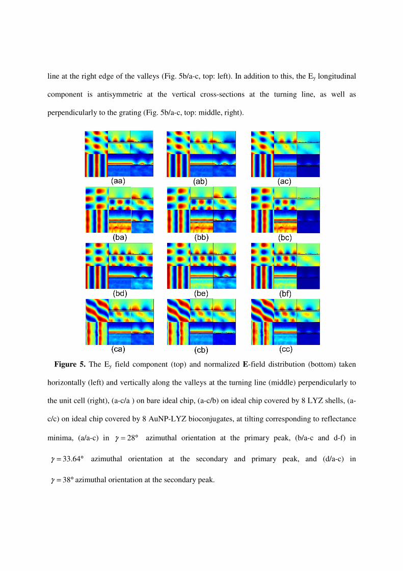
line at the right edge of the valleys (Fig. 5b/a-c, top: left). In addition to this, the Ey longitudinal
component is antisymmetric at the vertical cross-sections at the turning line, as well as
perpendicularly to the grating (Fig. 5b/a-c, top: middle, right).
Figure 5. The Ey field component (top) and normalized E-field distribution (bottom) taken
horizontally (left) and vertically along the valleys at the turning line (middle) perpendicularly to
the unit cell (right), (a-c/a ) on bare ideal chip, (a-c/b) on ideal chip covered by 8 LYZ shells, (a-
c/c) on ideal chip covered by 8 AuNP-LYZ bioconjugates, at tilting corresponding to reflectance
minima, (a/a-c) in °= 28γ azimuthal orientation at the primary peak, (b/a-c and d-f) in
°= 64.33γ azimuthal orientation at the secondary and primary peak, and (d/a-c) in
°= 38γ azimuthal orientation at the secondary peak.

These near-field phenomena prove that a two-fold antisymmetric long-range SPP mode
propagates along the edge of the valleys in this configuration, which ensure large interaction
cross-section with the covering biolayers.
In addition to this the E-field enhancement proves the co-existence of a glass side plasmon
(Fig. 5b/a-c, bottom: middle). The lateral extension of the E-field maxima ensures that both the
LYZ and the AuNP-LYZ bioconjugates at the bottom of valleys are inside areas shined with an
intense E-field originating from coupled LRSPPs (Fig. 5b/a-c, bottom: middle, right). The
secondary peak in reflectance is strongly sensitive to their presence, as a result the largest polar
angle shift is observable in this azimuthal orientation (Fig. 2d, 5b/a-c).
In contrast, at the primary peak appearing in °= 64.33_ chipidealγ azimuthal orientation smaller
areas correspond to global E-field maximal at the left border of the valleys, which are shifted
towards the valleys with smaller extent (Fig. 5b/d-f, bottom: left, right). The Ey longitudinal
component is antisymmetrical horizontally with a turning line at the right border of the valleys,
similarly to the secondary peak (Fig. 5b/d-f, top left). In contrast, the Ey longitudinal component
is hybrid on the vertical cross-sections at the turning line as well as perpendicularly to the grating
(Fig. 5b/d-f, top: middle, right). These near-field phenomena prove that a SRSPP mode
propagates along the edge of the valleys and is coupled with the LRSPP at the secondary peak in
this configuration. The smaller lateral extension of the E-field maxima makes possible smaller
spatial overlapping, and the SRSPPs can ensure smaller interaction cross-section with the LYZ
and the AuNP-LYZ conjugates (Fig. 5b/d-f, bottom: right). As a consequence, the primary peak
in reflectance is moderately sensitive to biomolecules presence, moreover a backward shift and
slight broadening is observable caused by their adherence (Fig. 2d, 5b/d-f).

Complementary studies revealed that adherence of larger amount of biomolecules can cause
disappearance of this peak, according to the critical nature of the configuration.
Finally, in the °= 38_ chipidealγ azimuthal orientation the E-field maxima cover completely the
left side of the valleys (Fig. 5c/a-c, bottom: left and right). The Ey longitudinal component is
hybrid horizontally as well as vertically at the turning lines at the valley edges and
perpendicularly to the grating (Fig. 5c/a-c, top). These field distributions indicate that SRSPP
modes exist in the valleys in this configuration. The location of both the LYZ and AuNP-LYZ
bioconjugates at the bottom of the valleys is inside the E-field intensity maxima. However, the
SRSPP in the valleys possesses a reduced interaction cross-section with the covering biolayers
caused by the inherently short propagation distance. As a consequence, the sensitivity of the
secondary reflectance peak is moderate in this azimuthal orientation.
The coupled two-fold antisymmetric LRSPP modes excited at the secondary peak in
°= 64.33_ chipidealγ azimuthal orientation are unique. Their E-field distribution and large
propagation distance makes possible sensitivity enhancement via enhanced interaction cross-
section with the LYZ shells and AuNP-LYZ bioconjugates, even if they are located at the bottom
of the valleys, not at the valley edge. The two-fold antisymmetric LRSPPs are secondary modes
at the edge of the valleys and hills, which originate from the intense laterally Bragg scattered
SPPs, accordingly their wave vector equals with the projection of the original SPP modes [23].
Comparison of the E-field and Ey distributions proves that the right edge of the valley can be
considered as the metal-dielectric interface of their propagation. The two-fold antisymmetry of
the Ey component ensures minimal attenuation of these modes.

The horizontal antisymmetry also holds for the modes coupled at the primary peak, however
they are accompanied by hybrid vertical Ey distribution at the edge of the valleys, as a
consequence they suffer larger attenuation.
The AuNPs in the bioconjugates are resonantly excited both at °= 64.33_ chipidealγ and
°= 38_chipideal
γ azimuthal angles, which results in further enhanced sensitivity. Although, the
local E-field enhancement is the largest in °= 38_ chipidealγ azimuthal orientation, i.e. when the E-
field maxima overlap better with the AuNP arrays, the SRSPPs suffer larger attenuation in the
valleys. As a consequence, larger enhancement was detected in the secondary peak shift in
°= 64.33_ chipidealγ azimuthal orientation, due to the enhanced interaction cross-section with the
unique LRSPPs (Multimedia corresponding to Fig. 5 about the Ey field distribution at the
secondary and primary minima in different azimuthal orientations).
3.6 Dispersion characteristics of the modes accompanying RGC-SPR
The dispersion characteristics of the ideal chip was mapped at °= 64.33_ chipidealγ azimuthal
orientation and shows that two anti-crossing plasmonic bands coexist. The lower branch is
similar to the usual unperturbed plasmonic band on flat dielectric-metal interface, however, it is
deformed caused by the splitting originating from rotated grating-coupling. The hybridized
modes are similar in their horizontally antisymmetrical components, which is in accordance with
the antisymmetric phase map formed during Bragg scattering at the optimal azimuthal
orientation. However, they differ in their vertical characteristics, which is antisymmetric and
hybrid at the turning lines along the edges of the valleys for the secondary and primary peak,
respectively (Fig. 5). On the ideal chip at the secondary peak a unique rotated grating coupled

LRSPP dominates due to the two-fold antisymmetry, this LRSPP is strongly coupled with a
SRSPP mode at the primary peak.
Figure 6. Dispersion characteristics in reflectance of (a ) bare ideal chip, (b) ideal chip covered
by 8 LYZ shells, (c) ideal chip covered by 8 AuNP-LYZ bioconjugates, in °= 64.33γ azimuthal
orientation. (d) Difference between reflectance modifications, (e, f) modifications of reflectance
on ideal chip caused by seeding with (e) 8 LYZ shells and (f) 8 AuNP-LYZ bioconjugates
compared to the bare ideal chip. Comparison of secondary and primary peak locations (g) 8
AuNP-LYZ to LYZ, (h/i) 8 LYZ / 8 AuNP-LYZ covered to bare ideal chip
Conclusion
Our study revealed that there is a definite maximum on the absorption spectrum of AuNP-LYZ
nanodispersion with mAu:mLYZ=1:5 around 532 nm wavelength. Accordingly, the SPR
measurements were performed on this dispersion and the numerical computations were realized
for seeding by LYZ shells and AuNP-LYZ bioconjugates with this mass ratio. The SPR
measurements performed in conical mounting proved that monitoring the shift of the secondary
peaks, which originate from rotated grating-coupling in the optimal azimuthal orientation, is
more suitable to detect adhered bio-moieties, than the observation of the broader primary peaks
shift. These results are demonstrated via FEM computations on a fitted and an ideal biochip.
Numerical computations on the fitted chip revealed that the measured secondary peak shifts
can be reproduced by seeding significantly larger number of protein shells and smaller number of
bioconjugates at the bottom of the valleys. Comparative studies on the effect of biomoieties
prove that the secondary peak is more strongly shifted, except in case of LYZ protein location at
the hill-sides. The sensitivity can be maximized by ensuring that the location of biomolecules is
coincident with the turning line of the longitudinal Ey field component at the right edge of the
valleys, where the two-fold antisymmetric coupled LRSSPs propagate. The optimal location still
ensures overlapping with the global E-field maximum on the left side of the valley as well.
The enhanced / moderate sensitivity of the resonance peaks is the consequence of the large /
small interaction cross-section achievable via long- / short-range modes propagating at the edges
of the valleys.
The near-field images taken on the ideal chip demonstrated that the E-field is confined onto
the hills / at the edge of the hills and valleys / and onto the left side of the valleys of the polymer
grating, when no primary peak shift / maximal secondary peak shift / moderate secondary peak
shift is observable. These E-field distributions are accompanied by hybrid / antisymmetrical
vertically as well as horizontally with respect to the valley edge / hybrid longitudinal Ey field-
component distribution. This proves that short-range / two-fold antisymmetric long-range /
short-range modes exist in these configurations in the valleys, where the biomoieties are adhered.
Each biosensing multilayer has its own optimal configuration, which can be qualified by the
optimal azimuthal orientation, capable of resulting in the largest polar angle shift. It was also
demonstrated that in presence of Au NPs the E-field confinement is further enhanced around
them. The presence of Au NPs enhanced the difference between polar angles corresponding to
reflectance peaks on bare and biomoieties covered chips. The larger shift in presence of Au NPs
reveals that these particles can be used to enhance detection sensitivity. This enhancement is
more pronounced for the secondary peak in the optimal azimuthal orientation. In conclusion,
considerable sensitivity enhancement is achievable in the critical RGC-SPR condition of the
ideal chip due to the secondary plasmonic modes originating from Bragg scattered of SPPs,
which is further enhanced, when bioconjugates are detected.
ASSOCIATED CONTENTS
Supporting Information
Movies: Modification of longitudinal Ey component at the secondary(left) and primary (right)
peak at different azimuhal orientation on horizontal (top) and vertical cross-sections (bottom).
AUTHOR INFORMATION
Corresponding Author
*36-62-544654, 36-62-544657, [email protected]

Funding Sources
The research was supported by National Research, Development and Innovation Office-NKFIH
through project “Optimized nanoplasmonics” K116362 and “Synthesis, structural and
thermodynamic characterization of nanohybrid systems at solid-liquid interfaces” K116323.
Mária Csete acknowledges that the project was supported by the János Bolyai Research
Scholarship of the Hungarian Academy of Sciences.
ACKNOWLEDGEMENT:
The authors would like to thank Hajnalka Milinszki and Lóránt Szabó for figures preparation.
REFERENCES:
(1) Elson, J. M. Light scattering from surfaces with a single dielectric overlayer. J. Opt. Soc.
Am. 1976, 66/7, 682-694.
(2) Giannattasio, A.; Hooper, I. R.; Barnes, W. L. Dependence on surface profile in grating-
assisted coupling of light to surface plasmon-polaritons. Opt. Comm. 2006, 261, 291-295.
(3) Wang, B.; Lalanne, P. Surface plasmon polaritons locally excited on the ridges of metallic
gratings. J. Opt. Soc. Am. 2010, 27/6, 1432-1441.
(4) Bozhevolnyi, S. I.; Sonergaard T. General properties of slow-plasmon resonant
nanostructures: nano-antennas and resonators. Optics Express 2007, 15/17, 10869.
(5) D’Aguanno G.; Mattiucci, N.; Alú, A.; Bloemer, M. J. Quenched optical transmission in
ultrathin subwavelength plasmonic gratings. Phys. Rev B 2011, 83, 035426.

(6) Chen, Y. J.; Koteles, E. S.; Seymour, R. J.; Sonek, G. J.; Ballantyne, J. M. Surface
plasmons on gratings: coupling in the minigap regions. Solid State Comm., 1983, 46/2, 95-99.
(7) Weber, M. G.; Mills, D. L. Determination of surface-polariton minigaps on grating
structures: A comparison between constant-frequency and constant-angle scans. Phys. Rev. B
1986, 34/4, 2893-2894.
(8) Barnes, W. L.; Preist, T. W.; Kitson, S. C.; Sambles, J. R.; Cotter, N. P. K.; Nash, D. J.
Photonic gaps in the dispersion of surface plasmons on gratings. Phys. Rev. B 1995, 51/16,
11164–11167.
(9) Ameling, R.; Giessen, H. Cavity Plasmonics: Large Normal Mode Splitting of Electric and
Magnetic Particle plasmons Induced by a Photonic Microcavity. Nano Letters 2010, 10, 4394.
(10) Ghoshal, A.; Divliansky, I.; Kik, P. G. Experimental observation of mode-selective
anticrossing in surface-plasmon coupled metal nanoparticle arrays. Appl. Phys. Lett. 2009, 94,
171108/3 pages.
(11) Mills, D. L. Interaction of surface polaritons with periodic surface structures; Rayleigh
waves and gratings. Phys. Rev. B 1977, 15, 3097–3118.
(12) Seshadri, S. R. Coupling of surface polaritons incident obliquely on a small amplitude
grating. J. Appl. Phys. 1985, 58/5, 1733-1738.
(13) Hibbins, A. P.; Sambles, J. R.; Lawrence, C. R. Azimuth-angle-dependent reflectivity data
from metallic gratings. Journal of Modern Optics 1998, 45/5, 1019-1028.
(14) Kretschmann, M.; Leskova, A.; Maradudin, A. A. Conical propagation of a surface
plasmon polariton across a grating. Optics Communications 2003, 215, 205-223.

(15) Romanato, F.; Hong, L. K.; Kang, H. K.; Wong, C. C.; Yun, Z.; Knoll, W. Azimuthal
dispersion and energy mode condensation of grating-coupled surface plasmon polaritons. Phys.
Rev. B 2008, 77, 245435.
(16) Csete, M.; Vass, Cs.; Kokavecz, J.; Goncalves, M.; Megyesi, V.; Bor, Zs.; Pietralla, M.;
Marti, O. Effect of sub-micrometer polymer gratings generated by two-beam interference on
surface plasmon resonance. Appl. Surf. Sci. 2005, 247/1-4, 477-485.
(17) Csete, M.; Szekeres, G.; Vass, Cs.; Maghelli, N.; Osvay, K.; Bor, Zs.; Pietralla, M.;
Marti, O. Surface plasmon resonance spectroscopy on rotated sub-micrometer polymer gratings
generated by UV laser based two-beam interference. Appl. Surf. Sci. 2006, 252/13, 4773-4780.
(18) Csete, M.; Kőházi-Kis, A.; Vass, Cs.; Sipos, Á.; Szekeres, G.; Deli, M.; Osvay, K.; Bor,
Zs. Atomic force microscopical and surface plasmon resonance spectroscopical investigation of
sub-micrometer metal gratings generated by UV laser based two-beam interference in Au-Ag
bimetallic layers. Appl. Surf. Sci. 2007, 253, 7662-7671.
(19) Szalai, A.; Szekeres, G.; Balázs, J.; Somogyi, A.; Csete, M. Rotated grating coupled
surface plasmon resonance on wavelength-scaled shallow rectangular gratings. Proc. SPIE 8809,
Plasmonics: Metallic Nanostructures and Their Optical Properties XI, 88092U
(20) Parisi, G.; Zilio, P.; Romanato, F. Complex Bloch-modes calculation of plasmonic crystal
slabs by means of finite element method. Optics Express 2010, 20/15, 16690-16703.
(21) Gazzola, E.; Brigo, L.; Zacco, G.; Zilio, P.; Ruffato, G.; Brusatin, G.; Romanato, F.
Coupled SPP Modes on 1D Plasmonic Gratings in Conical Mounting. Plasmonics 2013, 9, 867.

(22) Kim, D. Effect of the azimuthal orientation on the performance of grating-coupled
surface-plasmon resonance biosensors. Appl. Opt. 2005, 44/16, 3218-3223.
(23) Csete, M.; Kőházi-Kis, A.; Megyesi, V.; Osvay, K.; Bor, Zs.; Pietralla, M.; Marti, O.
Coupled surface plasmon resonance on bimetallic films covered by sub-micrometer polymer
gratings. Org. Electronics 2007, 8/2-3, 148-160.
(24) Csete, M.; Sipos, Á.; Kőházi-Kis, A.; Szalai, A.; Szekeres, G.; Mathesz, A.; Csákó, T.;
Osvay, K.; Bor, Zs.; Penke, B.; Deli, M. A.; Veszelka, Sz.; Schmatulla, A.; Marti, O.
Comparative study of sub-micrometer polymeric structures: dot-arrays, linear and crossed
gratings generated by UV laser based two-beam interference, as surfaces for SPR and AFM
based bio-sensing. Appl. Surf. Sci. 2007, 254/4, 1194-1205.
(25) Tóháti, H.; Sipos, Á.; Szekeres, G.; Mathesz, A.; Szalai, A.; Jójárt, P.; Budai, J.; Vass,
Cs.; Kőházi-Kis, A.; Csete, M.; Bor, Zs. Surface plasmon scattering on polymer-bimetal layer
covered fused silica gratings generated by laser-induced backside wet etching. Appl. Surf. Sci.
2009, 255/10, 5130-5137.
(26) Sipos, Á.; Tóháti, H.; Mathesz, A.; Szalai, A.; Veszelka, Sz.; Deli, M. A.; Fülöp, L.;
Kőházi-Kis, A.; Csete, M.; Bor, Zs. Effect of nanogold particles on coupled plasmon resonance
on biomolecule covered prepatterned multilayers. Sensor Lett. 2010, 8, 512-520.
(27) Romanato, F.; Lee, K. H.; Kang, H. K.; Ruffato G.; Wong, C. C. Sensitivity enhancement
in grating coupled surface plasmon resonance by azimuthal control Optics Express 2009, 17/14,
12145-12154.
(28) Brigo1, L.; Gazzola, E.; Cittadini, M.; Zilio, P.; Zacco, G.; Romanato, F.; Martucci, A.;
Guglielmi, M.; Brusatin, G. Short and long range surface polariton waveguides for xylene
sensing. Nanotechnology 2013, 24, 155502.
(29) González, M. U.; Weeber, J.-C.; Baudrion, A.-L.; Dereux, A.; Stepanov, A. L.; Krenn, J.
R.; Devaux, E.; Ebessen, T. W.; Design, near-field characterization, and modeling of 45°
surface-plasmon Bragg mirrors. Phys. Rev. B 2006, 73, 155416/3 pages.
(30) Randhawa, S.; González, M. U.; Renger, J.; Enoch, S.; Quidant, R. Design and properties
of dielectric surface plasmon Bragg mirrors. Opt. Exp. 2010, 18/14, 14496-14510.
(31) Homola, J.; Yee, S. S.; Gauglitz, G. Surface plasmon resonance sensors: review. Sens.
Actuators B 1999, 54, 3-15.
(32) Kim, D.; Shuler, M. L. Design and development of grating coupled optical biosensor to
detect animal pathogen. Proc of SPIE 2004, 5321, Biomedical Vibrational Spectroscopy and
Biohazard Detection Technologies, 309-314.
(33) Lukosz, W. Integrated optical chemical and direct biochemical sensors. Sens. Actuators B
1995, 29. 37-50.
(34) Ramsden, J. J.; Optical Biosensors. J. Mol. Recognit. 1997, 10, 109-120.
(35) Brockman, J. M.; Fernandez, S. M. Grating-coupled surface plasmon resonance for rapid,
label-free, array-based sensing. Am. Lab. 2001, 33. 37-40.
(36) Zieziulewicz, T. J.; Unfricht, D. W.; Hadjout, N.; Lynes, M. A.; Lawrence, D. A.
Shrinking the biologic world – nanobiotechnologies for toxicology. Toxicol. Sci. 2003, 74. 235-
244.

(37) Senlik, S. S.; Kocabas, A.; Aydinli, A.; Grating based plasmonic band gap cavities. Opt.
Exp. 2009, 17/18, 15541-15549.
(38) Wang B.; Wang, G. P. Plasmon Bragg reflectors and nanocavities on flat metallic
surfaces. Appl. Phys. Lett. 2005, 87, 013107.
(39) Shi, H.; Liu, Z.; Wang, X.; Guo, J.; Liu, L.; Luo, L.; Guo, J.; Ma, H.; Sun, S.; He, Y. A
symmetrical optical waveguide based surface plasmon resonance biosensing system. Sensors and
Actuators B 2013, 185, 91.
(40) Lal, S.; Link, S.; Halas, N. J. Nano-optics from sensing to waveguiding. Nature Photonics
2007, 1, 641-648.
(41) Anker, J. N.; Hall, W. P.; Lyandres, O.; Shah, N. C.; Zhao, J.; Van Duyne, R. P. Bio-
sensing with plasmonic nanosensors. Nature 2007, 7, 442-453.
(42) Luk'yanchuk, B.; Zheludev, N. I.; Maier, S. A.; Halas, N. J.; Nordlander, P.; Giessen, H.;
C. T. Chong Nature Materials 2010, 9, 707-715.
(43) Formoso, C.; Forster, L. S. Tryptophan fluorescence lifetimes in lysozyme. Journal of
Biological Chemistry 1975, 250, 3738-3745.
(44) Luo, Z.; Yuan, X.; Yu, Y.; Zhang, Q.; Leong, D. T.; Lee, J. Y.; Xie, J. From aggregation-
induced emission of Au(I)-thiolate complexes to ultrabright Au(0)@Au(I)-Thiolate core-shell
nanoclusters. J. Am. Chem. Soc. 2012, 134, 16662-16670.
(45) Wei, H.; Wang, Z.; Yang, L.; Tian, S.; Hou, C.; Lu, Y. Lysozyme-stabilized gold
fluorescent cluster: Synthesis and application as Hg2+ sensor, Analyst. 2010, 135, 1406-1410.
(46) Chen, W. Y.; Lin, J. Y.; Chen, W. J.; Luo, L.; Diau, E.W.G.; Chen, Y.C. Functional gold
nanoclusters as antimicrobal agents for antibiotic-resistant bacteria. Nanomedicine, 2010, 5, 755-
764.
(47) Eby, D. M.; Schaeublin, N. M.; Farrington, K. E.; Hussain, S. M.; Johnson, G. R.
Lysozyme catalyses the formation of antimicrobal silver nanoparticles. ACS Nano 2009, 3, 984-
994.
(48) Hornok, V.; Csapó, E.; Varga, N.; Ungor, D.; Sebők, D.; Janovák, L.; Laczkó, G.; Dékány
I. Controlled syntheses and structural characterization of plasmonic and red-emitting
gold/lysozyme nanohybrid dispersions. Collid. Polym. Sci. 2015, 294, 49-58.
(49) Arwin, H. Optical properties of thin layers of bovine serum albumin, γ-globulin, and
hemoglobin. Applied Spectroscopy, 1986, 40/3, 313-318.
(50) Johnson P. B.; Christy R. W. Optical constants of the noble metals. Phys. Rev. B 1972,
6/12, 4370-4379.