designing and using rna scaffolds to assemble proteins in vivo
TRANSCRIPT

©20
12 N
atu
re A
mer
ica,
Inc.
All
rig
hts
res
erve
d.
protocol
nature protocols | VOL.7 NO.10 | 2012 | 1797
IntroDuctIonSynthetic RNA scaffolds are engineered noncoding RNAs (ncRNAs) designed to bind and organize specific proteins1. They rely on a simple principle: a designed gene is transcribed in vivo into an ncRNA whose secondary structure enables the controlled scaf-folding of heterologous proteins in Escherichia coli via aptamer domains. In the present context, aptamers are small, naturally occurring RNA secondary structures that bind specific protein targets. Aptamers can also be evolved in vitro to bind a specific target by SELEX (systematic evolution of ligands by exponential enrichment)2,3. As aptamers have numerous applications in the pharmaceutical industry, the library of available protein-aptamer pairs is extensive and well characterized4.
Engineered RNA molecules serve as a more versatile, rationally programmable alternative to protein-based scaffolding strategies, allowing a new level of access and control over spatial organiza-tion of proteins1. The scaffold’s primary sequence can be ration-ally designed to control both the distance and orientation between bound proteins, as well as their stoichiometry and the size of the overall complexes. Engineered RNA scaffolds are widely applicable in basic research, nanotechnology and biotechnology.
Natural and engineered ncRNANatural ncRNA molecules derive their diverse range of behav-iors from their unique ability to fold into complex tertiary structures with recognition and even catalytic properties5. These properties in turn inspired the RNA hypothesis for the origins of life5, and much effort has been devoted to evolving novel RNA functions by SELEX2,3. Recently, RNA molecules were rationally designed to perform specific tasks both in vitro and in vivo6–9, with scaffolding being a compelling new application1. As our understanding about the causal relationship between primary sequence, secondary structure and function grows, RNA is now viewed as a modular molecule with extensive engi-neering potential.
Advantages and applications of synthetic RNA scaffoldsScaffolding is widely used in nature. For example, multienzyme pathways are often physically and spatially organized onto clusters
through protein domain interactions10, microcompartments11,12 or natural RNA scaffolds13–15. Spatial organization helps direct sub-strate flow between interacting enzymes, limiting cross-talk and increasing the yields of sequential metabolic reactions16–22.
Previously, synthetic protein scaffolds were developed to direct flux in synthetic metabolic pathways (e.g., improving titers of mevalonate in E. coli17). These scaffolds were built by fusing three eukaryotic protein-protein interaction domains (PDZ, SH3 and GBD) and coexpressing proteins to be scaffolded as fusions with their cognate binding domains. This strategy allows for the localiza-tion and stoichiometric control of a limited number of proteins and was successfully applied to improve yields of hydrogen and glucaric acid synthesis in E. coli20,21. Plasmid DNA was also recently used for scaffolding and improving the titer of resveratrol, 1,2-propanediol and melvanoate22.
The attraction of RNA scaffolds is their ability to be rationally programmed using the rules of base pairing. This offers access to larger scaffolds, in which hundreds of proteins are gathered to work together, and confers the ability to control not only stoichiometry but also the distance and orientation between interacting proteins. With hundreds of different, orthogonal, characterized aptamer domains4, and thus an expansive range of different binding domains, RNA scaffolds may bring together large complex pathways.
RNA scaffolds are applicable in many areas in which the spatial organization of biomolecules is desirable. In the synthetic biology realm, RNA scaffolds bring an added level of control by offering a tunable platform to control the spatial organization of proteins1. However, current tested designs are limited to the assembly of two distinct aptamers, but can be, in principle, enlarged either by using the discrete system (see Experimental design) or by mixing different aptamers in given ratios. Scaffold and assembly size can also be further controlled by adding nonpolymerizing ‘ends’ or by using the discrete system as described in the Experimental design. Furthermore, RNA scaffold libraries will also be expanded by applying future advances in the RNA nanotechnology field23–25 or by inspiration from naturally existing structures (e.g., Dsra RNA14), as well as by designing directed evolution selections for functional ncRNA-mediated assemblies.
Designing and using RNA scaffolds to assemble proteins in vivoCamille J Delebecque1–4, Pamela A Silver1,2 & Ariel B Lindner3,4
1Department of Systems Biology, Harvard Medical School, Boston, Massachusetts, USA. 2Wyss Institute for Biologically Inspired Engineering, Boston, Massachusetts, USA. 3Institut National de la Santé et de la Recherche Médicale, Paris, France. 4Faculty of Medicine, Paris Descartes University, Paris, France. Correspondence should be addressed to P.A.S. ([email protected]) or A.B.L. ([email protected]).
Published online 6 September 2012; doi:10.1038/nprot.2012.102
rna scaffolds are synthetic noncoding rna molecules with engineered 3D folding harnessed to spatially organize proteins in vivo. Here we provide a protocol to design, express and characterize rna scaffolds and their cognate proteins within 1 month. the rna scaffold designs described here are based on either monomeric or multimeric units harboring rna aptamers as protein docking sites. the scaffolds and proteins are cloned into inducible plasmids and expressed to form functional assemblies. rna scaffolds find applications in many fields in which in vivo organization of biomolecules is of interest. rna scaffolds provide extended flexibility compared with Dna or protein scaffolding strategies through programmed modulation of multiple protein stoichiometry and numbers, as well as the proteins’ relative distances and spatial orientations. For synthetic biology, rna scaffolds provide a new platform that can be used to increase yields of sequential metabolic pathways.

©20
12 N
atu
re A
mer
ica,
Inc.
All
rig
hts
res
erve
d.
protocol
1798 | VOL.7 NO.10 | 2012 | nature protocols
Experimental designThe general workflow for the design, induction and analysis of RNA scaffold expression is illustrated in Figure 1.
RNA scaffold design: RNA Designer (Steps 1–8). RNA scaffold design and optimization uses RNA folding software that relies on a free energy minimization algorithm to predict sequences that are amenable to in vivo scaffolding (Table 1). RNA Designer26 (http://www.rnasoft.ca/cgi-bin/RNAsoft/RNAdesigner/rnadesign.pl), for example, can be used to design the primary sequence of an RNA molecule that folds into a desired secondary structure (Fig. 2).
This software computes an RNA sequence that folds into the specified secondary structure given a number of optional sequence constraints. In the present context, the sequence constraints to be specified consist of the sequence of aptamers, terminator and restriction enzyme sites that will be used. The secondary struc-ture specified depends on the RNA folding scheme to be used. The simulation should be run at physiological temperature (e.g., 37 °C for E. coli) and with a target GC percentage matching the genomic content of the organism in which the RNA will be expressed (e.g., about 51% for E. coli).
RNA scaffold design: choosing aptamers to tether proteins to the RNA scaffolds (Steps 1 and 2). To choose the aptamers for the scaffold to be designed, a number of parameters should be consid-ered: (i) the binding affinity between the aptamer and its binding (adaptor) protein should be as high as possible (e.g., nanomolar range); (ii) the binding should also be as specific as possible, lead-ing to mutually orthogonal aptamers; and (iii) the binding protein sequence should be optimized for bacterial expression in terms of codon use and stability.
One such set of mutually orthogonal and extensively studied aptamers are those from MS2 and PP7 bacteriophages27,28 with dissociation constants of about 82 nM (F6 aptamer) and 1 nM, respectively29,30 (Fig. 3). This set of two aptamers will enable repeti-tive scaffolding of two different proteins from a chosen pathway (e.g., [Fe-Fe] hydrogenase and ferredoxin1).
RNA scaffold design: choosing a folding scheme (Steps 3–8). RNA scaffolds can be expressed in vivo as discrete (Fig. 4a,b) or poly-merizing molecules (Fig. 4c). The latter requires a more complex approach to both design and characterization but allows for more complex architectures and RNA-RNA interactions to be studied in vivo (see Step 3B). Discrete scaffolds are easier to engineer and can also be used as tags for mRNA expression studies31,32.
For metabolic engineering purposes, the scaffolds should be modular in length and number of docking sites, as well as easy to characterize. For this application, we suggest using an initial dis-crete scaffold. This ncRNA contains multiple copies of the chosen aptamer flanked by spacers that define the relative distance and orientation between the folded aptamers, as successfully used in previous work aiming at mRNA tagging31,32 (Fig. 4a). This design can be serially cloned to reach a desired scaffold length (typically 96-mer or more; Fig. 4b).
To achieve more complex scaffold geometries, scaffolds can instead be made of polymerizing RNA molecules. This relies on the molecular cross-assembly of RNA strands on the basis of princi-ples from the toolbox of RNA and DNA nanotechnology, including symmetry and kinetic considerations, as well as Watson-Crick and
noncanonical interactions24,33,34. Polymerizing scaffolds are made of short RNA strands cross-polymerizing to create extended struc-tures. Much exploration remains to be done to better understand and further expand the library of assembling RNAs and assem-bly schemes. Here, we give recommendations on how to use both symmetry and kinetic assembly, which are key to our published assembly scheme1.
Our polymerizing scaffold design strategy1 relies on two princi-ples. First, the use of palindromic sequences minimizes the number of different interacting strands necessary to form the extended structures. By using sequence symmetry, it is possible to design nanotubes or two-dimensional sheets with only one or two differ-ent polymerizing strands, respectively24,35,36. Second, kinetic assem-bly pathways37 enable the assembly to occur isothermally. In our design, RNA molecules assemble in a two-step process in which all noninteracting regions are locked into metastable assembly-intermediate hairpins. They only unfold upon cross-interaction through their dimerization region, leading to the polymerization of the RNA into extended structures1.
RNA scaffold optimization (Steps 9–11). Often a given sequence output from RNA Designer folds into a number of alternative sec-ondary structures. To further optimize the structure, these should be analyzed using another RNA secondary structure program such as mfold38 or NUPACK39 (Table 1). By screening the top RNA Designer outputs, one should pick the sequence that optimizes the thermodynamic gap between the desired folding and the next most favorable structure. Finally, the candidate sequences should be
Choose aptamer sequences (Steps 1 and 2)
RNA scaffold optimization(Steps 9–11)
Synthesize scaffold(Steps 12 and 13)
Clone RNA scaffold into expression system(Steps 14–24)
In vivopull-down
qRT–PCRIn vitro
assembly
Expression analysis(Step 27)
Design scaffold secondary structure and use RNAdesigner to compute a sequence
(Steps 3–8)
Terminator PromoterRestriction
sitesAptamers
Target proteins onto the RNA scaffold(Steps 28–30)
Induce scaffold expression(Steps 25 and 26)
Figure 1 | General workflow for design, induction, expression and experimental testing of the RNA scaffold. Orange boxes represent sequence constraints to be input into RNA Designer.

©20
12 N
atu
re A
mer
ica,
Inc.
All
rig
hts
res
erve
d.
protocol
nature protocols | VOL.7 NO.10 | 2012 | 1799
screened against any ribosome binding sites using an RBS calcula-tor40 (https://salis.psu.edu/software/) in order to avoid translation and against complete match of free single-stranded regions with any endogenous mRNA using the Basic Local Alignment Search Tool (BLAST) server (http://blast.ncbi.nlm.nih.gov/Blast.cgi). The workflow for RNA scaffold design and optimization is illustrated in Figure 2.
Cloning the designed RNA scaffold into an expression system (Steps 12–24). We have taken a modular cloning approach in which the key elements—promoters, assembly tags, aptamer-protein binding sites and terminators—are separated by unique restriction
sites to allow for tuning of the fundamental properties of the scaf-fold through quick and precise changes (Fig. 5a). This cloning strategy allows users to switch between the two different scaffold architectures: (i) the concatemerization of the aptamer module to create large discrete scaffolds and (ii) assembly by polymerization of discrete ncRNA molecules (Fig. 5b). Once the construct is veri-fied, it is cloned into commercial plasmids (e.g., pETDuet-1, EMD Chemicals) for in vivo expression.
Expression systems (Steps 25–27). For initial characterization of a candidate RNA scaffold, we recommend using a T7 expression system. This well-characterized expression system can be used for
table 1 | List of useful online RNA design tools.
tool Developer Website summary
mfold University of Albany http://mfold.rna.albany.edu/?q=mfold/RNA-Folding-Form
RNA folding software; folding temperature and ionic conditions are fixed
NUPACK California Institute of Technology
http://www.nupack.org/ RNA software suite for design and folding analysis with the option of designing RNA reaction pathways
RNA Designer University of British Columbia
http://www.rnasoft.ca/cgi-bin/RNAsoft/RNAdesigner/rnadesign.pl
RNA design tool using the dot-bracket format; temperature and GC content are adjustable
RBS Calculator Penn State University https://salis.psu.edu/software/ Predicts translation initiation rate in bacteria; takes into account RNA secondary structures for predictions
Nucleotide BLAST
National Center for Biotechnology Information
http://blast.ncbi.nlm.nih.gov/Blast.cgi
BLAST compares nucleotide sequences to sequence database and calculates the statistical significance of any match
Primer-BLAST National Center for Biotechnology Information
http://www.ncbi.nlm.nih.gov/tools/primer-blast/
Uses the popular primer3 engine to design primers; results are submitted to BLAST to check for unwanted endogenous match
BioNumbers Harvard Medical School http://bionumbers.hms.harvard.edu/
Registry of useful biological numbers, including genomic GC contents
genormPLUS Biogazelle http://www.biogazelle.com/genormplus/
Algorithm to determine the most stable reference genes from a set of tested candidate reference genes in a given qPCR sample panel
Sequence constraintsFormat: NNNNCUCAGGANNNN
Secondary sequenceFormat: (((((.....)))))
Aptamer TerminatorRestriction
sites
Run RNA Designer
Folding temperature Target GC content
Discretedesign
Polymerizingdesign
Optimize RNAscaffold sequences
No single-stranded regionsmatching endogenous DNA
(BLAST)
No strong translationinitiation sites
(RBS calculator)
No alternate secondary structuremore or as favorable
(mfold)
Figure 2 | Designing and optimizing RNA scaffolds using RNA Designer. The design process starts with choosing both the sequence constraints and the secondary structure folding scheme. Parameters to input are linked through dashed lines: for the sequence constraints, aptamer, terminator and restriction enzyme site sequences need to be input, whereas the secondary structure should be input in dot-bracket format and will vary according to whether a discrete or polymerizing folding scheme is chosen. Run RNA Designer taking into account the folding temperature and a target GC content. Optimizing the RNA scaffold sequence output is a crucial part of the design process, and key parameters to consider are annotated with dashed lines. Bioinformatic programs used to check these parameters are shown in brackets.

©20
12 N
atu
re A
mer
ica,
Inc.
All
rig
hts
res
erve
d.
protocol
1800 | VOL.7 NO.10 | 2012 | nature protocols
preliminary in vitro expression studies using commercially avail-able IVT systems (e.g., MEGAscript T7 Kit from Invitrogen), and can be tuned for a wide range of expression levels, yielding very high expression levels upon full induction. We thus suggest clon-ing the RNA scaffold expression cassette into a pET-based vec-tor (e.g., pETDuet-1, EMD Chemicals) and transforming it into BL21 DE3 E. coli cells. Scaffold concentration is tunable in vivo by using different amounts of the inducing molecule (e.g., isopropyl β-d-1-thiogalactopyranoside (IPTG) or arabinose). A number of compatible T7-based plasmids have been developed and can be used for the simultaneous expression of up to six different proteins in addition to the RNA scaffold (e.g., EMD Chemicals Duet Vectors system). It should be noted that the T7 system is the only expression system tested in ref. 1, and other systems may also be useful.
DNA capture probes for scaffold expression analysis by in vivo pull-down assay (Step 27B(iii)). The DNA capture probe has two functional regions with well-defined melting temperatures (Tm). The binding domain is designed to interact with an ~10-bases-long constant region that is single stranded and nonassembled (e.g., the region between RE4 and the start of the terminator, see Fig. 5). The annealing temperature of this region should be ~20 °C. The release region of the DNA capture probe should be designed to be about 10 bases. These extra 10 bases should be designed so that they do not form hairpins with the binding domain and bring the Tm of the 20 bases full DNA capture probe to ~35 °C. The probe should then be ordered as a 5′-biotinylated oligo.
Primer design for quantitative reverse-transcription PCR (qRT-PCR; Step 27C(iii)). qRT-PCR of small RNAs with complex secondary structures can be complicated. One strategy used by commercial kits is to add a poly-A tail to the RNA scaffold during retrotranscription41 (see Step 27C(ii)). This provides a specific site to anneal a reverse primer for the qPCR. A specific forward primer is equally important and we recommend designing three of them with a dedicated software program using a target Tm of 60 °C (e.g., primer-BLAST; Table 1), hybridizing between the RE4 and the end of the terminator stem and assessing their performance by incorpo-rating a melting curve analysis step at the end of the PCR program (refer to instrument manual for specific programming). Choose a primer giving a clear single peak in the melting curve graph.
Stability considerations. Different strategies can be considered to enhance the stability of the RNA scaffold. RNA turnover is a natural component of gene expression. Half-lives of most bacterial RNAs range from 40 s to 60 min (ref. 42). Steady-state transcript concen-trations are a result of degradation and synthesis rate43. Strategies to enhance RNA scaffold level include the use of highly efficient expression systems (e.g., T7-based expression system), but also the implementation of design considerations to minimize decay.
In E. coli, 5′- and 3′-end accessibility is of particular importance in initiating the decay process. For example, RNase E requires
MS2 F6 aptamer
UU
U
U
U
C
C
C
C
C
C
G
G
G
G
G
G
G
G
A
A
A
A
A
A
PP7 aptamer 1.0
0.8
0.6
0.4
0.2
0
Equilibrium
probability
5′-GACTCCCACAGT 5′-GGCACAGAAGATCACTGGGGAGTC-3′ ATGGCTTCGTGCC-3′
U C
C
G A
A
U
U
U
C
C
C
C
C
G
G
G
G
G
A
A
A
10 bpClamp
G C
Figure 3 | Compatible aptamer pair. Left, primary sequence and secondary structure of the MS2 F6 aptamer and PP7 aptamers. Both aptamers are orthogonal to each other and are usable for RNA scaffolds. A 10-bp clamp (as indicated on figure) is added to the stem of the 14-bp MS2 F6 aptamer to adjust its length to that of the PP7 aptamer. Right, visualization with NUPACK35: the color-coded scale depicts the base-pairing probabilities.
cPolymerizingscaffold
aDiscrete scaffold
bLargediscretescaffold
RNA monomerself-interaction RNA
polymer
Figure 4 | Examples of RNA scaffold folding scheme designs. (a) Computed secondary structure of a small discrete scaffold design comprising two aptamer-protein binding sites. (b) Secondary structure of an extended discrete scaffold expressed as a repeat of the small discrete scaffold. (c) Proposed secondary structure for a polymerizing scaffold. Small RNA modules interact through single-stranded domains and polymerize into the multimeric scaffold. Visualization with NUPACK35.
RE1 Promoter RE2-RE3 Aptamers RE4 Term RE5
5′ 3′
EcoNI Promoter EcoRI-XbaI Aptamers SpeI Term BlpI
5′ 3′
Aptamers repeat
Assembly tag
a
b
Figure 5 | Modular cloning approach to the design of RNA scaffolds. (a) Separation of promoter (gray box), aptamer (green box) and terminator (term; blue box) sequences by five unique restriction sites (RE; purple and yellow boxes). The dotted arrow indicates the suggested region to hybridize probes for the pull-down assay (see Step 27B(iii)). (b) Suggested restriction enzymes: RE1 = EcoNI and RE5 = BlpI enable cloning into the pETDuet-1 vector (EMD Chemicals); RE2 = EcoRI, RE3 = XbaI and RE4 = SpeI enable repeated cloning of the aptamer domain to create large discrete scaffolds or the cloning of an assembly tag (see Step 3B(i)) to create polymerizing RNA molecules.

©20
12 N
atu
re A
mer
ica,
Inc.
All
rig
hts
res
erve
d.
protocol
nature protocols | VOL.7 NO.10 | 2012 | 1801
5′ single-stranded RNA of at least four nucleotides in length for efficient binding, whereas RNase II and PNPase are unable to bind substrates with fewer than six to ten unpaired bases at the 3′ end43. Therefore, minimizing single-stranded regions and locking both ends of the RNA scaffold with hairpins is an important part of the design strategy44 (Fig. 4). In addition, using RNase E knockout strains for expression (e.g., BL21 DE3 Star, Invitrogen) may also help further stabilize the RNA transcripts43.
Targeting proteins onto the RNA scaffold (Steps 28–30). The chosen aptamer-binding proteins are fused to the proteins to be scaffolded. For this step, we recommend exploring the space of possible linker length and fusion orientation so as to optimize both
scaffolding and protein interactions. It is also useful to examine three-dimensional structures of the protein domains to be used to determine whether an N or C terminus can be used as a fusion junction without interfering with the function of the protein.
Controls. Impairing RNA polymerization (Step 27A(iii)): short DNA oligos are designed to match and hybridize to the dimerization region of the polymerizing scaffold to prevent RNA polymerization. qRT-PCR control (Step 27C(v)): GapA or MreB E. coli housekeep-ing genes1 are used as internal references to assess the relative RNAs given their scaffold concentration after induction. Impairing protein binding (Step (30)): RNA scaffolds with aptamers mutated to poly-T and poly-A sequences are designed to prevent protein binding.
MaterIalsREAGENTS crItIcal All solutions and buffers should be kept sterile and be stored according to the manufacturer’s recommendation.Chemicals and solvents
DNA ladder, 1 kb Plus (Invitrogen, cat. no. 10787-018)TBE-urea gels (6% (wt/vol), Novex gels; Invitrogen, cat. no. EC68652BOX)Acetic acid (Fisher Scientific, cat. no. A35-500) ! cautIon Acetic acid is corrosive. Avoid inhalation and exposure to skin and eyes.Ampicillin (Sigma-Aldrich, cat. no. A9518)Bacterial peptone (Fisher Scientific, cat. no. BP1420-2)Bacterial protein extraction reagent (Pierce, cat. no. 78243)ddH
2O, sterile
Dithiothreitol (Invitrogen, cat. no. 15508-013)Dynabeads M-280 streptavidin (Invitrogen, cat. no. 112-05D)EDTA (Invitrogen, cat. no. AM9261)Ethidium bromide (1% (wt/vol) solution; Sigma-Aldrich, cat. no. E-8751) ! cautIon Ethidium bromide is mutagenic; always use nitrile gloves.Glycerol (Sigma-Aldrich, cat. no. G5516-100ML)Isopropyl β-d-1-thiogalactopyranoside (IPTG; Anatrace, cat. no. I1002)Lysozyme (Sigma-Aldrich, cat. no. L6876-5G)Potassium chloride (KCl; Sigma-Aldrich, cat. no. P5405)SeaKem LE Agarose (Lonza Cologne, cat. no. 50002)Sodium chloride (Fisher Scientific, cat. no. S671-500)Tris (tris(hydroxymethyl)aminomethane; Fisher Scientific, cat. no. BP152-5)Yeast extract (VWR, cat. no. 97063-370)Luria-Bertani broth (LB; Sigma-Aldrich, cat. no. L2542-500ML)
Bacterial strains and vectors E. coli cloning strain (Turbo competent E. coli; NEB, cat. no. C2984H)E. coli expression strain (One Shot BL21 Star (DE3) chemically competent E. coli; Invitrogen, cat. no. C6010-03)pETDuet-1 T7 expression vector (EMD Chemicals, cat. no. 71146-3)pACYCDuet-1 T7 expression vector (EMD Chemicals, cat. no. 71147-3)pCOLADuet-1 T7 expression vector (EMD Chemicals, cat. no. 71406-3)pCDFDuet-1 T7 expression vector (EMD Chemicals, cat. no. 71340-3)
Kits MEGAscript T7 kit (Invitrogen, cat. no. AM1334M or Applied Biosystems)NCode VILO miRNA cDNA synthesis kit (Invitrogen, cat. no. A11193-050) crItIcal This kit allows for the detection and quantification of small RNAs by qRT-PCR by adding a poly-A tail before the retrotranscription step.
•••
••••••••
•••••••••
••
••••
••
QIAprep spin miniprep kit (Qiagen, cat. no. 27104)QIAquick gel extraction kit (Qiagen, cat. no. 28704)SYBR Green Supermix (Invitrogen, cat. no. 4309155)Total RNA purification 96-well kit (Norgen, cat. no. 24300) crItIcal This column-based purification kit allows for the purification of total RNA without a size cutoff.
Enzymes Restriction endonucleases: EcoNI and BlpI (Fermentas, cat. nos. FD1304 and FD0094, respectively)T4 DNA ligase (Promega, cat. no. M1801)
Buffers Assembly buffer: 150 mM KCl, 0.1 mM dithiothreitol, 0.1 mM EDTA, 10 mM Tris, in ddH
2O; the buffer is made fresh and adjusted to pH 7.4
Primers and probes PCR primers (Integrated DNA Technologies) crItIcal All PCR prim-ers should be designed using appropriate software (e.g., primer-BLAST; Table 1), in order to avoid the formation of self- or heterodimers, as well as to avoid complementarity to genomic DNA (in order to avoid nonspecific amplifications). Primers should be diluted to 100 µM and kept at − 20 °C until use.DNA oligos (Integrated DNA Technologies)Biotinylated DNA capture probes (Integrated DNA Technologies)
EQUIPMENTAsylum MFP-3D atomic force microscope (Asylum)Cuvettes (Semi-Micro cuvettes; BrandTech Scientific, cat. no. 2711010)Eppendorf centrifuge (Eppendorf AG, cat. no. 5702R)Eppendorf Mastercycler ep realplex (Eppendorf, cat. no. 6302 000.601)Etched silicon cantilever (Olympus, cat. no. OMCL-AC160TS)Highest-grade V1 mica, 25 × 76 mm (Ted Pella, cat. no. 56)Incubation facilities for bacterial cultureLiquid nitrogenSpectrophotometer (Ultrospec 3100 pro; GE Healthcare, cat. no. 80-2112-32)
Bioinformatics RNA Design tools (see Table 1) RNA Designer (ref. 33—http://www.rnasoft.ca/cgi-bin/RNAsoft/RNAdesigner/rnadesign.pl)mfold (ref. 34—http://mfold.rna.albany.edu/?q=mfold/RNA-Folding-Form)NUPACK (ref. 35—http://www.nupack.org/)RBS calculator (ref. 36—https://salis.psu.edu/software/)Biogazelle geNorm Plus (http://www.biogazelle.com/genormplus/)
••••
•
•
•
•
••
•••••••••
•
••••
proceDurerna scaffold design ● tIMInG 2 h1| Start by choosing an appropriate set of aptamers (e.g., MS2 and PP7, see Experimental design and Fig. 3).
2| If the aptamer stem loops are of different lengths, add complementary bases at both sides of the shorter stem’s root in order to equilibrate their length (see Fig. 4; use RNA Designer).

©20
12 N
atu
re A
mer
ica,
Inc.
All
rig
hts
res
erve
d.
protocol
1802 | VOL.7 NO.10 | 2012 | nature protocols
3| Choose an appropriate folding scheme (see Experimental design) and design the secondary structure of the scaffold. Use option A for designing discrete scaffolds made of single RNA strands and option B for designing polymerizing scaffolds made of cross-assembling RNA strands.(a) Discrete scaffold design (i) Design the scaffold RNA strand to be relatively short, up to 200–300 bases, so that it can be synthesized. (ii) Design the scaffold so that aptamer binding sites are protruding and spaced according to your choice (e.g., 11 bp per
one RNA helix; Fig. 6a). (iii) Design a complementary 3′ region of 20–30 bases that folds back on itself, thus minimizing single-stranded regions
and locking the whole structure into a duplex (Fig. 6a, discrete scaffold bases 80–106).(b) polymerizing scaffold design (i) Start by designing the scaffold domain responsible for polymerization, the ‘assembly tag’ (Fig. 5). The assembly tag
is divided into two functional regions, both palindromic. The dimerization region is involved in the first step of the assembly process and it should remain available until assembly. To prevent it from collapsing because of its symmetric sequence, design the polymerization region so that it partly folds back onto the dimerization region (Fig. 6b). This both stabilizes the dimerization region and makes it available for the first step of the assembly process. Upon cross-interaction, the polymerization domain becomes available by strand displacement, and tiles can then assemble. crItIcal step Stabilizing the dimerization region using the polymerization region might require the use of wobble base pairing and some shuffling. See Figure 6 for a detailed example.
4| By using the dot-bracket format (Fig. 2), input the secondary structure of the desired scaffold according to its folding scheme (from Step 3) into RNA Designer.
5| Select and input the required primary sequence elements into the constraint field of RNA Designer according to Figure 2. Specify the sequence constraints according to Figure 2, which should include the RNA aptamer sequences (from Steps 1 and 2) and the rho-independent terminator. ‘N’ denotes positions at which any bases can be used. crItIcal step If you are using the T7 promoter, the last three bases of the promoter are transcribed and should be considered in the sequence constraints.
6| Match target GC content to the organism into which the scaffold is to be expressed (a good database: http://bionumbers.hms.harvard.edu/) and adjust the simulation temperature to the temperature of organism growth.
7| Ensure that secondary and primary structure inputs match and are of equal length.
8| Run RNA Designer.? troublesHootInG
rna scaffold optimization ● tIMInG 1 h9| Check the predicted structure of the top RNA Designer results and their theoretical stabilities (free energies) using a tool such as the mfold web server (table 1). Evaluate the
RNA Designer inputa b
Structure: 5′-(((((..... )))))((((.....))))..... .....((((.....))))((((( .....)))))-3′
Constraint: nnnnnnnnnnnnnnnnnnnnnnnnnnnnnnnnnnnnnnnnnnnnnnnnnnnnnnnnnnnnnnnnnnn
RNA Designer input
Structure: 5′-(((( aptamer1 ((((((((((( aptamer2(((((....))))).))))))))))).))))-3′
Constraint: nnnnApt1nnnnnnnnnnnApt2nnnnnnnnnnnnnnnnnnnnnnnnnnnnnnn
RNA Designer output(With MS2 = Apt1 and PP7 = Apt2)GGGAGGACTCCCACAGTCACTGGGGAGTCCTCGAATACGAGCTGGGCACAGAAGATATGGCTTCGTGCCCAGGAAGTGTTCGCACTTCTCTCGTATTCGATTCCC
mfold: secondary structure check
RNA Designer outputSelect output, remove extra sequencesand complete palindromesUAAGC GCUUA GCUUA CUG UA CAG UAAGCGAUUG AUCC GGAU CAAUC CAAUC GAUUG
TweakingAim: Get to desired secondary structure keeping palindromic sequenceTools: Shuffling, Wobble base pairing
UAGGC GCCUA GCCUA AUGU ACAU UAAGUUAUUU UUCC GGAU GAAUA GAAUA UAUUC
NB: 56 bases; two Wobble pairs and twomismatches here
mfold: secondary structure check
How to read this chart - RNADesigner conventions
( ) Encodes 2 assembling bases... Denotes noninteracting bases
A
A5′3′ A
AAA
AAA
AA
A
A
A
AA
GU
UUUU
U
UA
C
G
G
G
G
G
G
G
G
G
20
30
1050
40
U
U
U
U
U
C
C
CC
C
CC
C
UU
U
UU
U
U
U
C
CG
G
G
G
G
GG
G
GG
G
G 60
80
20
405′3′ 100
G
G
GG
GG
G
GG
G
G
G
G
GGG
A
A
A AA
A
A
A
AG
UU
U
U
U
U
UA
AA
A
AA
AA
A
G
U
U
UU
UU
AA
AC
C
CC
CC
CC
C
CC
CC
C
C
C C
CCCCC
C
U
U
U
U UU
U
U
U
U
C
C
CC
A
U
Figure 6 | Example of RNA Designer input and output sequences and structures. (a) RNA Designer input and output for a discrete scaffold design (D0 in ref. 1). The example is taken here with 11 bp in between the aptamers (one RNA helix) to make them point in the same direction. (b) RNA Designer input and output required for a polymerizing scaffold design (D1 assembly tag in ref. 1). The folding scheme used here is 10 bp–18 bp–18 bp–10 bp, with the 10-bp domains (in red) being the polymerization region and the 18-bp domains (in blue) being the dimerization region. We first probe with RNA Designer to fold the polymerization domain onto the dimerization domain, thus creating the metastable intermediate. We also ask it to start making palindromes by asking for hairpins (in yellow—these extra sequences are added to enable RNA Designer to find a solution). Some tweaking might be necessary to keep the metastable intermediate folding once the palindromes are manually completed. This involves using G-U (Wobble pairs) or A-T base pairs instead of G-Cs (‘shuffling’) to weaken some regions until the correct folding is achieved in mfold.
©20
12 N
atu
re A
mer
ica,
Inc.
All
rig
hts
res
erve
d.
protocol
nature protocols | VOL.7 NO.10 | 2012 | 1803
probability of alternative secondary-structure formation by comparing the free energy of predicted alternative structures with that of the desired structure (see Experimental design).
10| Select the most stable primary sequence with the desired secondary structure, which optimizes the thermodynamic gap between the desired folding and the next most favorable structure.? troublesHootInG
11| Confirm that the selected primary sequence is lacking any strong ribosome binding sites using, for example, the RBS calculator web server (table 1).
cloning the designed rna scaffold into an expression system ● tIMInG 12–15 d12| Add the features enabling expression and cloning to the selected primary sequence from Step 11: the cloning site (RE1) and full promoter on the 5′ end, and the chosen cloning site (RE5) after the terminator on the 3′ end.
13| Order the full construct as a synthetic gene cloned into a high-copy cloning vector.? troublesHootInG
14| By using standard protocols45, transform competent bacterial cells (e.g., heat shock–competent E. coli Turbo cells from NEB) with about 0.1 ng of the high-copy plasmid containing the scaffold.
15| Spread the transformed bacteria on LB plates (100–150 µl of bacterial suspension per plate) containing the appropriate antibiotic for selection (e.g., 100 mg ml − 1 ampicillin for high-copy-number E. coli plasmids of the pUC or pBluescript series) and incubate them overnight at 37 °C.
16| Inoculate a colony into an individual aliquot of 5 ml of LB medium supplemented with the correct antibiotic (e.g., 100 µg ml − 1 ampicillin) and incubate the culture overnight at 37 °C.
17| Isolate the plasmid using a plasmid isolation kit (e.g., Qiagen miniprep kit) according to the manufacturer’s instructions.
18| Digest the plasmid using a suitable combination of restriction enzymes flanking the cassette (e.g., Fermentas FastDigest EcoNI and BlpI restriction enzymes; Fig. 5b) according to the manufacturer’s instructions.
19| Separate the restriction enzyme–digested samples by electrophoresis in 1.5–2% (wt/vol) agarose gels, using standard protocols45. Verify the fragment size by using a suitable DNA ladder.
20| Purify the cassette-containing DNA using an agarose gel purification kit according to the manufacturer’s instructions.? troublesHootInG
21| Digest the chosen expression vector (e.g., EMD Chemicals pETDuet-1 vector) with restriction enzymes corresponding to the 5′ and 3′ overhangs of the cassette (e.g., Fermentas FastDigest EcoNI and BlpI restriction enzymes) according to the manufacturer’s instructions.
22| Ligate the purified cassette from Step 20 into the digested expression vector from Step 21, using a T4 DNA ligase (e.g., Promega T4 DNA ligase) using standard protocols45.
23| Transform into competent bacterial cells (e.g., Invitrogen heat shock–competent E. coli BL21 DE2-star for the suggested T7-based expression system) using standard protocols45.
24| Identify correct clones by running purified and digested plasmids in a gel electrophoresis to confirm insert size corresponding to your scaffold using standard protocols45. This should be further confirmed by sequencing.? troublesHootInG pause poInt Correct clones can be kept as glycerol stocks at − 80 °C.
Induction of rna scaffold expression ● tIMInG 1–2 d25| Inoculate one colony of the scaffold-bearing strain from Step 23 into liquid growth medium (e.g., LB or a minimal growth medium for E. coli) with the appropriate antibiotic (e.g., 100 mg ml − 1 ampicillin) in a 5-ml culture tube.
©20
12 N
atu
re A
mer
ica,
Inc.
All
rig
hts
res
erve
d.
protocol
1804 | VOL.7 NO.10 | 2012 | nature protocols
26| After overnight culture at 37 °C, make a 1:20 dilution of the culture with fresh LB medium and the appropriate antibi-otic and let it grow until an optical density (OD) of ~0.3 at 600 nm (as measured by a spectrophotometer) is reached (about 2 h, see ref. 45) and induce scaffold production with the appropriate molecule (e.g., with 0.1–1 mM IPTG). crItIcal step Induction conditions are crucial. Cells should be incubated at the temperature at which the RNA scaffold was designed (i.e., 37 or 30 °C). Induce the cells when they reach the beginning of exponential phase (i.e., OD 0.2–0.3).
expression analysis27| Proceed with expression analysis using one or more of options A–C, depending on whether the RNA scaffold is designed to polymerize. Option A is suitable for studying RNA polymerization in vitro and relies on the choice of a T7, T3 or SP6 promoter in the design. Option B is suitable for purifying in vivo–produced RNA samples or RNA-protein complexes. It is a modified pull-down assay in which cross-linking is not necessary and biological samples are not denatured. Option C enables the precise quantification of in vivo RNA scaffolds.(a) In vitro assembly ● tIMInG 2–3 d (i) Set up an in vitro transcription reaction using an appropriate kit (e.g., MEGAscript T7 kit, Applied Biosystems). Use 500 ng
of linearized purified plasmid (e.g., digested with Fermentas FastDigest PstI and purified with the QIAquick PCR purifica-tion kit, according to the manufacturers’ protocols) containing the RNA scaffold expression cassette from Step 24. Perform in vitro transcription overnight at 37 °C according to the manufacturer’s instructions. crItIcal step Circular plasmid templates will generate extremely long heterogeneous RNA transcripts because RNA polymerases are processive and rho-independent terminators may not provide efficient termination in vitro. Thus, it is preferable to digest the plasmid with an appropriate restriction enzyme downstream of the RNA scaffold cassette. It is worthwhile to also gel-purify the linearized plasmid.
(ii) Purify the in vitro–transcribed RNA scaffold using the Norgen RNA purification kit according to the manufacturer’s instructions. pause poInt The purified in vitro–transcribed RNA scaffold may be stored at − 20 °C for 2–3 d. It is recommended that samples be placed at − 80 °C for long-term storage.
(iii) Deposit ~25 ng of the RNA onto freshly cleaved mica (2.0 cm2), and allow it to dry for 20 min at the appropriate assembly temperature (e.g., 30 or 37 °C). As an appropriate negative control, allow for assembly with a tenfold molar excess of DNA oligos designed to impair RNA interactions. These oligos should be fully complementary to the interacting regions of the RNA molecules (e.g., dimerization region in Fig. 6).
(iv) Visualize the presence of RNA assemblies (e.g., RNA polymers of length 10–100 nm) using an atomic force microscope (e.g., Asylum MFP-3D). Perform acquisition in air, at room temperature (~20 °C), using an etched silicon cantilever with a resonance frequency of ~300 kHz, a spring constant of ~42 N m − 1 and a tip radius of ~10 nm.
(b) In vivo pull-down assay ● tIMInG 2–3 d (i) Take 500 ml of induced cells from Step 26 and centrifuge the cells at 5,000g for 5 min at room temperature. (ii) Remove the supernatant and place the cells on ice. Perform cell lysis by adding 200 µl of ice-cold bacterial protein extraction
reagent and gently pipette up and down until the cell suspension is homogenous. Leave the suspension on ice for 5 min. (iii) Add 1 nmol of a DNA-biotinylated capture probe designed to specifically hybridize with the scaffold (see Experimental
design) and allow for interaction with the RNA scaffold at room temperature for 5 min. (iv) Add 1 mg of streptavidin-coated Dynabeads M-280 and allow for interaction with the sample for 5 min on ice. (v) Place the lysate on a magnet and rinse the beads twice with ice-cold assembly buffer made fresh. (vi) Resuspend the beads in 10 ml of assembly buffer complemented with 10 nM of a DNA probe fully complementary to
the capture probe. Incubate at 37 °C for 5 min with intermittent gentle hand shaking. (vii) Run on a 6% (wt/vol) TBE-urea gel and visualize the RNA scaffold with ethidium bromide staining according to the
manufacturer’s instructions. The purified scaffold should be visible as a clear single band. ? troublesHootInG
(c) In vivo expression analysis—qrt-pcr ● tIMInG 2–3 d (i) Purify RNA from 1 ml of induced cells from Step 26 and from 1 ml of noninduced cells from Step 23 using the Norgen
kit according to the manufacturer’s instructions. (ii) Prepare cDNA from 0.5 mg of purified total RNA using the NCode VILO kit according to the manufacturer’s instructions.
pause poInt cDNA may be kept in the provided buffer at − 20 °C. (iii) To a 4.5-ml aliquot of tenfold-diluted cDNA in ddH2O, add 3 ml of a 2 nM solution of a specific forward primer, the pro-
vided poly-A annealing reverse primer (according to the manufacturer’s instructions) and 7.5 ml of SYBR Green Supermix. (iv) Set up the PCR reaction using SYBR Green Supermix according to the manufacturer’s instructions and perform the qPCR
on an appropriate thermocycler (e.g., Eppendorf Mastercycler ep realplex) with the following program: 50 °C for 2 min, and then 95 °C for 5 min to activate the enzyme. The PCR cycles are then as follows: 15 s at 95 °C, 30 s at 60 °C and 30 s at 72 °C, repeated 45 times.
©20
12 N
atu
re A
mer
ica,
Inc.
All
rig
hts
res
erve
d.
protocol
nature protocols | VOL.7 NO.10 | 2012 | 1805
(v) Calculate the relative concentration of the RNA scaffold by using a stable mRNA internal reference (e.g., GapA or MreB for E. coli46): assess total RNA samples from Step 27C(i) for the concentration of the internal reference RNA. Next, calculate the relative concentration of each RNA sample (induced versus noninduced from Step 27C(i)) relative to the mRNA internal reference by using the Biogazelle qPCR software geNorm Plus (table 1) according to the manufacturer’s instructions. crItIcal step Good-quality, stable housekeeping genes are essential for high-quality RNA quantification by qRT-PCR46. We recommend designing primer pairs for at least three candidate housekeeping genes and assessing their stability in the assay conditions using dedicated software (e.g., geNorm; table 1). ? troublesHootInG
targeting proteins onto the rna scaffold ● tIMInG 2–3 d28| Make fusion proteins between the chosen aptamer proteins and the proteins to be scaffolded according to standard protocols (e.g., using the biobrick cloning system; see http://dspace.mit.edu/handle/1721.1/32535).
29| Clone the genes into compatible expression plasmids (e.g., Duet vector, EMD Chemicals) with appropriate restriction enzymes according to standard protocols45.
30| Co-transform the RNA scaffold plasmid from Step 23 alongside the protein-coding plasmid from Step 29 into competent bacterial cells (e.g., heat shock–competent E. coli BL21 DE2-star). Appropriate negative controls here are RNA scaffolds with mutated aptamers designed to prevent protein binding (e.g., modifying PP7 or MS2 stem loops to a stretch of T and A bases). Plate the transformed bacteria, pick colonies and identify correct clones according to standard protocols45. pause poInt Correct clones can be kept as glycerol stocks at − 80 °C and aliquots can be used for further experiments.
? troublesHootInGTroubleshooting advice can be found in table 2.
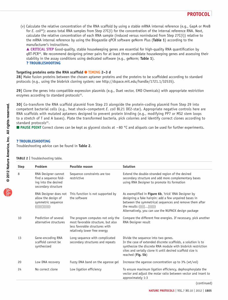
table 2 | Troubleshooting table.
step problem possible reason solution
8 RNA Designer cannot find a sequence fold-ing into the desired secondary structure
Sequence constraints are too restrictive
Extend the double-stranded region of the desired secondary structure and add more complementary bases using RNA Designer to promote its formation
RNA Designer does not allow the design of symmetric sequence (((((())))))
This function is not supported by the software
As exemplified in Figure 6b, ‘trick’ RNA Designer by designing a fake hairpin: add a few unpaired bases in between the symmetrical sequences and remove them after the results (((((...)))))) Alternatively, you can use the NUPACK design package
10 Prediction of several alternative structures
The program computes not only the most favorable structure, but also less favorable structures with relatively lower free energy
Compare the different free energies. If necessary, pick another RNA Designer result
13 Gene-encoding RNA scaffold cannot be synthesized
Long sequence with complicated secondary structures and repeats
Divide the sequence into two genes. In the case of extended discrete scaffolds, a solution is to synthesize the discrete RNA module with biobrick restriction sites and serially clone it until desired scaffold size is reached (Fig. 5b)
20 Low DNA recovery Fuzzy DNA band on the agarose gel Increase the agarose concentration up to 3% (wt/vol)
24 No correct clone Low ligation efficiency To ensure maximum ligation efficiency, dephosphorylate the vector and adjust the molar ratio between vector and insert to approximately 1:3
(continued)
©20
12 N
atu
re A
mer
ica,
Inc.
All
rig
hts
res
erve
d.
protocol
1806 | VOL.7 NO.10 | 2012 | nature protocols
● tIMInGSteps 1–8, RNA scaffold design: 2 hSteps 9–11, RNA scaffold optimization: 1 hSteps 12–24, cloning the designed RNA scaffold into an expression system: 12–15 d (Step 13 takes most of this time, typically 5–10 business days)Steps 25 and 26, induction of RNA scaffold expression: 1–2 dStep 27, expression analysis: 2–3 dSteps 28–30, targeting proteins onto the RNA scaffold: 2–3 d (typically Steps 28 and 29 can be done while waiting for the scaffold synthesis in Step 13)
antIcIpateD resultsThe described protocol results in a number of RNA scaffolds that are cloned and expressed in vivo and are capable of binding target proteins with prominent affinity and selectivity. Both polymerizing and discrete scaffolds can be designed, although, as discussed above, characterization is slightly more complex for polymerizing scaffolds as assembly efficiency should be assessed isothermally in vitro first (Step 27A). In vitro assembling candidates can then be evaluated for in vivo assembly and scaffolding efficiency by purifying them from cells (Step 27B). Polymerizing RNA scaffolds can be expected to reach tens of nanometers in size and gather hundreds of proteins. Discrete scaffolds can also gather hundreds of proteins depending on design choice (i.e., the number of aptamers). Aptamer occupancy was estimated to be of at least 70% for a repeat of 96 MS2 aptamers by Golding et al.31. It is expected to be equal or higher here because of the complementary 3′ region stabilizing the aptamers (Step 3A(iii)). Finally, depending on the expression system, fully induced cells can be expected to produce tens of thousands of RNA molecules without excessive metabolic burden1.
acknoWleDGMents We are indebted to F. Aldaye who had a leading role in this work. This work was supported by the Enerbio-Tuck Foundation and the Institut Français du Pétrole Energies Nouvelles (to C.J.D.); by the Agence Nationale de la Recherche France, Institut National de la Santé et de la Recherche Médicale (Unité 1001)–Institut National de Recherche en Informatique et en Automatique projet d’envergure, and an Axa Foundation Chair on Longevity (to A.B.L.); and by the Wyss Institute for Biologically Inspired Engineering and support from the Department of the Army W911NF-09-1-00226 (to P.A.S.).
autHor contrIbutIons All authors participated extensively in developing the protocol described in this paper.
coMpetInG FInancIal Interests The authors declare no competing financial interests.
Published online at http://www.nature.com/doifinder/10.1038/nprot.2012.102. Reprints and permissions information is available online at http://www.nature.com/reprints/index.html.
1. Delebecque, C.J., Lindner, A.B., Silver, P.A. & Aldaye, F.A. Organization of intracellular reactions with rationally designed RNA assemblies. Science 333, 470–474 (2011).
2. Ellington, A.D. & Szostak, J.W. In vitro selection of RNA molecules that bind specific ligands. Nature 346, 818–822 (1990).
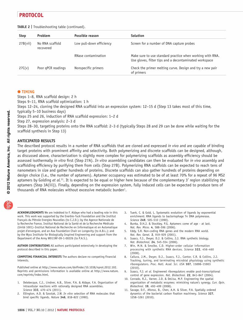
table 2 | Troubleshooting table (continued).
step problem possible reason solution
27B(vii) No RNA scaffold recovered
Low pull-down efficiency Screen for a number of DNA capture probes
RNase contamination Make sure to use standard practice when working with RNA. Use gloves, filter tips and a decontaminated workspace
27C(v) Poor qPCR readings Nonspecific primers Check the primer melting curve. Design and try a new pair of primers
3. Tuerk, C. & Gold, L. Systematic evolution of ligands by exponential enrichment: RNA ligands to bacteriophage T4 DNA polymerase. Science 249, 505–510 (1990).
4. Bunka, D.H.J. & Stockley, P.G. Aptamers come of age - at last. Nat. Rev. Micro. 4, 588–596 (2006).
5. Eddy, S.R. Non-coding RNA genes and the modern RNA world. Nat. Rev. Genet. 2, 919–929 (2001).
6. Isaacs, F.J., Dwyer, D.J. & Collins, J.J. RNA synthetic biology. Nat. Biotechnol. 24, 545–554 (2006).
7. Win, M.N. & Smolke, C.D. Higher-order cellular information processing with synthetic RNA devices. Science 322, 456–460 (2008).
8. Callura, J.M., Dwyer, D.J., Isaacs, F.J., Cantor, C.R. & Collins, J.J. Tracking, tuning, and terminating microbial physiology using synthetic riboregulators. Proc. Natl. Acad. Sci. USA 107, 15898–15903 (2010).
9. Isaacs, F.J. et al. Engineered riboregulators enable post-transcriptional control of gene expression. Nat. Biotechnol. 22, 841–847 (2004).
10. Conrado, R.J., Varner, J.D. & DeLisa, M.P. Engineering the spatial organization of metabolic enzymes: mimicking nature’s synergy. Curr. Opin. Biotechnol. 19, 492–499 (2008).
11. Savage, D.F., Afonso, B., Chen, A.H. & Silver, P.A. Spatially ordered dynamics of the bacterial carbon fixation machinery. Science 327, 1258–1261 (2010).

©20
12 N
atu
re A
mer
ica,
Inc.
All
rig
hts
res
erve
d.
protocol
nature protocols | VOL.7 NO.10 | 2012 | 1807
12. Burack, W.R. & Shaw, A.S. Signal transduction: hanging on a scaffold. Curr. Opin. Cell Biol. 12, 211–216 (2000).
13. Zappulla, D.C. & Cech, T.R. Yeast telomerase RNA: a flexible scaffold for protein subunits. Proc. Natl. Acad. Sci. USA 101, 10024–10029 (2004).
14. Cayrol, B. et al. A nanostructure made of a bacterial noncoding RNA. J. Am. Chem. Soc. 131, 17270–17276 (2009).
15. Shevtsov, S.P. & Dundr, M. Nucleation of nuclear bodies by RNA. Nat. Cell Biol. 13, 167–173 (2011).
16. Adam, G. & Delbriick, M. Reduction of dimensionality in biological diffusion processes. Struct. Chem. Mol. Biol. 198–215 (1968).
17. Dueber, J.E. et al. Synthetic protein scaffolds provide modular control over metabolic flux. Nat. Biotechnol. 27, 753–759 (2009).
18. Lee, H., DeLoache, W.C. & Dueber, J.E. Spatial organization of enzymes for metabolic engineering. Metab. Eng. 14, 242–251 (2012).
19. Park, S.-H., Zarrinpar, A. & Lim, W.A. Rewiring MAP kinase pathways using alternative scaffold assembly mechanisms. Science 299, 1061–1064 (2003).
20. Agapakis, C.M. et al. Insulation of a synthetic hydrogen metabolism circuit in bacteria. J. Biol. Eng. 4, 3–15 (2010).
21. Moon, T.S., Dueber, J.E., Shiue, E. & Prather, K.L.J. Use of modular, synthetic scaffolds for improved production of glucaric acid in engineered E. coli. Metab. Eng. 12, 298–305 (2010).
22. Conrado, R.J. et al. DNA-guided assembly of biosynthetic pathways promotes improved catalytic efficiency. Nucleic Acids Res. 40, 1879–1889 (2011).
23. Shu, D., Moll, W.-D., Deng, Z., Mao, C. & Guo, P. Bottom-up assembly of RNA arrays and superstructures as potential parts in nanotechnology. Nano Lett. 4, 1717–1723 (2004).
24. Guo, P. The emerging field of RNA nanotechnology. Nat. Nanotechnol. 5, 833–842 (2010).
25. Afonin, K.A. et al. In vitro assembly of cubic RNA-based scaffolds designed in silico. Nat. Nanotechnol. 5, 676–682 (2010).
26. Andronescu, M., Aguirre-Hernandez, R., Condon, A. & Hoos, H.H. RNAsoft: a suite of RNA secondary structure prediction and design software tools. Nucleic Acids Res. 31, 3416–3422 (2003).
27. Chao, J.A., Patskovsky, Y., Almo, S.C. & Singer, R.H. Structural basis for the coevolution of a viral RNA-protein complex. Nat. Struct. Mol. Biol. 15, 103–105 (2008).
28. Convery, M.A. et al. Crystal structure of an RNA aptamer-protein complex at 2.8 A resolution. Nat. Struct. Biol. 5, 133–139 (1998).
29. Lim, F., Downey, T.P. & Peabody, D.S. Translational repression and specific RNA binding by the coat protein of the Pseudomonas phage PP7. J. Biol. Chem. 276, 22507–22513 (2001).
30. Parrott, A.M. et al. RNA aptamers for the MS2 bacteriophage coat protein and the wild-type RNA operator have similar solution behaviour. Nucleic Acids Res. 28, 489–497 (2000).
31. Golding, I. & Cox, E.C. RNA dynamics in live Escherichia coli cells. Proc. Natl. Acad. Sci. USA 101, 11310–11315 (2004).
32. Valencia-Burton, M., McCullough, R.M., Cantor, C.R. & Broude, N.E. RNA visualization in live bacterial cells using fluorescent protein complementation. Nat. Methods 4, 421–427 (2007).
33. Aldaye, F.A., Palmer, A.L. & Sleiman, H.F. Assembling materials with DNA as the guide. Science 321, 1795–1799 (2008).
34. Seeman, N.C. An overview of structural DNA nanotechnology. Mol. Biotechnol. 37, 246–257 (2007).
35. Liu, H., Chen, Y., He, Y., Ribbe, A.E. & Mao, C. Approaching the limit: can one DNA oligonucleotide assemble into large nanostructures? Angew. Chem. Int. Ed. Engl. 45, 1942–1945 (2006).
36. Liu, H., He, Y., Ribbe, A.E. & Mao, C. Two-dimensional (2D) DNA crystals assembled from two DNA strands. Biomacromolecules 6, 2943–2945 (2005).
37. Yin, P., Choi, H.M.T., Calvert, C.R. & Pierce, N.A. Programming biomolecular self-assembly pathways. Nature 451, 318–322 (2008).
38. Zuker, M. Mfold web server for nucleic acid folding and hybridization prediction. Nucleic Acids Res. 31, 3406–3415 (2003).
39. Zadeh, J.N. et al. NUPACK: analysis and design of nucleic acid systems. J. Comput. Chem. 32, 170–173 (2011).
40. Salis, H.M., Mirsky, E.A. & Voigt, C.A. Automated design of synthetic ribosome binding sites to control protein expression. Nat. Biotechnol. 27, 946–950 (2009).
41. Benes, V. & Castoldi, M. Expression profiling of microRNA using real-time quantitative PCR, how to use it and what is available. Methods 50, 244–249 (2010).
42. Selinger, D.W. Global RNA half-life analysis in Escherichia coli reveals positional patterns of transcript degradation. Genome Res. 13, 216–223 (2003).
43. Richards, J., Sundermeier, T., Svetlanov, A. & Karzai, A.W. Quality control of bacterial mRNA decoding and decay. Biochimica et Biophysica Acta. 1779, 574–582 (2008).
44. Molinaro, M. & Tinoco, I. Use of ultra stable UNCG tetraloop hairpins to fold RNA structures: thermodynamic and spectroscopic applications. Nucleic Acids Res. 23, 3056–3063 (1995).
45. Tolia, N.H. & Joshua-Tor, L. Strategies for protein coexpression in Escherichia coli. Nat. Methods 3, 55–64 (2006).
46. Vandesompele, J. et al. Accurate normalization of real-time quantitative RT-PCR data by geometric averaging of multiple internal control genes. Genome Biol. 3, 1–12 (2002).