designer cells for stereocomplementary de novo … · designer cells for stereocomplementary de...
TRANSCRIPT

S1
Electronic Supplementary Information
Designer Cells for Stereocomplementary De
Novo Enzymatic Cascade Reactions Based on
Laboratory Evolution
Rubén Agudo and Manfred T. Reetz*
Philipps-Universität Marburg, Hans-Meerwein Str., 35032 Marburg, Germany
Max-Planck-Institut für Kohlenforschung, Kaiser-Wilhelm-Platz 1, 45470 Mülheim
an der Ruhr, Germany
*E-mail: [email protected]
Electronic Supplementary Material (ESI) for Chemical CommunicationsThis journal is © The Royal Society of Chemistry 2013

S2
Experimental Section
Reagents
E. coli BOU7301 electro-competent cells were prepared in-house according to standard protocols.
2 Restriction
enzyme DpnI was purchased from New England Biolabs. KOD hot start polymerase, dNTPs, MgSO4 were obtained from Novagen. GoTaq Hot Start was purchased from promega. DNAse I, carbenicillin and lysozyme were obtained from AppliChem. LB medium, in a form of premixed powder, and kanamycin were obtained from Roth. Chloramphenicol was obtained from Sigma. Glucose dehydrogenase (GDH) was purchased from Codexis. IPTG was obtained from Fermentas. NADP
+ was purchased from Calbiochem, Quick and Easy E. coli Gene Deletion Kit
were obtained from Gene Bridges. Compound 1 was obtained from Alfa Aesar. TB medium contained yeast extract (24 g/L), peptone (12 g/L), glycerol (4 mL/L), KH2PO4 (0.017 M) and K2HPO4 (0.072 M). Saturation mutagenesis and screening of P450-BM3 mutants
Saturation mutagenesis was carried out as described previously.1 It was performed by creating single residue libraries at positions P25, V26, F42, R47, Y51, S72, A74, L75, V78, F81, A82, F87, L181, L188, T260, I263, A264, E267, T268, A328, M354, L437 and T438 with NNK degeneration, and double residue libraries at positions F87/A328, A328/ P329, V78/L181 and V78/L437 with NDT degeneration. pRSF-P450BM31 vector was used as template for QuikChange PCR
3 in all cases. A comprehensive list of primers used appears in Table S1.
About 100 colonies were screened in the case of single amino acid libraries, while ca. 400 colonies were screened in the case of double amino acid libraries. These variants were subjected to the same UV-Vis screening based on NADPH consumption protocol published previously.
1 600 mutants showing the highest
spectrophotometric activity (initial NADPH consumption rates higher than the shown by P450 WT) were further subjected to GC analysis for production of compound 3 from starting material 1 following a procedure previously published elsewhere.
1
An additional single library created at position A328 with NNK degeneration using pRSF-P450-F87A vector (a pRSF-P450BM3 plasmid encoding F87A amino acid substitution previously created in our laboratory) as template, was also screened directly by this GC analysis protocol.
Screening of P450 mutants cloned in pETM11
After screening the above-mentioned P450 variants, best mutants found (i.e. F87A, F87P, F87V, F8W and A328N) showed only a slight improvement in the production of compound 3 from 1 relative to WT P450-BM3 (5-15% in comparison with 2-5%). Thus we decided to screen a set of additional P450-BM3 mutants from libraries created previously in our laboratory for a different purpose in which mutants were cloned into the pEMT11 vector
4
(a comprehensive list of mutants tested appears in Table S2). Aliquots of E.coli BL-21 Gold (DE3) glycerol stocks containing pETM11 plasmid harboring different P450-BM3 variants derived from F87A mutant were resuspended into 96-well plates containing LB medium (800 µL) with kanamycin (kan; 50 µg/mL). After overnight incubation at 37°C with gentle shaking, an aliquot (100 µL) was transferred to a new 96-well plate containing 900 µL TB medium with kan (50 µg/mL) and IPTG (0.2 mM). Cell cultures were incubated at 30°C during 20 h for overexpression of P450 variants. Cells were harvested by centrifugation (4000 r.p.m., 15 min) and pellets were resuspended in 500 µL of lysis buffer [phosphate buffer (pH 7.4, 100 mM), lysozyme (14 mg/mL), DNAse I (6 U/mL)], and incubated during 45 min at 37°C with agitation. After incubation, cellular debris was removed by centrifugation (4000 r.p.m., 30 min) and supernatants (250 µL) were transferred to 96-well plates containing reaction buffer (250 µL) [phosphate buffer (pH 7.4, 100 mM), compound 1 (2.5 mM in acetonitrile), glucose (100 mM), NADP
+ (250 µM) and
GDH (2 Units)]. After 20 h of incubation at 25°C with mild agitation, reaction mixtures were extracted with ethyl acetate (400 µL), and subjected to achiral GC analysis. Those variants which showed highest production of compound 3 (as average in three independent experiments) were chosen for further experiments (Table S3). The best P450 mutant in terms of regioselectivity was triple mutant encoding for amino acid substitutions V78L, A82F and F87A (termed P450-LFA). QuikChange mutagenesis on pRSF-P450-F87A
After screening P450 mutants cloned in pETM11 libraries, those variants showing the highest production of compound 3 were cloned into pRSF-P450-F87A vector by introduction of desired mutations by QuikChange mutagenesis.
3 PCR reactions contained 5 μL of 10 × KOD hot start polymerase buffer, 5 μL dNTPs (2 mM each),
1.25 μL of the appropriate forward and reverse primer (Table S4; 100 ng/μL each), 2 μL MgSO4 (25 mM), pRSF-P450BM3
as template (10-20 ng) and 0.5 μL of KOD polymerase in a final volume of 50 μL distilled water. PCR
1 R. Agudo, G. -D. Roiban, M. T. Reetz, ChemBioChem 2012, 13, 1465-1473.
2 J. Sambrook, D. W. Russell, ‘’The Condensed Protocols From Molecular Cloning: A Laboratory Manual’’ CSHL Press, NY, 2006.
3 a) QuikChange® Site-directed Mutagenesis Kit Instruction Manual, Stratagene, La Jolla, CA, USA; b) H. H. Hogrefe, J. Cline, G. L.
Youngblood, R. M. Allen, BioTechniques 2002, 33, 11581165. 4 S. Kille, F. E. Zilly, J. P. Acevedo, M. T. Reetz, Nat. Chem. 2011, 3, 738-743.
Electronic Supplementary Material (ESI) for Chemical CommunicationsThis journal is © The Royal Society of Chemistry 2013
S3
reaction started at 95°C (3 min), continued with 30 cycles of 95°C (1 min), 55°C as annealing temperature for all sets of primers used (1 min) and 68°C (10 min) and finished with 15 min at 68°C and storage at 4°C. After finishing, plasmid used as template in the reaction was removed by incubation of samples with 1.5 μL DpnI (10 U/ μL) overnight at 37°C. Digested products were purified with a QIAquick PCR purification spin column (Qiagen). An aliquot of 5 μL was used to transform 50 μL of electro-competent E. coli BOU730 cells. Transformation mixture was incubated with 900 μL LB medium at 37°C during 1 h with gentle shaking and then spread on LB-agar plates containing 50 μg/mL kan. Introduction of desired mutations in P450-BM3 coding region was confirmed for all constructions. Expression of desired P450-BM3 mutants was confirmed by SDS-PAGE analysis. Construction of pACYC-YqjM-C26D
Mutations encoding amino acid substitution C26D in YqjM protein from Bacillus subtilis were introduced by QuikChange mutagenesis PCR. Reactions contained 5 μL of 10 × KOD hot start polymerase buffer, 5 μL dNTPs (2 mM each), 1.25 μL of both YqjM-C26D_Fw and YqjM-C26D_rv primers (Table S5; 100 ng/μL each), 2 μL MgSO4
(25 mM), pBOU67804 as template (a derivative of pACYCDuet-1 plasmid harboring yqjM gene cloned between NcoI and AvrII sites)1 (10-20 ng) and 0.5 μL of KOD polymerase in a final volume of 50 μL distilled water. PCR reaction started at 95°C (3 min), continued with 20 cycles of denaturation [95°C (1 min)], annealing [53ºC (1 min)] and extension [68°C (10 min)]. Finally, an additional step of extension was performed [68°C (15 min)] and storage at 4°C. The sample was then subjected to digestion with 1.5 μL DpnI (10 U/ μL) overnight at 37°C for removal of the original template. Digested products were purified with a QIAquick PCR purification spin column (Qiagen) and an aliquot of 5 μL was used to transform 50 μL of electro-competent E. coli BOU730 cells. Transformation reaction was mixed with 900 μL LB medium and incubated at 37°C during 1 h. After incubation time, 100 µL were spread on LB-agar plates containing 28 μg/mL chloramphenicol (cam). Several individual colonies were grown and their plasmids extracted for confirming the introduction of the desired mutation by sequencing. Created plasmid was termed pACYC-YqjM-C26D. Expression of YqjM-C26D protein was confirmed by SDS-PAGE analysis.
Construction of pACYC-YqjM-C26D(gac) and pACYC-YqjM-C26G
Construction of a plasmid that encodes for YqjM-C26D protein, whose mutant codon is gac instead of gat, was performed exactly as described above except using YqjM-C26D(gac)_fw and YqjM-C26D(gac)_rv as primers (Table S5) for QuikChange PCR. For creation of a plasmid encoding YqjM-C26G protein, the same protocol described above was carried out, except using YqjM-C26G_fw and YqjM-C26G_rv as primers (Table S5) for QuikChange PCR. Created plasmids were termed pACYC-YqjM-C26D(gac) and pACYC-YqjM-C26G, respectively. Expression of both mutant proteins was confirmed by SDS-PAGE analysis. Constructions of E. coli BL21-Gold(DE3) ΔdkgA:: FRT-T7-gdh ΔnemA:: FRT-T7-yqjM_C26D [(R)-strain]
The (R)-strain was created by replacement of gene nemA from E.coli BOU730 for T7-yqjM_C26D cassette (YgjM gene from Bacillus subtilis with a mutated codon that encodes for amino acid substitution C26D placed downstream of T7 promoter sequence). T7-yqjM_C26D cassette was inserted in the place of nemA gene (a gene that encodes an endogenous flavoprotein belonging to Old Yellow Enzyme Family)
5 using Quick and Easy E. coli
Gene Deletion Kit. First, 5 μL (100 ng) of pBOU68408 vector (a plasmid derived from pACYC-Duet1 that encodes for FRT-PGK-
gb2-neo-FRT6-YqjM cassette which transcription is controlled by T7 promoter. Description of this plasmid is found
elsewhere)1 was amplified by QuikChange PCR to introduce the mutation encoding for C26D amino acid substitution (mutant gat codon instead of WT codon). PCR reaction was performed exactly as described above using primers YQJM_C26D_fw and YQJM_C26D_rv (Table S5). After reaction, the sample was purified, and transformed into electro competent BOU730 cells. Cells were spread on LB agar plates containing kan 50 μg/mL. Plasmids from 5 colonies were extracted and submitted to sequencing in order to confirm the desired mutation. Plasmid created was called pACYC-FRT-kan-FRT-T7-YqjM_C26D.
FRT-kan-FRT-T7-YqjM_C26D cassette was then amplified by PCR using pACYC-FRT-kan-FRT-T7-YqjM_C26D plasmid as template (100 ng) and primers Up-nemA-FRT (forward primer) and YqjM-Nem-rc (reverse primer) [PCR conditions: 3 μL of 10 × KOD buffer, 5 μL dNTPs (2 mM each), 1.25 μL of each primer (Table S5; 100 ng/μL each), 2 μL MgSO4 (25 mM) and 0.5 μL of KOD polymerase in a final volume of 30 μL distilled water. PCR reaction started at 95°C (3 min), continued with 30 cycles of 95°C (1 min), 55°C as annealing (1 min) 68°C (5 min) for elongation and finished with 15 min at 68°C and storage at 4°C]. The PCR product was purified using QIAquick Gel extraction kit (qiagen) and then transformed into electro competent BOU730 cells harboring pRedET (amp) vector (gene Bridges) that express proteins involved in λ –mediated recombination as described in Quick and Easy E. coli Gene Deletion Kit protocol. After transformation, cells were incubated at 37°C for 3 h in LB medium without antibiotics for elimination of pRedET (amp)
plasmid. Afterwards, 200 μL of cell culture were spread
5 a) K. Miura, Y. Tomioka, H. Suzuki, M. Yonezawa, T. Hishinuma, M. Mizugaki, Biol. Pharm. Bull. 1997, 20, 110-112; b) R. E. Williams, N. C.
Bruce, Microbiology 2002, 148, 1607-1614. 6 FRT-PGK-gb2-neo-FRT cassette confers resistance to kanamycin.
Electronic Supplementary Material (ESI) for Chemical CommunicationsThis journal is © The Royal Society of Chemistry 2013

S4
on a LB agar plate containing kan (15 µg/mL) and incubated at 37°C for 20 h. Twenty colonies from this plate were streaked on a new LB-agar plate containing kan (15 μg/mL) and screened by colony-PCR for checking the incorporation of FRT-kan-FRT-T7-YqjM_C26D cassette. [PCR conditions: 6 μL of 5 × GoTaq Hot Start buffer, 3 μL dNTPs (2 mM each), 0. 75 μL of primers Kan-Down-fw-test and YqjM-Nem-rc (Table S6; 100 ng/μL each), 1.2 μL MgSO4 (25 mM) and 0.3 μL of GoTaq Hot Start polymerase in a final volume of 30 μL distilled water. The PCR reaction started at 95°C (3 min), continued with 35 cycles of 95°C (1 min), 53°C as annealing temperature (1 min), 68°C for elongation (2 min) and finished with 15 min at 68°C and storage at 4°C]. Five single colonies were confirmed that contained the desired FRT-kan-FRT-T7-YqjM_C26D cassette inserted in the genome. After growing them in LB medium containing kan (50 μg/mL), four of them showed expression of YqjM mediated by T7 promoter after induction with 0.2 mM IPTG, as judged by SDS-PAGE analysis (Figure S1).
For removal of kanamycin selection marker, we proceeded as described in Quick and Easy E. coli Gene Deletion Kit protocol, transforming competent cells from the new created strain, E. coli BL21-Gold(DE3) ΔdkgA:: FRT-T7-gdh ΔnemA:: FRT-kan-FRT-T7-yqjM_C26D with 200 ng of pCP20 vector (Coli Genetic Stock Center, CGSC). Elimination of selection marker was confirmed by absence of resistance to kanamycin. Finally, genomic DNA from resulting strain was extracted and replacement of nemA gene for a single FRT site along with T7-yqjM_C26D was confirmed by PCR [conditions used were similar than described above, except using primers NemAr-fw and NemA-rc (Table S6)]. The resulting strain E. coli BL21-Gold(DE3) ΔdkgA:: FRT-T7-gdh ΔnemA:: FRT-T7-yqjM_C26D was termed (R)-strain. Constructions of E. coli BL21-Gold(DE3) ΔdkgA:: FRT-T7-gdh ΔnemA:: FRT-T7-yqjM_C26G [(S)-strain]
The (S)-strain was created following the same protocol described above for the (R)-strain except that in the first step pBOU68408 vector was amplified using primers YQJM_C26G_fw and YQJM_C26G_rv (Table S5).
Scale-up reactions of P450-BM3 mutants
In order to confirm results found in the screening of mutants cloned into pETM11, BOU730 cells containing best P450 variants cloned into pRSF-P450-F87A vector were grown to scale-up the model reaction 1 → 3. An individual colony of cells with the desired mutant was inoculated in LB medium (5 mL) containing kan (50 µg/mL). After 5 hours of incubation at 37ºC, this preinoculum was transferred to 50 mL of TB medium containing kan (50 µg/mL). The culture was maintained at the same temperature until an O.D. of 0.8-0.9 at 600 nm was reached. At that point, IPTG was added (0.2 mM final concentration) and the culture was incubated at 30ºC for 16-20 hours. Subsequently, 25 mL of cell culture were harvested at 4000 r.p.m. for 6 min at 4ºC and pellets were resuspended in reaction buffer [pH 7.4 (100 mM), NADP
+ (250 µM) and glucose (100 mM)]. The suspension was poured into a 100
mL flask and the reaction was started by addition of compound 1 (5 mM) and incubated at 25ºC overnight with mild agitation. After incubation, an aliquot of each reaction (700 µL) was extracted with the same volume of ethyl acetate and the organic phase was subjected to chiral and achiral GC analysis. Determination of the P450-BM3 concentration obtained after overexpression was performed as previously described elsewhere.
7 Final concentration values and activity obtained (measured as production of compound 3)
for each mutant appear in Table S3. Preliminary cascade reaction experiments
Several experiments were performed for setting up optimal conditions for all of the devised cascade reaction approaches. With this aim, an individual BOU730 colony containing either pRSF-P450-LFA (encoding for P450-LFA mutant), pACYC-YqjM-C26D, or both of them were inoculated in LB medium (5 mL) containing proper antibiotic [kan (50 µg/mL) or/and cam (28 μg/mL)]. After 5 hours of incubation at 37ºC, preinocula were transferred to 50 mL of TB medium supplemented with proper antibiotics as described above and cultures were incubated with gentle mixing at 37ºC until an O.D. of 0.8-0.9 at 600 nm was reached. At that point, 0.2 mM IPTG was added and the incubation temperature was decreased to 30ºC for 16-20 hours. After incubation, 25 mL of cell culture were harvested at 4000 r.p.m. for 6 min at 4ºC and pellets were resuspended in phosphate buffer (pH 7.4, 100 mM) containing different concentrations of NADP
+ (from 0 to 250 µM) and glucose (from 20 to 100 mM). Resting cells in
this buffer were poured into a 100 mL flask and the reaction was started by addition of different amounts of compound 1 (from 1 to 15 mM) and incubated at 25ºC for different time spans (from 1 to 18 hours). After incubation, an aliquot of each reaction (700 µL) was extracted with the same volume of ethyl acetate and the organic phase was subjected to chiral and achiral GC analysis.
For cascade reaction using approach (1) (i.e. P450 and YqjM proteins must be overexpressed separately and corresponding resting cells mixed in a one-pot manner) different volumes of phosphate buffer (2, 5, 10, 15 or 20 mL) were used to resuspend cells overexpressing either P450 or YqjM mutants. Different volume combinations of resting cells were mixed (at once, or sequentially starting with the one containing P450 protein) in an Erlenmeyer flask for finding optimum P450/YqjM ratio for cascade reaction 1→4. In these experiments it was also observed
7 F. P. Guengerich, M. V. Martin, C. D. Sohl, Q. Cheng, Nat. Protoc. 2009, 4, 1245-1251.
Electronic Supplementary Material (ESI) for Chemical CommunicationsThis journal is © The Royal Society of Chemistry 2013
S5
that the amount of compound 4 produced by YqjM mutant was dependent on the amount of compound 3 produced by P450 mutant.
Additional control experiments were performed to ensure that the three enzymes (i.e. GDH, P450 and YqjM) are in fact necessary for the production of compounds (S)/(R)-4. Briefly, an individual BOU730 colony empty, or containing either pACYC-YqjM-C26D or pRSF-P450-LFA plasmids alone, respectively, were grown, and subsequent cultures were incubated, induced, harvested and resuspended as described above in 5 mL of reaction buffer [(pH 7.4, 100 mM), NADP
+ (50 µM), glucose (100 mM). Reactions were started by addition of 1 mg of
compound 1 (1.5 mM, 7.5 µmol) and incubated at 25°C with mild agitation for 1 h in 100 mL Erlenmeyer flask with tight closure. Less than 3-4% of compound 4 was found in the three cases tested.
Likewise, we performed a similar experiment using E.coli BL21 (DE3) for checking the essential NADPH regeneration in the present cascade reaction system. Briefly: a single E.coli BL21 (DE3) colony containing pRSF-P450-LFA plasmid was inoculated in 5 mL LB medium containing kan 50 µg/mL, and the corresponding cell culture was grown and processed as described above. Cells were resuspended in reaction buffer (pH 7.4) containing NADP
+ (50 µM) and glucose (100 mM) and incubated in presence of 1 mg of compound 1 (1.5 mM, 7.5 µmol). In
this case, the amount of compound 3 observed after 1 hour incubation at 25ºC was less than 40% compare to 85% conversion into 3 using BOU730 cells under the same reaction conditions.
Control experiments demonstrated that exogenous addition of NADP+
was not necessary for carrying out any kind of enzymatic cascade reactions devised this study using 1 h of incubation time.
Multi-enzymatic cascade transformation approach (1)
For addressing this approach, both P450-BM3 and YqjM proteins were overexpressed separately in two independent cells. On the one hand, an individual BOU730 colony containing either pACYC-YqjM-C26D
8 or
pACYC-YqjM-C26G plasmids was inoculated in 5 mL of LB supplemented with 28 μg/mL cam and incubated for 5 h at 37ºC with gentle agitation. After incubation, the preculture was transferred to 50 mL of TB medium supplemented with 28 μg/mL cam and grown at 37ºC until an O.D. of 0.8-0.9 at 600 nm was reached. Then, IPTG was added (0.2 mM) and the culture was incubated for 16-20 additional hours at 30ºC with gentle agitation. 12 mL of each cell culture were centrifuged at 4000 r.p.m. for 6 min at 4ºC and pellets resuspended in 2 mL of reaction buffer [phosphate buffer (pH7.4, 100 mM) and glucose (100 mM, 67 equiv). Additionally, pellets can be stored for 2-3 hours at 4ºC before their resuspension in reaction buffer without any measurable loss of activity.
On the other hand, an individual BOU730 colony harboring plasmid pRSF-P450-LFA was inoculated in 5 mL of LB medium with kan (50 µg/mL). After 5 h of incubation at 37°C with shaking, this preculture was transferred to 50 mL of TB medium supplemented with kan (50 µg/mL) and the culture was grown at 37°C until an O.D. of 0.8-0.9 at 600 nm was reached. At this point, IPTG was added to a final concentration of 0.2 mM and the culture was grown at 30°C with vigorous agitation for 16-20 h. After incubation time, 25 mL of cell culture were centrifuged (6 min, 4000 r.p.m. at room temperature), the supernatant was discarded and the pellet resuspended in 5 mL of the same reaction buffer as described above. The resuspended solution was then poured into a 100 mL flask with tight closure (using rubber cap). The reaction was started by addition of compound 1 (1.5 mM, 7.5 µmol) and incubated at 25°C with mild agitation for 1 h.
After this incubation time, the 2 mL of resting cells expressing either YqjM-C26D or YqjM-C26G were added to the corresponding reaction flasks. After 15 min of additional incubation at the same conditions described above, an aliquot of 700 µL was extracted with ethyl acetate (700 µL) and the organic layer was subjected to GC analysis.
Similar experiments were performed using 5 mg of compound 1 as starting material (7.3 mM, 36.5 µmol). In this case, reactions in the presence of resting cells expressing P450 were incubated for 5 h before addition of resting cells expressing YqjM. Then the reaction was incubated for one more hour. Extraction of organic phase was performed as described above.
The amount of P450-BM3 protein was determined following the protocol described previously elsewhere.9
Average concentration of total P450-BM3 mutant V78L/A82F/F87A was 27.2 ±4.9 µM (three independent measurements), while average concentration value for active P450 fraction was 25.4 ±3.2 µM (three independent measurements), in agreement with P450-BM3 WT concentration (24.8±4.2 µM, Table S3). The concentration of YqjM protein was estimated by comparison of the samples containing unknown amount of YqjM with a standard curve of known protein (BSA). Analyses were performed by densitometry of samples run in SDS-PAGE. The concentration of YqjM-C26D and YqjM-C26G were 21.6 ±5.3 and 18.9 ±4.8 µM respectively (average of three independent measurements).
Multi-enzymatic cascade transformation approach (2)
For performing enzymatic reactions in an one-pot system using two plasmids, pRSF-P450-LFA vector and either pACYC-YqjM-C26D(gac)8 or pACYC-YqjM-C26G vectors were transformed into the same cell.
8 YqjM-C26D protein used in “Multi-enzymatic cascade transformation approach (1)” protocol is encoded by pACYC-YqjM-C26D plasmid,
while YqjM-C26D protein used in “Multi-enzymatic cascade transformation approach (2)” protocol is encoded by pACYC-YqjM-C26D(gac) plasmid. 9 F. P. Guengerich, M. V. Martin, C. D. Sohl, Q. Cheng, Nat. Protoc. 2009, 4, 1245-1251.
Electronic Supplementary Material (ESI) for Chemical CommunicationsThis journal is © The Royal Society of Chemistry 2013

S6
Exploratory experiments showed that the amount of YqjM protein obtained using pACYC-YqjM-C26D together with pRSF-P450-LFA was lower than using pACYC-YqjM-C26G together with pRSF-P450-LFA (data not shown). Thus, pACYC-YqjM-C26D(gac) plasmid was used to produce YqjM-C26D mutant in the cascade transformation approach (2).
An individual BOU730 colony transformed with pRSF-P450-LFA and either pACYC-YqjM-C26D(gac) or pACYC-YqjM-C26G plasmid was inoculated in 5 mL of LB supplemented with 28 μg/mL cam and 50 μg/mL kan, and incubated for 5 h at 37ºC with gentle agitation. After this time, the whole volume was transferred to 50 mL of TB medium supplemented with 28 μg/mL cam and 50 μg/mL kan and grown at 37ºC until an O.D. of 0.8-0.9 at 600 nm was reached. Then, IPTG was added to a final concentration of 0.2 mM and the culture grown for 16-20 additional hours at 30ºC with gentle agitation. 25 mL of cell culture was pelleted at 4000 r.p.m. for 6 min at 4ºC, supernatant discarded and the cell pellet resuspended in 5 mL of the same reaction buffer described above. The reaction was started by the addition of 1 mg of compound 1 (1.5 mM, 7.5 µmol) and incubation at 25°C with mild agitation for 1 h. After this incubation time, 700 µL of sample was extracted with ethyl acetate (700 µL) and the organic layer subjected to GC analysis.
Similar experiments were carried out following this cascade transformation approach but using lysates instead of resting cells. Briefly: After overexpression of P450-BM3 and YqjM mutant proteins, pellets were resuspended in 5 mL of lysis buffer [phosphate buffer (pH 7.4, 100 mM), lysozyme (14 mg/mL), DNAse I (6 U/mL)]. After 30 min of incubation at 37ºC, samples were centrifugated at 5000 r.p.m. for 15 min at 4ºC. Then 4.5 mL of supernatant were diluted to 5 mL with the same reaction buffer described above containing 250 µM NADP
+. Reaction was started,
incubated and organic and products extracted and analyzed as described above. Concentration of P450 and YqjM proteins present in cells used for cascade approach (2) was estimated by comparison of samples with unknown amount of P450 and YqjM with a standard curve of known protein (BSA). Analyses were performed by densitometry of samples run in SDS-PAGE.
The concentration of total P450-BM3 mutant V78L/A82F/F87A in cells used for approach (2) was 26.1 ±3.0 µM (Average of six independent experiments). The concentrations of YqjM-C26D and YqjM-C26G were 10.0 ±2.1 and 10.4 ±0.8 µM, respectively (average of three independent measurements).
Multi-enzymatic cascade transformation approach (3)
The pRSF-P450-LFA plasmid was transformed either into (S)- or (R)-strains in order to perform enzymatic reaction using one plasmid system with engineered cells containing YqjM genes inserted into the E. coli genome.
An individual colony of either (S)-strain or (R)-strain transformed with pRSF-P450-LFA was inoculated in 5 mL of LB medium supplemented with 50 μg/mL kan, and incubated for 5 h at 37ºC with gentle agitation. After incubation, 5 mL were transferred into 50 mL of TB medium supplemented with kan (50 μg/mL) and grown at 37ºC until an O.D. of 0.8-0.9 at 600 nm was reached. Then, IPTG was added to a final concentration of 0.2 mM and the culture grown for 16-20 additional hours at 30ºC with gentle agitation. Posterior centrifugation, resuspension in reaction buffer, enzymatic reaction and extraction with ethyl acetate were carried out as described above in “Multi-enzymatic cascade transformation approach (2)” protocol. The concentration of P450 and YqjM present in the cells used in this cascade transformation approach was determined as described above for cascade approach (2). The average concentration of total P450-BM3 mutant V78L/A82F/F87A in cells used for approach (3) was 27.3 ±3.1 µM µM (six independent experiments). The concentrations of YqjM-C26D and YqjM-C26G were 3.6 ±0.7 and 2.7 ±0.6 µM, respectively (average of three independent measurements).
Checking cell viability
The integrity of resting cells used in each cascade reaction approach was checked. After finishing the reaction, 0.1 and 1 µL aliquots of reaction mixture containing resting cells from each cascade reaction approach were inoculated in 4 mL of LB medium containing proper antibiotics (i.e. kan (50 mg/L) for cells overexpressing LFA P450 mutant, cam (28 mg/l) for cells transformed with pACYC and both antibiotics for cells cotransformated with both pACYC and pRSF plasmid derivatives). After overnight incubation at 37ºC, all bacterial cultures showed normal growth as judged by the observed turbidity. In control experiments, 0.1 and 1 µL of reaction mixture containing resting cells from non-transformed BOU730 cells were inoculated in 4 mL of antibiotic-free LB medium. After overnight incubation, the cultures showed normal growth with no differences regarding results indicated above.
Determination of conversions, yield and enantiomeric excess (ee)
Products obtained from biotransformation of compound 1 were analyzed by GC and GC/MS. Compounds 3, (S)-4 and (R)-4 were identified by comparison with previously published results1
,10 or by NMR analysis (see below).
10
D. J. Bougioukou, S. Kille, A. Taglieber, M. T. Reetz, Adv. Synth. Catal. 2009, 351, 3287–3305.
Electronic Supplementary Material (ESI) for Chemical CommunicationsThis journal is © The Royal Society of Chemistry 2013

S7
Yields were calculated on the basis of GC data using as standard a known amount of racemic sample of compound 4 (93% GC purity) synthesized as described previously.
11
11
M. Sugi, D. Sakuma, H. Togo, J. Org. Chem. 2003, 68, 7629-7633.
Electronic Supplementary Material (ESI) for Chemical CommunicationsThis journal is © The Royal Society of Chemistry 2013
S8
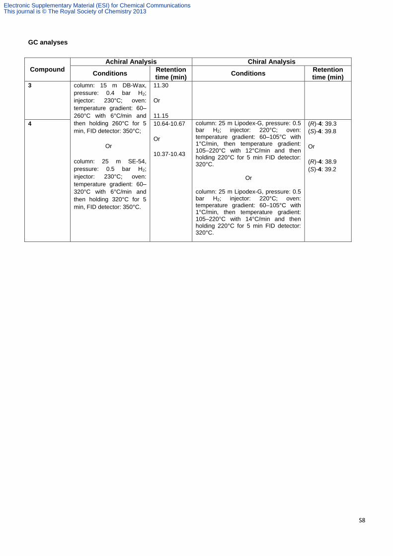
GC analyses
Compound
Achiral Analysis Chiral Analysis
Conditions Retention time (min)
Conditions Retention time (min)
3
column: 15 m DB-Wax,
pressure: 0.4 bar H2;
injector: 230°C; oven:
temperature gradient: 60–
260°C with 6°C/min and
then holding 260°C for 5
min, FID detector: 350°C;
Or
column: 25 m SE-54,
pressure: 0.5 bar H2;
injector: 230°C; oven:
temperature gradient: 60–
320°C with 6°C/min and
then holding 320°C for 5
min, FID detector: 350°C.
11.30
Or
11.15
4 10.64-10.67
Or
10.37-10.43
column: 25 m Lipodex-G, pressure: 0.5 bar H2; injector: 220°C; oven: temperature gradient: 60–105°C with 1°C/min, then temperature gradient: 105–220°C with 12°C/min and then holding 220°C for 5 min FID detector: 320°C. Or column: 25 m Lipodex-G, pressure: 0.5 bar H2; injector: 220°C; oven: temperature gradient: 60–105°C with 1°C/min, then temperature gradient: 105–220°C with 14°C/min and then holding 220°C for 5 min FID detector: 320°C.
(R)-4: 39.3
(S)-4: 39.8
Or
(R)-4: 38.9
(S)-4: 39.2
Electronic Supplementary Material (ESI) for Chemical CommunicationsThis journal is © The Royal Society of Chemistry 2013
S9
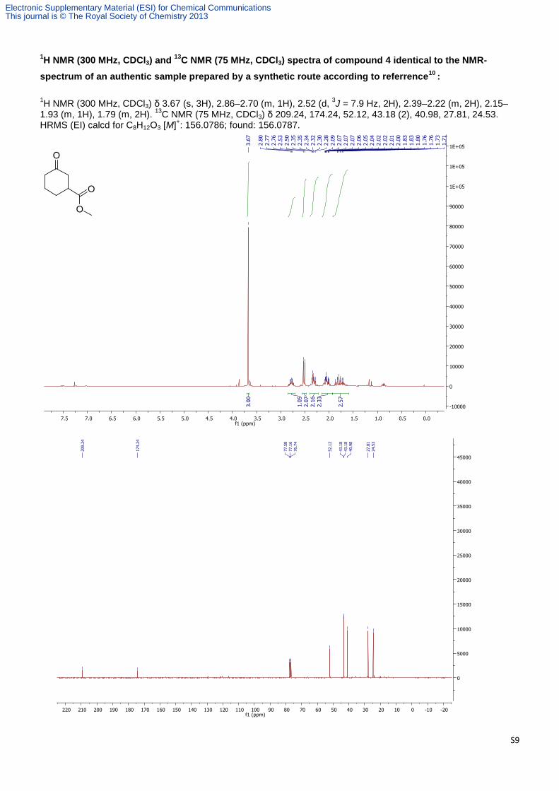
1H NMR (300 MHz, CDCl3) and
13C NMR (75 MHz, CDCl3) spectra of compound 4 identical to the NMR-
spectrum of an authentic sample prepared by a synthetic route according to referrence10
:
1H NMR (300 MHz, CDCl3) δ 3.67 (s, 3H), 2.86–2.70 (m, 1H), 2.52 (d,
3J = 7.9 Hz, 2H), 2.39–2.22 (m, 2H), 2.15–
1.93 (m, 1H), 1.79 (m, 2H). 13
C NMR (75 MHz, CDCl3) δ 209.24, 174.24, 52.12, 43.18 (2), 40.98, 27.81, 24.53. HRMS (EI) calcd for C8H12O3 [M]
+: 156.0786; found: 156.0787.
Electronic Supplementary Material (ESI) for Chemical CommunicationsThis journal is © The Royal Society of Chemistry 2013

S10
Supporting Tables
Table S1. Primers used for saturation mutagenesis in this study.
Name Sequence (5’→3’)
BM3_P25_nnk_fw CCGTTATTAAACACAGATAAANNKGTTCAAGCTTTGATG
BM3_P25_nnk_Rv CATCAAAGCTTGAACMNNTTTATCTGTGTTTAATAACGG
BM3_V26_nnk_fw CCGTTATTAAACACAGATAAACCGNNKCAAGCTTTGATG
BM3_V26_nnk_Rv CATCAAAGCTTGMNNCGGTTTATCTGTGTTTAATAACGG
BM3_F42_nnk_fw GGAGAAATCTTTAAANNKGAGGCGCCTGGTCG
BM3_F42_nnk_Rv CGACCAGGCGCCTCMNNTTTAAAGATTTCTCC
BM3_R47_nnk_fw CGAGGCGCCTGGTNNKGTAACGCGCTACTTATCAAG
BM3_R47_nnk_Rv CTTGATAAGTAGCGCGTTACMNNACCAGGCGCCTCG
BM3_Y51_nnk_fw CCTGGTCGTGTAACGCGCNNKTTATCAAGTCAGCGTC
BM3_Y51_nnk_Rv GACGCTGACTTGATAAMNNGCGCGTTACACGACCAGG
BM3_S72_nnk_fw CGCTTTGATAAAAACTTANNKCAAGCGCTTAAATTTGTACG
BM3_S72_nnk_Rv CGTACAAATTTAAGCGCTTGMNNTAAGTTTTTATCAAAGCG
BM3_S74_nnk_fw CGCTTTGATAAAAACTTAAGTCAANNKCTTAAATTTGTACG
BM3_S74_nnk_Rv CGTACAAATTTAAGMNNTTGACTTAAGTTTTTATCAAAGCG
BM3_S75_nnk_fw CGCTTTGATAAAAACTTAAGTCAAGCGNNKAAATTTGTACG
BM3_S75_nnk_Rv CGTACAAATTTMNNCGCTTGACTTAAGTTTTTATCAAAGCG
BM3_V78_ndt_fw GTCAAGCGCTTAAATTTNDTCGTGATTTTGCAGGAGAC
BM3_V78_ndt_Rv GTCTCCTGCAAAATCACGAMNAAATTTAAGCGCTTGAC
BM3_V78_nnk_fw GTCAAGCGCTTAAATTTNNKCGTGATTTTGCAGGAGAC
BM3_V78_nnk_Rv GTCTCCTGCAAAATCACGMNNAAATTTAAGCGCTTGAC
BM3_F81_nnk_fw CGCTTAAATTTGTACGTGATNNKGCAGGAGACGGG
BM3_F81_nnk_Rv CCCGTCTCCTGCMNNATCACGTACAAATTTAAGCG
BM3_A82_nnk_fw GCTTAAATTTGTACGTGATTTTNNKGGAGACGGGTTATTTACAAGC
BM3_A82_nnk_Rv GCTTGTAAATAACCCGTCTCCMNNAAAATCACGTACAAATTTAAGC
BM3_F87_ndt_fw GCAGGAGACGGGTTGNDTACAAGCTGGACG
BM3_F87_ndt_Rv CGTCCAGCTTGTAHNCAACCCGTCTCCTGC
BM3_F87_nnk_fw GCAGGAGACGGGTTGNNKACAAGCTGGACG
BM3_F87_nnk_Rv CGTCCAGCTTGTMNNCAACCCGTCTCCTGC
BM3_L181_ndt_fw CAAGTATGGTCCGTGCANDTGATGAAGCAATGAACAAGC
BM3_L181_ndt_Rv GCTTGTTCATTGCTTCATCAHNTGCACGGACCATACTTG
BM3_L181_nnk_fw CAAGTATGGTCCGTGCANNKGATGAAGCAATGAACAAGC
BM3_L181_nnk_Rv GCTTGTTCATTGCTTCATCMNNTGCACGGACCATACTTG
BM3_L188_nnk_fw GGATGAAGCAATGAACAAGNNKCAGCGAGCAAATCC
BM3_L188_nnk_Rv GGATTTGCTCGCTGMNNCTTGTTCATTGCTTCATCC
BM3_T260_nnk_fw CGCTATCAAATTATTNNKTTCTTAATTGCGGGACACG
BM3_T260_nnk_Rv CGTGTCCCGCAATTAAGAAMNNAATAATTTGATAGCG
BM3_I263_ndt_fw CGCTATCAAATTATTACATTCTTANDTGCGGGACACG
BM3_I263_ndt_Rv CGTGTCCCGCAHNTAAGAATGTAATAATTTGATAGCG
BM3_I263_nnk_fw CGCTATCAAATTATTACATTCTTANNKGCGGGACACG
BM3_I263_nnk_Rv CGTGTCCCGCMNNTAAGAATGTAATAATTTGATAGCG
BM3_A264_nnk_fw CGCTATCAAATTATTACATTCTTAATTNNKGGACACGAAACAACAAGTGG
BM3_A264_nnk_Rv CCACTTGTTGTTTCGTGTCCMNNAATTAAGAATGTAATAATTTGATAGCG
BM3_E267_nnk_fw GCGGGACACNNKACAACAAGTGGTCTTTTATCATTTGC
BM3_E267_nnk_Rv GCAAATGATAAAAGACCACTTGTTGTMNNGTGTCCCGC
BM3_T268_nnk_fw GCGGGACACGAANNKACAAGTGGTCTTTTATCATTTGC
BM3_T268_nnk_Rv GCAAATGATAAAAGACCACTTGTMNNTTCGTGTCCCGC
BM3_A328_ndt_fw GCGCTTATGGCCAACTNDTCCTGCGTTTTCCC
BM3_A328_ndt_Rv GGGAAAACGCAGGAHNAGTTGGCCATAAGCGC
BM3_A328_nnk_fw GCGCTTATGGCCAACTNNKCCTGCGTTTTCCC
BM3_A328_nnk_Rv GGGAAAACGCAGGMNNAGTTGGCCATAAGCGC
BM3_A328_P329_ndt_fw GCGCTTATGGCCAACTNDTNDTGCGTTTTCCC
BM3_A328_P329_ndt_Rv GGGAAAACGCAHNAHNAGTTGGCCATAAGCGC
BM3_M354_nnk_fw GGCGACGAACTANNKGTTCTGATTCCTCAGCTTCACC
BM3_M354_nnk_Rv GGTGAAGCTGAGGAATCAGAACMNNTAGTTCGTCGCC
Electronic Supplementary Material (ESI) for Chemical CommunicationsThis journal is © The Royal Society of Chemistry 2013

S11
BM3_L437_ndt_fw GGATATTAAAGAAACTNDTACGTTAAAACCTGAAGGC
BM3_L437_ndt_Rv GCCTTCAGGTTTTAACGTAHNAGTTTCTTTAATATCC
BM3_L437_nnk_fw GGATATTAAAGAAACTNNKACGTTAAAACCTGAAGGC
BM3_L437_nnk_Rv GCCTTCAGGTTTTAACGTMNNAGTTTCTTTAATATCC
BM3_T438_nnk_fw GAGCTGGATATTAAAGAAACTTTANNKTTAAAACCTGAAGGC
BM3_T438_nnk_Rv GCCTTCAGGTTTTAAMNNTAAAGTTTCTTTAATATCCAGCTC
Electronic Supplementary Material (ESI) for Chemical CommunicationsThis journal is © The Royal Society of Chemistry 2013

S12
Table S2. P450 mutants cloned in pETM11 used in this study. [a] Mutants highlighted in bold letters are those shown in Table S3.
Entry Position Mutant[a]
Entry Position Mutant[a]
1 87 F87A 45 78-82-87 V78A/A82F/F87A
2 82-87 A82D/F87A 46 78-82-87 V78T/A82F/F87A
3 78-82-87 V78M/A82D/F87A 47 78-82-87 V78W/A82F/F87A
4 82-87 A82Y/F87A 48 78-82-87 V78T/A82G/F87A
5 78-82-87 V78M/A82E/F87A 49 78-82-87 V78S/A82W/F87A
6 82-87 A82Q/F87A 50 78-82-87 V78M/A82W/F87A
7 78-82-87 V78I/A82E/F87A 51 78-82-87 V78V/A82W/F87A
8 82-87 A82N/F87A 52 78-82-87 V78C/A82W/F87A
9 78-82-87 V78I/A82Q/F87A 53 78-82-87 V78C/A82W/F87A
10 78-82-87 V78C/A82N/F87A 54 87-185-188 F87A/M185D/L188G
11 82-87 A82S/F87A 55 87-185-188 F87A/M185N/L188G
12 78-82-87 V78L/A82E/F87A 56 87-185-188 F87A/M185G/L188G
13 78-82-87 V78L/A82S/F87A 57 87-185-188 F87A/M185R/L188G
14 78-82-87 V78M/A82N/F87A 58 87-185-188 F87A/M185S/L188G
15 78-82-87 V78C/A82G/F87A 59 87-185-188 F87A/M185S/L188A
16 78-82-87 V78A/A82A/F87A 60 87-185-188 F87A/M185S/L188S
17 78-82-87 V78L/A82C/F87A 61 87-185-188 F87A/M185N/L188C
18 78-82-87 V78C/A82L/F87A 62 87-185-188 F87A/M185D/L188C
19 82-87 A82L/F87A 63 87-185-188 F87A/M185R/L188C
20 78-82-87 V78T/A82L/F87A 64 87-185-188 F87A/M185M/L188C
21 78-82-87 V78M/A82L/F87A 65 87-185-188 F87A/M185S/L188C
22 78-82-87 V78Y/A82M/F87A 66 87-185-188 F87A/M185G/L188C
23 82-87 A82M/F87A 67 87-185-188 F87A/M185S/L188D
24 78-82-87 V78M/A82M/F87A 68 87-185-188 F87A/M185G/L188T
25 78-82-87 V78L/A82M/F87A 69 87-185-188 F87A/M185G/L188V
26 78-82-87 V78T/A82M/F87A 70 87-185-188 F87A/M185D/L188V
27 78-82-87 V78S/A82M/F87A 71 87-185-188 F87A/M185G/L188L
28 78-82-87 V78L/A82H/F87A 72 87-185-188 F87A/M185G/L188I
29 78-82-87 V78L/A82F/F87A 73 87-185-188 F87A/M185R/L188S
30 78-82-87 V78I/A82F/F87A 74 87-185-188 F87A/M185G/L188S
31 78-82-87 V78M/A82F/F87A 75 87-185-188 F87A/M185R/L188N
32 78-82-87 V78T/A82F/F87A 76 87-185-188 F87A/M185H/L188C
33 78-82-87 V78T/A82N/F87A 77 87-185-188 F87A/M185N/L188N
34 78-82-87 V78W/A82V/F87A 78 87-185-188 F87A/M185S/L188F
35 78-82-87 V78W/A82T/F87A 79 87-185-188 F87A/M185G/L188Y
36 78-82-87 V78M/A82W/F87A 80 87-185-188 F87A/M185R/L188R
37 78-82-87 V78T/A82W/F87A 81 87-185-188 F87A/M185C/L188N
38 82-87 A82W/F87A 82 87-185-188 F87A/M185N/L188H
39 78-82-87 V78S/A82W/F87A 83 87-185-188 F87A/M185P/L188C
40 78-82-87 V78L/A82W/F87A 84 87-185-188 F87A/M185I/L188H
41 78-82-87 V78N/A82W/F87A 85 87-185-188 F87A/M185H/L188N
42 78-82-87 V78A/A82W/F87A 86 87-185-188 F87A/M185F/L188H
43 78-82-87 V78I/A82M/F87A 87 87-185-188 F87A/M185I/L188R
44 78-82-87 V78L/A82M/F87A
Electronic Supplementary Material (ESI) for Chemical CommunicationsThis journal is © The Royal Society of Chemistry 2013

S13
Table S3. Mutants found showing highest production of compound 3 from screening of P450 mutants cloned in pETM11 plasmid.
Entry Name Production of Compound 3 (%)[a]
P450 Concentration
[b]
1 WT 4 24.8±4.2
2 F87A 10 24.3±3.3
3 A82M/F87A 12 23.7±3.6
4 V78L/A82F/F87A 55 25.4±3.2
5 V78M/A82F/F87A 50 27.6±3.8
6 V78L/A82W/F87A 47 25.4±4.2
7 V78L/M185D/L188G 28 24.8±3.1
8 V78L/M185R/L188R 54 24.3±4.6
9 V78L/M185H/L188N 14 28.2±3.1
10 V78L/M185F/L188H 18 30.9±4.8
11 V78L/M185I/L188R 29 24.3±4.3
[a] Average of three independent experiments. [b] Concentration of active P450 fraction obtained after overexpression of was calculated as indicated above. Data obtained from P450 variants cloned into pRSF-P450-F87A vector and expressed in BOU730 cells.
Electronic Supplementary Material (ESI) for Chemical CommunicationsThis journal is © The Royal Society of Chemistry 2013

S14
Table S4. Primers used for QuikChange mutagenesis of pRSF-P450-F87A in the present work.
Name Sequence (5’→3’)
78V-82M_fw CGCTTAAATTTGTACGTGATTTTATGGGAGACGGGTTAGCTACAAGC
78V-82M_rv GCTTGTAGCTAACCCGTCTCCCATAAAATCACGTACAAATTTAAGCG
78L-82F_fw CGCTTAAATTTTTGCGTGATTTTTTCGGAGACGGGTTAGCTACAAGC
78L-82F_rv GCTTGTAGCTAACCCGTCTCCGAAAAAATCACGCAAAAATTTAAGCG
78M-82F_fw CGCTTAAATTTATGCGTGATTTTTTCGGAGACGGGTTAGCTACAAGC
78M-82F_rv GCTTGTAGCTAACCCGTCTCCGAAAAAATCACGCATAAATTTAAGCG
78L-82W_fw CGCTTAAATTTTTGCGTGATTTTTGGGGAGACGGGTTAGCTACAAGC
78L-82W_rv GCTTGTAGCTAACCCGTCTCCCCAAAAATCACGCAAAAATTTAAGCG
185D-188G_fw GCACTGGATGAAGCAGATAACAAGGGGCAGCGAGCAAATCC
185D-188G_rv GGATTTGCTCGCTGCCCCTTGTTATCTGCTTCATCCAGTGC
185R-188R_fw GCACTGGATGAAGCAAGGAACAAGAGGCAGCGAGCAAATCC
185R-188R_rv GGATTTGCTCGCTGCCTCTTGTTCCTTGCTTCATCCAGTGC
185H-188N_fw GCACTGGATGAAGCACATAACAAGAACCAGCGAGCAAATCC
185H-188N_rv GGATTTGCTCGCTGGTTCTTGTTATGTGCTTCATCCAGTGC
185F-188H_fw GCACTGGATGAAGCATTCAACAAGCATCAGCGAGCAAATCC
185F-188H_rv GGATTTGCTCGCTGATGCTTGTTGAATGCTTCATCCAGTGC
185I-188R_fw GCACTGGATGAAGCAATTAACAAGAGGCAGCGAGCAAATCC
185I-188R_rv GGATTTGCTCGCTGCCTCTTGTTAATTGCTTCATCCAGTGC
Electronic Supplementary Material (ESI) for Chemical CommunicationsThis journal is © The Royal Society of Chemistry 2013

S15
Table S5. Primers used for QuikChange mutagenesis of pACYC-YqjM plasmids in the present work.
Name Sequence (5’→3’)
YqjM-C26D_fw GCCAATGGATATGTATTCTTCTCATGAAAAGG
YqjM-C26D_rv ATACATATGCATTGGCACATGACAATGC
YqjM-C26G_fw GCCAATGGGCATGTATTCTTCTCATGAAAAGG
YqjM-C26G_rv ATACATGCCCATTGGCACATGACAATGC
YqjM-C26D(gac)_fw GCCAATGGACATGTATTCTTCTCATGAAAAGG
YqjM-C26D(gac)_rv ATACATCTGCATTGGCACATGACAATGC
Electronic Supplementary Material (ESI) for Chemical CommunicationsThis journal is © The Royal Society of Chemistry 2013
S16
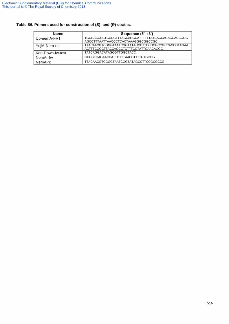
Table S6. Primers used for construction of (S)- and (R)-strains.
Name Sequence (5’→3’)
Up-nemA-FRT TGCGACGCCTGCCGTTTAGCAGGCATTTTTTATCACCAGACGACCGGGAGCCTTTAATTAACCCTCACTAAAGGGCGGCCGC
YqjM-Nem-rc TTACAACGTCGGGTAATCGGTATAGCCTTCCGCGCCGCCACCGTAGAAACTTTCGGCTTACCAGCCTCTTTCGTATTGAACAGGG
Kan-Down-fw-test TATCAGGACATAGCGTTGGCTACC
NemAr-fw GCCGTGAGAACCATTGTTTAACCTTTTGTGGCG
NemA-rc TTACAACGTCGGGTAATCGGTATAGCCTTCCGCGCCG
Electronic Supplementary Material (ESI) for Chemical CommunicationsThis journal is © The Royal Society of Chemistry 2013
S17
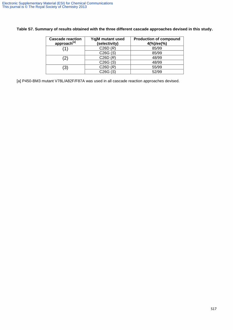
Table S7. Summary of results obtained with the three different cascade approaches devised in this study.
Cascade reaction approach
[a]
YqjM mutant used (selectivity)
Production of compound 4(%)/ee(%)
(1) C26D (R) 85/99
C26G (S) 85/99
(2) C26D (R) 48/99
C26G (S) 48/99
(3) C26D (R) 55/99
C26G (S) 52/99
[a] P450-BM3 mutant V78L/A82F/F87A was used in all cascade reaction approaches devised.
Electronic Supplementary Material (ESI) for Chemical CommunicationsThis journal is © The Royal Society of Chemistry 2013

S18
Figure S1. SDS-PAGE analysis of YqjM overexpressing from the five BOU730 cells that contain FRT-kan-FRT-T7-YqjM_C26D cassette inserted into genome judging by PCR (see details above). Red arrow indicates YqjM band. Molecular standards (line M) of 50 and 120 kDa are indicated.
Electronic Supplementary Material (ESI) for Chemical CommunicationsThis journal is © The Royal Society of Chemistry 2013

S19
Figure S2. Achiral GC analysis of compounds produced in a representative experiment of “Multi-enzymatic cascade transformation approach (1)” using P450 LFA and YqjM C26G mutants overexpressed in BOU730 cells and 1.5 mM of starting material 1. “*” Corresponds to unknown compounds of M.W. 170 (corresponding to an over-oxidation of compound 3).
Electronic Supplementary Material (ESI) for Chemical CommunicationsThis journal is © The Royal Society of Chemistry 2013

S20
Figure S3. Achiral GC analysis of compounds produced in a representative experiment of “Multi-enzymatic cascade transformation approach (1)” using P450 LFA and YqjM C26D mutants overexpressed in BOU730 cells and 1.5 mM of starting material 1. “*” Corresponds to unknown compounds of M.W. 170 (corresponding to an over-oxidation of compound 3).
Electronic Supplementary Material (ESI) for Chemical CommunicationsThis journal is © The Royal Society of Chemistry 2013
S21
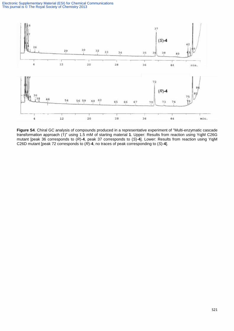
Figure S4. Chiral GC analysis of compounds produced in a representative experiment of “Multi-enzymatic cascade transformation approach (1)” using 1.5 mM of starting material 1. Upper: Results from reaction using YqjM C26G mutant [peak 36 corresponds to (R)-4, peak 37 corresponds to (S)-4]. Lower: Results from reaction using YqjM C26D mutant [peak 72 corresponds to (R)-4, no traces of peak corresponding to (S)-4].
Electronic Supplementary Material (ESI) for Chemical CommunicationsThis journal is © The Royal Society of Chemistry 2013
S22
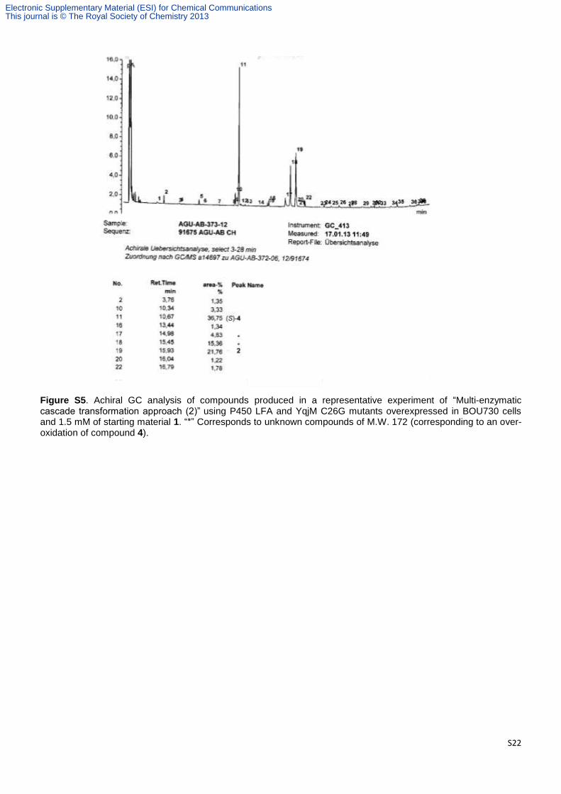
Figure S5. Achiral GC analysis of compounds produced in a representative experiment of “Multi-enzymatic cascade transformation approach (2)” using P450 LFA and YqjM C26G mutants overexpressed in BOU730 cells and 1.5 mM of starting material 1. “*” Corresponds to unknown compounds of M.W. 172 (corresponding to an over-oxidation of compound 4).
Electronic Supplementary Material (ESI) for Chemical CommunicationsThis journal is © The Royal Society of Chemistry 2013

S23
Figure S6. Achiral GC analysis of compounds produced in a representative experiment of “Multi-enzymatic cascade transformation approach (2)” using P450 LFA and YqjM C26D mutants overexpressed in BOU730 cells and 1.5 mM of starting material 1. “*” Corresponds to unknown compounds of M.W. 172 (corresponding to an over-oxidation of compound 4).
Electronic Supplementary Material (ESI) for Chemical CommunicationsThis journal is © The Royal Society of Chemistry 2013

S24
Figure S7. Chiral GC analysis of compounds produced in a representative experiment of “Multi-enzymatic cascade transformation approach (2)” using 1.5 mM of starting material 1. Upper: Results from reaction using YqjM C26G mutant [peak 20 corresponds to (S)-4, no traces of peak corresponding to (R)-4]. Lower: Results from reaction using YqjM C26D mutant [peak 21 corresponds to (R)-4, no traces of peak corresponding to (S)-4].
Electronic Supplementary Material (ESI) for Chemical CommunicationsThis journal is © The Royal Society of Chemistry 2013

S25
Figure S8. Achiral GC analysis of compounds produced in a representative experiment of “Multi-enzymatic cascade transformation approach (3)” using P450 LFA and YqjM C26G mutants overexpressed in “(S)-strain” and 1.5 mM of starting material 1. “*” Corresponds to unknown compounds of M.W. 172 (corresponding to an over-oxidation of compound 4).
Electronic Supplementary Material (ESI) for Chemical CommunicationsThis journal is © The Royal Society of Chemistry 2013

S26
Figure S9. Achiral GC analysis of compounds produced in a representative experiment of “Multi-enzymatic cascade transformation approach (3)” using P450 LFA and YqjM C26D mutants overexpressed in “(S)-strain” and 1.5 mM of starting material 1. “*” Corresponds to unknown compounds of M.W. 172 (corresponding to an over-oxidation of compound 4)
Electronic Supplementary Material (ESI) for Chemical CommunicationsThis journal is © The Royal Society of Chemistry 2013

S27
FigureS10. Chiral GC analysis of compounds produced in a representative experiment of “Multi-enzymatic cascade transformation approach (3)” using 1.5 mM of starting material 1. Upper: Results from reaction using YqjM C26G mutant produced by “(S)-strain” [peak 20 corresponds to (S)-4, no traces of peak corresponding to (R)-4]. Lower: Results from reaction using YqjM C26D mutant produced by “(S)-strain” [peak 22 corresponds to (R)-4, no traces of peak corresponding to (S)-4].
Electronic Supplementary Material (ESI) for Chemical CommunicationsThis journal is © The Royal Society of Chemistry 2013