design, synthesis, and biological evaluation of compounds
TRANSCRIPT

Design, Synthesis, and Biological Evaluation of Compounds for DNA Targeted
Therapy
by
Ahmed Hamdy Mahmoud Elmenoufy
A thesis submitted in partial fulfillment of the requirements for the degree of
Doctor of Philosophy
Department of Chemistry
University of Alberta
© Ahmed Hamdy Mahmoud Elmenoufy, 2020

ii
Abstract
At present, the number of cancer patients who develop resistance to conventional cancer
therapeutics, such as DNA damaging chemotherapeutics, or radiotherapy, is increasing.
A major reason for developing such resistance is that cancer cells have the ability to
repair their DNA damage caused by these therapies through various DNA repair
pathways. The most important of these damages are photo adducts produced by the UV
component of sunlight, such as cyclobutane pyrimidine dimers (CPD) and cisplatin-
DNA products, which cause intra-strand crosslinks (ICL). These bulky, helix distorting
lesions are repaired mainly by nucleotide excision repair (NER), a highly versatile
repair pathway that can recognize, verify, remove, and correct these damages.
ERCC1–XPF is a 5´-3´ structure-specific endonuclease that is involved in NER
and ICL repair pathways in mammalian cells. It plays a central role in NER because it
removes CPD and chemically induced helix-distorting lesions by incising the damaged
DNA strand 5´ and 3´, respectively. Therefore, ERCC1–XPF has become an interesting
therapeutic target to manipulate the DNA repair pathways, and its inhibition has the
potential of sensitizing cancer cells towards cytotoxic chemotherapeutic agents and
ionizing radiation (IR). One of the potential sites on ERCC–XPF to target for inhibiting
its endonuclease activity is the interaction site between ERCC and XPF which is
essential for the protein stability and activity. A few groups have developed hits that
interfere with the interaction site and inhibit its endonuclease activity successfully.
However, the activities of the reported hits to date are suboptimal in terms of clinical
properties, including potency and further optimization is required.

iii
In Chapter 2, we used the reported F06 (compound 1) as a reference hit that was
subjected in docking-based virtual screening. The in-silico screening results yielded
compounds with a better binding energy and ligand efficiency values using the XPF
interaction domain. Synthesis of seven novel analogues of F06 (1) is discussed. Two of
the new F06-based derivatives were shown to have a potent inhibitory effect on
ERCC1–XPF activity relative to F06 in vitro. The cell-based assays showed that
compound 4 significantly inhibited NER by inhibiting the removal of CPDs in UV-
irradiated cells. Also, it successfully showed a significant sensitization of colorectal
cancer cells to cyclophosphamide and UV radiation.
Chapter 3 describes the use of a multi-step CADD strategy to identify better
inhibitors than F06 (1) based on the modification of one site of F06 (1), methoxy-
acridine functional group. The in silico screening study identified two compounds that
showed improved inhibitory effect on the ERCC1–XPF nuclease activity compared to
the parent compound F06 (1). The in vitro endonuclease assay revealed that B9 has a
better ERCC1–XPF inhibition with an IC50 of 0.49 µM, showing 3-fold improvement
in inhibition activity compared to 1. Detailed analysis of the predicted binding mode of
B9 to XPF derived from molecular dynamics simulations is discussed. This analysis
showed that the hydroxyl group substituting the methoxy in F06 (1) was involved in a
new hydrogen bond interaction with side chain of V859, not observed for F06 (1) or
our previous inhibitor compound 4. Therefore, having a polar hydrogen bond donor
group in this site is responsible for the better in vitro activity of these two compounds
in comparison to F06 (1).

iv
Chapter 4 discusses an extensive structure activity relationship analysis that has
been conducted based on the previously identified hit compound, F06 (1), as a reference
compound. Two different series of generations have been developed by structure-based
design and were synthesized through various modifications on two different sites of
F06 (1), according to the corresponding pharmacophore model. Unfortunately, lack of
piperazine moiety in Gen C compounds was accompanied by a lost activity. Also,
replacing the acridine moiety with smaller aromatic group such as quinoline (Gen D
compounds) was responsible for losing the inhibitory effect on ERCC1-XPF.
Therefore, B9 has been selected for carrying out further cell-based/ cell-free assays. B9
elicit high binding affinity to the ERCC1–XPF complex with a Kd of 85 nM relative to
140 nM for 1. B9 also showed a significant sensitization of lung and colorectal cells
towards cisplatin, cyclophosphamide, and Mitomycin C. The proximity ligation assay
suggested that the mechanism of inhibition of B9 is mediated by heterodimerization
disruption.

v
Preface
Chapter 2 of this thesis has been published as Elmenoufy, A. H.; Gentile, F.; Jay, D.;
Karimi-Busheri, F.; Yang, X.; Soueidan, O. M.; Weilbeer, C.; Mani, R. S.; Barakat, K.
H.; Tuszynski, J. A.; Weinfeld, M.; West, F. G. Targeting DNA Repair in Tumor Cells
via Inhibition of ERCC1–XPF. Journal of Medicinal Chemistry 2019, 62, 7684-7696.
I was responsible for the synthesis and design of all compounds, data collection, and
manuscript composition. F.G. designed the computational studies of the compounds and
helped in mastering some of the graphs. D.J. expressed and purified ERCC1−XPF,
employed the in vitro ERCC–XPF1 assay, and conducted microscale thermophoresis
measurements. F.K.B and Y.X. carried out cell culture, UV dimer repair assay, and
clonogenic survival assay. O.S. helped in mentoring A.E in the synthesis and
characterization of the compounds. C.W. was responsible for purification and HPLC
analysis of compound 4. R.M. conducted the binding affinity studies of the compounds.
F.K.B. and J.T. supervised F.G. in designing VS studies. M.W. and F.W. drafted the
manuscript.
Chapter 3 of this thesis has been published as Gentile, F.; Elmenoufy, A. H.;
Ciniero, G.; Jay, D.; Karimi-Busheri, F.; Barakat, K. H.; Weinfeld, M.; West, F. G.;
Tuszynski, J. A. Computer-aided drug design of small molecule inhibitors of the
ERCC1–XPF protein–protein interaction. Chemical Biology & Drug Design 2020, 95,
460-471. F.G. and I contributed equally to this work. We were responsible for data
collection, data analysis, and manuscript writing. D.J. expressed and purified
ERCC1−XPF and employed the in vitro ERCC–XPF1 assay. F.K.B helped in the

vi
manuscript composition. K.B. supervised F.G. in designing the in silico studies. M.W.,
F.G., and J.T. drafted the manuscript.
Chapter 4 of this thesis will be published as Elmenoufy, A. H; Gentile, F;
Soueidan, O. M; Karimi-Busheri, F.; Jay, D.; J. Tuszynski; Barakat, K. H.; Weinfeld,
M.; West, F. G., “Design, synthesis and in vitro cell-free/cell-based biological
evaluations of novel ERCC1-XPF inhibitors targeting DNA repair pathway” I was
responsible for collecting and analyzing data as well as designing and writing the
manuscript. I was also responsible for the synthesis and characterization of all
compounds. F.G performed the computational work for predicting the binding mode of
Gen B9. D.J. carried out the in vitro ERCC–XPF1 endonuclease assay. F.K.B carried
out cell culture, UV dimer repair assay, and clonogenic survival assay. O.S. helped in
mentoring A.E in the synthesis and characterization of Gen B compounds. R.M.
conducted the binding affinity studies of the compounds. F.G. was involved in concept
formation and manuscript composition.

vii
Dedication
To my amazing wife Alshimaa Nassar, my adorable son Yaseen, my
great parents, as well as my whole family for their continuous years of
prayers, support and encouragement.

viii
Acknowledgements
Firstly, I would like to express my sincere gratitude to my supervisor, Dr. F. G. West,
for his continuous support, guidance, as well as his endless mentorship and patience.
Also, I am very thankful to you for accommodating me in your laboratory and providing
the appropriate atmosphere to manage my project flexibly; the freedom you offered me
in choosing a suitable project influenced the way I think and gave me the self-
confidence during my PhD studies. I would not be able to defend my thesis and achieve
such a milestone without your support and thoughtful advice. Thanks for being always
here for me.
Also, I am truly grateful to my Supervisory Committee Members, Dr. Jack
Tuszynski and Dr. Robert Campbell, for their support and dedication to help aid my
research with their valuable ideas and expertise during my graduate studies. I am also
truly thankful to all examiners for the PhD defence exam. I would like to express my
deepest sincere appreciation to them for enriching my knowledge in my discipline.
Words cannot express how grateful and thankful I am to my amazing
collaborators, Dr. Michael Weinfeld, Dr. Feridoun Karimi-Busheri, Dr. David Jay, Dr.
Rajam Mani, and Yang Xiaoyang, for providing me with a rich multi-disciplinary
environment. My chemical biology background was enriched through our usual
meetings and this knowledge was broadened through the fantastic scientific community
I was involved with through several symposiums organized by you. My thesis story
would not be significant without your guidance and help.
ix
I would like to thank the past and current members of the West group for
creating such an amazing environment to conduct research. I have acquired inevitable
research skills from the West group, which broadened my knowledge to reach such a
stage. I would like to express my deepest gratitude for Dr. Olivier Soueidan from whom
I learnt several things in my lab and life. He was such an amazing mentor for me
throughout my graduate studies. Also, I am thankful to Peter Ghaly for his support and
help in the lab. I want to thank my best brother, Yaseen Almehmadi, for his presence
all the time beside me, not only while working in the lab but also the fun time we share
outside the lab. I am grateful for my friends, Ahmed Oraby, Omar Torky and Ahmed
Elfaramawy for their support during my studies.
I am also extremely thankful to the staff members of the NMR, IR, Mass
Spectroscopy facility labs, and X-ray services. Research would not have been possible
without the support you provide. I am also very grateful to Dr. Anna D. Jordan for
reading my thesis and providing me with her valuable feedback and comments. This
thesis would not be composed in such high quality without your amazing touches on it.
Your dedication for lengthy hours editing my thesis is very much appreciated. Thank
you very much to every single one of you for being so approachable.
I am also grateful to the Department of Pharmaceutical Chemistry, Misr
University for Science and Technology in Egypt for giving me the academic leave to
be engaged in such a highly dynamic research environment at one the best universities
in North America, University of Alberta. I am greatly indebted to Dr. Soheir El-Ansary,
Dr. Mohamed Hussein and Dr. Akram Hifny for their encouragement and strong
support during and after my undergraduate studies.

x
Finally, words cannot express how grateful and thankful I am to my family and
relatives, especially to my mother Samia El-boghdady, my father Hamdy Elmenoufy,
my beloved brother Mahmoud Elmenoufy, and younger sisters Eman and Aya
Elmenoufy, as well as my beloved wife Alshimaa Nassar and son Yaseen Elmenoufy.
I would like to thank them for their endless prayers and support to pass stressful
moments throughout my studies. Indeed, I cannot thank you enough for the never-
ending support, unlimited love, and care.

xi
Table of Contents
Chapter 1: Introduction .............................................................................................. 1
1.1 Cancer .............................................................................................................................. 1
1.2 Sources of DNA Damage ................................................................................................. 2
1.3 Major DNA Repair Pathways and Cancer Therapy Resistance ....................................... 5
1.3.1 Mismatch Repair (MMR).......................................................................................... 5
1.3.2 Base Excision Repair (BER) ..................................................................................... 6
1.3.3 Double-strand Break Repair (DSB) .......................................................................... 6
1.3.4 Nucleotide Excision Repair (NER) ........................................................................... 7
1.3.4.1 Mechanism of NER ............................................................................................ 9
1.4 ERCC1–XPF and DNA Repair Pathways ..................................................................... 12
1.5 Patients with NER Deficiency and Mutated ERCC1–XPF............................................ 13
1.6 The Structure of ERCC1–XPF Endonuclease ............................................................... 14
1.6.1 Residues Essential for ERCC1–XPF Dimerization and DNA Binding .................. 15
1.7 Small Molecule Targets of the ERCC1–XPF Heterodimer ........................................... 17
1.7.1 ERCC1/XPA Binding Inhibitors ............................................................................. 17
1.7.2 XPF Endonuclease Inhibitors.................................................................................. 19
1.7.3 ERCC1–XPF Heterodimerization Inhibitors .......................................................... 23
1.8 Conclusion ..................................................................................................................... 32
1.9 References ...................................................................................................................... 36
Chapter 2: Targeting DNA Repair in Tumor Cells via Inhibition of ERCC1-XPF
...................................................................................................................................... 44
2.1 Introduction .................................................................................................................... 44
2.2 Results and Discussion .................................................................................................. 48
2.2.1 Computer-aided Drug Design of Compound 1 Analogues ..................................... 48
2.3.2 Synthesis of Compound 1-based Analogues ........................................................... 56
2.3.3 Inhibition of ERCC1–XPF Endonuclease Activity................................................. 57
2.3.4 Inhibitor Binding to ERCC1–XPF .......................................................................... 60
2.3.5 Inhibition of Cellular Repair of Cyclobutane Pyrimidine Dimers .......................... 64
2.3.6 Increased Sensitization to UV Radiation and Cyclophosphamide .......................... 65
2.3.7 Pharmacokinetic Properties of Compound 4 and Compound 1 .............................. 66
2.4 Conclusion ..................................................................................................................... 68
2.5 Methods and Experimental Section ............................................................................... 71

xii
2.5.1 General Experimental Procedures for Preparation of The Top Compound 1
Analogues ........................................................................................................................ 71
2.5.2 Synthesis and Characterization of Inhibitors (1-8) ................................................. 72
2.5.3 Molecular Docking of Compound 1 ....................................................................... 81
2.5.4 Characterization of The Binding Pocket and Pharmacophore Modeling ................ 81
2.5.5 Docking-based Virtual Screening of Compound 1 Analogues ............................... 82
2.5.6 Molecular Dynamics Simulations and MM/GBSA Rescoring of the Analogues ... 83
2.5.7 ERCC1–XPF Protein Preparation ........................................................................... 83
2.5.8 Microplate Fluorescence Incision Assay ................................................................ 84
2.5.9 Steady-state Fluorescence Assay ............................................................................ 84
2.5.10 Expression and Purification of Recombinant Truncated Forms of ERCC1 and
XPF .................................................................................................................................. 85
2.5.11 Microscale Thermophoresis (MST) Measurements .............................................. 85
2.5.12 Cell Culture ........................................................................................................... 86
2.5.13 Cellular Repair of Cyclobutane Pyrimidine Dimers ............................................. 86
2.5.14 Clonogenic Survival Assay ................................................................................... 87
2.5.14 PK Assessment of Compound 4 and Compound 1 ............................................... 88
2.10 References .................................................................................................................... 89
Chapter 3: Computer-Aided Drug Design of The Second-Generation ERCC1–
XPF Inhibitors ............................................................................................................ 94
3.1 Introduction .................................................................................................................... 94
3.2 Results and Discussion .................................................................................................. 98
3.2.1 Molecular Dynamics Simulations ........................................................................... 98
3.2.2 Interactions of F06 with The XPF Binding Site.................................................... 102
3.2.3 Virtual Screening of F06 (1) Analogues ............................................................... 104
3.2.4 Chemistry .............................................................................................................. 105
3.2.5 Inhibitory Effect of Gen B Compounds on ERCC1–XPF Activity ...................... 108
3.2.6 Predicted Binding Mode of B9 to XPF ................................................................. 110
3.3 Conclusion ................................................................................................................... 111
3.4 Methods and Experimental Section ............................................................................. 112
3.4.1 Molecular Dynamics of XPF HhH2 Domain Alone, F06-bound and ERCC1-bound
....................................................................................................................................... 112
3.4.2 Analysis of The Binding Mode of F06 ................................................................. 114
3.4.3 Computer-aided Design of F06 Analogues ........................................................... 114
3.4.4 Structure-based Virtual Screening ........................................................................ 115
3.4.5 Preparation of F06-based Analogues (Gen B Compounds) .................................. 117

xiii
3.4.6 ERCC1–XPF Protein Preparation ......................................................................... 125
3.4.7 Microplate Fluorescence Incision Assay .............................................................. 125
3.5 References .................................................................................................................... 126
Chapter 4: Design, Synthesis and In vitro Cell-free/Cell-based Biological
Evaluations of Novel ERCC1–XPF Inhibitors Targeting DNA Repair Pathway
.................................................................................................................................... 130
4.1 Introduction .................................................................................................................. 130
4.2 Results and Discussion ................................................................................................ 134
4.2.1 Chemistry .............................................................................................................. 134
4.2.2 Inhibition of ERCC1−XPF Endonuclease Activity .............................................. 137
4.2.2.1 Inhibitory Effect of Gen C Compounds on ERCC1–XPF Activity ............... 137
4.2.2.2 Inhibitory Effect of Gen D Compounds on ERCC1-XPF Activity ................ 138
4.2.3 Gen B9 Binding to ERCC1−XPF ......................................................................... 140
4.2.4 Inhibition of DNA Repair by B9 ........................................................................... 143
4.2.5 Cytotoxicity Profile of Inhibitors .......................................................................... 144
4.2.6 Increased Sensitization to UV Radiation and Cyclophosphamide ........................ 145
4.2.7 Pharmacokinetic Properties (PK) of B9 and F06 (1) ............................................ 146
4.2.8 Cell Death by a Combination of Compound 4 (Gen A) or B9 with
Chemotherapeutic Agents .............................................................................................. 147
4.2.9 Heterodimerization Inhibition of ERCC1–XPF by A4 and B9............................. 148
4.3 Conclusion ................................................................................................................... 149
4.4 Methods and Experimental Section ............................................................................. 151
4.4.1 Synthesis of C and D Compounds ........................................................................ 151
4.4.1.1 Synthesis of Gen C Compounds .................................................................... 153
4.4.1.2 Synthesis of Gen D Compounds .................................................................... 163
4.4.2 ERCC1−XPF Protein Preparation ......................................................................... 165
4.4.3 In vitro ERCC1-XPF Endonuclease Assay ........................................................... 165
4.4.4 Steady-State Fluorescence Assays ........................................................................ 166
4.4.5 Cell Culture ........................................................................................................... 166
4.4.6 Cellular Repair of Cyclobutane Pyrimidine Dimers ............................................. 166
4.4.7 Rapid Cell Viability Assay ................................................................................... 167
4.4.8 Clonogenic Survival Assay Following UV Treatment ......................................... 168
4.4.9 Combination Effect of B9 and Cisplatin and MMC ............................................. 168
4.4.10 Partial Pharmacokinetic Profile of Compounds Gen B9 and F06 (1) ................. 169
4.4.11 Partial Pharmacokinetic Profile of Compounds B9 and F06 (1) ......................... 169

xiv
4.4.12 Proximity Ligation Assay ................................................................................... 169
4.5 References .................................................................................................................... 171
Chapter 5 .................................................................................................................. 174
5.1 General Conclusions .................................................................................................... 174
5.2 Future Directions ......................................................................................................... 180
5.3 References .................................................................................................................... 183
Compiled References ............................................................................................... 184
Appendix I: Selected NMR Spectra ....................................................................... 204
Appendix II: HPLC chromatogram of Compound 4 ........................................... 221
Appendix III: Selected NMR Spectra .................................................................... 223
Appendix IV: HPLC chromatogram of B9 ........................................................... 234
Appendix V: Selected NMR Spectra ...................................................................... 236

xv
List of Figures
Chapter 1
Figure 1.1. Endogenous events as sources of DNA damage. ....................................... 3
Figure 1.2. DNA damage and repair pathways. ............................................................ 5
Figure 1.3. UV-induced formation of A) a cyclobutane pyrimidine dimer and B) a
pyrimidine-pyrimidone 6-4 photoproduct...................................................................... 8
Figure 1.4. A simplified model of steps of Nucleotide Excision Repair (NER) ......... 10
Figure 1.5. Structure of ERCC1–XPF.. ...................................................................... 14
Figure 1.6. Interaction of ERCC1 and XPF through their HhH2 domains ................. 16
Figure 1.7. Structure of UCN-01 as an ERCC1–XPA inhibitor. ................................ 18
Figure 1.8. The nuclease domain of XPF. Cartoon representation of XPF identifying
amino acids and their side chains................................................................................. 19
Figure 1.9. Structures of XPF endonuclease active site inhibitors E-X AS5-4 and E-X
AS7. ............................................................................................................................. 20
Figure 1.10. Structures of XPF endonuclease active site inhibitor
hydroxypyrimidinone derivative .................................................................................. 21
Figure 1.11. Metal chelators as ERCC1–XPF nuclease inhibitors. ............................ 21
Figure 1.12. Structures of ERCC1–XPF nuclease inhibitors procyanidine 8 and
hydroxy-naphthalene derivative 9. ............................................................................... 22
Figure 1.13. In silico screening for ERCC1–XPF interaction inhibitors .................... 24
Figure 1.14. In silico screened products as potential ERCC1–XPF heterodimerization
inhibitors. ..................................................................................................................... 25
Figure 1.15. Binding energy decomposition identified three interaction sites in
ERCC1-XPF ................................................................................................................ 27
Figure 1.16. Binding modes of the three hits, F02 (A), F03 (B), and F06 (C) ........... 28
Figure 1.17. Sensorgram for interaction between XPF814-905 and ERCC1.220-297 ........ 29

xvi
Figure 1.18. Fluorescence experiments for the interaction between XPF814-905 and F06
(1) ................................................................................................................................. 30
Figure 1.19. Potential modality of NER inactivation through ERCC1–XPF
heterodimerization inhibition. ...................................................................................... 32
Chapter 2
Figure 2.1. The structure of the reference hit, compound 1 (F06). ............................. 47
Figure 2.2. Visual analysis of 1 docking pose. ........................................................... 49
Figure 2.3. Compound 1 analogues functionalized with different substituents on the
piperazine ring ............................................................................................................. 51
Figure 2.4. Analysis of the binding mode of the top hits ............................................ 55
Figure 2.5. The structure of DNA stem loop substrate. .............................................. 57
Figure 2.6. In-vitro inhibition of ERCC1–XPF endonuclease activity ....................... 58
Figure 2.7. Time-dependent inactivation of ERCC1–XPF by compound 4.. ............. 60
Figure 2.8. Determination of the affinity (dissociation constant, Kd) between
compound 4 and the ERCC1–XPF complex ............................................................... 61
Figure 2.9. Microscale thermophoresis measurements ............................................... 63
Figure 2.10. Inhibition of cellular NER by compound 4. ........................................... 64
Figure 2.11. Survival of HCT-116, SW620, and XPF-/- cells treated with increasing
doses of compound 4 and compound 5 ........................................................................ 65
Figure 2.12. Sensitization of cells to UV and cyclophosphamide by compound 4 .... 66
Chapter 3
Figure 3.1. Involvement of ERCC1–XPF complex in three major DNA repair
pathways ...................................................................................................................... 96
Figure 3.2. Chemical structure of 1, ERCC1-XPF heterodimerization inhibitor. ...... 97
Figure 3.3. RMSDs analysis of F06 (1) and ERCC1 .................................................. 99

xvii
Figure 3.4. Structural differences between bound and unbound forms of XPF ........ 101
Figure 3.5. RMSF for the XPF backbone atoms calculated for the last 100 ns of MD
simulation for the unbound (red), F06-bound (blue) and ERCC1-bound (green) XPF
structure...................................................................................................................... 101
Figure 3.6. Protonated 3D structure of F06 (1). Polar atoms are labelled with their
names. ........................................................................................................................ 103
Figure 3.7. Poisson–Boltzmann electrostatic feature maps of the F06 binding site on
XPF.. .......................................................................................................................... 103
Figure 3.8. F06 (1) analogues. A) modification sites of F06 (1) explored using a
CADD strategy. B) structure of compound 4, best ERCC1–XPF inhibitor among Gen
A compounds. ............................................................................................................ 104
Figure 3.9. Pharmacophore model of the scaffold common to all the compounds,
including three aromatic features ............................................................................... 105
Figure 3.10. Structures of Gen B compounds including controls. ........................... 106
Figure 3.11. In-vitro inhibition of ERCC1−XPF endonuclease activity................... 109
Figure 3.12. Detailed analysis of the predicted binding mode of B9 to XPF derived
from the 2 ns-long MD simulation............................................................................. 110
Chapter 4
Figure 4.1. Structure of F06 (1) hit and the potential modification sites for hit to lead
optimization. .............................................................................................................. 134
Figure 4.2. Chemical structures of C analogues. ...................................................... 135
Figure 4.3. Chemical structures of D analogues. ...................................................... 137
Figure 4.4. In-vitro inhibition of ERCC1−XPF endonuclease activity by C
compounds (10 μM each). ......................................................................................... 138
Figure 4.5. In-vitro inhibition of ERCC1−XPF endonuclease activity by D
compounds (10 μM each). The increase of fluorescence signal as a function of time.
.................................................................................................................................... 139
Figure 4.6. Determination of the affinity (Kd) between ERCC1–XPF complex and B9
(active inhibitor) and B7 (negative control)............................................................... 140

xviii
Figure 4.7. The binding affinity measurement of B7 on the stem–loop DNA substrate.
.................................................................................................................................... 142
Figure 4.8. Inhibition of cellular NER by compound B9.......................................... 143
Figure 4.9. Crystal violet-based viability assay to assess the cytotoxicity profile of B9
(active inhibitor), B7 (negative control), and F06 (1) (hit). ....................................... 144
Figure 4.10. Sensitization of HCT 116 cells to UV and cyclophosphamide by B9.. 146
Figure 4.11. Survival of A549 (A) and HCT-116 (B) cells after exposure to alkylating
agents and NER inhibitors alone or in combination. ................................................. 148
Figure 4.12. Interaction between endogenous ERCC1 and XPF revealed by a
proximity ligation assay (red dots) in A549 cells incubated alone (NT) or in the
presence of A4 or B9 (2 µM) or cisplatin (20 µM) for 24 h. ..................................... 149
Chapter 5
Figure 5.1. Structural modifications on B9 or A4 for lead optimization. ................. 180

xix
List of Tables
Chapter 2
Table 2.1. Pharmacophore Features Built from the Docking Pose of 1 to the XPF Site
...................................................................................................................................... 50
Table 2.2. Computational Results for the Subset of Analogues Chosen for Synthesis
...................................................................................................................................... 52
Table 2.3. Decomposition of Total MM/GBSA Binding Energy in Different
Contributions for Compound 1 and the Two Top Analogues...................................... 53
Table 2.4. Half-Maximum Inhibitory Concentrations for The Compounds with The
Highest Inhibitory Potentials ....................................................................................... 59
Table 2.5. Pharmacokinetic Profile of Compound 4 and Compound 1 ...................... 67
Chapter 3
Table 3.1. Main Ligand–Receptor and Ligand–Solvent Hydrogen Bonds Detected for
the XPF–F06 Complex During the Last 100 ns of MD Simulationa ......................... 102
Table 3.2. Computational Results for the Series of F06 Analogues Selected for
Chemical Synthesisa ................................................................................................... 108
Chapter 4
Table 4.1. The Half-maximum Inhibitory Concentrations (IC50) and Binding Constants
Values of Inhibitors.................................................................................................... 141

xx
List of Schemes
Chapter 2
Scheme 2.1. Synthetic route of compounds 1–8 A) One-pot sequential addition
reaction. B) Main steps and intermediates included in the one-pot sequential addition
reaction. ........................................................................................................................ 56
Chapter 3
Scheme 3.1. General synthetic route of Gen B compounds...................................... 107
Chapter 4
Scheme 4.1. General synthetic route for Gen C compounds. ................................... 136

xxi
Abbreviations
°C Celsius
µL microliter
µM micromolar
6-4 PPs pryrimidine- (6-4)-pyrimidone
6-FAM 6-Carboxyfluorescein
Å angstrom
ADME absorption, distribution, metabolism and elimination
AP apurinic
APE apurinic endonuclease
ATCC American Type Culture Collection
BER base excision repair
CADD computer-aided drug design
CHAPS 3-[(3-cholamidopropyl)dimethylammonio]-1-propanesulfonate
CHK1 human checkpoint kinase
CO2 carbon dioxide
CPD cyclobutene pyrimidine dimer
CSA Cockayne syndrome
CYP cytochrome P
d doublet (spectral)
D aspartic acid
DCM dichloromethane
DMEM Dulbecco's Modified Eagle's Medium
DMSO dimethyl sulfoxide
DSB double-strand breaks
E glutamic acid
EDG electron donating group
EDTA ethylenediamine tetraacetic acid
Eq equivalent

xxii
ERCC1 excision repair cross-complementation group 1
Et ethanol
EWG electron withdrawing group
FBS fetal bovine serum
FEN flap endonuclease
GBVI/WSA Generalized Born Volume Integral/Weighted Surface Area
Gen generation
GG-NER global genome nucleotide excision repair
H histidine
HA heavy atom
HBA hydrogen bond acceptor
HBD hydrogen bond donor
HCl hydrochloric acid
HEPES N-2-hydroxyethylpiperazine-N-ethanesulfonic acid
HhH2 helix-hairpin-helix
HPLC high performance liquid chromatography
HR homologous recombination
HRMS high resolution mass spectrometry
HTS high throughput screening
Hz hertz
IC50 half maximal inhibitory concentration
ICL inter-strand crosslink
IgG immunoglobulin G
in vitro referring to the studies performed in living organisms
in vivo referring to the studies performed in cell culture
in silico computer simulation in reference to biological experiments
i-PrOH isopropyl alcohol
FTIR fourier transform infrared
IR ionizing radiation
J coupling constant (in NMR)
K lysin

xxiii
Kd dissociation constant
LC liquid chromatography
M moles per liter
m multiplet (spectral)
M methionine
m/z mass to charge ratio
MD molecular dynamics
Me methyl
MeCN acetonitrile
MeOH methanol
MHz megahertz
Min minute
mL milliliter
MM/GBSA Molecular Mechanics-Generalized Born Surface Area
MMC mitomycin C
MMR mismatch repair
MOE molecular operating environment
Mp melting point
MS mass spectrometry
MST microscale thermophoresis
NaCl sodium chloride
NER nucleotide excision repair
NHEJ non-homologous end joining
nm nanometer(s)
NMA normal mode anaylsis
NMR nuclear magnetic resonance
NSCLC Non-small cell lung cancer
OMe methoxy
PBS phosphate-buffered saline
PDB protein data bank
PK-C protein kinase-C

xxiv
PLA proximity ligation assay
Polb polymerase b
Ppm part per million
q quartet (spectral)
Q glutamine
R arginine
R generalized alkyl group of substituents
RFU relative fluorescence units
RMSD Root-mean-square deviation of atomic positions
ROS reactive oxygen species
s singlet (spectral)
SAM S-adenosylmethionine
SAR structure activity relationship
SD standard deviation
SEM standard error of mean
SNAr nucleophilic aromatic substitution
SPR surface plasmon resonance
TC-NER transcription-coupled nucleotide excision repair
TLC thin layer chromatography
UV ultraviolet
VS virtual screening
WT wild type
wt. weight
XP xeroderma pigmentosum
Y tyrosine
δ chemical shift in parts per million downfield from tetramethylsilane
wavelength

1
Chapter 1
1. Introduction
1.1 Cancer
Cancer is a global public health problem and is the second leading cause of death in the
United States.1 In 2017, of 1.7 million of cancer patients, about 0.6 million patients died
from the disease.1 Drug resistance, which results in ineffectiveness of the drug
treatment, is responsible for 90% of the cancer-related deaths.2-4 Cancer related drug
resistance is a widely known phenomenon that results from the tolerance of cancer to
the pharmaceutical treatment. Resistance to cancer therapeutics arises from a wide array
of factors, such as epigenetic changes and/or genetic mutations, upregulated drug efflux,
and several other molecular and cellular pathways. Currently, cancer therapy is managed
mainly by radiotherapy, chemotherapy, surgery, and immunotherapy.5-7 Despite the
successful achievements made in treating various cancers during the past decades, a
major problematic issue in cancer management arises due to the development of
resistance to either conventional chemotherapeutics or novel targeted therapies.8, 9
Several conventional chemotherapeutic anticancer agents, such as platinum-
containing drugs, cyclophosphamide, and mitomycin C elicit cytotoxicity to cancer cells
by damaging their DNA directly; however, such therapy lacks specificity and showed a
high toxicity to normal cells as well. Although many drugs targeting proliferation of
cancer cells provided significant benefit during initial treatment, the majority of cancer
patients develop resistance as treatment continues. For example, there is a 30%–55%
relapse occurrence in non-small cell lung cancer (NSCLC) patients who eventually die

2
from the disease.10 The ovarian adenocarcinomas relapse within the first year after
chemotherapy and surgery is 50%–70%.11 In addition, about 20% of pediatric patients
develop a recurrence of acute lymphoblastic leukemia.12 About 60% of colorectal cancer
patients develop relapses after two years of related chemotherapy treatment.13
Therefore, mechanisms and pathways underlying the drug resistance must be well-
understood, and there is an urgent need to facilitate the development of novel therapeutic
modalities that lead to better clinical outcomes.
1.2 Sources of DNA Damage
Like most biomolecules, DNA can undergo various chemical reactions; however, such
reactions often can lead to changes in the genetic code for various critical cellular
proteins (Figure 1.1). Intrinsic or extrinsic agents may cause DNA damage; however, in
general, most of the DNA modifications are endogenous in origin and are mediated by
spontaneous hydrolysis.14, 15 The N-glycosidic bond between the deoxyribose and DNA
base is especially prone to hydrolytic cleavage under acidic conditions. The products of
hydrolytic nucleobase loss from abasic or AP sites (apurinic/apyrimidinic sites) occur
at an approximate rate of ~10,000 per cell per day.15, 16 Moreover, abasic sites are
chemically labile and prone to β-elimination that may cause DNA strand scission.17
Another well known hydrolysis reaction is the deamination of DNA bases bearing
exocyclic amino groups.15, 18 Most of these lesions form uracil from cytosine, which is
estimated to occur 100–500 times per cell per day.19, 20 In addition, hypoxanthine and
xanthine could be formed by the spontaneous deamination of the

3
Figure 1.1. Endogenous events as sources of DNA damage.
adenine and guanine, respectively.21 The DNA strand also can be modified chemically
by reactive oxygen species (ROS), such as O2•−, H2O2, and •OH produced during normal
cellular metabolic processes.22, 23 ROS create more than one hundred different oxidative
DNA adducts, such as deoxyribose oxidation, base modification, DNA-protein cross-
links, and single- or double-strand breakage.24 Endogenous reactive nitrogen species,
such as nitric oxide (NO•) and its byproducts, also can create similar oxidative adducts.25
For example, 8-oxoguanine is the most commonly studied oxidative DNA lesion that is
used as a measure of oxidative DNA damage in biological systems.26 Alkylation, which
occurs primarily at O and N atoms of nucleobases, is another example of DNA damage

4
caused by endogenous reactive molecules, such as S-adenosylmethionine (SAM) and
methyl radicals produced by lipid peroxidation.14, 27
Cellular DNA also could be insulted by exogeneous DNA-damaging agents,
such as ionizing radiation (IR) and classical chemotherapeutic agents, which are used
widely in cancer therapy because of their capability of forming DNA lesions that
resulted from interference with various DNA-associated proteins and/or DNA breaks.
These, in turn, might cause disruption to normal cellular processes, such as replication,
transcription, and/or recombination, and thus lead to cell death.28, 29 The outcome of
treatment with such cancer therapeutics, whether the cell will survive or die, is
dependent on how the cells respond to the damaged DNA by activating DNA repair
streams or cell cycle arrest.30 Normal cells can reverse the DNA damage caused by many
chemotherapeutic drugs and IR by inducing appropriate repair mechanisms. Cancerous
cells have a similar capacity to recognize DNA lesions effectively and initiate DNA
repair pathways; however, this phenomenon leads to treatment resistance to various
cancer therapeutics.31 On the other hand, because of the inherited or somatic mutations
in certain DNA repair genes, cancer cells would depend on functioning DNA
mechanisms more than the normal cells.32 Therefore, therapeutics that are able to
manipulate the DNA repair pathways have the potential of sensitizing cancer cells to the
cytotoxic chemotherapeutic agents and IR. Several reports stated that the activation of
DNA repair pathways is responsible for the chemoresistance of cancer cells to DNA-
damaging agents and suppressing DNA repair pathways, leading to potentiation to these
cytotoxic agents.33, 34
5
1.3 Major DNA Repair Pathways and Cancer Therapy
Resistance
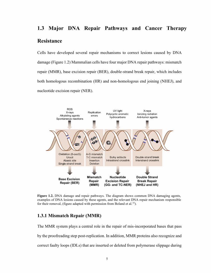
Cells have developed several repair mechanisms to correct lesions caused by DNA
damage (Figure 1.2) Mammalian cells have four major DNA repair pathways: mismatch
repair (MMR), base excision repair (BER), double-strand break repair, which includes
both homologous recombination (HR) and non-homologous end joining (NHEJ), and
nucleotide excision repair (NER).
Figure 1.2. DNA damage and repair pathways. The diagram shows common DNA damaging agents,
examples of DNA lesions caused by these agents, and the relevant DNA repair mechanism responsible
for their removal, (figure adapted with permission from Boland et al.35).
1.3.1 Mismatch Repair (MMR)
The MMR system plays a central role in the repair of mis-incorporated bases that pass
by the proofreading step post-replication. In addition, MMR proteins also recognize and
correct faulty loops (IDLs) that are inserted or deleted from polymerase slippage during

6
the replication of repetitive DNA sequences. The importance of this pathway is the fact
that MMR deficient cells are reported to display a mutator phenotype, which is
characterized by an elevated mutation frequency and a microsatellite instability.
Another more important point is that MMR genes that have germline mutations are
predisposed to a wide variety of cancers, especially the inherited non-polyposis colon
cancer, also known as Lynch syndrome.36 The MMR mechanism can be categorized
into three main steps: 1) a recognition step of mis-paired bases, 2) an excision step where
the damaged DNA strand is degraded, resulting in a gap, and 3) a repair synthesis step,
where the DNA is re-synthesized to fill the gap.37-39
1.3.2 Base Excision Repair (BER)
The base excision repair mechanism recognizes and accurately removes damaged bases
that are formed by oxidation, alkylation, ring saturation, or ionizing radiation,40 and also
eliminates the deaminated bases and DNA single strand breaks (SSBs). Base lesions are
excised by a damage-specific endonuclease, called DNA glycosylase, resulting in the
formation of potentially cytotoxic apyrimidinic or apurinic sites (AP sites)
intermediates. Afterwards, the AP sites are processed by an AP endonuclease (APE1),
which produces a strand break that is processed further by poly (ADP-ribose)
polymerase (PARP), DNA polymerase b (Polb), and ligase III. Subsequent repair steps
are followed and completed through further stream events.41
1.3.3 Double-strand Break Repair (DSB)
Double-strand breaks (DSBs) are one of the most biologically hazardous kinds of DNA
damage. For example, a single unrepaired DSB is often enough to cause cell death.

7
Moreover, inaccurate repair events can result in chromosomal aberrations or deletions,
which are associated with the development of some genomic instability syndromes or
cancer. Therefore, the repair of DSBs is vital for both the maintenance of genome
integrity and cell survival.42, 43 The two main pathways by which eukaryotic cells repair
DSBs are homologous recombination (HR) and non-homologous end-joining (NHEJ).
The difference between these two repair mechanisms is in their fidelity of DSB repair
and their requirement for a homologous template DNA. Generally, HR repair is an error-
free pathway because it utilizes the undamaged sister chromatid genetic information as
a template.44 On the contrary, NHEJ directed-repair normally is prone to errors, and the
elimination of DSBs is mediated by direct ligation of the broken ends.45 NHEJ is
reasoned to be the dominant mechanism in mammalian cells that operate in all phases
of the cell cycle, whereas HR is restricted to the G2 and late-S phases.
1.3.4 Nucleotide Excision Repair (NER)
NER is a highly versatile repair mechanism that can recognize, verify, and remove
various bulky, helix-distorting lesions from the DNA. The most important of these
damages are pyrimidine dimers, such as 6–4 photoproducts and cyclobutane pyrimidine
dimers (CPD), which are photoproducts generated by the UV component of sunlight
(Figure 1.3).

8
Figure 1.3. UV-induced formation of A) a cyclobutane pyrimidine dimer and B) a pyrimidine-
pyrimidone 6-4 photoproduct.
Another significant substrate of NER is cisplatin-DNA adducts, which cause
intra-strand crosslinks. NER is mediated by a sequence of assembled repair proteins at
the site of the DNA damage. Although mechanistically similar to the BER pathway, the
NER mechanism is much more complex because it requires about thirty different
proteins to carry out a multi-step ‘cut-and-patch’-like pathway. These steps include the
recognition of DNA lesion, opening of the DNA double helix around the local lesion,
and excision of a short single-strand sequence of DNA spanning the damage, followed
by a sequential repair synthesis and strand ligation steps.46-49 The biological importance
of NER is reasoned by the fact that defects in NER cause many human genetic diseases,
such as xeroderma pigmentosum, trichothiodystrophy, and Cockayne syndrome; these
conditions all cause extreme sun sensitivity. Furthermore, these disorders develop

9
symptoms that overlap with developmental delay, neurodegeneration, immunological
defects, premature aging, and cancer.50, 51
1.3.4.1 Mechanism of NER
The NER pathway consists of two related sub-pathways, termed transcription-coupled
NER (TC-NER) and global genome NER (GG-NER)49 (Figure 1.4). As the names
imply, TC-NER plays a preferential role in repairing lesions located on the coding strand
of genes that are transcribed actively, whereas GG-NER eliminates damaged DNA
throughout the genome. Both pathways are the same mechanistically, regardless of the
initial damage recognition step. In GG-NER, the main protein complex for damage
recognition is XPC/HR23B/CEN2 (XP complementation group C/Rad23 homolog
B/Centrin-2).52 CEN2 and HR23B are accessory proteins that increase both the
specificity and affinity of XPC binding to helix-distorting DNA damage. Additionally,
the binding affinity of DNA to XPC generally correlates with the extent of helical
distortion.53 For instance, XPC has a low affinity to DNA lesions formed by only minor
distortions, such as UV-induced CPDs. Therefore, the UV-damaged DNA binding
complex (UV-DDB), an auxiliary damage-recognizing complex that consists of two
subunits, XPE (DDB2) and DDB1, initially recognizes and detects these kinds of
lesions. The subsequent binding of UV-DDB to lesions result in only minor distortions
(i.e., DNA bending), which, in turn, facilitate the recruitment of the XPC complex to
the damaged site.54

10
Figure 1.4. A simplified model of steps of Nucleotide Excision Repair (NER). A) Recognition of DNA
damage that differs between transcription coupled- and global genomic NER (TC-NER and GG-,
respectively). B) dual incision of DNA strand, ligation and synthesis of DNA strand. (copied with
permission from Fousteri et al.55).

11
On the contrary, in TC-NER, the initial recognition of DNA damage is started
when an elongating RNA polymerase II (RNAPII) is arrested upon encountering a site
of DNA lesion.55 Afterwards, Cockayne syndrome A (CSA) and B (CSB), two TC-
NER-specific proteins, are believed to replace the stalled RNAPII to facilitate the access
of NER proteins to the damage.56 Subsequently, both TC-NER and GG-NER continue
via the common ‘core’ NER reactions. At first, either the XPC complex in CSB and
CSA in TC-NER or GG-NER recruit multi-functional transcription factor TFIIH and a
ten protein-complex (multi-subunit) to the lesion site. Next, the asymmetric unwinding
of the DNA helix is orchestrated by ATP-dependent helicases, such as XPB and XPD,
to produce a bubble consisting of ∼30 nucleotides flanking the damaged DNA site.
A second level of damage recognition is activated upon initial DNA unwinding
by having access of XPA to the region of the lesion to ensure that unaffected DNA is
not subjected to excision repair. Upon XPA binding to the damaged DNA, a
heterotrimeric single stranded DNA binding protein, called RPA (replication protein A),
is recruited for stabilizing the pre-incision complex and extension completion. In the
following step, ERCC1–XPF and XPG, two structure-specific endonucleases, cleave at
the 5´ position of a deoxyribose phosphodiester and XPG cleaves at the 3´ position of a
different deoxyribose phosphodiester, respectively, resulting in the excision of about 30
nucleotides from the lesion site. Finally, DNA polymerase δ or ε utilizes the undamaged
DNA strand as a template to re-synthesize the produced gap. Then, DNA ligase seals
the nick of the repaired strand, thus finalizing the NER process.49

12
1.4 ERCC1–XPF and DNA Repair Pathways
The ERCC1–XPF protein complex is a 5´-3´ structure-specific endonuclease that is
involved in several DNA repair mechanisms in mammalian cells. It is crucial for
nucleotide excision repair (NER) because it removes pyrimidine-(6,4)-pyrimidone
photoproducts (6-4PPs) and cyclobutane pyrimidine dimers (CPDs) induced by UV
irradiation by having the damaged DNA strand 5´ and 3´ incised, respectively. The
ERCC1–XPF heterodimer also plays a central role in the repair of chemically induced
helix-distorting and bulky DNA lesions, which are all substrates for the NER pathway.
ERCC1–XPF also has a key role in the repair of double-strand breaks (DSBs), induced
by free radicals, ionizing radiation, and chemotherapeutics such as the crosslinking
agents mitomycin C (MMC) and cisplatin, or the topoisomerase inhibitor etoposide,
which can be repaired by non-homologous end-joining (NHEJ) repair or homologous
recombination (HR). The role of ERCC1–XPF in DSB repair (DSBR) was shown
previously in budding yeast where mutations in RAD1 and RAD10, the yeast
orthologues of XPF and ERCC1, suppressed the HR repair pathway.57 Mammalian cells
containing mutant ERCC1–XPF were shown to be sensitive to DSBs,58 and both the
NHEJ and HR streams for DSBR were attenuated.59, 60 The principle activity of the
ERCC1–XPF heterodimer in both types of DSBR is its capability to remove non-
homologous 30 single-stranded flaps at broken ends before their rejoining.58
It is also noteworthy to mention that the ERCC1–XPF complex is involved in
inter-strand crosslink (ICL) repair. ERCC1-XPF endonuclease operates to remove
chemotherapeutic-induced crosslinks caused by psoralen, cisplatin, and MMC 61. Such
damages are particularly toxic because of their ability to prevent helix unwinding and,

13
in turn, block the transcription and replication phases. The sensitivity of mammalian
cells with ERCC1–XPF mutation to ICL DNA-damaging agents was found to be
significantly higher than the wild-type cells.61 In eukaryotes, the pathway of ICL
removal is dependent on the cell cycle phase in which the lesion is encountered.62
Incisions adjacent to an ICL are repaired completely in the G0 or G1 phase, whereas
ICL lesions persist into the S stage and will be repaired by the DSB pathway. The
ERCC1–XPF-dependent step is required not only in several models for ICL repair61, 62
to carry out the incision on either side of an ICL63 but also in the S-phase-dependent and
-independent ICL repair pathway.64, 65
1.5 Patients with NER Deficiency and Mutated ERCC1–XPF
Inherited defects in human NER genes lead to rare syndromes, such as Cockayne
syndrome (CS), xeroderma pigmentosum (XP), and trichothiodystrophy. While XP is
considered a repair syndrome, trichothiodystrophy and CS are regarded as transcription
syndromes.66 Diagnostic characteristics of XP are dry skin and abnormal pigmentation
that is patterned in areas exposed to sun, developing into severe photosensitivity. This
leads to an increase in the risk of developing UV-induced skin cancers by more than
1000-fold. Also, about 20–30% of XP patients have progressive neurological disorders,
emphasizing how NER is extremely important in repairing endogenous DNA damage.66
CS patients are photosensitive as well but do not develop pigmentation abnormalities or
an increased risk of having cancer;66, 67 however, they show neurological and
developmental defects.66

14
Mutations in XP patients are characterized by mutated ERCC168 and XPF
genes.69, 70 Mutations in the XPF or ERCC1 genes can result in a rare XF-E syndrome.71
XF-E patients exhibit similar characteristics of XP and CS and show additional
neurologic, musculoskeletal hepatobiliary and hematopoietic symptoms.71 XF-E
patients not only have a complete loss of GG- and TC-NER but also develop
hypersensitivity to ICL agents due to the central role of ERCC1–XPF in the repair of
ICL damage.71 This is what distinguishes XF-E syndrome from either XP or CS. 71
1.6 The Structure of ERCC1–XPF Endonuclease
The ERCC1 domain consists of 297 amino acids, with a central domain that is
catalytically inactive but plays a key role in interaction with both the XPA protein and
DNA,72, 73 and a helix-hairpin-helix (HhH2) domain required for heterodimerization
with XPF (Figure 1.5).72, 74, 75
Figure 1.5. Structure of ERCC1–XPF copied with permission from Tsodikov et al*.72 A) Crystal
structure of ERCC1–XPF heterodimeric HhH2 complex, B) A model for XPF∆655–ERCC1∆95 binding to
a splayed-arm DNA substrate. Light brown indicates XPF, and green indicates ERCC1. The cleavage
site is shown by the orange sphere. and thelinker between the nuclease and the HhH2 domains of XPF
by the dashed line. The XPA binding region of ERCC1 (residues 99–118) is shown in dark green.
* "Copyright (2005) National Academy of Sciences, U.S.A."
A) B)

15
On the other hand, the XPF protein domain has 916 amino acids, with key
residues (FANCQ) that are comprised of nuclease, helicase-like,76-78 and helix–hairpin–
helix (HhH2) domains.72, 74, 75 The main protein–protein interaction of ERCC1 and XPF
is the heterodimerization formation of their hydrophobic C-terminal domains to form a
stable heterodimer complex through the double helix–hairpin–helix motifs in their
HhH2 regions.74 It is believed that XPF acts as a scaffold for ERCC1 during protein
folding, and it is thought that ERCC1 lacks the ability to fold correctly in vitro without
the presence of XPF.74 It was shown that neither protein was stable in monomeric form
and rather forms aggregates after the exposure of their hydrophobic interaction domains,
leading to their rapid degradation.74, 75 It has been proven that without dimerization, the
endonuclease activity of the protein complex that came from the catalytic nuclease
domain within XPF was lost. In addition, it has been shown that the catalytically inactive
ERCC1 fragment remains indispensable for the heterodimer complex activity.74
1.6.1 Residues Essential for ERCC1–XPF Dimerization and DNA Binding
The HhH2 domains of ERCC1 and XPF have a predominant hydrophobic interacting
surface.72 XPF Phe905 and ERCC1 Phe293 are two crucial residues essential for
dimerization interaction, which allows the two proteins to be anchored together (Figure
1.6). In some of the reported mutational studies, the deletion of ERCC1 Phe293 led to
complete loss of catalytic activity, and no evidence of heterodimerization.69, 79
Therefore, this could explain the decreased levels of ERCC1

16
Figure 1.6. Interaction of ERCC1 and XPF through their HhH2 domains copied with permission from
Tsodikov et al.72 (A) Heterodimer of the HhH2 domains of ERCC1 (red) and XPF (blue). (B) Expanded
cartoon representation of the region boxed on XPF, identifying key interacting residues in the XPF pocket
for ERCC1 Phe293. (C) Expanded cartoon representation of the region boxed on ERCC1, identifying
key interacting residues in the ERCC1 pocket for XPF Phe905. Figure created using PyMOL v0.99 with
the ERCC1–XPF HhH2 domain crystal structure (PDB code 2A1J).
XPF complex and moderate sensitivity to crosslinking agents and UV observed in cells
from patients with XPF Phe905 mutations.80
It is also important to denote that the binding of the HhH2 fragment to DNA
significantly affects the endonuclease activity of the protein complex. It has been
reported that the HhH2 domains of ERCC1–XPF form two independent binding sites to

17
complex with ss-DNA.72, 74 This interaction is thought to be needed for an appropriate
orientation of the ERCC1–XPF complex at the ds- to ss-DNA junction.81 Tripsianes et
al.74 monitored chemical shift perturbations after DNA binding and observed that both
central and hairpin regions of ERCC1 and XPF were in contact with DNA; however,
the DNA interaction by XPF could not be detected under their experimental conditions.
Tsodikov et al.72 also observed similar DNA contacts with ERCC1 residues, but noticed
that XPF made DNA interactions via Gly857, Lys861, and Gly889 residues. They
demonstrated that the HhH2 domain of the recombinant truncated version of ERCC1-
XPF complex binds to two ss-DNA strands with a 6-fold preference over ds-DNA and
was assessed via the binding affinity measurements.72
1.7 Small Molecule Targets of the ERCC1–XPF Heterodimer
Various strategies for inhibition of ERCC1–XPF are described in the following sections.
However, the three targets with highest therapeutic potential on the ERCC1–XPF
heterodimer are the XPA-binding domain needed for NER complex recruitment, the
XPF endonuclease domain that is required for the catalytic activity of the ERCC1–XPF
complex, and the ERCC1–XPF interaction domain required for stability and catalytic
activity.
1.7.1 ERCC1/XPA Binding Inhibitors
Hong et al. reported that UCN-01 1 (Figure 1.7), a non-specific human checkpoint
kinase (CHK1) and protein kinase-C (PK-C) inhibitor, was able to inhibit the NER
pathway by reducing the ERCC1 binding to the XPA domain and showed an activity on
lung and colorectal cancer.82

18
Figure 1.7. Structure of UCN-01 as an ERCC1–XPA inhibitor.
Barakat et al. observed that upon DNA-lesion formation and UCN-01 treatment,
DNA-bound XPA accumulation was noticed; however, DNA-bound ERCC1 was
observed to have decreased. The binding energy of UCN-01 to ERCC1 was calculated
in silico with a value of 4.81 kcal/mol.83 UCN-01 also was shown to bind into the
ERCC1–XPA interaction site, resulting in disruption of the interaction between Tyr145
and Tyr152 in ERCC1 and capturing several hydrogen bond interactions that stabilize
the UCN-01/ERCC1 interaction.83 Potential ERCC1–XPA interaction inhibitors were
screened in silico, but the in vitro or in vivo activity of these compounds has not been
investigated yet.83 The in vitro and in vivo inhibition of this site has been demonstrated
by Tsodikov et al. with a synthetic XPA peptide that mimicked the interacting XPA
region.73 Therefore, ERCC1–XPA interaction inhibition is an attractive target for drug
design and discovery due to the known inhibitors and the availability of crystal
structures. However, an inhibitor of this interaction site would only cause disruption to
NER and would not have an effect on the role of ERCC1–XPF in either ICL or DSB
repair. Thus, use of an ERCC1/XPA inhibitor synergistically with a DNA crosslinking

19
agent, such as platinum containing compounds, would be of limited benefit, despite the
promising early evidence for inhibition of ERCC1–XPA interaction.
1.7.2 XPF Endonuclease Inhibitors
Inhibition of all well-known functions of ERCC1–XPF in DNA repair could be achieved
by inhibiting the nuclease catalytic domain of XPF (Figure 1.8). Because of the presence
of the divalent metal ion (Mg2+) in the active site, it provides a potential target for metal
ion chelation, which weakens XPF binding with DNA and thus inhibits its endonuclease
activity.
Figure 1.8. The nuclease domain of XPF. Cartoon representation of XPF identifying amino acids and
their side chains. Residues Asp687, Glu690, Asp715, and Glu725 are implicated in metal binding.84 No
metal ion has been shown. Figure created using PyMOL v0.99 with a homology model of XPF generated
using the Protein Homology/ analogY Recognition Engine v2.0 (PHYRE).85 Although the crystal
structure of XPF (PDB code 2BGW) from an archaebacterial strain86 has been used widely to produce a
human homology model to facilitate the development of active site inhibitors, there is no crystal structure
reported for the human XPF endonuclease domain to date, (copied with permission from McNeil et al.87).
McNeil et al.88 employed in silico and high throughput screening (HTS) to
identify inhibitors targeting the XPF catalytic active site. They were the first group to
identify nuclease active site inhibitors for ERCC1–XPF and to conduct a fuller series of

20
biophysical, biochemical, and cancer-related cell-based assays. Their results from cell-
free and cell-based assays showed that E-X AS5-4 2 and E-X AS7 3 (Figure 1.9)
displayed specificity for ∆ERCC1–∆XPF, truncated versions of XPF (residues 667–
916, containing the endonuclease and HhH2 domains) and ERCC1 (residues 96–297,
containing the central and HhH2 domains), over two other endonucleases, such as FEN
and DNAase enzymes in vitro, so that interfering with other signaling pathways
involved with these enzymes would be minimized; however, no binding affinity values
on ERCC1–XPF were reported for these compounds. The two active site inhibitors, E-
X AS5-4 and E-X AS7, were able to block the NER pathway in two independent assays
with micromolar IC50 values of 10 µM and 2 µM, respectively, and moderately
sensitized NER-proficient mouse and human cells to cisplatin by 1.2-fold and 1.6-fold
folds, respectively.
Figure 1.9. Structures of XPF endonuclease active site inhibitors E-X AS5-4 (2) and E-X AS7 (3).
Chapman et al.89 have shown that a N-hydroxyimide-containing compound 4
(Figure 1.10) with the metal-binding chemotype inhibited the endonuclease activity of
∆ERCC1–∆XPF; however, they showed that it has a high potency against seminal
endonuclease FEN-1. They further explored the structure-activity relationships (SARs)

21
around this nucleus that resulted in sub-micromolar inhibitors with enhanced selectivity
for the ∆ERCC1–∆XPF over FEN-1.
Figure 1.10. Structures of XPF endonuclease active site inhibitor hydroxypyrimidinone derivative.
Chapman and his co-workers90 also identified catechol-based compounds
(Figure 1.11) as ERCC1–XPF endonuclease inhibitors from HTS. Exploration of further
structure activity relationship studies within this chemotype yielded compound 5, which
hampered NER through ∆ERCC1–∆XPF inhibition with an IC50 of 0.6 µM and
sensitized A375 melanoma cells to cisplatin through ẟH2AX assays. Due to the known
toxicity of catechol groups, they screened other alternative fragments to the catechol
group. The screening results revealed 3-hydroxypyridones, which were able to inhibit
∆ERCC1–∆XPF and yielded two compounds 6 and 7, which showed inhibition of
ERCC1–XPF with an IC50 values of <10 µM.
Figure 1.11. Metal chelators as ERCC1–XPF nuclease inhibitors.

22
Further efforts have been conducted for identifying small molecule inhibitors of
ERCC1–XPF endonuclease activity. Recently, Arora et al.91 developed a fluorescence-
based assay suitable for HTS to identify ERCC1–XPF endonuclease inhibitors and able
to decrease DNA repair function. The screening results yielded two hits (Hit 1:
NSC143099 8 and Hit 2: NSC 16168 9) (Figure 1.12), which had a high affinity to the
purified ∆ERCC1–∆XPF enzyme and did not inhibit the DNA binding to it, resulting in
inhibiting the nuclease activity of the protein complex. However, in cell culture
experiments, Hit 1 lacked potency (IC50 ~15 µM for cisplatin efficacy). The authors
hypothesized that the weak cellular activity displayed by Hit 1 could be due to
significant off-target binding to unknown proteins, preventing the compound from
inhibiting the enzyme in a complex cellular environment effectively.
Figure 1.12. Structures of ERCC1–XPF nuclease inhibitors procyanidine 8 and hydroxy-naphthalene
derivative 9.
In contrast, 9 targeted the purified ∆ERCC1–∆XPF enzyme and also sensitized
cancer cells towards cisplatin in vitro. Furthermore, Arora et al. carried out an in vivo
experiment as a proof of principle using a xenograft of H460 lung cancer cells. The
results showed that cisplatin efficacy was potentiated against these cancer cells.
Although all previous studies were conducted to target the catalytic nuclease
active site of ERCC1–XPF, inhibition of such a domain of XPF is problematic due to

23
mechanistic similarity among several essential cellular endonucleases, such as Flap
Endonuclease 1 (FEN1), which is necessary in NER and BER,92-94 RAD51 recombinase,
which is involved in HR,95 and Apurinic/apyrimidinic Endonuclease (APE1) needed for
BER.96, 97 Thus, attaining the required specificity into inhibitors is likely to be very
challenging.
1.7.3 ERCC1–XPF Heterodimerization Inhibitors
ERCC1–XPF dimerization through interactions of their HhH2 domains is necessary for
the stability of the ERCC1–XPF complex and so is crucial for endonuclease activity.
Developing small molecule inhibitors of ERCC1–XPF heterodimerization interface
would be expected to potentiate DNA-damaging chemotherapeutics, whose damages
are repaired by ERCC1–XPF-dependent pathways in cancer cells. However, blocking
the interaction domains of HhH2 in ERCC1–XPF is very challenging and more difficult
than the enzyme active site inhibition because of their high binding affinity and the
hydrophobic nature of such interactions. The Kd of ERCC1–XPF binding has been
estimated to be 5 nM by a surface plasmon resonance (SPR) assay.87 Although the
development of small molecules to disrupt the ERCC1–XPF heterodimerization
interface has been challenging, developing inhibitors of strong protein–protein
interactions could be achievable. A study has shown the successful inhibition of
p53/MDM2, a strong bound gene complex associated with p53 inactivation, interaction
by a small molecule, nutlin. These results suggested that targeting strong protein–protein
interactions could be approachable.98 Moreover, mutagenesis experiments showed that
removal of the ERCC1 Phe293 interaction with XPF is enough for preventing the
heterodimer formation.69, 79 Furthermore, with the availability of an X-ray crystal

24
structure of ERCC1–XPF99 that clearly depicts the interactions of the HhH2 domains,
rational drug design should be possible.
McNeil et al. sought to identify the HhH2 interaction domains of the ERCC1–
XPF complex, thus disrupting the heterodimer formation and stability and resulting in
enhancing the sensitivity of cancer cells to platinum-based chemotherapeutics.
Previously, they identified the ERCC1 Phe293 site on XPF as a potential target.87 From
the current published ERCC1–XPF HhH2 crystal structure (PDB 2A1J),99 they
identified another two important interaction sites on XPF with ERCC1 Cys238 and
Ile264 residues (Figure 1.13).100
Figure 1.13. In silico screening for ERCC1–XPF interaction inhibitors. ERCC1–XPF HhH2
heterodimerization complex (PDB Code 2A1J), with ERCC1 on top. Also shown is the
heterodimerization surface of XPF (using the Connolly surface), identifying the binding pockets for
ERCC1 residues Cys238, Ile264, and Phe293, (copied with permission from McNeil et al.100 ).

25
They demonstrated the activity of ERCC1–XPF heterodimerization inhibitors by their
screening at 100 µM against the ∆XPF protein attached to the SPR surface. Four
compounds (Figure 1.14), E-X PPI1 to I4 10-13, from the in silico screens were shown
to be bound specifically to the immobilized ∆XPF, with Kd values of 17.8, 275, 537,
and 200 µM and stoichiometric ratios n∼1:1, 1:2, 1:2, and 1:3 for XPF binding,
respectively.
Figure 1.14. In silico screened products as potential ERCC1–XPF heterodimerization inhibitors.
The assessment of these compounds on their ability to inhibit NER has been
performed in vitro by using A375 melanoma cells established with a transfection-based
assay to evaluate and measure the NER of UV-damaged plasmid DNA. UV was selected

26
as the lesion agent over cisplatin because all UVC induced damages are substrates for
the NER pathway, whereas other repair mechanisms also are involved in the repair of
some DNA lesions caused by platinum containing compounds. The results revealed that
one of the four interaction disruptor compounds, E-X PPI2, showed a moderate activity
in the NER assay with an IC50 of 20 µM. Therefore, this was the only interaction
inhibitor that was investigated further.
The McNeil group further investigated the use of 11 in sensitizing cancer cells
in vitro. Although an ideal inhibitor should reduce the cisplatin IC50 by 10-fold,
according to their previous observations, when they used siRNA against ERCC1 or XPF
for isogenic ERCC1-proficient and -deficient mouse melanoma cells,101, 102 cisplatin
IC50 was reduced only slightly (1.3-fold) by 11. They also employed an in-situ proximity
ligation assay (PLA) with usage of monoclonal antibodies to ERCC1 and XPF to assess
whether E-X PPI2 could influence the heterodimer levels. Signals of PLA (nuclear foci)
are generated only when proteins are in close contact. They cultured A2780 human
ovarian cancer cells for five days and incubated with 75 µM E-X PPI2. The results
showed a significant reduction of 25% in the number of nuclear PLA foci.
To design and develop effective heterodimerization inhibitors, it is necessary to
understand the protein–protein interaction site and the manner in which small molecules
bind to it. Therefore, Jordheim et al.103 carried out MD simulations and binding energy
analysis via correlating the individual binding energy contributions with the positions
of different amino acid residues in the XPF interaction site. Upon mapping XPF sites
onto ERCC1, the results showed three interaction sites (Figure 1.15), with only one
binding pocket suitable for fitting drug candidates, even after carefully inspecting the

27
trajectory of the ERCC1–XPF MD simulation. Afterwards, they conducted in silico
screening using a library databank of compounds to target the Ile264 or Phe293 pockets
and pairwise pocket combinations. Next, their ranked compounds were tested for the
activity in vitro. The ERCC1–XPF endonuclease assay was not used in assessing the
interaction inhibitors because once the protein complex
Figure 1.15. Binding energy decomposition identified three interaction sites in ERCC1-XPF. The most
effective site and the one that was targeted in this study is interaction-site I (red), (copied with permission
from Jordheim et al.103).
forms, it would be harder to disrupt such an interaction to prevent complex formation
than targeting inhibitors to the interaction domains prior to ERCC1–XPF
heterodimerization. However, they performed physical binding studies on the XPF
target instead using surface plasmon resonance technique.
The Jordheim group103 also identified potential interaction inhibitors by
employing 20 virtual screening simulations of ∼3500 distinct structures from the NCI
diversity set and Drug-Bank with the use of 20 available NMR snapshots of the
protein.104, 105 The top hits from screening simulations were ranked according to their
binding energy values, and the top 73 hits were chosen to be validated further

28
experimentally after individual docking of all structures to 20 XPF targets. Most of their
hits were characteristic, with a side-chain that is embedded within the XPF binding that
buries Phe 293, a key residue for ERCC1–XPF interaction. Three hits, 14, 15, and 16
were shown to have a lower binding energy, and their binding modes have been studied.
The three hits’ binding modes indicate that compounds fit well in the XPF-binding
pocket, suggesting a high degree of complementarity in shape and feature (Figure 1.16).
Figure 1.16. Binding modes of the three hits, (copied with permission from Jordheim et al.103), F02 (A),
F03 (B), and F06 (C), and their chemical structures. Atomic colors are as follows: carbon in yellow,
nitrogen in blue, oxygen in red, and chlorine in green. Molecular surface grid is shown in pink around
the compounds to demonstrate their fitting within the interaction site I of XPF.
In addition, they performed synergy studies with cisplatin and potential
inhibitors on HCT116 (colon) and A549 (lung) cells treated with platinum containing
derivatives in a conventional MTT assay. Compounds that induced more than 80%
decrease in the cell survival (cytotoxicity) at 100 µM were selected to conduct this

29
synergy study. The results show that a synergy was observed between F06 and cisplatin
in HCT 116 cells and A549 cells. They also conducted cell survival assays after UVC
exposure to measure the NER activity in cells incubated with or without potential
inhibitors of the ERCC1–XPF complex. The survival of A549 cells upon exposure to
40 or 80 J/m2 showed that some compounds moderately sensitize such cells to UVC
light, especially 16 (1.8-fold to 80 J/m2; P = 0.02). To determine and confirm the correct
interaction fragments between ERCC1–XPF, Jordheim and co-workers used ∆ERCC1–
∆XPF and confirmed the correctness of their XPF814-905 model by revealing interaction
with the ERCC1220-297 domain (Figure 1.17).
Figure 1.17. Sensorgram for interaction between XPF814-905 and ERCC1.220-297 ERCC1220-297 was injected
at 10 µM in PBS containing 1 mM dithiothreitol on a chip with immobilized XPF814-905. Dotted line
indicates time of injection of ERCC1220-297, (copied with permission from Jordheim et al.103).
Afterwards, they assessed the potential interaction between chosen compounds
and XPF814-905, and they noticed a significant interaction for 16 by using fluorescence-
quenching experiments (Figure 1.18). Different concentrations of 16 were incubated
with XPF814-905, and a decrease in the intrinsic fluorescence caused by the tyrosine

30
residue of the protein fragment was observed, as shown by steady-state fluorescence.
The dissociation constant (Kd) of 16 on XPF814-905 were found to be 30 × 10-6 M
calculated from the Stern-Volmer plot.
Figure 1.18. Fluorescence experiments for the interaction between XPF814-905 and F06. (A) Emission
spectra of XPF814-905 (2 µM) alone (X) and in the presence of different concentrations of F06 (A: 10, B:
15, C: 20, D: 25, E: 27, and F: 30 µM). The excitation wavelength was 271 nm, (copied with permission
from Jordheim et al.103).
They also evaluated the ability of 16, their best NER inhibitor, to modify the
interaction between ERCC1 and XPF in a cellular level. They conducted proximity
ligation assays in A549 cells incubated with F06 and/or cisplatin. It was noticed that the
mean interactions per cells increased upon cisplatin treatment, detected and determined
by dot counts per cell (15.2 versus 35.3). The interaction between ERCC1 and XPF was
decreased after adding F06, both in the absence (6.9 dots/cell) and in the presence of
cisplatin (15.2 dots/cell), proving that F06 acts as a potent inhibitor of the ERCC1–XPF
interaction in a cellular context. This has been confirmed further with an
immunoprecipitation assay using cell extracts from A549 cells incubated with various

31
concentrations of F06 or DMSO as control. By using an anti-ERCC1 antibody as a
precipitating agent, they noticed that the quantity of co-immunoprecipitated XPF from
these cell extracts was decreased by F06, suggesting that F06 was able to inhibit
ERCC1–XPF interactions within an environment of several cellular proteins.
The McNeil and Jordheim groups extensively worked on developing ERCC1–
XPF heterodimerization inhibitors, and their preliminary in vitro assays confirmed that
their reported hits showed promising inhibitory activity for ERCC1–XPF and that it acts
synergistically with platinum containing drugs, such as cisplatin. However, the activity
of either 11 or 16 (best reported hits for ERCC1–XPF interaction inhibitors) was
suboptimal in terms of clinical properties, including potency and safety such as
moderate to high micromolar IC50, low binding affinity values, and weak to moderate
sensitizing agents. Therefore, a derivatization strategy suggested by Jordheim et al., 103
especially for F06, was adapted to optimize the action of this hit compound. Also, all
previous efforts on developing inhibitors for ERCC1–XPF activity were carried out on
the truncated version of the heterodimer and not the full-length protein.88, 90, 101, 103 It is
reported that the full length ERCC1–XPF protein was 15-fold more active than the
truncated form under standard reaction conditions.88, 99 Thus, further design and
development of potent inhibitors with optimized physio-chemical properties to disrupt
the heterodimerization between ERCC1–XPF using the full-length protein was required
and, in turn, will be expected to inhibit NER pathway (Figure 1.19) and sensitize cancer
cells towards DNA damaging agent.

32
Figure 1.19. Potential modality of NER inactivation through ERCC1–XPF heterodimerization inhibition.
1.8 Conclusion
Currently, radiotherapy, chemotherapy, surgery, and immunotherapy are the major
conventional treatments for cancer. Several chemotherapeutic agents act on damaging
the DNA machinery of cancer cells. However, such therapies fail in managing cancer
due to the ability of cancer cells to repair the DNA lesions formed by these therapies
through several DNA-repair pathways. These repair pathways are responsible for
developing the drug resistance and result in drug ineffectiveness. One major DNA repair
pathway is NER, which can recognize, verify, and remove several bulky, helix-
distorting lesions from the damaged DNA. This repair process requires a cascade of
signaling proteins to initiate the cellular repair for the damaged DNA.
ERCC1–XPF protein complex is one of the essential proteins involved in several
DNA repair pathways. However, it is very essential for the NER pathway because it

33
removes the bulky DNA and chemically induced helix-distorting lesions, which are all
substrates for the NER mechanism. Therefore, ERCC1–XPF has become an interesting
therapeutic target to manipulate the DNA repair pathways, and its inhibition has the
potential of potentiating the cytotoxic chemotherapeutic agents and other cross-linking
agents against cancer cells.
The activity of ERCC1–XPF heterodimer can be inhibited by developing either
nuclease (active site) inhibitors or heterodimerization inhibitors; several promising early
hits point to the possibility of developing effective inhibitors of heterodimerization that
could sensitize cancer cells to cisplatin or other crosslinking agents. However, the
activities of the reported hits to date are suboptimal in terms of clinical properties,
including potency and safety, and further optimization is required. In addition, all
previous efforts on developing inhibitors for ERCC1–XPF activity were carried out on
the truncated version of the heterodimer and not on the full-length protein, which is 15-
fold more active than the truncated form under standard reaction conditions. Thus,
further design and development of potent inhibitors with optimized physio-chemical
properties to disrupt the heterodimerization between ERCC1–XPF using the full-length
protein is required. In this thesis, the computer aided drug design (in collaboration with
F. Gentile), synthesis, and biological evaluations of improved inhibitors based upon the
preliminary results with F06, using full-length protein in a cell-free assay, and various
cell-based assays are described.
In Chapter 2, we used the reported F06 (16) as a reference hit, which was
subjected in docking-based virtual screening. The objectives of in silico studies is to 1)
identify the potential sites on F06 structure that can be functionalized further with
various functional groups to capture more interactions with the XPF residues, and 2)

34
propose compounds with better binding energy and ligand efficiency values using the
XPF interaction domain compared to F06. Afterwards, the synthesis of top
computationally ranked F06 derivatives, modified at the external part of piperazine ring
(generation A), using a facile, reproducible, and robust method is discussed. The
inhibitory activity of the synthesized compounds on ERCC1–XPF by using an in vitro
ERCC1–XPF endonuclease assay is described. The binding affinity of top ERCC1–XPF
inhibitors on the full-length protein is carried out, and their capacity to inhibit the NER
pathway and sensitize colorectal cancers cells to cyclophosphamide, cross linking agent,
and UV radiation is investigated using various cell-based assays.
Chapter 3 describes the use of multi-step CADD strategy to identify better
inhibitors than F06 based on the modification of a different site of F06, methoxy-
acridine functional group. The in silico screening study identified two compounds that
showed improved inhibitory effect on the ERCC1–XPF nuclease activity compared to
the parent compound,s F06. The synthesis of control compounds and top compounds
from the docking-based study is shown through a facile synthetic route. Detailed
analysis of the predicted binding mode of the best inhibitor to XPF derived from
molecular dynamics simulations is discussed.
Chapter 4 shows an extensive structure activity relationship analysis based on
the pharmacophore model that has been studied on the previously identified hit
compound, F06. Development of three different series of generations B, C, and D by
structure-based design and their synthesis through various modifications on three
different sites of F06 are discussed. Screening the inhibition ability of all generation
compounds on ERCC–XPF activity is done by using the in vitro endonuclease assay.

35
The screening results revealed that Gen B9 has a better ERCC1–XPF inhibition with
an improved IC50 compared to F06. The binding affinity of Gen B9 on the full-length
protein and the DNA substrate are described. Sensitization of colorectal cancer towards
cisplatin, cyclophosphamide, and Mitomycin C is discussed. In addition,
heterodimerization disruption by Gen B9 is confirmed by using the proximity ligation
assay.
Chapter 5 provides a general summary of the thesis findings about developing
potent inhibitors of ERCC1–XPF and the potential of improving cancer therapy using
DNA-repair inhibitors. Also, this chapter addresses the future work needed for our
improved inhibitors to ensure the potency, selectivity and safety of the optimized lead
compounds through in vivo studies.

36
1.9 References
1. Siegel, R. L.; Miller, K. D.; Jemal, A. Cancer statistics, 2020. CA: A Cancer Journal for
Clinicians 2020, 70, 7-30.
2. Holohan, C.; Van Schaeybroeck, S.; Longley, D. B.; Johnston, P. G. Cancer drug resistance: an
evolving paradigm. Nature Reviews Cancer 2013, 13, 714-726.
3. Housman, G.; Byler, S.; Heerboth, S.; Lapinska, K.; Longacre, M.; Snyder, N.; Sarkar, S. Drug
resistance in cancer: an overview. Cancers 2014, 6, 1769-1792.
4. Rueff, J.; Rodrigues, A. S. Cancer drug resistance: A brief overview from a genetic viewpoint.
In Cancer Drug Resistance, Springer: 2016; pp 1-18.
5. Urruticoechea, A.; Alemany, R.; Balart, J.; Villanueva, A.; Viñals, F.; Capella, G. Recent
advances in cancer therapy: an overview. Current Pharmaceutical Design 2010, 16, 3-10.
6. Baskar, R.; Lee, K. A.; Yeo, R.; Yeoh, K.-W. Cancer and radiation therapy: current advances
and future directions. International Journal of Medical Sciences 2012, 9, 193-199.
7. Khalil, D. N.; Smith, E. L.; Brentjens, R. J.; Wolchok, J. D. The future of cancer treatment:
immunomodulation, CARs and combination immunotherapy. Nature Reviews Clinical
Oncology 2016, 13, 273-290.
8. Alfarouk, K. O.; Stock, C.-M.; Taylor, S.; Walsh, M.; Muddathir, A. K.; Verduzco, D.; Bashir,
A. H. H.; Mohammed, O. Y.; Elhassan, G. O.; Harguindey, S. Resistance to cancer
chemotherapy: failure in drug response from ADME to P-gp. Cancer Cell International 2015,
15, 71-84.
9. Borst, P. Cancer drug pan-resistance: pumps, cancer stem cells, quiescence, epithelial to
mesenchymal transition, blocked cell death pathways, persisters or what? Open Biology 2012,
2, 120066-120075.
10. Uramoto, H.; Tanaka, F. Recurrence after surgery in patients with NSCLC. Translational Lung
Cancer Research 2014, 3, 242-249.
11. Castells, M.; Thibault, B.; Delord, J.-P.; Couderc, B. Implication of tumor microenvironment in
chemoresistance: tumor-associated stromal cells protect tumor cells from cell death.
International Journal of Molecular Sciences 2012, 13, 9545-9571.
12. O’Connor, D.; Sibson, K.; Caswell, M.; Connor, P.; Cummins, M.; Mitchell, C.; Motwani, J.;
Taj, M.; Vora, A.; Wynn, R. Early UK experience in the use of clofarabine in the treatment of
relapsed and refractory paediatric acute lymphoblastic leukaemia. British Journal of
Haematology 2011, 154, 482-485.
13. Yazilitas, D.; Özdemir, N.; Hocazade, C.; Demirci, N. S.; Zengin, N. The clinical and
pathological features affecting the time of relapse in patients with early stage colorectal cancer.
Journal of Cancer Research and Therapeutics 2016, 12, 1257-1260.

37
14. De Bont, R.; Van Larebeke, N. Endogenous DNA damage in humans: a review of quantitative
data. Mutagenesis 2004, 19, 169-185.
15. Lindahl, T. Instability and decay of the primary structure of DNA. Nature 1993, 362, 709-715.
16. Lindahl, T.; Nyberg, B. Rate of depurination of native deoxyribonucleic acid. Biochemistry
1972, 11, 3610-3618.
17. Sugiyama, H.; Fujiwara, T.; Ura, A.; Tashiro, T.; Yamamoto, K.; Kawanishi, S.; Saito, I.
Chemistry of thermal degradation of abasic sites in DNA. Mechanistic investigation on thermal
DNA strand cleavage of alkylated DNA. Chemical Research in Toxicology 1994, 7, 673-683.
18. Yonekura, S.-I.; Nakamura, N.; Yonei, S.; Zhang-Akiyama, Q.-M. Generation, biological
consequences and repair mechanisms of cytosine deamination in DNA. Journal of Radiation
Research 2008, 0811040073-0811040073.
19. Frederico, L. A.; Kunkel, T. A.; Shaw, B. R. A sensitive genetic assay for the detection of
cytosine deamination: determination of rate constants and the activation energy. Biochemistry
1990, 29, 2532-2537.
20. Krokan, H. E.; Drabløs, F.; Slupphaug, G. Uracil in DNA–occurrence, consequences and repair.
Oncogene 2002, 21, 8935-8948.
21. Kow, Y. W. Repair of deaminated bases in DNA. Free Radical Biology and Medicine 2002, 33,
886-893.
22. Apel, K.; Hirt, H. Reactive oxygen species: metabolism, oxidative stress, and signal
transduction. Annual Reviews Plant Biology 2004, 55, 373-399.
23. Marnett, L. J. Oxyradicals and DNA damage. Carcinogenesis 2000, 21, 361-370.
24. Cadet, J.; Berger, M.; Douki, T.; Ravanat, J. L. Oxidative damage to DNA: formation,
measurement, and biological significance. In Reviews of Physiology Biochemistry and
Pharmacology 1997, 131, 1-87.
25. Burney, S.; Caulfield, J. L.; Niles, J. C.; Wishnok, J. S.; Tannenbaum, S. R. The chemistry of
DNA damage from nitric oxide and peroxynitrite. Mutation Research/Fundamental and
Molecular Mechanisms of Mutagenesis 1999, 424, 37-49.
26. Ravanat, J.-L. Measuring oxidized DNA lesions as biomarkers of oxidative stress: an analytical
challenge. FABAD Journal of Pharmaceutical Sciences 2005, 30, 100-113.
27. Major, G. N.; Collier, J. D. Repair of DNA lesion O 6-methylguanine in hepatocellular
carcinogenesis. Journal of Hepato-Biliary-Pancreatic Surgery 1998, 5, 355-366.
28. Jackson, S. P.; Bartek, J. The DNA-damage response in human biology and disease. Nature
2009, 461, 1071-1078.
29. Kesari, S.; Advani, S. J.; Lawson, J. D.; Kahle, K. T.; Ng, K.; Carter, B.; Chen, C. C. DNA
damage response and repair: insights into strategies for radiation sensitization of gliomas.
Future Oncology 2011, 7, 1335-1346.

38
30. Vermund, H.; Gollin, F. F. Mechanisms of action of radiotherapy and chemotherapeutic
adjuvants: a review. Cancer 1968, 21, 58-76.
31. Zheng, H.-C. The molecular mechanisms of chemoresistance in cancers. Oncotarget 2017, 8,
59950-59964.
32. Berwick, M.; Vineis, P. Markers of DNA repair and susceptibility to cancer in humans: an
epidemiologic review. Journal of the National Cancer Institute 2000, 92, 874-897.
33. Longley, D. B.; Johnston, P. G. Molecular mechanisms of drug resistance. The Journal of
Pathology: A Journal of the Pathological Society of Great Britain and Ireland 2005, 205, 275-
292.
34. Zhou, B.-B. S.; Bartek, J. Targeting the checkpoint kinases: chemosensitization versus
chemoprotection. Nature Reviews Cancer 2004, 4, 216-225.
35. Boland, C. R.; Luciani, M. G.; Gasche, C.; Goel, A. Infection, inflammation, and gastrointestinal
cancer. Gut 2005, 54, 1321-1331.
36. Peltomäki, P. Deficient DNA mismatch repair: a common etiologic factor for colon cancer.
Human Molecular Genetics 2001, 10, 735-740.
37. Larrea, A. A.; Lujan, S. A.; Kunkel, T. A. SnapShot: DNA mismatch repair. Cell 2010, 141,
730-730.
38. Modrich, P. Mechanisms in eukaryotic mismatch repair. Journal of Biological Chemistry 2006,
281, 30305-30309.
39. Fukui, K. DNA mismatch repair in eukaryotes and bacteria. Journal of Nucleic Acids 2010,
260512-260528.
40. Chan, K. K. L.; Zhang, Q.-M.; Dianov, G. L. Base excision repair fidelity in normal and cancer
cells. Mutagenesis 2006, 21, 173-178.
41. Sancar, A.; Lindsey-Boltz, L. A.; Ünsal-Kaçmaz, K.; Linn, S. Molecular mechanisms of
mammalian DNA repair and the DNA damage checkpoints. Annual Review of Biochemistry
2004, 73, 39-85.
42. van Gent, D. C.; Hoeijmakers, J. H. J.; Kanaar, R. Chromosomal stability and the DNA double-
stranded break connection. Nature Reviews Genetics 2001, 2, 196-206.
43. Khanna, K. K.; Jackson, S. P. DNA double-strand breaks: signaling, repair and the cancer
connection. Nature Genetics 2001, 27, 247-254.
44. Li, X.; Heyer, W.-D. Homologous recombination in DNA repair and DNA damage tolerance.
Cell Research 2008, 18, 99-113.
45. Lieber, M. R. The mechanism of double-strand DNA break repair by the nonhomologous DNA
end-joining pathway. Annual Review of Biochemistry 2010, 79, 181-211.
46. Shuck, S. C.; Short, E. A.; Turchi, J. J. Eukaryotic nucleotide excision repair: from
understanding mechanisms to influencing biology. Cell Research 2008, 18, 64-72.

39
47. Costa, R. M. A.; Chiganças, V.; da Silva Galhardo, R.; Carvalho, H.; Menck, C. F. M. The
eukaryotic nucleotide excision repair pathway. Biochimie 2003, 85, 1083-1099.
48. Nouspikel, T. Nucleotide excision repair and neurological diseases. DNA Repair 2008, 7, 1155-
1167.
49. Lindahl, T.; Wood, R. D. Quality control by DNA repair. Science 1999, 286, 1897-1905.
50. Cleaver, J. E.; Lam, E. T.; Revet, I. Disorders of nucleotide excision repair: the genetic and
molecular basis of heterogeneity. Nature Reviews Genetics 2009, 10, 756-768.
51. Vermeulen, W.; De Boer, J.; Citterio, E.; Van Gool, A. J.; Van der Horst, G. T. J.; Jaspers, N.
G. J.; De Laat, W. L.; Sijbers, A. M.; Van der Spek, P. J.; Sugasawa, K. Mammalian nucleotide
excision repair and syndromes. Biochemical Society Transactions 1997, 25, 309-314.
52. Sugasawa, K.; Ng, J. M. Y.; Masutani, C.; Iwai, S.; van der Spek, P. J.; Eker, A. P. M.; Hanaoka,
F.; Bootsma, D.; Hoeijmakers, J. H. J. Xeroderma pigmentosum group C protein complex is the
initiator of global genome nucleotide excision repair. Molecular Cell 1998, 2, 223-232.
53. Sugasawa, K. XPC: its product and biological roles. In Molecular Mechanisms of Xeroderma
Pigmentosum 2008, 637, 47-56.
54. Sugasawa, K. Regulation of damage recognition in mammalian global genomic nucleotide
excision repair. Mutation Research/Fundamental and Molecular Mechanisms of Mutagenesis
2010, 685, 29-37.
55. Fousteri, M.; Mullenders, L. H. F. Transcription-coupled nucleotide excision repair in
mammalian cells: molecular mechanisms and biological effects. Cell Research 2008, 18, 73-84.
56. Hanawalt, P. C.; Spivak, G. Transcription-coupled DNA repair: two decades of progress and
surprises. Nature reviews Molecular Cell Biology 2008, 9, 958-970.
57. Schiestl, R. H.; Prakash, S. RAD10, an excision repair gene of Saccharomyces cerevisiae, is
involved in the RAD1 pathway of mitotic recombination. Molecular and Cellular Biology 1990,
10, 2485-2491.
58. Ahmad, A.; Robinson, A. R.; Duensing, A.; Van Drunen, E.; Beverloo, H. B.; Weisberg, D. B.;
Hasty, P.; Hoeijmakers, J. H. J.; Niedernhofer, L. J. ERCC1-XPF endonuclease facilitates DNA
double-strand break repair. Molecular and Cellular Biology 2008, 28, 5082-5092.
59. Niedernhofer, L. J.; Essers, J.; Weeda, G.; Beverloo, B.; De Wit, J.; Muijtjens, M.; Odijk, H.;
Hoeijmakers, J. H. J.; Kanaar, R. The structure‐specific endonuclease Ercc1—Xpf is required
for targeted gene replacement in embryonic stem cells. The EMBO Journal 2001, 20, 6540-
6549.
60. Al-Minawi, A. Z.; Saleh-Gohari, N.; Helleday, T. The ERCC1/XPF endonuclease is required
for efficient single-strand annealing and gene conversion in mammalian cells. Nucleic Acids
Research 2008, 36, 1-9.
61. Wood, R. D. Mammalian nucleotide excision repair proteins and interstrand crosslink repair.
Environmental and Molecular Mutagenesis 2010, 51, 520-526.

40
62. Deans, A. J.; West, S. C. DNA interstrand crosslink repair and cancer. Nature Reviews Cancer
2011, 11, 467-480.
63. Kuraoka, I.; Kobertz, W. R.; Ariza, R. R.; Biggerstaff, M.; Essigmann, J. M.; Wood, R. D.
Repair of an interstrand DNA cross-link initiated by ERCC1-XPF repair/recombination
nuclease. Journal of Biological Chemistry 2000, 275, 26632-26636.
64. Niedernhofer, L. J.; Odijk, H.; Budzowska, M.; Van Drunen, E.; Maas, A.; Theil, A. F.; De Wit,
J.; Jaspers, N. G. J.; Beverloo, H. B.; Hoeijmakers, J. H. J. The structure-specific endonuclease
Ercc1-Xpf is required to resolve DNA interstrand cross-link-induced double-strand breaks.
Molecular and Cellular Biology 2004, 24, 5776-5787.
65. Bhagwat, N.; Olsen, A. L.; Wang, A. T.; Hanada, K.; Stuckert, P.; Kanaar, R.; D'Andrea, A.;
Niedernhofer, L. J.; McHugh, P. J. XPF-ERCC1 participates in the Fanconi anemia pathway of
cross-link repair. Molecular and Cellular Biology 2009, 29, 6427-6437.
66. Friedberg, E. C.; Walker, G. C.; Siede, W.; Wood, R. D.; Schultz, R. A.; Ellenberger, T. DNA
Repair and Mutagenesis. 2nd edn ASM Press. Washington, DC 2006.
67. Hoeijmakers, J. H. J. DNA damage, aging, and cancer. New England Journal of Medicine 2009,
361, 1475-1485.
68. van Duin, M.; de Wit, J.; Odijk, H.; Westerveld, A.; Yasui, A.; Koken, M.; Hoeijmakers, J.;
Bootsma, D. Molecular characterization of the human excision repair gene ERCC-1: cDNA
cloning and aminoacid homology with the yeast DNA repair gene RAD10. Cell 1986, 44, 913-
923.
69. Sijbers, A. M.; van der Spek, P. J.; Odijk, H.; van den Berg, J.; van Duin, M.; Westerveld, A.;
Jaspers, N. G. J.; Bootsma, D.; Hoeijmakers, J. H. J. Mutational analysis of the human nucleotide
excision repair gene ERCC1. Nucleic Acids Research 1996, 24, 3370-3380.
70. Brookman, K. W.; Lamerdin, J. E.; Thelen, M. P.; Hwang, M.; Reardon, J. T.; Sancar, A.; Zhou,
Z.-Q.; Walter, C. A.; Parris, C. N.; Thompson, L. H. ERCC4 (XPF) encodes a human nucleotide
excision repair protein with eukaryotic recombination homologs. Molecular and Cellular
Biology 1996, 16, 6553-6562.
71. Niedernhofer, L. J.; Garinis, G. A.; Raams, A.; Lalai, A. S.; Robinson, A. R.; Appeldoorn, E.;
Odijk, H.; Oostendorp, R.; Ahmad, A.; Van Leeuwen, W. A new progeroid syndrome reveals
that genotoxic stress suppresses the somatotroph axis. Nature 2006, 444, 1038-1043.
72. Tsodikov, O. V.; Enzlin, J. H.; Schärer, O. D.; Ellenberger, T. Crystal structure and DNA
binding functions of ERCC1, a subunit of the DNA structure-specific endonuclease XPF–
ERCC1. Proceedings of the National Academy of Sciences 2005, 102, 11236-11241.
73. Tsodikov, O. V.; Ivanov, D.; Orelli, B.; Staresincic, L.; Shoshani, I.; Oberman, R.; Schärer, O.
D.; Wagner, G.; Ellenberger, T. Structural basis for the recruitment of ERCC1‐XPF to
nucleotide excision repair complexes by XPA. The EMBO journal 2007, 26, 4768-4776.

41
74. Tripsianes, K.; Folkers, G.; Eiso, A. B.; Das, D.; Odijk, H.; Jaspers, N. G. J.; Hoeijmakers, J. H.
J.; Kaptein, R.; Boelens, R. The structure of the human ERCC1/XPF interaction domains reveals
a complementary role for the two proteins in nucleotide excision repair. Structure 2005, 13,
1849-1858.
75. Choi, Y.-J.; Ryu, K.-S.; Ko, Y.-M.; Chae, Y.-K.; Pelton, J. G.; Wemmer, D. E.; Choi, B.-S.
Biophysical characterization of the interaction domains and mapping of the contact residues in
the XPF-ERCC1 complex. Journal of Biological Chemistry 2005, 280, 28644-28652.
76. Gaillard, P.-H. L.; Wood, R. D. Activity of individual ERCC1 and XPF subunits in DNA
nucleotide excision repair. Nucleic Acids Research 2001, 29, 872-879.
77. Sgouros, J.; Gaillard, P.-H. L.; Wood, R. D. A relationship between a DNA-
repair/recombination nuclease family and archaeal helicases. Trends in Biochemical Sciences
1999, 24, 95-97.
78. Nishino, T.; Komori, K.; Tsuchiya, D.; Ishino, Y.; Morikawa, K. Crystal structure and functional
implications of Pyrococcus furiosus hef helicase domain involved in branched DNA processing.
Structure 2005, 13, 143-153.
79. de Laat, W. L.; Sijbers, A. M.; Odijk, H.; Jaspers, N. G. J.; Hoeijmakers, J. H. J. Mapping of
interaction domains between human repair proteins ERCC1 and XPF. Nucleic Acids Research
1998, 26, 4146-4152.
80. Jaspers, N. G. J.; Raams, A.; Silengo, M. C.; Wijgers, N.; Niedernhofer, L. J.; Robinson, A. R.;
Giglia-Mari, G.; Hoogstraten, D.; Kleijer, W. J.; Hoeijmakers, J. H. J. First reported patient with
human ERCC1 deficiency has cerebro-oculo-facio-skeletal syndrome with a mild defect in
nucleotide excision repair and severe developmental failure. The American Journal of Human
Genetics 2007, 80, 457-466.
81. de Laat, W. L.; Appeldoorn, E.; Jaspers, N. G. J.; Hoeijmakers, J. H. J. DNA structural elements
required for ERCC1-XPF endonuclease activity. Journal of Biological Chemistry 1998, 273,
7835-7842.
82. Jiang, H.; Yang, L.-Y. Cell cycle checkpoint abrogator UCN-01 inhibits DNA repair:
association with attenuation of the interaction of XPA and ERCC1 nucleotide excision repair
proteins. Cancer Research 1999, 59, 4529-4534.
83. Barakat, K. H.; Huzil, J. T.; Luchko, T.; Jordheim, L.; Dumontet, C.; Tuszynski, J.
Characterization of an inhibitory dynamic pharmacophore for the ERCC1–XPA interaction
using a combined molecular dynamics and virtual screening approach. Journal of Molecular
Graphics and Modelling 2009, 28, 113-130.
84. Enzlin, J. H.; Schärer, O. D. The active site of the DNA repair endonuclease XPF–ERCC1 forms
a highly conserved nuclease motif. The EMBO Journal 2002, 21, 2045-2053.
85. Kelley, L. A.; Sternberg, M. J. E. Protein structure prediction on the Web: a case study using
the Phyre server. Nature Protocols 2009, 4, 363-371.

42
86. Newman, M.; Murray‐Rust, J.; Lally, J.; Rudolf, J.; Fadden, A.; Knowles, P. P.; White, M. F.;
McDonald, N. Q. Structure of an XPF endonuclease with and without DNA suggests a model
for substrate recognition. The EMBO Journal 2005, 24, 895-905.
87. McNeil, E. M.; Melton, D. W. DNA repair endonuclease ERCC1–XPF as a novel therapeutic
target to overcome chemoresistance in cancer therapy. Nucleic Acids Research 2012, 40, 9990-
10004.
88. McNeil, E. M.; Astell, K. R.; Ritchie, A.-M.; Shave, S.; Houston, D. R.; Bakrania, P.; Jones, H.
M.; Khurana, P.; Wallace, C.; Chapman, T.; Wear, M. A.; Walkinshaw, M. D.; Saxty, B.;
Melton, D. W. Inhibition of the ERCC1–XPF structure-specific endonuclease to overcome
cancer chemoresistance. DNA Repair 2015, 31, 19-28.
89. Chapman, T. M.; Wallace, C.; Gillen, K. J.; Bakrania, P.; Khurana, P.; Coombs, P. J.; Fox, S.;
Bureau, E. A.; Brownlees, J.; Melton, D. W.; Saxty, B. N-Hydroxyimides and
hydroxypyrimidinones as inhibitors of the DNA repair complex ERCC1–XPF. Bioorganic &
Medicinal Chemistry Letters 2015, 25, 4104-4108.
90. Chapman, T. M.; Gillen, K. J.; Wallace, C.; Lee, M. T.; Bakrania, P.; Khurana, P.; Coombs, P.
J.; Stennett, L.; Fox, S.; Bureau, E. A.; Brownlees, J.; Melton, D. W.; Saxty, B. Catechols and
3-hydroxypyridones as inhibitors of the DNA repair complex ERCC1-XPF. Bioorganic &
Medicinal Chemistry Letters 2015, 25, 4097-4103.
91. Arora, S.; Heyza, J.; Zhang, H.; Kalman-Maltese, V.; Tillison, K.; Floyd, A. M.; Chalfin, E. M.;
Bepler, G.; Patrick, S. M. Identification of small molecule inhibitors of ERCC1-XPF that inhibit
DNA repair and potentiate cisplatin efficacy in cancer cells. Oncotarget 2016, 7, 75104-75117.
92. Dorjsuren, D.; Kim, D.; Maloney, D. J.; Wilson Iii, D. M.; Simeonov, A. Complementary non-
radioactive assays for investigation of human flap endonuclease 1 activity. Nucleic Acids
Research 2011, 39, e11-e22.
93. Tumey, L. N.; Bom, D.; Huck, B.; Gleason, E.; Wang, J.; Silver, D.; Brunden, K.; Boozer, S.;
Rundlett, S.; Sherf, B. The identification and optimization of a N-hydroxy urea series of flap
endonuclease 1 inhibitors. Bioorganic & Medicinal Chemistry Letters 2005, 15, 277-281.
94. Tumey, L. N.; Huck, B.; Gleason, E.; Wang, J.; Silver, D.; Brunden, K.; Boozer, S.; Rundlett,
S.; Sherf, B.; Murphy, S. The identification and optimization of 2, 4-diketobutyric acids as flap
endonuclease 1 inhibitors. Bioorganic & Medicinal Chemistry Letters 2004, 14, 4915-4918.
95. Huang, F.; Motlekar, N. A.; Burgwin, C. M.; Napper, A. D.; Diamond, S. L.; Mazin, A. V.
Identification of specific inhibitors of human RAD51 recombinase using high-throughput
screening. ACS Chemical Biology 2011, 6, 628-635.
96. Rai, G.; Vyjayanti, V. N.; Dorjsuren, D.; Simeonov, A.; Jadhav, A.; Wilson Iii, D. M.; Maloney,
D. J. Synthesis, biological evaluation, and structure–activity relationships of a novel class of
apurinic/apyrimidinic endonuclease 1 inhibitors. Journal of Medicinal Chemistry 2012, 55,
3101-3112.

43
97. Manvilla, B. A.; Wauchope, O.; Seley-Radtke, K. L.; Drohat, A. C. NMR studies reveal an
unexpected binding site for a redox inhibitor of AP endonuclease 1. Biochemistry 2011, 50,
10540-10549.
98. Vassilev, L. T.; Vu, B. T.; Graves, B.; Carvajal, D.; Podlaski, F.; Filipovic, Z.; Kong, N.;
Kammlott, U.; Lukacs, C.; Klein, C. In vivo activation of the p53 pathway by small-molecule
antagonists of MDM2. Science 2004, 303, 844-848.
99. Tsodikov, O. V.; Enzlin, J. H.; Schärer, O. D.; Ellenberger, T. Crystal structure and DNA
binding functions of ERCC1, a subunit of the DNA structure-specific endonuclease XPF–
ERCC1. Proceedings of the National Academy of Sciences of the United States of America 2005,
102, 11236-11241.
100. McNeil, E. M.; Astell, K. R.; Ritchie, A.-M.; Shave, S.; Houston, D. R.; Bakrania, P.; Jones, H.
M.; Khurana, P.; Wallace, C.; Chapman, T. Inhibition of the ERCC1–XPF structure-specific
endonuclease to overcome cancer chemoresistance. DNA Repair 2015, 31, 19-28.
101. Arora, S.; Heyza, J.; Zhang, H.; Kalman-Maltese, V.; Tillison, K.; Floyd, A. M.; Chalfin, E. M.;
Bepler, G.; Patrick, S. M. Identification of small molecule inhibitors of ERCC1-XPF that inhibit
DNA repair and potentiate cisplatin efficacy in cancer cells. Oncotarget 2016.
102. Song, L.; Ritchie, A. M.; McNeil, E. M.; Li, W.; Melton, D. W. Identification of DNA repair
gene Ercc1 as a novel target in melanoma. Pigment Cell & Melanoma Research 2011, 24, 966-
971.
103. Jordheim, L. P.; Barakat, K. H.; Heinrich-Balard, L.; Matera, E.-L.; Cros-Perrial, E.; Bouledrak,
K.; El Sabeh, R.; Perez-Pineiro, R.; Wishart, D. S.; Cohen, R. Small molecule inhibitors of
ERCC1-XPF protein-protein interaction synergize alkylating agents in cancer cells. Molecular
Pharmacology 2013, 84, 12-24.
104. Lin, J.-H.; Perryman, A. L.; Schames, J. R.; McCammon, J. A. Computational drug design
accommodating receptor flexibility: the relaxed complex scheme. Journal of the American
Chemical Society 2002, 124, 5632-5633.
105. Amaro, R. E.; Baron, R.; McCammon, J. A. An improved relaxed complex scheme for receptor
flexibility in computer-aided drug design. Journal of Computer-Aided Molecular Design 2008,
22, 693-705.

44
Chapter 2
Targeting DNA Repair in Tumor Cells via
Inhibition of ERCC1-XPF *
2.1 Introduction
The ERCC1–XPF heterodimer is a critical DNA repair endonuclease. It plays a pivotal
role in nucleotide excision repair (NER) of bulky adducts and helix-distorting DNA
lesions, such as UV-induced pyrimidine-(6,4)-pyrimidone photoproducts (6-4PPs) and
CPDs.1-4 ERCC1–XPF also is involved in DNA interstrand crosslink (ICL) repair5 in
cells treated with platinum-based and other chemotherapeutic agents, such as
cyclophosphamide and mitomycin C (MMC).6 In a recent study, 72% of colorectal
cancer patients showed positive ERCC1 protein expression, which may be a useful
biomarker for colorectal cancer patients.7 There is also evidence that ERCC1–XPF
participates in DNA double-strand break (DSB) repair.8, 9 It thus contributes significantly
to the response of cancer cells to a range of DNA-damaging chemotherapeutic agents
and radiotherapy. In the ERCC1–XPF complex structure, ERCC1 is considered to be
catalytically inactive but rather regulates DNA–protein and protein–protein interactions,
whereas the endonuclease activity is provided by XPF, which also contains an inactive
helicase-like motif that is likely to be involved in protein–protein interactions and DNA
binding.10, 11
* The contents of this chapter have been copied and/or adapted from the following publication:
“Elmenoufy, A. H.; Gentile, F.; Jay, D.; Karimi-Busheri, F.; Yang, X.; Soueidan, O. M.; Weilbeer, C.;
Mani, R. S.; Barakat, K. H.; Tuszynski, J. A.; Weinfeld, M.; West, F. G. Targeting DNA Repair in
Tumor Cells via Inhibition of ERCC1–XPF. Journal of Medicinal Chemistry 2019, 62, 7684-7696.”

45
ERCC1–XPF is an attractive target for drug design due to the availability of
experimental structures and the presence of multiple sites that can be inhibited with small
molecules to stop the activity of the endonuclease. Our collaborator’s earlier drug design
studies focused on the XPA–ERCC1 interaction site12 and the XPF active site,13
fostering the use of computer-aided drug design to develop DNA repair inhibitors.14
However, due to the specificity of the ERCC1–XPF interaction and its general
involvement in all the ERCC1–XPF mediated repair processes, the dimerization
interface is perhaps the most promising domain to target pharmacologically.4
Dimerization and localization of ERCC1 and XPF is essential for the enzyme’s stability
and endonuclease activity.15 The dimerization of C-terminal regions of ERCC1 and XPF
is the key interaction to form a stable heterodimer. C-terminal regions dimerize through
the interaction of their double helix–hairpin–helix (HhH2) motifs.16, 17 It is thought that
XPF acts as a scaffold for ERCC1 during protein folding, and ERCC1 may exhibit
improper folding in vitro in the absence of XPF.17 It was demonstrated that without
dimerization the activity of ERCC1-XPF was abolished because neither protein was
stable, and therefore they were degraded rapidly.18, 19 As a result, development of small
molecule inhibitors that can disrupt the HhH2 domain interactions between ERCC1 and
XPF would be expected to sensitize cancer cells to chemotherapeutic treatments whose
DNA-damaging effects are repaired by ERCC1–XPF-dependent pathways.4 Moreover,
rational drug design methodology can be employed due to the availability of multiple
experimental structures of the dimerized HhH2 domains (for example, PDB code 2A1J
and 1Z00).19
Recently, McNeil et al. employed an in silico screening approach targeting three

46
sites on the XPF HhH2 domain to identify possible inhibitors of dimer formation. They
discovered a small molecule able to inhibit the NER activity in melanoma cells and
slightly sensitize them to cisplatin treatment. However, the reported Kd and IC50 values
for this compound were in a range (medium to high µM) which require further
optimization.20 In addition, Chapman et al.21, 22 and Arora et al.23 identified and
optimized different small molecules targeting the active site on the XPF nuclease
domain. These efforts resulted in several endonuclease inhibitors with IC50 in the
nanomolar range that were able to diminish NER activity and enhance the cytotoxicity
of platinum-based drugs in cancer cells. Although the specificity of these inhibitors to
the ERCC1–XPF endonuclease was assessed in some cases, similarities among active
sites of various endonucleases could produce off-target interactions from these
compounds; the lack of structural insights of the ligand–protein complexes in this case
also limits further development of this series. Most recently, Yang et al. proposed the
cellular delivery of therapeutic peptides mimicking the ERCC1 HhH2 domain (residues
220–297) as a promising alternative strategy to inhibit NER activity and sensitize cancer
cells to DNA-damaging agents.24
Development of small molecule inhibitors of the HhH2 domain interaction
would be expected to sensitize cells to therapies whose DNA-damaging effects are
repaired by ERCC1–XPF-dependent pathways. Jordheim et al., focused on developing
small molecule inhibitors of the NER pathway acting through the inhibition of ERCC1–
XPF heterodimerization and reported that compound 1 (also called F06, NSC-130813 or
NERI02, as depicted in Figure 2.1) interacted with XPF, repressed the interaction
between ERCC1 and XPF in vitro, and sensitized cancer cells to MMC and cisplatin.25

47
This affirms the hypothesis that targeting this protein–protein interaction can enhance
the cytotoxic activity of crosslinking agents, such as cisplatin. The initial hit, 1, arose
from a virtual screening (VS) of a large compound library, and the study also provided
a characterization of the XPF binding pocket and the binding mode of the compound to
it.25 Compound 1 was predicted to interfere with the heterodimerization of ERCC1 and
XPF, a necessary step to attain DNA repair activity.
Figure 2.1. The structure of the reference hit, compound 1 (F06).
Preliminary in vitro assays confirmed that compound 1 shows promising
inhibitory activity and acts synergistically with cisplatin. However, the activity of
compound 1 is suboptimal in terms of clinical properties, including potency and
pharmacokinetic profile, and a derivatization strategy, suggested by Jordheim et al,25
was adapted to optimize the action of the compound. Also, previous efforts on
developing inhibitors for ERCC1–XPF activity were carried out on the truncated version
of the heterodimer and not on the full-length protein.2, 25-27 It is reported that the full
length ERCC1–XPF protein was 15-fold more active than the truncated form under
standard reaction conditions.2, 19 To the best of our knowledge, I and my collaborators
were the first group to report inhibitors of the activity of the full-length protein. Using

48
compound 1 as a reference hit, we employed computer-aided drug design techniques,
such as electrostatic mapping of the compound 1 binding pocket, molecular docking,
pharmacophore modeling, and molecular dynamics (MD)-based rescoring, to rank
compound 1 derivatives based on their predicted binding affinities to the XPF domain.
The reference compound (compound 1) and top hits (compounds 2–7) were synthesized
and tested for their ability to inhibit the in vitro endonuclease activity of the full length
ERCC1–XPF protein, and the most active compound was assessed further as an inhibitor
of the repair of UV-induced thymidine dimers in colorectal cancer cells, as well as a
sensitizing agent not only to UV radiation but also to cyclophosphamide, which has been
used to reduce proliferation of metastatic colorectal cancer significantly.28
2.2 Results and Discussion
2.2.1 Computer-aided Drug Design of Compound 1 Analogues*
The first step was to investigate the binding mode of the hit compound, 1, to the pocket
on the XPF C-terminus, followed by functionalization and extension of the piperazine
ring in compound 1 in order to: 1) provide potential key interactions via hydrogen bond
formation or hydrophobic interactions, and 2) optimize the physicochemical properties
and binding affinity for better potency of the compound 1 based compounds.
The binding energy for the best docked conformation of 1 to the XPF structures
was –10.23 kcal/mol, as calculated by the Autodock scoring function. The compound
showed a high shape complementarity with the XPF pocket, which interacts with the
F293 residue of ERCC1 in the dimerized complex (Figure 2.2A).
* The design work was done by AE and FG in collaboration, and FG carried out all the computational
work.

49
Three ligand-receptor hydrogen bonds were observed in the docked pose,
namely, with the side chain of E829, the backbone of N834, and the backbone of K860.
In addition, the hydrophobic core of 1, comprised of three aromatic rings, was positioned
within a hydrophobic zone constituted by XPF residues Y833, Q838, M856, and H857
(Figure 2.2B). Also, the binding mode of the ligand was in accordance with the spatial
distribution of the affinity maps calculated in Molecular Operating Environment 2015
(MOE2015) (Figure 2.2C).
Figure 2.2. Visual analysis of 1 docking pose. A) Shape complementarity between the docked pose of 1
and the XPF binding site. The compound was docked to the XPF pocket that wraps around residue F293
of ERCC1 in the dimerization complex. B) Non-bonded interactions. Observed hydrogen bonds are
represented in purple. Residues involved in hydrogen bonding or hydrophobic interactions with the ligand
are represented explicitly (see text for more details. C) Affinity mapping of the 1 binding site on the XPF
HhH2 domain. Blue color indicates zones of interaction where a donor probe atom shows a potential of
–2 kcal/mol, therefore, where a ligand donor atom will contribute favourably to the free energy of
binding. Red indicates acceptor favorable zones, and white hydrophobic favorable zones. D)
Pharmacophore model designed for the lead compound binding to the XPF binding site. Aro features are
aromatic, Acc are hydrogen bond acceptors, and Don hydrogen bond donors. Original PDB structure for
XPF: 1Z00.
A) B)
C)
D)

50
To place the scaffold common to all the analogues within the binding site
correctly, a pharmacophore model based on the binding pose of 1 was built using MOE.
This model included six features, namely, four aromatic moieties, one hydrogen bond
donor, and one hydrogen bond acceptor features (Figure 2.2D) and Table 2.1.
Table 2.1. Pharmacophore Features Built from the Docking Pose of 1 to the XPF Site
Feature Type Radius (Å)
F1 Aromatic 1.5
F2 Aromatic 1.5
F3 Aromatic 1.5
F4 Donor 3
F5 Aromatic 1.5
F6 Acceptor 2
Upon generation of such features using MOE, we manually adjusted their radii
consistently with the spatial distribution of the affinity maps. The total number of
molecular structures that were docked was fifty-seven, accounting for all the different
states generated during the preparation step. After the docking was performed, just the
top scored pose for each analog was retained. The results were ranked according to their
Generalized Born Volume Integral/Weighted Surface Area (GBVI/WSA) score, as
calculated by MOE. Then, we performed the MD simulations of the complexes and
rescored the analogues using the Molecular Mechanics-Generalized Born Surface Area
(MM/GBSA) method.
A subset of the top ranked hits was selected for chemical synthesis, as shown in
Figure 2.3. According to the in silico screening (Table 2.2), seven compounds were

51
selected to be synthesized among the top hits due to their promising ligand efficiencies
and binding energy values in comparison to compound 1. Derivatization focused
primarily on extension of the piperazine ring with different functionalities (i.e., alicyclic
(compound 3), substituted aromatics with electron donating (EDG) or electron
withdrawing (EWG) groups (compounds 2, 5, 6 and 7), and an aliphatic chain installed
with a basic nitrogen atom (compound 4)), as shown in Figure 2.3. Compound 8 was
synthesized as a control to investigate whether the piperazine ring is crucial for the
activity of compounds.
Figure 2.3. Compound 1 analogues functionalized with different substituents on the piperazine ring.
The results of the docking-based VS for this subset are reported in Table 2.2. The
first two hits (compounds 3 and 4) were particularly interesting because of their binding

52
energies in the range of the lead compound, reasonable clogP values, and conserved
ligand efficiencies with respect to the hit structure. The decomposition of the binding
energies among the residues of the binding pocket revealed a similar pattern of ligand–
receptor interactions for the compound 3, 4 and 1, although some differences are
noticeable for compound 4.
Table 2.2. Computational Results for the Subset of Analogues Chosen for Synthesis
Compound GBVI/WSA (kcal/mol) Ligand efficiency
((kcal/mol)/HA)a
clogP MM/GBSAb (kcal/mol)
3 -7.06 -0.19 5.80 -21.73
4 -7.52 -0.20 2.61 -13.12
2 -6.83 -0.17 6.24 -11.60
7 -6.93 -0.18 5.50 -11.40
5 -7.57 -0.19 6.98 -9.62
6 -7.13 -0.15 7.63 -9.03
8 -6.44 -0.26 5.50 -4.46
1 -6.70 -0.20 4.10 -17.78
aThe ligand efficiency was calculated using the GBVI/WSA MOE score divided for the number of heavy atoms (HA) of each
molecule. bThe analogues are ranked based on their MM/GBSA score which includes the entropy contributions. The hit compound 1 is reported as the last entry.
The residues that contribute consistently to the binding of compound 1 analogues
were identified as Y833, M856, H857, and V859, where van der Waals interactions
dominated the binding. Electrostatic interactions were divided into highly-favorable
with acidic residues (D823, E825, E829, E831, D839, E864), and highly-unfavorable
with basic residues (K832, K843, K850, R853, H857, H858, K860) due, in part, to the
positive protonation state of the piperazine ring of the compounds (Figure 2.4A).
Noteworthy, both contributions were generally higher for compound 4, when compared
with compounds 1 and 3. This may be due to the addition of another positive R-

53
substituent moiety in compound 4, different from compounds 1 and 3 where
hydrophobic groups were present as R-substituents. A further confirmation of the
different nature of the binding was found when analyzing the different contributions of
the total binding energy. Indeed, while compounds 1 and 3 showed favorable
electrostatic interactions, a highly unfavorable value was observed for compound 4. An
opposite trend was observed for the polar solvation energy, highly favorable for
compound 4, and highly unfavorable for compounds 1 and 3. Van der Waals and non-
polar solvation contributions were similar for all the three compounds (Table 2.3).
Table 2.3. Decomposition of Total MM/GBSA Binding Energy in Different Contributions for Compound
1 and the Two Top Analogues
Compound MM/GBSA (kcal/mol)
Van der Waals Electrostatic Polar solvation Non-polar solvation
1 -44.55 -12.69 21.35 -3.41
3 -43.09 -25.99 29.10 -3.47
4 -43.76 63.57 -51.31 -3.12
A detailed analysis of the binding mode of compound 4 is shown in Figure 2.4B.
The conserved core structure of the analogue interacted similarly to compound 1 with
the residues constituting the binding site, although the backbone of V859 replaces K860
as hydrogen bond partner during the simulation. In addition, a network of water
molecules surrounding the binding site and interacting with the exposed substituent at
the distal nitrogen of piperazine ring of compound 4 was observed during the 2-ns
simulation (Figure 2.4C). These water–ligand interactions may be responsible for the
favorable polar solvation contribution to the binding energy of compound 4. Charge–

54
charge interactions with the E829 and E831 residues were maintained in terms of
average magnitude, when compared with compound 1. Based on our simulations and
because the branch containing the piperazine ring in compound 1 is exposed to the
solvent and cannot be accommodated in any additional cleft, we believe that adding a
hydrophilic extension such as for compound 4 strongly improves the ligand–solvent
interactions and cLogP. Consistent with the importance of limiting the hydrophobicity
of the piperazine ring extension, compound 1 analogues carrying aromatic rings in their
R-substituents showed reduced calculated binding affinities (Table 2.2). Importantly,
electrostatic charge-charge interactions with E829 and E831 residues of XPF were
maintained for compound 4, when compared with compound 1.

55
Figure 2.4. Analysis of the binding mode of the top hits. A) Per-residue decomposition of ligand–receptor
binding energy. Compounds 1* and 3 had similar patterns, while compound 4 showed a different pattern
of interaction energies. Blue cells indicate favorable interactions, while red cells indicate unfavorable
interactions. Refer to the text for more details. B) Lowest potential energy snapshot extracted from the
MD simulation of compound 4 bound to XPF. Hydrogen bonds are colored in purple. The highest pocket
contributors in terms of total interaction energy are labelled. C) Interaction diagram for compound 4 and
surrounding residues and water molecules. The non-bonded interactions established with water molecules
was responsible for a highly favorable polar solvation contribution to the binding energy. The diagram
refers to the lowest potential energy snapshot extracted from the MD simulation. Original PDB structure
for XPF: 1Z00.
* Compound 1 is represented in Figure 2.4A as F06.

56
2.3.2 Synthesis of Compound 1-based Analogues
Synthesis of compounds 1–8 was achieved through a one-pot sequential addition
reaction in three steps, as shown in Scheme 2.1.
Scheme 2.1. Synthetic route of compounds 1–8 A) One-pot sequential addition reaction. B) Main steps
and intermediates included in the one-pot sequential addition reaction.
This Mannich-type reaction of p-acetamidophenol with formaldehyde and the
appropriate secondary amine in 2-propanol was carried out under reflux for 12 h. The
solvent and the excess of unreacted formaldehyde from the resulting mixture were
removed under vacuum, and without isolating the compound, the resulting viscous
residue was treated with 6 M HCl to deacetylate the acetamido group and furnish the
primary amine, as depicted in Scheme 2.1. Afterwards, an equimolar amount of 6,9-

57
dichloro-2-methoxyacridine was added, affording, after heating, compounds 1–8 in
moderate to good yields after isolation. The sequence is general, facile, and
reproducible. All synthesized compounds 1–8 were characterized by 1H NMR, 13C
NMR, HRMS, IR as depicted in appendix 1, and the purity of the most active compound
4 was determined by HPLC (≥98% purity), as shown in appendix 2.
2.3.3 Inhibition of ERCC1–XPF Endonuclease Activity
Our collaborator, David Jay, used an in vitro real-time fluorescence-based assay to assess
the inhibitory effect of the synthesized compounds (except compound 2 due to its
instability over time caused by low solubility) on ERCC1–XPF endonuclease activity.
This assay has been described previously29 and utilizes a stem–loop substrate (composed
of a 10mer-duplex stem and a 20mer-oligodT single-strand loop), labeled on the 5′-
terminus with 6-FAM and 3′ with the quencher dabcyl (Q), as shown in Figure 2.5.
Figure 2.5. The structure of DNA stem loop substrate.
Upon ERCC1–XPF cleavage in the 10mer-duplex region, an 8-base 5′ FAM-
labelled product is released, resulting in an increased fluorescent signal (Figure 2.6A,
Control). The other tracings in Figure 2.6A indicate to what extent the different
compounds (8 µM each) could inhibit scission of the substrate. Nuclease inhibition

58
results in a decreased slope in the time-dependent increase of the fluorescence signal,
with stronger inhibitors leading to a larger decrease in slope (towards the x-axis).
Figure 2.6. In vitro inhibition of ERCC1–XPF endonuclease activity. A microplate fluorescent assay was
used to measure inhibition of ERCC1–XPF endonuclease activity by the different compounds. Incubation
of the DNA stem–loop substrate with ERCC1–XPF resulted in the release of a fluorescent 8-base
fragment. A) Shows the increase in fluorescence (FRU) with time. A representative tracing of the effect
of the different compounds (8 µM each) on the incision activity is shown. B) (inset) shows a
representative plot of enzyme rate (∆RFU/time) vs compound 4 concentration.
As shown here, three compounds exhibited a marked capacity to inhibit the
nuclease activity of ERCC1-XPF, i.e., compounds 1, 3 and 4. For these three
compounds, different concentrations of the drugs were plotted against the initial velocity
(Vo, change in relative fluorescence units (RFU)/time) of the enzyme. Figure 2.6B (inset)
shows an example of data obtained with compound 4. Half-maximum inhibitory

59
concentrations (IC50) for these compounds were estimated from at least three different
experiments for every compound (Table 2.4).
Table 2.4. Half-Maximum Inhibitory Concentrations for The Compounds with The Highest Inhibitory
Potentials and Their Binding Affinity Values.
Compound IC50 ± SD (µM) * Kd ± SD (nM)
1 1.86 ± 0.25 140 ± 5
3 0.38 ± 0.10 145 ± 5
4 0.33 ± 0.12 100 ± 5
*The data was obtained from at least three different experiments of Vo versus compound concentration.
Since acridine compounds are known to bind nucleic acids via intercalation, the
possibility existed that a disruption of the structure of the stem–loop substrate by
compound 4 could have been responsible for the inhibition observed in the ERCC1–XPF
kinetic assays. To rule out this possibility, the experiment shown in Figure 2.7 was
carried out. Pre-incubation of ERCC1–XPF with compound 4 (3 μM) in the reaction
medium resulted in a time-dependent loss of enzyme activity. The reactions were started
either with addition of the substrate (T0) or with substrate that had been pre-incubated
in the reaction medium in the presence of compound 4 (3 μM) for 12 min (T12). The
data indicate that the time-dependent inactivation of ERCC1–XPF by compound 4 was
the consequence of the interaction of the drug with the enzyme and was not altered by
addition of substrate that had been pre-incubated with the inhibitor. The data in Figure
2.7A were obtained with a concentration of compound 4 approximately 9 times higher
than the observed IC50 value of the compound and in the presence of 10% DMSO. When
the same test was carried out in the presence of 5% DMSO, a similar time-dependent
inhibition was observed, and the substrate exhibited comparable behavior (Figure 2.7B).

60
(The IC50 values reported in the present study were obtained by incubating ERCCI–XPF
in the presence of the indicated concentrations of the drug for 30 minutes in the reaction
medium containing 5% DMSO.)
Figure 2.7. Time-dependent inactivation of ERCC1–XPF by compound 4. The inactivation experiments
were carried out at 25 °C in the reaction medium, indicated in the Section 2.5, which also included 10%
or 5% DMSO final concentration (A or B, respectively). The reaction was started (T0) by the addition of
ERCC1–XPF (2.5 μg protein/mL), followed by the addition of 3 μM compound 4. 10 μL aliquots from
these media (25 ng protein) were withdrawn at the indicated times, and the activity (ΔRFU/time) was
measured for 6 min by adding 200 nM stem–loop substrate and additional compound 4 diluted in DMSO
(10% or 5% DMSO final concentration, A and B, respectively) to render final concentrations of 100 nM
stem–loop substrate and 3 μM compound 4 in 10 μL for a final total reaction volume of 20 μL. In T12,
the activity was measured by the addition of 200 nM stem–loop substrate that had been pre-incubated
with 3 μM compound 4 for 12 min to give the same final concentrations of reagents as indicated for T0.
2.3.4 Inhibitor Binding to ERCC1–XPF
Intrinsic fluorescence spectroscopy (of the protein tryptophan residues) was utilized to
study the binding affinity of compound 4 (active compound) and compound 5 (inactive
compound) to ERCC1–XPF (Figure 2.8). Addition of 2 µM of compound 5 had no
significant effect on protein fluorescence, and the observed fluorescence quenching at
330 nm was only 3 ± 1%, thus providing no evidence of any interaction (Figure 2.8A).
In contrast, addition of compound 4 (2 µM) induced nearly 23 ± 2% quenching of protein

61
fluorescence at 330 nm, clearly indicating interaction of compound 4 with the ERCC1–
XPF complex, as depicted in Figure 2.8B.
Figure 2.8. Determination of the affinity (dissociation constant, Kd) between compound 4 and the
ERCC1–XPF complex. Quenching of the intrinsic protein fluorescence of the tryptophan residues was
used to monitor the interaction between the compounds and the protein. A) Fluorescence of ERCC1–
XPF (13 nM and 20 nM) incubated with compound 5 (negative control), and B) compound 4 (active
inhibitor), respectively. C) Unimodal binding pattern and the binding affinity of compound 4 with
ERCC1–XPF (70 nM). The protein was excited at 295 nm and fluorescence intensity was monitored at
330 nm (see inset). The fraction bound (i.e., relative fluorescence) intensity versus ligand concentration
is plotted. D) Binding of Compound 4 to DNA stem–loop substrate of ERCC1–XPF complex. The
substrate of ERCC1–XPF was excited at 490 nm, and the fluorescence intensity at 520 nm was monitored
as a function of Compound 4 concentration. A unimodal binding pattern was observed and the binding
affinity of compound 4 to ERCC1–XPF substrate was 2.1 ± 0.1 µM.
Binding affinity (in terms of dissociation constant, Kd) of compound 4 for
ERCC1–XPF complex was determined by following fluorescence quenching (a measure
of ligand binding) as a function of ligand concentration. A representative plot of relative
fluorescence intensities versus the concentration of compound 4 is shown in Figure 2.8C

62
(inset). Nonlinear regression analysis (GraphPad Prism Software, San Diego, CA) of
the binding data was carried out, as described in an earlier paper by Mani et al.,30 and
revealed a unimodal binding with a Kd value of 100 ± 5 nM, also shown in Table 2.4.
Interestingly, compound 4 has a high binding affinity for ERCC1–XPF that is almost
20-fold greater than its binding affinity to the DNA stem–loop substrate used in the
assay, which has a unimodal binding with a Kd value of 2.1 ± 0.1 µM (Figure 2.8D). This
disparity in binding affinity strongly suggests that inhibition by 4 results from its
interaction with ERCC1–XPF rather than with the DNA substrate.
In an effort to identify a direct interaction between compound 4 and ERCC1–
XPF, we decided to utilize microscale thermophoresis (MST). We employed dERCC1
(ERCC1 codons 96–297) and dXPF (XPF codons 667–916) for this analysis because
these peptides contain the subunit interaction domains that compound 4 was designed
specifically to disrupt. Considering that dERCC1 is produced in low yields in E. coli and
dXPF, although highly expressed, it is generated mainly as an insoluble fraction in the
absence of detergents.2 A technique that can measure changes in the molecular properties
of biomolecules and detect their interactions at low nanomolar concentrations is needed,
and MST is such a technique that detects these changes in small amounts of proteins.31-
33 The expectation was that compound 4 would induce a larger change in the
thermophoretic properties of the truncated dimer as compared to each of the individual
peptides, provided the drug interacted with the enzyme according to the proposed
mechanism.
Figure 2.9 shows the thermophoretic property of the fluorescently labeled
proteins in the presence or absence of compound 4. Initiation of the temperature gradient

63
generated a depletion of the fluorescent signal in the case of dXPF (gray line). In the
case of dERCC1, after an initial depletion, the temperature gradient induced a general
accumulation of the fluorescent signal (light blue), a behavior that has been observed
before for other molecules.31 The dERCC1–dXPF dimer behaved in an intermediate
fashion (dark blue). Addition of compound 4 induced a reproducible minimal change in
the dERCC1 (orange) and dXPF (yellow) signals. Importantly, compound 4 changed
more dramatically the signal of the dERCC1–dXPF dimer (green vs dark blue),
indicating that compound 4 interacts with ERCC1–XPF in the subunit interaction
domain and alters the properties of this region.
Figure 2.9. Microscale thermophoresis measurements. MST was measured in capillaries with a total
volume of 10 µl. Peptides were incubated with compound 4 or DMSO in MST buffer as indicated in
Section 2.5. Relative fluorescence was measured as a function of time (seconds).

64
2.3.5 Inhibition of Cellular Repair of Cyclobutane Pyrimidine Dimers
ERCC1–XPF is required for repair of UV-induced CPDs; these lesions can be formed
by exposure of cells in culture to UV irradiation and be detected via an
immunofluorescence assay; we irradiated cells with just vehicle (control), either with 2
µM 4, or with 2 µM 5. Significant inhibition of NER determined by the removal of UV-
induced CPDs compared with control cells over 24 h is depicted in Figure 2.10A.
Immunofluorescent detection of CPDs after the exposure of HCT-116 colorectal cells
to 8 J/m2 treated with compound 2 µM 4 showed a significant inhibition of removal of
CPDs compared with control cells over 24 h. Based on our results and others,34
approximately 80% of CPDs are removed over 24 h after UV exposure of HCT-116
cells, but this was reduced to approximately 60% in the presence of compound 4. On
the other hand, compound 5, which demonstrated very limited inhibition in vitro, did
not show significant inhibition of cellular removal of CPDs relative to control. Figure
2.10B shows the relative quantification of CPDs after treatment with active (4) and
inactive (5) compounds.
Figure 2.10. Inhibition of cellular NER by compound 4. A) Immunofluorescence images of the UV-
based assay for detecting CPDs in HCT-116 cells treated with compounds 4 (2 µM) and 5 (2 µM). B)
Normalized fluorescence intensity of the treated cells (±S.E.M) based on quantitation of fluorescence
from 50 randomly selected cells per treatment. All the values obtained for treatment with compound 4 (2
µM) from 4–24 h post-irradiation were significantly different (p < 0.005, Student’s t-test) from the values
obtained with the control irradiated cells.

65
2.3.6 Increased Sensitization to UV Radiation and Cyclophosphamide
We wanted to look at the ability of 4 to sensitize cytotoxic therapies. However, first we
had to identify a concentration of 4 that would not be significantly cytotoxic on its own,
so we carried out a clonogenic survival assay and determined that ≤2 µM would be
acceptable as shown in Figure 2.11. Afterwards, we evaluated the effectiveness of
compound 4 (1 and 2 µM) to sensitize cells to UV irradiation based on clonogenic
survival, as shown in Figure 2.12A. In accord with the repair data, 2 µM compound 4
significantly reduced survival of the UV irradiated cells.
Figure 2.11. Survival of HCT-116, SW620, and XPF-/- cells treated with increasing doses of compound
4 and compound 5 (negative control) determined by the clonogenic survival assay. The survival curves
(±S.E.M) are based on three independent sets of determinations.
We then examined the combined effect of compound 4 (1 and 2 µM) on cellular
survival following exposure to the DNA interstrand crosslinking agent
cyclophosphamide using HCT 116 (WT) cells and XPF knockout cell lines. The
survival curves shown in Figure 2.12B indicate that, at a concentration of 2 µM,
compound 4 significantly sensitized the cells to cyclophosphamide (starting at 50 µM),
Compound (M)
Su
rviv
ing
Fracti
on
(%
)
0 1 2 3 410
100
Compound 4 - HCT 116
Compound 4 - SW 620
Compound 4 - 1-2-1 (XPF -/-)
Compound 5 - HCT 116
Compound 5 - SW 620
Compound 5 - 1-2-1 (XPF -/-)
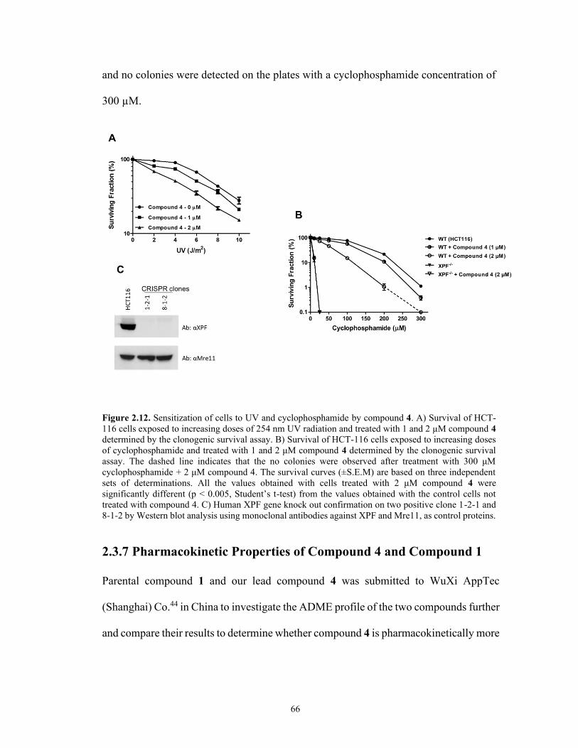
66
and no colonies were detected on the plates with a cyclophosphamide concentration of
300 µM.
Figure 2.12. Sensitization of cells to UV and cyclophosphamide by compound 4. A) Survival of HCT-
116 cells exposed to increasing doses of 254 nm UV radiation and treated with 1 and 2 µM compound 4
determined by the clonogenic survival assay. B) Survival of HCT-116 cells exposed to increasing doses
of cyclophosphamide and treated with 1 and 2 μM compound 4 determined by the clonogenic survival
assay. The dashed line indicates that the no colonies were observed after treatment with 300 μM
cyclophosphamide + 2 μM compound 4. The survival curves (±S.E.M) are based on three independent
sets of determinations. All the values obtained with cells treated with 2 μM compound 4 were
significantly different (p < 0.005, Student’s t-test) from the values obtained with the control cells not
treated with compound 4. C) Human XPF gene knock out confirmation on two positive clone 1-2-1 and
8-1-2 by Western blot analysis using monoclonal antibodies against XPF and Mre11, as control proteins.
2.3.7 Pharmacokinetic Properties of Compound 4 and Compound 1
Parental compound 1 and our lead compound 4 was submitted to WuXi AppTec
(Shanghai) Co.44 in China to investigate the ADME profile of the two compounds further
and compare their results to determine whether compound 4 is pharmacokinetically more

67
superior than compound 1. Compound 4 showed a lower logD (2.86) than 1 (3.86) at pH
7.4 (Table 2.5).
Table 2.5. Pharmacokinetic Profile of Compound 4 and Compound 1
Screening Test Compound Results
LogD at pH 7.4
Compound 1
3.86
Compound 4 2.86
Metabolic stability
in human liver
microsomes
Compound 1
53.7 (mL/min/kg)
Compound 4 44.0 (mL/min/kg)
Metabolic stability
in cryopreserved
human hepatocytes
Compound 1
<17.8 (mL/min/kg)
Compound 4 48.8 (mL/min/kg)
Serum protein
binding (% bound)
Compound 1
99.90
Compound 4 99.95
Permeability
(Efflux Ratio)
Compound 1
11.18
Compound 4 8.92
Cytochrome P450
(IC50)
Compound 1
CYP1A2,
CYP2C9
CYP2C19
CYP2D6
CYP3A4-M
14.1 µM 21.0 µM 15.2 µM 5.20 µM 7.45 µM
Compound 4
CYP1A2,
CYP2C9
CYP2C19
CYP2D6
CYP3A4-M
6.40 µM >50 µM >50 µM 16.0 µM 37.1 µM
Metabolism of compound 4, as measured by exposure to human liver
microsome, indicated that it has a metabolism in the midrange, while 1 has a more rapid

68
metabolism. Both compounds display an efflux ratio of higher than 2 (compound 4 =
8.92 and compound 1 = 11.18). Analysis of five CYP 450 tested enzymes indicates that
neither of the compounds has an IC50 lower than 1.00 µM, therefore, they are not potent
inhibitors of these enzymes. Compound 4, however, moderately inhibits CYP1A2 with
an IC50 of 6.40 µM. Compound 1 has the same moderate inhibitory effect on CYP2D6
and CYP3A4-M with an IC50 between 1 and 10 µM. Compound 4 acts as a weak
inhibitor of CYP2D6 and CYP3A4-M, while compound 1 is a weak inhibitor of three
CYP 450 enzymes (CYP1A2, CYP2C9, and CYP2C19) with an IC50 between 10 and
50 µM. Both compounds have similar behavior in terms of permeability and binding to
serum proteins, as shown in Table 2.5.
To conclude, our comparison of the ADME results between compound 1, the
parental compound, and compound 4 indicated that the latter has more favourable
pharmacological properties, particularly a lower logD than 1, which means that it has a
lower lipophilicity, and greater metabolic stability.
2.4 Conclusion
The ERCC1–XPF heterodimer is a structure-specific endonuclease, which is required
especially for NER and ICL DNA repair pathways. Although its action is essential to
maintain genome integrity and to protect against damage-induced mutations, as part of
the NER and ICL machinery it can counteract the effect of DNA damaging therapies,
such as platinum-based chemotherapy and radiotherapy. A promising approach to
enhance the effect of such therapies is to inhibit the action of DNA repair in cancer cells
using small molecules.

69
In this work, we used a computational drug design workflow to provide a rational
design for novel compound 1 analogues, a lead inhibitor targeting the dimerization
between XPF and ERCC1, which is required for endonuclease activity. We identified
seven compounds for which in silico simulations predicted attractive properties, namely,
binding affinities and ligand efficiency towards an XPF site on the dimerization
interface. The synthesis of the computationally designed compounds was carried out
successfully using a simple, robust, and reproducible synthetic strategy. Interestingly,
compound 4 has good physicochemical properties, i.e., reasonable logP and ligand
efficiency values and small molecular weight, and thus is a potential lead candidate for
further optimization. Following structure-activity relationship studies and in vitro
screening, this approach yielded compounds 3 and 4 as potent inhibitors of ERCC1–XPF
activity. An in vitro ERCC1–XPF endonuclease assay identified compound 4 as the best
ERCC1–XPF inhibitor with an IC50 value of 0.33 µM compared to 1.86 µM for
compound 1. In addition, the Kd value for this compound was measured experimentally
as 100 nM and interacts with ERCC1–XPF in the subunit interaction domain, altering
the related properties of this heterodimerization interface. Compound 4 also showed a
significant inhibition of the removal of cyclobutane pyrimidine dimers compared with
control cells after exposure of HCT-116 cells to UV radiation and sensitized the cells to
UV and cyclophosphamide-induced cytotoxicity, indicating inhibition of NER and ICL
repair. Furthermore, compound 4 possesses favourable pharmacokinetic properties,
particularly a lower logD than compound 1, which means that it has a lower lipophilicity
and greater metabolic stability. Our computational workflow successfully identified
superior compounds from a set of analogues differing by one substituent group.

70
Additionally, it provided detailed information about the ligand–protein interaction.
Compound 4 was our best in vitro analogue. Detailed analysis of the MM/GBSA binding
energies, together with the visual analysis of the simulation results, suggests a binding
mode for compound 4 where the conserved hydrophobic core of the analogue is buried
inside the XPF binding site and, different from the other active compounds (1 and 3),
the positive R-substituent is favorably exposed to the solvent. Although all of the
compounds share the acridine moiety, our assay showed that the inhibition activity of
compound 4 was the consequence of the interaction of the drug with the enzyme and not
the result of binding with DNA. The fact that compounds 4 and 5 (negative control)
show drastically different activity despite possessing the same acridine moiety also
provides strong evidence that inhibition is not mediated via DNA intercalation. In
summary, the use of in silico methods to design a superior compound to 1 led to
compound 4 that, potentially, can be used in combination with other existing DNA-
damaging therapies to amplify their effects by sensitizing cancer cells.
In future work, compound 4 will be the starting point for further optimization
towards a drug candidate compound to yield an optimized lead analogue of compound
4. Since polar side-chain installation at the exterior part of the piperazine position clearly
enhances activity, we intend to explore a wider range of polar groups on the remote end
of the piperazine. At the same time, we will retain the dimethylaminoethyl group and
explore structural changes in parts of the molecule that are intercalated more deeply into
the protein binding pocket. Different generations of compound 4 derivatives will be
synthesized according to the corresponding in silico studies and the current SAR results

71
in this study for the aim of obtaining a drug like candidate that has superior
physicochemical properties to compound 4.
2.5 Methods and Experimental Section*
2.5.1 General Experimental Procedures for Preparation of The Top
Compound 1 Analogues
Some reactions were carried out in flame-dried glassware under a positive nitrogen
atmosphere, unless otherwise stated. Transfer of anhydrous solvents and reagents was
accomplished with oven-dried syringes or cannulae. Chemicals were purchased from
Sigma Aldrich Inc. and were used without further purification. Thin layer
chromatography was performed on glass plates pre-coated with 0.25 mm silica gel. Flash
chromatography columns were packed with 230–400 mesh silica gel. Proton nuclear
magnetic resonance spectra (1H NMR) were recorded at 500 MHz, and coupling
constants (J) are reported in hertz (Hz). Standard notation was used to describe the
multiplicity of signals observed in 1H NMR spectra: broad (br), multiplet (m), singlet
(s), doublet (d), triplet (t), etc. Carbon nuclear magnetic resonance spectra (13C NMR)
were recorded at 125 MHz and are reported (ppm) relative to the center line of the triplet
from chloroform-d (77.0 ppm) or the center line of the septet from methanol-d4 (49.0
ppm). Infrared (IR) spectra were measured with a FT-IR 3000 spectrophotometer. Mass
spectra were determined on a high-resolution electrospray positive ion mode
spectrometer.
* I was responsible for the synthesis and design of all compounds. F.G. designed the computational studies
of the compounds. D.J. expressed and purified ERCC1−XPF, employed the in vitro ERCC–XPF1 assay,
and conducted microscale thermophoresis measurements. F.K.B and Y.X. carried out cell culture, UV
dimer repair assay, and clonogenic survival assay. R.M. conducted the binding affinity studies of the
compounds.

72
The high-performance liquid chromatography (HPLC) analyses were performed using
an Agilent 1100 LC/MSD instrument. Elution was done with a gradient of 10−95%
solvent B in solvent A (solvent A was 0.1% TFA in water, and solvent B was 0.1% acetic
acid in MeCN) through an Agilent column eclipse XDB- C18 (4.6 × 250 mm, 5 μm)
column at 1.0 mL/min. The area % purity was measured at 210 and 254 nm. The purity
of the most active compound 4 was assessed by HPLC (98%).
2.5.2 Synthesis and Characterization of Inhibitors (1-8)
4-((6-Chloro-2-methoxyacridin-9-yl)amino)-2-((4-methylpiperazin-1-
yl)methyl)phenol (1)
Synthesis of compound 1 was achieved through a one-pot sequential addition reaction
in three steps. A mixture of N-methyl piperazine (0.12 mL, 1.0 mmol), p-
acetamidophenol (0.15 g, 1.0 mmol) and 37% formaldehyde (0.10 mL, 5.0 mmol) in
isopropyl alcohol (2 mL) was taken in a 10 mL single neck round bottom flask. Then,
the reaction mixture was stirred and heated under reflux at 65 °C for 6 h. Upon
completion of the reaction (monitored by TLC with 15% MeOH/DCM eluent system),
the solvent was removed on a rotatory evaporator. Next, the residue was dissolved in 3

73
mL ethanol and 3 drops of 12 M HCl were added. Subsequently, the reaction mixture
was heated at 90 °C under reflux for 90 min. Afterwards, 6,9-dichloro-methoxyacridine
(0.28 g, 1.0 mmol) was added to the mixture and further stirred at 90 °C under reflux,
and the course of reaction followed by TLC until little or no starting material was
detected (around 12 h). On cooling to room temperature, the reaction mixture was
diluted with cold water and neutralized to pH of 8–9 with 28% v/v ammonia solution.
The alkaline solution was extracted with dichloromethane. The organic layer was
washed with brine, concentrated in vacuum, and purified by column chromatography
(gradient elution with 5% to 10% MeOH:DCM system) to afford compound 1 as an
orange reddish semisolid (0.08 g) in 78% yield; Rf 0.50 (2:8, MeOH:DCM); IR (cast
film) νmax = 3272, 2923, 1629, 1254, 1231, 1032, 926, 816, 815, 775 cm-1; 1H NMR
(500 MHz, CDCl3) δ 7.99 (s, 1H), 7.91 (d, J = 8.7 Hz, 1H), 7.80 (d, J = 9.2 Hz, 1H),
7.29 (dd, J = 9.4, 2.6 Hz, 1H), 7.11 (d, J = 10.7 Hz, 1H), 7.03 (s, 1H), 6.77 – 6.70 (m,
2H), 6.49 (d, J = 2.5 Hz, 1H), 3.63 (s, 3H), 3.53 (s, 2H), 2.75 – 2.32 (m, 8H), 2.27 (s,
3H), OH and NH protons were not observed; 13C NMR (125 MHz, CDCl3) δ 155.9,
153.2, 147.8, 143.9, 137.0, 135.0, 125.0, 124.9, 122.0, 121.8, 120.0, 119.9, 119.7,
117.6, 116.8, 116.5, 116.1, 115.9, 100.3, 61.1, 55.2, 54.8 (2C), 52.4 (2C), 45.8; HRMS
(ESI) calcd for C26H28ClN4O2 [M + H]+ 463.1895; found 463.1890.

74
4-((6-Chloro-2-methoxyacridin-9-yl)amino)-2-((4-(3-methylbenzyl)piperazin-1-
yl)methyl)phenol (2)
The previous method was employed to synthesize compound 2 with the following
stoichiometric amounts: 1-(3-methylbenzyl)piperazine (0.19 g, 1.0 mmol) and 6,9-
dichloroacridine (0.28 g, 1.0 mmol) to afford it as an orange reddish semisolid (0.14 g)
in 73% yield; Rf 0.44 (1:9, MeOH:DCM); IR (cast film) νmax = 3258, 2921, 1629, 1560,
1493, 1253, 1133, 1007, 927, 826, 775 cm-1; 1H NMR (500 MHz, CDCl3) δ 8.05 (s,
1H), 7.95 (d, J = 9.2 Hz, 1H), 7.83 (d, J = 9.2 Hz, 1H), 7.32 (d, J = 7.6 Hz, 1H), 7.21
(t, J = 7.5 Hz, 1H), 7.17 – 7.06 (m, 5H), 6.87 (d, J = 7.1 Hz, 1H), 6.77 (d, J = 8.6 Hz,
1H), 6.62 (d, J = 1.9 Hz, 1H), 3.70 (s, 3H), 3.59 (s, 2H), 3.49 (s, 2H), 2.79 – 2.40 (m,
8H), 2.35 (s, 3H). OH and NH protons were not observed; 13C NMR (125 MHz, CDCl3)
δ 156.1, 153.9, 150.4, 146.8, 144.5, 137.9, 137.6, 136.3, 135.5, 129.9, 128.2, 127.9,
126.2, 125.4, 125.0, 122.1, 121.8, 120.6, 120.3, 119.2, 116.9, 116.5, 116.1, 115.9,
100.4, 62.8, 61.2, 55.4, 52.8 (2C), 52.5 (2C), 21.4; HRMS (ESI) calcd for C33H34ClN4O2
[M + H]+ 553.2357; found 553.2365.

75
4-((6-Chloro-2-methoxyacridin-9-yl)amino)-2-((4-cyclohexylpiperazin-1-
yl)methyl)phenol (3)
The previous method was employed to synthesize compound 3 with the following
stoichiometric amounts: 1-cyclohexyl piperazine (0.17 g, 1.0 mmol) and 6,9-
dichloroacridine (0.28 g, 1.0 mmol) to afford it as an orange reddish semisolid (0.13 g)
in 81% yield; Rf 0.52 (0.5:9.5, MeOH: EtOAc); IR (cast film) νmax = 3313, 2918, 1736,
1560, 1468, 1255, 1235, 1181, 1032, 929, 829, 722 cm-1; 1H NMR (500 MHz, CDCl3)
δ 8.03 (d, J = 1.4 Hz, 1H), 7.92 (d, J = 9.6 Hz, 1H), 7.80 (d, J = 9.5 Hz, 1H), 7.28 (d, J
= 5.7 Hz, 1H), 7.11 (d, J = 9.1 Hz, 2H), 6.91 (d, J = 9.3 Hz, 1H), 6.77 (d, J = 8.5 Hz,
1H), 6.69 (s, 1H), 3.71 (s, 3H), 3.60 (s, 2H), 2.73 – 2.22 (m, 8H), 1.83 (m, 4H), 1.63 (d,
J = 12.8 Hz, 1H), 1.26 – 1.18 (m, 6H). OH and NH protons were not observed; 13C
NMR (125 MHz, CDCl3) δ 156.1, 153.8, 146.9, 144.5, 136.3, 135.5, 125.4 – 116.8 (10
C), 101.3, 100.4, 98.8, 63.4, 61.3, 52.9 (2 C), 48.7 (2 C), 29.7, 28.9 (2 C), 26.2, 25.8 (2
C); HRMS (ESI) calcd for C31H35ClN4O2 [M + H]+ 531.2521; found 531.2527.

76
4-((6-Chloro-2-methoxyacridin-9-yl)amino)-2-((4-(2-(dimethylamino)ethyl)
piperazin-1-yl) methyl) phenol (4)
The previous method was employed to synthesize compound 4 with the following
stoichiometric amounts: 1-[2-(dimethylamino)ethyl] piperazine (0.16 g, 1.0 mmol) and
6,9-dichloroacridine (0.28 g, 1.0 mmol) to afford it as an orange reddish semisolid (0.13
g) in 80 % yield; Rf 0.40 (1.3: 8.5: 0.2, MeOH: DCM: Et3N); IR (cast film) νmax = 3361,
2918, 1736, 1562, 1467, 1295, 1255, 1143, 1032, 945, 828, 763, 722 cm-1; 1H NMR
(500 MHz, CDCl3) δ 8.10 (s, 1H), 8.00 (s, 1H), 7.89 (d, J = 9.1 Hz, 1H), 7.40 (d, J =
9.8 Hz, 1H), 7.24 (d, J = 8.0 Hz, 1H), 7.09 (s, 1H), 6.85 (dd, J = 8.6, 2.7 Hz, 1H), 6.78
(d, J = 8.6 Hz, 1H), 6.56 (d, J = 2.3 Hz, 1H), 3.74 (s, 3H), 3.59 (s, 2H), 2.70 – 2.40 (m,
12H), 2.27 (s, 6H). OH and NH protons were not observed; 13C NMR (125 MHz,
CDCl3) δ 156.2, 153.6, 147.5, 144.0, 136.6, 135.2, 125.3, 125.1, 124.8, 122.0, 120.3,
120.2, 120.0, 119.8, 119.6, 117.5, 116.9, 116.3, 100.2, 61.2, 56.7, 56.7, 55.3, 53.3 (2C),
52.4 (2C), 45.8 (2C); HRMS (ESI) calcd for C29H35ClN5O2 [M + H]+ 520.2474; found
520.2473.

77
4-((6-Chloro-2-methoxyacridin-9-yl)amino)-2-((4-(3,4-dichlorophenyl)piperazin-
1-yl)methyl)phenol (5)
The previous method was employed to synthesize compound 5 with the following
stoichiometric amounts: 1-(3,4-dichlorophenyl) piperazine (0.23 g, 1.0 mmol), p-
acetamidophenol (0.15 g, 1.0 mmol), 37% formaldehyde (0.10 mL, 5 mmol) and 6,9-
dichloroacridine (0.28 g, 1.0 mmol) to afford it as an orange reddish semisolid (0.16 g)
in 71% yield; Rf 0.56 (0.5:9.5, MeOH:DCM); IR (cast film) νmax = 3271, 2918, 1736,
1563, 1227, 1180, 1080, 1031, 803, 721 cm-1; 1H NMR (500 MHz, CDCl3) δ 8.09 (s,
1H), 8.00 (s, 1H), 7.88 (d, J = 9.2 Hz, 1H), 7.40 (d, J = 9.3 Hz, 1H), 7.28 (d, J = 8.7 Hz,
1H), 7.24 (d, J = 9.0 Hz, 1H), 7.09 (s, 1H), 6.95 (d, J = 2.8 Hz, 1H), 6.87 (dd, J = 8.6,
2.7 Hz, 1H), 6.80 (d, J = 8.6 Hz, 1H), 6.73 (dd, J = 8.9, 2.8 Hz, 1H), 6.54 (d, J = 2.6
Hz, 1H), 3.74 (s, 3H), 3.63 (s, 2H), 3.27 – 3.15 (m, 4H), 2.72 – 2.62 (m, 4H), OH and
NH protons were not observed; 13C NMR (125 MHz, CDCl3) δ 156.3, 153.1, 150.2,
147.9, 143.3, 137.0, 135.0, 132.9, 130.5, 125.2, 124.7, 122.9, 121.6, 121.4, 120.2,
120.0, 119.7, 117.9, 117.6, 117.0, 116.7, 116.2, 115.6, 115.5, 100.0, 61.2, 55.4 (2C),

78
52.2 (2C), 48.7; HRMS (ESI) calcd for C31H28Cl3N4O2 [M + H]+ 593.1272; found
593.1272.
2-((4-(Bis(4-fluorophenyl)methyl)piperazin-1-yl)methyl)-4-((6-chloro-2-
methoxyacridin-9-yl)amino)phenol (6)
The previous method was employed to synthesize compound 6 with the following
stoichiometric amounts: 1-bis (4-fluorophenyl)methylpiperazine (0.29 g, 1.0 mmol), p-
acetamidophenol (0.15 g, 1.0 mmol), 37% formaldehyde (0.10 mL, 5 mmol) and 6,9-
dichloroacridine (0.28 g, 1.0 mmol) to afford it as an orange reddish semisolid (0.19 g)
in 65% yield; Rf 0.56 (0.5:9.5, MeOH:DCM); IR (cast film) νmax = 3275, 2919, 1737,
1566, 1255, 1217, 1181, 1032, 828, 722 cm-1; 1H NMR (500 MHz, CDCl3) δ 8.07 (s,
1H), 7.98 (d, J = 9.0 Hz, 1H), 7.84 (d, J = 9.2 Hz, 1H), 7.34 (m, 6H), 7.18 (d, J = 8.0
Hz, 1H), 7.06 (d, J = 1.8 Hz, 1H), 6.97 (m, 5H), 6.83 (dd, J = 8.5, 2.6 Hz, 1H), 6.75 (d,
J = 8.6 Hz, 1H), 6.53 (d, J = 2.2 Hz, 1H), 4.23 (s, 1H), 3.69 (s, 3H), 3.57 (s, 2H), 2.74
– 2.21 (m, 8H); 13C NMR (125 MHz, CDCl3) δ 161.9 (2C), 156.2, 153.5, 150.3, 147.7,

79
143.7, 137.8 (2C), 136.7, 135.0, 129.2 (4C), 125.2, 124.8, 121.9, 121.7, 120.2, 119.9,
119.7, 117.6, 116.8, 116.5, 116.1, 115.5 (4C), 100.0, 74.3, 61.1, 55.3, 52.6 (2C), 51.5
(2C); HRMS (ESI) calcd for C38H34ClF2N4O2 [M + H]+ 651.2333; found 651.2328.
4-((6-Chloro-2-methoxyacridin-9-yl)amino)-2-((4-(4-hydroxyphenyl)piperazin-1-
yl)methyl)phenol (7)
The previous method was employed to synthesize compound 7 with the following
stoichiometric amounts: 1-(4-hydroxyphenyl)piperazine (0.30 g, 1.0 mmol), p-
acetamidophenol (0.15 g, 1.0 mmol), 37% formaldehyde (0.10 mL, 5 mmol) and 6,9-
dichloroacridine (0.28 g, 1.0 mmol) to afford it as an orange reddish semisolid (0.17 g)
in 59% yield; Rf 0.58 (1.5:8.5, MeOH:DCM); IR (cast film) νmax = 3334, 2918, 1737,
1564, 1468, 1235, 1181, 1031, 930, 830, 722 cm-1; 1H NMR (500 MHz, CD3OD) δ 8.07
(d, J = 9.3 Hz, 1H), 7.87 (d, J = 1.8 Hz, 1H), 7.82 (d, J = 9.3 Hz, 1H), 7.51– 7.46 (m,
1H), 7.41 (d, J = 2.5 Hz, 1H), 7.29 – 7.24 (m, 1H), 7.04 (dd, J = 8.7, 2.4 Hz, 1H), 6.93
– 6.89 (m, 1H), 6.88 – 6.81 (m, 3H), 6.70 (dd, J = 8.9, 2.3 Hz, 2H), 3.72 (s, 3H), 3.61
(s, 2H), 3.11 – 2.99 (m, 4H), 2.82 – 2.67 (m, 4H), OH and NH protons were not

80
observed; 13C NMR (125 MHz, CDCl3) δ 156.3, 153.5, 152.1, 150.2, 150.1, 145.3,
145.2, 144.0, 125.3, 124.7, 122.5, 121.9, 121.5, 120.4, 120.0, 118.8 (2C), 118 (2C),
118.4, 118.1, 117.0, 116.5, 116.1, 115.9, 61.3, 55.4, 52.7 (2C), 50.7 (2C); HRMS (ESI)
calcd for C31H30ClN4O3 [M + H]+ 541.2001; found 541.2010.
4-((6-Chloro-2-methoxyacridin-9-yl)amino)phenol (8)
Synthesis of compound 8 was carried out according to the reported procedures 21 with
minor modifications. The synthesis was performed by mixing 4-aminophenol (0.11 g,
1.0 mmol) and 6,9-dichloroacridine (0.28 g, 1.0 mmol) in a 10 mL single neck round
bottom flask and stirred at 90 °C under reflux. The course of the reaction was followed
by TLC until little or no starting material was detected (around 12 h). On cooling to
room temperature, the reaction mixture was diluted with cold water and neutralized to
pH of 8–9 with 28% v/v ammonia solution. The alkaline solution was extracted with
dichloromethane. The organic layer was washed with brine, concentrated in vacuum,
and purified by column chromatography (gradient elution with 1% to 5% MeOH:DCM
system) to afford it as an orange reddish semisolid in 73% yield; Rf 0.47 (1.5:8.5,
MeOH:DCM); IR (cast film) νmax = 3325, 2918, 1737, 1562, 1468, 1236, 1180, 1103,
1050, 722 cm-1; 1H NMR (500 MHz, CD3OD) δ 8.12 (d, J = 9.4 Hz, 1H), 7.84 – 7.83

81
(m, 1H), 7.78 (d, J = 9.3 Hz, 1H), 7.61 (dd, J = 9.3, 2.6 Hz, 1H), 7.48 (d, J = 1.5 Hz,
1H), 7.35 (dd, J = 9.4, 2.1 Hz, 1H), 7.27 – 7.22 (m, 2H), 6.95 – 6.91 (m, 2H), 3.88 (s,
1H), 3.69 (s, 3H). OH proton was not observed; 13C NMR (125 MHz, CD3OD) δ 158.9,
157.7, 141.7, 137.9, 133.2, 129.8, 128.4, 127.8 (2C), 126.6, 125.7, 122.2, 120.1, 119.3,
117.8 (2C), 116.1, 113.3, 104.5, 56.1.; HRMS (ESI) calcd for C20H16ClN2O2 [M + H]+
351.0908; found 351.0896.
2.5.3 Molecular Docking of Compound 1
The first step of our VS study was to investigate the binding mode of the lead compound
125 to the pocket on the XPF C-terminus. We used twenty XPF structures as single
targets for a relaxed complex scheme (RCS) docking protocol in order to account for
protein flexibility accurately.35, 36 These structures were extracted from the Protein Data
Bank (PDB)37 entry 1Z00,17 reporting the NMR ensemble of the dimerization complex
between the ERCC1 and XPF HhH2 domains. The XPF structures were optimized with
a minimization process. The binding site of each target was defined as the geometric
center of the residues Y833, N834, P837, Q838, M856, K860, N861, and I862 on the
XPF HhH2 domain, as reported in our previous study,25 where the residues are
numbered according to the 1Z00 PDB file. Molecular docking simulations were
performed using the Lamarckian Genetic Algorithm (LGA) 38 and the built-in scoring
function39 of Autodock4.40
2.5.4 Characterization of The Binding Pocket and Pharmacophore
Modeling
To characterize the binding site and the binding mode of compound 1, electrostatic

82
maps were generated in the MOE2015 software package,41 based on the docked pose
of compound 1, using –2 kcal/mol for the potential iso-surfaces of hydrophobic,
acceptor, and donor probe atoms, and the Poisson–Boltzmann equation to compute the
potentials. The maps were built within 4.5 Å of the ligand pose obtained from docking.
Using the information from the docking and electrostatic mapping, a pharmacophore
model for compound 1 was built with the MOE Pharmacophore Query Editor and the
EHT scheme.42
2.5.5 Docking-based Virtual Screening of Compound 1 Analogues
Fifty-seven analogues of compound 1 structure were designed by replacement of the
piperazine N-methyl group with other moieties expected to capture additional binding
interactions of the piperazine ring. The structures were prepared using Schrödinger
LigPrep43 to account for multiple tautomers, protonation states, and low-energy ring
conformations, using the same approach as for compound 1. The molecular docking
simulations were performed with MOE Dock. Only the XPF structure from the best
compound 1 complex was used as a target for the VS study. Different conformations of
the analogues were obtained using Conformation Import. The previously built
pharmacophore model was used for the placement step, in which 30 poses are returned
according to the London dG scoring method.44 To account for the local arrangement of
the pocket residues upon ligand binding, the Induced Fit method was selected for the
refinement step, where the side chains of the binding pocket were left to move freely.
At the end, one pose scored with the Generalized Born Volume Integral/Weighted
Surface Area (GBVI/WSA) function was returned.44 Water/octanol partition
coefficients (logP) of the molecules were calculated in MOE using the SlogP function,45

83
which takes into account the given protonation state of the molecule under examination.
Ligand efficiencies were calculated as the ratio between the computed binding energies
and the number of heavy atoms of each analog.
2.5.6 Molecular Dynamics Simulations and MM/GBSA Rescoring of
the Analogues
To calculate an average binding energy for the hits, we performed 2 ns of MD
simulations for the top ranked compound-XPF complexes. Free energy calculations
were performed over the trajectories with the MM/GBSA method using the
MMPBSA.py script.46 The calculations were performed on snapshots extracted every
10 ps from the MD trajectories, and per-residue decompositions of the binding energies
also were performed for the residues within 10 Å of any analog atom at the beginning
of the simulations. We also calculated the entropic contribution of ligand binding using
the normal mode analysis (NMA) method.47 The final binding energies used to rank the
compounds were calculated combining the MM/GBSA and entropy contributions.
2.5.7 ERCC1–XPF Protein Preparation
David Jay used human ERCC1–XPF wild-type protein that was obtained as previously
described.29 Basically, the recombinant protein was expressed from a bi-cistronic
plasmid (kindly provided by Dr. Richard Wood, University of Texas MD Anderson
Cancer Center, Smithville, TX) in the E. coli BL21 (DE3) strain. Since both XPF and
ERCC1 contained a polyhistidine (His-6) tag, the proteins extracted from E. coli were
incubated with a ProBond Nickel-Chelating Resin (Thermo Fisher Scientific). Protein
eluted from the Ni affinity column subsequently was loaded into a Hi-trap heparin

84
column (GE Healthcare). Fractions recovered from the heparin column that contained
ERCC1-XPF were dialyzed, concentrated, and stored at -80 ºC in 10 mM HEPES pH
7.4, 2.5 mM β-mercaptoethanol, 0.01% CHAPS, 0.25 mM EDTA, 50% glycerol, and
25 mM NaCl.
2.5.8 Microplate Fluorescence Incision Assay
David Jay followed a previously described protocol.2, 29 Briefly, reactions were carried
out in a 384-well black, flat-bottomed microtiter plates (OptiPlate – 384 F, Perkin
Elmer) in a total volume of 20 µl containing the indicated concentrations of inhibitor
compounds, 100 nM stem–loop substrate [6-FAM-5′-
CAGCGCTCGG(20T)CCGAGCGCTG-3′-dabcyl], 25 ng ERCC1-XPF, 50 mM Tris-
Cl pH 8, 20 mM NaCl, 0.5 mM DDT, and 0.75 mM MnCl2 at 25 ºC. Fluorescent
readings were obtained using a FLUOstar Optima fluorimeter (BMG Labtech) with
Optima software at an excitation of and emission wavelengths of 485 and 520 nm,
respectively, for 12 min.
2.5.9 Steady-state Fluorescence Assay
Steady-state fluorescence spectra were measured at room temperature on a Perkin-
Elmer LS-55 spectrofluorometer (Freemont, CA) with 5 nm spectral resolution for
excitation and emission using 30–80 nM solution of purified recombinant ERCC1–XPF
protein complex. Protein fluorescence was excited at 295 nm, and fluorescence
emission spectra were recorded in the 300–400 nm range: changes in fluorescence
intensity were monitored at the emission maximum (330 nm). In studying the effects
of inhibitors on protein fluorescence intensities, additions to protein samples were made
from inhibitors stock solutions, keeping the protein dilution below 3%.

85
2.5.10 Expression and Purification of Recombinant Truncated Forms
of ERCC1 and XPF
Synthetic DNA sequences encoding for human ERCC1 codons 96–297 (dERCC1) and
XPF codons 667–916 (dXPF) inserted into separate pET-28a expression plasmids were
used to transform BL21(DE3) cells and express the corresponding recombinant
proteins.2 Since the truncated form of the proteins both contained N-terminal
polyhistidine-tags (His-tags), they were purified utilizing a nickel-chelating resin
(ThermoFisher Scientific) under conditions similar to the ones described.2 Both
truncated peptides were solubilized finally to final concentrations for the experiments
of 40 nM dXPF and 20 nM dERCC1 in a buffer (MST buffer) containing 20 mM
HEPES pH 7.4, 50 mM NaCl, 1 mM DTT, 2 mM MgCl2, 5% glycerol, and 0.01%
CHAPS.
2.5.11 Microscale Thermophoresis (MST) Measurements
We tried to remove the His-tag from one of the truncated peptides (preferentially
dERCC1 because of its low yield) to carry out MST equilibrium experiments. However,
the peptides proved to be resistant to thrombin treatment, so we decided to follow a
different qualitative approach. Both, dXPF and dERCC1 were labelled with 20 nM and
10 nM RED-tris-NTA (NanoTemper Technologies), respectively, following the
manufacturer instructions. Individual peptides (20 nM final concentration) or equimolar
amounts of dERCC1 and dXPF (20 nM final concentration of the dimer) were incubated
in MST buffer in the presence of compound 4 (10 µM) or DMSO. After 20 minutes of
incubation time at room temperature, 10 µl of the samples were placed in standard
capillaries (NanoTemper Technologies), and the emitted red fluorescence (670 nm) was

86
measured in a Monolith NT.115 instrument (NanoTemper Technologies) with a High
MST-Power, 70% Excitation Power, and a Nano-RED Excitation type. Pretest and
binding check experiments did not show any change in fluorescence of the RED-tris-
NTA dye induced by compound 4. In addition, the peak shape did not indicate any
adsorption of the components of the solution to the surface of the capillaries.
2.5.12 Cell Culture
The human colorectal cancer HCT-116 cell line was obtained from the American Type
Culture Collection (ATCC). The cell population was expanded immediately after
arrival, aliquoted, and stored frozen in liquid nitrogen. Freshly thawed cells were used
for each experiment. The cells were cultured in a 1:1 DMEM/F12 media supplemented
with 10% FBS, 50 units/mL penicillin, 50 μg/mL streptomycin, mM l-glutamine, 0.1
mM nonessential amino acids, and1 mM sodium pyruvate, and maintained under 5%
CO2 in a humidifier incubator at 37 °C. All the supplies for cell culture were obtained
from Gibco/BRL.
2.5.13 Cellular Repair of Cyclobutane Pyrimidine Dimers
We followed the protocol of Mirzayans et al. with minor modifications.34
Approximately 1 x 10⁵ HCT-116 cells were seeded on each coverslip and allowed to
attach overnight. Then, the medium was removed, and the cells were treated for 1 h
with the desired compound. The medium was removed, and the cells were exposed to
8 J/m2 UV-C radiation (G15T8 254 nm lamp, Ushio America Inc, Cypress, CA),
followed by adding fresh medium containing the compound. The plates were incubated
at 37 °C for different periods of time up to 24 h and fixed in a 50:50 methanol/1X
phosphate-buffered saline (PBS) solution, followed by replacing the methanol/PBS

87
solution with 100% methanol and incubation in –20 °C. After 20 min, the methanol was
removed, and the cells were treated with PBS for 5 min at room temperature. After
fixing, the cells were permeabilized in 0.5% Triton/PBS and washed with PBS,
denatured in 2 N HCl, and neutralized by twice washing with 0.1 M borate buffer pH
8.5. Cells on cover slips were washed once with PBS followed by blocking with 5%
non-fat dry milk/PBS for 30 min. Mouse anti-thymine dimer monoclonal antibody (Cat.
No. MC-062, Kamiya Biomedical Company, Seattle, WA) was applied to the cover
slips for 1 h in the dark and at room temperature. Cells were then washed with PBS
containing 0.1% Tween-20 and incubated with rabbit anti-mouse IgG-Alexafluor
antibody (Invitrogen, Carlsbad, CA) followed by two washes with PBS/(0.1%)Tween-
20. Cells on coverslips were then washed with PBS, rinsed with water, and mounted on
slides using DAPI glycerol mounting solution. Slides were kept at 4 °C before
fluorescent microscopic evaluation and measurement of fluorescence intensity with
MetaXpress Version: 6.2.1.704 software (Molecular Devices, Sunnyvale, California).
2.5.14 Clonogenic Survival Assay
UV treatment: HCT-116 cells (100–800 cells depending on the UV dose) were plated
in triplicate in 60-mm petri dishes. The cells were incubated overnight at 37 °C in a
humidified atmosphere containing 5% CO2 to allow for cell attachment. Then, the
medium was removed, and the cells were treated with 1 or 2 µM compound 4 for 1 h.
The medium was removed again, and the cells were exposed to increasing doses (0–10
J/m2) of UV-C radiation and then incubated for a further 10 days in the presence of
inhibitor at 37 °C in a 5% CO2 atmosphere to allow for colony formation. After this
period, the plates were stained with crystal violet and colonies were counted using a

88
Colcount instrument (Oxford Optronix, Abingdon UK). Finally, plating efficiency and
surviving fraction were calculated.
Cyclophosphamide treatment: A similar protocol was followed as described for
the UV treatment, except that cells were treated with 1 or 2 µM compound 4 for 4 h,
followed by addition of increasing doses of cyclophosphamide (0–300 µM). After 24 h,
the medium was replaced with fresh medium containing compound 4 alone. The plates
were incubated for another 8 days at 37 °C in a 5% CO2 atmosphere for colony
formation. After this period the plates were stained with crystal violet, colonies were
counted, and plating efficiency and surviving fraction were calculated.
2.5.14 PK Assessment of Compound 4 and Compound 1
Standard protocols were used for assessing the absorption, distribution, metabolism,
and excretion (ADME) profile of our lead compound 4 and compound 1 (reference hit).
Screening were performed at WuXi AppTec (Shanghai) Co48 to conduct the following
assays according to the standard protocols: distribution coefficient (Log D at pH 7.4),
aqueous solubility (Kinetic), metabolic stability in human liver microsomes and
cryopreserved human hepatocytes, bidirectional permeability in Caco-2 cells, serum
protein binding, and cytochromes P450 (CYPs) inhibition (CYP1A2, CYP2C9,
CYP2C19, CYP2D6, CYP3A4-M).

89
2.10 References
1. Sijbers, A. M.; de Laat, W. L.; Ariza, R. R.; Biggerstaff, M.; Wei, Y.-F.; Moggs, J. G.; Carter,
K. C.; Shell, B. K.; Evans, E.; de Jong, M. C. Xeroderma pigmentosum group F caused by a
defect in a structure-specific DNA repair endonuclease. Cell 1996, 86, 811-822.
2. McNeil, E. M.; Astell, K. R.; Ritchie, A.-M.; Shave, S.; Houston, D. R.; Bakrania, P.; Jones, H.
M.; Khurana, P.; Wallace, C.; Chapman, T.; Wear, M. A.; Walkinshaw, M. D.; Saxty, B.;
Melton, D. W. Inhibition of the ERCC1–XPF structure-specific endonuclease to overcome
cancer chemoresistance. DNA Repair 2015, 31, 19-28.
3. Friedberg, E. C.; Walker, G. C.; Siede, W.; Wood, R. D.; Schultz, R. A.; Ellenberger, T. DNA
Repair and Mutagenesis. 2nd edn ASM.; Press. Washington, DC 2006.
4. McNeil, E. M.; Melton, D. W. DNA repair endonuclease ERCC1–XPF as a novel therapeutic
target to overcome chemoresistance in cancer therapy. Nucleic Acids Research 2012, 40, 9990-
10004.
5. Douwel, D. K.; Hoogenboom, W. S.; Boonen, R. A. C. M.; Knipscheer, P. Recruitment and
positioning determine the specific role of the XPF‐ERCC1 endonuclease in interstrand crosslink
repair. The EMBO Journal 2017, 36, 2034-2046.
6. Yagi, T.; Katsuya, A.; Koyano, A.; Takebe, H. Sensitivity of group F xeroderma pigmentosum
cells to UV and mitomycin C relative to levels of XPF and ERCC1 overexpression. Mutagenesis
1998, 13, 595-599.
7. Gajjar, K. K.; Yadav, D. K.; Kobawala, T. P.; Trivedi, T. I.; Vora, H. H.; Ghosh, N. R. ERCC1
expression in patients with colorectal cancer: a pilot study. Journal of Cancer Metastasis and
Treatment 2016, 2, 471-476.
8. McHugh, P. J.; Spanswick, V. J.; Hartley, J. A. Repair of DNA interstrand crosslinks: molecular
mechanisms and clinical relevance. The Lancet Oncology 2001, 2, 483-490.
9. Ahmad, A.; Robinson, A. R.; Duensing, A.; van Drunen, E.; Beverloo, H. B.; Weisberg, D. B.;
Hasty, P.; Hoeijmakers, J. H. J.; Niedernhofer, L. J. ERCC1-XPF endonuclease facilitates DNA
double-strand break repair. Molecular and Cellular Biology 2008, 28, 5082-5092.
10. De Laat, W. L.; Appeldoorn, E.; Jaspers, N. G. J.; Hoeijmakers, J. H. J. DNA structural elements
required for ERCC1-XPF endonuclease activity. Journal of Biological Chemistry 1998, 273,
7835-7842.
11. Gaillard, P.-H. L.; Wood, R. D. Activity of individual ERCC1 and XPF subunits in DNA
nucleotide excision repair. Nucleic Acids Research 2001, 29, 872-879.

90
12. Gentile, F.; Tuszynski, J. A.; Barakat, K. H. New design of nucleotide excision repair (NER)
inhibitors for combination cancer therapy. Journal of Molecular Graphics and Modelling 2016,
65, 71-82.
13. Gentile, F.; Barakat, K. H.; Tuszynski, J. A. Computational Characterization of Small Molecules
Binding to the Human XPF Active Site and Virtual Screening to Identify Potential New DNA
Repair Inhibitors Targeting the ERCC1-XPF Endonuclease. International Journal of Molecular
Sciences 2018, 19, 1328−1341 .
14. Gentile, F.; A Tuszynski, J.; H Barakat, K. Modelling DNA repair pathways: recent advances
and future directions. Current Pharmaceutical Design 2016, 22, 3527-3546.
15. Ahmad, A.; Enzlin, J. H.; Bhagwat, N. R.; Wijgers, N.; Raams, A.; Appledoorn, E.; Theil, A.
F.; Hoeijmakers, J. H. J.; Vermeulen, W.; Jaspers, N. G. J. Mislocalization of XPF-ERCC1
nuclease contributes to reduced DNA repair in XP-F patients. PLoS Genetics 2010, 6, e1000871-
e1000882.
16. De Laat, W. L.; Sijbers, A. M.; Odijk, H.; Jaspers, N. G. J.; Hoeijmakers, J. H. J. Mapping of
interaction domains between human repair proteins ERCC1 and XPF. Nucleic Acids Research
1998, 26, 4146-4152.
17. Tripsianes, K.; Folkers, G.; Eiso, A. B.; Das, D.; Odijk, H.; Jaspers, N. G. J.; Hoeijmakers, J. H.
J.; Kaptein, R.; Boelens, R. The structure of the human ERCC1/XPF interaction domains reveals
a complementary role for the two proteins in nucleotide excision repair. Structure 2005, 13,
1849-1858.
18. Choi, Y.-J.; Ryu, K.-S.; Ko, Y.-M.; Chae, Y.-K.; Pelton, J. G.; Wemmer, D. E.; Choi, B.-S.
Biophysical characterization of the interaction domains and mapping of the contact residues in
the XPF-ERCC1 complex. Journal of Biological Chemistry 2005, 280, 28644-28652.
19. Tsodikov, O. V.; Enzlin, J. H.; Schärer, O. D.; Ellenberger, T. Crystal structure and DNA
binding functions of ERCC1, a subunit of the DNA structure-specific endonuclease XPF–
ERCC1. Proceedings of the National Academy of Sciences of the United States of America 2005,
102, 11236-11241.
20. McNeil, E. M.; Astell, K. R.; Ritchie, A.-M.; Shave, S.; Houston, D. R.; Bakrania, P.; Jones, H.
M.; Khurana, P.; Wallace, C.; Chapman, T. Inhibition of the ERCC1–XPF structure-specific
endonuclease to overcome cancer chemoresistance. DNA Repair 2015, 31, 19-28.
21. Chapman, T. M.; Gillen, K. J.; Wallace, C.; Lee, M. T.; Bakrania, P.; Khurana, P.; Coombs, P.
J.; Stennett, L.; Fox, S.; Bureau, E. A. Catechols and 3-hydroxypyridones as inhibitors of the

91
DNA repair complex ERCC1-XPF. Bioorganic & Medicinal Chemistry Letters 2015, 25, 4097-
4103.
22. Chapman, T. M.; Wallace, C.; Gillen, K. J.; Bakrania, P.; Khurana, P.; Coombs, P. J.; Fox, S.;
Bureau, E. A.; Brownlees, J.; Melton, D. W. N-Hydroxyimides and hydroxypyrimidinones as
inhibitors of the DNA repair complex ERCC1–XPF. Bioorganic & Medicinal Chemistry Letters
2015, 25, 4104-4108.
23. Arora, S.; Heyza, J.; Zhang, H.; Kalman-Maltese, V.; Tillison, K.; Floyd, A. M.; Chalfin, E. M.;
Bepler, G.; Patrick, S. M. Identification of small molecule inhibitors of ERCC1-XPF that inhibit
DNA repair and potentiate cisplatin efficacy in cancer cells. Oncotarget 2016, 7, 75104-75117.
24. Yang, L.; Ritchie, A.-M.; Melton, D. W. Disruption of DNA repair in cancer cells by
ubiquitination of a destabilising dimerization domain of nucleotide excision repair protein
ERCC1. Oncotarget 2017, 8, 55246-55264.
25. Jordheim, L. P.; Barakat, K. H.; Heinrich-Balard, L.; Matera, E.-L.; Cros-Perrial, E.; Bouledrak,
K.; El Sabeh, R.; Perez-Pineiro, R.; Wishart, D. S.; Cohen, R. Small molecule inhibitors of
ERCC1-XPF protein-protein interaction synergize alkylating agents in cancer cells. Molecular
Pharmacology 2013, 84, 12-24.
26. Chapman, T. M.; Gillen, K. J.; Wallace, C.; Lee, M. T.; Bakrania, P.; Khurana, P.; Coombs, P.
J.; Stennett, L.; Fox, S.; Bureau, E. A.; Brownlees, J.; Melton, D. W.; Saxty, B. Catechols and
3-hydroxypyridones as inhibitors of the DNA repair complex ERCC1-XPF. Bioorganic &
Medicinal Chemistry Letters 2015, 25, 4097-4103.
27. Arora, S.; Heyza, J.; Zhang, H.; Kalman-Maltese, V.; Tillison, K.; Floyd, A. M.; Chalfin, E. M.;
Bepler, G.; Patrick, S. M. Identification of small molecule inhibitors of ERCC1-XPF that inhibit
DNA repair and potentiate cisplatin efficacy in cancer cells. Oncotarget 2016, 7, 75104−75117
28. Scurr, M.; Pembroke, T.; Bloom, A.; Roberts, D.; Thomson, A.; Smart, K.; Bridgeman, H.;
Adams, R.; Brewster, A.; Jones, R.; Gwynne, S.; Blount, D.; Harrop, R.; Hills, R.; Gallimore,
A.; Godkin, A. Low-Dose Cyclophosphamide Induces Antitumor T-Cell Responses, which
Associate with Survival in Metastatic Colorectal Cancer. Clinical Cancer Research : an Official
Journal of the American Association for Cancer Research 2017, 23, 6771-6780.
29. Bowles, M.; Lally, J.; Fadden, A. J.; Mouilleron, S.; Hammonds, T.; McDonald, N. Q.
Fluorescence-based incision assay for human XPF–ERCC1 activity identifies important
elements of DNA junction recognition. Nucleic Acids Research 2012, 40, e101-e101.

92
30. Mani, R. S.; Karimi-Busheri, F.; Fanta, M.; Caldecott, K. W.; Cass, C. E.; Weinfeld, M.
Biophysical characterization of human XRCC1 and its binding to damaged and undamaged
DNA. Biochemistry 2004, 43, 16505-16514.
31. Jerabek-Willemsen, M.; Wienken, C. J.; Braun, D.; Baaske, P.; Duhr, S. Molecular interaction
studies using microscale thermophoresis. Assay and Drug Development Technologies 2011, 9,
342-353.
32. Wienken, C. J.; Baaske, P.; Rothbauer, U.; Braun, D.; Duhr, S. Protein-binding assays in
biological liquids using microscale thermophoresis. Nature communications 2010, 1, 100-107.
33. Zillner, K.; Jerabek-Willemsen, M.; Duhr, S.; Braun, D.; Längst, G.; Baaske, P. Microscale
thermophoresis as a sensitive method to quantify protein: nucleic acid interactions in solution.
In Functional Genomics, Springer: 2012; pp 241-252.
34. Mirzayans, R.; Pollock, S.; Scott, A.; Gao, C. Q.; Murray, D. Metabolic labeling of human cells
with tritiated nucleosides results in activation of the ATM-dependent p53 signaling pathway and
acceleration of DNA repair. Oncogene 2003, 22, 5562-5571.
35. Philippopoulos, M.; Lim, C. Exploring the dynamic information content of a protein NMR
structure: Comparison of a molecular dynamics simulation with the NMR and X‐ray structures
of Escherichia coli ribonuclease HI. Proteins: Structure, Function, and Bioinformatics 1999,
36, 87-110.
36. Ishima, R.; Torchia, D. A. Protein dynamics from NMR. Nature Structural & Molecular Biology
2000, 7, 740-743.
37. Berman, H. M.; Westbrook, J.; Feng, Z.; Gilliland, G.; Bhat, T. N.; Weissig, H.; Shindyalov, I.
N.; Bourne, P. E. The Protein Data Bank. Nucleic Acids Research 2000, 28, 235-242.
38. Morris, G. M.; Goodsell, D. S.; Halliday, R. S.; Huey, R.; Hart, W. E.; Belew, R. K.; Olson, A.
J. Automated docking using a Lamarckian genetic algorithm and an empirical binding free
energy function. Journal of Computational Chemistry 1998, 19, 1639-1662.
39. Huey, R.; Morris, G. M.; Olson, A. J.; Goodsell, D. S. A semiempirical free energy force field
with charge‐based desolvation. Journal of Computational Chemistry 2007, 28, 1145-1152.
40. Morris, G. M.; Huey, R.; Lindstrom, W.; Sanner, M. F.; Belew, R. K.; Goodsell, D. S.; Olson,
A. J. AutoDock4 and AutoDockTools4: Automated docking with selective receptor flexibility.
Journal of Computational Chemistry 2009, 30, 2785-2791.
41. Chemical Computing Group, I. Molecular Operating Environment (MOE), 2013.08; 2017.
42. Lin, A. Overview of Pharmacophore Applications in MOE. In 2004.

93
43. Schrödinger. LigPrep. In 2013.
44. Labute, P. The generalized Born/volume integral implicit solvent model: estimation of the free
energy of hydration using London dispersion instead of atomic surface area. Journal of
Computational Chemistry 2008, 29, 1693-1698.
45. Wildman, S. A.; Crippen, G. M. Prediction of physicochemical parameters by atomic
contributions. Journal of Chemical Information and Computer Sciences 1999, 39, 868-873.
46. Miller Iii, B. R.; McGee Jr, T. D.; Swails, J. M.; Homeyer, N.; Gohlke, H.; Roitberg, A. E.
MMPBSA. py: an efficient program for end-state free energy calculations. Journal of Chemical
Theory and Computation 2012, 8, 3314-3321.
47. Kassem, S.; Ahmed, M.; El-Sheikh, S.; Barakat, K. H. Entropy in bimolecular simulations: A
comprehensive review of atomic fluctuations-based methods. Journal of Molecular Graphics
and Modelling 2015, 62, 105-117.
48. WuXi AppTech Inc., https://www.wuxiapptec.com

94
Chapter 3
Computer-Aided Drug Design of The Second-
Generation ERCC1–XPF Inhibitors*
3.1 Introduction
Genomic instability is associated with the aging process1 and an enabling characteristic
to acquire the hallmarks of cancer.2 Therefore, it is not surprising that cells require a
well-functioning DNA repair apparatus to be protected from endogenous and exogenous
damaging agents and to preserve their healthy status. Likewise, defective DNA repair
pathways are associated with rare genetic diseases, such as xeroderma pigmentosum,
Cockayne’s syndrome, and trichothiodystrophy, whose affected individuals show a
propensity for cancer, neurological deficiencies, and physical abnormalities.3, 4
Although DNA repair is essential to maintain genomic stability, these pathways can
interfere with cancer therapies and repair the damage inflicted on tumor cells, allowing
cancer to progress. Combination cancer therapy is a relatively new strategy in which a
DNA-damaging agent (e.g., cisplatin) and a DNA repair inhibitor are administered
jointly to the patient in order to potentiate the effect of the former.5
The therapeutic potential of DNA repair inhibitors was demonstrated recently
by the successful development of inhibitors for poly(ADP-ribose) polymerase
* The contents of this chapter have been copied and/or adapted from the following publication:
Gentile, F.a; Elmenoufy, A. H.a; Ciniero, G.; Jay, D.; Karimi-Busheri, F.; Barakat, K. H.; Weinfeld, M.;
West, F. G.; Tuszynski, J. A. Computer-aided drug design of small molecule inhibitors of the ERCC1-
XPF protein–protein interaction. Chemical Biology & Drug Design 2020, 95, 460-471. aAuthors equally contributed to this work.

95
(PARP) enzymes with several molecules that either entered clinical trials or are FDA-
approved.6 Despite the increasing interest in DNA repair proteins as potential drug
targets, there are very few known inhibitors besides the aforementioned PARP
inhibitors. In particular, there are a number of other, complementary DNA repair
mechanisms that could be exploited for anti-cancer therapy.7
The heterodimeric protein ERCC1–XPF, made up of the DNA excision repair
protein ERCC1 and the DNA repair endonuclease XPF, is a 5´-3´ structure-specific
endonuclease, which cleaves double-strand/single-strand DNA junctions.8 The catalytic
activity of ERCC1–XPF is essential for the nucleotide excision repair (NER) pathway
in order to repair helix-distorting damage, such as UV-induced cyclobutane pyrimidine
dimers, and it also is involved in the repair of double-strand breaks (DSBs) and inter-
strand crosslinks (ICLs)9 (Figure 3.1).
NER and ICL repair are the main mechanisms responsible for removing damage
induced by platinum-based and other crosslinking chemotherapy and modulation or
downregulation of ERCC1, XPF, and interacting partners. Inhibiting this process
sensitizes cancer cells to chemotherapy,8, 11, 12 hence, ERCC1–XPF is an attractive target
for developing DNA repair inhibitors.8 Due to its central role in mediating therapeutic
damage inflicted as part of cancer treatment, ERCC1–XPF has been the subject of a
number of studies aimed at developing inhibitors of the endonuclease activity.13 A first
line of research was devoted to the discovery of inhibitors of the NER-specific
interaction between the DNA repair protein complementing XP-A cells (XPA) and
ERCC1.13, 14 ERCC1–XPF is recruited to the

96
Figure 3.1. Involvement of ERCC1–XPF complex in three major DNA repair pathways: a) in NER,
adduct-caused distortions of the double helix are detected and the double stranded filament opened and
stabilized by the repair machinery; then, ERCC1–XPF and XPG endonucleases are recruited at 5′ and
3′, respectively, for the cleavage and removal of the single-strand lesion. Next, the free 3′-OH terminal
is used as a primer for the synthesis of the missing fragment. b) ICLs seriously hinder the replication
machinery, eventually leading to formation of double-strand breaks. ERCC1–XPF is involved in the
incision either side of an ICL from one strand allowing the passage of polymerases and the filling of the
gaps. c) In DSB repair pathway the damaged DNA can be fixed either by homologous recombination or
microhomology-mediated end joining. Independently of the repair mechanism, ERCC1–XPF is needed
to remove 3′ single strand flaps formed by non-homologous fragments at the edges of the former break
to allow ligases to seal gaps and restore the integrity of the double helix, (copied with permission from
Gentile et al.10).
damaged zone through this interaction in NER, and some XPA–ERCC1 inhibitors
showed promising pre-clinical potential to enhance the effect of cisplatin on tumors,
although this approach would be effective on NER only without affecting other
ERCC1–XPF-mediated pathways not involving XPA.14 The catalytic site of the
endonuclease has been explored as a target for inhibition as well. The site is on the XPF
nuclease domain and contains a series of charged residues and metal ions, which make
it an attractive target for binding by small molecules. Recently, several inhibitors were
discovered, some of which have been shown to enhance chemotherapy in cell and
xenograft tumor models.11, 15-18 However, the similarity between catalytic sites of
several human divalent cation-based DNA-cleaving enzymes constitutes an obstacle to
designing XPF-specific inhibitors, and an X-ray structure of the human XPF nuclease

97
domain has not been solved yet.8 Lastly, the ERCC1–XPF protein–protein interaction
represents another interesting target for pharmacological inhibition since dimerization
is required to generate a functional endonuclease. Despite the high-affinity,
hydrophobic-driven interaction between the two HhH2 domains of ERCC1 and XPF,
the interaction is specific to the endonuclease, and the deletion of a single residue (F293)
at the ERCC1 side is enough to abrogate dimerization and nuclease activity. These
characteristics make the ERCC1–XPF dimerization interface a promising, yet
challenging, drug target for DNA repair inhibition.8
Jordheim et al. previously reported 1 (Figure 3.2), the first ERCC1–XPF
dimerization inhibitor. 1 is a small molecule, which binds at the XPF pocket interacting
with ERCC1’s F293, disrupts the protein–protein interaction, selectively blocks
ERCC1–XPF-mediated DNA repair, and sensitizes cancer cell lines to UV and DNA-
damaging drugs, such as cisplatin and mitomycin C.19
Figure 3.2. Chemical structure of 1, ERCC1-XPF heterodimerization inhibitor.
Chapter 2 describes a successful way to combine a computer-aided drug design
(CADD) with a structure-activity relationship approach to extend the piperazine ring of
1 with different substituents in order to improve the biological activities of this class of

98
compounds. In this way, my collaborator, Francesco Gentile (F.G.) and I assessed 1 as
a suitable scaffold for developing a DNA repair inhibitor drug candidate.20 Building on
our previous results, F.G. report a new CADD strategy for further improvement of 1,
which uses molecular dynamics (MD) to characterize in detail the structural changes
introduced by 1 binding to XPF compared to XPF free form and ERCC1–XPF
heterodimer. During the simulation, F.G. also evaluated the hydrogen bond network of
1 with XPF in order to drive the design of 1 analogues with enhanced protein
interactions. In particular, B9 showed improved inhibition of the ERCC1–XPF incision
activity in vitro. In contrast to the work was described in Chapter 2 where we modified
a solvent-exposed group of 1,20 here, we focused on a different modification, such as
truncation of the lateral branch of 1 and substitution of the methoxy group substituted
by hydrogen bond acceptor groups to improve buried ligand–receptor interactions.
Through our CADD-driven medicinal chemistry strategy, I obtained analogues showing
3-fold increased potencies in vitro. Our results confirm 1 as a suitable scaffold for
designing DNA repair drugs and suggest a rational approach to improving its target
affinity.
3.2 Results and Discussion
3.2.1 Molecular Dynamics Simulations
The MD simulation of the unbound form of XPF revealed a dramatic change from the
initial structure extracted from the complex with ERCC1, with an ~8 Å of difference in
RMSD (shown in red in Figure 3.3A) with the ERCC1-bound form (shown in

99
Figure 3.3. RMSDs analysis of F06 and ERCC1. a) RMSDs of backbone atoms of unbound (red), F06
(1)-bound (blue) and ERCC1-bound XPF (green) from MD simulations. The initial structure was the
same in all three cases, i.e., the XPF conformation bound to ERCC1. The systems reached a plateau after
~150 ns of simulation in explicit solvent. Binding of F06, despite the reduced size of the ligand compared
to the ERCC1 HhH2 domain, partially restored the RMSD trend to the ERCC1-bound form, indicating a
stabilizing effect upon binding to the F293-interacting pocket of XPF, and confirming F06 (1) as a
dimerization inhibitor able to replace ERCC1 as a binding partner. b) RMSD of F06 (1) heavy atoms.
The ligand maintained a stable pose within the binding pocket of XPF, with an average displacement of
~1 Å from the starting structure.
green in Figure 3.3A). Remarkably, despite the small size of F06 (1) compared to the
ERCC1 HhH2 domain, the binding of the ligand to the XPF cavity (hosting ERCC1’s
F293 in the heterodimer) led to a stable XPF conformation, which diverged from the
ERCC1-bound equilibrated structure by only ~2 Å in terms of RMSD (shown in blue
in Figure 3.3A). MD results suggest that the binding of 1 to the F293 hotspot-interacting

100
pocket on XPF8, 21-23 can substitute for ERCC1 as a binding partner of XPF and induce
an XPF conformation similar to the ERCC1-bound one. The RMSD of the heavy atoms
of F06 during the production MD simulation fluctuated around an average value of 1 Å
from the starting conformation, indicating a stable pose of the ligand within the binding
site of XPF (Figure 3.3B).
To assess further the structural differences between the three XPF models, we
extracted and superposed the lowest potential energy structures of XPF from the MD
trajectories. The ERCC1-bound (shown in green in Figure 3.4A) and F06-bound
(shown in blue in Figure 3.4A) form showed a similar tertiary structure, suggesting
again that 1 may mimic the natural ERCC1 partner and, subsequently, cause a similar
conformation of XPF to be maintained. As anticipated by the reduced size of 1, the F06
(1)–XPF complex was less stable compared to ERCC1–XPF, with higher fluctuations
of the XPF backbone, as visible from the RMSF trends for the last 100 ns of MD
simulation (Figure 3.5). The unbound XPF (shown in red in Figure 3.4A) showed a less
ordered conformation compared to the other two structures. The main structural change
observable in the unbound form was the disruption of the H1 alpha helix (residue 834-
845) and the loss of the HhH2 typical domain structure (Figure 3.4B), as revealed also
by the DSSP analysis carried on the last 100 ns of MD simulation (Figure 3.4C).
Importantly, H1 is part of the F06 binding site. Overall, the ERCC1-bound and the F06-
bound structures showed more globular shapes compared to the unbound form, with
less exposure of the hydrophobic regions devoted to interact with ERCC1, in agreement
with previous experimental observations.8, 23

101
Figure 3.4. Structural differences between bound and unbound forms of XPF. a) ERCC1-bound (green)
and F06 (1)-bound (blue) displayed a similar tertiary structure of the domain, with a well-formed HhH2
domain, despite the difference in size and interaction interface between ERCC1 and F06 (1). The unbound
form (red) lost the HhH2 structure and adopted a less globular conformation. b) Residues constituting
H1 showed an alpha helical structure for the two bound forms, while a more disordered structure was
observed in the unbound form. H1 is part of the binding site of F06 (1). C) DSSP analysis of H1 residues
in the last 50000 frames (100 ns) of the MD simulations, confirming the presence of alpha helical
conformations for the XPF bound forms and less organized conformations for the unbound form.
Figure 3.5. RMSF for the XPF backbone atoms calculated for the last 100 ns of MD simulation for the
unbound (red), F06-bound (blue) and ERCC1-bound (green) XPF structure. Over the equilibrated parts
of the trajectories the F06-bound model was the highest-fluctuating one.

102
3.2.2 Interactions of F06 with The XPF Binding Site
In Chapter 2, we synthesized and tested F06 (1) analogues where a hydrophobic,
solvent-exposed moiety was modified. In this chapter, we focused on polar groups of
F06 (1). We carried out hydrogen bond analysis of the last 100 ns of the XPF–F06 (1)
trajectory in order to assess polar interactions of the ligand with the receptor and the
solvent (Table 3.1).
Table 3.1. Main Ligand–Receptor and Ligand–Solvent Hydrogen Bonds Detected for the XPF–F06
Complex During the Last 100 ns of MD Simulationa
Donor Acceptor Ratiob Average distance (Å) Average angle (º)
F06@N2 E831 0.37 2.88 160.24
F06@N3 V859 0.15 2.98 159.68
F06@N3 M856 0.11 3.19 142.63
F06@N2 S830 0.08 2.98 140.18
F06@O1 Solventc 0.92 2.78 160.81
F06@N3 Solventc 0.75 3.02 160.20
F06@N2 Solventc 0.68 2.95 155.90
N834 F06@N4 0.09 3.10 155.05
Solventd F06@N4 0.96 3.02 154.51
Solventd F06@N1 0.82 2.94 160.17
Solventd F06@CL1 0.49 3.28 138.81
Solventd F06@O1 0.41 3.07 148.57
Solventd F06@N3 0.13 3.24 145.08
a Only the bonds present in at least 5% of the trajectory are reported. Names of F06 polar atoms follow
the scheme of Figure 3.3.2.1. bRatio = (frames where the interaction was present)/(total number of
frames). cWater, Na+ and Cl–. dWater.
The charge-assisted hydrogen bond between the N2-H atoms of F06 (Figure 3.6)
and the negatively charged side chain of E831 of XPF was the most prevalent
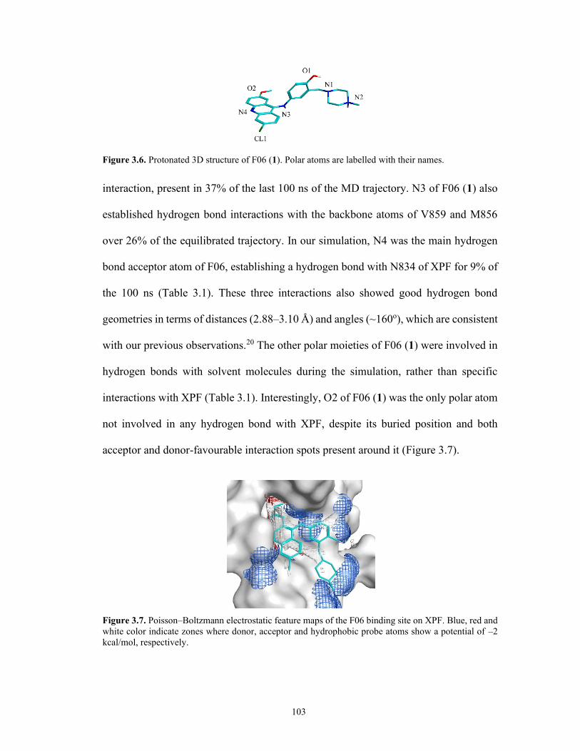
103
Figure 3.6. Protonated 3D structure of F06 (1). Polar atoms are labelled with their names.
interaction, present in 37% of the last 100 ns of the MD trajectory. N3 of F06 (1) also
established hydrogen bond interactions with the backbone atoms of V859 and M856
over 26% of the equilibrated trajectory. In our simulation, N4 was the main hydrogen
bond acceptor atom of F06, establishing a hydrogen bond with N834 of XPF for 9% of
the 100 ns (Table 3.1). These three interactions also showed good hydrogen bond
geometries in terms of distances (2.88–3.10 Å) and angles (~160o), which are consistent
with our previous observations.20 The other polar moieties of F06 (1) were involved in
hydrogen bonds with solvent molecules during the simulation, rather than specific
interactions with XPF (Table 3.1). Interestingly, O2 of F06 (1) was the only polar atom
not involved in any hydrogen bond with XPF, despite its buried position and both
acceptor and donor-favourable interaction spots present around it (Figure 3.7).
Figure 3.7. Poisson–Boltzmann electrostatic feature maps of the F06 binding site on XPF. Blue, red and
white color indicate zones where donor, acceptor and hydrophobic probe atoms show a potential of –2
kcal/mol, respectively.

104
3.2.3 Virtual Screening of F06 (1) Analogues
F.G and I generated a virtual library of analogues by modifying two groups of F06 (1)
(Figure 3.2), namely 1) the methoxy group of F06 (Gen B), as we expected
modifications of this moeity to generate an additional favourable hydrogen bond and 2)
the lateral branch of the acridine scaffold of F06 to reduce the solvent-exposed portion
of the compound (Gen A). We also included the piperazine ring extension of compound
4, the most active F06 (1) analogue identified in the previous medicinal chemistry
campaign.
Figure 3.8. F06 (1) analogues. A) modification sites of F06 (1) explored using a CADD strategy. The
choice of sites was driven by the analysis of the binding mode of the original compound F06 (1).
B) structure of compound 4, best ERCC1–XPF inhibitor among Gen A compounds.
The resulting library includes 5650 structures. Because different features were
introduced in the analogues, we defined an essential pharmacophore model, based on
the conserved acridine core, with three aromatic features accounting for the aromatic
rings anchoring the compounds to the hydrophobic floor of the binding site (Figure 3.9),
and we used it for pharmacophore-assisted docking in MOE, followed by MD-
MM/GBSA-based rescoring. Among the top ranked virtual hits, we selected five
compounds for synthesis, based on visual inspection of the predicted binding modes,
MM/GBSA score, and synthetic chemistry feasibility.

105
Figure 3.9. Pharmacophore model of the scaffold common to all the compounds, including three
aromatic features, each one centered at one of the acridine rings.
3.2.4 Chemistry
In Chapter 1, we reported the first generation (Gen A) of F06 (1) analogues based on
the modification of the piperazine ring with solvent-exposed moieties, according to our
reported computational studies.20 In this chapter, we focused on further modifying
different sites of F06 (1), according to our suggested pharmacophore model, and
extensively investigating their structure activity relationship.
For the second generation series (Gen B) including controls, B2, B3 (lacks the
aminophenol linked with piperazine), and B4 which lacks acridine (Figure 3.10), we
modified the acridine-methoxy group of F06 with polar hydrogen bond donor groups,
such as OH (B2) or its functionalization such as esterification (B6) or alkylation ( B7
and 8) to gain more insight into the role of this group on the activity of ERCC1–XPF.

106
Figure 3.10. Structures of Gen B compounds including controls.
The general synthesis of the Gen B compounds was achieved through a
nucleophilic substitution reaction of B5 and acyl chloride or benzyl chloride derivatives
to afford compounds B6, B7 and B8, respectively, as depicted in Scheme 3.1. The
synthesis of Gen B2 was achieved through dealkylation of Gen B3 (2-methoxy-6,9-
dichloroacrdine) using BBr3 in dichloromethane under nitrogen for 8 h at room
temperature. The synthesis of F06 (1), B5, and B9 was performed according to our

107
reported procedure20 with minor modifications, which was through a one-pot three
sequential addition reaction protocol. The first step was the Mannich-like reaction
Scheme 3.1. General synthetic route of Gen B compounds.
of p-acetamidophenol with formaldehyde, followed by reaction with the appropriate
secondary amine in 2-propanol carried out under reflux for 12 h. The solvent and excess
unreacted formaldehyde from the resulting mixture were removed under vacuum, and,
without isolating the compound, the resulting viscous residue was treated with 6 M HCl
to deacetylate the acetamido group to furnish the primary amine. Afterwards, an
equimolar amount of 2-methoxy 6,9 chloroacridine or 2-hydroxy 6,9 chloroacridine was
added, affording, after heating, compounds F06 (1) and B5, respectively, in moderate
to good yields after isolation. The synthesis is general, facile, and reproducible. Binding

108
energies and other computationally derived values for F06 and the series of analogues
are reported in Table 3.2.
Table 3.2. Computational Results for the Series of F06 Analogues Selected for Chemical Synthesisa
Compound
GBVI/WSAb
(kcal/mol)
Ligand efficiencyc (kcal/mol/ha)
MM/GBSA
(kcal/mol)
LogP
F06 -6.70 -0.20 -17.78 4.10
B6 -8.56 -0.21 -14.19 5.31
B9 -7.00 -0.19 -12.44 2.31
B7 -5.17 -0.13 -8.15 5.93
B2 -5.21 -0.31 -7.11 4.40
B5 -5.90 -0.18 -3.79 3.79
aThe list is in ascending order based on MM/GBSA scores. bFrom MOE Dock. cDocking binding
energy/heavy atoms (ha).
3.2.5 Inhibitory Effect of Gen B Compounds on ERCC1–XPF Activity
Two compounds, B5 and B9, exhibited a marked capacity to inhibit the nuclease
activity of ERCC1−XPF compared to F06 (Figure 3.11). The inhibitory activity of B5
is superior to that of F06, which is probably attributable to the hydrogen bond donor
phenolic group installed in replacement of the acridine-methoxy group of F06. This
suggestion is also consistent with B2 (methoxy acridine) and B3 (hydroxy acridine),
although they are not as active as B5 or B9. Removal of the piperazine substituted
aminophenol as in B2 or acridine as in B4 resulted in complete loss of inhibition. To
measure the half-maximum inhibitory concentrations (IC50) for the best inhibitor B9 on
the full-length protein, different concentrations of the drug were plotted against the
initial velocity (Vo, change in relative fluorescence units (RFU)/time) of the enzyme
(Figure. 3.11B). The IC50 of B9 was estimated as 0.49 ± 0.04 μM, which was

109
approximately four times more efficient than F06, which has an estimated IC50 of 1.86
± 0.25 μM.20 Although compound 4 has superior inhibitory activity on ERCC1–XPF
relative to B9 (Figure 3.12), the improved inhibitory activity of B9 suggests the
replacement of the acridine-methoxy group with a hydrogen bond donor group together
with the conservation or extension of the piperazine ring enhances the activity of this
generation of compounds in comparison of F06 (1).
Figure 3.11. In vitro inhibition of ERCC1−XPF endonuclease activity. A microplate fluorescent assay
was used to measure inhibition of ERCC1−XPF endonuclease activity by the different compounds.
Incubation of the DNA stem–loop substrate with ERCC1−XPF resulted in the release of a fluorescent 8-
base fragment accompanied by an increase in fluorescence (RFU) with time. A) A representative tracing
of the effect of the different compounds (10 μM each) on the incision activity is shown. B) Plot of enzyme
rate (ΔRFU/time) vs B9 concentration.
A

110
3.2.6 Predicted Binding Mode of B9 to XPF
Since B9 was the most active analogue of the series, we carried out a detailed analysis
of its predicted binding pose, based on the 2 ns-long MD simulation and MM-GBSA
calculations, and compared it with the analysis for F06. The lowest potential energy
conformation of the XPF-B9 complex extracted from the 2ns MD simulation
(Figure 3.12A) showed a hydrophobic stacking of the acridine moiety of B9 between
Figure 3.12. Detailed analysis of the predicted binding mode of B9 to XPF derived from the 2 ns-long
MD simulation. a) lowest potential energy XPF (white), B9 (yellow) conformation extracted from the
simulation. The acridine moiety was stacked between the side chains of Y833 and K860. Hydrogen bonds
between B9 and N834 and V859 were observed. b) Ligand interaction diagram of the lowest potential
energy conformation; the lateral branch of B9 containing the piperazine ring extension was solvent-
exposed (blue clouds) thanks to its polar and charged groups that favourably interacted with the
surrounding water molecules. c) Per-residue decomposition of the total MM/GBSA binding energy of
B9 (blue) and F06 (orange).

111
the side chains of Y833 and K860 of F06 (Figure 3.12A and B), resulting in more
favourable contributions to the overall binding affinity by these residues compared to
XPF–F06 (Figure 3.12C). N834 was involved in a hydrogen bond with B9, similarly to
F06 (1) (Figure 3.12A and B). Interestingly, the hydroxyl group substituting the
methoxy in B9 was involved in a new hydrogen bond established with the side chain of
V859, which was not observed for F06 or our previous compound 4.20 Consistent with
our previous observations, the acridine moiety was buried stably inside the XPF pocket,
while the piperazine ring extension derived from compound 4 resulted in enhanced
interactions with the solvent and E829 (Figure 3.12B), although the latter was less
strong than the one observed for F06 (Figure 3.12C).
3.3 Conclusion
Interest is growing in the role of the ERCC1–XPF endonuclease as a drug target in
cancer treatment due to its critical role in repairing DNA damage inflicted by platinum-
based and cross-linking chemotherapy. Abnormal regulation of levels of XPF, ERCC1,
or related proteins has been linked with modulation of the response to DNA-damaging
treatments, either showing resistance in the case of over-regulation or sensitization in
the case of down-regulation. Thus, ERCC1–XPF inhibition is a viable strategy to
circumvent DNA damage-related drug resistance and improve the outcome of cancer
therapy.
In this work we focused on the disruption of the ERCC1–XPF protein–protein
interaction with small molecules, as heterodimerization of ERCC1 and XPF is required
to constitute a functional complex,8 by developing ERCC1–XPF dimerization inhibitors

112
using a multi-step CADD strategy. Two compounds, B5 and B9, have been identified
by the in vitro endonuclease assay with improved inhibition of the ERCC1–XPF
incision activity compared to the parent compound F06 (1). Compound B1, a truncated
version of B9 and B5, also may be a promising starting structure for further
modifications due to the observation of inhibitory activity, despite the replacement of
the bulky lateral branch with a chlorine.
To conclude, the workflow we introduced here provides a blueprint for future
modification of other sites of F06 (1) in order to advance towards a preclinical drug
candidate for DNA repair inhibition. Likewise, the set of analogues described in this
study, especially B5, and B9, and their structure-activity relationships, provide
important information for the lead optimization process of F06 (1).
3.4 Methods and Experimental Section*
3.4.1 Molecular Dynamics of XPF HhH2 Domain Alone, F06-bound and
ERCC1-bound
We computationally predicted F06 binding to a pocket of the human XPF double helix–
hairpin–helix (HhH2) domain, replacing the side chain of F293 from ERCC1 in the
dimer.19 Using classical MD simulations, we compared the unbound, F06-bound and
ERCC1-bound form of the HhH2 domain of XPF (residues 822–905). For the F06-XPF
complex, we used the structure previously obtained from docking simulations.20
* I was responsible for the synthesis and design of all compounds. F.G. carried out the computational
studies on the ERCC1-XPF heterodimer and the virtual screening of compounds. D.J. expressed and
purified ERCC1−XPF, employed the in vitro ERCC–XPF1 assay.

113
The unbound and ERCC1-bound forms were extracted from the NMR ensemble of
ERCC1–XPF dimerized HhH2 domains (PDB 1Z00),23 where the bound ERCC1
structure was excluded and included in the simulation, respectively.
The protonation states of the systems were assigned using the H++ server by
setting pH = 7, a salinity of 0.15 M, a dielectric constant of 10 for the solute, and a
solvent dielectric constant of 80 as input parameters.24 In AmberTools14 tleap,25 we
assigned Amber ff14SB force field parameters to the proteins26 and the 12-6-4
parameters for mono- and divalent ions in TIP3P water to the ions, as previously
recommended.27, 28 We assigned the parameters of the Generalized Amber Force Field
(GAFF)29 and AM1-BCC charges30 to F06 using Antechamber.29 The three systems
were solvated with octahedral boxes of TIP3P explicit water molecules31 with 15 Å of
buffer. The required numbers of Na+ and Cl- ions were added to neutralize the overall
charge and establish a 0.15 M physiological ionic concentration. We then ran MD
simulations of the systems using pmemd.cuda32 with the following procedure:
relaxation of ions and water molecules through 1000 steps of steepest descent and 1000
steps of conjugate gradient minimization, keeping protein and ligand atoms
harmonically restrained with a force constant of 500 kcal/mol/Å2; 2000 steps of steepest
descent, followed by 3000 conjugate gradient steps without any restraint; gradual
heating from 0 to 300 K over 100 ps using the Langevin thermostat, keeping the
backbone and ligand heavy atoms harmonically restrained (2 kcal/mol/Å2), and using a
time step of 0.5 fs and NVT conditions; gradual release of the restraints in four NPT
phases of 50 ps where the force constant was reduced by 0.5 kcal/mol/Å2 at each phase

114
with a time step of 2 fs and 250 ns of NPT production simulation with a time step of 2
fs. The cut-off for long-range interactions was set to 9 Å.
After the simulations, we calculated the mass-weighted root-mean square
deviation (RMSD) of protein backbone and ligand heavy atoms using cpptraj from
AmberTools. We also evaluated the root-mean square fluctuation (RMSF) of the XPF
residues over the equilibrated portions of the three MD trajectories. We also carried out
a secondary structure analysis over the MD trajectories using the dictionary of protein
secondary structure (DSSP) method from cpptraj.33
3.4.2 Analysis of The Binding Mode of F06
In order to investigate the dynamics of the F06–XPF interactions, we carried out the
analysis of hydrogen bonds occurring between F06 and the receptor and F06 and
solvent, respectively, during the equilibrated part of the MD trajectory using cpptraj.
We defined as hydrogen bonds non-bonded interactions occurring between donor-
hydrogen and acceptor atoms that deviated no more than 60° from linearity and with a
maximum distance of 3.5 Å between the two heavy atoms involved in the interaction.
Using the MOE (Molecular Operating Environment) Pharmacophore Query
Editor and the EHT scheme, we generated a set of essential pharmacophore features of
F06, based on the docking pose.34 Electrostatic maps of the binding site were generated
as reported previously.20
3.4.3 Computer-aided Design of F06 Analogues
We used the binding pose of F06 previously obtained as a starting point for our
computer-aided generation of analogues. We created an extensive, non-redundant

115
collection of multi-conformational fragments by merging the MOE Linker library,35 the
ChEMBL fragment database,35 the Cambridge Structure Linker database,36 and a
collection of fragments derived from the ZINC15 3D database.37 This last library was
generated using the MOE 2015 SD Pipelining Command tools to obtain different
protonation states and tautomers, remove non-lead-like, reactive, and large-ring
structures, and generate molecular fragments. Up to five conformations for each
ZINC15-derived fragment were obtained using Conformation Import and imposing a
strain limit of 4 kcal/mol. We then used the entire collection of fragments for MOE2015
Scaffold Replacement35 of F06 substructures. We also used the MOE MedChem
transformation tool (175 modification rules)35 on the same F06 groups, with six
iterations and a protein–ligand clash energy limit of 0.5 kcal/mol/ligand atom. For both
Scaffold Replacement and MedChem transformations, we ran minimization of the
resulting poses with protein side chains free to move and generalized Born Volume
Integral/Weighted Surface Area (GBVI/WSA) function for scoring.38
3.4.4 Structure-based Virtual Screening
We adopted the same structure-based virtual screening (VS) strategy used before for
F06 analogues because of its proven ability to identify highly active compounds.20
Molecular docking simulations were run with MOE Dock.35 The pharmacophore model
was used for the placement step, in which 30 high-scoring poses were returned, based
on the London dG scores.38 Poses were minimized together with the side chains of the
binding site (Induced Fit), and one pose was returned as the top-scored by the
GBVI/WSA scoring function. We calculated log P values of the molecules from the

116
SlogP function in MO39 as well as docking ligand efficiencies (final score/number of
ligand heavy atoms).
We ran MD simulations of the top 200 analogue-XPF complexes resulting from
docking. The systems were prepared in the same way as the F06–XPF system, and
simulation setups were the same, except for the length of the production simulations (2
ns). For MD snapshots extracted every 10 ps from the production simulations, we
calculated the enthalpic portion of the binding energy using the Molecular
Mechanics/Generalized Born Surface Area (MM/GBSA) method40 implemented in the
MMPBSA.py script.41 In MM/GBSA, the free energy change due to ligand binding is
calculated as:
Gbind,solv = GMM,vac + Gsolv,complex - Gsolv,ligand - Gsolv,protein – TS Eq. 1
where GMM,vac includes averaged non-bonded molecular mechanics terms
(electrostatic and van der Waals) occurring between protein and ligand. Solvation terms
are modeled as:
Gsolv = Gsolv,polar + Gsolv,npolar Eq. 2
where the polar contribution of the solvent is calculated by the Generalized Born
equation.42 Here, we used the GBOCBII model and the modified radii from Onufriev et
al.,40 and an ionic concentration of 0.15 M. The hydrophobic contribution to the
solvation free energy is calculated as:
Gsolv,npolar = •SASA Eq. 3
with (surface tension) as 0.005 kcal/mol/Å2, and the solvent-accessible surface area
(SASA) is calculated using the linear combinations of pairwise overlaps (LCPO) model
method.43 Averaged values were estimated for the ligand–receptor (LR) complex,

117
receptor (R), and ligand (L) alone, and final values were calculated as a difference
between these three values (LR-R-L).
The entropy, S, is represented in the free energy by the TS term. It was
calculated for each trajectory using the Amber normal mode analysis (NMA) method
for the LR, R, and L systems and then calculating the difference between the three
systems. 44 Pairwise decomposition of binding energies also was carried out for the top
compounds and the binding pocket residues.
3.4.5 Preparation of F06-based Analogues (Gen B Compounds)
3.4.5.1 4-((6-Chloro-2-methoxyacridin-9-yl)amino)-2-((4-methylpiperazin-
1- l)methyl)phenol (F06(1))
The hit compound F06 (1) has been synthesized according to our previously reported
procedures.20
3.4.5.2 Synthesis of Gen B Compounds
3.4.5.2.1 6,9-dichloroacridin-2-ol (B2)
Synthesis of B2 was achieved by the reported procedures45 but with minor
modifications: a mixture of the commercially available 6,9-dichloro-methoxyacridine,
B3, (0.28 g, 1.0 mmol) and BBr3 in DCM solvent (2.87 mL, 2.87 mmol) was taken in a

118
10 mL single neck round bottom flask under nitrogen conditions. Then, the reaction
mixture was stirred at –78 °C for 20 min and heated to room temperature overnight.
Upon completion of the reaction (monitored by TLC with 20% MeOH/DCM eluent
system), the reaction was quenched by adding a saturated solution of NaHCO3 to yield
a yellowish solid. Next, the solid residue was filtered and washed with H2O and a
saturated solution of NaHCO3 three times, followed by drying overnight under high
vacuum to yield a pure yellowish solid (B2), as reported.
3.4.5.2.2 4-amino-2-((4-methylpiperazin-1-yl)methyl)phenol (B4)
B4 was synthesized according to the reported procedures 46 with minor changes: A
mixture of N-methyl piperazine (0.12 mL, 1.0 mmol), p-acetaminophenol (0.15 g, 1.0
mmol), and 37% formaldehyde (0.10 mL, 5 mmol) in isopropyl alcohol (2 ml) was
taken in a 10 mL single neck round bottom flask. Then, the reaction mixture was stirred
and heated under reflux at 65 °C for 6 h. Upon completion of the reaction (monitored
by TLC with 15% MeOH/DCM eluent system), the solvent was removed on a rotatory
evaporator. Next, the residue was dissolved in 3 mL ethanol, and 3 drops of 12 M HCl
were added. Subsequently, the reaction mixture was heated at 110 °C under reflux for
90 min to yield an orange reddish solution. Afterwards, the reaction was left to cool
down to room temperature, the reaction mixture was diluted with cold water, and

119
neutralized to pH of 8–9 with 28% v/v ammonia solution. The alkaline solution was
extracted with dichloromethane. The organic layer was washed with brine, concentrated
in vacuum, and purified by column chromatography (gradient elution with 5% to 10%
MeOH:DCM system) to afford B4 as a brownish oil, which is the same as reported
before.
3.4.5.2.3 6-chloro-9-((4-hydroxy-3-((4-methylpiperazin-
1-yl)methyl)phenyl)amino)acridin- 2-ol (B5)
The previous method of F06 synthesis was employed to synthesize B5 with the
following stoichiometric amounts: N-methyl piperazine (0.12 mL, 1.0 mmol), p-
acetaminophenol (0.15 g, 1.0 mmol), 37% formaldehyde (0.10 mL, 5 mmol), and 6,9-
dichloro-hydroxyacridine B2 (0.26 g, 1.0 mmol) to afford it as an orange reddish
semisolid (0.25 g) in 56% yield; Rf 0.40 (1:9, MeOH:DCM); IR (cast film) νmax = 3266,
2923, 1628, 1572, 1496, 1250, 1138, 1002, 929, 826, 778 cm-1; 1H NMR (500 MHz,
CD3OD) δ 8.02 (d, J = 9.3 Hz, 1H), 7.88 (d, J = 2.1 Hz, 1H), 7.83 (d, J = 9.3 Hz, 1H),
7.36 (d, J = 9.3 Hz, 1H), 7.24 (d, J = 2.6 Hz, 1H), 7.18 (dd, J = 9.3, 2.1 Hz, 1H), 6.84
(dd, J = 8.6, 1.8 Hz, 1H), 6.71 (d, J = 8.6 Hz, 1H), 6.64 (d, J = 2.6 Hz, 1H), 3.54 (s,
2H), 2.88 – 2.29 (m, 8H), 2.27 (s, 3H). OH and NH protons were not observed; 13C

120
NMR (126 MHz, CD3OD) δ 155.0, 153.9, 148.2, 147.2, 146.1, 138.7, 136.4, 129.7,
127.2, 126.3, 124.9, 123.8, 122.1, 121.4, 121.3, 118.2, 117.3, 105.9, 60.4, 55.8 (2C),
53.0 (2C), 49.0, 45.8; HRMS (ESI) calcd for C25H26ClN4O2 [M + H]+ 449.1739; found
449.1739.
3.4.5.2.4 6-chloro-9-((4-hydroxy-3-((4-methylpiperazin-
1-yl)methyl)phenyl)amino)acridin-2-yl benzoate (B6)
The synthesis of B6 was achieved by mixing B5 (0.05 g, 0.1 mmol) with benzoyl
chloride (0.01 mL, 0.1 mmol) in 3 mL of anhydrous solvent mixture DCM/Et3N (20:1)
and stirred for 1 h at room temperature. Afterwards, the reaction mixture was diluted
with water, and the organic layer was extracted with DCM. The organic layer was
washed with brine, concentrated in vacuum, and purified by column chromatography
(gradient elution with 2% to 15% MeOH:DCM system) to afford it as an orange reddish
semisolid (0.04 g) in 74% yield; Rf 0.45 (1:9, MeOH:DCM); IR (cast film) νmax = 3268,
2926, 1692, 1628, 1582, 1490, 1238, 1135, 1002, 923, 828, 772 cm-1; 1H NMR (500
MHz, CD3OD) δ 8.09 – 8.05 (m, 2H), 7.88 (d, J = 9.2 Hz, 1H), 7.73 – 7.62 (m, 4H),
7.54 – 7.47 (m, 3H), 7.04 (dd, J = 9.2, 2.1 Hz, 1H), 6.83 (dd, J = 8.5, 2.6 Hz, 1H), 6.73
(d, J = 8.5 Hz, 1H), 6.70 (d, J = 2.6 Hz, 1H), 3.55 (s, 2H), 2.70 – 2.29 (m, 8H), 2.26 (s,

121
3H). OH and NH protons were not observed; 13C NMR (126 MHz, CD3OD) δ 166.5,
155.1, 151.2, 147.4, 146.9, 145.5, 139.5, 138.1, 135.1, 131.0 (2C), 130.4, 129.9 (2C),
128.3, 128.1, 126.2, 124.1, 124.0, 123.4, 123.2, 122.8, 118.7, 118.0, 117.7, 116.9, 60.3,
55.5 (2C), 52.8 (2C), 45.6; HRMS (ESI) calcd for C32H30ClN4O3 [M + H]+ 553.2001;
found 553.1995.
3.4.5.2.5 4-((2-(benzyloxy)-6-chloroacridin-9-yl)amino)-2-((4-methylpiperazin-
1-yl) methyl)phenol (B7)
The synthesis of B7 was achieved as follows: NaH (60% dispersion in mineral oil
0.15 g, 0.4 mmol) in DMF (2 mL) was added dropwise to a stirred solution of Gen
B5 (0.08 g, 0.2 mmol) in DMF (2 mL) and stirred for 10 min prior to the addition of
benzyl bromide (0.05 mL, 0.4 mmol). After 1.5 h stirring, the reaction mixture was
concentrated under vacuum, and the residue was diluted with DCM prior to washing
with saturated solution of NH4Cl. The organic layer was extracted with DCM, washed
with brine, concentrated in vacuum, and purified by column chromatography (gradient
elution with 3% to 10% MeOH:DCM system) to afford it as a light orange reddish
semisolid (0.03 g) in 30% yield; Rf 0.40 (1:9, MeOH:DCM); IR (cast film) νmax = 3268,

122
2926, 1692, 1628, 1582, 1490, 1238, 1135, 1002, 923, 828, 772 cm-1; 1H NMR (500
MHz, CD3OD) δ 8.10 (d, J = 9.3 Hz, 1H), 7.88 (d, J = 2.1 Hz, 1H), 7.83 (d, J = 9.3 Hz,
1H), 7.45 (dd, J = 9.4, 2.7 Hz, 1H), 7.39 – 7.29 (m, 6H), 7.24 (dd, J = 9.3, 2.1 Hz, 1H),
6.89 (dd, J = 8.5, 2.7 Hz, 1H), 6.80 – 6.75 (m, 2H), 4.84 (s, 2H), 3.59 (s, 2H), 2.73 –
2.28 (m, 8H), 2.24 (s, 3H). OH and NH protons were not observed; 13C NMR (126
MHz, CD3OD) δ 165.2, 155.8, 154.6, 149.1, 140.7, 138.5, 137.9, 137.0, 129.5 (2C),
129.1, 128.7 (2C), 127.2, 126.7, 125.9, 125.0, 124.0, 123.3, 122.8, 119.5, 117.93, 117.4,
104.6, 103.8, 71.2, 60.5, 55.7 (2C), 53.0 (2C), 45.7; HRMS (ESI) calcd for
C32H32ClN4O2 [M + H]+ 539.2208; found 539.2216.
3.4.5.2.6 2-(benzyloxy)-N-(4-(benzyloxy)-3-((4-methylpiperazin-
1-yl)methyl)phenyl)-6-chloroacridin-9-amine (B8)
The synthesis of Gen B8 followed the synthesis of B7 with minor changes as follows:
NaH (60% dispersion in mineral oil 0.15 g, 0.4 mmol) in DMF (2 mL) was added
dropwise to a stirred solution of B5 (0.08 g, 0.2 mmol) in DMF (2 mL), and the reaction
was further stirred for 10 min prior to the addition of benzyl bromide (0.05 mL, 0.4
mmol). After 2.5 h stirring, the reaction mixture was concentrated under vacuum, and
the residue was diluted with DCM prior to washing with a saturated solution of NH4Cl.

123
The organic layer was extracted with DCM, washed with brine, concentrated in
vacuum, and purified by column chromatography (gradient elution with 3% to 10%
MeOH:DCM system) to afford it as a light orange reddish semisolid (0.04 g) in 40%
yield; Rf 0.50 (1:9, MeOH:DCM); IR (cast film) νmax = 3159, 2906, 1687, 1623, 1577,
1480, 1222, 1125, 1001, 922, 825, 769 cm-1; 1H NMR (500 MHz, CD3OD) δ 8.09 (d, J
= 9.2 Hz, 1H), 7.96 – 7.82 (m, 2H), 7.47 – 7.40 (m, 3H), 7.37 – 7.21 (m, 10H), 7.04 (d,
J = 8.7 Hz, 1H), 6.97 (dd, J = 8.7, 2.8 Hz, 1H), 6.86 (d, J = 2.8 Hz, 1H), 5.08 (s, 2H),
4.83 (s, 2H), 3.48 (s, 2H), 2.48 – 2.19 (m, 8H), 2.16 (s, 3H). OH and NH protons were
not observed; 13C NMR (126 MHz, CD3OD) δ 171.9, 156.0, 154.2, 138.8, 137.9, 136.7,
129.5, 129.5, 129.0 (2C), 128.9 (2C), 128.7 (4C), 128.5 (4C), 128.2, 127.3, 126.6,
125.9, 125.6, 123.6, 121.2, 120.3, 118.6, 114.7, 100.2, 71.7, 70.7, 56.7, 55.6 (2C), 53.4
(2C), 45.8; HRMS (ESI) calcd for C39H38ClN4O2 [M + H]+ 629.2678; found 629.2687.
3.4.5.2.7 6-chloro-9-((3-((4-(2-(dimethylamino)ethyl)piperazin-1-yl)methyl)-
4-hydroxyphenyl)amino)acridin-2-ol (B9)
Synthesis of B9 was performed according to our previous method for compound 4
synthesis with minor modifications:20 The reaction was carried out in a one pot
sequential addition in three steps. A mixture of 1-[2-(dimethylamino)ethyl] piperazine

124
(0.16 g, 1.0 mmol), p-acetaminophenol (0.15 g, 1.0 mmol) and 37% formaldehyde (0.10
mL, 5 mmol) in isopropyl alcohol (2 ml) was taken in a 10 mL single neck round bottom
flask. Then, the reaction mixture was stirred and heated under reflux at 65 °C for 6 h.
Upon completion of the reaction (monitored by TLC with 15% MeOH/DCM eluent
system), the solvent was removed on a rotatory evaporator. Next, the residue was
dissolved in 3 mL ethanol and 3 drops of 12 M HCl were added. Subsequently, the
reaction mixture was heated at 90 °C under reflux for 90 min. Afterwards, 6,9-dichloro-
hydroxyyacridine B2 (0.26 g, 1.0 mmol) was added to the mixture and stirred further at
90 °C under reflux, and the course of reaction followed by TLC until little or no starting
material was detected (around 12 h). On cooling to room temperature, the reaction
mixture was diluted with cold water and neutralized to pH of 8–9 with 28% v/v
ammonia solution. The alkaline solution was extracted with dichloromethane. The
organic layer was washed with brine, concentrated in vacuum, and purified by column
chromatography (gradient elution with 5% to 10% MeOH:DCM system) to afford B9
as an orange reddish semisolid (0.35 g) in 70 % yield; Rf 0.48 (2:8, MeOH:DCM); IR
(cast film) νmax = 3030, 2946, 2876, 2820, 1621, 1494, 1254, 1150, 1008, 932, 816, 736
cm-1; 1H NMR (500 MHz, CD3OD) δ 7.97 – 7.90 (m, 1H), 7.85 – 7.77 (m, 2H), 7.34
(dd, J = 9.3, 1.5 Hz, 1H), 7.12 – 7.06 (m, 1H), 6.81 – 6.75 (m, 1H), 6.71 – 6.64 (m, 1H),
6.63 – 6.42 (m, 2H), 3.47 (s, 2H), 2.60 – 2.31 (m, 12H), 2.24 (s, 6H). OH and NH
protons were not observed; 13C NMR (125 MHz, CD3OD) δ 155.3, 153.9, 148.1, 146.9,
146.1, 138.6, 136.2, 129.7, 127.2, 126.5, 126.4, 124.8, 123.7, 121.9, 121.3, 121.2,
118.3, 118.1, 117.3, 60.5, 57.0, 56.5, 54.3 (2C), 53.1 (2C), 45.7 (2C); HRMS (ESI)
calcd for C28H33ClN5O2 [M + H]+ 506.2317; found 506.2307.

125
3.4.6 ERCC1–XPF Protein Preparation
David Jay conducted this experiment using human ERCC1–XPF wild-type protein was
obtained as described in section 2.5.7 in chapter 2.20, 47
3.4.7 Microplate Fluorescence Incision Assay
ERCC1–XPF incision was measured in a microplate incision assay by David Jay, as
described in section 2.5.8 in chapter 2.20, 47

126
3.5 References
1. López-Otín, C.; Blasco, M. A.; Partridge, L.; Serrano, M.; Kroemer, G. The hallmarks of aging.
Cell 2013, 153, 1194-1217.
2. Hanahan, D.; Weinberg, R. A. Hallmarks of cancer: the next generation. Cell 2011, 144, 646-
674.
3. Bukowska, B.; Karwowski, B. T. Actual state of knowledge in the field of diseases related with
defective nucleotide excision repair. Life Sciences 2018, 195, 6-18.
4. Cleaver, J. E.; Lam, E. T.; Revet, I. Disorders of nucleotide excision repair: the genetic and
molecular basis of heterogeneity. Nature Reviews Genetics 2009, 10, 756-768.
5. Helleday, T.; Petermann, E.; Lundin, C.; Hodgson, B.; Sharma, R. A. DNA repair pathways as
targets for cancer therapy. Nature Reviews Cancer 2008, 8, 193-204.
6. Ohmoto, A.; Yachida, S. Current status of poly (ADP-ribose) polymerase inhibitors and future
directions. OncoTargets and Therapy 2017, 10, 5195-5208.
7. Brown, J. S.; O'Carrigan, B.; Jackson, S. P.; Yap, T. A. Targeting DNA repair in cancer: beyond
PARP inhibitors. Cancer Discovery 2017, 7, 20-37.
8. McNeil, E. M.; Melton, D. W. DNA repair endonuclease ERCC1–XPF as a novel therapeutic
target to overcome chemoresistance in cancer therapy. Nucleic Acids Research 2012, 40, 9990-
10004.
9. Faridounnia, M.; Folkers, G. E.; Boelens, R. Function and interactions of ERCC1-XPF in DNA
damage response. Molecules 2018, 23, 3205-323.
10. Gentile, F.; Elmenoufy, A. H.; Ciniero, G.; Jay, D.; Karimi-Busheri, F.; Barakat, K. H.;
Weinfeld, M.; West, F. G.; Tuszynski, J. A. Computer-aided drug design of small molecule
inhibitors of the ERCC1-XPF protein–protein interaction. Chemical Biology & Drug Design
2020, 95, 460-471.
11. Arora, S.; Heyza, J.; Zhang, H.; Kalman-Maltese, V.; Tillison, K.; Floyd, A. M.; Chalfin, E. M.;
Bepler, G.; Patrick, S. M. Identification of small molecule inhibitors of ERCC1-XPF that inhibit
DNA repair and potentiate cisplatin efficacy in cancer cells. Oncotarget 2016, 7, 75104-75117.
12. Mendoza, J.; Martínez, J.; Hernández, C.; Pérez-Montiel, D.; Castro, C.; Fabián-Morales, E.;
Santibáñez, M.; González-Barrios, R.; Díaz-Chávez, J.; Andonegui, M. A. Association between
ERCC1 and XPA expression and polymorphisms and the response to cisplatin in testicular germ
cell tumours. British Journal of Cancer 2013, 109, 68-75.
13. Gentile, F.; Tuszynski, J. A.; Barakat, K. H. New design of nucleotide excision repair (NER)
inhibitors for combination cancer therapy. Journal of Molecular Graphics and Modelling 2016,
65, 71-82.

127
14. Barakat, K. H.; Jordheim, L. P.; Perez-Pineiro, R.; Wishart, D.; Dumontet, C.; Tuszynski, J. A.
Virtual screening and biological evaluation of inhibitors targeting the XPA-ERCC1 interaction.
PloS One 2012, 7.
15. Chapman, T. M.; Wallace, C.; Gillen, K. J.; Bakrania, P.; Khurana, P.; Coombs, P. J.; Fox, S.;
Bureau, E. A.; Brownlees, J.; Melton, D. W. N-Hydroxyimides and hydroxypyrimidinones as
inhibitors of the DNA repair complex ERCC1–XPF. Bioorganic & Medicinal Chemistry Letters
2015, 25, 4104-4108.
16. Chapman, T. M.; Gillen, K. J.; Wallace, C.; Lee, M. T.; Bakrania, P.; Khurana, P.; Coombs, P.
J.; Stennett, L.; Fox, S.; Bureau, E. A. Catechols and 3-hydroxypyridones as inhibitors of the
DNA repair complex ERCC1-XPF. Bioorganic & Medicinal Chemistry Letters 2015, 25, 4097-
4103.
17. McNeil, E. M.; Astell, K. R.; Ritchie, A.-M.; Shave, S.; Houston, D. R.; Bakrania, P.; Jones, H.
M.; Khurana, P.; Wallace, C.; Chapman, T. Inhibition of the ERCC1–XPF structure-specific
endonuclease to overcome cancer chemoresistance. DNA Repair 2015, 31, 19-28.
18. Gentile, F.; Barakat, K. H.; Tuszynski, J. A. Computational characterization of small molecules
binding to the human XPF active site and virtual screening to identify potential new DNA repair
inhibitors targeting the ERCC1-XPF endonuclease. International Journal of Molecular Sciences
2018, 19, 1328-1341.
19. Jordheim, L. P.; Barakat, K. H.; Heinrich-Balard, L.; Matera, E.-L.; Cros-Perrial, E.; Bouledrak,
K.; El Sabeh, R.; Perez-Pineiro, R.; Wishart, D. S.; Cohen, R. Small molecule inhibitors of
ERCC1-XPF protein-protein interaction synergize alkylating agents in cancer cells. Molecular
Pharmacology 2013, 84, 12-24.
20. Elmenoufy, A. H.; Gentile, F.; Jay, D.; Karimi-Busheri, F.; Yang, X.; Soueidan, O. M.;
Weilbeer, C.; Mani, R. S.; Barakat, K. H.; Tuszynski, J. A.; Weinfeld, M.; West, F. G. Targeting
DNA Repair in Tumor Cells via Inhibition of ERCC1–XPF. Journal of Medicinal Chemistry
2019, 62, 7684-7696.
21. de Laat, W. L.; Sijbers, A. M.; Odijk, H.; Jaspers, N. G. J.; Hoeijmakers, J. H. J. Mapping of
interaction domains between human repair proteins ERCC1 and XPF. Nucleic Acids Research
1998, 26, 4146-4152.
22. Sijbers, A. M.; van der Spek, P. J.; Odijk, H.; van den Berg, J.; van Duin, M.; Westerveld, A.;
Jaspers, N. G. J.; Bootsma, D.; Hoeijmakers, J. H. J. Mutational analysis of the human nucleotide
excision repair gene ERCC1. Nucleic Acids Research 1996, 24, 3370-3380.
23. Tripsianes, K.; Folkers, G.; Eiso, A. B.; Das, D.; Odijk, H.; Jaspers, N. G. J.; Hoeijmakers, J. H.
J.; Kaptein, R.; Boelens, R. The structure of the human ERCC1/XPF interaction domains reveals
a complementary role for the two proteins in nucleotide excision repair. Structure 2005, 13,
1849-1858.

128
24. Anandakrishnan, R.; Aguilar, B.; Onufriev, A. V. H++ 3.0: automating p K prediction and the
preparation of biomolecular structures for atomistic molecular modeling and simulations.
Nucleic Acids Research 2012, 40, W537-W541.
25. Case, D. A.; Babin, V.; Berryman, J.; Betz, R. M.; Cai, Q.; Cerutti, D. S.; Cheatham Iii, T. E.;
Darden, T. A.; Duke, R. E.; Gohlke, H. AMBER 14, 2014. University of California, San
Francisco 2014.
26. Maier, J. A.; Martinez, C.; Kasavajhala, K.; Wickstrom, L.; Hauser, K. E.; Simmerling, C.
ff14SB: improving the accuracy of protein side chain and backbone parameters from ff99SB.
Journal of Chemical Theory and Computation 2015, 11, 3696-3713.
27. Li, P.; Roberts, B. P.; Chakravorty, D. K.; Merz Jr, K. M. Rational design of particle mesh Ewald
compatible Lennard-Jones parameters for+ 2 metal cations in explicit solvent. Journal of
Chemical Theory and Computation 2013, 9, 2733-2748.
28. Li, P.; Song, L. F.; Merz Jr, K. M. Systematic parameterization of monovalent ions employing
the nonbonded model. Journal of Chemical Theory and Computation 2015, 11, 1645-1657.
29. Wang, J.; Wolf, R. M.; Caldwell, J. W.; Kollman, P. A.; Case, D. A. Development and testing
of a general amber force field. Journal of Computational Chemistry 2004, 25, 1157-1174.
30. Jakalian, A.; Jack, D. B.; Bayly, C. I. Fast, efficient generation of high‐quality atomic charges.
AM1‐BCC model: II. Parameterization and validation. Journal of Computational Chemistry
2002, 23, 1623-1641.
31. Jorgensen, W. L.; Chandrasekhar, J.; Madura, J. D.; Impey, R. W.; Klein, M. L. Comparison of
simple potential functions for simulating liquid water. The Journal of Chemical Physics 1983,
79, 926-935.
32. Salomon-Ferrer, R.; Gotz, A. W.; Poole, D.; Le Grand, S.; Walker, R. C. Routine microsecond
molecular dynamics simulations with AMBER on GPUs. 2. Explicit solvent particle mesh
Ewald. Journal of Chemical Theory and Computation 2013, 9, 3878-3888.
33. Kabsch, W.; Sander, C. Dictionary of protein secondary structure: pattern recognition of
hydrogen‐bonded and geometrical features. Biopolymers: Original Research on Biomolecules
1983, 22, 2577-2637.
34. Lin, A. Overview of Pharmacophore Applications in MOE. In 2004.
35. Inc., C. C. G. Molecular Operating Environment (MOE). Chemical Computing Group Inc.,
Canada website: http://www.chemcomp.com 2015.
36. Groom, C. R.; Bruno, I. J.; Lightfoot, M. P.; Ward, S. C. The Cambridge structural database.
Acta Crystallographica Section B: Structural Science, Crystal Engineering and Materials 2016,
72, 171-179.
37. Sterling, T.; Irwin, J. J. ZINC 15–ligand discovery for everyone. Journal of Chemical
Information and Modeling 2015, 55, 2324-2337.

129
38. Labute, P. The generalized Born/volume integral implicit solvent model: estimation of the free
energy of hydration using London dispersion instead of atomic surface area. Journal of
Computational Chemistry 2008, 29, 1693-1698.
39. Wildman, S. A.; Crippen, G. M. Prediction of physicochemical parameters by atomic
contributions. Journal of Chemical Information and Computer Sciences 1999, 39, 868-873.
40. Onufriev, A.; Bashford, D.; Case, D. A. Exploring protein native states and large‐scale
conformational changes with a modified generalized born model. Proteins: Structure, Function,
and Bioinformatics 2004, 55, 383-394.
41. Miller III, B. R.; McGee Jr, T. D.; Swails, J. M.; Homeyer, N.; Gohlke, H.; Roitberg, A. E.
MMPBSA. py: an efficient program for end-state free energy calculations. Journal of Chemical
Theory and Computation 2012, 8, 3314-3321.
42. Genheden, S.; Ryde, U. The MM/PBSA and MM/GBSA methods to estimate ligand-binding
affinities. Expert Opinion on Drug Discovery 2015, 10, 449-461.
43. Weiser, J.; Shenkin, P. S.; Still, W. C. Approximate atomic surfaces from linear combinations
of pairwise overlaps (LCPO). Journal of Computational Chemistry 1999, 20, 217-230.
44. Kassem, S.; Ahmed, M.; El-Sheikh, S.; Barakat, K. H. Entropy in bimolecular simulations: A
comprehensive review of atomic fluctuations-based methods. Journal of Molecular Graphics
and Modelling 2015, 62, 105-117.
45. Carlson, W. W.; Cretcher, L. H. The Hydroxyethyl Analog of Quinacrine. Journal of the
American Chemical Society 1948, 70, 597-599.
46. Sriram, D.; Devakaram, R. V.; Dinakaran, M.; Yogeeswari, P. Aromatic amino analogues of
artemisinin: synthesis and in vivo antimalarial activity. Medicinal Chemistry Research 2010,
19, 524-532.
47. Bowles, M.; Lally, J.; Fadden, A. J.; Mouilleron, S.; Hammonds, T.; McDonald, N. Q.
Fluorescence-based incision assay for human XPF–ERCC1 activity identifies important
elements of DNA junction recognition. Nucleic Acids Research 2012, 40, e101-e101.

130
Chapter 4
Design, Synthesis and In vitro Cell-free/Cell-based
Biological Evaluations of Novel ERCC1–XPF
Inhibitors Targeting DNA Repair Pathway
4.1 Introduction
The ERCC1–XPF heterodimer is a highly conserved structure-specific endonuclease
that plays a central function in various DNA repair pathways, including interstrand
crosslink (ICL) repair, double-strand break (DSB) repair, and nucleotide excision
repair.1-3 As such, it has a pivotal role in the response of various cancers to a wide array
of DNA-damaging chemotherapeutics, such as platinum-containing compounds,
cyclophosphamide, and mitomycin C (MMC).4, 5 The enzyme also has the important
role of removing bulky adducts and helix-distorting DNA lesions, such as UV-induced
pyrimidine-(6,4)-pyrimidone photoproducts (6-4PPs) and cyclobutane pyrimidine
dimmers, CPDs.6 A recent study revealed that 72% of patients with colorectal cancer
exhibited high expression levels of ERCC1 protein,7 raising the potential importance of
developing inhibitors of ERCC1–XPF that could increase the sensitivity of cancer cells
towards conventional cancer therapeutics, which would impact the survival rate of
cancer patients in the clinic positively. Thanks to the availability of molecular structures
of ERCC1–XPF and the multiple sites that could be targeted by small molecules to
inhibit the endonuclease activity,8 the enzyme complex has become an attractive target
for drug design studies.

131
In the ERCC1–XPF heterodimer, the ERCC1 is catalytically inactive but
regulates DNA–protein and protein–protein interactions, whereas XPF possesses the
endonuclease active site as well as an inactive helicase-like motif and is involved in
additional protein–protein interactions and DNA binding.9 However, enzymatic activity
requires the dimerization of the two proteins because neither protein alone is stable, and
both are degraded rapidly.8, 10, 11 Dimerization between ERCC1 and XPF is mediated by
the interaction of their double helix−hairpin−helix (HhH2) motifs in their C-terminal
domains. As a result, small molecule inhibitors that can disrupt the interaction domains
linking XPF and ERCC1 would be expected to sensitize cancer cells to conventional
DNA crosslinking chemotherapeutic treatments and radiotherapy.
The availability of several experimental structures of the dimerized HhH2
domains (for example, PDB code 2A1J and 1Z00) allows rational drug design studies
to be employed to develop small molecules that disrupt the heterodimerization interface.
Our initial drug design studies, which focused on the XPA active site12 and the
XPA−ERCC1 interaction site,13 validated the use of computer-aided drug design
approaches to develop and optimize DNA repair inhibitors;14 however, due to the
contribution of ERCC1–XPF interaction in many DNA repair pathways, targeting the
dimerization interface is regarded as more promising.9
Several studies have been carried out recently to develop ERCC1–XPF
inhibitors. Yang et al.15 proposed a cellular delivery approach of therapeutic peptides
that mimic the ERCC1 HhH2 domain (residues 220−297) as a potential alternative
strategy to inhibit the NER pathway and potentiate DNA-damaging agents to cancer
cells. However, the translation of this modality into the clinic is challenging in terms of

132
the cost and applicability. Arora et al.16 and Chapman et al.17, 18 have identified and
optimized various small molecules targeting the XPF endonuclease active site. Their
results revealed that some of the endonuclease inhibitors, with IC50 in the nanomolar
range, were capable of diminishing NER activity and enhancing the cytotoxicity of
platinum-containing drugs in cancer cells. However, specific inhibition of the XPF
endonuclease through metal ion chelation with inhibitors is challenging due to the
similarities among various endonuclease active sites that could lead to off-target
interactions from these inhibitors; thus, the development of this series will be limited
due to the lack of structural insights of the compound–protein complexes.9 McNeil et
al. conducted an in silico screening approach that targeted only three different sites on
the XPF HhH2 domain with the aim of identifying potential inhibitors that could stably
bind to it and hinder the interaction between ERCC1 and XPF domains but also disrupt
the XPF endonuclease domain.19 They demonstrated that one of their compounds that
had the ability of binding to the XPF domain in vitro also was able to inhibit the NER
pathway in melanoma cells and sensitize them to cisplatin. However, the reported Kd
and IC50 values for this compound, in the medium to high micromolar range, were
suboptimal. Jordheim et al.20 developed small molecule ERCC1–XPF inhibitors acting
through the XPF interaction domain, which led to heterodimerization disruption in vitro.
They also reported that 1 (also called NSC-130813 or NERI02) sensitized cancer cells
to cisplatin and MMC. This affirmed that enhancing the cytotoxicity of cross-linking
agents, such as cisplatin, could be achieved via targeting ERCC1–XPF protein–protein
interaction. However, the clinical properties of 1 were suboptimal in terms of toxicity

133
and potency, and a derivatization strategy, suggested by Jordheim et al.,20 was required
to optimize the compound.
Most of the previous efforts to develop ERCC1–XPF inhibitors were tested
using a truncated form of the heterodimer and not the full-length protein.17-19, 21 It has
been shown that the truncated form of ERCC1–XPF is ~15-fold less active than the full
length ERCC1−XPF protein under standard reaction conditions.9, 10 Using 1 as a
reference hit, we employed computer-aided drug design techniques, such as electrostatic
mapping of the compound 1 binding pocket, pharmacophore modeling, molecular
docking, and molecular dynamics (MD)- based rescoring, to rank compound 1
analogues based on their predicted binding affinities to the XPF domain.22
Interestingly, one of the derivatives that has good physiochemical properties
showed significant inhibition of the removal of CPDs in UV-irradiated cells and
increased capacity to sensitize colorectal cancer cells to UV radiation and
cyclophosphamide relative to the initial hit.22 More recently, our group has conducted a
study that followed a multi-step computational approach to investigate one of the
potential sites of compound 1 and propose potential candidates to be synthesized and
tested in vitro.23 The results of the previous study in Chapter 3 yielded one compound
that showed an improved IC50 value of 0.49 µM for the inhibition of ERCC1–XPF
endonuclease activity compared to the IC50 value of 1.86 M for 1. Having a potential
lead compound and a solid computational approach, previously mentioned in Chapter
3, stimulated us to develop DNA repair inhibitors further. Therefore, in this study, we
have conducted an extensive structure activity relationship analysis through synthesis

134
of different series of compounds based on modification of specific sites of 1 in
comparison with B9.
All the compounds were synthesized and tested for their capability to inhibit the
endonuclease activity of the full-length ERCC1–XPF in vitro. The top active compound
was assessed further for its inhibition of repair of UV-induced thymidine dimers in
human colorectal cancer cells as well as a sensitizing agent to UV irradiation and
cyclophosphamide. Cyclophosphamide is being examined now as a part of
chemotherapeutic combination for colorectal cancer.24, 25
4.2 Results and Discussion
4.2.1 Chemistry
In Chapter 2, we synthesized the first generation (Gen A) of F06 (1) analogues based
on the modification of the piperazine ring with solvent-exposed moieties, according to
our reported computational studies.22 In this chapter, we focused on further modifying
two different sites of F06 (1), according to our suggested pharmacophore model, and
extensively investigating their structure activity relationship (Figure 4.1).
Figure 4.1. Structure of F06 (1) hit and the potential modification sites for hit to lead optimization.

135
For the third generation (Gen C), compounds were modified with different
substituted anilines that are considered to be medicinally favorable, such as heterocyclic,
gem dimethyl, aliphatic, and hydrophobic groups (Figure 4.2). Also, this series lacks the
piperazine moiety to contrast their activity on ERCC1–XPF as compared to the second-
generation analogues, which retain the piperazine group. General synthesis of Gen C
compounds was carried out through nucleophilic aromatic substitution of B2 and
different substituted aniline derivatives, as seen in Scheme 4.1.
Figure 4.2. Chemical structures of Gen C analogues.

136
Scheme 4.1. General synthetic route for Gen C compounds.
Regarding Gen D compounds (Figure 4.3), their design was based on the
modification of the acridine moiety, such as installing quinoline and phenyl groups to
investigate the role of these functionalities on the activity of ERCC1–XPF compared to
the hit compound. D2 and D3 were used as controls. D2 was synthesized via
nucleophilic aromatic substitution of D1 (4-chloro-7-methoxyquinoline) and 4-
aminophenol. D1 and D3 and were purchased from Sigma Aldrich Inc. in high purity.
D4 and D5 and were synthesized with the same experimental procedure of B9, except
for the last SNAr step that occurred between the top aniline intermediate of B9 and D1
or D1 in TFA solvent to yield D4 and D5, respectively.

137
Figure 4.3. Chemical structures of Gen D analogues.
4.2.2 Inhibition of ERCC1−XPF Endonuclease Activity
We previously22 used an in vitro assay based on real-time fluorescence to evaluate the
inhibitory effect of the synthesized derivatives on ERCC1−XPF endonuclease activity.
This assay has been described in Chapter 2 and utilizes a stem–loop substrate (composed
of a 10mer-duplex stem and a 20mer-oligo-dT single-strand loop), labeled on the 3′-
terminus with the quencher dabcyl and the 5′-terminus with 6-FAM.26 Upon cleavage
of the 10mer-duplex region by ERCC1−XPF, a product consisting of an 8-base 5′-
labeled FAM fragment is released, which leads to an increase in the fluorescent signal.
4.2.2.1 Inhibitory Effect of Gen C Compounds on ERCC1–XPF Activity
The purpose for designing Gen C compounds was to study the aniline structure activity
relationship without the piperazine moiety in order to simplify the structures but to retain
the acridine with the phenolic group and compare their activity with the hit F06 (1) and

138
the best inhibitor from the B series of compounds, B9. Different aniline substituents
were selected for their known favourable medicinal features.27
Unfortunately, the inhibitory effect of Gen C compounds on ERCC1–XPF
activity decreased relative to either B9 (structure is shown in Chapter 3, Figure 3.10) or
F06 (1), as depicted in Figure 4.4. However, C7 showed modest inhibition of the
endonuclease activity. These results support the idea that the presence of the piperazine
ring is critical for maintaining inhibitory activity, regardless of the substitution on the
aminophenol ring.
Figure 4.4. In vitro inhibition of ERCC1−XPF endonuclease activity by Gen C compounds (10 μM
each).
4.2.2.2 Inhibitory Effect of Gen D Compounds on ERCC1-XPF Activity
Our previous computational studies of F06 (1) indicated that the planar aromatic rings
of the acridine moiety buried in the XPF binding pocket engaged in π–π interactions
and contributed to the binding energy and ligand efficiency of F06 (1).22, 23 However,

139
this has not been investigated experimentally to determine whether reduced aromaticity
would impact the inhibition of ERCC1–XPF. Therefore, we chose to replace the
acridine moiety with a quinolone ring, which is a smaller unit that provides compounds
much less likely to intercalate into the DNA substrate. As can be seen in Figure 4.5, D1,
D2, and D5 (the salt form of D4 for enhanced solubility) failed to inhibit ERCC1–XPF.
Figure 4.5. In vitro inhibition of ERCC1−XPF endonuclease activity by Gen D compounds (10 μM
each). The increase of fluorescence signal as a function of time.
Although D3 and D4 displayed modest inhibitory activity, it was less than 1 and
B9, indicating the importance of retaining an extended aromatic feature as in acridine
to maintain high inhibition. Interestingly, D4 bears the same dimethylaminoethyl
substituent on the piperazine ring as in B9, supporting its role in inhibiting ERCC1–
XPF. Since B9 displayed the most effective inhibition in vitro it was subjected to further
analysis.
0
500
1000
1500
2000
2500
0 2 4 6
RF
U
minutes
Gen D1
Gen D2
Gen D5
Control
Gen D3
Gen D4
F06 (1)
Gen B9

140
4.2.3 Gen B9 Binding to ERCC1−XPF
Intrinsic fluorescence spectroscopy (of the protein tryptophan residues) was utilized, as
previously described,22 to study the binding affinity of B9 (active compound) and B7
(inactive compound) (structure shown in Chapter 3, Figure 3.10) to ERCC1−XPF.
Addition of 4 μM B7 had no significant effect on protein fluorescence (Figure 4.6A),
Figure 4.6. Determination of the affinity (Kd) between ERCC1–XPF complex and B9 (active inhibitor)
and B7 (negative control). Quenching of the intrinsic protein fluorescence of the tryptophan residues
was used to monitor the interaction between the compounds and the protein. Fluorescence of ERCC1–
XPF (50 nM) incubated with (A) B7 (negative control) and (B) B9 (active inhibitor). (C) Unimodal
binding pattern and the binding affinity of B9 with ERCC1–XPF (85 nM). The protein was excited at
295 nm, and the fluorescence intensity was monitored at 345 nm (see inset). The fraction bound (i.e.,
relative fluorescence intensity) versus ligand concentration is plotted. (D) Unimodal binding of B9 to
DNA substrate of the ERCC1−XPF protein. The stem–loop substrate (lacking the quenching group) of
the ERCC1−XPF complex (100 nM) was excited at 490 nm and the intensity of fluorescence signal at
520 nm was monitored as a function of B9 concentration. The binding affinity of B9 to ERCC1−XPF
substrate was 1.05 ± 0.05 μM, as shown in Table 4.1.
300 325 350 375 4000
100
200
300
400
500
ERCC1.XPF + B7 (4M)
ERCC1.XPF
Wavelength, nm
Rel. F
luo
r. In
ten
sit
y
300 325 350 375 4000
200
400
600
ERCC1.XPF + B9 (4M)
ERCC1.XPF
Wavelength. nm
Rela
tive.
Flu
or.
In
ten
sit
y
0 500 1000 1500 2000 2500 30000.0
0.5
1.0
[Compound B9], nM
Fra
cti
on
Bo
un
d0 500 1000 1500 2000 2500 3000
100
150
200
250
[Compound B9], nM
Re
l. F
luo
r. In
ten
sit
y
0 2000 4000 6000 80000.0
0.5
1.0
[Compound B9], nM
Fra
cti
on
Bo
un
d
0 2000 4000 6000 8000150
200
250
300
350
[Compound B9], nM
Re
l. F
luo
r. In
ten
sit
y
Kd = 85 5 nM
A
B
C
D
Kd = 1.05 ± 0.05 µM

141
and the observed fluorescence quenching at 330 nm was less than 2%, thus providing
no evidence of any interaction. In contrast, addition of B9 (4 μM) induced nearly 25%
± 3% quenching of protein fluorescence at 330 nm, clearly indicating interaction of B9
with the ERCC1−XPF complex, as depicted in Figure 4.6B. The binding affinity (in
terms of dissociation constant, Kd) of B9 for ERCC1−XPF complex was determined by
following fluorescence quenching (a measure of ligand binding) as a function of ligand
concentration. A representative plot of relative fluorescence intensity versus the
concentration of B9 is shown in Figure 4.6C (inset). Nonlinear regression analysis
(GraphPad Prism Software, San Diego, CA) of the binding data was carried out, as
described in our earlier paper,28 and revealed a unimodal binding with a Kd value of 85
± 5 nM (Table 4.1).
Table 4.1. The Half-maximum Inhibitory Concentrations (IC50) and Binding Constants Values of
Inhibitorsa.
Compound Kd protein (nM) Kd DNA (nM) IC50 (µM)
F06 (1) 140 ± 5 N.D.* 1.86 ± 0.25
Gen B7 N.D. 200 ± 10 N.D.
Gen B9 85 ± 5 1050 ± 50 0.49 ± 0.04
aThe IC50 data was obtained from at least three different experiments of Vo versus compound
concentration. * Not determined.
All the B compounds, except B4 (structure is shown in Chapter 3, Figure 3.10),
share an acridine moiety that is known to bind nucleic acids as intercalators. To
determine if the binding to the DNA substrate plays a major role in the inhibitory

142
activity of B9, we compared the interaction of B9 and B7 with the stem–loop substrate
(lacking the dabcyl quencher) by following the change in fluorescence with increasing
concentration of inhibitor. We observed that B9 has a binding affinity for the DNA
stem–loop (Kd = 1.05 ± 0.05 µM, Figure 4.6D) that is almost 12-fold lower than for
ERCC1−XPF. This disparity in binding affinity values strongly suggests that inhibition
by B9 results from its interaction with ERCC1−XPF rather than with the DNA substrate.
Regarding B7 binding to the DNA stem–loop but is not an effective substrate, it showed
a unimodal binding with a Kd value of 200 ± 10 nM (Figure 4.7).
Figure 4.7. The binding affinity measurement of B7 on the stem–loop DNA substrate.
This means that B7 (negative control) binds more tightly to the DNA than B9
but is not an inhibitor of ERCC1–XPF, although they share the same acridine moiety.
This provides further support that inhibition is not a result of the compounds binding to
DNA. In addition, as shown in Chapter 2 using microscale thermophoresis, another
0 2500 5000 7500 100000.0
0.5
1.0
[Compound B7], nM
Fra
cti
on
Bo
un
d
0 2500 5000 7500 10000200
250
300
350
[Compound B7], nM
Re
l. F
luo
r. In
ten
sit
y
Kd = 200 ± 10 nM

143
compound with a similar structure to B9 interacted with ERCC1−XPF in the subunit
interaction domain.22 This provides additional evidence that B9 inhibition of ERCC1–
XPF endonuclease activity is mediated by disruption of heterodimerization between
ERCC1 and XPF.
4.2.4 Inhibition of DNA Repair by B9
To test for inhibition of nucleotide excision repair, we monitored the cellular removal
of UVC-induced cyclobutane pyrimidine dimers (CPDs) in HCT 116 cells over a 24
hour period using an immunochemical assay. The results (Fig. 4.8) show that B9
significantly inhibited the removal of CPDs compared with control cells or cells treated
with B7. Twenty-four hours after irradiation, the residual percentage of CPDs in the
cells treated with B7 was 35% compared to 16% in the untreated cells.
Figure 4.8. Inhibition of cellular NER by compound B9. (A) Immunofluorescence images of the UV-
based assay for detecting CPDs in HCT-116 colorectal cancer cells treated with compounds B9 (at 2 µM)
and compared it to negative control B7 and the control. (B) Normalized fluorescence intensity of the
treated cells (±SEM) based on quantitation of fluorescence from 100 randomly selected cells per
treatment. Treated cells with active inhibitor B9 were significantly different from the non-inhibitory
negative control B7 from 4 to 24 h post-irradiation (p <0.005, Student’s t test).

144
4.2.5 Cytotoxicity Profile of Inhibitors
Crystal violet viability assays were carried out to assess the cellular toxicity of these
compounds to HCT 116 cells and to choose relatively non-cytotoxic concentrations to
be utilized further in sensitizing cells to UV radiation or cyclophosphamide. As can be
seen in Figure 4.9, B7 as a negative control is the least cytotoxic compound to HCT 116
cells. The cytotoxicity of the hit F06 (1) to the cells is more than B9, especially at
concentrations ≥5 µM. In the case of F06 (1), about 35% of the cells survived after three
days exposure of F06 (1) to a dose of 5 µM, whereas approximately 95% of the HCT116
cells survived exposure to B9 at the same dose. As a result, we selected 2 µM as a
maximum low-cytotoxic concentration for F06 (1), while 4 µM of Gen B9 and Gen B7
was chosen as a maximum concentration to be used in the following assay.
Figure 4.9. Crystal violet-based viability assay to assess the cytotoxicity profile of B9 (active inhibitor),
B7 (negative control), and F06 (1) (hit).

145
4.2.6 Increased Sensitization to UV Radiation and Cyclophosphamide
The effectiveness of B9 to sensitize HCT 116 cells to UV radiation and
cyclophosphamide was evaluated. As depicted in Fig. 4.10A, neither 2 nor 4 µM B7
(negative control) had a significant sensitisation effect on HCT 116 cells to UV
radiation, which is consistent with the failure of B7 to inhibit ERCC1–XPF
endonuclease. In contrast, 2 µM F06 (1) displayed a modest sensitization capacity with
increasing dose (Figure 4.10B), while 2 and 4 µM B9 significantly reduced the survival
of cells irradiated with UV (Figure 4.10C). We then measured the effect of B9 on the
survival of cells exposed to the DNA interstrand cross-linking agent cyclophosphamide.
The survival curves shown in Figure 10D, indicate that B9, at a concentration of 2 µM,
significantly sensitized the HCT 116 cells to cyclophosphamide (starting at 50 µM),
whereas there was no significant sensitization effect with B7 (at a higher dose of 4 µM),
which is in accordance with the UV survival data.

146
Figure 4.10. Sensitization of HCT 116 cells to UV and cyclophosphamide. A) Cellular survival upon
exposure to increasing doses of 254 nm UV radiation and treated with 2 and 4 μM B7, B) 2 μM F06 (1),
and C) 2 and 4 μM B9 determined by the clonogenic survival assay. D) HCT 116 cell survival following
exposure to increasing doses of cyclophosphamide and treated with 4 and 2 μM of B7 and B9,
respectively, determined by the clonogenic survival assay. The cell survival curves (±SEM) are based on
three independent sets of experiments. All the values related to cells treated with 2 μM B9 were
significantly different (p < 0.005, Student’s test) from the values obtained with the control cells not
treated with B9.
4.2.7 Pharmacokinetic Properties (PK) of B9 and F06 (1)
The results obtained from PK properties were compared to the parental compound F06
(1)22 to determine if compound B9 is pharmacokinetically superior to compound F06
(1). B9 showed a lower log D (2.01) than F06 (1) (3.86) at pH 7.4. Metabolism of B9,
as measured by exposure to human liver microsomes, indicated that it has a moderate
metabolism, while compound F06 (1) has a more rapid metabolism and high clearance
UV (J/m2)
Su
rviv
ing
Fra
cti
on
(%
)
0 2 4 6 8 101
10
100
Control
B7 - 2 µM
B7 - 4 µM
UV (J/m2)
Su
rviv
ing
Fra
cti
on
(%
)
0 2 4 6 8 101
10
100
Control
B9 - 2 µM
B9 - 4 µM
UV (J/m2)
Su
rviv
ing
Fra
cti
on
(%
)
0 2 4 6 8 101
10
100
Control
F06-2 µM
Cyclophosphamide (µM)
Su
rviv
ing
Fra
cti
on
(%
)0 50 100 150 200 250 300
0.01
0.1
1
10
100
Control
B7 - 4 µM
B9 - 2 µM
A
C D
B

147
from the liver. Both compounds display an efflux ratio greater than 2 (B9 = >43.39 and
F06 (1) = 11.18).
4.2.8 Cell Death by a Combination of Compound 4 (Gen A) or B9 with
Chemotherapeutic Agents
As can be seen clearly in Figure 4.11A, cisplatin alone induced A549 cell death with a
rate of 50% or less at different concentrations. However, B9 caused more than 80% of
cell death when it was used in a combination with cisplatin. Also, the same effect was
observed for B9 when it is incubated with Mitomycin C (MMC) relative to the MMC
alone. In HCT 116 cells (Figure 4.11B), using cisplatin alone at 50 µM caused about
40% cell death only, while incubating B9 with cisplatin at the same concentration
induced more than 80% of HCT 116 cell death. On the other hand, a combination of A4
and cisplatin or MMC did not cause a significant cell death relative to either cisplatin
or MMC alone treated cells.

148
Figure 4.11. Survival of A549 (A) and HCT-116 (B) cells after exposure to alkylating agents and NER
inhibitors alone or in combination. Cisplatine and MMC were used at indicated concentrations, anB9 and
A4 at 1 µM. Graphs show mean values of three independent experiments performed in duplicate, and
error bars are standard deviations.
4.2.9 Heterodimerization Inhibition of ERCC1–XPF by A4 and B9
To confirm the inhibition of ERCC1–XPF interaction in a cellular context, our
collaborators, Dr. L P. Jordheim and G. Ciniero, performed a proximity ligation assay
(Figure 4.12) in A549 cells incubated with B9 or A4 and/or cisplatin. As expected,
cisplatin increased the mean interactions per cells. The addition of either B9 or A4
decreased the interaction between ERCC1 and XPF in the presence of cisplatin,
showing that B9 is a potent inhibitor of the ERCC1–XPF interaction in whole cells.
A
B

149
Figure 4.12. Interaction between endogenous ERCC1 and XPF revealed by a proximity ligation assay
(red dots) in A549 cells incubated alone (NT) or in the presence of A4 or B9 (2 µM) or cisplatin (20 µM)
for 24 h.
4.3 Conclusion
The ERCC1−XPF complex is a 5′−3′ structure-specific endonuclease that has a central
role in different DNA repair pathways, especially NER and ICL. It is required to protect
the integrity of the genome as well as to prevent damage-induced mutations by different
insults; however, it hampers the efficacy of genotoxic cancer therapeutics, such as
radiotherapy or chemotherapy based on platinum containing compounds. Therefore,
inhibiting the mechanism of DNA repair in cancer cells by small molecules is a
promising approach to potentiate the effect of such therapies.
In Chapter 3, we employed a computational drug design workflow to design
novel compounds based on the hit F06 (1) rationally, resulting in a pharmacophore
model that suggested further optimization to modulate the chemical space of different
sites on F06 (1). Herein, we further explored different modifications on the suggested
sites on F06 (1), which yielded three different series of compounds, and dissected their

150
structure activity relationships with regard to their ability to inhibit the ERCC1–XPF
heterodimer and sensitize colorectal cancer cells to either UV radiation or
cyclophosphamide.
All the designed compounds were synthesized via robust, facile, and
reproducible methods. The inhibitory effects of all compounds on the full-length
ERCC1–XPF protein activity were assessed in vitro. The results in Chapter 3 revealed
that the in vitro ERCC1–XPF endonuclease assay identified B9 as a potent inhibitor of
the ERCC1–XPF nuclease activity with an IC50 value of 0.49 ± 0.04 μM compared to
1.86 ± 0.25 μM for F06 (1). This result suggests that incorporating the piperazine ring
with a polar side-chain on the exterior face of the piperazine and a large hydrophobic
moiety (acridine) is important for optimized activity, which is in accord with the results
of the effect of Gen C and D on the protein complex. However, installing a hydrogen
bond donor group on the acridine, as in B9, increased the inhibitory effect by almost
two-fold compared to having a hydrogen bond acceptor, such as the methoxy group in
F06 (1). The docking studies indicated that this resulted in additional hydrogen bond
interaction with the side chain of the buried V859 residue the XPF binding pocket. The
binding constant for B9 was measured experimentally as 85 nM, which has a higher
affinity for ERCC1–XPF relative to F06 (1) with a Kd value of 140 nM. The fact that
Gen B7 (negative control) and B9 show disparity in their activity and binding profiles
on ERCC1–XPF, despite having the same acridine moiety, also provides strong
evidence that the protein complex inhibition is not mediated through DNA intercalation.
B9 elicited inhibition of CPDs removal upon UV irradiation and sensitized the HCT
116 cells to cyclophosphamide- and UV-induced cytotoxicity, indicating inhibition of

151
ICL and NER. Furthermore, comparison of the partial pharmacokinetics screening
results of B9 and F06 (1) indicates that our lead second generation compound, B9, has
more favorable qualities than the F06 (1), by showing a moderate clearance from the
liver, a better permeability, and a lower log D, which means that it has a lower
lipophilicity and greater metabolic stability. In addition, B9 showed a significant
synergistic effect when used with cisplatin, which caused an 80% cell death relative to
the effect of cisplatin alone. Furthermore, the B9 inhibitory activity is attributed to the
heterodimerization disruption of ERCC1–XPF on a cellular level.
Having shown that hydrogen bond donor group installation on the acridine
moiety clearly enhances the activity, we intend to explore a wider range of polar
hydrogen bond donor groups to be installed on the hydrophobic acridine structure and
contrast their ERCC1–XPF inhibitory activity with our best lead compound, B9. In
summary, the analysis of the conducted comprehensive SAR studies identified B9,
which can be combined with other existing DNA-damaging therapies to synergize their
effects by sensitizing cancer cells.
4.4 Methods and Experimental Section*
4.4.1 Synthesis of C and D Compounds
Reactions were conducted in flame-dried glassware. Transfer of anhydrous reagents and
solvents was done with cannula or oven-dried syringes. Chemicals were purchased from
Sigma Aldrich Inc. and were used without further purification.
* I was responsible for the synthesis and design of all compounds. F.G. carried out the computational
studies of the compounds. D.J. expressed and purified ERCC1−XPF, employed the in vitro ERCC–XPF1
assay. F.K.B and Y.X. carried out cell culture, UV dimer repair assay, and clonogenic survival assay. R.M.
conducted the binding affinity studies of the compounds. G.C conducted the synergy experiment and PLA
assay.

152
Thin layer chromatography (TLC) was performed on glass plates pre-coated with 0.25
mm silica gel. Flash chromatography columns were packed with 230-400 mesh silica
gel. Proton nuclear magnetic resonance spectra (1H NMR) were recorded at 500 MHz,
and coupling constants (J) are reported in hertz (Hz). Standard notation was used to
describe the multiplicity of signals observed in 1H NMR spectra: broad (br), multiplet
(m), singlet (s), doublet (d), triplet (t), etc. Carbon nuclear magnetic resonance spectra
(13C NMR) were recorded at 125 MHz and are reported (ppm) relative to the center line
of the triplet from chloroform-d (77.0 ppm) or the center line of the septet from
methanol-d4 (49.0 ppm). Infrared (IR) spectra were measured with a FT-IR 3000
spectrophotometer. Mass spectra were determined on a high-resolution electrospray
positive ion mode spectrometer.
4-((6-Chloro-2-methoxyacridin-9-yl)amino)-2-((4-methylpiperazin-
1- l)methyl)phenol (F06(1))
The hit compound F06 (1) has been synthesized according to our previously reported
procedures.22

153
4.4.1.1 Synthesis of Gen C Compounds
6-chloro-9-((4-isopropylphenyl)amino)acridin-2-ol (C1)
Synthesis of C1 was performed by mixing 6,9-dichloro-hydroxyacridine B2 (0.26 g,
1.0 mmol) and 4-isopropylaniline (0.14 g, 1.0 mmol) in 8 mL ethanol (95%), and 5
drops of 12 M HCl were added. Subsequently, the reaction mixture was heated at 90 °C
under reflux for 12 h. Then, a reddish orange solid was formed after the reaction
completion, which was monitored by TLC (0.5:9.5, MeOH:DCM). On cooling to room
temperature, the solid precipitate was filtered, washed with cold ethanol 2–3 times, and
dried overnight under high vacuum to afford D1 as an orange reddish semisolid (0.24
g) in 67% yield; Rf 0.40 (0.5:9.5, MeOH:DCM); mp 222–224 °C; IR (cast film) νmax =
3100, 3032, 2960, 2925, 1628, 1582, 1512, 1266, 1162, 1086, 936, 829, 765 cm-1; 1H
NMR (500 MHz, CD3OD) δ 8.03 (d, J = 9.4 Hz, 1H), 7.87 (d, J = 2.1 Hz, 1H), 7.83 (d,
J = 9.2 Hz, 1H), 7.62 (dd, J = 9.2, 2.5 Hz, 1H), 7.50 (d, J = 2.5 Hz, 1H), 7.42 – 7.36 (m,
2H), 7.34 – 7.26 (m, 3H), 3.00 (hept, J = 6.9 Hz, 1H), 1.30 (d, J = 6.9 Hz, 6H). OH and
NH protons were not observed; 13C NMR (125 MHz, CD3OD) δ 156.21, 155.17,
150.24, 141.85, 141.34, 139.66, 136.50, 129.61, 129.23, 128.64 (2C), 125.58, 125.42
(2C), 121.95, 119.08, 117.47, 113.24, 107.60, 35.10, 24.32 (2C); HRMS (ESI) calcd
for C22H20ClN2O [M + H]+ 363.1259; found 363.1254. The purity of C1 was 99.5 %,
was determined by HPLC.

154
6-chloro-9-((4-isopropylphenyl)amino)acridin-2-ol (C2)
The previous method was employed to synthesize C2 with the following stoichiometric
amounts: 6,9-dichloro-hydroxyacridine B2 (0.26 g, 1.0 mmol) and 4-butylaniline (0.15
g, 1.0 mmol) to afford C2 as an orange reddish solid (0.27 g) in 72% yield; Rf 0.50
(0.5:9.5, MeOH:DCM); mp 233–237 °C; IR (cast film) νmax = 3084, 2954, 2928, 1628,
1581, 1510, 1266, 1162, 1085, 936, 832, 765 cm-1; 1H NMR (700 MHz, DMSO-d6) δ
10.42 (s, 1H), 8.17 – 8.12 (m, 2H), 8.04 (d, J = 9.2 Hz, 1H), 7.69 (dd, J = 9.2, 2.5 Hz,
1H), 7.47 (d, J = 2.5 Hz, 1H), 7.40 (dd, J = 9.4, 2.1 Hz, 1H), 7.32 – 7.25 (m, 4H), 2.64
(t, J = 7.7 Hz, 2H), 1.63 – 1.56 (m, 2H), 1.33 (h, J = 7.4 Hz, 2H), 0.91 (t, J = 7.4 Hz,
3H). OH proton was not observed; 13C NMR (176 MHz, DMSO-d6) δ 154.1, 152.8,
141.3, 139.4, 138.6, 138.4, 134.8, 129.5 (2C), 128.2, 127.6, 123.9 (2C), 123.84, 120.91,
117.8, 115.9, 111.9, 106.7, 34.3, 32.9, 21.6, 13.7; HRMS (ESI) calcd for C23H22ClN2O
[M + H]+ 377.1415; found 377.1414. The purity of D2 was 99.3%, was determined by
HPLC.

155
6-chloro-9-((4-(trifluoromethyl)phenyl)amino)acridin-2-ol (C3)
The previous method was employed to synthesize C3 with the following stoichiometric
amounts: 6,9-dichloro-hydroxyacridine B2 (0.26 g, 1.0 mmol) and 4-
(trifluoromethyl)aniline (0.16 g, 1.0 mmol) to afford C3 as an orange reddish solid (0.29
g) in 74% yield; Rf 0.45 (0.5:9.5, MeOH:DCM); mp 272–274 °C; IR (cast film) νmax =
3086, 2926, 1629, 1583, 1518, 1434, 1325, 1267, 1165, 1119, 1068, 936, 837, 764 cm-
1; 1H NMR (700 MHz, DMSO-d6) δ 10.61 (s, 1H), 8.31 (d, J = 9.3 Hz, 1H), 8.27 (d, J
= 2.1 Hz, 1H), 8.17 (d, J = 9.2 Hz, 1H), 7.73 (dd, J = 9.0, 2.8 Hz, 3H), 7.55 (dd, J = 9.3,
2.2 Hz, 1H), 7.42 (d, J = 8.2 Hz, 2H), 7.39 (d, J = 2.6 Hz, 1H). OH proton was not
observed; 13C NMR (176 MHz, DMSO-d6) δ 154.8, 151.3, 145.7, 139.4, 138.7, 135.5,
129.2, 127.5, 126.6, 126.6 (q, JC-F = 3.7 Hz), 126.6, 126.5, 125.2, 125.0, 123.5, 121.6,
118.5, 118.2, 114.7, 106.0; HRMS (ESI) calcd for C20H13ClF3N2O [M + H]+ 389.0663;
found 389.0659. The purity of C3 was 99.2 %, was determined by HPLC.

156
6-chloro-9-((4-morpholinophenyl)amino)acridin-2-ol (C4)
The previous method was employed to synthesize C4 with the following stoichiometric
amounts: 6,9-dichloro-hydroxyacridine B2 (0.26 g, 1.0 mmol) and 4-morpholinoaniline
(0.18 g, 1.0 mmol) to afford C4 as a reddish brown solid (0.33 g) in 81% yield; Rf 0.40
(0.5:9.5, MeOH:DCM); mp 281–285 °C; IR (cast film) νmax = 3360, 3075, 2955, 1627,
1583, 1512, 1450, 1375, 1229, 1119, 1086, 935, 815, 762 cm-1; 1H NMR (500 MHz,
CD3OD) δ 8.05 (d, J = 9.4 Hz, 1H), 7.83 (d, J = 2.1 Hz, 1H), 7.78 (d, J = 9.2 Hz, 1H),
7.59 (dd, J = 9.2, 2.5 Hz, 1H), 7.50 (d, J = 2.5 Hz, 1H), 7.34 – 7.22 (m, 3H), 7.11 – 7.05
(m, 2H), 3.88 – 3.82 (m, 4H), 3.27 – 3.20 (m, 4H). OH and NH protons were not
observed; 13C NMR (126 MHz, CD3OD) δ 155.9, 155.1, 152.4, 141.6, 141.3, 136.3,
133.0, 129.2, 128.6, 126.6 (2C), 125.3, 121.8, 118.9, 117.4 (2C), 117.0, 112.8, 107.70,
67.8 (2C), 50.9 (2C); HRMS (ESI) calcd for C23H21ClN3O2 [M + H]+ 406.1317; found
406.1244. The purity of C4 was 94.2 %, was determined by HPLC.

157
9-([1,1'-biphenyl]-4-ylamino)-6-chloroacridin-2-ol (C5)
The previous method was employed to synthesize C5 with the following stoichiometric
amounts– 6,9-dichloro-hydroxyacridine B2 (0.26 g, 1.0 mmol) and 4-aminobiphenyl
(0.17 g, 1.0 mmol) to afford C5 as a reddish orange solid (0.33 g) in 84% yield; Rf 0.60
(0.5:9.5, MeOH:DCM); mp 269–272 °C; IR (cast film) νmax = 3396, 3214, 2923, 1627,
1581, 1510, 1437, 1403, 1253, 1161, 1086, 1008, 936, 866, 763 cm-1; 1H NMR (700
MHz, DMSO-d6) δ 10.48 (s, 1H), 8.26 (d, J = 9.4 Hz, 1H), 8.16 (d, J = 2.1 Hz, 1H),
8.07 (d, J = 9.2 Hz, 1H), 7.82 – 7.78 (m, 2H), 7.77 – 7.73 (m, 2H), 7.71 (dd, J = 9.2,
2.5 Hz, 1H), 7.55 (d, J = 2.5 Hz, 1H), 7.52 – 7.47 (m, 3H), 7.44 – 7.36 (m, 3H). OH
proton was not observed; 13C NMR (176 MHz, DMSO-d6) δ 154.3, 152.8, 140.5, 139.5,
138.9, 138.7, 137.8, 135.2, 129.0 (2C), 128.5, 127.6 (2C), 127.6, 127.6, 126.4 (2C),
124.3 (2C), 123.6, 121.2, 118.1, 116.6, 112.8, 106.5; HRMS (ESI) calcd for
C25H18ClN2O [M + H]+ 397.1102; found 397.1089. The purity of C5 was 99.2 %,
determined by HPLC.

158
6-chloro-9-((4-hydroxy-2-nitrophenyl)amino)acridin-2-ol (C6)
The previous method was employed to synthesize C6 with the following stoichiometric
amounts: 6,9-dichloro-hydroxyacridine B2 (0.26 g, 1.0 mmol) and 4-amino-3-
nitrophenol (0.16 g, 1.0 mmol) to afford C6 as an orange reddish solid (0.26 g) in 68%
yield; Rf 0.40 (0.5:9.5, MeOH:DCM); mp 276–280 °C; IR (cast film) νmax = 3107, 2956,
1628, 1584, 1532, 1443, 1345, 1242, 1164, 1086, 936, 814, 763 cm-1; 1H NMR (500
MHz, CD3OD) δ 8.08 (d, J = 9.4 Hz, 1H), 7.91 (d, J = 2.0 Hz, 1H), 7.85 (d, J = 9.2 Hz,
1H), 7.64 (dd, J = 9.2, 2.6 Hz, 2H), 7.43 (dd, J = 9.4, 2.2 Hz, 1H), 7.38 – 7.30 (m, 2H),
7.19 (dd, J = 8.7, 2.9 Hz, 1H). OH and NH protons were not observed; 13C NMR (126
MHz, CD3OD) δ 159.4, 156.5, 145.9, 142.0, 141.1, 136.6, 130.2, 129.9, 127.7, 126.4,
123.3, 122.8, 122.2, 119.9, 119.3, 117.2, 113.3, 109.0, 106.9; HRMS (ESI) calcd for
C19H11ClN3O4 [M - H]- 380.0444; found 380.0434. The purity of C6 was 93.4 %,
determined by HPLC.

159
2-((6-chloro-2-hydroxyacridin-9-yl)amino)-4,5-dimethoxybenzo-
nitrile (C7)
The previous method was employed to synthesize C7 with the following stoichiometric
amounts: 6,9-dichloro-hydroxyacridine B2 (0.26 g, 1.0 mmol) and 2-amino-4,5-
dimethoxybenzonitrile (0.18 g, 1.0 mmol) to afford C7 as an orange yellowish solid
(0.33 g) in 82% yield; Rf 0.45 (0.5:9.5, MeOH:DCM); mp 285–288 °C; IR (cast film)
νmax = 3541, 3131, 2851, 2777, 2225, 1626, 1584, 1555, 1467, 1355, 1269, 1165, 1083,
937, 857, 769 cm-1; 1H NMR (500 MHz, CD3OD) δ 8.11 (d, J = 9.4 Hz, 1H), 7.95 (d, J
= 2.0 Hz, 1H), 7.89 (d, J = 9.2 Hz, 1H), 7.66 (dd, J = 9.2, 2.5 Hz, 1H), 7.47 (dd, J = 9.4,
2.1 Hz, 1H), 7.34 (d, J = 2.5 Hz, 2H), 7.25 (s, 1H), 3.93 (s, 3H), 3.90 (s, 3H). OH and
NH protons were not observed; 13C NMR (126 MHz, CD3OD) δ 156.6, 156.1, 155.0,
150.4, 142.1, 140.9, 139.3, 136.8, 130.2, 127.6, 126.6, 122.2, 119.3, 117.58, 117.36,
115.7, 113.8, 110.9, 106.8, 101.2, 57.1, 57.0; HRMS (ESI) calcd for C22H15ClN3O3 [M
- H]- 404.0807; found 404.0801. The purity of C7 was 96.8%, determined by HPLC.

160
Ethyl 2-(4-((6-chloro-2-hydroxyacridin-9-yl)amino)phenyl)acetate (C8)
The previous method was employed to synthesize C8 with the following stoichiometric
amounts: 6,9-dichloro-hydroxyacridine B2 (0.26 g, 1.0 mmol) and 4-
aminophenylacetic acid (0.15 g, 1.0 mmol) to afford C8 as a reddish orange solid (0.27
g) in 67% yield; Rf 0.65 (0.5:9.5, MeOH:DCM); mp 274–277 °C; IR (cast film) νmax =
3361, 3186, 2906, 1724, 1628, 1583, 1509, 1466, 1343, 1226, 1184, 1087, 1029, 936,
860, 760 cm-1; 1H NMR (700 MHz, DMSO-d6) δ 10.46 (s, 1H), 8.16 (dd, J = 5.8, 3.5
Hz, 2H), 8.06 (d, J = 9.2 Hz, 1H), 7.70 (dd, J = 9.2, 2.5 Hz, 1H), 7.50 (d, J = 2.5 Hz,
1H), 7.42 (dd, J = 9.4, 2.1 Hz, 1H), 7.38 – 7.35 (m, 2H), 7.33 – 7.27 (m, 2H), 4.10 (q,
J = 7.1 Hz, 2H), 3.74 (s, 2H), 1.20 (t, J = 7.1 Hz, 3H). OH proton was not observed; 13C
NMR (176 MHz, DMSO-d6) δ 170.9, 154.2, 152.7, 139.7, 139.4, 138.6, 134.9, 132.9,
130.7 (2C), 128.4, 127.6 (2C), 124.0, 123.5, 121.0, 117.9, 116.3, 112.3, 106.58, 60.98,
40.0, 14.0; HRMS (ESI) calcd for C23H18ClN2O3 [M - H]- 405.1011; found 405.1009.

161
1-(4-((6-chloro-2-hydroxyacridin-9-yl)amino)phenyl)ethan-1-one (C9)
The previous method was employed to synthesize C9 with the following stoichiometric
amounts: 6,9-dichloro-hydroxyacridine B2 (0.26 g, 1.0 mmol) and 4-
aminoacetophenone (0.15 g, 1.0 mmol) to afford C9 as a reddish orange solid (0.29 g)
in 82% yield; Rf 0.65 (0.5:9.5, MeOH:DCM); mp 295–296 °C; IR (cast film) νmax =
3322, 3214, 2908, 1684, 1629, 1582, 1509, 1473, 1406, 1265, 1089, 1029, 937, 859,
761 cm-1; 1H NMR (700 MHz, DMSO-d6) δ 10.60 (s, 1H), 8.30 – 8.24 (m, 2H), 8.16
(d, J = 9.2 Hz, 1H), 8.01 – 7.95 (m, 2H), 7.74 (dd, J = 9.2, 2.5 Hz, 1H), 7.57 (dd, J =
9.4, 2.1 Hz, 1H), 7.43 (d, J = 2.5 Hz, 1H), 7.36 – 7.31 (m, 2H), 2.57 (s, 3H). OH proton
was not observed; 13C NMR (176 MHz, DMSO-d6) δ 196.5, 154.8, 151.1, 146.4, 139.6,
138.7, 136.1, 132.9, 129.9 (2C), 129.2, 127.5 (2C), 125.2, 122.0, 120.9, 118.7, 118.3,
114.8, 106.0, 26.5; HRMS (ESI) calcd for C21H14ClN2O2 [M - H]- 361.0749; found
361.0747. The purity of C9 was 99.4%, determined by HPLC.

162
6-chloro-9-((4-mercaptophenyl)amino)acridin-2-ol (C10)
The previous method was employed to synthesize C10 with the following
stoichiometric amounts: 6,9-dichloro-hydroxyacridine B2 (0.26 g, 1.0 mmol) and 4-
aminothiophenol (0.13 g, 1.0 mmol) to afford C10 as an orange brownish solid (0.26 g)
in 75% yield; Rf 0.55 (0.5:9.5, MeOH:DCM); mp 223–226 °C; IR (cast film) νmax =
3309, 3131, 2851, 2777, 2225, 1622, 1581, 1461, 1395, 1262, 1165, 1081, 936, 857,
765 cm-1; 1H NMR (500 MHz, CD3OD) δ 8.21 (dd, J = 8.4, 1.3 Hz, 1H), 8.09 – 7.99
(m, 1H), 7.85 – 7.72 (m, 1H), 7.65 – 7.51 (m, 2H), 7.46 – 7.35 (m, 4H), 7.35 – 7.25 (m,
2H), 7.23 – 7.08 (m, 2H); 13C NMR (126 MHz, CD3OD) δ 178.9, 154.1, 142.6, 140.4,
136.5, 131.2, 129.8, 129.2, 128.3, 126.0, 123.1, 122.6, 121.8, 119.8, 119.2, 118.9,
117.3, 117.3, 109.2; HRMS (ESI) calcd for C19H14ClN2OS [M + H]+ 353.0510; found
353.0505.

163
4.4.1.2 Synthesis of Gen D Compounds
4-((7-methoxyquinolin-4-yl)amino)phenol (D2)
Synthesis of D2 was carried out by the same protocol used for C1 with minor
modification. The synthesis was performed by mixing 4-hydroxyaniline (0.11 g, 1.0
mmol) and the commercially available 4-chloro-7-methoxyquinoline, D1, (0.19 g, 1.0
mmol) in 11 mL ethanol (95%), and 5 drops of 12 M HCl were added. Subsequently,
the reaction mixture was heated at 90 °C under reflux for 12 h. Then, a yellowish solid
was formed after the reaction completion, which was monitored by TLC (0.5:9.5,
MeOH:DCM). On cooling to room temperature, the solid precipitate was filtered,
washed with cold ethanol 2–3 times, and dried overnight under high vacuum to afford
D2 as a yellowish solid (0.16 g) in 62% yield; Rf 0.55 (0.5:9.5, MeOH:DCM); mp 270–
272 °C; IR (cast film) νmax = 3153, 3095, 3020, 2967, 2913, 2827, 2355, 2206, 1623,
1571, 1513, 1265, 1240, 1171, 1020, 831, 771, 698cm-1; 1H NMR (500 MHz, CD3OD)
δ 8.40 (d, J = 9.4 Hz, 1H), 8.19 (d, J = 7.1 Hz, 1H), 7.35 (dd, J = 9.4, 2.5 Hz, 1H), 7.26
– 7.20 (m, 3H), 6.96 – 6.91 (m, 2H), 6.62 (d, J = 7.1 Hz, 1H), 4.01 (s, 3H).OH and NH
protons were not observed; 13C NMR (125 MHz, CD3OD) δ 165.5, 158.7, 157.6, 142.5,
142.1, 129.6, 128.4 (2C), 125.7, 119.8, 117.6 (2C), 112.6, 100.6, 100.30, 56.72; HRMS
(ESI) calcd for C16H15N2O2 [M + H]+ 267.1128; found 267.1126.

164
2-((4-(2-(dimethylamino)ethyl)piperazin-1-yl)methyl)-4-
((7-methoxyquinolin-4-yl)amino)phenol (Gen D4 and Gen D5-TFA salt)
The synthesis of D4 was performed according to our previous method for B9 synthesis
with the following stoichiometric amounts: 1-[2-(dimethylamino)ethyl] piperazine
(0.16 g, 1.0 mmol) and 4-chloro-7-methoxyquinoline (0.19 g, 1.0 mmol) to afford D4
as a light brownish solid (0.30 g) in 70% yield; Rf 0.52 (2:8, MeOH:DCM); mp 185–
188 °C; IR (cast film) νmax = 3035, 2940, 2871, 2804, 1616, 1479, 1252, 1170, 1014,
936, 826, 738 cm-1; 1H NMR (500 MHz, CDCl3) δ 8.38 (d, J = 5.4 Hz, 1H), 7.80 (d, J
= 9.1 Hz, 1H), 7.33 (d, J = 2.6 Hz, 1H), 7.09 (dd, J = 8.9, 2.6 Hz, 2H), 6.92 (d, J = 2.6
Hz, 1H), 6.85 (d, J = 8.5 Hz, 1H), 6.69 (s, 1H), 6.53 (d, J = 5.5 Hz, 1H), 3.90 (s, 3H),
3.68 (s, 2H), 2.71 – 2.37 (m, 12H), 2.23 (s, 6H). OH and NH protons were not observed;
13C NMR (125 MHz, CDCl3) δ 160.6, 155.8, 151.0, 150.6, 149.3, 130.8, 125.6, 125.4,
122.3, 121.0, 117.7, 117.1, 113.6, 108.0, 100.1, 61.3, 56.9, 56.5, 55.5, 53.4 (2C), 52.5
(2C), 46.0 (2C); HRMS (ESI) calcd for C25H34N5O2 [M + H]+ 436.2707; found
436.2701. As for D5, some of the crude sample prior to its purification has been purified
by HPLC with the same conditions of B9 purity measurements and yielded an eluted
D4 as a TFA salt.

165
4.4.2 ERCC1−XPF Protein Preparation
Human ERCC1−XPF wildtype protein was prepared as previously described by David
Jay .22, 26 Briefly, the expression of recombinant protein was performed using a
bicistronic plasmid (kindly provided by Dr. Richard Wood, University of Texas, MD
Anderson Cancer Center, Smithville, TX) in the E. coli BL21(DE3) strain. The proteins
extracted from E. coli were incubated with a ProBond NickelChelating Resin (Thermo
Fisher Scientific) to purify ERCC1 and XPF containing a polyhistidine (His-6) tag.
Afterwards, protein eluted from the Ni affinity column was loaded onto a Hi-trap
heparin column (GE Healthcare). Fractions recovered from the heparin column that
contained ERCC1−XPF were dialyzed, concentrated, and stored at −80 °C in 10 mM
HEPES, pH 7.4, 2.5 mM β-mercaptoethanol, 0.01% CHAPS, 0.25 mM EDTA, 50%
glycerol, and 25 mM NaCl.
4.4.3 In vitro ERCC1-XPF Endonuclease Assay
A previous described protocol was followed to conduct the fluorescence-based DNA
incision assay by David Jay.22, 31 Basically, reactions were performed in 384-well black,
flat-bottomed microtiter plates (OptiPlate −384 F, PerkinElmer) in a total volume of
20 μL containing 100 nM DNA stem–loop substrate [6-FAM-5′-
CAGCGCTCGG(20T)CCGAGCGCTG-3′-dabcyl], the indicated concentrations of
incubated inhibitors, 25 ng of ERCC1−XPF, 50 mM Tris-Cl, pH 8, 20 mM NaCl,
0.5 mM DDT, and 0.75 mM MnCl2 at 25 °C. A FLUOstar Optima fluorimeter (BMG
Labtech) with Optima software was used to monitor the enzyme activity at excitation
and emission wavelengths of 485 and 520 nm, respectively. The fluorescent signal was
followed during the times indicated in the Section 4.2.

166
4.4.4 Steady-State Fluorescence Assays
Fluorescence spectra were measured by Rajam Mani, as previously described,22 on a
PerkinElmer LS-55 spectrofluorometer (Freemont, CA) with 5 nm spectral resolution
for excitation and emission using a 30–80 nM solution of purified recombinant
ERCC1−XPF protein complex at room temperature. Excitation of protein fluorescence
was at 295 nm, and the fluorescence emission spectra were obtained in the 300−400 nm
range; the change in the intensity of the fluorescence signal was monitored at the
emission maximum of 330 nm. To study effect of the inhibitors on the intensities of the
protein fluorescence, inhibitor stock solutions were added to protein samples to keep
the protein dilution below 3%. For DNA binding, the substrate was excited at 490 nm,
and the fluorescent intensity was monitored at 520 nm.
4.4.5 Cell Culture
The human colorectal cancer HCT-116 cell line was obtained from the American Type
Culture Collection (ATCC). Cells were expanded in a 1:1 DMEM/F12 medium
supplemented with 10% FBS, 50 units/mL penicillin, 50 μg/mL streptomycin, 2.5 mM
L-glutamine, 0.1 mM non-essential amino acids, and 1 mM sodium pyruvate, and
maintained under 5% CO2 in a humidifier incubator at 37 °C. All the products were
obtained from Gibco/BRL.
4.4.6 Cellular Repair of Cyclobutane Pyrimidine Dimers
Repair or the inhibition of the repair of cyclobutane pyrimidine dimers (CPD) was
detected following a protocol that has been described previously, carried out by
Xiaoyang Yang.22, 32 Briefly, approximately 1 × 10⁵ HCT-116 cells were seeded on each

167
coverslip and allowed to attach overnight, followed by treatment with the inhibitors for
1 h. Then, the cells were exposed to 8 J/m2 UV-C radiation, followed by addition of
fresh medium containing the compounds. The plates were incubated at 37 °C for
different periods of time up to 24 h and hand fixed in 50:50 methanol/1 × phosphate-
buffered saline (PBS) solution, followed by replacement of the methanol/PBS solution
with 100% methanol and incubation at −20 °C. After 20 min, the methanol was removed
and the cells were fixed and then permeabilized in 0.5% Triton/PBS, washed, denatured
in 2 N HCl, and neutralized by twice washing with 0.1 M borate buffer, pH 8.5. Next,
the Cells were blocked with 5% non-fat dry milk/PBS for 30 min and then treated with
mouse anti-thymine dimer monoclonal antibody (cat. no. MC-062, Kamiya Biomedical
Company, Seattle, WA) for 1 h in the dark and at room temperature. Then, the cells
were washed with PBS containing 0.1% Tween-20 and incubated with rabbit anti-
mouse IgG−Alexafluor antibody (Invitrogen, Carlsbad, CA), followed by two washes
with PBS/ (0.1%) Tween-20. Cells on coverslips were mounted on slides using DAPI
glycerol mounting solution. The slides were kept at 4 °C before fluorescent microscopic
evaluation and measurement of fluorescence intensity with MetaXpress, version
6.2.1.704, software (Molecular Devices, Sunnyvale, California).
4.4.7 Rapid Cell Viability Assay
The crystal violet assay was used to determine the cell viability of HCT 116 cells
following exposure to the inhibitors according to a published procedure, conducted by
Xiaoyang Yang.33 Briefly, 3 x 10³ cells were seeded in each well of a 96-well plate.
After 18–24 h, the medium was removed and replaced with 100 µL medium
supplemented with appropriate drug concentrations of the inhibitory compounds. After

168
72 h, the medium was removed, 50 µL of 0.5% crystal violet staining solution was
added to each well, and the dishes were held at room temperature for 20 min. Then, the
wells were washed four times with water and allowed to air-dry before adding 200 µL
of methanol and incubating for 20 min. The absorbance at 570 nm was tobtained using
a microplate reader, and the corrected absorbance (after subtracting the average OD570
of the wells without cells) was plotted against the inhibitor concentration.
4.4.8 Clonogenic Survival Assay Following UV Treatment
Similar to what was described previously22 and depending on the UV dose, 100 to 800
HCT-116 cells were plated in triplicate in 60mm Petri dishes and incubated overnight
at 37 °C in a 5% CO2 atmosphere to allow the cells to attach. Then, the medium was
removed, and the cells were treated with 2 or 4 μM Gen B9 or Gen B7, or 2 M F06
(1) for 4 h. After this period, the medium was removed, and the cells were exposed to
increasing doses of UV-C radiation (0−10 J/m2) and incubated for a further 8 days in
the presence of inhibitors at 37 °C in a 5% CO2 atmosphere to allow for colony
formation. After this period, the plates were stained with crystal violet and the colonies
were counted using a Colcount instrument (Oxford Optronix, Abingdon UK). Finally,
the plating efficiency and surviving fraction were calculated.
4.4.9 Combination Effect of B9 and Cisplatin and MMC
A similar protocol was followed as described for the UV treatment, except that cells
were treated with 2 μM compounds B9 and 4 M B7 (as negative control) for 4 h,
followed by addition of increasing doses of cyclophosphamide (0−300 μM). After 24
h, the medium was replaced with fresh medium containing compounds B9 and B7

169
alone. The plates were incubated at 37 °C in a 5% CO2 atmosphere for colony
formation. After 8 days, the plates were stained with crystal violet, the colonies were
counted, and the plating efficiency and surviving fraction were determined.
4.4.10 Partial Pharmacokinetic Profile of Compounds Gen B9 and F06 (1)
B9 was screened by WuXi AppTec (Shanghai) Co in China for the following assays
according to the standard protocols:34 distribution coefficient (log D at pH 7.4),
metabolic stability in human liver microsomes, and bidirectional permeability in Caco-
2 cells.
4.4.11 Partial Pharmacokinetic Profile of Compounds B9 and F06 (1)
The cells (50,000 per well) were seeded in complete media in 24-well plates and left
overnight to adhere before the compounds (cisplatine, mitomycine C, A4, B9) were
added at indicated concentrations. After 48 h, the cells were washed with PBS and
stained with AnnexinV-Fluos Staining kit (Roche) as indicated by the manufacturer.
The cells were analyzed by FACS (Fortessa, BD Biosciences), and the percentage of
alive cells (AnnexinV negative and propidium iodide negative) was used to measure the
activity of drugs and combinations.
4.4.12 Proximity Ligation Assay
A549 cells were incubated in the absence or presence of A4 or B9 (2 µM) and cisplatin
(20 µM) for 24 h and, subsequently, treated for Duolink assay (Olink Bioscience,
Uppsala, Sweden), with the use of ERCC1 antibody (FL-297, 1 mg; Santa Cruz
Biotechnology, Santa Cruz, CA) and XPF antibody (LS-C33719, 1/1000; LifeSpan
BioSciences, Seattle, WA). Dots corresponding to protein–protein interaction were

170
counted using the Blobfinder software (The Center for Image Analysis, Uppsala
University, Uppsala, Sweden) in at least 150 cells from at least five microscopic fields,
and the results are mean values from two independent experiments.

171
4.5 References
1. Manandhar, M.; Boulware, K. S.; Wood, R. D. The ERCC1 and ERCC4 (XPF) genes and gene
products. Gene 2015, 569, 153-161.
2. Staresincic, L.; Fagbemi, A. F.; Enzlin, J. H.; Gourdin, A. M.; Wijgers, N.; Dunand-Sauthier, I.;
Giglia-Mari, G.; Clarkson, S. G.; Vermeulen, W.; Schärer, O. D. Coordination of dual incision
and repair synthesis in human nucleotide excision repair. The EMBO Journal 2009, 28, 1111-
1120.
3. Ahmad, A.; Robinson, A. R.; Duensing, A.; van Drunen, E.; Beverloo, H. B.; Weisberg, D. B.;
Hasty, P.; Hoeijmakers, J. H. J.; Niedernhofer, L. J. ERCC1-XPF endonuclease facilitates DNA
double-strand break repair. Molecular and Cellular Biology 2008, 28, 5082-5092.
4. McHugh, P. J.; Spanswick, V. J.; Hartley, J. A. Repair of DNA interstrand crosslinks: molecular
mechanisms and clinical relevance. The Lancet Oncology 2001, 2, 483-490.
5. Yagi, T.; Katsuya, A.; Koyano, A.; Takebe, H. Sensitivity of group F xeroderma pigmentosum
cells to UV and mitomycin C relative to levels of XPF and ERCC1 overexpression. Mutagenesis
1998, 13, 595-599.
6. Sijbers, A. M.; de Laat, W. L.; Ariza, R. R.; Biggerstaff, M.; Wei, Y.-F.; Moggs, J. G.; Carter,
K. C.; Shell, B. K.; Evans, E.; de Jong, M. C. Xeroderma pigmentosum group F caused by a
defect in a structure-specific DNA repair endonuclease. Cell 1996, 86, 811-822.
7. Gajjar, K. K.; Yadav, D. K.; Kobawala, T. P.; Trivedi, T. I.; Vora, H. H.; Ghosh, N. R. ERCC1
expression in patients with colorectal cancer: a pilot study. Journal of Cancer Metastasis and
Treatment 2016, 12, 471-476.
8. Tripsianes, K.; Folkers, G.; Eiso, A. B.; Das, D.; Odijk, H.; Jaspers, N. G. J.; Hoeijmakers, J. H.
J.; Kaptein, R.; Boelens, R. The structure of the human ERCC1/XPF interaction domains reveals
a complementary role for the two proteins in nucleotide excision repair. Structure 2005, 13,
1849-1858.
9. McNeil, E. M.; Melton, D. W. DNA repair endonuclease ERCC1–XPF as a novel therapeutic
target to overcome chemoresistance in cancer therapy. Nucleic Acids Research 2012, 40, 9990-
10004.
10. Tsodikov, O. V.; Enzlin, J. H.; Schärer, O. D.; Ellenberger, T. Crystal structure and DNA
binding functions of ERCC1, a subunit of the DNA structure-specific endonuclease XPF–
ERCC1. Proceedings of the National Academy of Sciences 2005, 102, 11236-11241.
11. Choi, Y.-J.; Ryu, K.-S.; Ko, Y.-M.; Chae, Y.-K.; Pelton, J. G.; Wemmer, D. E.; Choi, B.-S.
Biophysical characterization of the interaction domains and mapping of the contact residues in
the XPF-ERCC1 complex. Journal of Biological Chemistry 2005, 280, 28644-28652.
12. Gentile, F.; Barakat, K. H.; Tuszynski, J. A. Computational Characterization of Small Molecules
Binding to the Human XPF Active Site and Virtual Screening to Identify Potential New DNA

172
Repair Inhibitors Targeting the ERCC1-XPF Endonuclease. International Journal of Molecular
Sciences 2018, 19, 1328-1340.
13. Gentile, F.; Tuszynski, J. A.; Barakat, K. H. New design of nucleotide excision repair (NER)
inhibitors for combination cancer therapy. Journal of Molecular Graphics and Modelling 2016,
65, 71-82.
14. Gentile, F.; A Tuszynski, J.; H Barakat, K. Modelling DNA repair pathways: recent advances
and future directions. Current Pharmaceutical Design 2016, 22, 3527-3546.
15. Yang, L.; Ritchie, A.-M.; Melton, D. W. Disruption of DNA repair in cancer cells by
ubiquitination of a destabilising dimerization domain of nucleotide excision repair protein
ERCC1. Oncotarget 2017, 8, 55246-55264.
16. Arora, S.; Heyza, J.; Zhang, H.; Kalman-Maltese, V.; Tillison, K.; Floyd, A. M.; Chalfin, E. M.;
Bepler, G.; Patrick, S. M. Identification of small molecule inhibitors of ERCC1-XPF that inhibit
DNA repair and potentiate cisplatin efficacy in cancer cells. Oncotarget 2016, 7, 75104-75117.
17. Chapman, T. M.; Wallace, C.; Gillen, K. J.; Bakrania, P.; Khurana, P.; Coombs, P. J.; Fox, S.;
Bureau, E. A.; Brownlees, J.; Melton, D. W. N-Hydroxyimides and hydroxypyrimidinones as
inhibitors of the DNA repair complex ERCC1–XPF. Bioorganic & Medicinal Chemistry Letters
2015, 25, 4104-4108.
18. Chapman, T. M.; Gillen, K. J.; Wallace, C.; Lee, M. T.; Bakrania, P.; Khurana, P.; Coombs, P.
J.; Stennett, L.; Fox, S.; Bureau, E. A. Catechols and 3-hydroxypyridones as inhibitors of the
DNA repair complex ERCC1-XPF. Bioorganic & Medicinal Chemistry Letters 2015, 25, 4097-
4103.
19. McNeil, E. M.; Astell, K. R.; Ritchie, A.-M.; Shave, S.; Houston, D. R.; Bakrania, P.; Jones, H.
M.; Khurana, P.; Wallace, C.; Chapman, T.; Wear, M. A.; Walkinshaw, M. D.; Saxty, B.;
Melton, D. W. Inhibition of the ERCC1–XPF structure-specific endonuclease to overcome
cancer chemoresistance. DNA Repair 2015, 31, 19-28.
20. Jordheim, L. P.; Barakat, K. H.; Heinrich-Balard, L.; Matera, E.-L.; Cros-Perrial, E.; Bouledrak,
K.; El Sabeh, R.; Perez-Pineiro, R.; Wishart, D. S.; Cohen, R. Small molecule inhibitors of
ERCC1-XPF protein-protein interaction synergize alkylating agents in cancer cells. Molecular
Pharmacology 2013, 84, 12-24.
21. Arora, S.; Heyza, J.; Zhang, H.; Kalman-Maltese, V.; Tillison, K.; Floyd, A. M.; Chalfin, E. M.;
Bepler, G.; Patrick, S. M. Identification of small molecule inhibitors of ERCC1-XPF that inhibit
DNA repair and potentiate cisplatin efficacy in cancer cells. Oncotarget 2016, 46, 75104-75117.
22. Elmenoufy, A. H.; Gentile, F.; Jay, D.; Karimi-Busheri, F.; Yang, X.; Soueidan, O. M.;
Weilbeer, C.; Mani, R. S.; Barakat, K. H.; Tuszynski, J. A.; Weinfeld, M.; West, F. G. Targeting
DNA Repair in Tumor Cells via Inhibition of ERCC1–XPF. Journal of Medicinal Chemistry
2019, 62, 7684-7696.

173
23. Gentile, F.; Elmenoufy, A. H.; Ciniero, G.; Jay, D.; Karimi-Busheri, F.; Barakat, K. H.;
Weinfeld, M.; West, F. G.; Tuszynski, J. A. Computer-Aided Drug Design of Small Molecule
Inhibitors of the ERCC1-XPF Protein-Protein Interaction. Chemical Biology & Drug Design
2020, 00, 1-12.
24. Kelley, R. K.; Hwang, J.; Magbanua, M. J. M.; Watt, L.; Beumer, J. H.; Christner, S. M.;
Baruchel, S.; Wu, B.; Fong, L.; Yeh, B. M.; Moore, A. P.; Ko, A. H.; Korn, W. M.; Rajpal, S.;
Park, J. W.; Tempero, M. A.; Venook, A. P.; Bergsland, E. K. A phase 1 trial of imatinib,
bevacizumab, and metronomic cyclophosphamide in advanced colorectal cancer. British
Journal of Cancer 2013, 109, 1725-1734.
25. Cremolini, C.; Marmorino, F.; Bergamo, F.; Aprile, G.; Salvatore, L.; Masi, G.; Dell’Aquila, E.;
Antoniotti, C.; Murgioni, S.; Allegrini, G. Phase II randomised study of maintenance treatment
with bevacizumab or bevacizumab plus metronomic chemotherapy after first-line induction with
FOLFOXIRI plus Bevacizumab for metastatic colorectal cancer patients: the MOMA trial.
European Journal of Cancer 2019, 109, 175-182.
26. Bowles, M.; Lally, J.; Fadden, A. J.; Mouilleron, S.; Hammonds, T.; McDonald, N. Q.
Fluorescence-based incision assay for human XPF–ERCC1 activity identifies important
elements of DNA junction recognition. Nucleic Acids Research 2012, 40, 101-112.
27. Kourounakis, A. P.; Xanthopoulos, D.; Tzara, A. Morpholine as a privileged structure: A review
on the medicinal chemistry and pharmacological activity of morpholine containing bioactive
molecules. Medicinal Research Reviews 2019, 1-44.
28. Mani, R. S.; Karimi-Busheri, F.; Fanta, M.; Caldecott, K. W.; Cass, C. E.; Weinfeld, M.
Biophysical characterization of human XRCC1 and its binding to damaged and undamaged
DNA. Biochemistry 2004, 43, 16505-16514.
29. Carlson, W. W.; Cretcher, L. H. The Hydroxyethyl Analog of Quinacrine. Journal of the
American Chemical Society 1948, 70, 597-599.
30. Sriram, D.; Devakaram, R. V.; Dinakaran, M.; Yogeeswari, P. Aromatic amino analogues of
artemisinin: synthesis and in-vivo antimalarial activity. Medicinal Chemistry Research 2010, 19,
524-532.
31. McNeil, E. M.; Astell, K. R.; Ritchie, A.-M.; Shave, S.; Houston, D. R.; Bakrania, P.; Jones, H.
M.; Khurana, P.; Wallace, C.; Chapman, T. Inhibition of the ERCC1–XPF structure-specific
endonuclease to overcome cancer chemoresistance. DNA Repair 2015, 31, 19-28.
32. Mirzayans, R.; Pollock, S.; Scott, A.; Gao, C. Q.; Murray, D. Metabolic labeling of human cells
with tritiated nucleosides results in activation of the ATM-dependent p53 signaling pathway and
acceleration of DNA repair. Oncogene 2003, 22, 5562-5571.
33. Feoktistova, M.; Geserick, P.; Leverkus, M. Crystal violet assay for determining viability of
cultured cells. Cold Spring Harbor Protocols 2016, 4, pdb-prot087379.
34. WuXi AppTech Inc., https://www.wuxiapptec.com

174
Chapter 5
General Conclusions and Future Directions
5.1 General Conclusions
As outlined in the thesis, major conventional cancer therapies, such as radiotherapy and
chemotherapy, face major issues due to the problem of resistance caused by the capacity
of cancer cells to repair the DNA damage produced by such therapies. This is one of
the leading causes for relapses and cancer related deaths. A significant portion of these
therapeutics work by causing DNA damage to cancer cells; however, the damage also
takes place from endogenous events in origin, such as spontaneous hydrolysis,
oxidation by reactive oxygen species (ROS) or reactive nitrogen species, and alkylation
to external factors, such as IR and chemotherapeutics. Although normal cells are able
to repair such lesions to their DNA by initiating the corresponding repair pathways,
cancerous cells have a similar capacity to recognize, verify, and repair these DNA
damages effectively, a phenomenon that results in treatment resistance to several cancer
therapeutics. Several repair pathways can be initiated by cells to repair different types
of damages caused by different DNA damaging agents, such as 1) mismatch repair
(MMR), which correct the mis-incorporated nucleobases, 2) base excision repair
(BER), which remove and repair bases with lesions that are caused by oxidation,
alkylation, ring saturation or ionizing radiation, 3) double-strand break repair, which
includes both homologous recombination (HR) and non-homologous end joining
(NHEJ), and 4) nucleotide excision repair (NER).

175
In this thesis work, we focused on inhibiting NER through targeting an essential
DNA-repair heterodimer, ERCC1–XPF, because it is a highly versatile repair pathway
that can recognize, verify, and remove several bulky, helix-distorting lesions from the
damaged DNA. One of the crucial substrates in NER pathway is cisplatin-DNA
products, which cause intra-strand crosslinks, and cyclobutane pyrimidine dimers
(CPD) and pyrimidine dimers, such as 6–4 photoproducts, which are photo adducts
produced by the UV component of sunlight.
ERCC1–XPF heterodimer plays a central role in nucleotide excision repair
(NER) of bulky adducts and helix-distorting DNA lesions, such as UV-induced
pyrimidine-(6,4)-pyrimidone photoproducts (6-4PPs) and CPDs. ERCC1–XPF also is
involved in a different repair pathway, DNA inter-strand crosslink (ICL) repair in cells
treated with platinum containing compounds and other chemotherapeutic agents, such
as cyclophosphamide and mitomycin C (MMC). Therefore, it contributes significantly
to the response of cancer cells to a wide array of DNA-damaging chemotherapeutic
agents and radiotherapy.
The heterodimerization interface is probably the most promising domain to be
targeted pharmacologically, as previously reported. Furthermore, dimerization and
localization of ERCC1 and XPF fragments is crucial for the enzyme’s stability and
endonuclease activity. To form a stable heterodimer, the C-terminal regions of ERCC1
and XPF (HhH2 domains) should be dimerized. It was illustrated that without the
dimerization being formed, the activity of ERCC1–XPF was abolished due to losing the
stability of the protein; therefore, they were rapidly degraded.

176
As a result, developing of ERCC1–XPF heterodimerization inhibitors would be
expected to potentiate chemotherapeutic treatments whose DNA-damaging effects are
repaired by ERCC1–XPF-dependent pathways to cancer cells. Moreover, computer
aided drug design methods can be employed due to the availability of several
experimental structures of the dimerized HhH2 domains (for example, PDB code 2A1J
and 1Z00). In this thesis work, we report the design and development of potent
inhibitors with optimized physio-chemical properties to disrupt the interaction domain
between ERCC1–XPF using the full-length protein and their ability to sensitize cancer
cells towards contemporary cancer therapies, such as chemotherapy or radiotherapy.
In Chapter 2 of this thesis, we used the reported ERCC1–XPF inhibitor hit,
compound 1 (F06), to optimize and develop more potent inhibitors with enhanced
physicochemical properties. My collaborator, Francesco Gentile, employed a
computational drug design workflow to provide a rational model for novel compound
1 analogues with the aim of finding a lead inhibitor targeting the dimerization between
XPF and ERCC1, and thus inhibiting the endonuclease activity. Seven compounds were
identified by a virtual screening study on the XPF interaction domain. Additionally, in
silico simulations predicted attractive properties, such as binding affinities and ligand
efficiency towards an XPF site on the interaction interface. The synthesis of selected
computationally designed compounds was carried out successfully using a simple,
robust, and reproducible synthetic approach. Interestingly, compound 4 showed
reasonable cLog P, ligand efficiency values and small molecular weight; therefore, it
was considered to be a potential lead candidate for further optimization. Following
structure-activity relationship studies in this generation and in vitro screening, the

177
results yielded compounds 3 and 4 as potent inhibitors of ERCC1–XPF activity. An in
vitro ERCC1–XPF endonuclease assay identified compound 4 as the best ERCC1–XPF
inhibitor with an IC50 value of 0.33 µM compared to 1.86 µM for 1. In addition, the
binding affinity value for this compound was measured experimentally as 100 nM. It
interacts with ERCC1–XPF in the subunit interaction domain and altered the related
properties of this heterodimerization interface. The lead compound 4 also showed not
only a significant inhibition of NER pathway by inhibiting the removal of cyclobutane
pyrimidine dimers compared with control cells after exposure of HCT-116 cells to UV
radiation but also sensitized the cells to UV and cyclophosphamide-induced
cytotoxicity. In addition, compound 4 possessed a favorable pharmacokinetic profile,
particularly a lower log D than the hit compound 1, which indicated that it has a lower
lipophilicity and greater metabolic stability. Our computational design workflow
successfully identified superior compounds from a set of analogues differing by one
substituent group, and it is providing detailed information about the ligand–protein
interaction. Compound 4 was our best in vitro analogue. Detailed analysis of the
MM/GBSA binding energies, together with the visual analysis of the simulation results,
suggested a binding mode for compound 4 where the conserved hydrophobic core of
the analogue is buried inside the XPF binding site and, differently to the other active
compounds (1 and 3), the positively charged R-substituent is exposed favorably to the
solvent that was engaged in the formation of solvent (H2O) interaction in the binding
pocket. Although all of the compounds share the acridine moiety, our assay showed that
the inhibition activity of the best 1 analogue, compound 4, was the consequence of the
interaction of the drug with the enzyme instead of the binding with DNA. The fact that

178
compounds 4 and 5 (negative control) show drastically different activity despite
possessing the same acridine moiety also provides strong evidence that inhibition is not
mediated via DNA intercalation.
In Chapter 3, we focused on using a multi-step CADD strategy to identify better
inhibitors than 1 by considering the substitution of a methoxy-acridine group by
different polar moieties. These in silico screened inhibitors are expected also to disrupt
the ERCC1–XPF heterodimerization interface, as this interaction between ERCC1 and
XPF is required to constitute a functional stable complex. The virtual screening
identified two compounds, which were subjected to the cell-free assay and showed
improved inhibition of the ERCC1-XPF incision activity compared to the parent
compound 1.
In Chapter 4, the workflow we introduced provided a blueprint for further
modification of other sites of 1 in order to advance towards a preclinical drug candidate
for DNA repair inhibition. We further explored two different sites modifications
(acridine hydrophobic and aniline moieties) on 1, according to the pharmacophore
model, which yielded two series of compounds (C and D), and dissected their structure
activity relationships with regard to their ability to inhibit the ERCC1–XPF heterodimer
and sensitize colorectal cancer cells to either UV radiation or other clinical
chemotherapeutic agents, such as cyclophosphamide, cisplatin, and MMC. All the
designed compounds were synthesized via a robust, facile, and reproducible strategy.
The inhibitory effects of all compounds on the full-length ERCC1–XPF protein activity
were assessed in vitro. The results of the in vitro ERCC1–XPF endonuclease assay
identified B9 as a potent inhibitor of the ERCC1–XPF nuclease activity with an IC50

179
value of 0.49 ± 0.04 μM compared to 1.86 ± 0.25 μM for 1. This result suggested that
incorporating the piperazine ring with a polar side-chain on the exterior face of the
piperazine and a large hydrophobic moiety (acridine) is essential for optimized activity,
which is in accord with the results of the effect of C and D compounds on the protein
complex. However, installing a hydrogen bond donor group on the acridine, as in Gen
B9, increased the inhibitory effect by almost two-fold compared to having a hydrogen
bond acceptor, such as the methoxy group in 1. The docking studies indicated that this
resulted in additional hydrogen bond interaction with the side chain of the buried V859
residue in the XPF binding pocket. The binding constant for B9 was measured
experimentally as 85 nM, which has a higher affinity for ERCC1–XPF relative to 1 with
a Kd value of 140 nM. The fact that B7 (negative control) and B9 show disparity in their
activity and binding profiles on ERCC1–XPF, despite having the same acridine moiety,
also provides strong evidence that the protein complex inhibition is not mediated
through DNA intercalation. B9 elicited inhibition of CPDs removal upon UV irradiation
and sensitized the HCT 116 cells to cyclophosphamide- and UV- induced cytotoxicity,
indicating inhibition of ICL and NER pathways. Furthermore, comparison of the partial
pharmacokinetics screening results of B9 and F06 indicates that our lead second
generation compound B9 has more favorable properties than the 1, particularly by
showing a moderate clearance from the liver, a better permeability, and a lower log D,
which means that it has a lower lipophilicity and greater metabolic stability. In addition,
B9 also showed a significant synergistic effect when used with cisplatin, causing 80%
cell death relative to the effect of cisplatin alone. Furthermore, the B9 inhibitory activity
is attributed to the heterodimerization disruption of ERCC1–XPF on a cellular level. In

180
summary, the analysis of the conducted comprehensive SAR studies identified B9,
which can be combined with other existing DNA-damaging therapies to synergize their
effects by sensitizing cancer cells.
5.2 Future Directions
Future work is needed to explore a wider range of polar hydrogen bond donor groups
to be installed on the hydrophobic acridine structure (Figure 5.1) since hydrogen bond
donor group installation on the acridine moiety clearly enhances the activity and to
contrast their ERCC1–XPF inhibitory activity with our best lead compound B9.
Furthermore, exchange the toxophore, phenolic group on the acridine moiety, with other
polar hydrogen bond groups such as NH2 or SH might reduce the toxicity of these
compounds in vivo by avoid formation the quinone like structures. In addition, structural
modifications on the N, N dimethyl ethyl group installed at the distal nitrogen atom of
the piperazine ring is important. This could be attained by installing different linkers or
by changing the heteroatom and its substituents (Figure 5.1). By doing these
modifications, we will get a better understanding and deep insight of which part of this
group is crucial for the activity and the potential of optimizing our lead compounds to
have enhanced activity and better ADME profile.
Figure 5.1. Structural modifications on B9 or A4 for lead optimization.

181
A recent study has indicated that using DNA-damaging drugs along with P53
reactivators has a significant synergistic effect on ovarian cancer.1 Thus, the studying
of the in vitro synergy effects of the lead compounds with regard to ERCC1–XPF
inhibition in the presence and absence of P53 mutants and their activators is ongoing
with our collaborators, Gloria Ciniero and co-workers at Lyon University, France. The
aim of this joint work is to assess the synergistic ability of compound 4 and B9 to
potentiate conventional chemotherapies towards different cell lines with P53 positive
or negative mutants.
In addition, finding an active targeting modality to selectively target compound
4 and B9 to the cancer cells is required urgently. The importance of this approach is to
reduce the off-target toxicity of inhibitors, thus reducing a wide array of adverse effects
associated with such off-targeting. To address this issue, we plan to implement a
nanoparticle-based drug delivery approach. Our collaborator, Dr. Afsaneh Lavasanifar
at the Faculty of Pharmacy, University of Alberta, has developed polymeric micelle
nanoparticles grafted with an active targeting peptide GE11 to target the epidermal
growth factor receptor (EGFR) on HCT 116, colorectal cancer cells.2 These
nanoparticles will be employed as nanocarriers for selective delivery of compound 4
and B9 to colorectal cancer cells in vitro and in vivo using the modified nanocarrier
previously reported by Igor and co-workers.2 Currently, the in vitro cellular uptake and
release studies of the nanoparticles encapsulated with compound 4 are ongoing.
Future work is still needed to validate the in vitro results of the active inhibitors
by performing in vivo studies using nude mice with +/+ or -/- ERCC1–XPF xenografts.
Assessing the in vivo safety and toxicity of the lead compounds is required, and
182
monitoring the tumor growth after administration of inhibitors, along with conventional
chemotherapeutics is needed. Our collaborator, Dr. Jirik, at the University of Calgary,
has developed different mice models to be subjected to the previous-mentioned assays.
These in vivo studies will soon be started after a successful nano-formulation with the
active inhibitors.

183
5.3 References
1. Fransson, Å.; Glaessgen, D.; Alfredsson, J.; Wiman, K. G.; Bajalica-Lagercrantz, S.; Mohell,
N. Strong synergy with APR-246 and DNA-damaging drugs in primary cancer cells from
patients with TP53 mutant High-Grade Serous ovarian cancer. Journal of Ovarian Research
2016, 9, 27-37.
2. Paiva, I.; Mattingly, S.; Wuest, M.; Leier, S.; Vakili, M. R.; Weinfeld, M.; Lavasanifar, A.;
Wuest, F. Synthesis and analysis of 64Cu-labeled GE11-modified polymeric micellar
nanoparticles for EGFR-targeted molecular imaging in a colorectal cancer model. Molecular
Pharmaceutics 2020, Just Accpeted.

184
Compiled References
Chapter 1
1. Siegel, R. L.; Miller, K. D.; Jemal, A. Cancer statistics, 2020. CA: A Cancer Journal for
Clinicians 2020, 70, 7-30.
2. Holohan, C.; Van Schaeybroeck, S.; Longley, D. B.; Johnston, P. G. Cancer drug resistance: an
evolving paradigm. Nature Reviews Cancer 2013, 13, 714-726.
3. Housman, G.; Byler, S.; Heerboth, S.; Lapinska, K.; Longacre, M.; Snyder, N.; Sarkar, S. Drug
resistance in cancer: an overview. Cancers 2014, 6, 1769-1792.
4. Rueff, J.; Rodrigues, A. S. Cancer drug resistance: A brief overview from a genetic viewpoint.
In Cancer Drug Resistance, Springer: 2016; pp 1-18.
5. Urruticoechea, A.; Alemany, R.; Balart, J.; Villanueva, A.; Viñals, F.; Capella, G. Recent
advances in cancer therapy: an overview. Current Pharmaceutical Design 2010, 16, 3-10.
6. Baskar, R.; Lee, K. A.; Yeo, R.; Yeoh, K.-W. Cancer and radiation therapy: current advances
and future directions. International Journal of Medical Sciences 2012, 9, 193-199.
7. Khalil, D. N.; Smith, E. L.; Brentjens, R. J.; Wolchok, J. D. The future of cancer treatment:
immunomodulation, CARs and combination immunotherapy. Nature Reviews Clinical
Oncology 2016, 13, 273-290.
8. Alfarouk, K. O.; Stock, C.-M.; Taylor, S.; Walsh, M.; Muddathir, A. K.; Verduzco, D.; Bashir,
A. H. H.; Mohammed, O. Y.; Elhassan, G. O.; Harguindey, S. Resistance to cancer
chemotherapy: failure in drug response from ADME to P-gp. Cancer Cell International 2015,
15, 71-84.
9. Borst, P. Cancer drug pan-resistance: pumps, cancer stem cells, quiescence, epithelial to
mesenchymal transition, blocked cell death pathways, persisters or what? Open Biology 2012,
2, 120066-120075.
10. Uramoto, H.; Tanaka, F. Recurrence after surgery in patients with NSCLC. Translational Lung
Cancer Research 2014, 3, 242-249.
11. Castells, M.; Thibault, B.; Delord, J.-P.; Couderc, B. Implication of tumor microenvironment in
chemoresistance: tumor-associated stromal cells protect tumor cells from cell death.
International Journal of Molecular Sciences 2012, 13, 9545-9571.
12. O’Connor, D.; Sibson, K.; Caswell, M.; Connor, P.; Cummins, M.; Mitchell, C.; Motwani, J.;
Taj, M.; Vora, A.; Wynn, R. Early UK experience in the use of clofarabine in the treatment of
relapsed and refractory paediatric acute lymphoblastic leukaemia. British Journal of
Haematology 2011, 154, 482-485.

185
13. Yazilitas, D.; Özdemir, N.; Hocazade, C.; Demirci, N. S.; Zengin, N. The clinical and
pathological features affecting the time of relapse in patients with early stage colorectal cancer.
Journal of Cancer Research and Therapeutics 2016, 12, 1257-1260.
14. De Bont, R.; Van Larebeke, N. Endogenous DNA damage in humans: a review of quantitative
data. Mutagenesis 2004, 19, 169-185.
15. Lindahl, T. Instability and decay of the primary structure of DNA. Nature 1993, 362, 709-715.
16. Lindahl, T.; Nyberg, B. Rate of depurination of native deoxyribonucleic acid. Biochemistry
1972, 11, 3610-3618.
17. Sugiyama, H.; Fujiwara, T.; Ura, A.; Tashiro, T.; Yamamoto, K.; Kawanishi, S.; Saito, I.
Chemistry of thermal degradation of abasic sites in DNA. Mechanistic investigation on thermal
DNA strand cleavage of alkylated DNA. Chemical Research in Toxicology 1994, 7, 673-683.
18. Yonekura, S.-I.; Nakamura, N.; Yonei, S.; Zhang-Akiyama, Q.-M. Generation, biological
consequences and repair mechanisms of cytosine deamination in DNA. Journal of Radiation
Research 2008, 0811040073-0811040073.
19. Frederico, L. A.; Kunkel, T. A.; Shaw, B. R. A sensitive genetic assay for the detection of
cytosine deamination: determination of rate constants and the activation energy. Biochemistry
1990, 29, 2532-2537.
20. Krokan, H. E.; Drabløs, F.; Slupphaug, G. Uracil in DNA–occurrence, consequences and repair.
Oncogene 2002, 21, 8935-8948.
21. Kow, Y. W. Repair of deaminated bases in DNA. Free Radical Biology and Medicine 2002, 33,
886-893.
22. Apel, K.; Hirt, H. Reactive oxygen species: metabolism, oxidative stress, and signal
transduction. Annual Reviews Plant Biology 2004, 55, 373-399.
23. Marnett, L. J. Oxyradicals and DNA damage. Carcinogenesis 2000, 21, 361-370.
24. Cadet, J.; Berger, M.; Douki, T.; Ravanat, J. L. Oxidative damage to DNA: formation,
measurement, and biological significance. In Reviews of Physiology Biochemistry and
Pharmacology 1997, 131, 1-87.
25. Burney, S.; Caulfield, J. L.; Niles, J. C.; Wishnok, J. S.; Tannenbaum, S. R. The chemistry of
DNA damage from nitric oxide and peroxynitrite. Mutation Research/Fundamental and
Molecular Mechanisms of Mutagenesis 1999, 424, 37-49.
26. Ravanat, J.-L. Measuring oxidized DNA lesions as biomarkers of oxidative stress: an analytical
challenge. FABAD Journal of Pharmaceutical Sciences 2005, 30, 100-113.
27. Major, G. N.; Collier, J. D. Repair of DNA lesion O 6-methylguanine in hepatocellular
carcinogenesis. Journal of Hepato-Biliary-Pancreatic Surgery 1998, 5, 355-366.
28. Jackson, S. P.; Bartek, J. The DNA-damage response in human biology and disease. Nature
2009, 461, 1071-1078.

186
29. Kesari, S.; Advani, S. J.; Lawson, J. D.; Kahle, K. T.; Ng, K.; Carter, B.; Chen, C. C. DNA
damage response and repair: insights into strategies for radiation sensitization of gliomas.
Future Oncology 2011, 7, 1335-1346.
30. Vermund, H.; Gollin, F. F. Mechanisms of action of radiotherapy and chemotherapeutic
adjuvants: a review. Cancer 1968, 21, 58-76.
31. Zheng, H.-C. The molecular mechanisms of chemoresistance in cancers. Oncotarget 2017, 8,
59950-59964.
32. Berwick, M.; Vineis, P. Markers of DNA repair and susceptibility to cancer in humans: an
epidemiologic review. Journal of the National Cancer Institute 2000, 92, 874-897.
33. Longley, D. B.; Johnston, P. G. Molecular mechanisms of drug resistance. The Journal of
Pathology: A Journal of the Pathological Society of Great Britain and Ireland 2005, 205, 275-
292.
34. Zhou, B.-B. S.; Bartek, J. Targeting the checkpoint kinases: chemosensitization versus
chemoprotection. Nature Reviews Cancer 2004, 4, 216-225.
35. Boland, C. R.; Luciani, M. G.; Gasche, C.; Goel, A. Infection, inflammation, and gastrointestinal
cancer. Gut 2005, 54, 1321-1331.
36. Peltomäki, P. Deficient DNA mismatch repair: a common etiologic factor for colon cancer.
Human Molecular Genetics 2001, 10, 735-740.
37. Larrea, A. A.; Lujan, S. A.; Kunkel, T. A. SnapShot: DNA mismatch repair. Cell 2010, 141,
730-730.
38. Modrich, P. Mechanisms in eukaryotic mismatch repair. Journal of Biological Chemistry 2006,
281, 30305-30309.
39. Fukui, K. DNA mismatch repair in eukaryotes and bacteria. Journal of Nucleic Acids 2010,
260512-260528.
40. Chan, K. K. L.; Zhang, Q.-M.; Dianov, G. L. Base excision repair fidelity in normal and cancer
cells. Mutagenesis 2006, 21, 173-178.
41. Sancar, A.; Lindsey-Boltz, L. A.; Ünsal-Kaçmaz, K.; Linn, S. Molecular mechanisms of
mammalian DNA repair and the DNA damage checkpoints. Annual Review of Biochemistry
2004, 73, 39-85.
42. van Gent, D. C.; Hoeijmakers, J. H. J.; Kanaar, R. Chromosomal stability and the DNA double-
stranded break connection. Nature Reviews Genetics 2001, 2, 196-206.
43. Khanna, K. K.; Jackson, S. P. DNA double-strand breaks: signaling, repair and the cancer
connection. Nature Genetics 2001, 27, 247-254.
44. Li, X.; Heyer, W.-D. Homologous recombination in DNA repair and DNA damage tolerance.
Cell Research 2008, 18, 99-113.
45. Lieber, M. R. The mechanism of double-strand DNA break repair by the nonhomologous DNA
end-joining pathway. Annual Review of Biochemistry 2010, 79, 181-211.

187
46. Shuck, S. C.; Short, E. A.; Turchi, J. J. Eukaryotic nucleotide excision repair: from
understanding mechanisms to influencing biology. Cell Research 2008, 18, 64-72.
47. Costa, R. M. A.; Chiganças, V.; da Silva Galhardo, R.; Carvalho, H.; Menck, C. F. M. The
eukaryotic nucleotide excision repair pathway. Biochimie 2003, 85, 1083-1099.
48. Nouspikel, T. Nucleotide excision repair and neurological diseases. DNA Repair 2008, 7, 1155-
1167.
49. Lindahl, T.; Wood, R. D. Quality control by DNA repair. Science 1999, 286, 1897-1905.
50. Cleaver, J. E.; Lam, E. T.; Revet, I. Disorders of nucleotide excision repair: the genetic and
molecular basis of heterogeneity. Nature Reviews Genetics 2009, 10, 756-768.
51. Vermeulen, W.; De Boer, J.; Citterio, E.; Van Gool, A. J.; Van der Horst, G. T. J.; Jaspers, N.
G. J.; De Laat, W. L.; Sijbers, A. M.; Van der Spek, P. J.; Sugasawa, K. Mammalian nucleotide
excision repair and syndromes. Biochemical Society Transactions 1997, 25, 309-314.
52. Sugasawa, K.; Ng, J. M. Y.; Masutani, C.; Iwai, S.; van der Spek, P. J.; Eker, A. P. M.; Hanaoka,
F.; Bootsma, D.; Hoeijmakers, J. H. J. Xeroderma pigmentosum group C protein complex is the
initiator of global genome nucleotide excision repair. Molecular Cell 1998, 2, 223-232.
53. Sugasawa, K. XPC: its product and biological roles. In Molecular Mechanisms of Xeroderma
Pigmentosum 2008, 637, 47-56.
54. Sugasawa, K. Regulation of damage recognition in mammalian global genomic nucleotide
excision repair. Mutation Research/Fundamental and Molecular Mechanisms of Mutagenesis
2010, 685, 29-37.
55. Fousteri, M.; Mullenders, L. H. F. Transcription-coupled nucleotide excision repair in
mammalian cells: molecular mechanisms and biological effects. Cell Research 2008, 18, 73-84.
56. Hanawalt, P. C.; Spivak, G. Transcription-coupled DNA repair: two decades of progress and
surprises. Nature reviews Molecular Cell Biology 2008, 9, 958-970.
57. Schiestl, R. H.; Prakash, S. RAD10, an excision repair gene of Saccharomyces cerevisiae, is
involved in the RAD1 pathway of mitotic recombination. Molecular and Cellular Biology 1990,
10, 2485-2491.
58. Ahmad, A.; Robinson, A. R.; Duensing, A.; Van Drunen, E.; Beverloo, H. B.; Weisberg, D. B.;
Hasty, P.; Hoeijmakers, J. H. J.; Niedernhofer, L. J. ERCC1-XPF endonuclease facilitates DNA
double-strand break repair. Molecular and Cellular Biology 2008, 28, 5082-5092.
59. Niedernhofer, L. J.; Essers, J.; Weeda, G.; Beverloo, B.; De Wit, J.; Muijtjens, M.; Odijk, H.;
Hoeijmakers, J. H. J.; Kanaar, R. The structure‐specific endonuclease Ercc1—Xpf is required
for targeted gene replacement in embryonic stem cells. The EMBO Journal 2001, 20, 6540-
6549.
60. Al-Minawi, A. Z.; Saleh-Gohari, N.; Helleday, T. The ERCC1/XPF endonuclease is required
for efficient single-strand annealing and gene conversion in mammalian cells. Nucleic Acids
Research 2008, 36, 1-9.

188
61. Wood, R. D. Mammalian nucleotide excision repair proteins and interstrand crosslink repair.
Environmental and Molecular Mutagenesis 2010, 51, 520-526.
62. Deans, A. J.; West, S. C. DNA interstrand crosslink repair and cancer. Nature Reviews Cancer
2011, 11, 467-480.
63. Kuraoka, I.; Kobertz, W. R.; Ariza, R. R.; Biggerstaff, M.; Essigmann, J. M.; Wood, R. D.
Repair of an interstrand DNA cross-link initiated by ERCC1-XPF repair/recombination
nuclease. Journal of Biological Chemistry 2000, 275, 26632-26636.
64. Niedernhofer, L. J.; Odijk, H.; Budzowska, M.; Van Drunen, E.; Maas, A.; Theil, A. F.; De Wit,
J.; Jaspers, N. G. J.; Beverloo, H. B.; Hoeijmakers, J. H. J. The structure-specific endonuclease
Ercc1-Xpf is required to resolve DNA interstrand cross-link-induced double-strand breaks.
Molecular and Cellular Biology 2004, 24, 5776-5787.
65. Bhagwat, N.; Olsen, A. L.; Wang, A. T.; Hanada, K.; Stuckert, P.; Kanaar, R.; D'Andrea, A.;
Niedernhofer, L. J.; McHugh, P. J. XPF-ERCC1 participates in the Fanconi anemia pathway of
cross-link repair. Molecular and Cellular Biology 2009, 29, 6427-6437.
66. Friedberg, E. C.; Walker, G. C.; Siede, W.; Wood, R. D.; Schultz, R. A.; Ellenberger, T. DNA
Repair and Mutagenesis. 2nd edn ASM Press. Washington, DC 2006.
67. Hoeijmakers, J. H. J. DNA damage, aging, and cancer. New England Journal of Medicine 2009,
361, 1475-1485.
68. van Duin, M.; de Wit, J.; Odijk, H.; Westerveld, A.; Yasui, A.; Koken, M.; Hoeijmakers, J.;
Bootsma, D. Molecular characterization of the human excision repair gene ERCC-1: cDNA
cloning and aminoacid homology with the yeast DNA repair gene RAD10. Cell 1986, 44, 913-
923.
69. Sijbers, A. M.; van der Spek, P. J.; Odijk, H.; van den Berg, J.; van Duin, M.; Westerveld, A.;
Jaspers, N. G. J.; Bootsma, D.; Hoeijmakers, J. H. J. Mutational analysis of the human nucleotide
excision repair gene ERCC1. Nucleic Acids Research 1996, 24, 3370-3380.
70. Brookman, K. W.; Lamerdin, J. E.; Thelen, M. P.; Hwang, M.; Reardon, J. T.; Sancar, A.; Zhou,
Z.-Q.; Walter, C. A.; Parris, C. N.; Thompson, L. H. ERCC4 (XPF) encodes a human nucleotide
excision repair protein with eukaryotic recombination homologs. Molecular and Cellular
Biology 1996, 16, 6553-6562.
71. Niedernhofer, L. J.; Garinis, G. A.; Raams, A.; Lalai, A. S.; Robinson, A. R.; Appeldoorn, E.;
Odijk, H.; Oostendorp, R.; Ahmad, A.; Van Leeuwen, W. A new progeroid syndrome reveals
that genotoxic stress suppresses the somatotroph axis. Nature 2006, 444, 1038-1043.
72. Tsodikov, O. V.; Enzlin, J. H.; Schärer, O. D.; Ellenberger, T. Crystal structure and DNA
binding functions of ERCC1, a subunit of the DNA structure-specific endonuclease XPF–
ERCC1. Proceedings of the National Academy of Sciences 2005, 102, 11236-11241.

189
73. Tsodikov, O. V.; Ivanov, D.; Orelli, B.; Staresincic, L.; Shoshani, I.; Oberman, R.; Schärer, O.
D.; Wagner, G.; Ellenberger, T. Structural basis for the recruitment of ERCC1‐XPF to
nucleotide excision repair complexes by XPA. The EMBO journal 2007, 26, 4768-4776.
74. Tripsianes, K.; Folkers, G.; Eiso, A. B.; Das, D.; Odijk, H.; Jaspers, N. G. J.; Hoeijmakers, J. H.
J.; Kaptein, R.; Boelens, R. The structure of the human ERCC1/XPF interaction domains reveals
a complementary role for the two proteins in nucleotide excision repair. Structure 2005, 13,
1849-1858.
75. Choi, Y.-J.; Ryu, K.-S.; Ko, Y.-M.; Chae, Y.-K.; Pelton, J. G.; Wemmer, D. E.; Choi, B.-S.
Biophysical characterization of the interaction domains and mapping of the contact residues in
the XPF-ERCC1 complex. Journal of Biological Chemistry 2005, 280, 28644-28652.
76. Gaillard, P.-H. L.; Wood, R. D. Activity of individual ERCC1 and XPF subunits in DNA
nucleotide excision repair. Nucleic Acids Research 2001, 29, 872-879.
77. Sgouros, J.; Gaillard, P.-H. L.; Wood, R. D. A relationship between a DNA-
repair/recombination nuclease family and archaeal helicases. Trends in Biochemical Sciences
1999, 24, 95-97.
78. Nishino, T.; Komori, K.; Tsuchiya, D.; Ishino, Y.; Morikawa, K. Crystal structure and functional
implications of Pyrococcus furiosus hef helicase domain involved in branched DNA processing.
Structure 2005, 13, 143-153.
79. de Laat, W. L.; Sijbers, A. M.; Odijk, H.; Jaspers, N. G. J.; Hoeijmakers, J. H. J. Mapping of
interaction domains between human repair proteins ERCC1 and XPF. Nucleic Acids Research
1998, 26, 4146-4152.
80. Jaspers, N. G. J.; Raams, A.; Silengo, M. C.; Wijgers, N.; Niedernhofer, L. J.; Robinson, A. R.;
Giglia-Mari, G.; Hoogstraten, D.; Kleijer, W. J.; Hoeijmakers, J. H. J. First reported patient with
human ERCC1 deficiency has cerebro-oculo-facio-skeletal syndrome with a mild defect in
nucleotide excision repair and severe developmental failure. The American Journal of Human
Genetics 2007, 80, 457-466.
81. de Laat, W. L.; Appeldoorn, E.; Jaspers, N. G. J.; Hoeijmakers, J. H. J. DNA structural elements
required for ERCC1-XPF endonuclease activity. Journal of Biological Chemistry 1998, 273,
7835-7842.
82. Jiang, H.; Yang, L.-Y. Cell cycle checkpoint abrogator UCN-01 inhibits DNA repair:
association with attenuation of the interaction of XPA and ERCC1 nucleotide excision repair
proteins. Cancer Research 1999, 59, 4529-4534.
83. Barakat, K. H.; Huzil, J. T.; Luchko, T.; Jordheim, L.; Dumontet, C.; Tuszynski, J.
Characterization of an inhibitory dynamic pharmacophore for the ERCC1–XPA interaction
using a combined molecular dynamics and virtual screening approach. Journal of Molecular
Graphics and Modelling 2009, 28, 113-130.

190
84. Enzlin, J. H.; Schärer, O. D. The active site of the DNA repair endonuclease XPF–ERCC1 forms
a highly conserved nuclease motif. The EMBO Journal 2002, 21, 2045-2053.
85. Kelley, L. A.; Sternberg, M. J. E. Protein structure prediction on the Web: a case study using
the Phyre server. Nature Protocols 2009, 4, 363-371.
86. Newman, M.; Murray‐Rust, J.; Lally, J.; Rudolf, J.; Fadden, A.; Knowles, P. P.; White, M. F.;
McDonald, N. Q. Structure of an XPF endonuclease with and without DNA suggests a model
for substrate recognition. The EMBO Journal 2005, 24, 895-905.
87. McNeil, E. M.; Melton, D. W. DNA repair endonuclease ERCC1–XPF as a novel therapeutic
target to overcome chemoresistance in cancer therapy. Nucleic Acids Research 2012, 40, 9990-
10004.
88. McNeil, E. M.; Astell, K. R.; Ritchie, A.-M.; Shave, S.; Houston, D. R.; Bakrania, P.; Jones, H.
M.; Khurana, P.; Wallace, C.; Chapman, T.; Wear, M. A.; Walkinshaw, M. D.; Saxty, B.;
Melton, D. W. Inhibition of the ERCC1–XPF structure-specific endonuclease to overcome
cancer chemoresistance. DNA Repair 2015, 31, 19-28.
89. Chapman, T. M.; Wallace, C.; Gillen, K. J.; Bakrania, P.; Khurana, P.; Coombs, P. J.; Fox, S.;
Bureau, E. A.; Brownlees, J.; Melton, D. W.; Saxty, B. N-Hydroxyimides and
hydroxypyrimidinones as inhibitors of the DNA repair complex ERCC1–XPF. Bioorganic &
Medicinal Chemistry Letters 2015, 25, 4104-4108.
90. Chapman, T. M.; Gillen, K. J.; Wallace, C.; Lee, M. T.; Bakrania, P.; Khurana, P.; Coombs, P.
J.; Stennett, L.; Fox, S.; Bureau, E. A.; Brownlees, J.; Melton, D. W.; Saxty, B. Catechols and
3-hydroxypyridones as inhibitors of the DNA repair complex ERCC1-XPF. Bioorganic &
Medicinal Chemistry Letters 2015, 25, 4097-4103.
91. Arora, S.; Heyza, J.; Zhang, H.; Kalman-Maltese, V.; Tillison, K.; Floyd, A. M.; Chalfin, E. M.;
Bepler, G.; Patrick, S. M. Identification of small molecule inhibitors of ERCC1-XPF that inhibit
DNA repair and potentiate cisplatin efficacy in cancer cells. Oncotarget 2016, 7, 75104-75117.
92. Dorjsuren, D.; Kim, D.; Maloney, D. J.; Wilson Iii, D. M.; Simeonov, A. Complementary non-
radioactive assays for investigation of human flap endonuclease 1 activity. Nucleic Acids
Research 2011, 39, e11-e22.
93. Tumey, L. N.; Bom, D.; Huck, B.; Gleason, E.; Wang, J.; Silver, D.; Brunden, K.; Boozer, S.;
Rundlett, S.; Sherf, B. The identification and optimization of a N-hydroxy urea series of flap
endonuclease 1 inhibitors. Bioorganic & Medicinal Chemistry Letters 2005, 15, 277-281.
94. Tumey, L. N.; Huck, B.; Gleason, E.; Wang, J.; Silver, D.; Brunden, K.; Boozer, S.; Rundlett,
S.; Sherf, B.; Murphy, S. The identification and optimization of 2, 4-diketobutyric acids as flap
endonuclease 1 inhibitors. Bioorganic & Medicinal Chemistry Letters 2004, 14, 4915-4918.
95. Huang, F.; Motlekar, N. A.; Burgwin, C. M.; Napper, A. D.; Diamond, S. L.; Mazin, A. V.
Identification of specific inhibitors of human RAD51 recombinase using high-throughput
screening. ACS Chemical Biology 2011, 6, 628-635.

191
96. Rai, G.; Vyjayanti, V. N.; Dorjsuren, D.; Simeonov, A.; Jadhav, A.; Wilson Iii, D. M.; Maloney,
D. J. Synthesis, biological evaluation, and structure–activity relationships of a novel class of
apurinic/apyrimidinic endonuclease 1 inhibitors. Journal of Medicinal Chemistry 2012, 55,
3101-3112.
97. Manvilla, B. A.; Wauchope, O.; Seley-Radtke, K. L.; Drohat, A. C. NMR studies reveal an
unexpected binding site for a redox inhibitor of AP endonuclease 1. Biochemistry 2011, 50,
10540-10549.
98. Vassilev, L. T.; Vu, B. T.; Graves, B.; Carvajal, D.; Podlaski, F.; Filipovic, Z.; Kong, N.;
Kammlott, U.; Lukacs, C.; Klein, C. In vivo activation of the p53 pathway by small-molecule
antagonists of MDM2. Science 2004, 303, 844-848.
99. Tsodikov, O. V.; Enzlin, J. H.; Schärer, O. D.; Ellenberger, T. Crystal structure and DNA
binding functions of ERCC1, a subunit of the DNA structure-specific endonuclease XPF–
ERCC1. Proceedings of the National Academy of Sciences of the United States of America 2005,
102, 11236-11241.
100. McNeil, E. M.; Astell, K. R.; Ritchie, A.-M.; Shave, S.; Houston, D. R.; Bakrania, P.; Jones, H.
M.; Khurana, P.; Wallace, C.; Chapman, T. Inhibition of the ERCC1–XPF structure-specific
endonuclease to overcome cancer chemoresistance. DNA Repair 2015, 31, 19-28.
101. Arora, S.; Heyza, J.; Zhang, H.; Kalman-Maltese, V.; Tillison, K.; Floyd, A. M.; Chalfin, E. M.;
Bepler, G.; Patrick, S. M. Identification of small molecule inhibitors of ERCC1-XPF that inhibit
DNA repair and potentiate cisplatin efficacy in cancer cells. Oncotarget 2016.
102. Song, L.; Ritchie, A. M.; McNeil, E. M.; Li, W.; Melton, D. W. Identification of DNA repair
gene Ercc1 as a novel target in melanoma. Pigment Cell & Melanoma Research 2011, 24, 966-
971.
103. Jordheim, L. P.; Barakat, K. H.; Heinrich-Balard, L.; Matera, E.-L.; Cros-Perrial, E.; Bouledrak,
K.; El Sabeh, R.; Perez-Pineiro, R.; Wishart, D. S.; Cohen, R. Small molecule inhibitors of
ERCC1-XPF protein-protein interaction synergize alkylating agents in cancer cells. Molecular
Pharmacology 2013, 84, 12-24.
104. Lin, J.-H.; Perryman, A. L.; Schames, J. R.; McCammon, J. A. Computational drug design
accommodating receptor flexibility: the relaxed complex scheme. Journal of the American
Chemical Society 2002, 124, 5632-5633.
105. Amaro, R. E.; Baron, R.; McCammon, J. A. An improved relaxed complex scheme for receptor
flexibility in computer-aided drug design. Journal of Computer-Aided Molecular Design 2008,
22, 693-705.

192
Chapter 2
1. Sijbers, A. M.; de Laat, W. L.; Ariza, R. R.; Biggerstaff, M.; Wei, Y.-F.; Moggs, J. G.; Carter,
K. C.; Shell, B. K.; Evans, E.; de Jong, M. C. Xeroderma pigmentosum group F caused by a
defect in a structure-specific DNA repair endonuclease. Cell 1996, 86, 811-822.
2. McNeil, E. M.; Astell, K. R.; Ritchie, A.-M.; Shave, S.; Houston, D. R.; Bakrania, P.; Jones, H.
M.; Khurana, P.; Wallace, C.; Chapman, T.; Wear, M. A.; Walkinshaw, M. D.; Saxty, B.;
Melton, D. W. Inhibition of the ERCC1–XPF structure-specific endonuclease to overcome
cancer chemoresistance. DNA Repair 2015, 31, 19-28.
3. Friedberg, E. C.; Walker, G. C.; Siede, W.; Wood, R. D.; Schultz, R. A.; Ellenberger, T. DNA
Repair and Mutagenesis. 2nd edn ASM.; Press. Washington, DC 2006.
4. McNeil, E. M.; Melton, D. W. DNA repair endonuclease ERCC1–XPF as a novel therapeutic
target to overcome chemoresistance in cancer therapy. Nucleic Acids Research 2012, 40, 9990-
10004.
5. Douwel, D. K.; Hoogenboom, W. S.; Boonen, R. A. C. M.; Knipscheer, P. Recruitment and
positioning determine the specific role of the XPF‐ERCC1 endonuclease in interstrand crosslink
repair. The EMBO Journal 2017, 36, 2034-2046.
6. Yagi, T.; Katsuya, A.; Koyano, A.; Takebe, H. Sensitivity of group F xeroderma pigmentosum
cells to UV and mitomycin C relative to levels of XPF and ERCC1 overexpression. Mutagenesis
1998, 13, 595-599.
7. Gajjar, K. K.; Yadav, D. K.; Kobawala, T. P.; Trivedi, T. I.; Vora, H. H.; Ghosh, N. R. ERCC1
expression in patients with colorectal cancer: a pilot study. Journal. of Cancer Metastasis and
Treatment 2016, 2, 471-476.
8. McHugh, P. J.; Spanswick, V. J.; Hartley, J. A. Repair of DNA interstrand crosslinks: molecular
mechanisms and clinical relevance. The Lancet Oncology 2001, 2, 483-490.
9. Ahmad, A.; Robinson, A. R.; Duensing, A.; van Drunen, E.; Beverloo, H. B.; Weisberg, D. B.;
Hasty, P.; Hoeijmakers, J. H. J.; Niedernhofer, L. J. ERCC1-XPF endonuclease facilitates DNA
double-strand break repair. Molecular and Cellular Biology 2008, 28, 5082-5092.
10. De Laat, W. L.; Appeldoorn, E.; Jaspers, N. G. J.; Hoeijmakers, J. H. J. DNA structural elements
required for ERCC1-XPF endonuclease activity. Journal of Biological Chemistry 1998, 273,
7835-7842.
11. Gaillard, P.-H. L.; Wood, R. D. Activity of individual ERCC1 and XPF subunits in DNA
nucleotide excision repair. Nucleic Acids Research 2001, 29, 872-879.
12. Gentile, F.; Tuszynski, J. A.; Barakat, K. H. New design of nucleotide excision repair (NER)
inhibitors for combination cancer therapy. Journal of Molecular Graphics and Modelling 2016,
65, 71-82.

193
13. Gentile, F.; Barakat, K. H.; Tuszynski, J. A. Computational Characterization of Small Molecules
Binding to the Human XPF Active Site and Virtual Screening to Identify Potential New DNA
Repair Inhibitors Targeting the ERCC1-XPF Endonuclease. International Journal of Molecular
Sciences 2018, 19, 1328−1341 .
14. Gentile, F.; A Tuszynski, J.; H Barakat, K. Modelling DNA repair pathways: recent advances
and future directions. Current Pharmaceutical Design 2016, 22, 3527-3546.
15. Ahmad, A.; Enzlin, J. H.; Bhagwat, N. R.; Wijgers, N.; Raams, A.; Appledoorn, E.; Theil, A.
F.; Hoeijmakers, J. H. J.; Vermeulen, W.; Jaspers, N. G. J. Mislocalization of XPF-ERCC1
nuclease contributes to reduced DNA repair in XP-F patients. PLoS Genetics 2010, 6, e1000871-
e1000882.
16. De Laat, W. L.; Sijbers, A. M.; Odijk, H.; Jaspers, N. G. J.; Hoeijmakers, J. H. J. Mapping of
interaction domains between human repair proteins ERCC1 and XPF. Nucleic Acids Research
1998, 26, 4146-4152.
17. Tripsianes, K.; Folkers, G.; Eiso, A. B.; Das, D.; Odijk, H.; Jaspers, N. G. J.; Hoeijmakers, J. H.
J.; Kaptein, R.; Boelens, R. The structure of the human ERCC1/XPF interaction domains reveals
a complementary role for the two proteins in nucleotide excision repair. Structure 2005, 13,
1849-1858.
18. Choi, Y.-J.; Ryu, K.-S.; Ko, Y.-M.; Chae, Y.-K.; Pelton, J. G.; Wemmer, D. E.; Choi, B.-S.
Biophysical characterization of the interaction domains and mapping of the contact residues in
the XPF-ERCC1 complex. Journal of Biological Chemistry 2005, 280, 28644-28652.
19. Tsodikov, O. V.; Enzlin, J. H.; Schärer, O. D.; Ellenberger, T. Crystal structure and DNA
binding functions of ERCC1, a subunit of the DNA structure-specific endonuclease XPF–
ERCC1. Proceedings of the National Academy of Sciences of the United States of America 2005,
102, 11236-11241.
20. McNeil, E. M.; Astell, K. R.; Ritchie, A.-M.; Shave, S.; Houston, D. R.; Bakrania, P.; Jones, H.
M.; Khurana, P.; Wallace, C.; Chapman, T. Inhibition of the ERCC1–XPF structure-specific
endonuclease to overcome cancer chemoresistance. DNA Repair 2015, 31, 19-28.
21. Chapman, T. M.; Gillen, K. J.; Wallace, C.; Lee, M. T.; Bakrania, P.; Khurana, P.; Coombs, P.
J.; Stennett, L.; Fox, S.; Bureau, E. A. Catechols and 3-hydroxypyridones as inhibitors of the
DNA repair complex ERCC1-XPF. Bioorganic & Medicinal Chemistry Letters 2015, 25, 4097-
4103.
22. Chapman, T. M.; Wallace, C.; Gillen, K. J.; Bakrania, P.; Khurana, P.; Coombs, P. J.; Fox, S.;
Bureau, E. A.; Brownlees, J.; Melton, D. W. N-Hydroxyimides and hydroxypyrimidinones as
inhibitors of the DNA repair complex ERCC1–XPF. Bioorganic & Medicinal Chemistry Letters
2015, 25, 4104-4108.

194
23. Arora, S.; Heyza, J.; Zhang, H.; Kalman-Maltese, V.; Tillison, K.; Floyd, A. M.; Chalfin, E. M.;
Bepler, G.; Patrick, S. M. Identification of small molecule inhibitors of ERCC1-XPF that inhibit
DNA repair and potentiate cisplatin efficacy in cancer cells. Oncotarget 2016, 7, 75104-75117.
24. Yang, L.; Ritchie, A.-M.; Melton, D. W. Disruption of DNA repair in cancer cells by
ubiquitination of a destabilising dimerization domain of nucleotide excision repair protein
ERCC1. Oncotarget 2017, 8, 55246-55264.
25. Jordheim, L. P.; Barakat, K. H.; Heinrich-Balard, L.; Matera, E.-L.; Cros-Perrial, E.; Bouledrak,
K.; El Sabeh, R.; Perez-Pineiro, R.; Wishart, D. S.; Cohen, R. Small molecule inhibitors of
ERCC1-XPF protein-protein interaction synergize alkylating agents in cancer cells. Molecular
Pharmacology 2013, 84, 12-24.
26. Chapman, T. M.; Gillen, K. J.; Wallace, C.; Lee, M. T.; Bakrania, P.; Khurana, P.; Coombs, P.
J.; Stennett, L.; Fox, S.; Bureau, E. A.; Brownlees, J.; Melton, D. W.; Saxty, B. Catechols and
3-hydroxypyridones as inhibitors of the DNA repair complex ERCC1-XPF. Bioorganic &
Medicinal Chemistry Letters 2015, 25, 4097-4103.
27. Arora, S.; Heyza, J.; Zhang, H.; Kalman-Maltese, V.; Tillison, K.; Floyd, A. M.; Chalfin, E. M.;
Bepler, G.; Patrick, S. M. Identification of small molecule inhibitors of ERCC1-XPF that inhibit
DNA repair and potentiate cisplatin efficacy in cancer cells. Oncotarget 2016, 7, 75104−75117
28. Scurr, M.; Pembroke, T.; Bloom, A.; Roberts, D.; Thomson, A.; Smart, K.; Bridgeman, H.;
Adams, R.; Brewster, A.; Jones, R.; Gwynne, S.; Blount, D.; Harrop, R.; Hills, R.; Gallimore,
A.; Godkin, A. Low-Dose Cyclophosphamide Induces Antitumor T-Cell Responses, which
Associate with Survival in Metastatic Colorectal Cancer. Clinical Cancer Research : an Official
Journal of the American Association for Cancer Research 2017, 23, 6771-6780.
29. Bowles, M.; Lally, J.; Fadden, A. J.; Mouilleron, S.; Hammonds, T.; McDonald, N. Q.
Fluorescence-based incision assay for human XPF–ERCC1 activity identifies important
elements of DNA junction recognition. Nucleic Acids Research 2012, 40, e101-e101.
30. Mani, R. S.; Karimi-Busheri, F.; Fanta, M.; Caldecott, K. W.; Cass, C. E.; Weinfeld, M.
Biophysical characterization of human XRCC1 and its binding to damaged and undamaged
DNA. Biochemistry 2004, 43, 16505-16514.
31. Jerabek-Willemsen, M.; Wienken, C. J.; Braun, D.; Baaske, P.; Duhr, S. Molecular interaction
studies using microscale thermophoresis. Assay and Drug Development Technologies 2011, 9,
342-353.
32. Wienken, C. J.; Baaske, P.; Rothbauer, U.; Braun, D.; Duhr, S. Protein-binding assays in
biological liquids using microscale thermophoresis. Nature communications 2010, 1, 100-107.
33. Zillner, K.; Jerabek-Willemsen, M.; Duhr, S.; Braun, D.; Längst, G.; Baaske, P. Microscale
thermophoresis as a sensitive method to quantify protein: nucleic acid interactions in solution.
In Functional Genomics, Springer: 2012; pp 241-252.

195
34. Mirzayans, R.; Pollock, S.; Scott, A.; Gao, C. Q.; Murray, D. Metabolic labeling of human cells
with tritiated nucleosides results in activation of the ATM-dependent p53 signaling pathway and
acceleration of DNA repair. Oncogene 2003, 22, 5562-5571.
35. Philippopoulos, M.; Lim, C. Exploring the dynamic information content of a protein NMR
structure: Comparison of a molecular dynamics simulation with the NMR and X‐ray structures
of Escherichia coli ribonuclease HI. Proteins: Structure, Function, and Bioinformatics 1999,
36, 87-110.
36. Ishima, R.; Torchia, D. A. Protein dynamics from NMR. Nature Structural & Molecular Biology
2000, 7, 740-743.
37. Berman, H. M.; Westbrook, J.; Feng, Z.; Gilliland, G.; Bhat, T. N.; Weissig, H.; Shindyalov, I.
N.; Bourne, P. E. The Protein Data Bank. Nucleic Acids Research 2000, 28, 235-242.
38. Morris, G. M.; Goodsell, D. S.; Halliday, R. S.; Huey, R.; Hart, W. E.; Belew, R. K.; Olson, A.
J. Automated docking using a Lamarckian genetic algorithm and an empirical binding free
energy function. Journal of Computational Chemistry 1998, 19, 1639-1662.
39. Huey, R.; Morris, G. M.; Olson, A. J.; Goodsell, D. S. A semiempirical free energy force field
with charge‐based desolvation. Journal of Computational Chemistry 2007, 28, 1145-1152.
40. Morris, G. M.; Huey, R.; Lindstrom, W.; Sanner, M. F.; Belew, R. K.; Goodsell, D. S.; Olson,
A. J. AutoDock4 and AutoDockTools4: Automated docking with selective receptor flexibility.
Journal of Computational Chemistry 2009, 30, 2785-2791.
41. Chemical Computing Group, I. Molecular Operating Environment (MOE), 2013.08; 2017.
42. Lin, A. Overview of Pharmacophore Applications in MOE. In 2004.
43. Schrödinger. LigPrep. In 2013.
44. Labute, P. The generalized Born/volume integral implicit solvent model: estimation of the free
energy of hydration using London dispersion instead of atomic surface area. Journal of
Computational Chemistry 2008, 29, 1693-1698.
45. Wildman, S. A.; Crippen, G. M. Prediction of physicochemical parameters by atomic
contributions. Journal of Chemical Information and Computer Sciences 1999, 39, 868-873.
46. Miller Iii, B. R.; McGee Jr, T. D.; Swails, J. M.; Homeyer, N.; Gohlke, H.; Roitberg, A. E.
MMPBSA. py: an efficient program for end-state free energy calculations. Journal of Chemical
Theory and Computation 2012, 8, 3314-3321.
47. Kassem, S.; Ahmed, M.; El-Sheikh, S.; Barakat, K. H. Entropy in bimolecular simulations: A
comprehensive review of atomic fluctuations-based methods. Journal of Molecular Graphics
and Modelling 2015, 62, 105-117.

196
Chapter 3
1. López-Otín, C.; Blasco, M. A.; Partridge, L.; Serrano, M.; Kroemer, G. The hallmarks of aging.
Cell 2013, 153, 1194-1217.
2. Hanahan, D.; Weinberg, R. A. Hallmarks of cancer: the next generation. Cell 2011, 144, 646-
674.
3. Bukowska, B.; Karwowski, B. T. Actual state of knowledge in the field of diseases related with
defective nucleotide excision repair. Life Sciences 2018, 195, 6-18.
4. Cleaver, J. E.; Lam, E. T.; Revet, I. Disorders of nucleotide excision repair: the genetic and
molecular basis of heterogeneity. Nature Reviews Genetics 2009, 10, 756-768.
5. Helleday, T.; Petermann, E.; Lundin, C.; Hodgson, B.; Sharma, R. A. DNA repair pathways as
targets for cancer therapy. Nature Reviews Cancer 2008, 8, 193-204.
6. Ohmoto, A.; Yachida, S. Current status of poly (ADP-ribose) polymerase inhibitors and future
directions. OncoTargets and Therapy 2017, 10, 5195-5208.
7. Brown, J. S.; O'Carrigan, B.; Jackson, S. P.; Yap, T. A. Targeting DNA repair in cancer: beyond
PARP inhibitors. Cancer Discovery 2017, 7, 20-37.
8. McNeil, E. M.; Melton, D. W. DNA repair endonuclease ERCC1–XPF as a novel therapeutic
target to overcome chemoresistance in cancer therapy. Nucleic Acids Research 2012, 40, 9990-
10004.
9. Faridounnia, M.; Folkers, G. E.; Boelens, R. Function and interactions of ERCC1-XPF in DNA
damage response. Molecules 2018, 23, 3205-323.
10. Gentile, F.; Elmenoufy, A. H.; Ciniero, G.; Jay, D.; Karimi-Busheri, F.; Barakat, K. H.;
Weinfeld, M.; West, F. G.; Tuszynski, J. A. Computer-aided drug design of small molecule
inhibitors of the ERCC1-XPF protein–protein interaction. Chemical Biology & Drug Design
2020, 95, 460-471.
11. Arora, S.; Heyza, J.; Zhang, H.; Kalman-Maltese, V.; Tillison, K.; Floyd, A. M.; Chalfin, E. M.;
Bepler, G.; Patrick, S. M. Identification of small molecule inhibitors of ERCC1-XPF that inhibit
DNA repair and potentiate cisplatin efficacy in cancer cells. Oncotarget 2016, 7, 75104-75117.
12. Mendoza, J.; Martínez, J.; Hernández, C.; Pérez-Montiel, D.; Castro, C.; Fabián-Morales, E.;
Santibáñez, M.; González-Barrios, R.; Díaz-Chávez, J.; Andonegui, M. A. Association between
ERCC1 and XPA expression and polymorphisms and the response to cisplatin in testicular germ
cell tumours. British Journal of Cancer 2013, 109, 68-75.
13. Gentile, F.; Tuszynski, J. A.; Barakat, K. H. New design of nucleotide excision repair (NER)
inhibitors for combination cancer therapy. Journal of Molecular Graphics and Modelling 2016,
65, 71-82.

197
14. Barakat, K. H.; Jordheim, L. P.; Perez-Pineiro, R.; Wishart, D.; Dumontet, C.; Tuszynski, J. A.
Virtual screening and biological evaluation of inhibitors targeting the XPA-ERCC1 interaction.
PloS One 2012, 7.
15. Chapman, T. M.; Wallace, C.; Gillen, K. J.; Bakrania, P.; Khurana, P.; Coombs, P. J.; Fox, S.;
Bureau, E. A.; Brownlees, J.; Melton, D. W. N-Hydroxyimides and hydroxypyrimidinones as
inhibitors of the DNA repair complex ERCC1–XPF. Bioorganic & Medicinal Chemistry Letters
2015, 25, 4104-4108.
16. Chapman, T. M.; Gillen, K. J.; Wallace, C.; Lee, M. T.; Bakrania, P.; Khurana, P.; Coombs, P.
J.; Stennett, L.; Fox, S.; Bureau, E. A. Catechols and 3-hydroxypyridones as inhibitors of the
DNA repair complex ERCC1-XPF. Bioorganic & Medicinal Chemistry Letters 2015, 25, 4097-
4103.
17. McNeil, E. M.; Astell, K. R.; Ritchie, A.-M.; Shave, S.; Houston, D. R.; Bakrania, P.; Jones, H.
M.; Khurana, P.; Wallace, C.; Chapman, T. Inhibition of the ERCC1–XPF structure-specific
endonuclease to overcome cancer chemoresistance. DNA Repair 2015, 31, 19-28.
18. Gentile, F.; Barakat, K. H.; Tuszynski, J. A. Computational characterization of small molecules
binding to the human XPF active site and virtual screening to identify potential new DNA repair
inhibitors targeting the ERCC1-XPF endonuclease. International Journal of Molecular Sciences
2018, 19, 1328-1341.
19. Jordheim, L. P.; Barakat, K. H.; Heinrich-Balard, L.; Matera, E.-L.; Cros-Perrial, E.; Bouledrak,
K.; El Sabeh, R.; Perez-Pineiro, R.; Wishart, D. S.; Cohen, R. Small molecule inhibitors of
ERCC1-XPF protein-protein interaction synergize alkylating agents in cancer cells. Molecular
Pharmacology 2013, 84, 12-24.
20. Elmenoufy, A. H.; Gentile, F.; Jay, D.; Karimi-Busheri, F.; Yang, X.; Soueidan, O. M.;
Weilbeer, C.; Mani, R. S.; Barakat, K. H.; Tuszynski, J. A.; Weinfeld, M.; West, F. G. Targeting
DNA Repair in Tumor Cells via Inhibition of ERCC1–XPF. Journal of Medicinal Chemistry
2019, 62, 7684-7696.
21. de Laat, W. L.; Sijbers, A. M.; Odijk, H.; Jaspers, N. G. J.; Hoeijmakers, J. H. J. Mapping of
interaction domains between human repair proteins ERCC1 and XPF. Nucleic Acids Research
1998, 26, 4146-4152.
22. Sijbers, A. M.; van der Spek, P. J.; Odijk, H.; van den Berg, J.; van Duin, M.; Westerveld, A.;
Jaspers, N. G. J.; Bootsma, D.; Hoeijmakers, J. H. J. Mutational analysis of the human nucleotide
excision repair gene ERCC1. Nucleic Acids Research 1996, 24, 3370-3380.
23. Tripsianes, K.; Folkers, G.; Eiso, A. B.; Das, D.; Odijk, H.; Jaspers, N. G. J.; Hoeijmakers, J. H.
J.; Kaptein, R.; Boelens, R. The structure of the human ERCC1/XPF interaction domains reveals
a complementary role for the two proteins in nucleotide excision repair. Structure 2005, 13,
1849-1858.

198
24. Anandakrishnan, R.; Aguilar, B.; Onufriev, A. V. H++ 3.0: automating p K prediction and the
preparation of biomolecular structures for atomistic molecular modeling and simulations.
Nucleic Acids Research 2012, 40, W537-W541.
25. Case, D. A.; Babin, V.; Berryman, J.; Betz, R. M.; Cai, Q.; Cerutti, D. S.; Cheatham Iii, T. E.;
Darden, T. A.; Duke, R. E.; Gohlke, H. AMBER 14, 2014. University of California, San
Francisco 2014.
26. Maier, J. A.; Martinez, C.; Kasavajhala, K.; Wickstrom, L.; Hauser, K. E.; Simmerling, C.
ff14SB: improving the accuracy of protein side chain and backbone parameters from ff99SB.
Journal of Chemical Theory and Computation 2015, 11, 3696-3713.
27. Li, P.; Roberts, B. P.; Chakravorty, D. K.; Merz Jr, K. M. Rational design of particle mesh Ewald
compatible Lennard-Jones parameters for+ 2 metal cations in explicit solvent. Journal of
Chemical Theory and Computation 2013, 9, 2733-2748.
28. Li, P.; Song, L. F.; Merz Jr, K. M. Systematic parameterization of monovalent ions employing
the nonbonded model. Journal of Chemical Theory and Computation 2015, 11, 1645-1657.
29. Wang, J.; Wolf, R. M.; Caldwell, J. W.; Kollman, P. A.; Case, D. A. Development and testing
of a general amber force field. Journal of Computational Chemistry 2004, 25, 1157-1174.
30. Jakalian, A.; Jack, D. B.; Bayly, C. I. Fast, efficient generation of high‐quality atomic charges.
AM1‐BCC model: II. Parameterization and validation. Journal of Computational Chemistry
2002, 23, 1623-1641.
31. Jorgensen, W. L.; Chandrasekhar, J.; Madura, J. D.; Impey, R. W.; Klein, M. L. Comparison of
simple potential functions for simulating liquid water. The Journal of Chemical Physics 1983,
79, 926-935.
32. Salomon-Ferrer, R.; Gotz, A. W.; Poole, D.; Le Grand, S.; Walker, R. C. Routine microsecond
molecular dynamics simulations with AMBER on GPUs. 2. Explicit solvent particle mesh
Ewald. Journal of Chemical Theory and Computation 2013, 9, 3878-3888.
33. Kabsch, W.; Sander, C. Dictionary of protein secondary structure: pattern recognition of
hydrogen‐bonded and geometrical features. Biopolymers: Original Research on Biomolecules
1983, 22, 2577-2637.
34. Lin, A. Overview of Pharmacophore Applications in MOE. In 2004.
35. Inc., C. C. G. Molecular Operating Environment (MOE). Chemical Computing Group Inc.,
Canada website: http://www.chemcomp.com 2015.
36. Groom, C. R.; Bruno, I. J.; Lightfoot, M. P.; Ward, S. C. The Cambridge structural database.
Acta Crystallographica Section B: Structural Science, Crystal Engineering and Materials 2016,
72, 171-179.
37. Sterling, T.; Irwin, J. J. ZINC 15–ligand discovery for everyone. Journal of Chemical
Information and Modeling 2015, 55, 2324-2337.

199
38. Labute, P. The generalized Born/volume integral implicit solvent model: estimation of the free
energy of hydration using London dispersion instead of atomic surface area. Journal of
Computational Chemistry 2008, 29, 1693-1698.
39. Wildman, S. A.; Crippen, G. M. Prediction of physicochemical parameters by atomic
contributions. Journal of Chemical Information and Computer Sciences 1999, 39, 868-873.
40. Onufriev, A.; Bashford, D.; Case, D. A. Exploring protein native states and large‐scale
conformational changes with a modified generalized born model. Proteins: Structure, Function,
and Bioinformatics 2004, 55, 383-394.
41. Miller III, B. R.; McGee Jr, T. D.; Swails, J. M.; Homeyer, N.; Gohlke, H.; Roitberg, A. E.
MMPBSA. py: an efficient program for end-state free energy calculations. Journal of Chemical
Theory and Computation 2012, 8, 3314-3321.
42. Genheden, S.; Ryde, U. The MM/PBSA and MM/GBSA methods to estimate ligand-binding
affinities. Expert Opinion on Drug Discovery 2015, 10, 449-461.
43. Weiser, J.; Shenkin, P. S.; Still, W. C. Approximate atomic surfaces from linear combinations
of pairwise overlaps (LCPO). Journal of Computational Chemistry 1999, 20, 217-230.
44. Kassem, S.; Ahmed, M.; El-Sheikh, S.; Barakat, K. H. Entropy in bimolecular simulations: A
comprehensive review of atomic fluctuations-based methods. Journal of Molecular Graphics
and Modelling 2015, 62, 105-117.
45. Carlson, W. W.; Cretcher, L. H. The Hydroxyethyl Analog of Quinacrine. Journal of the
American Chemical Society 1948, 70, 597-599.
46. Sriram, D.; Devakaram, R. V.; Dinakaran, M.; Yogeeswari, P. Aromatic amino analogues of
artemisinin: synthesis and in vivo antimalarial activity. Medicinal Chemistry Research 2010,
19, 524-532.
47. Bowles, M.; Lally, J.; Fadden, A. J.; Mouilleron, S.; Hammonds, T.; McDonald, N. Q.
Fluorescence-based incision assay for human XPF–ERCC1 activity identifies important
elements of DNA junction recognition. Nucleic Acids Research 2012, 40, e101-e101.
Chapter 4
1. Manandhar, M.; Boulware, K. S.; Wood, R. D. The ERCC1 and ERCC4 (XPF) genes and gene
products. Gene 2015, 569, 153-161.
2. Staresincic, L.; Fagbemi, A. F.; Enzlin, J. H.; Gourdin, A. M.; Wijgers, N.; Dunand-Sauthier, I.;
Giglia-Mari, G.; Clarkson, S. G.; Vermeulen, W.; Schärer, O. D. Coordination of dual incision
and repair synthesis in human nucleotide excision repair. The EMBO Journal 2009, 28, 1111-
1120.

200
3. Ahmad, A.; Robinson, A. R.; Duensing, A.; van Drunen, E.; Beverloo, H. B.; Weisberg, D. B.;
Hasty, P.; Hoeijmakers, J. H. J.; Niedernhofer, L. J. ERCC1-XPF endonuclease facilitates DNA
double-strand break repair. Molecular and Cellular Biology 2008, 28, 5082-5092.
4. McHugh, P. J.; Spanswick, V. J.; Hartley, J. A. Repair of DNA interstrand crosslinks: molecular
mechanisms and clinical relevance. The Lancet Oncology 2001, 2, 483-490.
5. Yagi, T.; Katsuya, A.; Koyano, A.; Takebe, H. Sensitivity of group F xeroderma pigmentosum
cells to UV and mitomycin C relative to levels of XPF and ERCC1 overexpression. Mutagenesis
1998, 13, 595-599.
6. Sijbers, A. M.; de Laat, W. L.; Ariza, R. R.; Biggerstaff, M.; Wei, Y.-F.; Moggs, J. G.; Carter,
K. C.; Shell, B. K.; Evans, E.; de Jong, M. C. Xeroderma pigmentosum group F caused by a
defect in a structure-specific DNA repair endonuclease. Cell 1996, 86, 811-822.
7. Gajjar, K. K.; Yadav, D. K.; Kobawala, T. P.; Trivedi, T. I.; Vora, H. H.; Ghosh, N. R. ERCC1
expression in patients with colorectal cancer: a pilot study. Journal of Cancer Metastasis and
Treatment 2016, 12, 471-476.
8. Tripsianes, K.; Folkers, G.; Eiso, A. B.; Das, D.; Odijk, H.; Jaspers, N. G. J.; Hoeijmakers, J. H.
J.; Kaptein, R.; Boelens, R. The structure of the human ERCC1/XPF interaction domains reveals
a complementary role for the two proteins in nucleotide excision repair. Structure 2005, 13,
1849-1858.
9. McNeil, E. M.; Melton, D. W. DNA repair endonuclease ERCC1–XPF as a novel therapeutic
target to overcome chemoresistance in cancer therapy. Nucleic Acids Research 2012, 40, 9990-
10004.
10. Tsodikov, O. V.; Enzlin, J. H.; Schärer, O. D.; Ellenberger, T. Crystal structure and DNA
binding functions of ERCC1, a subunit of the DNA structure-specific endonuclease XPF–
ERCC1. Proceedings of the National Academy of Sciences 2005, 102, 11236-11241.
11. Choi, Y.-J.; Ryu, K.-S.; Ko, Y.-M.; Chae, Y.-K.; Pelton, J. G.; Wemmer, D. E.; Choi, B.-S.
Biophysical characterization of the interaction domains and mapping of the contact residues in
the XPF-ERCC1 complex. Journal of Biological Chemistry 2005, 280, 28644-28652.
12. Gentile, F.; Barakat, K. H.; Tuszynski, J. A. Computational Characterization of Small Molecules
Binding to the Human XPF Active Site and Virtual Screening to Identify Potential New DNA
Repair Inhibitors Targeting the ERCC1-XPF Endonuclease. International Journal of Molecular
Sciences 2018, 19, 1328-1340.
13. Gentile, F.; Tuszynski, J. A.; Barakat, K. H. New design of nucleotide excision repair (NER)
inhibitors for combination cancer therapy. Journal of Molecular Graphics and Modelling 2016,
65, 71-82.
14. Gentile, F.; A Tuszynski, J.; H Barakat, K. Modelling DNA repair pathways: recent advances
and future directions. Current Pharmaceutical Design 2016, 22, 3527-3546.

201
15. Yang, L.; Ritchie, A.-M.; Melton, D. W. Disruption of DNA repair in cancer cells by
ubiquitination of a destabilising dimerization domain of nucleotide excision repair protein
ERCC1. Oncotarget 2017, 8, 55246-55264.
16. Arora, S.; Heyza, J.; Zhang, H.; Kalman-Maltese, V.; Tillison, K.; Floyd, A. M.; Chalfin, E. M.;
Bepler, G.; Patrick, S. M. Identification of small molecule inhibitors of ERCC1-XPF that inhibit
DNA repair and potentiate cisplatin efficacy in cancer cells. Oncotarget 2016, 7, 75104-75117.
17. Chapman, T. M.; Wallace, C.; Gillen, K. J.; Bakrania, P.; Khurana, P.; Coombs, P. J.; Fox, S.;
Bureau, E. A.; Brownlees, J.; Melton, D. W. N-Hydroxyimides and hydroxypyrimidinones as
inhibitors of the DNA repair complex ERCC1–XPF. Bioorganic & Medicinal Chemistry Letters
2015, 25, 4104-4108.
18. Chapman, T. M.; Gillen, K. J.; Wallace, C.; Lee, M. T.; Bakrania, P.; Khurana, P.; Coombs, P.
J.; Stennett, L.; Fox, S.; Bureau, E. A. Catechols and 3-hydroxypyridones as inhibitors of the
DNA repair complex ERCC1-XPF. Bioorganic & Medicinal Chemistry Letters 2015, 25, 4097-
4103.
19. McNeil, E. M.; Astell, K. R.; Ritchie, A.-M.; Shave, S.; Houston, D. R.; Bakrania, P.; Jones, H.
M.; Khurana, P.; Wallace, C.; Chapman, T.; Wear, M. A.; Walkinshaw, M. D.; Saxty, B.;
Melton, D. W. Inhibition of the ERCC1–XPF structure-specific endonuclease to overcome
cancer chemoresistance. DNA Repair 2015, 31, 19-28.
20. Jordheim, L. P.; Barakat, K. H.; Heinrich-Balard, L.; Matera, E.-L.; Cros-Perrial, E.; Bouledrak,
K.; El Sabeh, R.; Perez-Pineiro, R.; Wishart, D. S.; Cohen, R. Small molecule inhibitors of
ERCC1-XPF protein-protein interaction synergize alkylating agents in cancer cells. Molecular
Pharmacology 2013, 84, 12-24.
21. Arora, S.; Heyza, J.; Zhang, H.; Kalman-Maltese, V.; Tillison, K.; Floyd, A. M.; Chalfin, E. M.;
Bepler, G.; Patrick, S. M. Identification of small molecule inhibitors of ERCC1-XPF that inhibit
DNA repair and potentiate cisplatin efficacy in cancer cells. Oncotarget 2016, 46, 75104-75117.
22. Elmenoufy, A. H.; Gentile, F.; Jay, D.; Karimi-Busheri, F.; Yang, X.; Soueidan, O. M.;
Weilbeer, C.; Mani, R. S.; Barakat, K. H.; Tuszynski, J. A.; Weinfeld, M.; West, F. G. Targeting
DNA Repair in Tumor Cells via Inhibition of ERCC1–XPF. Journal of Medicinal Chemistry
2019, 62, 7684-7696.
23. Gentile, F.; Elmenoufy, A. H.; Ciniero, G.; Jay, D.; Karimi-Busheri, F.; Barakat, K. H.;
Weinfeld, M.; West, F. G.; Tuszynski, J. A. Computer-Aided Drug Design of Small Molecule
Inhibitors of the ERCC1-XPF Protein-Protein Interaction. Chemical Biology & Drug Design
2020, 00, 1-12.
24. Kelley, R. K.; Hwang, J.; Magbanua, M. J. M.; Watt, L.; Beumer, J. H.; Christner, S. M.;
Baruchel, S.; Wu, B.; Fong, L.; Yeh, B. M.; Moore, A. P.; Ko, A. H.; Korn, W. M.; Rajpal, S.;
Park, J. W.; Tempero, M. A.; Venook, A. P.; Bergsland, E. K. A phase 1 trial of imatinib,

202
bevacizumab, and metronomic cyclophosphamide in advanced colorectal cancer. British
Journal of Cancer 2013, 109, 1725-1734.
25. Cremolini, C.; Marmorino, F.; Bergamo, F.; Aprile, G.; Salvatore, L.; Masi, G.; Dell’Aquila, E.;
Antoniotti, C.; Murgioni, S.; Allegrini, G. Phase II randomised study of maintenance treatment
with bevacizumab or bevacizumab plus metronomic chemotherapy after first-line induction with
FOLFOXIRI plus Bevacizumab for metastatic colorectal cancer patients: the MOMA trial.
European Journal of Cancer 2019, 109, 175-182.
26. Bowles, M.; Lally, J.; Fadden, A. J.; Mouilleron, S.; Hammonds, T.; McDonald, N. Q.
Fluorescence-based incision assay for human XPF–ERCC1 activity identifies important
elements of DNA junction recognition. Nucleic Acids Research 2012, 40, 101-112.
27. Kourounakis, A. P.; Xanthopoulos, D.; Tzara, A. Morpholine as a privileged structure: A review
on the medicinal chemistry and pharmacological activity of morpholine containing bioactive
molecules. Medicinal Research Reviews 2019, 1-44.
28. Mani, R. S.; Karimi-Busheri, F.; Fanta, M.; Caldecott, K. W.; Cass, C. E.; Weinfeld, M.
Biophysical characterization of human XRCC1 and its binding to damaged and undamaged
DNA. Biochemistry 2004, 43, 16505-16514.
29. Carlson, W. W.; Cretcher, L. H. The Hydroxyethyl Analog of Quinacrine. Journal of the
American Chemical Society 1948, 70, 597-599.
30. Sriram, D.; Devakaram, R. V.; Dinakaran, M.; Yogeeswari, P. Aromatic amino analogues of
artemisinin: synthesis and in-vivo antimalarial activity. Medicinal Chemistry Research 2010, 19,
524-532.
31. McNeil, E. M.; Astell, K. R.; Ritchie, A.-M.; Shave, S.; Houston, D. R.; Bakrania, P.; Jones, H.
M.; Khurana, P.; Wallace, C.; Chapman, T. Inhibition of the ERCC1–XPF structure-specific
endonuclease to overcome cancer chemoresistance. DNA Repair 2015, 31, 19-28.
32. Mirzayans, R.; Pollock, S.; Scott, A.; Gao, C. Q.; Murray, D. Metabolic labeling of human cells
with tritiated nucleosides results in activation of the ATM-dependent p53 signaling pathway and
acceleration of DNA repair. Oncogene 2003, 22, 5562-5571.
33. Feoktistova, M.; Geserick, P.; Leverkus, M. Crystal violet assay for determining viability of
cultured cells. Cold Spring Harbor Protocols 2016, 4, pdb-prot087379.
34. WuXi AppTech Inc., https://www.wuxiapptec.com

203
Chapter 5
1. Fransson, Å.; Glaessgen, D.; Alfredsson, J.; Wiman, K. G.; Bajalica-Lagercrantz, S.; Mohell,
N. Strong synergy with APR-246 and DNA-damaging drugs in primary cancer cells from
patients with TP53 mutant High-Grade Serous ovarian cancer. Journal of Ovarian Research
2016, 9, 27-37.
2. Paiva, I.; Mattingly, S.; Wuest, M.; Leier, S.; Vakili, M. R.; Weinfeld, M.; Lavasanifar, A.;
Wuest, F. Synthesis and analysis of 64Cu-labeled GE11-modified polymeric micellar
nanoparticles for EGFR-targeted molecular imaging in a colorectal cancer model. Molecular
Pharmaceutics 2020, Just Accpeted.

204
Appendix I: Selected NMR Spectra
(Chapter 2)

205

206

207

208

209

210

211

212
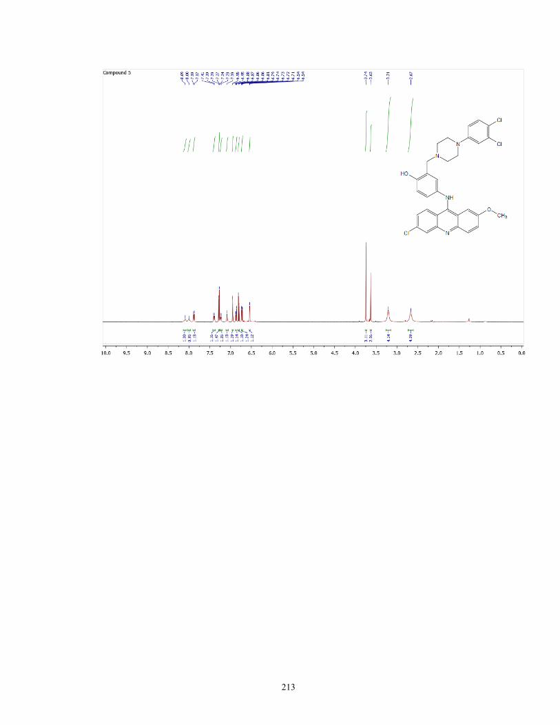
213

214

215

216

217

218

219

220

221
Appendix II: HPLC chromatogram of
Compound 4
(Chapter 2)

222
HPLC chromatogram of A4, yielded a purity of 98%.

223
Appendix III: Selected NMR Spectra
(Chapter 3)

224
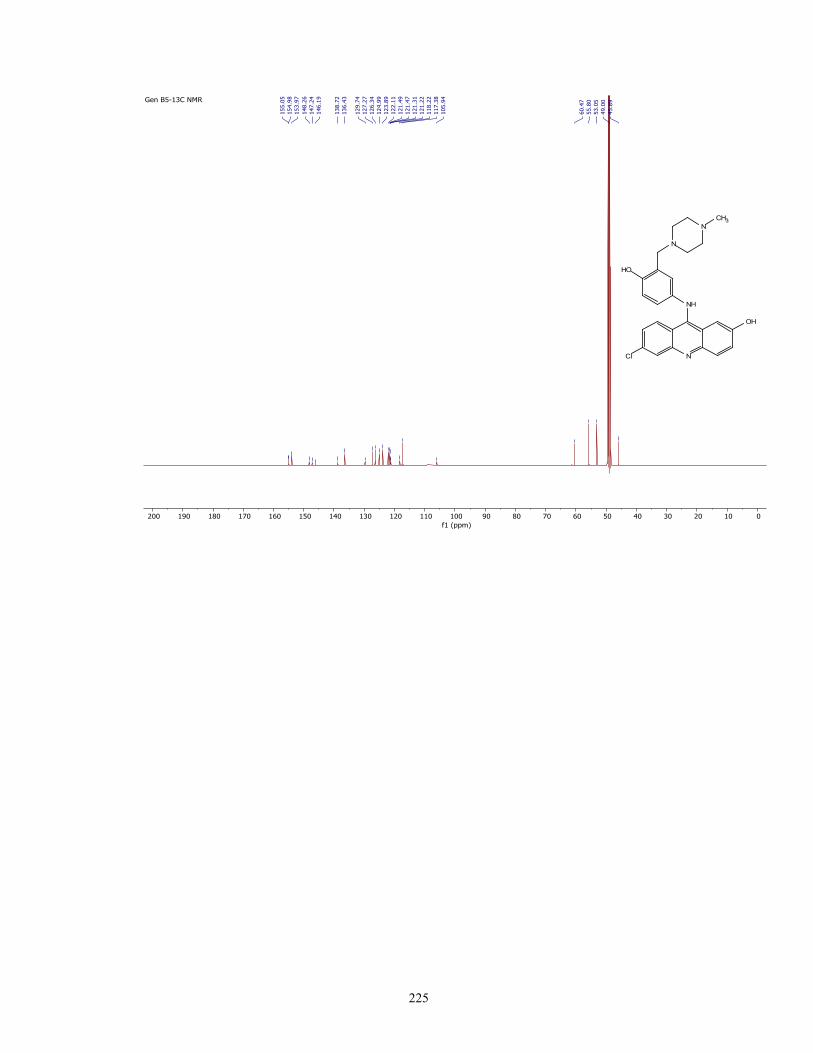
225

226

227

228

229

230

231

232

233

234
Appendix IV: HPLC chromatogram of B9
(Chapter 3)

235
HPLC chromatogram of B9, yielded a purity of 96.9%.

236
Appendix V: Selected NMR Spectra
(Chapter 4)

237
1H/13C NMR spectra of Gen C compounds

238

239

240

241

242

243

244

245

246

247

248

249

250

251

252

253

254

255

256
1H/13C NMR spectra of Gen D compounds

257

258

259