design framework of the mua remodeling signal that confers
TRANSCRIPT

Design framework of the MuA remodeling signal thatconfers preferential complex disassembly by the AAA+
unfoldase ClpXby
Lorraine LingB.A. Molecular and Cell Biology,
University of California, Berkeley (2007)
Submitted to the Department of Biologyin partial fulfillment of the requirements for the degree of
Doctor of Philosophy
at the
MASSACHUSETTS INSTITUTE OF TECHNOLOGY
September 2014
© 2014 Lorraine Ling. All rights reserved.
The author hereby grants to MIT permission to reproduce and todistribute publicly paper and electronic copies of this thesis document in
whole or in part in any medium now known or hereafter created.
Signature of Author . . . . . . . . . . . . . . . . . . . . . . . . . . . . . . . . . . . . . . . . . . . . . . . . . . . . . .Department of Biology
July 22, 2014Certified by. . . . . . . . . . . . . . . . . . . . . . . . . . . . . . . . . . . . . . . . . . . . . . . . . . . . . . . . . . . . . . .
Tania A. BakerE. C. Whitehead Professor of Biology
Thesis Supervisor
Accepted by . . . . . . . . . . . . . . . . . . . . . . . . . . . . . . . . . . . . . . . . . . . . . . . . . . . . . . . . . . . . . .Michael Hemann
Associate Professor of BiologyCo-Chair, Biology Graduate Committee

2
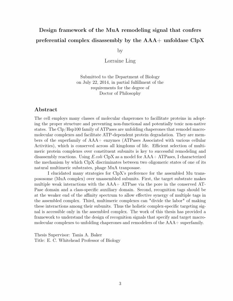
Design framework of the MuA remodeling signal that confers
preferential complex disassembly by the AAA+ unfoldase ClpX
by
Lorraine Ling
Submitted to the Department of Biologyon July 22, 2014, in partial fulfillment of the
requirements for the degree ofDoctor of Philosophy
Abstract
The cell employs many classes of molecular chaperones to facillitate proteins in adopt-ing the proper structure and preventing non-functional and potentially toxic non-nativestates. The Clp/Hsp100 family of ATPases are unfolding chaperones that remodel macro-molecular complexes and facilitate ATP-dependent protein degradation. They are mem-bers of the superfamily of AAA+ enzymes (ATPases Associated with various cellularActivities), which is conserved across all kingdoms of life. Efficient selection of multi-meric protein complexes over constituent subunits is key to successful remodeling anddisassembly reactions. Using E.coli ClpX as a model for AAA+ ATPases, I characterizedthe mechanism by which ClpX discriminates between two oligomeric states of one of itsnatural multimeric substrates, phage MuA tranposase.
I elucidated many strategies for ClpX’s preference for the assembled Mu trans-pososome (MuA complex) over unassembled subunits. First, the target substrate makesmultiple weak interactions with the AAA+ ATPase via the pore in the conserved AT-Pase domain and a class-specific auxiliary domain. Second, recognition tags should beat the weaker end of the affinity spectrum to allow effective synergy of multiple tags inthe assembled complex. Third, multimeric complexes can "divide the labor" of makingthese interactions among their subunits. Thus the holistic complex-specific targeting sig-nal is accessible only in the assembled complex. The work of this thesis has provided aframework to understand the design of recognition signals that specify and target macro-molecular complexes to unfolding chaperones and remodelers of the AAA+ superfamily.
Thesis Supervisor: Tania A. BakerTitle: E. C. Whitehead Professor of Biology
3

4
Acknowledgments
I am a confidant scientist today thanks in large part my advisor Tania Baker. She ismy mentor and role model. Even as an established professor many years away from thebench, Tania answered my nitty-gritty questions and would offer technical help in myMuA assays. Her encyclopedic memory saved me many times from repeating experimentswith negative/ uninterpretable results because hardly anyone publishes inconclusive data.Her guidance and enthusiasm help build my confidence as a researcher. She has helpedme become a better scientific writer. As a role model, Tania has shown me how to handlestress and life’s complications with grace and perseverance. She is like the CEO of theTania Baker company. I thank Bob Sauer, my co-advisor, for great advice at our monthlySJ meetings and reminding me to look at the bigger picture in my research.
Thank you to my thesis committee members, Amy Keating and Mike Laub, whohave been with me since the start of my thesis research. They provided wonderfulconstructive criticism at all my thesis meetings, helped me pivot my project at a criticaltime, and ensured that I graduated in a timely manner. Thank you to Jodi Camberg forsitting on my thesis defense committee and suggesting improvements to this thesis.
As an undergraduate who majored in Genetics, biochemistry at the graduate levelseemed intimidating. I thank all my biochemistry teachers at MIT who taught the subjectin an engaging, thought-provoking, and accessible manner. Bob and Frank Solomontaught Graduate Biochemistry. Amy and Bob taught Special Topics in Biochemistry.They all helped me unlock my "biochemistry" power.
Many thanks to all past and present members of the Baker Lab. I am so lucky tobe colleagues with this group of smart, witty, and compassionate people. Special thanksto fellow graduate student Ben Stein, with whom I shared the lab room. I will miss our’Party-time’ music-science mash ups. Although I didn’t have the pleasure of overlappingwith Aliaa whose research my thesis work has built upon, Aliaa was so generous answeringmy emails. I thank Anne Meyer who was my rotation mentor. She was uncertain, maybeeven skeptical, that I would join the Baker lab due to my wildly different rotations butI did!
Lastly, thank you to my family and friends who provide a wonderful counter-balance to graduate school.
5

6

Contents
1 Introduction 11
1.1 Protein homeostasis in the cell . . . . . . . . . . . . . . . . . . . . . . . . 12
1.2 Clp/Hsp100 ATPases . . . . . . . . . . . . . . . . . . . . . . . . . . . . . 17
1.3 Structural features of Clp/Hsp100 family . . . . . . . . . . . . . . . . . . 20
1.3.1 ATPase domain . . . . . . . . . . . . . . . . . . . . . . . . . . . . 20
1.3.2 Auxiliary domain . . . . . . . . . . . . . . . . . . . . . . . . . . . 22
1.4 Substrate selection by Clp/Hsp100 ATPases . . . . . . . . . . . . . . . . 22
1.4.1 Direct recognition . . . . . . . . . . . . . . . . . . . . . . . . . . . 23
1.4.2 Assisted recognition . . . . . . . . . . . . . . . . . . . . . . . . . . 25
1.5 Remodeling enzymes in AAA+ superfamily . . . . . . . . . . . . . . . . 27
1.6 The virus, Bacteriophage Mu . . . . . . . . . . . . . . . . . . . . . . . . 28
1.6.1 MuA transposase . . . . . . . . . . . . . . . . . . . . . . . . . . . 30
1.6.2 Transposition pathway . . . . . . . . . . . . . . . . . . . . . . . . 30
1.6.3 Transpososome remodeling by ClpX . . . . . . . . . . . . . . . . . 33
1.7 Motivation for thesis research . . . . . . . . . . . . . . . . . . . . . . . . 34
2 Design logic of a multivalent recognition signal confers preferential com-
plex disassembly by the AAA+ unfoldase ClpX 37
2.1 Abstract . . . . . . . . . . . . . . . . . . . . . . . . . . . . . . . . . . . . 38
2.2 Introduction . . . . . . . . . . . . . . . . . . . . . . . . . . . . . . . . . . 39
2.3 Results . . . . . . . . . . . . . . . . . . . . . . . . . . . . . . . . . . . . . 43
2.3.1 Identification of a region critical for enhanced recognition of trans-
pososomes by ClpX . . . . . . . . . . . . . . . . . . . . . . . . . . 43
7

2.3.2 A peptide encompassing the critical region interacts with the N-
terminal zinc-binding domain of ClpX . . . . . . . . . . . . . . . 47
2.3.3 Step-wise loss of the Enhancement tag trends with ClpX’s weaker
affinity for complexes . . . . . . . . . . . . . . . . . . . . . . . . . 49
2.3.4 Mu pore-binding tag is an intrinsically poor ClpX signal without
adaptor-like contacts . . . . . . . . . . . . . . . . . . . . . . . . . 54
2.3.5 Transpososomes with a strong pore-binding tag do not require En-
hancement tags . . . . . . . . . . . . . . . . . . . . . . . . . . . . 54
2.4 Discussion . . . . . . . . . . . . . . . . . . . . . . . . . . . . . . . . . . . 58
2.5 Methods . . . . . . . . . . . . . . . . . . . . . . . . . . . . . . . . . . . . 64
2.6 Appendix: Geometry experiments on MuA monomer variants . . . . . . 67
2.6.1 Introduction . . . . . . . . . . . . . . . . . . . . . . . . . . . . . . 67
2.6.2 Results . . . . . . . . . . . . . . . . . . . . . . . . . . . . . . . . . 68
2.6.3 Discussion . . . . . . . . . . . . . . . . . . . . . . . . . . . . . . . 69
3 Conclusion & Future Directions 71
3.1 Conclusion . . . . . . . . . . . . . . . . . . . . . . . . . . . . . . . . . . . 72
3.2 Future Directions . . . . . . . . . . . . . . . . . . . . . . . . . . . . . . . 74
8

List of Figures
1-1 A simplified protein life cycle . . . . . . . . . . . . . . . . . . . . . . . . 13
1-2 Prokaryotic Heat Shock Protein chaperones . . . . . . . . . . . . . . . . . 16
1-3 Model of AAA+ ATPase unfolding and translocation cycles . . . . . . . 18
1-4 Domain structure of bacterial Clp/Hsp100s . . . . . . . . . . . . . . . . . 19
1-5 ClpX structure . . . . . . . . . . . . . . . . . . . . . . . . . . . . . . . . 20
1-6 Mechanism of replicative transposition . . . . . . . . . . . . . . . . . . . 29
1-7 Domain structure of MuA transposase . . . . . . . . . . . . . . . . . . . 30
1-8 Phage Mu in vivo replicative transposition . . . . . . . . . . . . . . . . . 33
1-9 Structure of type1 and type 2 Mu transpososomes . . . . . . . . . . . . . 34
2-1 In vitro assays for Mu complex assembly and recognition by ClpX . . . . 41
2-2 Mutation of a sequence region R622-S624 reduces disassembly and degra-
dation rates . . . . . . . . . . . . . . . . . . . . . . . . . . . . . . . . . . 44
2-3 Comparison of all reaction rates for Mu aspartate variants . . . . . . . . 45
2-4 Residues P623 S624 form a critical interaction between MuA complex and
ClpX . . . . . . . . . . . . . . . . . . . . . . . . . . . . . . . . . . . . . . 46
2-5 The N-terminal zinc-binding domain of ClpX binds to the enhancement
peptide . . . . . . . . . . . . . . . . . . . . . . . . . . . . . . . . . . . . . 48
2-6 Mu transpososome is an asymmetric complex . . . . . . . . . . . . . . . 50
2-7 Tools for making homogeneous mixed mutant complexes . . . . . . . . . 51
2-8 Chimeric complexes disassembly controls . . . . . . . . . . . . . . . . . . 52
2-9 All four subunits can provide the Enhancement tag in MuA complexes . 53
2-10 Mu pore-binding tag is a weak ClpX recognition signal . . . . . . . . . . 55
9

2-11 Mu complexes with a strong pore-binding tag are recognized as well as
native Mu complexes . . . . . . . . . . . . . . . . . . . . . . . . . . . . . 56
2-12 Mu∆8ssrA monomer degradation by ClpXP . . . . . . . . . . . . . . . . 57
2-13 Permutations of tag engagement in Mu transpososome by ClpX . . . . . 61
2-14 MuSspB monomer degradation by ClpXP . . . . . . . . . . . . . . . . . 69
10

Chapter 1
Introduction
11

Overview
The foundational theme of this thesis is understanding the mechanism and reg-
ulation of substrate selection by the Hsp100/Clp chaperones. In the cell, chaperones
are molecular machines that facilitate a change in a protein’s functions by altering the
structure of proteins. Depending at which stage of the protein life cycle that the target
protein is in, chaperones aid in both folding and unfolding of the target protein to bring
about changes in structure.
This introduction will focus on the chaperones found in prokaryotes. The first
section situates the many roles of chaperones within the protein life cycle. The next
few sections introduce the Hsp100/Clp family of chaperones and chaperone-linked pro-
teases found in E.coli, their structure, some interacting partners which aid in substrate
recognition, and a sampling of biological targets. The last section introduces phage MuA
transposase, one of many substrates of ClpX, an E.coli Hsp100/Clp unfolding chaper-
one. The ClpX-MuA transposase interaction is my model system to study mechanisms
of specificity and design of recognition signals.
1.1 Protein homeostasis in the cell
Inside the densely packed environment of a cell, proteins perform many roles; as structural
macromolecules providing support and shape, as enzymes catalyzing chemical reactions,
and as signaling molecules and receptors communicating information between the extra-
cellular environment and the cell. For these diverse roles, proteins must adopt the correct
structure leading to the biochemists’ axiom “structure equals function.” For ideal cellu-
lar function, proteins must be properly folded into their native three-dimensional forms,
associated with appropriate partners or oligomerize if required, and disposed of when
no longer needed or damaged. Nature has evolved machinery, referred to as molecular
chaperones, to guide proteins through these major milestones in the protein life cycle.
The life cycle of a protein begins at its birth by the protein-making factory called a
ribosome, itself a protein-RNA complex. The newly synthesized polypeptide contains all
the information needed within its primary amino acid sequence to define its final folded
12
Foldedprotein
Protein Complex
Unfolded protein
Aggregatedprotein
Degraded protein
Holdases
Clp/Hsp100 proteases
Clp/Hsp100proteasesHsp90
Clp/Hsp100
Hsp60Hsp70Hsp90
Clp/Hsp100Hsp70
Clp/Hsp
100
protease
s
Non-nativestate
Clp/Hsp100Hsp70
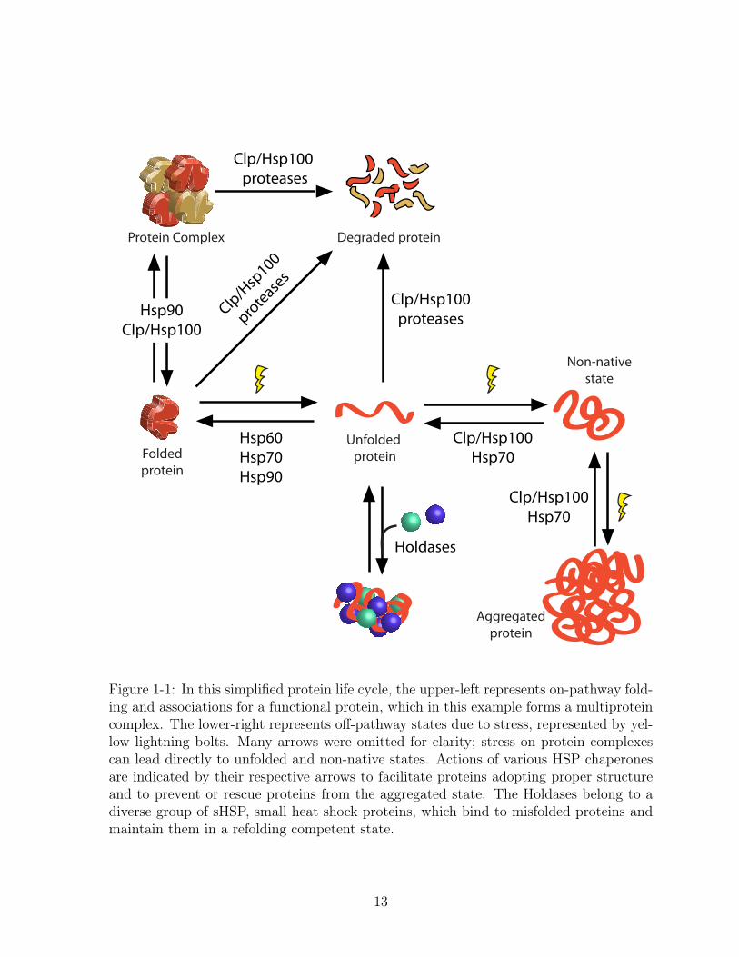
Figure 1-1: In this simplified protein life cycle, the upper-left represents on-pathway fold-ing and associations for a functional protein, which in this example forms a multiproteincomplex. The lower-right represents off-pathway states due to stress, represented by yel-low lightning bolts. Many arrows were omitted for clarity; stress on protein complexescan lead directly to unfolded and non-native states. Actions of various HSP chaperonesare indicated by their respective arrows to facilitate proteins adopting proper structureand to prevent or rescue proteins from the aggregated state. The Holdases belong to adiverse group of sHSP, small heat shock proteins, which bind to misfolded proteins andmaintain them in a refolding competent state.
13

(native) structure (Anfinsen, 1973). However, in the crowded intracellular environment,
nascent polypeptides need the help of molecular chaperones to properly fold and to avoid
non-functional non-native and aggregated states (Figure 1-1). Chaperones do not mold
proteins into their native structure but simply facilitate the self-directed folding process
(Hartl, 2011; Mayer, 2013).
In many cases, proteins combine with other proteins to form large macromolecular
complexes which are the biologically active states. Individual proteins within a larger
complex are referred to as subunits. When these complexes need to perform new func-
tions, the complex may gain, lose, or exchange subunits. These changes in quaternary
structure very often reflect changes in biological function. Once again, chaperones fa-
cilitate the assembly, alteration, and deactivation of macromolecular complexes. These
multi-faceted processes performed by chaperones are encompassed in the term “remodel-
ing.”
Several molecular chaperones participating at initial protein folding stages of the
protein life cycle come from the highly conserved heat shock protein (Hsp) family, dis-
covered in their roles in heat-shock response (Parsell & Lindquist, 1993). The Hsp60s
group includes E.coli GroEL and its co-chaperone GroES (Figure 1-2A). Together they
oligomerize into a barrel-shaped structure reminiscent of a basket with a domed lid to
provide a defined and isolated environment for the polypeptide to fold and avoid aggre-
gation prior to reaching its native state (Braig et al., 1994; Xu et al., 1997; Ellis, 2003).
GroEL has a preference for binding stretches of hydrophobic amino acids, an elegant way
to corral unfolded or non-native proteins (Fenton et al., 1994; Mayhew et al., 1996). Using
non-specific substrate binding strategies and sequestration mechanisms, GroEL/GroES
aids in folding of at least half of the proteins in the cell(Houry et al., 1999; Viitanen
et al., 1992).
Another group of Hsp, Hsp70s also help proteins reach their native state. In E.coli,
the Hsp70 member is DnaK (Figure 1-2B). Working with a co-chaperone,DnaJ, and a
nucleotide exchange factor GrpE, the DnaK/ DnaJ/ GrpE chaperone team works at
the site of the ribosome, to protect emerging polypeptides and help in folding (Deuerling
et al., 1999; Teter et al., 1999). At this stage, these emerging polypeptides may have very
14

little folded structure, so stretches of exposed hydrophobic amino acids are particularly
susceptible to aggregation. DnaK chaperone team binds non-specifically to stretches
of hydrophobic residues, shielding them from neighbors. Then, powered by ATP, the
chaperone releases the chain when it is ready to fold (Fourie et al., 1994; Flynn et al.,
1989). Thus, both Hsp60s and Hsp70s use similar non-specific binding mechanisms to
prevent inappropriate interactions and facilitate on-pathway folding.
Although not required for de novo folding of most proteins, the Hsp90s and
Hsp100/Clp chaperones use the energy from ATP to aid the final maturation of selected
proteins substrates ("clients") that function as a multicomponent or larger oligomeric
complex. In most eukaryotes, Hsp90 is an essential chaperone and its clients include
the steroid hormone receptors, protein kinases, and transcription factors (Li & Buch-
ner, 2013). The growing list of Hsp90-associated co-factors enables the chaperone to
have a broad substrate repertoire (Eckl & Richter, 2013). Clp/Hsp100 unfolding chap-
erones facilitate protein complex remodeling and disassembly. Similar to Hsp90 group,
Clp/Hsp100 chaperones utilize different co-factors to enlarge substrate range. However,
many Clp/Hsp100 unfolding chaperones target just one protein complex; often remodel-
ing these multiprotein complexes for the goal of recycling the subunits.
In addition to functions during normal growth, chaperones are key protective
agents during times of stress. If proteins adopt non-native conformations due to damage
or environmental stress, they may form insoluble aggregates which deactivate protein
functions and are often toxic to the cell. Unfolding chaperones in the Hsp100/Clp group
working as disaggregases can re-solubilize aggregates (Squires et al., 1991). Then the
misfolded polypeptide has a chance to refold into its native conformation, often with
help from Hsp60 and Hsp70 folding chaperones, or be degraded(McCarthy et al., 1998;
Laskowska et al., 1996).
Lastly at the end of a protein’s useful “life”, proteins are degraded into their
amino acid building blocks. Whether caused by regulatory responses or quality con-
trol mechanisms, proteins which are at the end of their lives are funneled to intracel-
lular chaperone-linked proteases also from the Hsp100/Clp group (Figure 1-2C). Unlike
Hsp60s and Hsp70s, the Hsp100/Clp group of unfolding chaperones generally use specific
15

A. Hsp60 group : GroEL/ES
B. Hsp70 group : DnaK
C. Hsp100 group : HslU
GroES
GroEL
HslV
HslU
GroEL
~800kDa ~70kDa~900kDa
top view
side view side viewDavid Goodsell & RSCB Protein Data BankMolecule of the Month Series
Figure 1-2: Atomic structures of members of the Hsp family of chaperones illustrated byDavid Goodsell. Figures are not to scale.A. Seven GroEL proteins (Hsp60 chaperone) form a ring. Depending on its nucleotidestate, the double stacked GroEL rings are capped by a GroES particle (a 7-mer).B. DnaK, ATP-binding domain on the left, peptide binding domain on the right with abound peptide colored in red.C. HslUV compartmentalized protease. HslU unfoldase forms a hexameric ring. HslVpeptidase is a “double donut” of hexameric rings.
16

substrate selection mechanisms and actively unfold their substrates for two biological
outcomes: remodeling and degradation. Because the Hsp100/Clp group deactivate com-
plexes and often promote irreversible protein degradation, these destructive powers must
be tightly regulated at multiple levels, such as target binding, spatial location, and de-
velopmental timing.
1.2 Clp/Hsp100 ATPases
Clp/Hsp100 ATPase family belong to the larger AAA+ (ATPases associated with various
cellular activities) superfamily of proteins. These enzymes use ATP hydrolysis to drive
repetitive conformational changes that perform mechanical work in the cell (reviewed in
Hanson & Whiteheart, 2005). Many AAA+ enzymes function by translocating protein
polypeptides or nucleic-acid polymers. Examples include DNA/RNA helicases, protein-
secretion translocation machinery, and viral packaging motors.
The Clp/Hsp100 ATPases actively unfold proteins for two biological outcomes:
remodeling and degradation. Protein substrates are targeted to Clp ATPases by short
peptide sequences, called tags or degrons (discussed in section 1.4.1). Cycles of ATP
binding and hydrolysis drive protein unfolding (Figure 1-3A). For the outcome of degra-
dation, the Clp/Hsp100 ATPase partners with a peptidase to form a compartmentalized
protease (Figure 1-3B). The Clp/Hsp100 ATPases are ring hexamers, containing one or
two ATPase modules per polypeptide (Figure 1-4). In E.coli, the first Clp family member
identified was ClpA (Caseinolytic protease A) named for its role as the ATP-dependent
regulatory subunit which partners with ClpP peptidase to form the ClpAP protease that
degrades casein(Katayama et al., 1988). ClpP is unrelated to the AAA+ superfamily.
Later a paralog of ClpA, ClpX was discovered which can also partner with ClpP to form
ClpXP protease. Similarly, the HslUV protease is comprised of the ATPase Heat shock
locus HslU (also known as ClpY) and the the peptidase HslV (also known as ClpQ).
Two additional proteases, Lon and FtsH, have the ATPase and peptidase components
encoded on a single polypeptide instead of existing as separate subunits (reviewed in
Sauer & Baker, 2011). Lon was the first bacterial energy-dependent protease discovered.
17

A. Unfolding for remodeling/disassembly
tag
AAA+ unfoldase
ATP ATP
AAA+ unfoldase
peptidase
ATP ATP
B. Unfolding for degradation
Figure 1-3: A. A recognition signal (tag) in a native substrate is initially recognized by theAAA+ unfoldase. Repetitive cycles of ATP hydrolysis then power unfolding of substratesand translocation through the enzyme’s central channel. This leads to unfolding and/orremodeling of the protein complex. B. When the AAA+ unfoldase is associated with acompartmental peptidase, translocation of the polypeptide into the degradation chamberleads to protein destruction. (Adapted from Sauer and Baker, 2011)
Lon’s ability to degrade partially folded proteins has led to its designation as the major
protease responsible for protein quality control in the cell (Chung & Goldberg, 1981).
Although deletion of other AAA+ chaperones and proteases leads to severe pleiotropic
phenotypes, FtsH is the only essential protease in E.coli (Ogura et al., 1991). FtsH
is anchored to the inner membrane and degrades membrane and cytoplasmic proteins
(Tomoyasu et al., 1993).
ClpB cannot partner with ClpP and thus has only chaperone and no coupled pro-
tease functions. It is essential for thermotolerance and can solubilize almost any protein
that becomes aggregated after severe stress (Squires et al., 1991). ClpB collaborates with
the DnaK chaperone system (not members of AAA+ superfamily) to reactive proteins
from insoluble aggregates. Studies of these chaperones’ activities in vitro have clarified
18

FtsH
LonA
HslU
ClpX
ClpA/ClpC
ClpB
ClpP
ClpP
HslV
AAA+ module AAA+ module
protease
protease
protease
protease
protease
N
M
N
I
N1 N2
TM
N
Class-speci�cAuxiliary domains
Figure 1-4: The Clp/Hsp100 family of chaperones and compartmentalized proteases con-tain a conserved ATPase domain, the hallmark feature of the AAA+ superfamily. ClpB,ClpA, and ClpC contain two AAA+ modules. ClpX, HslU, FtsH and Lon contain onlyone AAA+ module. Each ATPase has class-specific auxiliary domains that are not con-served. Proteases that associate with Clps are shown on the right. ClpP and HslV areseparate proteins while FtsH and LonA each contain a protease domain. Adapted from(Sauer and Baker, 2011)
their individual mechanisms but a combined mechanism with wide consensus has not
yet been established. One model is that ClpB breaks apart large protein aggregates into
smaller ones by extracting and unfolding polypeptides from the aggregate (Weibezahn
et al., 2004). The released polypeptide can spontaneously refold or interact with the
folding chaperones such as DnaK or GroEL/GroES. DnaK may also act earlier in the
ClpB-mediated remodeling reaction by helping ClpB bind to aggregates or regulating
ATP hydrolysis by ClpB (Doyle & Wickner, 2009).
The Gram-positive bacterium, Bascillus subtilis, shares many Clp/Hsp100 or-
thologs with E.coli. However B.subtilis has a species-specific ClpC ATPase and lacks
both ClpA and ClpB. ClpC chaperone can associate with ClpP peptidase to form ClpCP
protease (Molière & Turgay, 2009).
19
1.3 Structural features of Clp/Hsp100 family
1.3.1 ATPase domain
Clp/Hsp100 enzymes are members of the AAA+ superfamily. The hallmark feature of
the AAA+ superfamily is a structurally conserved ATPase module, which performs cycles
of nucleotide binding, hydrolysis and release to convert chemical energy into mechanical
work. The ATPase domain contains a conserved ATP binding pocket. Two conserved
sequence motifs which interact with ATP are the Walker A and Walker B motifs, which
bind and hydrolyze ATP, respectively (Wendler et al., 2012). Most oligomerize into rings
with a central pore.
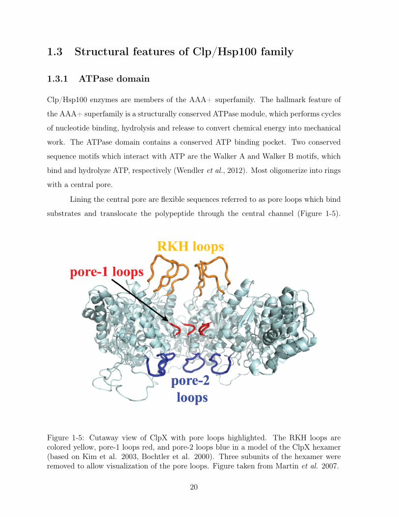
Lining the central pore are flexible sequences referred to as pore loops which bind
substrates and translocate the polypeptide through the central channel (Figure 1-5).
Figure 1-5: Cutaway view of ClpX with pore loops highlighted. The RKH loops arecolored yellow, pore-1 loops red, and pore-2 loops blue in a model of the ClpX hexamer(based on Kim et al. 2003, Bochtler et al. 2000). Three subunits of the hexamer wereremoved to allow visualization of the pore loops. Figure taken from Martin et al. 2007.
20

Located in the center of the channel, the pore-1 loops are the most conserved, with a
nearly invariant Aromatic-hydrophic sequence motif, YVG, among all AAA+ ATPases.
Mutagenesis studies and structures of ClpX with different nucleotide states argue that
the pore-1 loop is key to translocation (Siddiqui et al., 2004; Martin et al., 2008b; Glynn
et al., 2009). The RKH and pore-2 loops are conserved among ClpX orthologs and are
located at the mouth of the channel and bottom of the channel, respectively. Both RKH
and pore-2 loops play a role in initial binding and translocation of substrates (Farrell
et al., 2007; Martin et al., 2007, 2008a). Pore-2 loops also mediate ClpP binding and
communication (Martin et al., 2007).
Because there are no atomic structures of AAA+ ATPases with bound substrates,
the mechanism of coupling ATP hydrolysis, enzyme conformation changes and mechani-
cal work is under active investigation. Studies of ClpX have led to a model mechanism in
which cycles of ATP hydrolysis are coupled to ClpX subunit conformational changes and
thus the orientation of pore loops leading to a net movement of the substrate polypep-
tide (Stinson et al., 2013). Crystal structures of HslU and ClpX reveal that the pore-1
loop adopts a dynamic range of confirmations that position the tip of the loop through-
out the length of the channel (Bochtler et al., 2000; Sousa et al., 2000; Glynn et al.,
2009). Repetitive cycles of ATP hydrolysis by ClpX may be needed to unfold the protein
and translocate the polypeptide(Martin et al., 2008c). Translocation of the polypep-
tide is proposed to pull the attached folded protein against the entrance to the axial
pore, thereby generating a denaturing force because the pore is smaller than the folded
protein (Baker & Sauer, 2006). The observable result is that Clp/Hsp100 chaperones
and chaperone-linked proteases exert a pulling force on the polypeptide which leads to
cooperative unfolding of the target protein (Aubin-Tam et al., 2011).
There appears to be no obligatory directionality to translocation, as ClpXP can
degrade substrates starting either from the N-terminus or from the C-terminus (Gottes-
man et al., 1998; Gonciarz-Swiatek et al., 1999; Lee et al., 2001; Flynn et al., 2003;
Hoskins et al., 2002; Kenniston et al., 2005; Farrell et al., 2007). Once ClpX engages
with the protein substrate, the translocation process is processive and remarkably toler-
ant to the structural identity of the polypeptide. ClpX exhibits very little preference for
21
side group size, charge, or chirality and will translocate peptide bonds spaced with long
hydrophobic carbon chains (Barkow et al., 2009).
1.3.2 Auxiliary domain
A second feature of Clp/Hsp100 ATPases are auxiliary domains specific to each member.
In general these auxiliary domains are not conserved between members, dispensable for
ATPase function but play roles in substrate recognition (Sauer & Baker, 2011). Often
these member-specific domains provide a docking platform for delivery proteins, called
adaptors (discussed in section 1.4.2). ClpA, ClpC, and ClpX all have N-terminal auxiliary
domains. However the structure of each auxiliary domain and the connection of each to
their respective ATPase module differs among the three Clp ATPases (Zeth et al., 2002;
Wang et al., 2011; Park et al., 2007) HslU has an I-domain, which is an insertion of 140
residues in the AAA+ module (Bochtler et al., 2000). The I-domain domain is thought to
play a role in substrate binding and allosterically regulates ATPase activity (Sundar et al.,
2012). The M-domain (middle domain) of ClpB forms a propeller-shaped coiled-coil and
has been proposed to act like a “crowbar” to break apart large aggregates, though the
crowbar model has not been experimentally validated (Lee et al., 2003). Interestingly, the
M-domain serves as the site for species-specific interaction with the DnaK/DnaJ/GrpE
chaperone team (Miot et al., 2011).
1.4 Substrate selection by Clp/Hsp100 ATPases
Clp ATPases employ two modes of substrate recognition. The first is direct substrate
recognition by binding to short peptide sequences, known as tags, on the substrate. The
tag interacts with the pore of the ATPase and subsequent engagement results in un-
folding. The second mode termed “assisted recognition” is when another protein aids
the ATPase in recognition of the substrate. Additional mechanisms for substrate selec-
tion not discussed below are subcellular relocalization of either substrates or Clp/Hsp100
ATPases, phosphorylation of proteins turning them into substrates, and regulating ex-
pression levels of substrates or ATPases by developmental timing.
22

1.4.1 Direct recognition
Recognition tags are found often at the N- or C-termini of substrates. Tags that bind
directly to the unfoldase fall into three classes: intrinsic, latent, and co-translational.
intrinsic class
Intrinsic signals are encoded in the primary sequence of a substrate protein. The
tags are often present at the N or C-termini but may not be accessible to proteases until
a conformational change or loss of a shielding binding partner. Examples of substrates
with N-terminal tags include Dps (DNA-binding protein from starved cells), a stationary
phase nucleoid protein that sequesters iron and protects DNA from damage (Flynn et al.,
2003), and bacteriophage 𝜆 O replication initiator protein when not bound to orilambda
DNA (Gonciarz-swiatek et al., 1999). Additionally, 𝜆O may have an internal intrinsic
signal. Residues Q49-M67 bind strongly to ClpX N-domain and a peptide containing this
sequence can compete with degradation of the full-length protein by ClpXP (Thibault
et al., 2006).
Remarkably the stability of a protein can be attributed to a single amino acid at
its N-terminus, known as the N-end rule. Present in both prokaryotes and eukaryotes,
the N-end rule tags are often the large hydrophobic residues. In E.coli, an N-terminal
leucine, phenylalanine, tryptophan or tyrosine directly targets proteins to intracellular
proteases (Varshavsky, 1996).
An example of an intrinsic C-terminal tag comes from Supressor of lon (SulA)
identified in a screen to suppress the lon - sensitivity to ultraviolet radiation (Gayda
et al., 1976). SulA is an inhibitor of cell division and upregulated during SOS response
otherwise during normal growth it is degraded by Lon and HslUV proteases (Gottesman
et al., 1981; Mizusawa, 1983; Seong et al., 1999). The last eight C-terminal residues are
crucial for recognition by Lon as a truncated SulA variant was stabilized both in vivo
ans in vitro. (Higashitani et al., 1997)
A proteomic based screen for in vivo substrates of ClpXP revealed five classes
of recognition signals. The screen utilized a tagged and catalytically inactive variant
of ClpP (ClpPtrap) to receive and contain proteins translocated by ClpX (Flynn et al.,
23
2003). Analysis of the >50 captured proteins led to two classes of motifs located at
the C-terminus and three classes located at the N-terminus. About one quarter of the
trapped proteins contain potential intrinsic ClpX recognition signals at both the N-
terminus and C-terminus. Similarly to phage 𝜆 O replication initiator protein, many N-
terminal sequences from the pool of trapped proteins directly bound to ClpX’s N-domain
on a peptide array. However with so few examples of confirmed N-domain interacting
sequences, it was difficult to establish a consensus motif (Flynn et al., 2003). It is
unclear whether both signals are engaged by the ATPase domain of ClpX or contribute
to additional enzyme-binding interactions via the auxiliary domain similarly to adaptor
proteins (see section 1.4.2).
latent class
Latent signals are also encoded in the primary sequence but require processing of
the protein to make the tag accessible. Often an endopeptidic cleavage reveals a new
termini containing the tag. Examples include LexA and RseA. LexA is a transcriptional
repressor of genes involved in DNA-damage response. During the SOS response to DNA
damage, LexA undergoes RecA-stimulated autocleavage between the N-terminal DNA
binding domain and the C-terminal dimerization domain. Both fragments are rapidly
degraded by Lon and ClpXP (Little, 1983; Neher et al., 2003a). The cleavage reveals a
tag at the new C-terminus with residues VAA-CO2 which is similar to the region of the
ssrA tag (LAA-CO2discussed below) recognized by ClpX (Neher et al., 2003a).
RseA is the anti-sigma factor to 𝜎E which activates expression of genes involved
in the bacterial extracytoplasmic stress response. RseA is an inner membrane-spanning
protein. The N-terminal cytoplasmic portion binds 𝜎E and sequesters the sigma fac-
tor from its target promoters. In response to extracytoplasmic stress, RseA undergoes
sequential cleavage steps by proteases in the periplasm and cytoplasm which results in
an N-terminal fragment of RseA and frees 𝜎E (Lima et al., 2013). The released 𝜎E can
activate its regulon, while the N-terminal fragment of RseA, revealing residues VAA at
the new C-terminus, is degraded by ClpXP (Flynn et al., 2004).
co-translational class
24
In contrast to the two previous classes, the co-translation class is not encoded in
the primary sequence of the substrate protein. The only member of this class is the ssrA
tag which marks proteins for degradation by multiple AAA+ proteases (ClpAP, ClpXP,
FtsH, Lon) (Keiler et al., 1996). When ribosomes get stuck on an mRNA, a rescue system
of tmRNA (trans-messenger RNA) displaces the offending mRNA. Translation continues
on the tmRNA, which encodes an eleven amino acid sequence (AANDENYALAA) and a
stop codon. The ssrA tag is appended onto the truncated polypeptide and marks these
incomplete translation products for destruction. It is estimated that 0.5% of translation
products receive an ssrA tag (Lies & Maurizi, 2008). Thus the ssrA-tagging system
rescues stalled ribosomes and destroys potentially dangerous protein fragments (reviewed
in Karzai et al., 2000).
1.4.2 Assisted recognition
Clp ATPases also use a second mode termed “assisted recognition” in which accessory
proteins called adaptor proteins modulate substrate choice and often give rise to higher-
affinity enzyme-adaptor-substrate complexes. Various mechanisms for adaptors have
been observed from acting as delivery vehicles to directly affecting chaperone and pro-
tease activity (reviewed in Kirstein et al., 2009). Adaptor proteins are themselves not
degradation substrates and thus participate in multiple rounds of delivery or modulation
of enzyme activity. A general overview of three E.coli adaptors (SspB, RssB, ClpS) and
one B.subtilis adaptor (MecA) represents the diversity of mechanisms to deliver target
proteins and modulate chaperone-protease activities yet highlights one common theme
of docking to the enzyme’s auxiliary domain.
SspB
Stringent starvation protein B (SspB) is the best characterized E.coli adaptor
protein. SspB enhances degradation of ssrA-tagged substrates by ClpXP via a tethering
mechanism. Here, the adaptor binds to both the substrate and the AAA+ unfoldase
to increase the effective concentration of the tag near the enzyme’s active center (Wah
et al., 2003). SspB binds to ClpX specifically through the unfoldase’s N-domain (Park
25

et al., 2007). In E.coli, ClpX and SspB bind distinct portions of the ssrA tag. ClpX
recognizes the last three residues and the carboxyl group whereas SspB recognizes the
first four and seventh residue (Levchenko et al., 2000; Flynn et al., 2001; Levchenko et al.,
2003; Song & Eck, 2003). As mentioned above, ssrA-tagged substrates are also degraded
by ClpAP protease. However, the same adaptor SspB inhibits this reaction because
ClpA and SspB bind overlapping residues in the ssrA tag (Flynn et al., 2001). Although
the biological consequence of this inhibition remains unclear, a plausible outcome is to
turn other substrates into high-priority ClpAP targets, leaving ClpXP to clean up ssrA-
tagged polypeptides. SspB also recognizes and delivers the N-terminal fragment of RseA
to ClpXP for degradation (Flynn et al., 2004).
RssB
Regulator of sigma-S protein B (RssB) also known as stationary-phase regulator
(SprE) is essential for turnover of stationary phase sigma factor, 𝜎S (Muffler et al., 1996;
Pratt & Silhavy, 1996). RssB is a ClpXP-specific adaptor and is activated by phospho-
rylation. Phospho-RssB protein binds 𝜎S which exposes a latent tag in the N-terminal
region of 𝜎S.
ClpS
ClpS adaptor delivers N-end rule substrates to ClpAP protease for degradation
(Dougan et al., 2002). Because both ClpS and ClpA recognize the same N-terminal
amino acid, ClpS employs a more involved mechanism than simple tethering. ClpA
may recognize additional tag features that are not directly bound to ClpS. ClpA, ClpS
and the N-end rule substrate form a high-affinity ternary complex (Román-Hernández
et al., 2011). Within this complex, ClpA engages the unstructured N-terminal section of
ClpS which causes a conformational change and hand-off of the N-end rule residue from
the ClpS binding pocket to the pore of ClpA (personal communication Izarys Rivera-
Rivera). ClpS escapes degradation to catalyze another round of delivery because the core
substrate binding domain of ClpS carries structural elements that are non-denaturable
by ClpA (personal communication Izarys Rivera-Rivera). ClpS interacts with ClpA via
the unfoldase’s N-terminal domain. This interaction is necessary for delivery of N-end
26

rule substrates (Zeth et al., 2002).
MecA
Medium-independent expression of competence (MecA), the best characterized
adaptor protein of B.subtilis, was identified in a screen for genes involved in regulation
of competence (Dubnau & Roggiani, 1990). MecA binds and inhibits the transcriptional
activator of competence, ComK. Furthermore MecA targets ComK for degradation by
ClpCP. Unlike the previous adaptor examples, MecA has a unique mechanism to modu-
late ClpC activity. ClpC is an inactive monomer on it own. MecA triggers the oligomer-
ization of ClpC into the active hexameric chaperone which then can associate with ClpP
to form an active protease. This adapter-mediated oligomerization requires MecA to
bind to the N-domain of ClpC (Kirstein et al., 2006).
1.5 Remodeling enzymes in AAA+ superfamily
In this section, four examples of remodeling enzymes in the AAA+superfamily showcase
the breadth of important biological transitions promoted by remodeling. Katanin and
Spastin are eukaryotic AAA+ ATPases which remodel microtubules (McNally & Vale,
1993). Microtubules provide support to organelles, shape the cell, and organize into
a distinct structure called the spindle that is essential for cell replication and division.
They are made up of tubulin subunits, which polymerize into long and branched dynamic
polymers. Many factors regulate microtubule assembly and disassembly at the termini
of polymers (reviewed in Gardner et al., 2013). However, Katanin and Spastin modulate
the dynamics of microtubule from the middle of a polymer. These “microtubule severing
enzymes” preferentially unfold and abstract tubulin subunits from the lattice (Roll-Mecak
& McNally, 2010). This process of microtubule severing requires the C-terminal tails of
tubulin and and the pore loops of Katanin and Spastin (White & Lauring, 2007; Roll-
Mecak & Vale, 2008).
Another eukaryotic AAA+ family member, N-ethylmalemide sensitive fusion pro-
tein (NSF) is an essential factor in intracellular membrane trafficking. NSF in concert
with SNAPs (soluble NSF attachment proteins) disassemble SNARE complexes, thus
27

freeing SNARE subunits for additional rounds of membrane fusion (Whiteheart et al.,
2001).
Although most studied for their functions in intracellular proteolysis, both E.coli
ClpA and ClpX have been observed in ClpP-independent remodeling reactions. In vitro,
ClpA activates the phage P1 replication initiator protein RepA by remodeling inactive
RepA dimers into monomers that are competent to bind DNA (Wickner et al., 1994).
ClpX plays a key role in the phage Mu lytic cycle (Mhammedi-Alaoui et al.,
1994; Kruklitis et al., 1996). The biochemical steps in phage Mu transposition and the
remodeling of the Mu transposase-DNA complex by ClpX have been extensively studied
and are summarized in the next section. This well-characterized protein has provided me
an ideal model substrate to address questions of target specificity and design principles
of ClpX recognition signals.
1.6 The virus, Bacteriophage Mu
Bacteriophage Mu is a virus that propagates its genome within a bacterial host using
a mechanism of replicative transposition. In replicative transposition, the mobile DNA
element cuts, copies itself to a new location, and leaves behind the copy at the previous
genomic location. This movement occurs using a branched DNA intermediate called a
Shapiro structure (Shapiro, 1979). Extensive study of phage Mu has led to a deeper
understanding of the molecular mechanism and regulation of transposition by mobile
DNA elements. Phage Mu encodes two proteins necessary for in vivo transposition,
MuA and MuB. MuA is the transposase and MuB is a regulatory factor. Together they
recombine the correct DNA sequences, transpose at the correct time during the phage
lifecycle, and avoid disrupting phage Mu’s own genome (Figure 1-6).
Phage Mu regulates this sequence of events by evolving a vectorial process that
uses increasingly stable nucleoprotein complexes and co-opting a host chaperone to direct
a key transition point. A highly tractable in vitro system was developed early which
allowed for biochemical analysis of the transposition reaction. This biochemical system
minimally contains the sequences of Mu genomic ends on a supercoiled plasmid, MuA,
28

transposon
donor DNA
target DNA
3’OH
3’OHdonor DNA cleavageof transferred strand
strand transfer
replication forkassembly (at left gap)
replication throughthe transposon
Ligation
Cointegrate with 2 copies of transposon
5’
3’OH
3’
3’OH
lagging strand
leading strand
Shapiro Intermediate
gap
gap
Figure 1-6: The transpososome introduces a single-strand nick at each end of the endsof the transposon DNA (green). The liberated 3’OH groups then attack the target DNAand become joined to the target by DNA strand transfer. At each end of the transposon,only one strand is transferred into the target at this point, resulting in the formation ofa doubly-branched DNA structure, the Shapiro intermediate. The replication apparatusassembles at one of these "forks" (the left one in this figure). Replication continuesthrough the transposon sequence. The resulting product, called a cointegrate, has the twostarting circular DNA molecules joined by two copies of the transposon. The ssDNA gapsin the branched intermediate give rise to the target site duplications. These duplicationsare not shown in the cointegrate for clarity. (Adapted from Figure 11-22 of Watson etal. Molecular Biology of the Gene, 6th ed.)
the phage-encoded transposase, host-encoded DNA bending proteins and divalent metal
ions (Craigie et al., 1985). Although not required for the core steps of transposition,
MuB protein as a key regulator helps ensure transposition into sensible target DNA sites
(Reyes et al., 1987).
29
1.6.1 MuA transposase
MuA transposase is part of the DDE family of recombinase enzymes which include HIV
integrase, Tn5 transposase, and RSV integrase (Rice & Baker, 2001). MuA transposase
has a mass of 75,000Da and is monomeric in the absence of phage DNA (Baker & Mizu-
uchi, 1992). It is organized into three structurally and functionally distinct domains: a
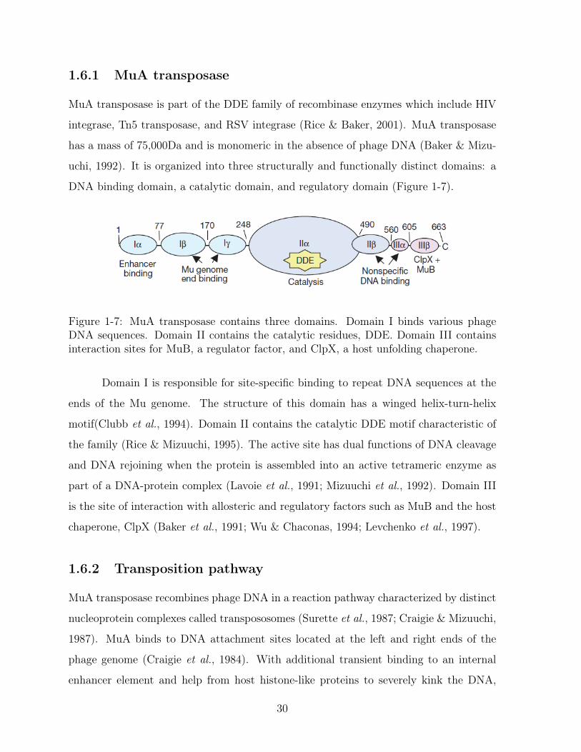
DNA binding domain, a catalytic domain, and regulatory domain (Figure 1-7).
Figure 1-7: MuA transposase contains three domains. Domain I binds various phageDNA sequences. Domain II contains the catalytic residues, DDE. Domain III containsinteraction sites for MuB, a regulator factor, and ClpX, a host unfolding chaperone.
Domain I is responsible for site-specific binding to repeat DNA sequences at the
ends of the Mu genome. The structure of this domain has a winged helix-turn-helix
motif(Clubb et al., 1994). Domain II contains the catalytic DDE motif characteristic of
the family (Rice & Mizuuchi, 1995). The active site has dual functions of DNA cleavage
and DNA rejoining when the protein is assembled into an active tetrameric enzyme as
part of a DNA-protein complex (Lavoie et al., 1991; Mizuuchi et al., 1992). Domain III
is the site of interaction with allosteric and regulatory factors such as MuB and the host
chaperone, ClpX (Baker et al., 1991; Wu & Chaconas, 1994; Levchenko et al., 1997).
1.6.2 Transposition pathway
MuA transposase recombines phage DNA in a reaction pathway characterized by distinct
nucleoprotein complexes called transpososomes (Surette et al., 1987; Craigie & Mizuuchi,
1987). MuA binds to DNA attachment sites located at the left and right ends of the
phage genome (Craigie et al., 1984). With additional transient binding to an internal
enhancer element and help from host histone-like proteins to severely kink the DNA,
30
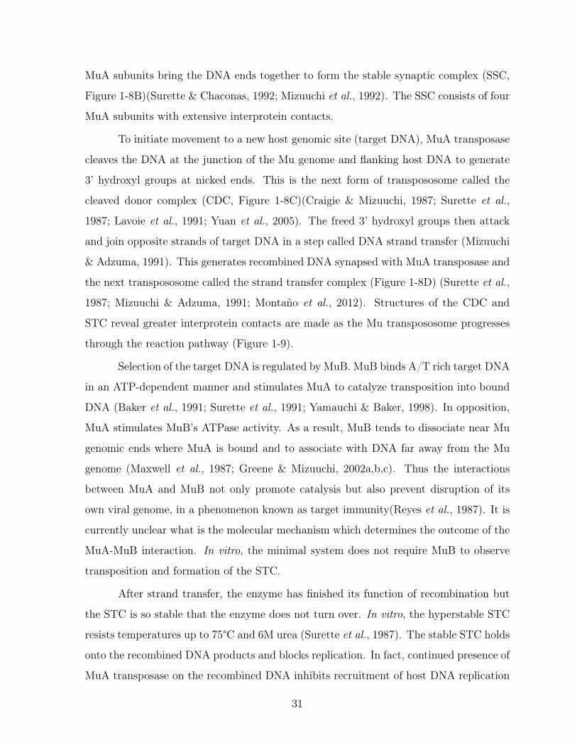
MuA subunits bring the DNA ends together to form the stable synaptic complex (SSC,
Figure 1-8B)(Surette & Chaconas, 1992; Mizuuchi et al., 1992). The SSC consists of four
MuA subunits with extensive interprotein contacts.
To initiate movement to a new host genomic site (target DNA), MuA transposase
cleaves the DNA at the junction of the Mu genome and flanking host DNA to generate
3’ hydroxyl groups at nicked ends. This is the next form of transpososome called the
cleaved donor complex (CDC, Figure 1-8C)(Craigie & Mizuuchi, 1987; Surette et al.,
1987; Lavoie et al., 1991; Yuan et al., 2005). The freed 3’ hydroxyl groups then attack
and join opposite strands of target DNA in a step called DNA strand transfer (Mizuuchi
& Adzuma, 1991). This generates recombined DNA synapsed with MuA transposase and
the next transpososome called the strand transfer complex (Figure 1-8D) (Surette et al.,
1987; Mizuuchi & Adzuma, 1991; Montaño et al., 2012). Structures of the CDC and
STC reveal greater interprotein contacts are made as the Mu transpososome progresses
through the reaction pathway (Figure 1-9).
Selection of the target DNA is regulated by MuB. MuB binds A/T rich target DNA
in an ATP-dependent manner and stimulates MuA to catalyze transposition into bound
DNA (Baker et al., 1991; Surette et al., 1991; Yamauchi & Baker, 1998). In opposition,
MuA stimulates MuB’s ATPase activity. As a result, MuB tends to dissociate near Mu
genomic ends where MuA is bound and to associate with DNA far away from the Mu
genome (Maxwell et al., 1987; Greene & Mizuuchi, 2002a,b,c). Thus the interactions
between MuA and MuB not only promote catalysis but also prevent disruption of its
own viral genome, in a phenomenon known as target immunity(Reyes et al., 1987). It is
currently unclear what is the molecular mechanism which determines the outcome of the
MuA-MuB interaction. In vitro, the minimal system does not require MuB to observe
transposition and formation of the STC.
After strand transfer, the enzyme has finished its function of recombination but
the STC is so stable that the enzyme does not turn over. In vitro, the hyperstable STC
resists temperatures up to 75°C and 6M urea (Surette et al., 1987). The stable STC holds
onto the recombined DNA products and blocks replication. In fact, continued presence of
MuA transposase on the recombined DNA inhibits recruitment of host DNA replication
31

Bacterial genome
integrated Mu genome
target site DNA
MuA
MuB
original Mu genome
Polymerase
replicated Mu genome
Stable Synaptic Complex(SSC)
Cleaved Donor Complex(CDC)
Strand Transfer Complex(STC)
remodeled fragile complex
ClpX remodelling
left end right end
replication
replication machinery
A
B
C
D
E
F
G H
32
machinery and thus lytic growth (Nakai & Kruklitis, 1995). At this critical transition,
phage Mu switches from its own proteins to ClpX, a host-encoded chaperone, to resolve
the replication block (Figure 1-8E).
1.6.3 Transpososome remodeling by ClpX
ClpX remodels the stable STC into a fragile complex which then recruits host replication
machinery to complete amplification of the phage genome (Figure 1-8F) (Levchenko
et al., 1995; Kruklitis et al., 1996; Jones et al., 1998). Deleting ClpX inhibits phage Mu
replication in vivo almost completely, but deleting ClpP has almost no effect (Mhammedi-
Alaoui et al., 1994). In fact full length ClpX is required for in vivo Mu replication as
ClpX lacking its N-terminal zinc-binding domain could not support phage lytic growth
(Wojtyra et al., 2003). Thus, it is the chaperone rather than degradation function that
is necessary for the Mu lytic cycle. ClpP is irrelevant for transpososome remodeling in
vivo. Purification of a host factor that enabled transpososome remodeling led to a single
protein fraction with high-specific activity which turned out to be ClpX (Levchenko et al.,
1995; Kruklitis et al., 1996).
Further in vitro studies of purified ClpX enzyme and transpososomes showed that
ClpX was sufficient to disassemble the stable STC and that the last eight C-terminal
residues comprised an intrinsic recognition tag (Levchenko et al., 1997). Transpososomes
biased to have only one subunit with a tag were sufficient to be destabilized by ClpX
(Burton et al., 2001). Highlighting that the unfolding process and not degradation of a
Figure 1-8 (preceding page): The in vivo replicative transposition of phage Mu beginswith an integrated Mu genome and proceeds through multiple nucleoprotein complexescalled transpososomes (A). MuA transposase binds to the left and right ends and bringsthem together to from the SSC while MuB binds to target site DNA (B). MuA trans-posase cleaves the DNA to form the CDC while MuB brings the target site closer (C).Recombination into target DNA occurs to form the STC (D). ClpX remodels the hy-perstable STC into a fragile complex by unfolding a MuA subunit (E). This remodeledfragile complex then recruits host replication machinery and remaining MuA subunitsare released (F). Replication of phage Mu DNA (G) results in two copies of the Mugenome integrated into the bacterial chromosome (H) and the transposition-replicationcycle repeats.
33
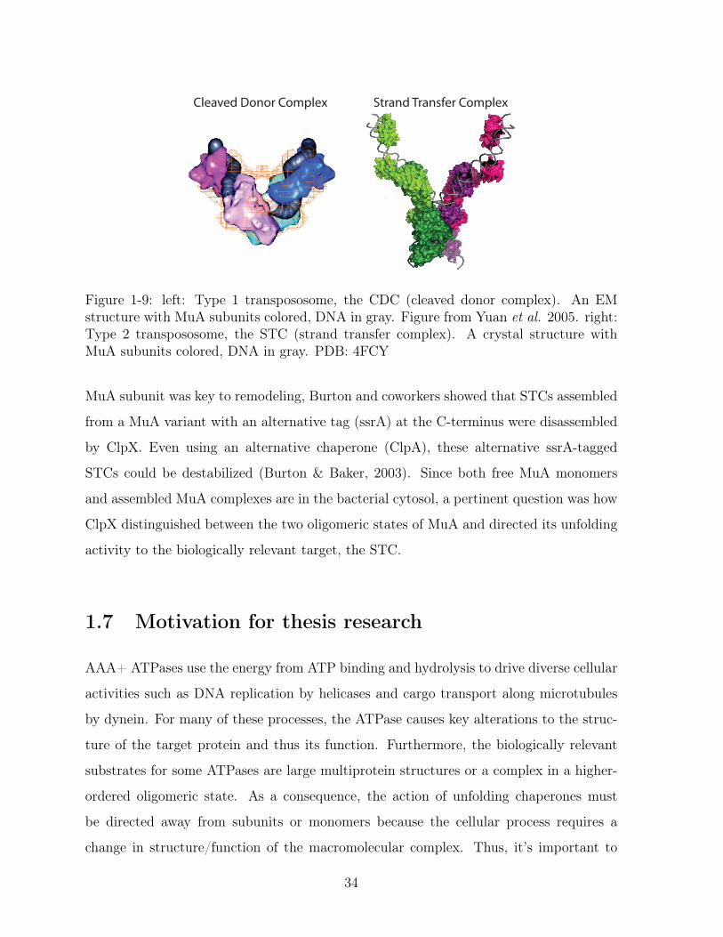
Cleaved Donor Complex Strand Transfer Complex
Figure 1-9: left: Type 1 transpososome, the CDC (cleaved donor complex). An EMstructure with MuA subunits colored, DNA in gray. Figure from Yuan et al. 2005. right:Type 2 transpososome, the STC (strand transfer complex). A crystal structure withMuA subunits colored, DNA in gray. PDB: 4FCY
MuA subunit was key to remodeling, Burton and coworkers showed that STCs assembled
from a MuA variant with an alternative tag (ssrA) at the C-terminus were disassembled
by ClpX. Even using an alternative chaperone (ClpA), these alternative ssrA-tagged
STCs could be destabilized (Burton & Baker, 2003). Since both free MuA monomers
and assembled MuA complexes are in the bacterial cytosol, a pertinent question was how
ClpX distinguished between the two oligomeric states of MuA and directed its unfolding
activity to the biologically relevant target, the STC.
1.7 Motivation for thesis research
AAA+ ATPases use the energy from ATP binding and hydrolysis to drive diverse cellular
activities such as DNA replication by helicases and cargo transport along microtubules
by dynein. For many of these processes, the ATPase causes key alterations to the struc-
ture of the target protein and thus its function. Furthermore, the biologically relevant
substrates for some ATPases are large multiprotein structures or a complex in a higher-
ordered oligomeric state. As a consequence, the action of unfolding chaperones must
be directed away from subunits or monomers because the cellular process requires a
change in structure/function of the macromolecular complex. Thus, it’s important to
34

understand how these enzymes discriminate and prioritize macromolecular complexes
over constituent subunits.
Using the extensively characterized Mu transpososome remodeling process, I elu-
cidated at the molecular level, the intrinsic recognition signal in transpososomes evolved
for disassembly. Then, I uncovered a mechanism used to discriminate between assembled
MuA complexes and constituent subunits. Additionally, I articulated the different roles
for the multiple intrinsic recognition tags in MuA transposase. Lastly, I engineered MuA
variants with different recognition tags to probe the effect of the tags themselves and of
the architecture of MuA complex on the holistic “ClpX remodeling” signal. Through this
work I present an underlying design framework for how AAA+ enzymes achieve speci-
ficity for macromolecular complexes, the biologically relevant targets of remodeling and
disassembly reactions.
35

36

Chapter 2
Design logic of a multivalent
recognition signal confers preferential
complex disassembly by the AAA+
unfoldase ClpX
This chapter has been written as a manuscript for publication. A draft is currently being
reviewed by collaborators. I performed all experiments for all figures except Figure2-10,
which was contributed by A. Abdelhakim. I.Levchenko synthesized the peptides used in
Figure 2-5. S.P. Montano and P.A. Rice designed and cloned the initial Sin15Mu chimeric
protein and Sin-attachment site DNA oligo, both of which I modified for research in this
chapter.
37

2.1 Abstract
AAA+ enzymes are present in all kingdoms and use the chemical energy of ATP to
remodel protein complexes and catalyze substrate protein unfolding. How these power-
ful enzymes recognize protein complexes and aggregates is poorly understood. Efficient
selection of multimeric protein complexes over constituent subunits is key to success-
ful disassembly. Here, we use E.coli ClpX, a AAA+ unfoldase, and the tetrameric
MuA transpososome, to investigate how preferential specificity for an assembled com-
plex is achieved. We demonstrate that the MuA tetramer employs a multivalent set
of recognition peptides to ensure that the complex has the tightest affinity for ClpX.
The critical recognition components are the weak ClpX pore-binding peptide at the C-
terminus of MuA and a second peptide ∼ 40 residues away from the pore signal that
binds the ClpX N-terminal domain. By constructing chimeric SinMuA proteins with
altered DNA-binding specificity we investigated multiple variant complexes carrying dif-
ferent geometries of mutant signals and determined how each subunit contributes to
complex-specific recognition. Although individually, the two key recognition peptides
bind weakly (70 − 400𝜇M) to ClpX , together within the assembled MuA tetramer they
impart an affinity of ∼1𝜇M. All four subunits in the tetramer can donate the ClpX N-
domain-binding peptide and optimal recognition is achieved when all four are present. In
contrast, only two specifically located subunits can donate the pore-binding signal. Im-
portantly, the N-domain-binding peptides become unnecessary for complex recognition
when the native weak pore-binding signal is replaced with a much stronger compact pore-
binding tag. Thus, we conclude that the design of signals that are specific for assembled
complexes depends on collaboration between multiple weak protein-unfoldase-interaction
peptides and that strong-binding signals can prevent multimer-specific recognition.
38

2.2 Introduction
Cells are densely packed with proteins performing structural and/or enzymatic roles es-
sential for life. To help respond to environmental changes, manage protein turnover and
protein quality control, these cells employ energy-dependent unfoldases/disaggregases
and proteases from the AAA+ family (ATPases associated with various cellular activ-
ities). Powered by cycles of nucleotide binding, hydrolysis, and release, these ATPases
remodel complexes, solubilize aggregates, and degrade proteins (when coupled with part-
ner peptidases). E. coli ClpX is arguably the best-characterized AAA+ unfoldase and is
known to disassemble complexes and unfolds proteins (Sauer & Baker, 2011). ClpX acts
alone as a protein-remodeling enzyme as well as in complex with ClpP peptidase to make
the ClpXP protease. In ClpXP, ClpX recognizes and unfolds many substrate proteins
and translocates the unfolded chain to ClpP peptidase where it is degraded. Because
of its destructive power, ClpX’s selection of substrates must be exquisite. The protein
signals (recognition sequences) and design logic governing recognition of different classes
of substrates is being actively investigated.
Most bacteria have no ubiquitination system and recognition of protein targets
for degradation or disassembly is mediated by a diverse set of short unstructured peptide
sequences, or tags. These recognition tags are often located at the termini of otherwise
native substrate proteins (Sauer et al., 2004). Examples of substrates with N-terminal
recognition tags are 𝜆O, a DNA replication origin-binding protein from phage 𝜆, and
UmuD, a subunit of DNA polymerase V, a DNA-repair/tolerance polymerase (Gonciarz-
swiatek et al., 1999; Gonzalez et al., 2000) and proteins recognized by the N-end rule
pathway (Varshavsky, 1996). The best characterized tag is for one class of substrates:
truncated polypeptides from stalled ribosomes. These incompletely translated products
are marked at their C-termini with an 11 amino acid sequence called the ssrA tag and
targeted for degradation principally by ClpXP (Gottesman et al., 1998). However, some
substrates may have more complicated, multicomponent recognition signals. A screen of
in vivo ClpXP substrates revealed many target proteins carried multiple ClpX-recognition
sequences. Furthermore, the screen also indicated that many ClpX substrates ( 60%) are
39

multimeric or subunits in multiprotein complexes (Flynn et al., 2003). For substrates in
multiprotein complexes that are remodeled or disassembled by ClpX, how the enzyme
distinguishes between assembled complexes versus subunits is not understood.
To investigate how preference for an assembled complex is achieved over con-
stituent subunits, we used the MuA transposase, a natural disassembly substrate of
ClpX. Phage Mu duplicates its genome by replicative transposition. During transposi-
tion, MuA binds DNA sites located at the ends of the Mu genome, forms a tetramer
that brings the two ends of the DNA together, and catalyzes the DNA cleavage and join-
ing reactions core to transposition (Craigie et al., 1984; Kuo et al., 1991; Lavoie et al.,
1991). This recombination phase of transposition does not require external energy (such
as ATP) and is driven forward by proceeding through a series of nucleoprotein complexes
(transpososomes) that increase in stability to a final hyperstable DNA product-bound
transpososome (Surette et al., 1987). Remodeling converts the hyperstable transposo-
some (MuA complex) into a fragile complex, which facilitates both disassembly and the
recruitment of DNA-replication machinery (Levchenko et al., 1995; Nakai & Kruklitis,
1995; Jones et al., 1998). Completion of a reaction cycle therefore requires that the stable
MuA complex be remodeled by ClpX (Mhammedi-Alaoui et al., 1994).
MuA transpososome assembly, recombination, and ClpX remodeling have all been
reconstituted in vitro. On a native agarose gel, the stable transpososome is observed as
a slower migrating band (asterisk, lane 2) as compared to supercoiled substrate plasmid
(arrow, lane 1) (Figure 2-1A). In contrast, the fragile complex is unstable to gel elec-
trophoresis and the liberated DNA transposition products are visible as a characteristic
series of topoisomerases (Figure 2-1B, bracket). As both monomeric and DNA-bound
tetrameric MuA are present in the cytoplasm we sought a molecular understanding to
explain how ClpX recognized the transpososome as a high-priority target.
Previous analysis revealed that there was information throughout MuA protein
that guided ClpX recognition and that information in domain III is most central. MuA
contains a C-terminal sequence (RRKKAI) that is necessary for ClpX recognition of both
monomeric MuA and assembled transpososomes (Levchenko et al., 1995, 1997; Abdel-
hakim et al., 2008). It is also established that interaction between the transpososome
40

C. ClpX-catalyzed degradation
MuA
ClpXPSDS-PAGEtime (min)
0 2 4 6 8 10 15
MuA
MuA
L2 L1 R1 R2
pMini-Mu
1 supercoiledplasmid (sc)
+
Transpososome(Tr)
2
A. Transpososome assembly
≈ ≈
R1L1
L2R2 ≈
≈ R1
R2
≈ ≈
≈ ≈
L1
L2
targetDNA
B. ClpX-catalyzed disassembly
Fragile Complex recombinedDNA products
3
ClpXelectrophoresis
Transpososome2
≈≈ R1
R2
≈ ≈
≈ ≈
L2
≈
≈
≈
≈
≈
≈
≈
≈ R1
R2
≈ ≈
≈ ≈
L1
L2
DNALadder
*
1 2
* 3
Native agarose geltime (min)
0.5 1 2 3 +SDS
sc scTr
Native agarose gel
Figure 2-1: In vitro assays for Mu complex assembly and recognition by ClpXA. MuA transposase monomers and host protein HU are incubated with a plasmid sub-strate (“pMini-Mu”) containing “left” and “right” phage Mu attachment sites ( L1, L2, R1,R2). Mu catalyzes DNA cleavage and recombination with target DNA to form the trans-pososome, a stable complex. When visualized on a native agarose gel, the transpososomeappears as a band (“Tr”, asterisk) that migrates slower than supercoiled plasmid alone(“sc”, black arrow)B. ClpX remodels the transpososome (MuA complex) by unfolding a subunit bound toL1 or R1 attachment site to produce the fragile complex. The fragile complex falls apartduring gel electrophoresis and produces a stereotypical series of recombined topoisomers(white arrows). Addition of SDS disrupts all inter-protein and protein-DNA interactionswithin the MuA complex and serves as the “100% disassembly” control. Rates of MuAcomplex disassembly by ClpX were assayed by measuring the rate of appearance of thelowermost disassembly DNA product on a native agarose gel.C. Schematic of monomeric MuA degradation by ClpXP protease. Rates of proteindegradation were assayed by measuring the rate of disappearance of MuA on SDS-PAGE.
41

and the unfoldase is not simply due to avidity contributed by the four C-terminal tags
(Mu pore-binding tag). This conclusion is supported by the findings that additional mu-
tations in MuA domain III antagonize only MuA transpososome disassembly by ClpX
and not degradation (Abdelhakim et al., 2008). Furthermore, the N-domain of ClpX is
exceedingly important for transpososome remodeling but has little role in MuA monomer
degradation (Abdelhakim et al., 2008).
As previous studies established that ClpX recognizes short peptide-like signals, we
characterized whether the remodeling-specific protein-protein contacts were also peptide-
like or not. Second, we sought to understand at the molecular level how MuA assembly
into a DNA-bound tetramer modulates recognition by ClpX. Because recognition de-
pends on architectural features of the complex, we designed a hybrid Mu protein with
novel DNA-binding specificity to assist in specifically placing subunits with altered recog-
nition tags within assembled complexes. Lastly, we modulated the binding affinity of the
C-terminal tag to understand the extent of cooperation among the different recognition
peptides that together comprise the Mu transpososome remodeling signal “recognon”.
Thus, here we establish molecular interactions between MuA and ClpX that enable ClpX
to preferentially target the assembled Mu tetramer. This work also elucidates principles
attractive to the general problem of designing recognition mechanisms that favor assem-
bled, multimeric protein complexes.
42
2.3 Results
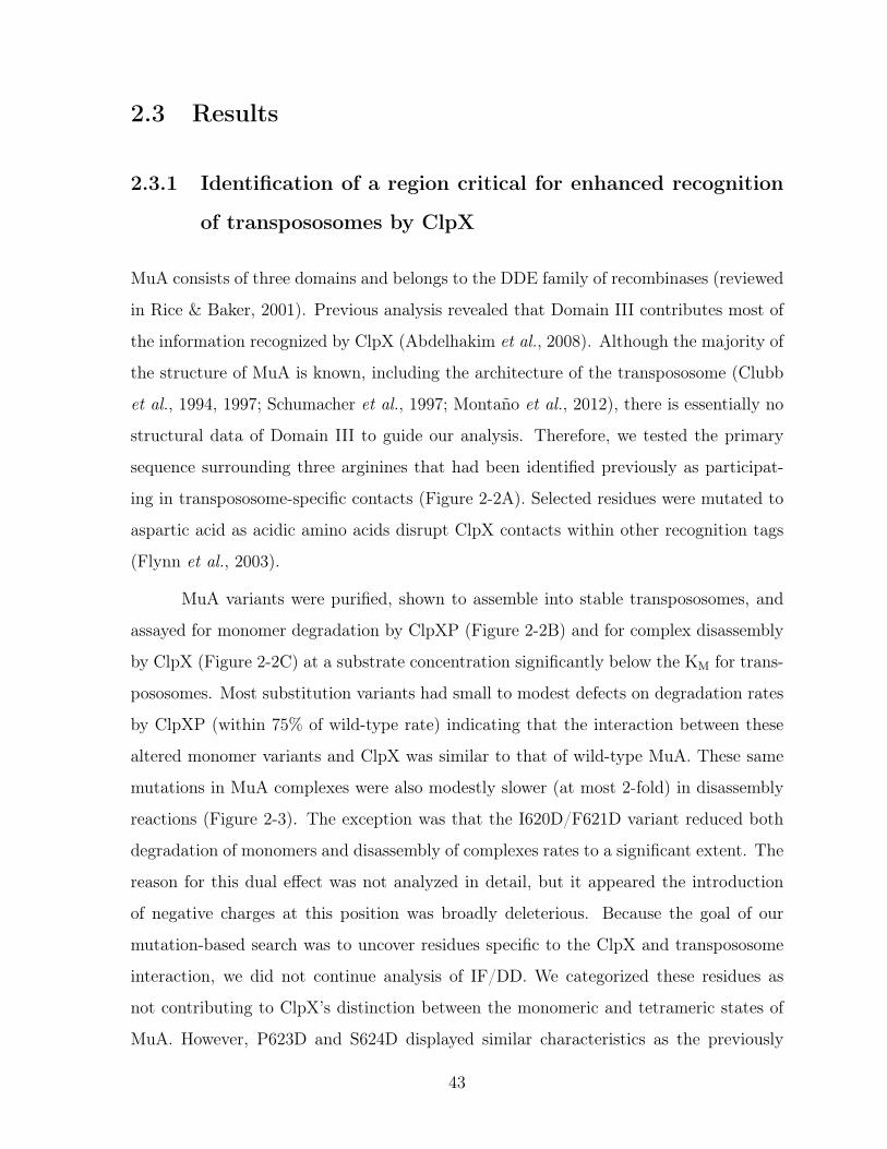
2.3.1 Identification of a region critical for enhanced recognition
of transpososomes by ClpX
MuA consists of three domains and belongs to the DDE family of recombinases (reviewed
in Rice & Baker, 2001). Previous analysis revealed that Domain III contributes most of
the information recognized by ClpX (Abdelhakim et al., 2008). Although the majority of
the structure of MuA is known, including the architecture of the transpososome (Clubb
et al., 1994, 1997; Schumacher et al., 1997; Montaño et al., 2012), there is essentially no
structural data of Domain III to guide our analysis. Therefore, we tested the primary
sequence surrounding three arginines that had been identified previously as participat-
ing in transpososome-specific contacts (Figure 2-2A). Selected residues were mutated to
aspartic acid as acidic amino acids disrupt ClpX contacts within other recognition tags
(Flynn et al., 2003).
MuA variants were purified, shown to assemble into stable transpososomes, and
assayed for monomer degradation by ClpXP (Figure 2-2B) and for complex disassembly
by ClpX (Figure 2-2C) at a substrate concentration significantly below the KM for trans-
pososomes. Most substitution variants had small to modest defects on degradation rates
by ClpXP (within 75% of wild-type rate) indicating that the interaction between these
altered monomer variants and ClpX was similar to that of wild-type MuA. These same
mutations in MuA complexes were also modestly slower (at most 2-fold) in disassembly
reactions (Figure 2-3). The exception was that the I620D/F621D variant reduced both
degradation of monomers and disassembly of complexes rates to a significant extent. The
reason for this dual effect was not analyzed in detail, but it appeared the introduction
of negative charges at this position was broadly deleterious. Because the goal of our
mutation-based search was to uncover residues specific to the ClpX and transpososome
interaction, we did not continue analysis of IF/DD. We categorized these residues as
not contributing to ClpX’s distinction between the monomeric and tetrameric states of
MuA. However, P623D and S624D displayed similar characteristics as the previously
43
1 77 243 490 575 615 663Ia Ib IIa IIb IIIa IIIb
Domain I Domain II Domain III
DNA binding Catalysis MuA tag ( ClpX recognition )
P A A P E S R I V G I F R P S G N T E R V K N Q E R D D E Y E T E R D E Y L N H S L D I L E Q N R R K K A I
610635 663
634
A. MuA domain structure
C. Complex Disassembly
WT
PS
*
time (min)0 2 4 6 8
*
+SDS
0
0.2
0.4
0.6
0.8
0 2 4 6 8 10
WTIVG
IFPS
GN
DILEQN
time (min)
Frac
tion
disa
ssem
bled 0.1µM ClpX6
0.2
0.4
0.6
0.8
1.0
0 2 4 6 8 10
WT
GNDILPS
time (min)
Frac
tion
of s
tart
WTPS
0 2 4 6 8time (min)
B. Monomer degradation
0.3µM ClpX6
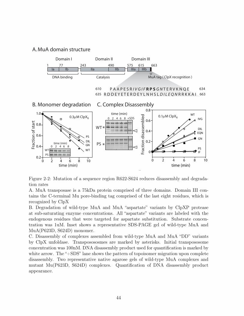
E. Figure 2-2: Mutation of a sequence region R622-S624 reduces disassembly and degrada-tion ratesA. MuA transposase is a 75kDa protein comprised of three domains. Domain III con-tains the C-terminal Mu pore-binding tag comprised of the last eight residues, which isrecognized by ClpXB. Degradation of wild-type MuA and MuA “aspartate” variants by ClpXP proteaseat sub-saturating enzyme concentrations. All “aspartate” variants are labeled with theendogenous residues that were targeted for aspartate substitution. Substrate concen-tration was 1uM. Inset shows a representative SDS-PAGE gel of wild-type MuA andMuA(P623D, S624D) monomer.C. Disassembly of complexes assembled from wild-type MuA and MuA “DD” variantsby ClpX unfoldase. Transposososmes are marked by asterisks. Initial transpososomeconcentration was 100nM. DNA disassembly product used for quantification is marked bywhite arrow. The “+SDS” lane shows the pattern of topoisomer migration upon completedisassembly. Two representative native agarose gels of wild-type MuA complexes andmutant Mu(P623D, S624D) complexes. Quantification of DNA disassembly productappearance.
44

0
20
40
60
80
100
WT Δ8 IVG IF R PS GN DIL EQN617 620 622 623 625 653 656
DD DDDDDDD DDD DDDA
Rate
rela
tive
to W
T (%
)
Monomer degradation
Complex disassembly
Figure 2-3: Comparison of all reaction rates for Mu aspartate variantsQuantification of differences in degradation and disassembly rates of MuA variants whoseindicated sequences were mutated to alanine or aspartic acid relative to wild-type MuA.Reactions with error bars were performed in triplicate. Error bars are the standard errorof the mean.
identified transpososome-specific contact residue R622 in which substitution specifically
slowed disassembly rates by as much as 10-fold at the sub-saturating protein concentra-
tions (Figure 2-3).
We determined the functional interaction of ClpX with double mutant (P623D
S624D) variant complexes during disassembly by measuring the concentration of enzyme
for half-maximal velocity (KM). Because it is difficult to obtain transpososomes at high
concentration, we started with a fixed substrate concentration, varied the concentration
of ClpX, measured the rate of appearance of DNA transposition product released by
disassembly, and analyzed these data as previously described to obtain apparent KM
values (Pyle & Green, 1994; Abdelhakim et al., 2008). For many ClpX substrates, the
KM is nearly equivalent to the KD because catalysis is relatively slower that the binding
reactions. Therefore the apparent KM is a measure of the functional affinity of ClpX for
transpososomes. The apparent KM for disassembly of PS→DD double-mutant complexes
was about 8-fold weaker than that of wild-type MuA complex (Figure 2-4). Additionally
45
0
0.5
1
1.5
2
2.5
0 5 10 15 20 25
WT
PS
ClpX (µΜ)6
Reac
tion
Rate
(min
-1) KM
app V appmax
( µM ) (min )
10.6 2.7 ±
1.4 0.2 ± 2.7 0.1 ±
0.6 0.1 ±
WT
PS
−1
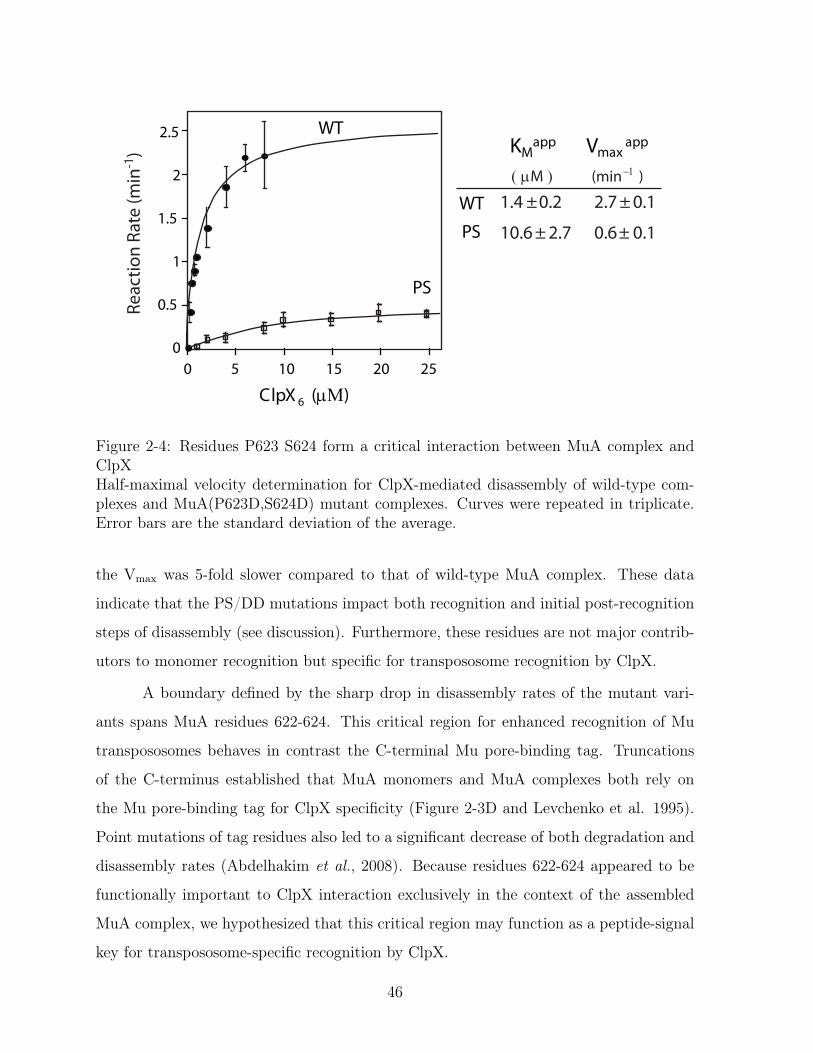
Figure 2-4: Residues P623 S624 form a critical interaction between MuA complex andClpXHalf-maximal velocity determination for ClpX-mediated disassembly of wild-type com-plexes and MuA(P623D,S624D) mutant complexes. Curves were repeated in triplicate.Error bars are the standard deviation of the average.
the Vmax was 5-fold slower compared to that of wild-type MuA complex. These data
indicate that the PS/DD mutations impact both recognition and initial post-recognition
steps of disassembly (see discussion). Furthermore, these residues are not major contrib-
utors to monomer recognition but specific for transpososome recognition by ClpX.
A boundary defined by the sharp drop in disassembly rates of the mutant vari-
ants spans MuA residues 622-624. This critical region for enhanced recognition of Mu
transpososomes behaves in contrast the C-terminal Mu pore-binding tag. Truncations
of the C-terminus established that MuA monomers and MuA complexes both rely on
the Mu pore-binding tag for ClpX specificity (Figure 2-3D and Levchenko et al. 1995).
Point mutations of tag residues also led to a significant decrease of both degradation and
disassembly rates (Abdelhakim et al., 2008). Because residues 622-624 appeared to be
functionally important to ClpX interaction exclusively in the context of the assembled
MuA complex, we hypothesized that this critical region may function as a peptide-signal
key for transpososome-specific recognition by ClpX.
46

2.3.2 A peptide encompassing the critical region interacts with
the N-terminal zinc-binding domain of ClpX
Recognition of Mu transpososomes is strongly influenced by the presence of the N-
terminal zinc-binding domain (the N-domain) of ClpX (Abdelhakim et al., 2008). The
N-domain binds peptide sequences donated by adaptor proteins, which assist ClpX in
recognizing some substrates. For example, the adaptor protein, SspB, enhances degrada-
tion of ssrA-tagged substrates by ClpXP (Levchenko et al., 2000). The key interaction
for this stimulation is a C-terminal sequence of SspB, known as the ClpX Binding (XB)
peptide that binds the N-domain of ClpX (Wah et al., 2003; Song & Eck, 2003). Because
Mu transpososome is a multimeric complex and depends on the N-domain for efficient
disassembly, we hypothesized that one or more MuA subunit(s) may provide adaptor-like
contacts to enhance recognition of MuA complexes by ClpX.
In the previous section we provide evidence for a second ClpX-binding sequence in
MuA, beyond the C-terminal Mu pore-binding tag, which functions in ClpX’s enhanced
recognition of the MuA complex. If the critical region (residues 622-624) behaves as an
adaptor-like signal similar to the XB peptide, then we expect this sequence to bind to
N-domain of ClpX.
We tested for a direct interaction between the critical region of MuA and the N-
domain using both peptide blot and solution binding assays. We designed a MuA peptide
blot, wherein each “spot” was a 20 amino acid sequence from MuA, and each neighboring
spot was a related peptide, shifted three residues over (C-terminal). In this manner the
entire sequence of MuA domain III could be tested for binding in one experiment. The
blot was probed with S35 radiolabeled N-domain. By this semi-qualitative measure, we
observed binding at spots in three major regions of domain III (Figure 2-5A). One of
these regions contained sequences that corresponded to the critical region identified by
mutagenesis in section 1 (Figure 2-5A, purple box).
To test interaction between this MuA peptide and the ClpX N-domain in solution,
an 18-amino acid peptide (Mu-peptide 614-633) containing the critical region 622-624 was
synthesized and labeled with fluorescein at its N-terminus. When assayed by fluorescence
47

A A G R E Y R R R Q K Q L K S A T K A A
APeptide BlotMu Domain III
B56
0 L
563
V
566
N
569
A
572
R
575
R
578
Q
581
L
584
A
587
A
590
K
620
I
617
I
614
E
611
A
608
A
593
K
596
D
599
E
602
E
605
P
644
D
641
T
638
E
635
R
632
N
629
R
626
N
623
P
E S R I V G I F R P S G N T E R V K N Q
XB p
eptid
e
D E Y L N H S L D I L E Q N R R K K A I
X X X X X X X X
644
569
614
Tr. enhancement peptide
XB peptide
ESRIVGIFRPSGNTERVKNQ
RGGRPALRVVKKD=13± 3µΜ
KD≈ 3000µΜAni
sotr
opy
(a.u
.)
N-domain of ClpX (µΜ)
KD= 380 ± 90µΜ
Mutated control peptideESRIVGIFDDDGNTERVKNQ
0.04
0.08
0.12
0 500 1000
Figure 2-5: The N-terminal zinc-binding domain of ClpX binds to the enhancementpeptideA. Peptide array of sequences from Domain III of MuA (residues 560-663). Each spotrepresents a 20-amino-acid-long peptide shifted by three residues. The first amino acidof each peptide is labeled with the position number and single letter. Spots marked withan X have no peptides. The XB peptide serves as a positive control and is derived froma known ClpX-binding sequence in the adapter protein, SspB. Blot was probed withradiolabelled S35 N-domain of ClpX. The sequence of a peptide spot from each of threeregions is shown.B. Solution binding of N-terminal Fluorescein-labeled peptides and purified ClpX N-domain. A fixed amount (200nM) of Fluorescein-labeled peptides with the indicatedsequences were incubated with increasing concentrations of purified N-domain. Errorsrepresent standard deviation of the average.
48

anisotropy, the binding of N-domain with peptide, albeit weak (∼400𝜇M), was observed
(Figure 2-5B). For comparison, the XB peptide binds to the N-domain with a much
tighter affinity, KD ∼13𝜇M. Mutation of the genetically-identified RPS residues (622-624)
to aspartates in the Mu-peptide 614-633 essentially abolished binding to the N-domain,
verifying that the interaction between the critical region of MuA and the N-domain was
specific, although weak. These results establish that the critical region is an N-domain-
binding signal in addition to its transpososome-specific signaling attribute. These two
features of the critical region suggest that MuA complex makes adapter-like contacts with
ClpX. We will refer to the critical region as the N-domain-binding tag or the Enhancement
tag for its property of imparting enhanced recognition of MuA complex.
The above results, taken together with previous data establish that the natural
remodeling signal for transpososomes contains at least two distinct peptide tags recog-
nized by ClpX. These two type of tags are each present on all four subunits in MuA
complex. The geometry of this cohort of eight tags in the context of the assembled
MuA complex produces the remodeling signal that allows ClpX to discriminate trans-
pososomes from monomers. However, previous work hinted that this cohort of eight tags
do not contribute equally to the remodeling signal. Transpososomes with only one active
MuA77-663 subunit carrying the pore-binding tag and three tag-truncated subunits are
remodeled by ClpX (Burton et al., 2001). Furthermore, one subunit out of the four within
the transpososome carrying the pore-binding tag was sufficient to allow ClpX-mediated
remodeling (Abdelhakim et al., 2010). To continue to understand transpososome recog-
nition, we addressed how the architecture of MuA complexes affects the Enhancement
tag’s contribution to the remodeling signal.
2.3.3 Step-wise loss of the Enhancement tag trends with ClpX’s
weaker affinity for complexes
A Mu transpososome has rotational symmetry around its vertical axis. However the sub-
units in the complex adopt two very different conformations and thus form two classes
of subunits (Yuan et al., 2005; Montaño et al., 2012). Class 1 are the catalytic subunits;
49
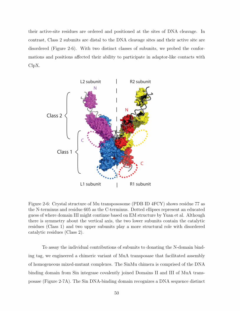
their active-site residues are ordered and positioned at the sites of DNA cleavage. In
contrast, Class 2 subunits are distal to the DNA cleavage sites and their active site are
disordered (Figure 2-6). With two distinct classes of subunits, we probed the confor-
mations and positions affected their ability to participate in adaptor-like contacts with
ClpX.
Class 1
Class 2
N
C
N
C
R1 subunit
R2 subunit
L1 subunit
L2 subunit
Figure 2-6: Crystal structure of Mu transpososome (PDB ID 4FCY) shows residue 77 asthe N-terminus and residue 605 as the C-terminus. Dotted ellipses represent an educatedguess of where domain III might continue based on EM structure by Yuan et al. Althoughthere is symmetry about the vertical axis, the two lower subunits contain the catalyticresidues (Class 1) and two upper subunits play a more structural role with disorderedcatalytic residues (Class 2).
To assay the individual contributions of subunits to donating the N-domain bind-
ing tag, we engineered a chimeric variant of MuA transposase that facilitated assembly
of homogeneous mixed-mutant complexes. The SinMu chimera is comprised of the DNA
binding domain from Sin integrase covalently joined Domains II and III of MuA trans-
posase (Figure 2-7A). The Sin DNA-binding domain recognizes a DNA sequence distinct
50

Sin R1 R1 Sin
pSinRRSin
Chimeric Complex
MuA
≈ ≈
≈ ≈
SinMu+ MuA
SinMu
MuA : SinMu
1:11:0 0:1
*
+SDS
plas
mid
SinMu chimera253 575 615 663
Sin (147-200)His6 domain 2 domain 3
A.
B.
C.
Figure 2-7: Tools for making homogeneous mixed mutant complexesA. Domain structure of the SinMu chimera. The chimera has the DNA binding domainof Sin recombinase substituting for Mu DNA binding domain.B. Schematic of assembly of chimeric SinMu complexes on the altered- specificity plasmidsubstrate pSinRRSin. pSinRRSin has the Mu attachment sites, L2 and R2, swapped outfor Sin-specific DNA binding sites.C. Assembly of chimeric complexes on pSinRRSin plasmid requires the correct ratio ofMuA and SinMu proteins. Lane 1 contains un-reacted pSinRRSin supercoiled plasmid(black arrow). A native agarose gel of the in vitro assembly reaction shows the bandassociated with assembled complexes (asterisk) in lane 4 with the correct 1:1 proteinratio, but not in lanes 2 and 3 which have incorrect protein ratios. The characteristicpattern of recombined DNA disassembly products (white arrow) can be seen with additionof SDS in lane 5.
from that of the MuA DNA binding domain. We derived a set of altered-specificity plas-
mids from the original pMiniMu by substituting Mu attachment sites individually for the
Sin-specific DNA sequences (Figure 2-7B).
Assembly of chimeric transpososomes onto a pSinRRSin plasmid depended on
the presence of both MuA and SinMu proteins. Transposososmes assembled efficiently
on two Mu DNA (or hybrid) right ends and symmetrical complexes have been widely
51
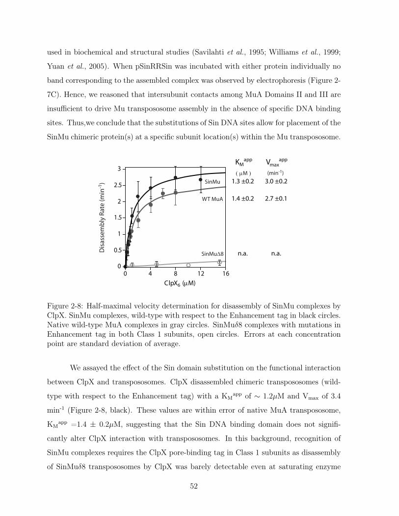
used in biochemical and structural studies (Savilahti et al., 1995; Williams et al., 1999;
Yuan et al., 2005). When pSinRRSin was incubated with either protein individually no
band corresponding to the assembled complex was observed by electrophoresis (Figure 2-
7C). Hence, we reasoned that intersubunit contacts among MuA Domains II and III are
insufficient to drive Mu transpososome assembly in the absence of specific DNA binding
sites. Thus,we conclude that the substitutions of Sin DNA sites allow for placement of the
SinMu chimeric protein(s) at a specific subunit location(s) within the Mu transpososome.
ClpX6 (µM)
Dis
asse
mbl
y Ra
te (m
in-1
)
0
0.5
1
1.5
2
2.5
3
0 4 8 12 16
WT MuA
SinMu
SinMu∆8
KMapp V app
max
( µM ) (min-1)3.0 ±0.21.3 ±0.2
2.7 ±0.11.4 ±0.2
n.a.n.a.
Figure 2-8: Half-maximal velocity determination for disassembly of SinMu complexes byClpX. SinMu complexes, wild-type with respect to the Enhancement tag in black circles.Native wild-type MuA complexes in gray circles. SinMu𝛿8 complexes with mutations inEnhancement tag in both Class 1 subunits, open circles. Errors at each concentrationpoint are standard deviation of average.
We assayed the effect of the Sin domain substitution on the functional interaction
between ClpX and transpososomes. ClpX disassembled chimeric transpososomes (wild-
type with respect to the Enhancement tag) with a KMapp of ∼ 1.2𝜇M and Vmax of 3.4
min-1 (Figure 2-8, black). These values are within error of native MuA transpososome,
KMapp =1.4 ± 0.2𝜇M, suggesting that the Sin DNA binding domain does not signifi-
cantly alter ClpX interaction with transpososomes. In this background, recognition of
SinMu complexes requires the ClpX pore-binding tag in Class 1 subunits as disassembly
of SinMu𝛿8 transpososomes by ClpX was barely detectable even at saturating enzyme
52

concentrations (Figure 2-8, dark gray).
0
0.5
1
1.5
2
2.5
3
0 10 20
KMapp V app
max( µM )
4.6 ± 1.6 2.2 ± 0.2
4.4 ± 1.7 1.1 ± 0.1
11.5 ± 1.3 0.5 ± 0.02
ClpX6 (µM)
(min )−1
Dis
asse
mbl
y Ra
te (m
in-1
)
PS PS
PS PS
PS PS
PS PS
3.0 ±0.21.3 ±0.2
SinMu ∆8
SinMu“wild type”
PS in Class 1
PS in Class 2
PS in all four
Figure 2-9: All four subunits can provide the Enhancement tag in MuA complexes.Half-maximal velocity determination for disassembly of chimeric complexes with differentnumbers of subunits mutated at the Enhancement tag (PS/DD). Curves were repeatedin triplicate. Error bars at each concentration point are the standard deviation of theaverage. Errors of the KM are standard deviation of average of three fits.
We also mutated the Enhancement tag using the double mutation, P623D/S624D,
in Class 1 subunits, Class 2 subunits, and in all four subunits. The mutant subunits
in either Class 1 catalytic or Class 2 structural subunits resulted in a weaker KMapp,
4.5𝜇M and 4.4𝜇M, respectively (Figure 2-9). The similar magnitude of these KMapp
values indicated that the two conformations do not differently present the Enhancement
tag. Thus, the Enhancement tag is accessible by ClpX (at least in this conformation of
the transpososome) from either class of subunits. Only when the Enhancement tag is
mutated in all four subunits was the weakest apparent affinity observed, KMapp = 11.5
± 1.5𝜇M (Figure 2-9, black triangles). This value is striking because the KMapp of the
chimeric complex with all four mutant subunits is within error of the KMapp of wild-type
Mu monomers (10.5 ± 2.7 𝜇M) (Abdelhakim et al., 2008), revealing that the 10-fold
53
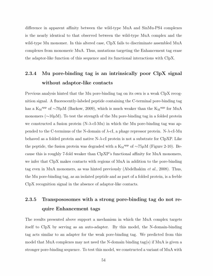
difference in apparent affinity between the wild-type MuA and SinMu-PS4 complexes
is the nearly identical to that observed between the wild-type MuA complex and the
wild-type Mu monomer. In this altered case, ClpX fails to discriminate assembled MuA
complexes from monomeric MuA. Thus, mutations targeting the Enhancement tag erase
the adaptor-like function of this sequence and its functional interactions with ClpX.
2.3.4 Mu pore-binding tag is an intrinsically poor ClpX signal
without adaptor-like contacts
Previous analysis hinted that the Mu pore-binding tag on its own is a weak ClpX recog-
nition signal. A fluorescently-labeled peptide containing the C-terminal pore-binding tag
has a KMapp of ∼70𝜇M (Barkow, 2009), which is much weaker than the KM
app for MuA
monomers (∼10𝜇M). To test the strength of the Mu pore-binding tag in a folded protein
we constructed a fusion protein (N-𝜆-cI-Mu) in which the Mu pore-binding tag was ap-
pended to the C-terminus of the N-domain of 𝜆-cI, a phage repressor protein. N-𝜆-cI-Mu
behaved as a folded protein and native N-𝜆-cI protein is not a substrate for ClpXP. Like
the peptide, the fusion protein was degraded with a KMapp of ∼75𝜇M (Figure 2-10). Be-
cause this is roughly 7-fold weaker than ClpXP’s functional affinity for MuA monomers,
we infer that ClpX makes contacts with regions of MuA in addition to the pore-binding
tag even in MuA monomers, as was hinted previously (Abdelhakim et al., 2008). Thus,
the Mu pore-binding tag, as an isolated peptide and as part of a folded protein, is a feeble
ClpX recognition signal in the absence of adaptor-like contacts.
2.3.5 Transpososomes with a strong pore-binding tag do not re-
quire Enhancement tags
The results presented above support a mechanism in which the MuA complex targets
itself to ClpX by serving as an auto-adapter. By this model, the N-domain-binding
tag acts similar to an adapter for the weak pore-binding tag. We predicted from this
model that MuA complexes may not need the N-domain binding tag(s) if MuA is given a
stronger pore-binding sequence. To test this model, we constructed a variant of MuA with
54
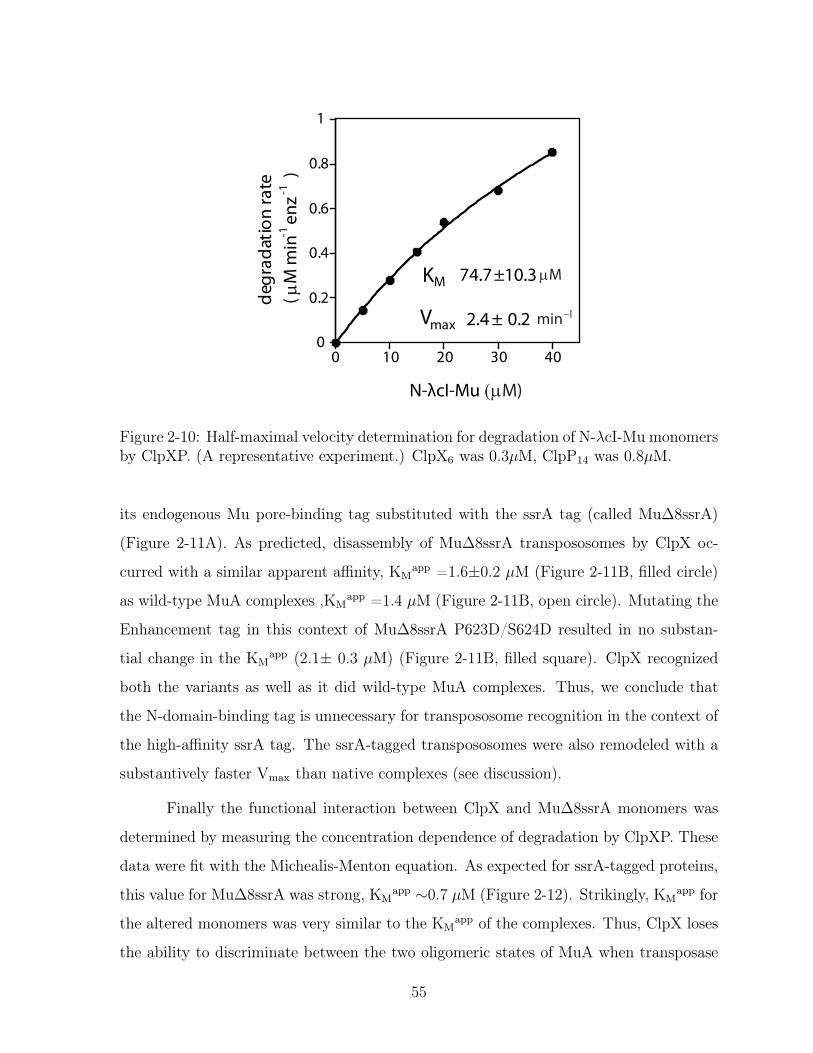
0
0.2
0.4
0.6
0.8
1
0 10 20 30 40
N-λcI-Mu (µM)
degr
adat
ion
rate
( µM
min
-1 e
nz -1
)KM
Vmax
µM
2.4 ± 0.2
74.7±10.3
min−1
Figure 2-10: Half-maximal velocity determination for degradation of N-𝜆cI-Mu monomersby ClpXP. (A representative experiment.) ClpX6 was 0.3𝜇M, ClpP14 was 0.8𝜇M.
its endogenous Mu pore-binding tag substituted with the ssrA tag (called Mu∆8ssrA)
(Figure 2-11A). As predicted, disassembly of Mu∆8ssrA transpososomes by ClpX oc-
curred with a similar apparent affinity, KMapp =1.6±0.2 𝜇M (Figure 2-11B, filled circle)
as wild-type MuA complexes ,KMapp =1.4 𝜇M (Figure 2-11B, open circle). Mutating the
Enhancement tag in this context of Mu∆8ssrA P623D/S624D resulted in no substan-
tial change in the KMapp (2.1± 0.3 𝜇M) (Figure 2-11B, filled square). ClpX recognized
both the variants as well as it did wild-type MuA complexes. Thus, we conclude that
the N-domain-binding tag is unnecessary for transpososome recognition in the context of
the high-affinity ssrA tag. The ssrA-tagged transpososomes were also remodeled with a
substantively faster Vmax than native complexes (see discussion).
Finally the functional interaction between ClpX and Mu∆8ssrA monomers was
determined by measuring the concentration dependence of degradation by ClpXP. These
data were fit with the Michealis-Menton equation. As expected for ssrA-tagged proteins,
this value for Mu∆8ssrA was strong, KMapp ∼0.7 𝜇M (Figure 2-12). Strikingly, KM
app for
the altered monomers was very similar to the KMapp of the complexes. Thus, ClpX loses
the ability to discriminate between the two oligomeric states of MuA when transposase
55

N-domain binding tag
615 666
Mu∆8ssrA
ssrA tag
S R I V G I F R P S G N T E R V K N Q E R D D E Y E T E R D E Y L N H S L D I L E A A N D E N Y A L A A
615645 666
S R I V G I F R D D G N T E R V K N Q E R D D E Y E T E R D E Y L N H S L D I L E A A N D E N Y A L A A
615645 666
Mu∆8ssrAPS DD
A. Mu∆8ssrA Domain IIIβ sequence
0
4
8
12
0 4 8 12ClpX6 (µM)
Dis
asse
mbl
y Ra
te (m
in-1
)
B. Complex disassembly by ClpX
10.6 ±2.7
1.4 ±0.2 2.7 ±0.1
0.6 ±0.1
11.4 ±0.52.1 ±0.3
12.0 ±0.41.6 ±0.2
( µM ) (min )−1
V appmaxKM
app
Mu∆8ssrA
PS DD
MuA - wildtype
MuA PS DD
Mu∆8ssrA
Figure 2-11: Mu complexes with a strong pore-binding tag are recognized as well asnative Mu complexesA. Close up of DomainIII𝛽 structure of the variant, Mu∆8ssrA. Enhancement tag isunderlined. The ssrA tag is in grey box. Sequence changes are indicated to the right.B. Half-maximal velocity determination for disassembly of wild-type MuA, MuA(PS/DD), Mu∆8ssrA, and Mu∆8ssrA (PS/DD) complexes by ClpX. Reactions wererepeated four times. Error bars at each concentration point are standard deviation ofthe average. Error of the KM are standard deviation of the average of four fits.
56

carries a strong C-terminal pore-binding tag.
0
1
2
3
4
0 3 6 9
Deg
rada
tion
Rate
(µ
M m
in-1
enz-1
)
Mu∆8ssrA (µM)
KMapp
V appmax
0.71 µM
3.6 min-1
Figure 2-12: Half-maximal velocity determination for degradation of Mu∆8ssrAmonomers by ClpXP. ClpX6 was 0.3𝜇M, ClpP14 was 0.8𝜇M. A representative experi-ment.
57

2.4 Discussion
Here we elucidate a substantial feature of the molecular basis of recognition of the Mu
transpososome, a multimeric substrate of the unfoldase ClpX. Our work provides insight
into a framework governing the design of recognition signals specialized to favor dis-
assembly/remodeling. Mu transpososome’s disassembly signal is comprised of multiple
weak-affinity peptide-like sequences (tags) that are distributed throughout the assembled
complex. One class of tag interacts with the central pore whereas the others interact
with the N-domain of ClpX. A specific sequence region that interacts with the N-domain
of ClpX has a principle role in enhancing recognition specifically of the assembled Mu
transpososome. Our results support a strategy of prioritizing selection of an assembled
complex over constituent subunits by employing (1) an intrinsically weak pore-binding
tag with multiple enhancing signals, and (2) distributing these tags among the subunits
of the complex.
We propose an auto-adaptor mechanism in which Mu transposase’s disassembly
signal is constructed using distinct interactions with the pore and N-domain of ClpX.
This multivalent recognition signal is comprised of several short peptide-like sequences
(tags). A previously characterized recognition element located at the C-terminus of MuA
(Mu pore-binding tag) is accessible to ClpX. This tag is both sufficient and required for
unfolding MuA (monomers) by ClpX, thus we infer the Mu pore-binding tag directly
interacts with the central pore of ClpX. In this study, we identified and characterized
another sequence comprising residues R622-S624 and located in MuA domain III that
we term the “Enhancement tag.” Importantly, in contrast to the C-terminal Mu pore-
binding tag, the Enhancement tag contributes solely (or nearly so) to recognition of MuA
complexes by ClpX. Mutations in the Enhancement tag did not appreciably disrupt ClpX-
MuA monomer interaction (within 75% of wild-type degradation rate) but displayed a
serious defect in ClpX-MuA complex interaction (5-8% of wild-type disassembly rate)
at sub-saturating concentrations of enzyme (Figure2-3). Peptide interaction blots and
solution fluorescence anisotropy binding assays establish that the Enhancement tag binds
weakly but specifically to the ClpX N-domain (Figure 2-5).
58
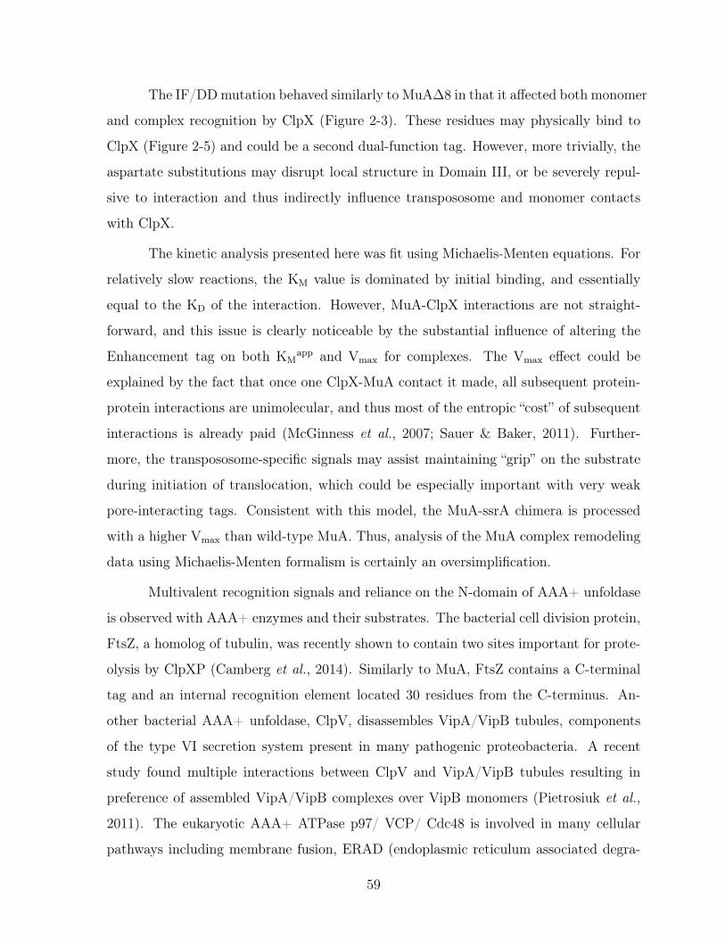
The IF/DD mutation behaved similarly to MuA∆8 in that it affected both monomer
and complex recognition by ClpX (Figure 2-3). These residues may physically bind to
ClpX (Figure 2-5) and could be a second dual-function tag. However, more trivially, the
aspartate substitutions may disrupt local structure in Domain III, or be severely repul-
sive to interaction and thus indirectly influence transpososome and monomer contacts
with ClpX.
The kinetic analysis presented here was fit using Michaelis-Menten equations. For
relatively slow reactions, the KM value is dominated by initial binding, and essentially
equal to the KD of the interaction. However, MuA-ClpX interactions are not straight-
forward, and this issue is clearly noticeable by the substantial influence of altering the
Enhancement tag on both KMapp and Vmax for complexes. The Vmax effect could be
explained by the fact that once one ClpX-MuA contact it made, all subsequent protein-
protein interactions are unimolecular, and thus most of the entropic “cost” of subsequent
interactions is already paid (McGinness et al., 2007; Sauer & Baker, 2011). Further-
more, the transpososome-specific signals may assist maintaining “grip” on the substrate
during initiation of translocation, which could be especially important with very weak
pore-interacting tags. Consistent with this model, the MuA-ssrA chimera is processed
with a higher Vmax than wild-type MuA. Thus, analysis of the MuA complex remodeling
data using Michaelis-Menten formalism is certainly an oversimplification.
Multivalent recognition signals and reliance on the N-domain of AAA+ unfoldase
is observed with AAA+ enzymes and their substrates. The bacterial cell division protein,
FtsZ, a homolog of tubulin, was recently shown to contain two sites important for prote-
olysis by ClpXP (Camberg et al., 2014). Similarly to MuA, FtsZ contains a C-terminal
tag and an internal recognition element located 30 residues from the C-terminus. An-
other bacterial AAA+ unfoldase, ClpV, disassembles VipA/VipB tubules, components
of the type VI secretion system present in many pathogenic proteobacteria. A recent
study found multiple interactions between ClpV and VipA/VipB tubules resulting in
preference of assembled VipA/VipB complexes over VipB monomers (Pietrosiuk et al.,
2011). The eukaryotic AAA+ ATPase p97/ VCP/ Cdc48 is involved in many cellular
pathways including membrane fusion, ERAD (endoplasmic reticulum associated degra-
59
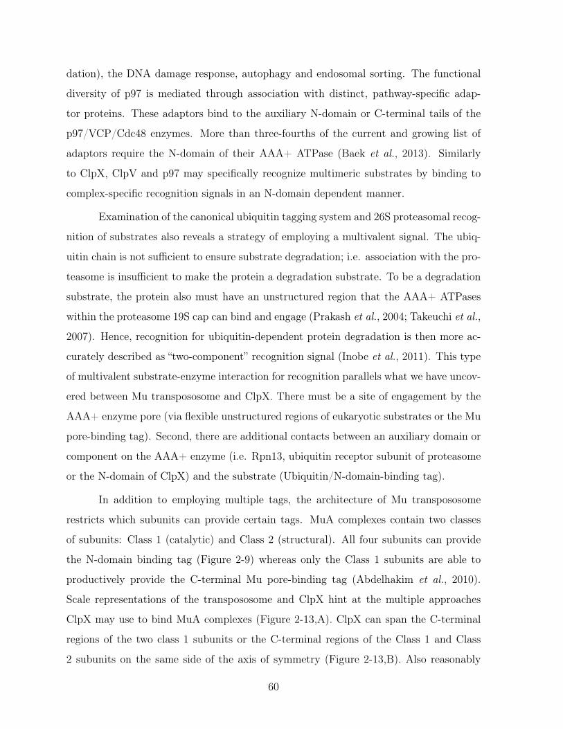
dation), the DNA damage response, autophagy and endosomal sorting. The functional
diversity of p97 is mediated through association with distinct, pathway-specific adap-
tor proteins. These adaptors bind to the auxiliary N-domain or C-terminal tails of the
p97/VCP/Cdc48 enzymes. More than three-fourths of the current and growing list of
adaptors require the N-domain of their AAA+ ATPase (Baek et al., 2013). Similarly
to ClpX, ClpV and p97 may specifically recognize multimeric substrates by binding to
complex-specific recognition signals in an N-domain dependent manner.
Examination of the canonical ubiquitin tagging system and 26S proteasomal recog-
nition of substrates also reveals a strategy of employing a multivalent signal. The ubiq-
uitin chain is not sufficient to ensure substrate degradation; i.e. association with the pro-
teasome is insufficient to make the protein a degradation substrate. To be a degradation
substrate, the protein also must have an unstructured region that the AAA+ ATPases
within the proteasome 19S cap can bind and engage (Prakash et al., 2004; Takeuchi et al.,
2007). Hence, recognition for ubiquitin-dependent protein degradation is then more ac-
curately described as “two-component” recognition signal (Inobe et al., 2011). This type
of multivalent substrate-enzyme interaction for recognition parallels what we have uncov-
ered between Mu transpososome and ClpX. There must be a site of engagement by the
AAA+ enzyme pore (via flexible unstructured regions of eukaryotic substrates or the Mu
pore-binding tag). Second, there are additional contacts between an auxiliary domain or
component on the AAA+ enzyme (i.e. Rpn13, ubiquitin receptor subunit of proteasome
or the N-domain of ClpX) and the substrate (Ubiquitin/N-domain-binding tag).
In addition to employing multiple tags, the architecture of Mu transpososome
restricts which subunits can provide certain tags. MuA complexes contain two classes
of subunits: Class 1 (catalytic) and Class 2 (structural). All four subunits can provide
the N-domain binding tag (Figure 2-9) whereas only the Class 1 subunits are able to
productively provide the C-terminal Mu pore-binding tag (Abdelhakim et al., 2010).
Scale representations of the transpososome and ClpX hint at the multiple approaches
ClpX may use to bind MuA complexes (Figure 2-13,A). ClpX can span the C-terminal
regions of the two class 1 subunits or the C-terminal regions of the Class 1 and Class
2 subunits on the same side of the axis of symmetry (Figure 2-13,B). Also reasonably
60

spaced is ClpX spanning the C-terminal regions of Class 1 and Class 2 subunits on
opposite sides of the axis of symmetry. Our data support these orientations of ClpX
~106Å
~113Å
Transpososome
ClpX
A.
Subunits on the same side of symmetry axis
Subunits on the opposite side of symmetry axis
P-tag E-tag P-tagE-tag
E-tag
P-tag
E-tag
P-tag
E-tag
P-tag
E-tag
P-tag
B.
Figure 2-13: Permutations of tag engagement in Mu transpososome by ClpXA. Crystal structures of Mu transpososome (4FCY), ClpX ATPase domain hexamer(3HWS), and ClpX N-domain (2DS6) shown at the same scale were modeled to indicatehow ClpX may interact simultaneously with multiple subunits in the MuA complex.Transpososome subunits are colored in shades of green and purple. ClpX hexamer iscolored in blue. N-domain is colored in light blue.B. For simplicity this diagram considers only two subunits at a time although in principlemultiple N-domains of ClpX may interact simultaneously with multiple subunits in theMuA complex. Each subunit contributes either the ClpX-pore-binding tag (P-tag) or theEnhancement a.k.a. N-domain-binding tag (E-tag).
61

as being sufficient to lead to recognition for complex disassembly. By making the N-
domain binding tag accessible on all four subunits, the architecture of the transpososome
maximizes the permutations for successful recognition with six possible orientations of
ClpX relative to MuA complex. A stricter division of labor in which only Class2 subunits
can provide the N-domain-binding tag would result in four possible orientations; likewise
if only Class 1 subunits provide both the N-domain-binding tag and the pore-binding
tag there would be only two acceptable ClpX “attack” orientations. Multiple N-domain-
interacting subunits may also be beneficial as the regulator protein MuB also binds
the C-terminal region MuA transposase, and ClpX therefore may be able to make initial
contacts with MuA complexes prior to the exit of MuB. Thus the fact that MuA presents
the high-affinity multivalent signal only in the context of the assembled complex may have
multiple advantages and indicates that the transpososome, by using an intricate set of
signals, has evolved to be a high-priority ClpX target.
Our work supports a design principle of tuning the strength of recognition signals
with modular weakly-binding tags. On its own, the C-terminal Mu pore-binding tag is
a poor ClpX recognition signal with an apparent affinity of ∼70𝜇M for ClpX (Figure
2-10; Barkow 2009). In comparison, the ssrA tag has an apparent affinity of ∼1.8𝜇M
(Levchenko et al., 2000). However in the context of the assembled MuA complex, the
Mu pore-binding tag benefits from multiple “diffuse” ClpX-interacting contacts, one of
which is the N-domain binding tag identified in this study. Together, these peptide
tags act synergistically to form a high-affinity ClpX recognition signal comparable in
affinity to the ssrA tag. According to this model, weak pore-interacting tags favor heavy
dependence on accessory recognition elements. In converse, we predicted that accessory
recognition elements provide little or no additional benefit to strong, compact ClpX
recognition signals. This hypothesis is supported by the MuA-ssrA hybrid proteins,
which gained essentially no benefit from the N-domain-binding tag. These observations
are consistent with the protein engineering studies of McGuiness et al. who demonstrated
that a weakened pore-binding tag gains a much larger magnitude benefit from an adapter
(McGinness et al., 2006). This type of “tag tuning” is used during the biologically relevant
recognition of MuA by ClpX, as ClpX is no longer able to discriminate between the
62

tetrameric and monomeric states when carrying a strong pore-binding tag. Thus, this
analysis of recognition of MuA lays a theoretical framework to understand the design
logic of complex-targeting recognition signals employed by AAA+ enzymes.
63

2.5 Methods
Buffers
Buffer L1, W20, W250 contained 25mM HEPES-KOH pH 7.6, 100mM KCl,
400mM NaCl, 10mM beta-mercaptoethanol, 10% glycerol, and imidazole at concentra-
tions of 10mM, 20mM, and 250mM, respectively.
Buffer A contained 25mM HEPES-KOH pH 7.6, 0.1mM EDTA, 1mM DTT, 10% glyc-
erol, 0.3M KCl.
Buffer B contained 25mM HEPES-KOH pH 7.6, 0.1mM EDTA, 1mM DTT, 10% glyc-
erol, 1M KCl.
PD50 buffer contained 25mM HEPES-KOH ph 7.6, 50mM KCl, 5mM MgCl2, 0.032%
NP-40, 10% glycerol.
Protein and peptide purification
Wild-type and mutant variants of MuA proteins (Baker et al., 1991) , E.coli ClpX
(Neher et al., 2003b), ClpX∆N (residue 47-424) (Abdelhakim et al., 2008), HU (Baker
et al., 1994), ClpP (Kim et al., 2000), N-domain of ClpX (residue 1-64) with a cleaveable
N-terminal His tag (Chowdhury et al., 2010) were purified as previously described.
SinMu chimera was cloned based on a plasmid gift, pSin15Mu, from S.P.M and
P.A.R. The plasmid, pSin15Mu is residues 147-200 of Sin recombinase followed by a
ten-residue SG repeat followed by MuA (residues 253-605). SinMu was generated by
appending the remaining MuA transposase sequence such that the construct ends at
the natural C-terminus of MuA, (residue 663), cloned into a pET3a vector via NdeI
and BamHI restriction sites, and transformed into E. coli strain BL21(DE3). Cells
were grown at 37°C to O.D.600nm ≈0.6 in Luria-Bertani broth containing 100𝜇g/mL
ampicillin. Protein expression was induced for 3 hours by addition of 0.4mM IPTG.
The culture was harvested by centrifugation, resuspended in 10mL of BufferL1 per liter
of initial cell culture, and lysed by French press. The lysate was treated with PMSF
(phenylmethylsulfonyl fluoride), cleared by centrifugation for 30min at 30,000g 4°C and
incubated with Ni-NTA agarose beads equilibrated in BufferL1 for 1 hour at 4°C. The
beads were transferred to a column, washed with Buffer W20, and bound protein was
64

eluted using Buffer W250. Fractions containing SinMu variants were identified by SDS-
PAGE, buffer-exchanged into Buffer A using PD-10 desalting columns. The eluate was
further purified by anion exchange chromatography, MonoS equilibrated with Buffer A,
and eluted by gradient to Buffer B. Fractions containing SinMu variants were identified
by SDS-PAGE, pooled, and concentrated using Amicon (MWCO 5k) filter tubes, and
the protein concentration by determined by Bradford reagent.
Mu∆8ssrA was generated from pTB1, a pET3d containing MuA tranposase. The
last eight C-terminal residues were replaced with the sequence for ssrA tag, gener-
ated by PCR with 5’-phosphate primers LLO62: aactacgctttagcagctTAAGGATCCG-
GCTGCTAACAAAGCC and LLO63: ttcgtcgtttgcggcTTCCAGAATATCCAGCGAAT-
GATTCAGATA. The variant Mu∆8ssrA (P623D S624D) was cloned by PCR using
5’-phosphate primers LLO64: gaTgaCGGTAATACGGAACGGGTGAAG and LLO55:
CCGGAAAATACCAACAATTCGTGA. Both Mu∆8ssrA and Mu∆8ssrA (PS/DD) pro-
teins were expressed and purified using the protocol for wild-type MuA.
Fluorescein-labeled peptides were synthesized by FMOC technique on an Apex
396 solid-phase synthesizer and purified on a reverse-phage C12 column running a gra-
dient of 0-100% Acetonitrile by HPLC. Peptides were verified by MALDI-TOF mass
spectrometry.
DNA for transposition
pSinRRSin was generated from miniMu plasmid, pMK586. pMK586 was digested
with ClaI and EcoN1 to remove the phage left-end attachment sites, treated with Antar-
tic phosphatase, and ligated to 5’-phospshate annealed oligonucleotides: LLO37:
CCAAGGAAGCTTGAAGCGGCGCACGAAAAACGCGAAAGCcgtatgattagggtAT LLO38:
CGATaccctaatcatacgGCTTTCGCGTTTTTCGTGCGCCGCTTCAAGCTTCCTTG con-
taining the R1-Sin binding sites with appropriate overhangs. The right-end R2 binding
site was replaced with Sin attachment sote sequence, generated by PCR with 5’-phosphate
primers LLO46: tcatacgGCTTTCGCGTTTTTCGTGCGC and LLO47: ttagggtCTT-
TAGCTTTCGCGCTTCAAATG.
Transpososome Assembly
65
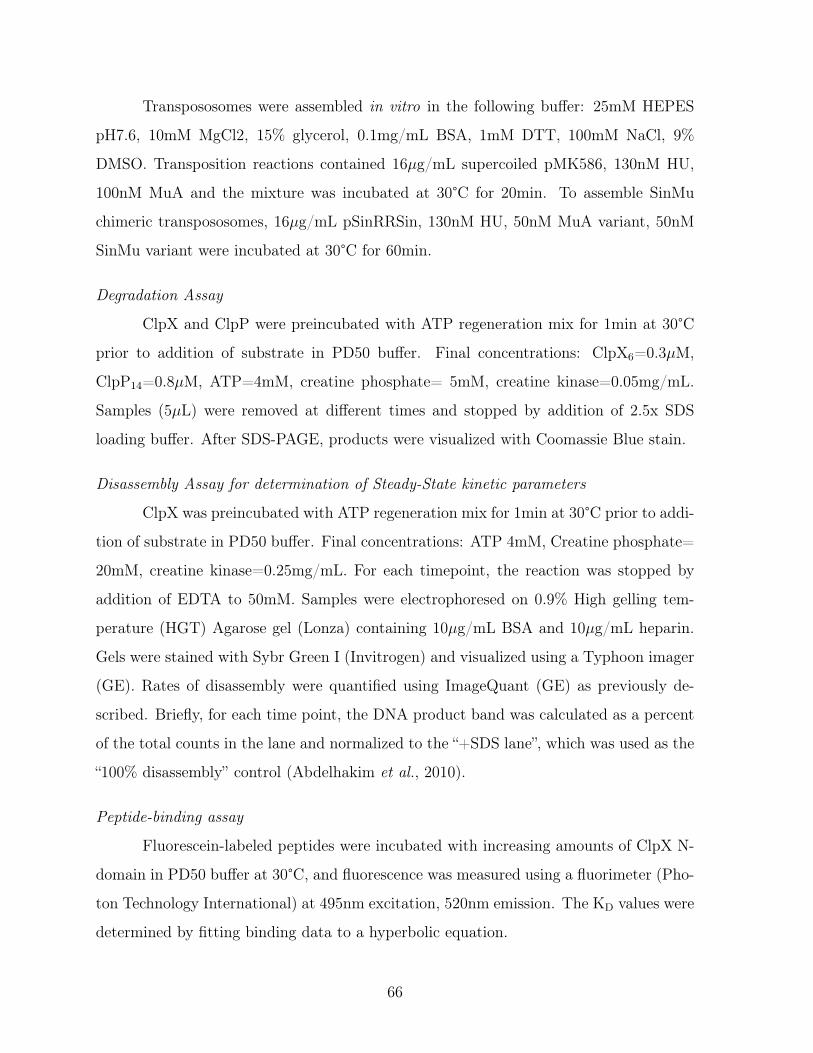
Transpososomes were assembled in vitro in the following buffer: 25mM HEPES
pH7.6, 10mM MgCl2, 15% glycerol, 0.1mg/mL BSA, 1mM DTT, 100mM NaCl, 9%
DMSO. Transposition reactions contained 16𝜇g/mL supercoiled pMK586, 130nM HU,
100nM MuA and the mixture was incubated at 30°C for 20min. To assemble SinMu
chimeric transpososomes, 16𝜇g/mL pSinRRSin, 130nM HU, 50nM MuA variant, 50nM
SinMu variant were incubated at 30°C for 60min.
Degradation Assay
ClpX and ClpP were preincubated with ATP regeneration mix for 1min at 30°C
prior to addition of substrate in PD50 buffer. Final concentrations: ClpX6=0.3𝜇M,
ClpP14=0.8𝜇M, ATP=4mM, creatine phosphate= 5mM, creatine kinase=0.05mg/mL.
Samples (5𝜇L) were removed at different times and stopped by addition of 2.5x SDS
loading buffer. After SDS-PAGE, products were visualized with Coomassie Blue stain.
Disassembly Assay for determination of Steady-State kinetic parameters
ClpX was preincubated with ATP regeneration mix for 1min at 30°C prior to addi-
tion of substrate in PD50 buffer. Final concentrations: ATP 4mM, Creatine phosphate=
20mM, creatine kinase=0.25mg/mL. For each timepoint, the reaction was stopped by
addition of EDTA to 50mM. Samples were electrophoresed on 0.9% High gelling tem-
perature (HGT) Agarose gel (Lonza) containing 10𝜇g/mL BSA and 10𝜇g/mL heparin.
Gels were stained with Sybr Green I (Invitrogen) and visualized using a Typhoon imager
(GE). Rates of disassembly were quantified using ImageQuant (GE) as previously de-
scribed. Briefly, for each time point, the DNA product band was calculated as a percent
of the total counts in the lane and normalized to the “+SDS lane”, which was used as the
“100% disassembly” control (Abdelhakim et al., 2010).
Peptide-binding assay
Fluorescein-labeled peptides were incubated with increasing amounts of ClpX N-
domain in PD50 buffer at 30°C, and fluorescence was measured using a fluorimeter (Pho-
ton Technology International) at 495nm excitation, 520nm emission. The KD values were
determined by fitting binding data to a hyperbolic equation.
66

2.6 Appendix: Geometry experiments on MuA monomer
variants
2.6.1 Introduction
ClpX unfoldase recognizes two oligomeric states of phage MuA transposase. The first
state is the biologically-active tetrameric complex synapsed with phage genomic ends
and host DNA in the core of the complex (transpososome). The second state is the
catalytically-incompetent and unassembled MuA monomeric subunits. However, ClpX
has a clear preference to target the transpososome as the substrate for its unfolding
activity . In vitro, ClpX has an apparent affinity of ∼1𝜇M for transpososome disassembly
while it recognizes MuA monomers with a ten-fold weaker apparent affinity of ∼10𝜇M
(Abdelhakim et al., 2008). In my thesis research, I strove to uncover the molecular
mechanism for ClpX’s ability to discriminate between the two oligomeric states of MuA
transposase and the resulting preference for the assembled MuA complex over MuA
monomers.
Before the crystal structure of the transpososome was solved, the only available
structural reference for an assembled MuA complex was a low-resolution EM (electron
microscope) structure of a type 1 transpososome, or CDC (Yuan et al., 2005). In Chap-
ter 2, I identified another ClpX recognition signal, the Enhancement tag, which con-
tributed principally and substantially to transpososome recognition and insignificantly
to monomer recognition. With the Mu pore-binding tag, there were now a total of
two identified tags that contributed to the holistic remodeling signal. One hypothesis
I considered was that these two tags in MuA transposase fit into the "two-component
recognition signal" model (Inobe et al., 2011). Studies on the intracellular protease, 26S
proteasome, an eukaryotic compartmentalized protease parallel to bacterial ClpXP, and
the ubiquitin-tagging system enumerated two minimal components for ubiquitin- tagged
proteasome substrates (Prakash et al., 2004).
1) a proteasome-binding site (Ubiquitin or UBL(ubiquitin-like) domains)
2) a flexible unstructured region
67

Futher protein engineering studies by the Matoushek group showed efficient sub-
strate degradation occurred when the initiation region is of a certain minimal length
and is appropriately separated in space from the proteasome-binding tag (Inobe & Ma-
touschek, 2014). Inspired by their work on the geometry/ spacing of two-component
recognition signals, I probed the spacing of the two tags in MuA transposase.
2.6.2 Results
I hypothesized that spatial constraints prohibited simultaneous engagement of the pore-
binding tag and the N-domain-binding tag in MuA monomer. Assuming that both tags
are accessible in MuA monomer, I tested if increasing the spatial separation between the
two tags would improve ClpX’s affinity for MuA monomers.
I designed variants of MuA transposase with additional amino acids to increase
separation of the C-terminal pore-binding tag from the N-domain-binding tag. The ad-
ditional amino acids were taken from the sequence in the adaptor protein SspB, a flexible
region that connects the folded substrate-binding domain and the XB peptide. I inserted
15 or 25-long sequences just before the Mu pore-binding tag, naming them after the
number of inserted amino acids, Mu-SspB15L and Mu-SspB25L. (Figure2-14A). Circular
dichroism established that there was no discernible changes in the overall fold of the Mu-
SspB variants as compared to wild-type. I assayed ClpXP-mediated degradation of these
Mu-SspB variants. Degradation of Mu-SspB15L and Mu-SspB25L was barely detectable,
roughly 3-4% of the wild-type degradation rate at the low enzyme concentration assayed
(Figure2-14B).
As the Mu-SspB variants seem folded overall, I checked if the inserted SspB linker
was somehow evolved to be "undegradable." I then modified the Mu-SspB15L and Mu-
SspB25L genes by removing the Mu pore-binding tag sequence and replacing it with
the sequence for the ssrA tag. I was only able to recover clones of Mu-SspB25L-ssrA.
Degradation of Mu-SspB25L-ssrA by ClpXP was detectable but slow at ∼25% of the rate
for wild-type native MuA monomers.
68
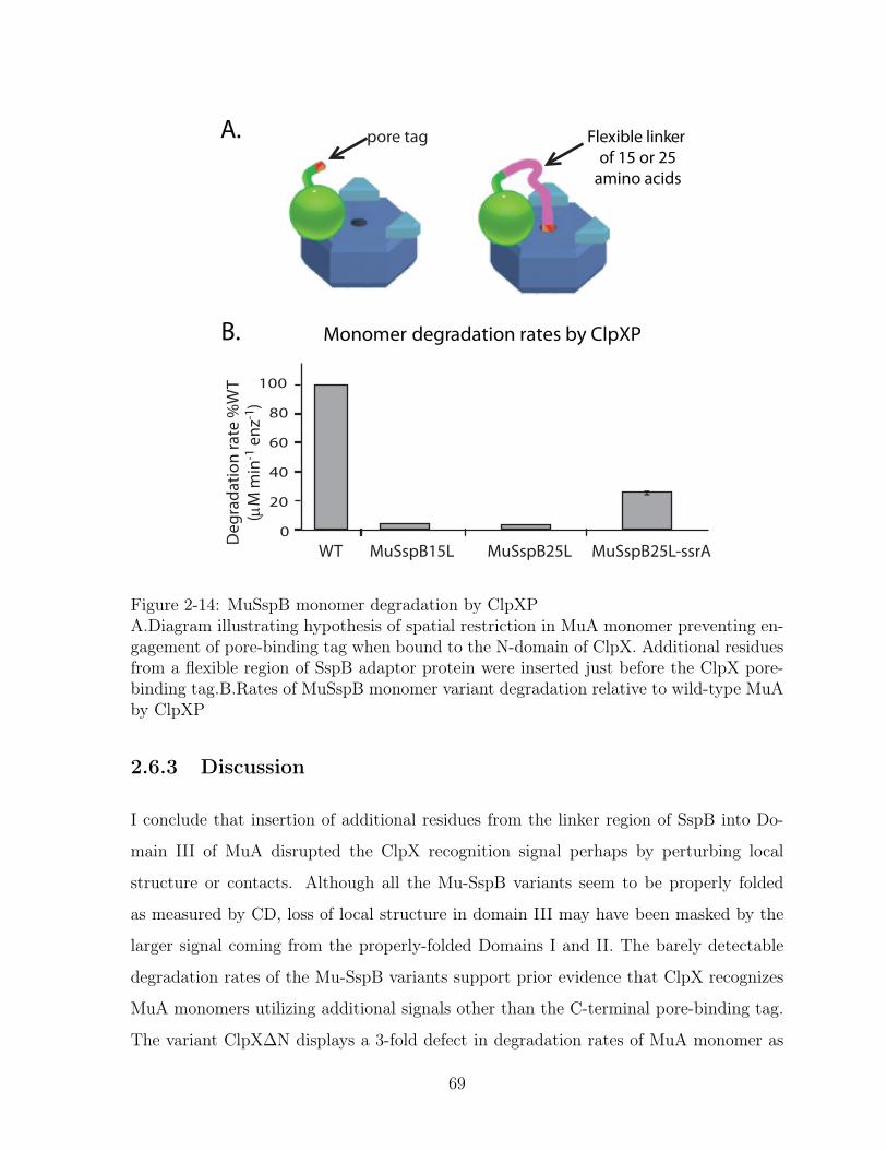
Monomer degradation rates by ClpXP
Flexible linker of 15 or 25
amino acids
A.
B.
0
20
40
60
80
100
Deg
rada
tion
rate
%W
T(µ
M m
in-1
enz
-1)
WT MuSspB15L MuSspB25L MuSspB25L-ssrA
pore tag
Figure 2-14: MuSspB monomer degradation by ClpXPA.Diagram illustrating hypothesis of spatial restriction in MuA monomer preventing en-gagement of pore-binding tag when bound to the N-domain of ClpX. Additional residuesfrom a flexible region of SspB adaptor protein were inserted just before the ClpX pore-binding tag.B.Rates of MuSspB monomer variant degradation relative to wild-type MuAby ClpXP
2.6.3 Discussion
I conclude that insertion of additional residues from the linker region of SspB into Do-
main III of MuA disrupted the ClpX recognition signal perhaps by perturbing local
structure or contacts. Although all the Mu-SspB variants seem to be properly folded
as measured by CD, loss of local structure in domain III may have been masked by the
larger signal coming from the properly-folded Domains I and II. The barely detectable
degradation rates of the Mu-SspB variants support prior evidence that ClpX recognizes
MuA monomers utilizing additional signals other than the C-terminal pore-binding tag.
The variant ClpX∆N displays a 3-fold defect in degradation rates of MuA monomer as
69

compared to full-length ClpX (Abdelhakim et al., 2008).
Surprisingly, Mu-SspB25L-ssrA monomer variant was degraded slower than wild-
type MuA monomers at substrate concentrations well below the KM for degradation.
Most likely this difference is reflective of the method used to detect loss of protein;
Coomassie Blue staining of SDS-PAGE gels. Coomassie Blue has a smaller dynamic
signal range than other protein stains. Thus monitoring fast reactions with Coomassie
Blue leads to greater errors and slower-than expected initial rates. For a more reliable
comparison between these two variants, I would assay Mu-SspB25L-ssrA degradation by
ClpXP over a range of substrate concentrations to get Michaelis-Menten kinetic param-
eters and would follow degradation using a more sensitive stain like SYPRO Orange or
radiolabelled test substrate.
70

Chapter 3
Conclusion & Future Directions
71
3.1 Conclusion
E.coli ClpX is a member of the Clp/Hsp100 family of ATPases that remodel multi-
component complexes and facilitate ATP-dependent protein degradation. Previous ex-
tensive studies on the interaction of ClpX remodeling Mu transpososomes led to progress
toward a molecular understanding of how ClpX recognized MuA complexes. The work of
this thesis deepens this understanding by identifying the molecular mechanism for ClpX’s
distinction between transpososomes and MuA monomers and thereby the specificity for
transpososomes.
MuA transposase exists as inactive monomers and as an assembled transpososome,
a nucleoprotein complex with four subunits synapsed with recombined DNA. ClpX rec-
ognizes the biologically relevant transpososome (MuA complex) with a 10-fold tighter ap-
parent affinity compared to monomeric MuA. I identified a critical region (RPS) in MuA
transposase expanding upon a previously identified arginine residue that contributed
greatly to the recognition of transpososomes by ClpX and very little to recognition of
MuA monomers. This peptide-like signal (residues 622-624) forms a ClpX recognition
tag that interacts weakly but specifically to the N-domain of ClpX. Mutation of this
Enhancement tag from all four subunits resulted in a 10-fold weaker apparent affinity for
disassembly (KMapp ∼10𝜇M), which is the same magnitude as ClpX’s affinity for MuA
monomers. This newly identified tag (Enhancement tag) act synergistically with the
previously characterized C-terminal Mu pore-binding tag (a.k.a "Mu degron" in other
studies) to form the holistic remodeling signal imparting ClpX specificity for transposo-
somes over monomers.
I then investigated the geometry of these two recognition tags in the context of the
assembled MuA complex. To this end, I engineered a chimeric protein (SinMu) with al-
tered DNA binding specifity by modifying a fusion protein (Sin15Mu) that was initially
designed and constructed by Montano and Rice. I also created a library of Mini-mu
plasmids with different substitutions in phage attachment sites using the oligo sequence
specific for Sin15Mu binding also designed by Montano and Rice. I was able to deter-
mine the contributions of Mu subunits to each type of tag. All four subunits in the
72

transpososome can effectively contribute an N-domain-binding tag. Optimal recognition
is achieved when all four are present. In contrast, only the catalytic Class 1 subunits pro-
ductively provide the pore-binding tag. I further showed that for the variant, Mu∆8ssrA,
the N-domain-binding tag becomes unnecessary for complex recognition because the na-
tive pore-binding tag is replaced by the ssrA tag, a stronger ClpX recognition signal.
Complexes of Mu∆8ssrA with and without mutation of the Enhancement tag were rec-
ognized by ClpX just as well as native transpososomes. However, ClpX also recognized
Mu∆8ssrA monomers with nearly the same apparent affinity, KMapp, as assembled com-
plexes.
From the above results, I propose the following design framework for recognition
signals to target assembled protein complexes to unfolding chaperones and remodelers
of the AAA+ superfamily. First, the target substrate makes multiple weak interactions
with the AAA ATPase. One type of interaction must be with the pore of the ATPase for
engagement and subsequent translocation/unfolding. Another type of interaction occurs
with an auxiliary domain of the AAA+ unfoldase. This binding may be direct with the
multicomponent substrate such as in the case for Mu transpososome or may be indirect
through adaptor proteins.
Second, recognition tags should be at the weaker end of the affinity spectrum to
allow effective synergy of multiple tags in the assembled complex. As shown in Chapter
2, ClpX failed to discriminate between the tetrameric and monomeric states of a protein
that has a strong pore-binding tag.
Third, multi-subunit complexes can "divide the labor" of making these interac-
tions among their subunits. The specific architecture of each multimeric complex will
determine the extent of this division of labor. Unassembled subunits or monomers are
therefore incompetent to provide all the tags available in assembled complexes. Confor-
mational changes within subunits that accompany formation of the assembled complex
are a likely mechanism to reveal recognition tags. A reverse-strategy is employed in
examples when protein complexes fall apart and a specific subunit is targeted for degra-
dation by a revealed tag. Thus, collaboration between multiple weak substrate-unfoldase
signals is an attractive general mechanism for targeting assembled protein complexes to
73

AAA+ enzymes.
3.2 Future Directions
The Enhancement tag was described and referred to as a peptide-like signal due to
its contiguous nature and ability to interact with purified N-domain when synthesized
as a 20-aa peptide. Several secondary structure prediction algorithms (Jpred, CFSSP,
NPS@SOPMA) assign these residues to a flexible unstructured linker or random coil
immediately following a predicted helix. However, these observations don’t exclude that
the residues may be part of a binding surface. Definitive answers would come from a
co-crystral structure of ClpX and transpososome assembled using a MuA construct that
extends to MuA’s C-terminus as the current crystal structure of the STC transpososome
utilized a truncated MuA protein (Montaño et al., 2012). Furthermore, I propose extend-
ing the lysine-acetylation footprinting experiments from Abdelhakim et al. 2008 to the
SinMu chimeric complexes. This method would likely reveal which residues or surface
areas on which subunits are protected in the presence of ClpX and ATP𝛾S compared to
SinMu chimeric complexes alone.
To further support the insights gained from the transpososome remodeling signal
and develop more robust design principles of ClpX recognition signals, I propose that
future researchers investigate substrates in a systematic and high-throughput approach.
As mentioned in Chapter 1, our lab performed a proteomic screen for in vivo ClpXP
substrates and sorted the trapped proteins into five classes of recognition motifs. In light
of identifying two distinct classes of recognition tags in Mu transpososome, the results
from the 2003 screen may benefit from re-examination with the goal of categorizing
the sequences as pore-binding motifs or N-domain-binding motifs. Furthermore, almost
60% of these trapped substrates perform their biological function as subunits within a
complex. It would be interesting to elucidate how many of those trapped substrates
are recognized by ClpX in an adaptor or N-domain dependent mechanism. Does ClpX
recognize these proteins in the context of the assembled complex like Mu transpososome
or in the context of a monomeric subunit post-disassembly? To address these questions,
74
I propose a modified version of the original proteomic screen, in which ClpX∆N is used
as the partnering unfoldase for the ClpPtrap (Flynn et al., 2003). Secondly, I would re-
probe the peptide array from Figure 4 with isolated N-domain of ClpX to observe if these
residues/polypeptide regions may be classified as N-domain-binding tags. I would confirm
the interaction with solution-based binding assays. Lastly, I would seek to understand if
a consensus N-domain-binding motif emerges from these sequences.
75

76
Bibliography
Abdelhakim, A. H., Oakes, E. C., Sauer, R. T., & Baker, T. A. (2008). Unique contactsdirect high-priority recognition of the tetrameric Mu transposase-DNA complex by theAAA+ unfoldase ClpX.. Mol. Cell 30(1), 39–50.
Abdelhakim, A. H., Sauer, R. T., & Baker, T. A. (2010). The AAA+ ClpX machineunfolds a keystone subunit to remodel the Mu transpososome.. Proc. Natl. Acad. Sci.U. S. A. 107(6), 2437–42.
Anfinsen, C. B. (1973). Principles that govern the folding of protein chains.. Science181(4096), 223–30.
Aubin-Tam, M.-E., Olivares, A. O., Sauer, R. T., Baker, T. A., & Lang, M. J. (2011).Single-molecule protein unfolding and translocation by an ATP-fueled proteolytic ma-chine.. Cell 145(2), 257–67.
Baek, G. H., Cheng, H., Choe, V., Bao, X., Shao, J., Luo, S., & Rao, H. (2013). Cdc48:A Swiss Army Knife of Cell Biology.. J. Amino Acids 2013, 183421.
Baker, T. A., Kremenstova, E., & Luo, L. (1994). Complete transposition requires fouractive monomers in the mu transposase tetramer.. Genes Dev. 8(20), 2416–2428.
Baker, T. A. & Mizuuchi, K. (1992). DNA-promoted assembly of the active tetramer ofthe Mu transposase.. Genes Dev. 6(11), 2221–32.
Baker, T. A., Mizuuchi, M., & Mizuuchi, K. (1991). MuB protein allosterically activatesstrand transfer by the transposase of phage Mu.. Cell 65(6), 1003–13.
Baker, T. A. & Sauer, R. T. (2006). ATP-dependent proteases of bacteria: recognitionlogic and operating principles.. Trends Biochem. Sci. 31(12), 647–53.
Barkow, S. R. (2009). Mechanistic Studies of the AAA + Molecular Motor ClpXP PhDthesis Massachusetts Institute of Technology.
Barkow, S. R., Levchenko, I., Baker, T. A., & Sauer, R. T. (2009). Polypeptide translo-cation by the AAA+ ClpXP protease machine.. Chem. Biol. 16(6), 605–12.
Bochtler, M., Hartmann, C., Song, H. K., Bourenkov, G. P., Bartunik, H. D., & Huber, R.(2000). The structures of HsIU and the ATP-dependent protease HsIU-HsIV.. Nature403(6771), 800–5.
77

Braig, K., Otwinowski, Z., Hegde, R., Boisvert, D. C., Joachimiak, A., Horwich, A. L.,& Sigler, P. B. (1994). The crystal structure of the bacterial chaperonin GroEL at 2.8A.. Nature 371(6498), 578–86.
Burton, B. M. & Baker, T. A. (2003). Mu Transpososome Architecture Ensures thatUnfolding by ClpX or Proteolysis by ClpXP Remodels but Does Not Destroy theComplex. Chem. Biol. 10, 463–472.
Burton, R. E., Siddiqui, S. M., Kim, Y. I., Baker, T. A., & Sauer, R. T. (2001). Effectsof protein stability and structure on substrate processing by the ClpXP unfolding anddegradation machine.. EMBO J. 20(12), 3092–100.
Camberg, J. L., Viola, M. G., Rea, L., Hoskins, J. R., & Wickner, S. (2014). Location ofDual Sites in E. coli FtsZ Important for Degradation by ClpXP; One at the C-Terminusand One in the Disordered Linker.. PLoS One 9(4), e94964.
Chowdhury, T., Chien, P., Ebrahim, S., Sauer, R. T., & Baker, T. A. (2010). Versatilemodes of peptide recognition by the ClpX N domain mediate alternative adaptor-binding specificities in different bacterial species.. Protein Sci. 19(2), 242–54.
Chung, C. H. & Goldberg, A. L. (1981). The product of the lon (capR) gene in Escherichiacoli is the ATP-dependent protease, protease La.. Proc. Natl. Acad. Sci. U. S. A. 78(8),4931–5.
Clubb, R. T., Omichinski, J. G., Savilahti, H., Mizuuchi, K., Gronenborn, A. M., &Clore, G. M. (1994). A novel class of winged helix-turn-helix protein: the DNA-bindingdomain of Mu transposase.. Structure 2(11), 1041–8.
Clubb, R. T., Schumacher, S., Mizuuchi, K., Gronenborn, a. M., & Clore, G. M. (1997).Solution structure of the I gamma subdomain of the Mu end DNA-binding domain ofphage Mu transposase.. J. Mol. Biol. 273(1), 19–25.
Craigie, R., Arndt-Jovin, D. J., & Mizuuchi, K. (1985). A defined system for the DNAstrand-transfer reaction at the initiation of bacteriophage Mu transposition: proteinand DNA substrate requirements.. Proc. Natl. Acad. Sci. U. S. A. 82(22), 7570–4.
Craigie, R. & Mizuuchi, K. (1987). Transposition of Mu DNA: joining of Mu to targetDNA can be uncoupled from cleavage at the ends of Mu.. Cell 51(3), 493–501.
Craigie, R., Mizuuchi, M., & Mizuuchi, K. (1984). Site-specific recognition of the bacte-riophage Mu ends by the Mu A protein.. Cell 39(2 Pt 1), 387–94.
Deuerling, E., Schulze-Specking, A., Tomoyasu, T., Mogk, A., & Bukau, B. (1999).Trigger factor and DnaK cooperate in folding of newly synthesized proteins.. Nature400(6745), 693–6.
Dougan, D. a., Reid, B. G., Horwich, A. L., & Bukau, B. (2002). ClpS, a substratemodulator of the ClpAP machine.. Mol. Cell 9(3), 673–83.
78
Doyle, S. M. & Wickner, S. (2009). Hsp104 and ClpB: protein disaggregating machines..Trends Biochem. Sci. 34(1), 40–8.
Dubnau, D. & Roggiani, M. (1990). Growth medium-independent genetic competencemutants of Bacillus subtilis.. J. Bacteriol. 172(7), 4048–55.
Eckl, J. M. & Richter, K. (2013). Functions of the Hsp90 chaperone system: lifting clientproteins to new heights.. Int. J. Biochem. Mol. Biol. 4(4), 157–65.
Ellis, R. (2003). Protein Folding: Importance of the Anfinsen Cage. Curr. Biol. 13(22),R881–R883.
Farrell, C. M., Baker, T. A., & Sauer, R. T. (2007). Altered specificity of a AAA+protease.. Mol. Cell 25(1), 161–6.
Fenton, W. A., Kashi, Y., Furtak, K., & Horwich, A. L. (1994). Residues in chaperoninGroEL required for polypeptide binding and release.. Nature 371(6498), 614–9.
Flynn, G. C., Chappell, T. G., & Rothman, J. E. (1989). Peptide binding and release byproteins implicated as catalysts of protein assembly.. Science 245(4916), 385–90.
Flynn, J. M., Levchenko, I., Sauer, R. T., & Baker, T. A. (2004). Modulating substratechoice: the SspB adaptor delivers a regulator of the extracytoplasmic-stress responseto the AAA+ protease ClpXP for degradation.. Genes Dev. 18(18), 2292–301.
Flynn, J. M., Levchenko, I., Seidel, M., Wickner, S. H., Sauer, R. T., & Baker, T. A.(2001). Overlapping recognition determinants within the ssrA degradation tag allowmodulation of proteolysis.. Proc. Natl. Acad. Sci. U. S. A. 98(19), 10584–9.
Flynn, J. M., Neher, S. B., Kim, Y. I., Sauer, R. T., & Baker, T. A. (2003). Proteomicdiscovery of cellular substrates of the ClpXP protease reveals five classes of ClpX-recognition signals.. Mol. Cell 11(3), 671–83.
Fourie, a. M., Sambrook, J. F., & Gething, M. J. (1994). Common and divergent peptidebinding specificities of hsp70 molecular chaperones.. J. Biol. Chem. 269(48), 30470–8.
Gardner, M. K., Zanic, M., & Howard, J. (2013). Microtubule catastrophe and rescue..Curr. Opin. Cell Biol. 25(1), 14–22.
Gayda, R. C., Yamamoto, L. T., & Markovitz, a. (1976). Second-site mutations incapR (lon) strains of Escherichia coli K-12 that prevent radiation sensitivity and allowbacteriophage lambda to lysogenize.. J. Bacteriol. 127(3), 1208–16.
Glynn, S. E., Martin, A., Nager, A. R., Baker, T. A., & Sauer, R. T. (2009). Structures ofasymmetric ClpX hexamers reveal nucleotide-dependent motions in a AAA+ protein-unfolding machine.. Cell 139(4), 744–56.
79
Gonciarz-swiatek, M., Um, S.-j., Learn, B. A., Mcmacken, R., Kelley, W. L., Chem, J. B.,Wawrzynow, A., Georgopoulos, C., Sliekers, O., & Zylicz, M. (1999). Recognition, Targeting , and Hydrolysis of the 𝜆 O Replication Protein by the Recognition ,Targeting , and Hydrolysis of the O Replication Protein by the ClpP / ClpX Protease*. J. Biol. Chem. 274, 13999–14005.
Gonzalez, M., Rasulova, F., Maurizi, M. R., & Woodgate, R. (2000). Subunit-specificdegradation of the UmuD/D’ heterodimer by the ClpXP protease: the role of transrecognition in UmuD’ stability.. EMBO J. 19(19), 5251–8.
Gottesman, S., Halpern, E., & Trisler, P. (1981). Role of sulA and sulB in filamentationby lon mutants of Escherichia coli K-12. J. Bacteriol. 148(1).
Gottesman, S., Roche, E., Zhou, Y., & Sauer, R. T. (1998). The ClpXP and ClpAPproteases degrade proteins with carboxy-terminal peptide tails added by the SsrA-tagging system. Genes Dev. 12(9), 1338–1347.
Greene, E. C. & Mizuuchi, K. (2002a). Direct observation of single MuB polymers:evidence for a DNA-dependent conformational change for generating an active targetcomplex.. Mol. Cell 9(5), 1079–89.
Greene, E. C. & Mizuuchi, K. (2002b). Dynamics of a protein polymer: the assemblyand disassembly pathways of the MuB transposition target complex.. EMBO J. 21(6),1477–86.
Greene, E. C. & Mizuuchi, K. (2002c). Target immunity during Mu DNA transposition.Transpososome assembly and DNA looping enhance MuA-mediated disassembly of theMuB target complex.. Mol. Cell 10(6), 1367–78.
Hanson, P. I. & Whiteheart, S. W. (2005). AAA+ proteins: have engine, will work.. Nat.Rev. Mol. Cell Biol. 6(7), 519–29.
Hartl, F. U. (2011). Chaperone-assisted protein folding: the path to discovery from apersonal perspective.. Nat. Med. 17(10), 1206–10.
Higashitani, A., Ishii, Y., Kato, Y., & Koriuchi, K. (1997). Functional dissection of acell-division inhibitor, SulA, of Escherichia coli and its negative regulation by Lon..Mol. Gen. Genet. 254(4), 351–7.
Houry, W. A., Frishman, D., Eckerskorn, C., Lottspeich, F., & Hartl, F. U. (1999).Identification of in vivo substrates of the chaperonin GroEL.. Nature 402(6758), 147–54.
Inobe, T., Fishbain, S., Prakash, S., & Matouschek, A. (2011). Defining the geometry ofthe two-component proteasome degron.. Nat. Chem. Biol. 7(3), 161–7.
Inobe, T. & Matouschek, A. (2014). Paradigms of protein degradation by the proteasome..Curr. Opin. Struct. Biol. 24, 156–64.
80

Jones, J. M., Welty, D. J., Chem, J. B., & Nakai, H. (1998). Versatile Action of Es-cherichia coli ClpXP as Protease or Molecular Chaperone for Bacteriophage Mu Trans-position Versatile Action of Escherichia coli ClpXP as Protease or Molecular Chaperonefor Bacteriophage Mu Transposition *. J. Bacteriol. 273, 459–465.
Karzai, A. W., Roche, E. D., & Sauer, R. T. (2000). The SsrA-SmpB system for proteintagging, directed degradation and ribosome rescue.. Nat. Struct. Biol. 7(6), 449–55.
Katayama, Y., Gottesman, S., Pumphreyll, J., Rudikoffl, S., Clark, W. P., & Maurizi,M. R. (1988). The Two-component , ATP-dependent Clp Protease of Escherichia coli.J. Biol. Chem. 263(29), 15226–15236.
Keiler, K. C., Waller, P. R., & Sauer, R. T. (1996). Role of a peptide tagging system indegradation of proteins synthesized from damaged messenger RNA.. Science 271(5251),990–3.
Kim, Y. I., Burton, R. E., Burton, B. M., Sauer, R. T., & Baker, T. A. (2000). Dynamicsof substrate denaturation and translocation by the ClpXP degradation machine.. Mol.Cell 5(4), 639–48.
Kirstein, J., Molière, N., Dougan, D. a., & Turgay, K. (2009). Adapting the machine:adaptor proteins for Hsp100/Clp and AAA+ proteases.. Nat. Rev. Microbiol. 7(8),589–99.
Kirstein, J., Schlothauer, T., Dougan, D. a., Lilie, H., Tischendorf, G., Mogk, A., Bukau,B., & Turgay, K. (2006). Adaptor protein controlled oligomerization activates theAAA+ protein ClpC.. EMBO J. 25(7), 1481–91.
Kruklitis, R., Welty, D. J., & Nakai, H. (1996). ClpX protein of Escherichia coli activatesbacteriophage Mu transposase in the strand transfer complex for initiation of Mu DNAsynthesis.. EMBO J. 15(4), 935–44.
Kuo, C. F., Zou, a. H., Jayaram, M., Getzoff, E., & Harshey, R. (1991). DNA-proteincomplexes during attachment-site synapsis in Mu DNA transposition.. EMBO J. 10(6),1585–91.
Laskowska, E., Kuczyńska-Wiśnik, D., Skórko-Glonek, J., & Taylor, A. (1996). Degrada-tion by proteases Lon, Clp and HtrA, of Escherichia coli proteins aggregated in vivo byheat shock; HtrA protease action in vivo and in vitro.. Mol. Microbiol. 22(3), 555–71.
Lavoie, B. D., Chan, B. S., Allison, R. G., & Chaconas, G. (1991). Structural aspects ofa higher order nucleoprotein complex: induction of an altered DNA structure at theMu - host junction of the Mu Type 1 transpososome. EMBO 10(10), 3051–3059.
Lee, S., Sowa, M. E., Watanabe, Y.-h., Sigler, P. B., Chiu, W., Yoshida, M., & Tsai,F. T. (2003). The Structure of ClpB. Cell 115(2), 229–240.
81

Levchenko, I., Grant, R. a., Wah, D. a., Sauer, R. T., & Baker, T. A. (2003). Structureof a delivery protein for an AAA+ protease in complex with a peptide degradationtag.. Mol. Cell 12(2), 365–72.
Levchenko, I., Luo, L., & Baker, T. A. (1995). Disassembly of the Mu transposasetetramer by the ClpX chaperone.. Genes Dev. 9(19), 2399–2408.
Levchenko, I., Seidel, M., Sauer, R. T., & Baker, T. A. (2000). A Specificity-EnhancingFactor for the ClpXP Degradation Machine. Science 289(5488), 2354–2356.
Levchenko, I., Yamauchi, M., & Baker, T. A. (1997). ClpX and MuB interact withoverlapping regions of Mu transposase: implications for control of the transpositionpathway.. Genes Dev. 11(12), 1561–1572.
Li, J. & Buchner, J. (2013). Structure, function and regulation of the hsp90 machinery..Biomed. J. 36(3), 106–17.
Lies, M. & Maurizi, M. R. (2008). Turnover of endogenous SsrA-tagged proteins mediatedby ATP-dependent proteases in Escherichia coli.. J. Biol. Chem. 283(34), 22918–29.
Lima, S., Guo, M. S., Chaba, R., Gross, C. a., & Sauer, R. T. (2013). Dual molecularsignals mediate the bacterial response to outer-membrane stress.. Science 340(6134),837–41.
Little, J. W. (1983). The SOS Regulatory System: Control of its State by the Level ofRecA Protease. J. Mol. Biol. 167, 791–808.
Martin, A., Baker, T. A., & Sauer, R. T. (2007). Distinct static and dynamic interactionscontrol ATPase-peptidase communication in a AAA+ protease.. Mol. Cell 27(1), 41–52.
Martin, A., Baker, T. A., & Sauer, R. T. (2008a). Diverse pore loops of the AAA+ClpX machine mediate unassisted and adaptor-dependent recognition of ssrA-taggedsubstrates.. Mol. Cell 29(4), 441–50.
Martin, A., Baker, T. A., & Sauer, R. T. (2008b). Pore loops of the AAA+ ClpX machinegrip substrates to drive translocation and unfolding.. Nat. Struct. Mol. Biol. 15(11),1147–51.
Martin, A., Baker, T. A., & Sauer, R. T. (2008c). Protein unfolding by a AAA+ proteaseis dependent on ATP-hydrolysis rates and substrate energy landscapes.. Nat. Struct.Mol. Biol. 15(2), 139–45.
Maxwell, A., Craigie, R., & Mizuuchi, K. (1987). B protein of bacteriophage mu is anATPase that preferentially stimulates intermolecular DNA strand transfer.. Proc. Natl.Acad. Sci. U. S. A. 84(3), 699–703.
Mayer, M. P. (2013). Hsp70 chaperone dynamics and molecular mechanism.. TrendsBiochem. Sci. 38(10), 507–14.
82

Mayhew, M., da Silva, A. C., Martin, J., Erdjument-Bromage, H., Tempst, P., & Hartl,F. U. (1996). Protein folding in the central cavity of the GroEL-GroES chaperonincomplex.. Nature 379(6564), 420–6.
McCarthy, D., Kramer, G., & Hardesty, B. (1998). Reactivation of thermally inactivatedpre-beta-lactamase by DnaK, DnaJ, and GrpE.. Protein Sci. 7(5), 1164–71.
McGinness, K. E., Baker, T. A., & Sauer, R. T. (2006). Engineering controllable proteindegradation.. Mol. Cell 22(5), 701–7.
McGinness, K. E., Bolon, D. N., Kaganovich, M., Baker, T. a., & Sauer, R. T. (2007). Al-tered tethering of the SspB adaptor to the ClpXP protease causes changes in substratedelivery.. J. Biol. Chem. 282(15), 11465–73.
McNally, F. J. & Vale, R. D. (1993). Identification of katanin, an ATPase that seversand disassembles stable microtubules.. Cell 75(3), 419–29.
Mhammedi-Alaoui, A., Pato, M., Gama, M.-J., & Toussaint, A. (1994). A new componentof bacteriophage Mu replicative transposition machinery: the Escherichia coli CipXprotein. Mol. Microbiol. 11, 1109–1116.
Miot, M., Reidy, M., Doyle, S. M., Hoskins, J. R., Johnston, D. M., Genest, O., Vitery,M.-C., Masison, D. C., & Wickner, S. (2011). Species-specific collaboration of heatshock proteins (Hsp) 70 and 100 in thermotolerance and protein disaggregation.. Proc.Natl. Acad. Sci. U. S. A. 108(17), 6915–20.
Mizusawa, S. (1983). Protein degradation in Escherichia coli : The Lon gene controls thestability of sulA protein. Proc. Natl. Acad. Sci. U. S. A. 80(January), 358–362.
Mizuuchi, K. & Adzuma, K. (1991). Inversion of the phosphate chirality at the target siteof Mu DNA strand transfer: evidence for a one-step transesterification mechanism..Cell 66(1), 129–40.
Mizuuchi, M., Baker, T. a., & Mizuuchi, K. (1992). Assembly of the active form ofthe transposase-Mu DNA complex: a critical control point in Mu transposition.. Cell70(2), 303–11.
Molière, N. & Turgay, K. (2009). Chaperone-protease systems in regulation and proteinquality control in Bacillus subtilis.. Res. Microbiol. 160(9), 637–44.
Montaño, S. P., Pigli, Y. Z., & Rice, P. a. (2012). The 𝜇 transpososome structure shedslight on DDE recombinase evolution.. Nature 491(7424), 413–7.
Muffler, a., Fischer, D., Altuvia, S., Storz, G., & Hengge-Aronis, R. (1996). The re-sponse regulator RssB controls stability of the sigma(S) subunit of RNA polymerasein Escherichia coli.. EMBO J. 15(6), 1333–9.
Nakai, H. & Kruklitis, R. (1995). Disassembly of the bacteriophage Mu transposase forthe initiation of Mu DNA replication. J. Biol. Chem. 270(33), 19591–19598.
83

Neher, S. B., Flynn, J. M., Sauer, R. T., & Baker, T. a. (2003a). Latent ClpX-recognitionsignals ensure LexA destruction after DNA damage.. Genes Dev. 17(9), 1084–9.
Neher, S. B., Sauer, R. T., & Baker, T. a. (2003b). Distinct peptide signals in the UmuDand UmuD’ subunits of UmuD/D’ mediate tethering and substrate processing by theClpXP protease.. Proc. Natl. Acad. Sci. U. S. A. 100(23), 13219–24.
Ogura, T., Tomoyasu, T., Yuki, T., Morimura, S., Begg, K. J., Donachie, W. D., Mori,H., Niki, H., & Hiraga, S. (1991). Structure and function of the ftsH gene in Escherichiacoli.. Res. Microbiol. 142(2-3), 279–82.
Park, E. Y., Lee, B.-G., Hong, S.-B., Kim, H.-W., Jeon, H., & Song, H. K. (2007).Structural basis of SspB-tail recognition by the zinc binding domain of ClpX.. J. Mol.Biol. 367(2), 514–26.
Parsell, D. A. & Lindquist, S. (1993). The Function of Heat-shock Proteins in StressTolerance: Degradation and Reactivation of Damaged Proteins. Annu. Rev. Genet.27, 437–496.
Pietrosiuk, A., Lenherr, E. D., Falk, S., Bönemann, G., Kopp, J., Zentgraf, H., Sinning,I., & Mogk, A. (2011). Molecular basis for the unique role of the AAA+ chaperoneClpV in type VI protein secretion.. J. Biol. Chem. 286(34), 30010–21.
Prakash, S., Tian, L., Ratliff, K. S., Lehotzky, R. E., & Matouschek, A. (2004). Anunstructured initiation site is required for efficient proteasome-mediated degradation..Nat. Struct. Mol. Biol. 11(9), 830–7.
Pratt, L. A. & Silhavy, T. J. (1996). regulator SprE controls the stability of RpoS. Proc.Natl. Acad. Sci. U. S. A. 93(March), 2488–2492.
Pyle, A. M. & Green, J. B. (1994). Building a kinetic framework for group II intron ri-bozyme activity: quantitation of interdomain binding and reaction rate.. Biochemistry33(9), 2716–25.
Reyes, O., Beyou, A., Mignotte-Vieux, C., & Richaud, F. (1987). Mini-Mu transduction:cis-inhibition of the insertion of Mud transposons.. Plasmid 18(3), 183–92.
Rice, P. A. & Baker, T. A. (2001). Comparative architecture of transposase and integrasecomplexes.. Nat. Struct. Biol. 8(5), 302–7.
Rice, P. A. & Mizuuchi, K. (1995). Structure of the bacteriophage Mu transposase core:a common structural motif for DNA transposition and retroviral integration.. Cell82(2), 209–20.
Roll-Mecak, A. & McNally, F. J. (2010). Microtubule-severing enzymes.. Curr. Opin.Cell Biol. 22(1), 96–103.
Roll-Mecak, A. & Vale, R. D. (2008). Structural basis of microtubule severing by thehereditary spastic paraplegia protein spastin.. Nature 451(7176), 363–7.
84
Román-Hernández, G., Hou, J. Y., Grant, R. A., Sauer, R. T., & Baker, T. A. (2011).The ClpS adaptor mediates staged delivery of N-end rule substrates to the AAA+ClpAP protease.. Mol. Cell 43(2), 217–28.
Sauer, R. T. & Baker, T. a. (2011). AAA+ proteases: ATP-fueled machines of proteindestruction.. Annu. Rev. Biochem. 80, 587–612.
Sauer, R. T., Bolon, D. N., Burton, B. M., Burton, R. E., Flynn, J. M., Grant, R. A.,Hersch, G. L., Joshi, S. A., Kenniston, J. A., Levchenko, I., Neher, S. B., Oakes, E. S.,Siddiqui, S. M., Wah, D. A., & Baker, T. A. (2004). Sculpting the Proteome withAAA+ Proteases and Disassembly Machines. Cell 119(1), 9–18.
Savilahti, H., Rice, P. A., & Mizuuchi, K. (1995). The phage Mu transpososome core:DNA requirements for assembly and function.. EMBO J. 14(19), 4893–903.
Schumacher, S., Clubb, R. T., Cai, M., Mizuuchi, K., Clore, G. M., & Gronenborn, a. M.(1997). Solution structure of the Mu end DNA-binding ibeta subdomain of phage Mutransposase: modular DNA recognition by two tethered domains.. EMBO J. 16(24),7532–41.
Seong, I. S., Oh, J. Y., Yoo, S. J., Seol, J. H., & Chung, C. H. (1999). ATP-dependentdegradation of SulA, a cell division inhibitor, by the HslVU protease in Escherichiacoli.. FEBS Lett. 456(1), 211–4.
Shapiro, J. A. (1979). Molecular Model for the transposition and replication of bacte-riophage Mu and other transposable elements. Proc. Natl. Acad. Sci. U. S. A. 76,1933–1937.
Siddiqui, S. M., Sauer, R. T., & Baker, T. A. (2004). Role of the processing pore of theClpX AAA+ ATPase in the recognition and engagement of specific protein substrates..Genes Dev. 18(4), 369–74.
Song, H. K. & Eck, M. J. (2003). Structural basis of degradation signal recognition bySspB, a specificity-enhancing factor for the ClpXP proteolytic machine.. Mol. Cell12(1), 75–86.
Sousa, M. C., Trame, C. B., Tsuruta, H., Wilbanks, S. M., Reddy, V. S., & McKay, D. B.(2000). Crystal and Solution Structures of an HslUV Protease–Chaperone Complex.Cell 103(4), 633–643.
Squires, C. L., Pedersen, S., Ross, B. M., & Squires, C. (1991). ClpB is the Escherichiacoli heat shock protein F84.1.. J. Bacteriol. 173(14), 4254–62.
Stinson, B. M., Nager, A. R., Glynn, S. E., Schmitz, K. R., Baker, T. A., & Sauer, R. T.(2013). Nucleotide binding and conformational switching in the hexameric ring of aAAA+ machine.. Cell 153(3), 628–39.
85
Sundar, S., Baker, T. A., & Sauer, R. T. (2012). The I domain of the AAA+ HslUV pro-tease coordinates substrate binding, ATP hydrolysis, and protein degradation.. ProteinSci. 21(2), 188–98.
Surette, M. G., Buch, S. J., & Chaconas, G. (1987). Transpososomes: stable protein-DNA complexes involved in the in vitro transposition of bacteriophage Mu DNA. Cell49, 253–262.
Surette, M. G. & Chaconas, G. (1992). The Mu transpositional enhancer can function intrans: requirement of the enhancer for synapsis but not strand cleavage.. Cell 68(6),1101–8.
Surette, M. G., Harkness, T., & Chaconas, G. (1991). Stimulation of the Mu A Protein-mediated Strand Cleavage Reaction by the Mu B Protein, and the Requirement ofDNA Nicking for Stable Type 1 Transpososome Formation. J. Biol. Chem. 266(15),3118–3124.
Takeuchi, J., Chen, H., & Coffino, P. (2007). Proteasome substrate degradation requiresassociation plus extended peptide.. EMBO J. 26(1), 123–31.
Teter, S. A., Houry, W. A., Ang, D., Tradler, T., Rockabrand, D., Fischer, G., Blum, P.,Georgopoulos, C., & Hartl, F. U. (1999). Polypeptide flux through bacterial Hsp70:DnaK cooperates with trigger factor in chaperoning nascent chains.. Cell 97(6), 755–65.
Thibault, G., Yudin, J., Wong, P., Tsitrin, V., Sprangers, R., Zhao, R., & Houry, W. a.(2006). Specificity in substrate and cofactor recognition by the N-terminal domain ofthe chaperone ClpX.. Proc. Natl. Acad. Sci. U. S. A. 103(47), 17724–9.
Tomoyasu, T., Kunitoshi, Y., Murata, K., Suzaki, T., Bouloc, P., Kato, A., Niki, H.,Hiraga, S., & Ogura, T. (1993). Topology and Subcellular Localization of FtsH Proteinin Escherichia coli. J. Bacteriol. 175(5), 1352–1357.
Varshavsky, A. (1996). The N-end rule: functions, mysteries, uses.. Proc. Natl. Acad.Sci. U. S. A. 93(22), 12142–9.
Viitanen, P. V., Gatenby, A. A., & Lorimer, G. H. (1992). Purified chaperonin 60 (groEL)interacts with the nonnative states of a multitude of Escherichia coli proteins.. ProteinSci. 1(3), 363–9.
Wah, D. A., Levchenko, I., Rieckhof, G. E., Bolon, D. N., Baker, T. a., & Sauer, R. T.(2003). Flexible linkers leash the substrate binding domain of SspB to a peptide modulethat stabilizes delivery complexes with the AAA+ ClpXP protease.. Mol. Cell 12(2),355–63.
Wang, F., Mei, Z., Qi, Y., Yan, C., Hu, Q., Wang, J., & Shi, Y. (2011). Structureand mechanism of the hexameric MecA-ClpC molecular machine.. Nature 471(7338),331–5.
86

Weibezahn, J., Tessarz, P., Schlieker, C., Zahn, R., Maglica, Z., Lee, S., Zentgraf, H.,Weber-ban, E. U., Dougan, D. A., Tsai, F. T. F., Mogk, A., & Bukau, B. (2004).Thermotolerance Requires Refolding of Aggregated Proteins by Substrate Transloca-tion through the Central Pore of ClpB. Cell 119(5), 653–665.
Wendler, P., Ciniawsky, S., Kock, M., & Kube, S. (2012). Structure and function of theAAA+ nucleotide binding pocket.. Biochim. Biophys. Acta 1823(1), 2–14.
White, S. R. & Lauring, B. (2007). AAA+ ATPases: achieving diversity of function withconserved machinery.. Traffic 8(12), 1657–67.
Whiteheart, S. W., Schraw, T., & Matveeva, E. A. (2001). N-ethylmaleimide sensitivefactor (NSF) structure and function.. Int. Rev. Cytol. 207, 71–112.
Wickner, S., Gottesman, S., Skowyra, D., Hoskins, J., McKenney, K., & Maurizi, M. R.(1994). A molecular chaperone, ClpA, functions like DnaK and DnaJ.. Proc. Natl.Acad. Sci. U. S. A. 91(25), 12218–22.
Williams, T. L., Jackson, E. L., Carritte, A., & Baker, T. A. (1999). Organization anddynamics of the Mu transpososome : recombination by communication between twoactive sites. Genes Dev. 13(20), 2725–2737.
Wojtyra, U. a., Thibault, G., Tuite, A., & Houry, W. a. (2003). The N-terminal zinc bind-ing domain of ClpX is a dimerization domain that modulates the chaperone function..J. Biol. Chem. 278(49), 48981–90.
Wu, Z. & Chaconas, G. (1994). Characterization of a region in phage Mu transposase thatis involved in interaction with the Mu B protein.. J. Biol. Chem. 269(46), 28829–33.
Xu, Z., Horwich, A. L., & Sigler, P. B. (1997). The crystal structure of the asymmetricGroEL-GroES-(ADP)7 chaperonin complex.. Nature 388(6644), 741–50.
Yamauchi, M. & Baker, T. A. (1998). An ATP-ADP switch in MuB controls progressionof the Mu transposition pathway.. EMBO J. 17(18), 5509–18.
Yuan, J. F., Beniac, D. R., Chaconas, G., & Ottensmeyer, F. P. (2005). 3D reconstructionof the Mu transposase and the Type 1 transpososome: a structural framework for MuDNA transposition.. Genes Dev. 19(7), 840–52.
Zeth, K., Ravelli, R. B., Paal, K., Cusack, S., Bukau, B., & Dougan, D. A. (2002).Structural analysis of the adaptor protein ClpS in complex with the N-terminal domainof ClpA.. Nat. Struct. Biol. 9(12), 906–11.
87