depletion of kinesin 5b affects lysosomal distribution and ... of kinesin 5b.pdf · depletion of...
TRANSCRIPT
Depletion of Kinesin 5B Affects Lysosomal Distributionand Stability and Induces Peri-Nuclear Accumulation ofAutophagosomes in Cancer CellsCarla M. P. Cardoso1, Line Groth-Pedersen2, Maria Høyer-Hansen2, Thomas Kirkegaard2, Elizabeth
Corcelle, Jens S. Andersen3, Marja Jaattela2.*, Jesper Nylandsted2.*
1 Center for Neuroscience and Cell Biology, Faculty of Medicine, University of Coimbra, Coimbra, Portugal, 2 Apoptosis Department and Centre for Genotoxic Stress
Research, Institute of Cancer Biology, Danish Cancer Society, Copenhagen, Denmark, 3 Centre for Experimental Bioinformatics (CEBI), University of Southern Denmark,
Odense, Denmark
Abstract
Background: Enhanced lysosomal trafficking is associated with metastatic cancer. In an attempt to discover cancer relevantlysosomal motor proteins, we compared the lysosomal proteomes from parental MCF-7 breast cancer cells with those fromhighly invasive MCF-7 cells that express an active form of the ErbB2 (DN-ErbB2).
Methodology/Principal Findings: Mass spectrometry analysis identified kinesin heavy chain protein KIF5B as the onlymicrotubule motor associated with the lysosomes in MCF-7 cells, and ectopic DN-ErbB2 enhanced its lysosomal association.KIF5B associated with lysosomes also in HeLa cervix carcinoma cells as analyzed by subcellular fractionation. The depletionof KIF5B triggered peripheral aggregations of lysosomes followed by lysosomal destabilization, and cell death in HeLa cells.Lysosomal exocytosis in response to plasma membrane damage as well as fluid phase endocytosis functioned, however,normally in these cells. Both HeLa and MCF-7 cells appeared to express similar levels of the KIF5B isoform but the deathphenotype was weaker in KIF5B-depleted MCF-7 cells. Surprisingly, KIF5B depletion inhibited the rapamycin-inducedaccumulation of autophagosomes in MCF-7 cells. In KIF5B-depleted cells the autophagosomes formed and accumulated inthe close proximity to the Golgi apparatus, whereas in the control cells they appeared uniformly distributed in thecytoplasm.
Conclusions/Significance: Our data identify KIF5B as a cancer relevant lysosomal motor protein with additional functions inautophagosome formation.
Citation: Cardoso CMP, Groth-Pedersen L, Høyer-Hansen M, Kirkegaard T, Corcelle E, et al. (2009) Depletion of Kinesin 5B Affects Lysosomal Distribution andStability and Induces Peri-Nuclear Accumulation of Autophagosomes in Cancer Cells. PLoS ONE 4(2): e4424. doi:10.1371/journal.pone.0004424
Editor: Andreas Bergmann, UT MD Anderson Cancer Center, United States of America
Received November 3, 2008; Accepted December 18, 2008; Published February 10, 2009
Copyright: � 2009 Cardoso et al. This is an open-access article distributed under the terms of the Creative Commons Attribution License, which permitsunrestricted use, distribution, and reproduction in any medium, provided the original author and source are credited.
Funding: CMP Cardoso was a recipient of a grant from the Portuguese Foundation for Science and Technology (SFRH/BPD/14448/2003). This work wassupported by grants from the Danish Cancer Society (MJ), the Danish National Research Foundation (MJ), the Danish Medical Research Council (JN and MJ), theMeyer Foundation (MJ), the M.L. JA?rgensen and Gunnar Hansens Foundation (MJ), the Novo Foundation (MJ and MHH), the Vilhelm Pedersen Foundation (JNand MJ), the Danish Cancer Research Foundation (MJ) and the European Commission FP7 APO-SYS consortium (MJ and JSA). The funders had no role in studydesign, data collection and analysis, decision to publish, or preparation of the manuscript.
Competing Interests: The authors have declared that no competing interests exist.
* E-mail: [email protected] (MJ); [email protected] (JN)
. These authors contributed equally to this work.
Introduction
Lysosomes are membrane-bound dynamic organelles that
represent the final destination for endocytic, secretory and
autophagic pathways [1]. The physiological importance of
lysosomes is highlighted by a number of diseases resulting from
defects in the lysosomal biogenesis and function [2]. On the
contrary, the enhanced synthesis, trafficking and extracellular
release of lysosomal proteases (cathepsins), are important hall-
marks of malignancy and associate with the invasive and
metastatic capacity of cancer cells [3,4]. Interestingly, the
lysosomal changes associated with immortalization and transfor-
mation of cancer cells also sensitize cancer cells to programmed
cell death pathways involving lysosomal membrane permeabiliza-
tion [5,6]. Once triggered, lysosomal membrane permeabilization
results in the release of cathepsins and other lysosomal hydrolases
to the cytosol, where they can trigger the mitochondrial outer
membrane permeabilization followed by caspase-mediated apo-
ptosis [7,8] or mediate caspase-independent programmed cell
death [9]. Thus, the inhibition of lysosomal trafficking/exocytosis
appears as a promising target for cancer therapy. It would not only
inhibit the cathepsin-mediated invasion but also obstruct the
general trafficking and possibly result in the accumulation of
lysosomes destined for secretion and therefore further sensitize
cancer cells to lysosomal cell death pathways. This hypothesis is
supported by data showing that vincristine, a microtubule-
destabilizing anti-cancer drug, not only inhibits lysosome traffick-
ing but also induces a rapid increase in the volume of the
lysosomal compartment followed by lysosomal leakage and
cathepsin-dependent cell death [10].
PLoS ONE | www.plosone.org 1 February 2009 | Volume 4 | Issue 2 | e4424
Because drugs that disturb the microtubule network show high
general toxicity, we speculated that a more specific interference
with lysosome trafficking could result in anti-cancer strategies with
fewer side effects. Accordingly, we wanted to identify and
characterize motor proteins important for lysosome transport in
cancer cells. Motor proteins utilizing the cytoskeleton as substrate
for movement are divided into myosin motors that move along
actin microfilaments and kinesin/dynein motors that use micro-
tubules through the interaction with tubulin for their movement
[11]. Motor proteins are powered by the hydrolysis of ATP and
convert chemical energy into mechanical work enabling them to
move cargo (vesicles, proteins and lipids) over long distances.
Microtubule specific motors consist of two basic types of
microtubule motors: plus-end motors and minus-end motors,
depending on the direction in which they move along the filaments
within the cell [12].
The truncated form of the ErbB2 receptor is frequently found
over-expressed in breast cancer and its expression and activity
correlates with increased invasiveness, motility and poor prognosis
[13]. Accordingly, the ectopic expression of DN-ErbB2 in MCF-7
breast cancer cells renders them highly mobile and invasive [14]
(Our unpublished observation). Prompted by the finding that the
DN-ErbB2-induced invasive phenotype was associated with
altered lysosomal trafficking and a several fold increase in the
expression and activity of lysosomal proteases, we chose this model
system to search for cancer relevant lysosomal motor proteins. We
applied a quantitative proteomic analysis on purified lysosomes
from DN-ErbB2 MCF-7 and control cells showing that some
motor protein levels were significantly up-regulated following DN-
ErbB2 induction. Interestingly, we found that DN-ErbB2
increased the expression of kinesin 5B (KIF5B), a motor protein
implicated in lysosomal and mitochondrial transport [15,16]. In
line with this, KIF5B mRNA has been reported to be up-regulated
in several types of cancer tissues including bladder cancer
(GDS1479 record), advanced gastric cancer (GDS1210 record),
squamous cell carcinoma (GDS2200 record), sporadic basal-like
breast cancer and BRCA1-associated breast cancer (GDS2250
record) (Data obtained from NCBI: http://www.ncbi.nlm.nih.
gov/sites/entrez; Gene Expression Omnibus). KIF5B is a N-
kinesin (Plus-end motor) belonging to the super family of kinesin-1
molecular motor proteins that together with cytoplasmic dynein is
responsible for microtubule-dependent transport of cargo in
eukaryotic cells [17]. To elucidate the role of KIF5B in cancer
cells we examined its function in various lysosomal pathways
including the lysosomal cell death pathway, the resealing response
after plasma membrane damage (exocytosis) and macroautophagy.
Materials and Methods
Cell culture and treatmentsMCF-7, HeLa and U2OS cells originate from human breast
carcinoma, cervix carcinoma and osteosarcoma, respectivly.
MCF-7-eGFP-LC3 cell line is a single cell clone of MCF-7 cells
expressing a fusion protein consisting of enhanced green
fluorescent protein (eGFP) and rat LC3 [18]. MCF-7-DNErbB2
and MCF-7-pTRE cell lines are single cell clones of MCF-7
expressing the tetracycline transactivator transfected with pTRE-
DNErbB2 and pTRE, respectively [14]. HeLa-LIMP1-eGFP cells
are HeLa cells expressing eGFP-tagged lysosome integral
membrane protein-1 (LIMP-1) [19] (kindly provided by Dr. J.P.
Luzio, University of Cambridge). The cancer cells and their
transfected variants were propagated in RPMI 1640 (Invitrogen)
supplemented with 6% heat-inactivated fetal calf serum (FCS;
Biological Industries) and penicillin-streptomycin. The medium of
MCF-7-DNErbB2 and MCF-7-pTRE was further supplemented
with 5 mg/ml tetracycline. To induce the DN-ErbB2 expression,
tetracycline (5 mg/ml) was removed and the cells were washed 5
times in PBS before plating. All cells were kept at 37uC in a
humidified air atmosphere at 5% CO2.
Analysis of lysosome-associated proteins by massspectrometry using stable isotope labeling with aminoacids in cell culture (SILAC)
MCF-7-DNErbB2 and MCF-7-pTRE were grown in custom-
synthesized RPMI 1640 medium with either normal lysine
12C614N2 (Lys0) or isotope labeled L-lysine 13C615N2 (Lys8)
(Sigma-Isotec, St. Louis, MO) supplemented with 10% dialyzed
foetal calf serum (Invitrogen) for at least 5 cell divisions to fully
incorporate the labeled amino acids. Lysosomes were purified by
Iron-Dextran (FeDex) fractionation according to a protocol
published previously [20]. Briefly, cells (80–906106 in total)
preincubated with FeDex (8 h) were lysed mechanically in a dounce
homogenizer and the light membrane fraction was loaded on a
MiniMachs column attached to a magnet (MACS Separator
system, Miltenyi Biotec). Lysosomes trapped on the column were
eluted in sucrose extraction buffer (250 mM sucrose, 20 mM
Hepes, 10 mM KCl, 1.5 mM MgCl2, 1 mM EDTA, 1 mM
EGTA, and 1 mM pefabloc, pH 7.5) by removing the column
from the magnet and flush out lysosomes by a plunger. Lysosomes
were dissolved and proteins separated by electrophoresis on
NuPAGE Bis-Tris 4–12% gradient gels (Invitrogen) and stained
with Comassie Blue. Gel slices were cut into small pieces and
incubated with 12.5 ng/ml trypsin at 37uC overnight. The resulting
peptides were analyzed by liquid chromatography (Agilent HP1100)
combined with tandem mass spectrometry (LC MS/MS) using a
linear ion-trap Fourier-transform ion-cyclotron resonance mass
spectrometer (LTQ-FT-ICR, Thermo-Finnigan). Peak list were
extracted using an in-house developed scripts (DTA-supercharge),
combined for each gel slides, and used for protein database searches.
Stringent criteria were required for protein identification in the
International Protein Index database using the Mascot program
(Matrix Science): at least two matching peptides per protein, a mass
accuracy within 3 p.p.m., a Mascot score for individual peptides of
better than 20, and a delta score of better than 5. MS-Quant
(http://msquant.sourceforge.net/), an in-house developed software
program was used to calculate peptide abundance ratio and to
evaluate the certainty in peptide identification.
siRNAs and transfectionsThree siRNAs were designed to target KIF5B mRNA: 59-
CCAUCAUCAUACAAUGAGUCUGAAA-39 (KIF5B-1), 59-
CGGCGACAAGUACAUCGCCAAGUUU-39 (KIF5B-2), and
59-CAUCUACCAGAAGGGAUCAAGACAA-39 (KIF5B-3). All
siRNAs were purchased from Invitrogen. In every siRNA
experiment a control sample treated with the transfection agent
alone and/or a KIF5B mismatch oligo, 59-CGGAACACAUGG-
CUAAACCGGCUUU-39 (MM), were included. MCF-7 and
HeLa cells were transfected with 25 nM of siRNA applying
oligofectamine (Invitrogen) as the transfection agent.
Measurement of cell viability and microscopic analysisViable cells were measured by their ability to reduce the
tetrazolium salt 3-(4,5-dimethylthiazole-2-y)-2,5-diphenyltetraso-
diumbromide (MTT; Sigma) to a formazan dye detectable by
spectrophotometric analysis in a VersaMax microplate reader
(Molecular Devices Ltd., Wokingham, United Kingdom) as
described previously [9]. Phase contrast pictures of cell lines were
KIF5B in Death and Autophagy
PLoS ONE | www.plosone.org 2 February 2009 | Volume 4 | Issue 2 | e4424
taken with an inverted Olympus IX-70 microscope connected to
an Olympus DP70 digital camera. Time lapse microscopy was
performed with a Carl Zeiss Axiovert 200M fluorescence
microscope using MetaMorph software.
Analysis of GFP-LC3 translocationAutophagy was induced by incubating MCF-7-LC3-eGFP cells
with 2.5 mM rapamycin (Sigma-Aldrich, St. Louis, MO, USA) for
24 h. The percentage of cells with eGFP-LC3 translocation into
dots (a minimum of 100 cells/sample) was counted in eGFP-LC3
expressing cells fixed in 3.7% formaldehyde and 0.19% picric acid
(vol/vol) applying Zeiss Axiovert 100 M Confocal Laser Scanning
Microscope. Cells with $5 green cytosolic vesicles were consid-
ered positive.
Measurement of enzyme activitiesCaspase-3-like (DEVD-AFC, Enxzyme System Products),
cysteine cathepsin (zFR-AFC, Enzyme System Products), acid
phosphatase and ß-N-acetyl-glucosaminidase (NAG) activities were
determined essentially as previously described [6,9]. Briefly, the
cytoplasmic fraction was extracted with 20–35 mg/ml digitonin
and the total cellular fraction with 200 mg/ml digitonin and the
rate of the appropriate substrate hydrolysis Vmax was measured
over 20 min at 30uC on a Spectramax Gemini fluorometer
(Molecular Devices, Sunnyvale, CA, USA). Lactate dehydroge-
nase (LDH) activity of the cytosol determined by a cytotoxicity
detection kit (Roche) was used as an internal standard.
Immunoblot analysis and immunocytochemistryProteins were separated by SDS-PAGE and transferred to
nitrocellulose membranes. Primary antibodies raised against
KIF5B (SUK4 from Developmental Studies Hybridoma Bank
(DSHB), University of Iowa) and ab5629 (from Abcam), lysosome-
associated membrane protein-2 (LAMP-2; clone H4B4 from
DSHB), GRP75 (SPA825 from Stressgene,), cathepsin B (Ab1
from Oncogene), p70 S6 kinase 1 (p70S6K1; #9202) and phospo-
p70S6K1(#9206) (from Cell Signalling Technology), and glyceral-
dehyde-3-phosphate (GAPDH, Biogenesis, Poole, UK) followed
by appropriate peroxidase-conjugated secondary antibodies from
DAKO A/S (Glostrup, Denmark).
For immunocytochemistry, cells on coverslips were fixed using
ice-cold methanol for 10 min or 3,7% formaldehyde for 30 min at
25uC. Cells were stained with the indicated primary antibodies
including mouse anti sea urchin KIF5B (1:20; SUK4), mouse anti-
human cytochrome c (clone 556432 at 1:350, BD PharMingen,
San Diego, CA), goat anti-human c-tubulin (SC-7396, Santa Cruz
Biotechnology) and mouse anti-human LAMP-2 (1:100). After
washing, samples were incubated with the appropriate Alexa
Fluor-488– and Alexa- Fluor-546/594-coupled secondary anti-
bodies (Molecular Probes). Confocal images were taken using a
Zeiss Axiovert 100 M Confocal Laser Scanning Microscope
equipped with LSM 510 system (Carl Zeiss MicroImaging, Inc.).
RNA extraction, cDNA synthesis and reversetranscription-PCR (RT-PCR)
The RNA was harvested from cell culture with RNeasy columns
(QIAGEN) and cDNA synthesis was made with the TaqMan RT
Kit (Roche) using oligo-(dT)16 primers. The PCR reactions were
performed according to standard conditions with the following
primers:
KIF1A-forw:GACACGCTGGTCTGAGATGA. KIF1A-rev:-
TGGCTTAGGCACTCCTCACT; KIF3A-forw:GACTATGCT-
GAGGCTGCAA. KIF3A-rev:TGTCTTTGGCCTTGCTTTC;
KIF5A-forw:CAGCTTGACGACAAGGATGA. KIF5A-rev: GG-
TGTCCACTGACCTCCTGT; KIF5B-forw:GATGGATCGGA-
AGTGAGCAT. KIF5B-rev:ATCACGACCGTGTCTTCTCC;
KIF5C-forw:GCAACTGGAACAGGAGAAGC.
KIF5C-rev:ACCTCACCCAAACACTCCAG. PBGD-forw:
CATGTCTGGTAACGGCAATG; PBGD-rev:AGGGCATG-
TTCAAGCTCCTT. Porphobilinogen deaminase (PBGD;
PubMed entry BC000520) was used as internal control together
with the gene of interest. PCR products were size-separated on a
1,5%-agarose gel containing ethidium bromide, visualized under
UV light, photographed using Polaroid film.
Subcellular fractionationFor density gradient fractionation cells were pooled in ice-cold
homogenization buffer (250 mM sucrose, 20 mM Hepes and
1 mM EDTA, pH 7.4) and lysed in a dounce homogenizer on ice.
Homogenates were centrifuged and the supernatant spun down at
3000 g for 10 min at 4uC and the pellet was discarded. The
supernatant was centrifuged at 17000 g for 20 min at 4uC.
Iodixanol gradients were formed by sequential addition of 4, 10,
16 and 24% solutions in homogenization buffer at 25uC for
1 hour, resulting in the formation of a continuous gradient. The
final pellet was resuspended in homogenization buffer and loaded
onto a continuous 4–24% iodixanol gradient and centrifuged at
20000 g in a SW41Ti rotor (Beckman) for 17 h at 4uC. Gradients
were separated into a total of twenty 500 ml fractions, collected
from the bottom. The density of each fraction was determined by
measuring OD at 244 nm. Cathepsins B/L, N-acetylglucosamini-
dase (NAG) and acidic phosphatase activities were measured for
each fraction after addition of digitonin.
Analysis of exocytosis activity upon plasma membranewounding
Membrane wounding by electroporation was performed as
described previously [21]. Briefly, cells were suspended in hanks
balanced salt solution (HBSS) (Gibco, Invitrogen), subjected to
electroporation at 200 V with variable levels of capacitance in a
0.2-cm electrode gene pulser cuvette (Bio-Rad), and incubated for
1 min at 37uC. Cells were then incubated with anti-LAMP-1 (sc-
20011, Santa Cruz Biotechnology) antibody on ice for 30 min,
washed, fixed, and stained with Alexa Fluor 488 secondary
antibodies (Molecular Probes). Flow cytometry on 10000 cells per
sample was performed with a FACS (Becton Dickinson) and data
were analyzed by CELLQUEST software (Becton Dickinson). To
measure ionomycin induced exocytosis activity cells were incu-
bated in HBSS containing 10 mM ionomycin (Sigma). A cathepsin
B specific probe, zfr-AMC (VWR International) was added to
each well at a final concentration of 100 mM at time 0 and 10 min.
The rate of substrate hydrolysis, as measured by the liberation of
AMC (excitation wavelength, 400 nm; emission wavelength,
489 nm) at 30uC on a Spectramax Gemini fluorometer (Molecular
Devices, Sunnyvale, CA, USA).
Results
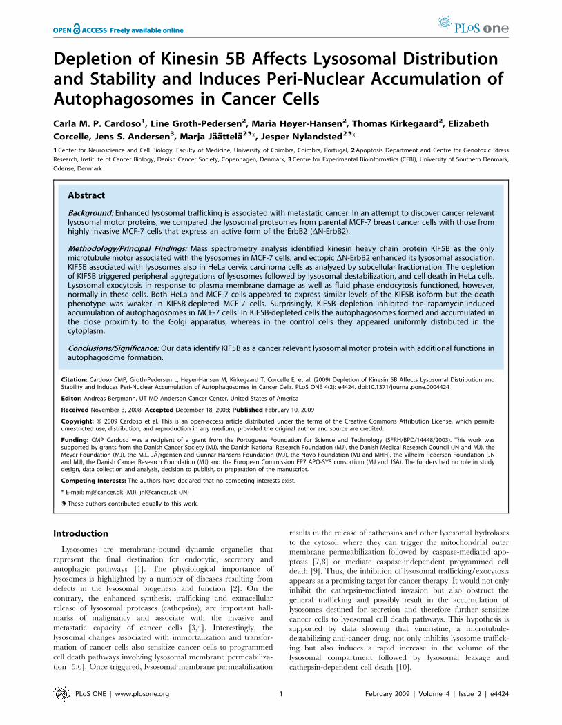
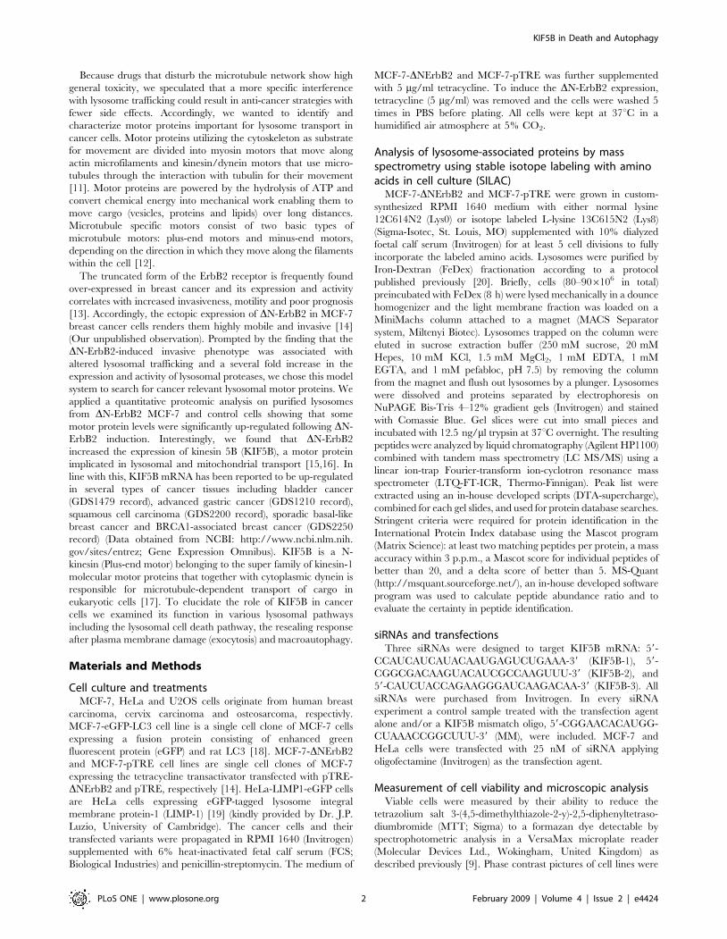
DNErbB2 increases the level of KIF5B in lysosomesMCF-7 breast carcinoma cells expressing amino-terminally
truncated constitutively active form of ErbB2 receptor tyrosine
kinase (DNErbB2) display a highly motile phenotype characterized
by extensive membrane ruffling, plasma membrane projections
and scattering of the cells (Fig. 1A). Furthermore, DNErbB2
expression induces the localization of lysosomes to the filopodia
(Fig. 1A) and a 3-4-fold up-regulation of lysosomal cysteine
cathepsin activity [6] suggesting that DN-ErbB2 changes the
KIF5B in Death and Autophagy
PLoS ONE | www.plosone.org 3 February 2009 | Volume 4 | Issue 2 | e4424
KIF5B in Death and Autophagy
PLoS ONE | www.plosone.org 4 February 2009 | Volume 4 | Issue 2 | e4424
lysosomal trafficking and content. In order to identify motor
proteins involved in lysosomal trafficking in cancer cells, we
compared the proteomes of lysosomes isolated from control MCF-
7 and MCF-7-DNErbB2 cells by stable-isotope labeling by amino
acids (Lys0/Lys8) in cell culture (SILAC) followed by mass
spectrometry analysis [22]. Six myosin motors and one microtu-
bule specific kinesin motor could be detected by this approach as
lysosome-associated motor proteins (Table 1 and Dataset S1). The
lysosomal association of three of the identified motor proteins
(Myosin Ib, Myosin Ic and kinesin heavy chain KIF5B) was up-
regulated by more than 25% upon ectopic DN-ErbB2 expression
in MCF-7 cells. To characterize the functional significance of these
three motors with regard to growth, survival and lysosomal
distribution we depleted them in MCF-7 and HeLa cervix
carcinoma cells by RNA interference. Only the siRNAs specific
for KIF5B affected these parameters (Fig. 2 and data not shown).
Thus, we chose to study the role of KIF5B on lysosomal function
in more detail.
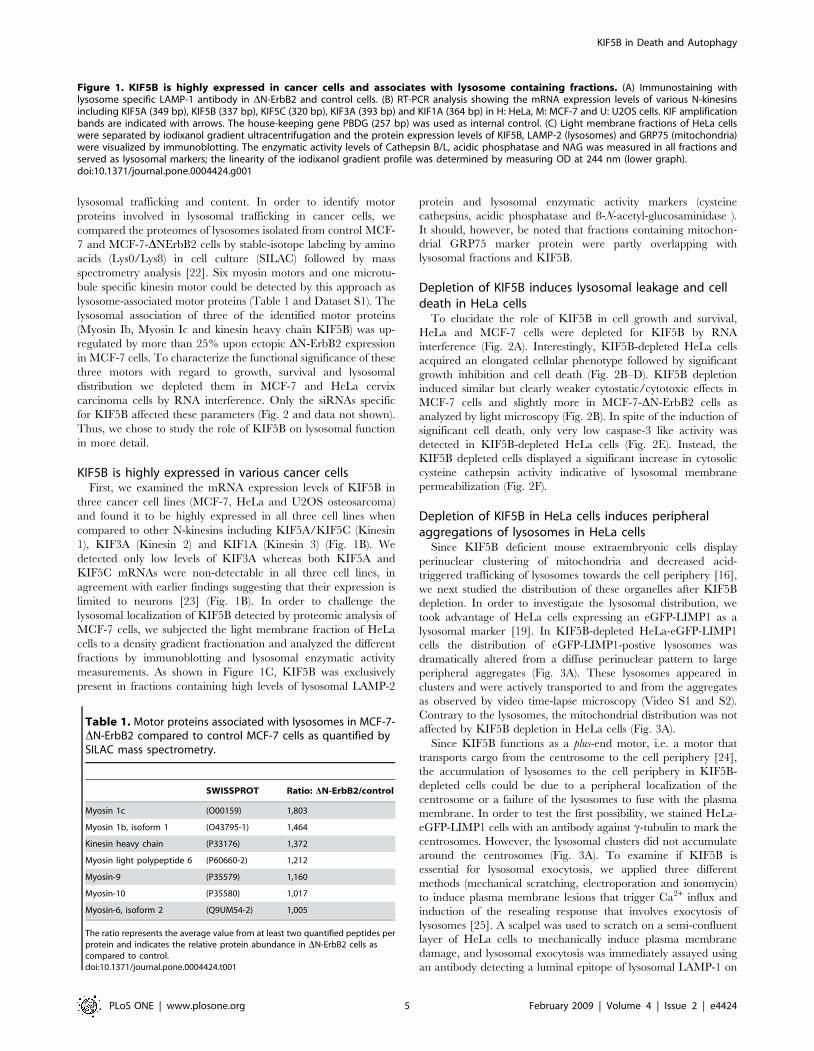
KIF5B is highly expressed in various cancer cellsFirst, we examined the mRNA expression levels of KIF5B in
three cancer cell lines (MCF-7, HeLa and U2OS osteosarcoma)
and found it to be highly expressed in all three cell lines when
compared to other N-kinesins including KIF5A/KIF5C (Kinesin
1), KIF3A (Kinesin 2) and KIF1A (Kinesin 3) (Fig. 1B). We
detected only low levels of KIF3A whereas both KIF5A and
KIF5C mRNAs were non-detectable in all three cell lines, in
agreement with earlier findings suggesting that their expression is
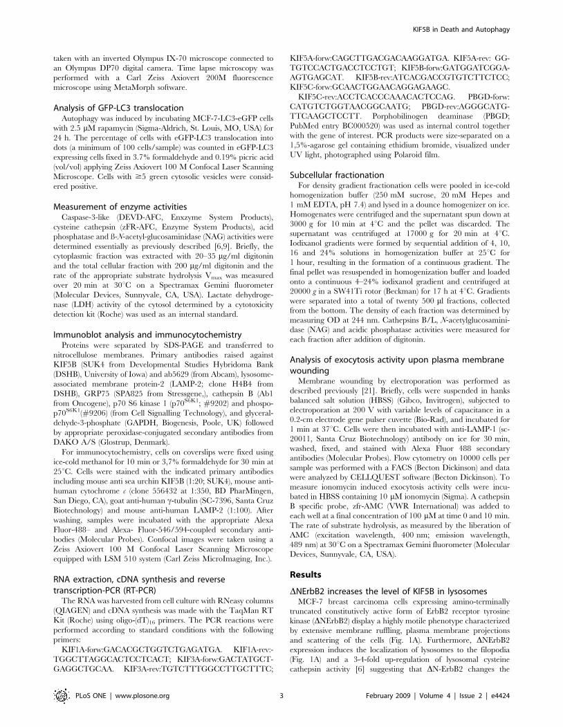
limited to neurons [23] (Fig. 1B). In order to challenge the
lysosomal localization of KIF5B detected by proteomic analysis of
MCF-7 cells, we subjected the light membrane fraction of HeLa
cells to a density gradient fractionation and analyzed the different
fractions by immunoblotting and lysosomal enzymatic activity
measurements. As shown in Figure 1C, KIF5B was exclusively
present in fractions containing high levels of lysosomal LAMP-2
protein and lysosomal enzymatic activity markers (cysteine
cathepsins, acidic phosphatase and ß-N-acetyl-glucosaminidase ).
It should, however, be noted that fractions containing mitochon-
drial GRP75 marker protein were partly overlapping with
lysosomal fractions and KIF5B.
Depletion of KIF5B induces lysosomal leakage and celldeath in HeLa cells
To elucidate the role of KIF5B in cell growth and survival,
HeLa and MCF-7 cells were depleted for KIF5B by RNA
interference (Fig. 2A). Interestingly, KIF5B-depleted HeLa cells
acquired an elongated cellular phenotype followed by significant
growth inhibition and cell death (Fig. 2B–D). KIF5B depletion
induced similar but clearly weaker cytostatic/cytotoxic effects in
MCF-7 cells and slightly more in MCF-7-DN-ErbB2 cells as
analyzed by light microscopy (Fig. 2B). In spite of the induction of
significant cell death, only very low caspase-3 like activity was
detected in KIF5B-depleted HeLa cells (Fig. 2E). Instead, the
KIF5B depleted cells displayed a significant increase in cytosolic
cysteine cathepsin activity indicative of lysosomal membrane
permeabilization (Fig. 2F).
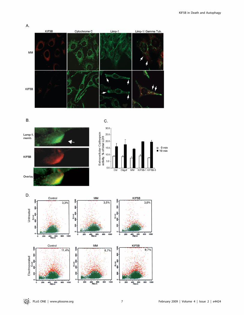
Depletion of KIF5B in HeLa cells induces peripheralaggregations of lysosomes in HeLa cells
Since KIF5B deficient mouse extraembryonic cells display
perinuclear clustering of mitochondria and decreased acid-
triggered trafficking of lysosomes towards the cell periphery [16],
we next studied the distribution of these organelles after KIF5B
depletion. In order to investigate the lysosomal distribution, we
took advantage of HeLa cells expressing an eGFP-LIMP1 as a
lysosomal marker [19]. In KIF5B-depleted HeLa-eGFP-LIMP1
cells the distribution of eGFP-LIMP1-postive lysosomes was
dramatically altered from a diffuse perinuclear pattern to large
peripheral aggregates (Fig. 3A). These lysosomes appeared in
clusters and were actively transported to and from the aggregates
as observed by video time-lapse microscopy (Video S1 and S2).
Contrary to the lysosomes, the mitochondrial distribution was not
affected by KIF5B depletion in HeLa cells (Fig. 3A).
Since KIF5B functions as a plus-end motor, i.e. a motor that
transports cargo from the centrosome to the cell periphery [24],
the accumulation of lysosomes to the cell periphery in KIF5B-
depleted cells could be due to a peripheral localization of the
centrosome or a failure of the lysosomes to fuse with the plasma
membrane. In order to test the first possibility, we stained HeLa-
eGFP-LIMP1 cells with an antibody against c-tubulin to mark the
centrosomes. However, the lysosomal clusters did not accumulate
around the centrosomes (Fig. 3A). To examine if KIF5B is
essential for lysosomal exocytosis, we applied three different
methods (mechanical scratching, electroporation and ionomycin)
to induce plasma membrane lesions that trigger Ca2+ influx and
induction of the resealing response that involves exocytosis of
lysosomes [25]. A scalpel was used to scratch on a semi-confluent
layer of HeLa cells to mechanically induce plasma membrane
damage, and lysosomal exocytosis was immediately assayed using
an antibody detecting a luminal epitope of lysosomal LAMP-1 on
Figure 1. KIF5B is highly expressed in cancer cells and associates with lysosome containing fractions. (A) Immunostaining withlysosome specific LAMP-1 antibody in DN-ErbB2 and control cells. (B) RT-PCR analysis showing the mRNA expression levels of various N-kinesinsincluding KIF5A (349 bp), KIF5B (337 bp), KIF5C (320 bp), KIF3A (393 bp) and KIF1A (364 bp) in H: HeLa, M: MCF-7 and U: U2OS cells. KIF amplificationbands are indicated with arrows. The house-keeping gene PBDG (257 bp) was used as internal control. (C) Light membrane fractions of HeLa cellswere separated by iodixanol gradient ultracentrifugation and the protein expression levels of KIF5B, LAMP-2 (lysosomes) and GRP75 (mitochondria)were visualized by immunoblotting. The enzymatic activity levels of Cathepsin B/L, acidic phosphatase and NAG was measured in all fractions andserved as lysosomal markers; the linearity of the iodixanol gradient profile was determined by measuring OD at 244 nm (lower graph).doi:10.1371/journal.pone.0004424.g001
Table 1. Motor proteins associated with lysosomes in MCF-7-DN-ErbB2 compared to control MCF-7 cells as quantified bySILAC mass spectrometry.
SWISSPROT Ratio: DN-ErbB2/control
Myosin 1c (O00159) 1,803
Myosin 1b, isoform 1 (O43795-1) 1,464
Kinesin heavy chain (P33176) 1,372
Myosin light polypeptide 6 (P60660-2) 1,212
Myosin-9 (P35579) 1,160
Myosin-10 (P35580) 1,017
Myosin-6, isoform 2 (Q9UM54-2) 1,005
The ratio represents the average value from at least two quantified peptides perprotein and indicates the relative protein abundance in DN-ErbB2 cells ascompared to control.doi:10.1371/journal.pone.0004424.t001
KIF5B in Death and Autophagy
PLoS ONE | www.plosone.org 5 February 2009 | Volume 4 | Issue 2 | e4424
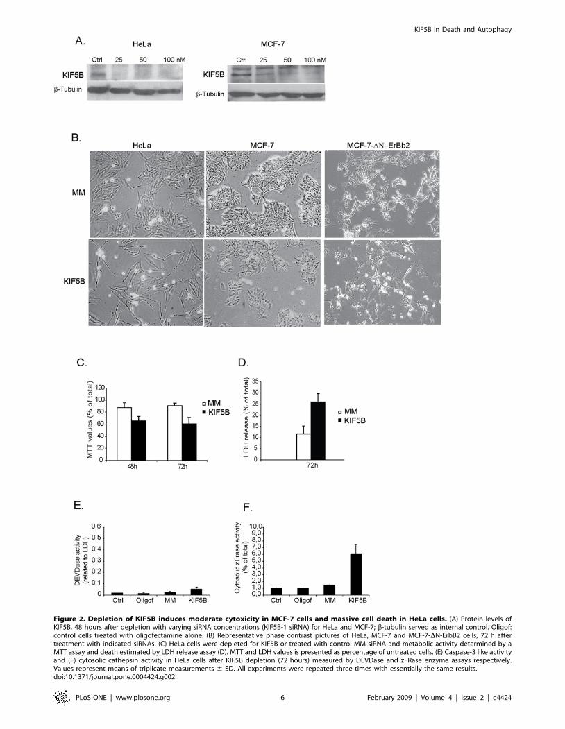
Figure 2. Depletion of KIF5B induces moderate cytoxicity in MCF-7 cells and massive cell death in HeLa cells. (A) Protein levels ofKIF5B, 48 hours after depletion with varying siRNA concentrations (KIF5B-1 siRNA) for HeLa and MCF-7; b-tubulin served as internal control. Oligof:control cells treated with oligofectamine alone. (B) Representative phase contrast pictures of HeLa, MCF-7 and MCF-7-DN-ErbB2 cells, 72 h aftertreatment with indicated siRNAs. (C) HeLa cells were depleted for KIF5B or treated with control MM siRNA and metabolic activity determined by aMTT assay and death estimated by LDH release assay (D). MTT and LDH values is presented as percentage of untreated cells. (E) Caspase-3 like activityand (F) cytosolic cathepsin activity in HeLa cells after KIF5B depletion (72 hours) measured by DEVDase and zFRase enzyme assays respectively.Values represent means of triplicate measurements 6 SD. All experiments were repeated three times with essentially the same results.doi:10.1371/journal.pone.0004424.g002
KIF5B in Death and Autophagy
PLoS ONE | www.plosone.org 6 February 2009 | Volume 4 | Issue 2 | e4424
KIF5B in Death and Autophagy
PLoS ONE | www.plosone.org 7 February 2009 | Volume 4 | Issue 2 | e4424
the cell surface. The surface fluorescence of LAMP-1 was
significantly increased at the damage site indicative of lysosomal
membrane resealing, and an additional co-localization and
accumulation of KIF5B at the damage site was observed
suggesting that KIF5B is involved in this response (Fig. 3B). Since
this method is not suitable for quantitative studies, we used
electroporation to induce small hydrophilic pores in the plasma
membrane to investigate if KIF5B was essential in the transport
process of lysosomes to the damage sites. The method is widely
used to introduce proteins and DNA into cells and depends on the
cells ability to reseal their plasma membrane after electroporation
[26]. HeLa cells were electroporated with increasing capacitance
and immediately after they were stained for surface LAMP-1
(Fig. 3D). Quantification of LAMP-1 exposed on the plasma
membrane by flow cytometry revealed a detectable level of
LAMP-1 on 3,3% of untreated cells. In contrast, when cells were
electroporated at 125 and 250 mF, LAMP-1 was detected on the
surface in 11,4 and 21% of the cells respectively. Cells depleted for
KIF5B and exposed to 125 mF did not display any significant
change in surface LAMP-1 as compared to control treated cells
exposed to 125 mF (Fig. 3D). Similarly, the ionomycin-induced
lysosomal exocytosis of luminal proteases was unaffected by
KIF5B depletion (Fig 3C). These data demonstrate that KIF5B is
not crucial for the lysosomal exocytosis and plasma membrane
resealing. Furthermore, the uptake of Alexa Flour 488-Dextran
(10 kDa) was not affected by KIF5B depletion indicating that
KIF5B is not required for fluid phase endocytosis (data not shown).
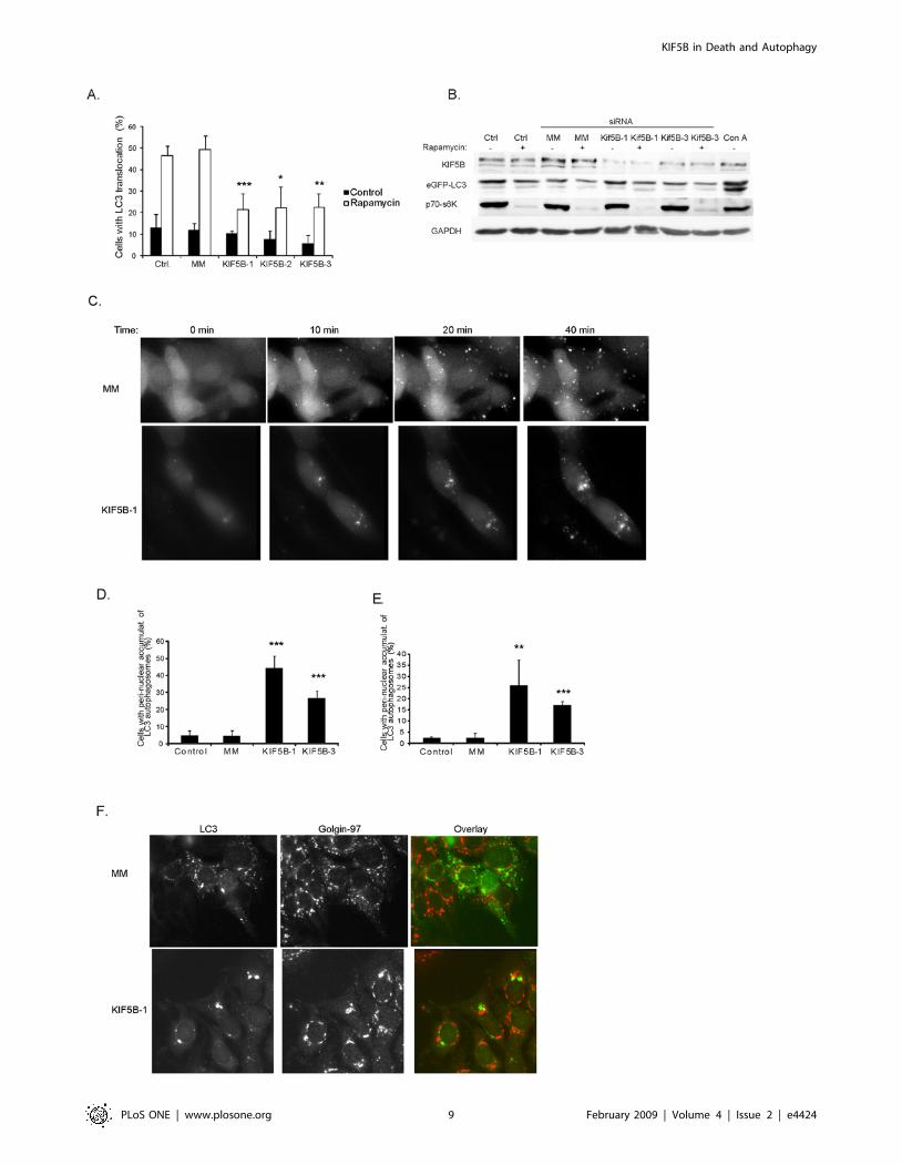
Depletion of KIF5B induces peri-nuclear accumulation ofautophagosomes
Next, we examined if KIF5B plays a role in autophagy, the
major lysosomal degradation pathway. For this purpose, we
treated MCF-7 cells stably expressing the autophagosome-
associated LC3 protein fused to the enhanced green fluorescence
protein (MCF-7-LC3-eGFP) with either rapamycin that induces
autophagy by inactivating the mammalian target of rapamycin
complex 1 (mTORC1) or concanamycin A that inhibits the
vacuolar V-ATPase activity in lysosomes resulting in reduced
turnover of autophagosomes as well as induction of autophago-
some formation via inhibition of mTORC1 [27]. Interestingly, the
depletion of KIF5B by three non-overlapping siRNAs significantly
decreased the ability of rapamycin to trigger the formation of LC3-
postive autophagic vesicles (Fig. 4A). This effect was not brought
about by changes in the ability of rapamycin to inhibit mTORC1
since the depletion of KIF5B did not have any influence on
mTORC1 activity as analyzed by the phosphorylation status of
p70 S6 kinase 1 (p70S6K1) (Fig. 4B). To explore the phenomenon
further, we followed the autophagosome formation in MCF-7-
LC3-eGFP cell treated with rapamycin (not shown) or concana-
mycin A by time lapse video microscopy for 45 min. Surprisingly,
the distribution of autophagosomes was dramatically altered by
KIF5B depletion. In KIF5B depleted cells the autophagosomes
appeared and accumulated mainly around the nucleus (Fig. 4C–E;
Video S3) whereas in cells treated with control siRNA they were
distributed diffusely throughout the cytoplasm (Fig. 4C; Video S4).
The perinuclear autophagosomes in KIF5B depleted cells were
located in close proximity to the golgi apparatus as visualized by
staining with an antibody against a trans-Golgi network membrane
protein Golgin-97 (Fig. 4F and Video S5). Since the distribution of
Golgi was not affected by KIF5B depletion, these data suggest that
KIF5B may transport a component(s) involved in the formation
and/or localization of autophagosomes in the cytoplasm.
Discussion
The data presented here suggest that KIF5B is implicated in
several pathways involving lysosomes and that its highly expressed
in all cancer cell lines tested, as compared to other N-Kinesins
including KIF5A/KIF5C (Kinesin 1 family), KIF3A (Kinesin 2
family) and KIF1A (Kinesin 3 family). The three KIF5 subfamily
members (KIF5A, KIF5B and KIF5C) display high similarity in
the amino acid sequence and probably share functional redun-
dancy and similar properties. However, at present it is unknown
how the individual KIF5 members might contribute or co-operate
in various transport mechanisms and why they differ in their
expression patterns in neurons and non-neuronal cells. Density
fractionation methodology from HeLa cells revealed, that KIF5B
is mainly represented in light-membrane fractions containing
lysosomes and to some extent in mitochondria.
MCF-7 and MCF-7-DN-ErbB2 cells depleted for KIF5B were
inhibited in their growth but displayed a less pronounced death
phenotype as observed in HeLa cells. Moreover, we were unable
to detect any changes in lysosomal or mitochondrial distribution in
MCF-7 cells, whereas HeLa cells displayed a distinct change in
lysosomal distribution followed by significant death. This discrep-
ancy between the two cell lines may be ascribed to differences in
motor protein expression levels and HeLa cells are probably more
dependent on KIF5B for plus-end directed motor protein activity.
The aggregation of lysosomes in the pericellular area preceding
death in HeLa cells suggested that KIF5B might play a role in
trafficking lysosomes proximal to the plasma membrane. Howev-
er, we were unable to detect any reduction in exocytosis activity
triggered either by ionomycin, or electroporation induced
membrane damage, following KIF5B depletion. Nevertheless, a
significant KIF5B translocation was observed, when HeLa cells
were exposed to mechanical induced plasma membrane lesions to
induce the resealing response and facilitate lysosomal exocytosis.
Here, we observed a considerable recruitment of LAMP-1 positive
lysosomes to reseal the damage site and significant colocalization
with KIF5B. These data signify that KIF5B could play a role in
transporting lysosomes to the plasma membrane destined for
exocytosis, but functional redundancy probably exists that
implicates other motor proteins as well. The aggregation of
lysosomes following KIF5B depletion can also be explained by an
alternative possibility: that KIF5B besides its role as a transporter
plays a role in positioning lysosomes at distinct sites in the
cytoplasm (predominantly perinuclear). Accordingly, depletion of
KIF5B would liberate lysosomes allowing their transport by other
motor proteins including N-kinesins towards the cortical areas of
Figure 3. KIF5B depletion induces pericellular aggregation of lysosomes in HeLa cells but has no impact on exocytosis activity. (A)Representative confocal pictures of either HeLa cells or HeLa stably expressing LIMP-1-EGFP transfected with KIF5B or MM siRNA, and stained withindicated antibodies. (B) HeLa cells seeded on coverslips (80% confluency) were membrane wounded with a scalpel and immediately after stained forsurface LAMP-1; cells were subsequently fixated and stained for KIF5B. (C) HeLa cells transfected with indicated siRNAs were (after 48 h) stimulated toexocytose with 10 mM ionomycin. Extracellular secretion of lysosomal cathepsins was measured by a zFR-AMC enzyme assay and values (means oftriplicate measurements 6 SD) were expressed as percent of total cellular LDH content. (D) Quantification of surface LAMP-1 in electroporated HeLacells by flow cytometry. Red and green indicates cells in two different gates. The percentage of cells in the red gate was used to estimate the amountof surface-exposed LAMP-1 +/2 electroporation. FL1-H: fluorescence intensity. FSC-H: forward side scatter.doi:10.1371/journal.pone.0004424.g003
KIF5B in Death and Autophagy
PLoS ONE | www.plosone.org 8 February 2009 | Volume 4 | Issue 2 | e4424
KIF5B in Death and Autophagy
PLoS ONE | www.plosone.org 9 February 2009 | Volume 4 | Issue 2 | e4424
the cell resulting in lysosomal aggregations. This implies that
recruitment of lysosomes by KIF5B to microtubules may localize
or queue them and not necessarily facilitate long distance travel.
The death pathway induced after KIF5B depletion in HeLa
cells triggered the aggregation of lysosomes followed by lysosomal
destabilization and subsequent release of lysosomal cathepsins to
the cytosol. We observed only limited caspase-3 like activation
suggesting that the classical caspase mediated death pathway plays
a minor role in the death mode observed. Lysosomal cathepsins
function as effective mediators of programmed cell death but the
pathways leading to LMP are, however, poorly understood. We
have recently shown that vincristine, a compound that destabilizes
microtubules and is frequently used in cancer therapy, induce
dramatic aggregations of lysosomes and induces LMP and death in
HeLa cells [10]. The two treatments might have similar
consequences for lysosomal distribution resulting in lysosomes
that are brought together in an uncontrolled manner inducing
aggregations and subsequent destabilization of lysosomes resulting
in death.
The more pronounced death phenotype observed in HeLa cells
when compared to MCF-7/MCF-7-DN-ErbB2 cells could be
explained by a higher dependency on proper KIF5B motor
protein activity basically to deal with a high metabolic activity and
growth rate. Alternatively, MCF-7 cells might encompass more
functional redundancy through expression of various motor
proteins than in HeLa cells. Most of the knowledge about KIF5
mediated transport is based on studies in neurons where KIF5
family members can transport various cargoes and its activity
seems to be dominant over other motor activities [12], however
the data in non-neuronal cells are still limited.
Depletion of KIF5B by RNA interference in MCF-7 cells had a
surprising impact on autophagosome formation/localization as
determined by the translocation of LC3-eGFP to autophagosomes.
We estimated autophagic activity by scoring LC3-eGFP positive
autophagosomes after stimulation with rapamycin and observed a
significant reduction upon KIF5B depletion. This finding
prompted us to follow autophagosome formation in real time by
time lapse microscopy using higher concentrations of either
rapamycin or concanamycin A enabling us to follow the process in
a shorter time frame. The remarkable accumulations of autopha-
gosomes around the nucleus in cells depleted for KIF5B suggested
that KIF5B could be involved directly in the transport of
autophagosomes along microtubule tracks to the cytoplasm.
However, our recent quantitative mass-spectometry analysis on
membrane associated proteins purified from autophagosomes
(from MCF-7 cells treated with rapamycin or concanamycin A)
revealed that KIF5B is not directly associated with autophago-
somes (MHH, JA, JO, unpublished data). Alternatively, KIF5B
might be involved in the transport of critical factor(s) important for
initiation of the autophagic process and proper distribution of
autophagosomes in the cytoplasm. Accordingly, removal of KIF5B
might prevent the transport of initiation factor(s) and result in
formation of autophagosomes accumulating close to the microtu-
bule organizing center and nucleus. Since the autophagosomes
appeared mainly at one site of the nucleus (Fig. 4C and Video S3)
and close to the trans-golgi network (Fig. 4F and Video S5)
suggests, that the putative initiation factor(s) transported by KIF5B
could be golgi derived and affect the distribution of autophago-
somes after KIF5B depletion. In addition, KIF5B depletion did
not have any influence on the activity of mTOR (as determined by
the phosphorylation status of p70 S6 kinase) indicating that the
motor protein is acting down-stream of mTOR.
Our data demonstrate that KIF5B is highly expressed in cancer
cells and plays a significant role in growth and survival of HeLa
cells. Additionally, we show that KIF5B is very abundant at the
site of plasma membrane damage co localizing with lysosomes
destined for exocytosis although it’s not essential suggesting that
functional redundancy probable exist. Moreover, we provide data
indicating that KIF5B is involved in the initial formation/
localization of autophagosomes and might transport component(s)
important for the autophagic process.
Supporting Information
Dataset S1 Motor proteins associated with lysosomes in MCF-
7-deltaN-ErbB2 compared to control MCF-7 cells as quantified by
SILAC mass spectrometry.
Found at: doi:10.1371/journal.pone.0004424.s001 (1.32 MB
XLS)
Video S1 Control of HeLa-LIMP1-eGFP cells
Found at: doi:10.1371/journal.pone.0004424.s002 (1.58 MB
MOV)
Video S2 HeLa-LIMP1-eGFP cells depleted for KIF5B (after
72h) and imaged by time-lapse microscopy.
Found at: doi:10.1371/journal.pone.0004424.s003 (8.62 MB
MOV)
Video S3 MCF-7-LC3-eGFP cells depleted for KIF5B were
stimulated with 6 nM Concanamycin A and followed by time-
lapse microscopy.
Found at: doi:10.1371/journal.pone.0004424.s004 (8.90 MB
MOV)
Video S4 MCF-7-LC3-eGFP cells treated with control siRNA
(MM, 72h) were stimulated with 6 nM Concanamycin A and
followed by time-lapse microscopy.
Found at: doi:10.1371/journal.pone.0004424.s005 (9.23 MB
MOV)
Video S5 MCF-7-LC3-eGFP cells co-expressing dsRED-Golgi
(trans Golgi stack) and depleted for KIF5B were stimulated with 6
nM Concanamycin A and imaged by time-lapse microscopy.
Found at: doi:10.1371/journal.pone.0004424.s006 (8.48 MB
MOV)
Figure 4. KIF5B depletion suppresses autophagy and induces nuclear accumulation of autophagosomes. (A) MCF-7-LC3-eGFP cellstreated with oligofectamine or transfected with indicated siRNAs were left untreated or stimulated with rapamycin for 24 h. The percentage of cellswith LC3-eGFP localized to $five cytosolic granular structures was estimated by counting a minimum of 100 cells/sample. Values represent means ofthree independent experiments 6 SD. (B) Immunoblots showing the protein levels after depletion (72 h) with indicated siRNAs or treatment with1 mM rapamycin for 12 h. p70-s6K: phosphorylated form of p70 S6 kinase. eGFP-LC3: eGFP antibody specific for eGFP (fused to LC3) (C)Representative phase contrast pictures adapted from time lapse movies of MCF-7-LC3-eGFP cells treated with Concanamycin A (6 nM) to induceautophagy and LC3-eGFP translocation to autophagosomes, 72 h after transfection with indicated siRNAs. (D and E) MCF-7-LC3-eGFP cellstransfected with indicated siRNAs were (after 72 h) imaged by time lapse microscopy during treatment with either 6 nM concanamycin A (D) or 4 mMrapamycin (E). The percentage of cells displaying nuclear accumulation of LC3-eGFP autophagosomes were scored after 45 min incubation. (F)Immuno staining of trans-Golgi (Golgin-97 Ab) in MCF-7-LC3-eGFP cells depleted for KIF5B and incubated for 45 min with concanamycin A. Valuesrepresent means of 3–4 independent experiments. P-values: MM/KIF5B. *: p,0,05; **: p,0,01; ***: p,0,001 (student’s T-test).doi:10.1371/journal.pone.0004424.g004
KIF5B in Death and Autophagy
PLoS ONE | www.plosone.org 10 February 2009 | Volume 4 | Issue 2 | e4424
Acknowledgments
We thank Jane Hinriksen for expert technical assistance. We are grateful
for the monoclonal antibody SUK 4 developed by J. Scholey and the
monoclonal antibody H4B4 developed by J.T. August and J.E.K. Hildreth
(both obtained from the Developmental Studies Hybridoma Bank
developed under the auspices of the NICHD and maintained by The
University of Iowa, Department of Biological Sciences, Iowa City, IA
52242).
Author Contributions
Conceived and designed the experiments: CMPC LGP MHH TK EC JSA
MJ. Performed the experiments: CMPC LGP MHH TK EC JSA JN.
Analyzed the data: CMPC JSA MJ JN. Wrote the paper: CMPC MJ JN.
References
1. Eskelinen EL, Tanaka Y, Saftig P (2003) At the acidic edge: emerging functionsfor lysosomal membrane proteins. Trends Cell Biol 13: 137–145.
2. Gieselmann V (1995) Lysosomal storage diseases. Biochim Biophys Acta 1270:103–136.
3. Gocheva V, Joyce JA (2007) Cysteine cathepsins and the cutting edge of cancerinvasion. Cell Cycle 6: 60–64.
4. Jedeszko C, Sloane BF (2004) Cysteine cathepsins in human cancer. Biol Chem
385: 1017–1027.5. Fehrenbacher N, Gyrd-Hansen M, Poulsen B, Felbor U, Kallunki T, et al.
(2004) Sensitization to the lysosomal cell death pathway upon immortalizationand transformation. Cancer Res 64: 5301–5310.
6. Fehrenbacher N, Bastholm L, Kirkegaard-Sorensen T, Rafn B, Bottzauw T, et
al. (2008) Sensitization to the lysosomal cell death pathway by oncogene-induceddown-regulation of lysosome-associated membrane proteins 1 and 2. Cancer Res
68: 6623–6633.7. Roberg K, Kagedal K, Ollinger K (2002) Microinjection of cathepsin d induces
caspase-dependent apoptosis in fibroblasts. Am J Pathol 161: 89–96.8. Boya P, Andreau K, Poncet D, Zamzami N, Perfettini JL, et al. (2003)
Lysosomal membrane permeabilization induces cell death in a mitochondrion-
dependent fashion. J Exp Med 197: 1323–1334.9. Foghsgaard L, Wissing D, Mauch D, Lademann U, Bastholm L, et al. (2001)
Cathepsin B acts as a dominant execution protease in tumor cell apoptosisinduced by tumor necrosis factor. J Cell Biol 153: 999–1010.
10. Groth-Pedersen L, Ostenfeld MS, Hoyer-Hansen M, Nylandsted J, Jaattela M
(2007) Vincristine induces dramatic lysosomal changes and sensitizes cancer cellsto lysosome-destabilizing siramesine. Cancer Res 67: 2217–2225.
11. Mallik R, Gross SP (2004) Molecular motors: strategies to get along. Curr Biol14: R971–R982.
12. Hirokawa N, Takemura R (2005) Molecular motors and mechanisms ofdirectional transport in neurons. Nat Rev Neurosci 6: 201–214.
13. Ross JS, Fletcher JA (1998) The HER-2/neu Oncogene in Breast Cancer:
Prognostic Factor, Predictive Factor, and Target for Therapy. Oncologist 3:237–252.
14. Egeblad M, Mortensen OH, Jaattela M (2001) Truncated ErbB2 receptorenhances ErbB1 signaling and induces reversible, ERK-independent loss of
epithelial morphology. Int J Cancer 94: 185–191.
15. Nakata T, Hirokawa N (1995) Point mutation of adenosine triphosphate-binding
motif generated rigor kinesin that selectively blocks anterograde lysosome
membrane transport. J Cell Biol 131: 1039–1053.
16. Tanaka Y, Kanai Y, Okada Y, Nonaka S, Takeda S, et al. (1998) Targeted
disruption of mouse conventional kinesin heavy chain, kif5B, results in abnormal
perinuclear clustering of mitochondria. Cell 93: 1147–1158.
17. Goldstein LS, Yang Z (2000) Microtubule-based transport systems in neurons:
the roles of kinesins and dyneins. Annu Rev Neurosci 23: 39–71.
18. Hoyer-Hansen M, Bastholm L, Szyniarowski P, Campanella M, Szabadkai G, et
al. (2007) Control of macroautophagy by calcium, calmodulin-dependent kinase
kinase-beta, and Bcl-2. Mol Cell 25: 193–205.
19. Bampton ET, Goemans CG, Niranjan D, Mizushima N, Tolkovsky AM (2005)
The dynamics of autophagy visualized in live cells: from autophagosome
formation to fusion with endo/lysosomes. Autophagy 1: 23–36.
20. Diettrich O, Mills K, Johnson AW, Hasilik A, Winchester BG (1998) Application
of magnetic chromatography to the isolation of lysosomes from fibroblasts of
patients with lysosomal storage disorders. FEBS Lett 441: 369–372.
21. Huynh C, Roth D, Ward DM, Kaplan J, Andrews NW (2004) Defective
lysosomal exocytosis and plasma membrane repair in Chediak-Higashi/beige
cells. Proc Natl Acad Sci U S A 101: 16795–16800.
22. Andersen JS, Lam YW, Leung AK, Ong SE, Lyon CE, et al. (2005) Nucleolar
proteome dynamics. Nature 433: 77–83.
23. Kanai Y, Okada Y, Tanaka Y, Harada A, Terada S, et al. (2000) KIF5C, a
novel neuronal kinesin enriched in motor neurons. J Neurosci 20: 6374–6384.
24. Kasprzak AA, Hajdo L (2002) Directionality of kinesin motors. Acta Biochim
Pol 49: 813–821.
25. Reddy A, Caler EV, Andrews NW (2001) Plasma membrane repair is mediated
by Ca(2+)-regulated exocytosis of lysosomes. Cell 106: 157–169.
26. Potter H (2001) Transfection by electroporation. Curr Protoc Immunol Chapter
10: Unit.
27. Ostenfeld MS, Hoyer-Hansen M, Bastholm L, Fehrenbacher N, Olsen OD, et
al. (2008) Anti-cancer agent siramesine is a lysosomotropic detergent that
induces cytoprotective autophagosome accumulation. Autophagy 4: 487–499.
KIF5B in Death and Autophagy
PLoS ONE | www.plosone.org 11 February 2009 | Volume 4 | Issue 2 | e4424