department of prosthodontics pdm preclinical i l5 - engl · 3/21/2020 · mandibular movements....
TRANSCRIPT

1
Department of Prosthodontics
PDM_Preclinical_I_L5 -_Engl
Functional – mechanical equilibrium of the periodontium. Muscle
forces. Masticatory pressure. Periodontal pressure. Biomechanics of
Mandibular movements. Basic movements. Theory of Mandibular movements
Slide 1
The masticatory system is a complex musculoskeletal system, which consists of the
jaws.And anatomically complex of muscles, involved in mastication. These muscles bring about
mandibular motion and generate forces, which can influence morphology and Material properties
of the osseous components. The activation and coordination of the jaw muscles determine the
direction of jaw movement, control occlusal force, and load the bones of the skull in various
ways. The jaw muscles are involved in a much broader range of motor tasks.
Slide 2
2
Under physiological conditions, the muscle is activated until nervous stimulation ceases.
The level of muscle activation, and with it the amount of force generated, is controlled by two
major mechanisms: rate modulation, altering the frequency of neuronal action potentials at the
neuromuscular junction and recruitment modulation, altering the number of active motor units in
the muscle.
Slide 3
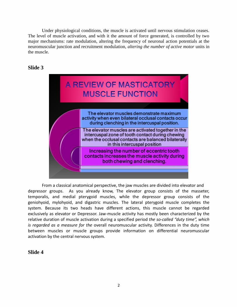
From a classical anatomical perspective, the jaw muscles are divided into elevator and
depressor groups. As you already know, The elevator group consists of the masseter, temporalis, and medial pterygoid muscles, while the depressor group consists of the geniohyoid, mylohyoid, and digastric muscles. The lateral pterygoid muscle completes the system. Because its two heads have different actions, this muscle cannot be regarded exclusively as elevator or Depressor. Jaw-muscle activity has mostly been characterized by the relative duration of muscle activation during a specified period the so-called “duty time”, which is regarded as a measure for the overall neuromuscular activity. Differences in the duty time between muscles or muscle groups provide information on differential neuromuscular activation by the central nervous system.
Slide 4

3
Slide 5
Dental absence interferes in the physiological functioning of the masticatory system, promoting occlusal and functional alterations. These data evidence the strong influence of

4
dental loss over the maximal bite force and small correlation between bite force and electromyographic activity. Changes in vertical jaw opening may affect the relative contributions of various masticatory muscles to bite force production. EMG activity usualy has recorded simultaneously from the masseter, and anterior, middle, and posterior temporalis muscles, during controlled isometric biting at different force levels and vertical jaw openings.
Slide 6
Every individual muscle group develop muscle activity with a certain force. The size of
the masticatory pressure is proportional to the size of this aggregate and absolute muscle strength
and it is inverse to the size of the occlusal surface. The Absolute muscle force is in direct
proportion to the mass of muscle groups. This slide shows data for the masticatory muscles as
follows: The thickness determines the absolute muscle strength, which includes the synergetic
action of these symmetrical muscles.
Slide 7
5
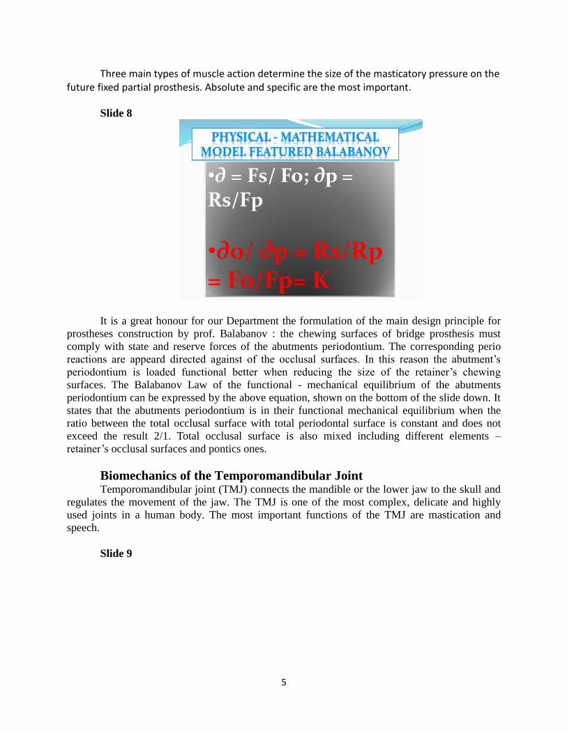
Three main types of muscle action determine the size of the masticatory pressure on the future fixed partial prosthesis. Absolute and specific are the most important.
Slide 8
It is a great honour for our Department the formulation of the main design principle for
prostheses construction by prof. Balabanov : the chewing surfaces of bridge prosthesis must
comply with state and reserve forces of the abutments periodontium. The corresponding perio
reactions are appeard directed against of the occlusal surfaces. In this reason the abutment’s
periodontium is loaded functional better when reducing the size of the retainer’s chewing
surfaces. The Balabanov Law of the functional - mechanical equilibrium of the abutments
periodontium can be expressed by the above equation, shown on the bottom of the slide down. It
states that the abutments periodontium is in their functional mechanical equilibrium when the
ratio between the total occlusal surface with total periodontal surface is constant and does not
exceed the result 2/1. Total occlusal surface is also mixed including different elements –
retainer’s occlusal surfaces and pontics ones.
Biomechanics of the Temporomandibular Joint Temporomandibular joint (TMJ) connects the mandible or the lower jaw to the skull and
regulates the movement of the jaw. The TMJ is one of the most complex, delicate and highly
used joints in a human body. The most important functions of the TMJ are mastication and
speech.
Slide 9

6
TMJ is a bi-condylar joint in which the condyles, the movable round upper ends of the
Mandible, function at the same time. Between the condyle and the articular fossa is a disc made
of fibrocartilage that acts as a cushion to absorb stress and allows the condyle to move easily
when the mouth opens and closes. Mandibular movement around the horizontal axis is an
opening and closing motion. It is referred to as a hinge movement, and the horizontal axis
around which it occurs is therefore referred to as the hinge axis (Figure 4-2). The hinge
movement is probably the only example of mandibular activity in which a “pure” rotational
movement occurs.
Slide 10
Slide 11

7
Slide 12

8
Slide 13
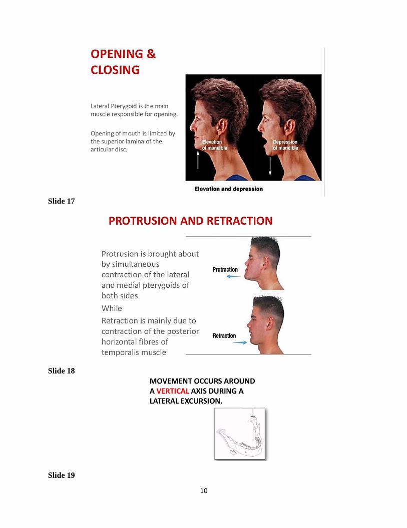
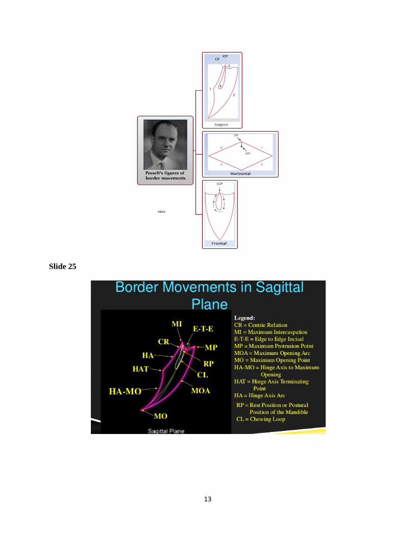
Mandibular movement refers to the muscle- and ligament-activated border and/or
intraborder movements of the lower jaw. There are five types of mandibular movements
including rotational, horizontal axis, frontal axis, sagittal axis, and translational. Mandibular
movement is affected by several factors such as the muscles used in suspending the jaw,
mandibular articulation, and the synovial joint system. Study of this movement is important for
the fields of dentistry and orthodontics as it describes the concepts related to dental occlusion
and the masticatory processes of the jaw.
Slide 14

9
Slide 15
Slide 16
10
Slide 17
Slide 18
Slide 19

11
Slide 20
Slide 21

12
Slide 22
Slide 23
Slide 24
13
Slide 25

14
Slide 26
Slide 27

15
Slide 28
Functional conditions of the mandible: theory and physiology
The functional conditions of the mandible are differentiated according to the number of
kinematic degrees of freedom assigned to each mandibular movement. As mandibular
movements reflect the functional morphology of temporomandibular joint (TMJ), the occlusal
morphology of each tooth may be functionally related to its antagonist, to the TMJ and to the
other components of the stomatognathic system in reasonably precise ways.
Slide 29
16
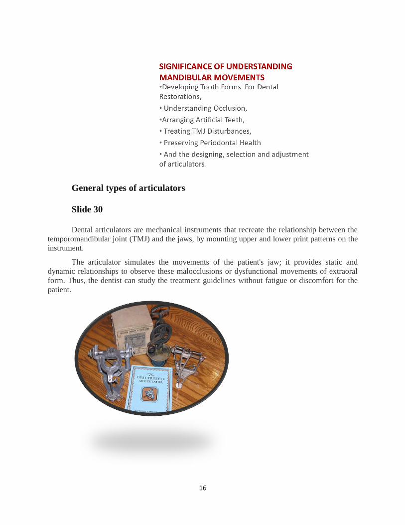
General types of articulators
Slide 30
Dental articulators are mechanical instruments that recreate the relationship between the
temporomandibular joint (TMJ) and the jaws, by mounting upper and lower print patterns on the
instrument.
The articulator simulates the movements of the patient's jaw; it provides static and
dynamic relationships to observe these malocclusions or dysfunctional movements of extraoral
form. Thus, the dentist can study the treatment guidelines without fatigue or discomfort for the
patient.
17
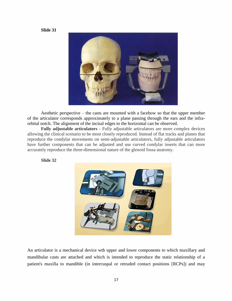
Slide 31
Aesthetic perspective – the casts are mounted with a facebow so that the upper member
of the articulator corresponds approximately to a plane passing through the ears and the infra-
orbital notch. The alignment of the incisal edges to the horizontal can be observed.
Fully adjustable articulators - Fully adjustable articulators are more complex devices
allowing the clinical scenario to be most closely reproduced. Instead of flat tracks and planes that
reproduce the condylar movements on semi-adjustable articulators, fully adjustable articulators
have further components that can be adjusted and use curved condylar inserts that can more
accurately reproduce the three-dimensional nature of the glenoid fossa anatomy.
Slide 32
An articulator is a mechanical device wth upper and lower components to which maxillary and
mandibular casts are attached and which is intended to reproduce the static relationship of a
patient's maxilla to mandible (in intercuspal or retruded contact positions [RCPs]) and may

18
provide to a limited extent for lateral and protrusive movements. Semi adjustable articulators are
the most often used in dental practice.
Articulators are used for the following:
•
Study individual teeth and full dental arches for diagnosis and treatment planning
•
Allow adjustment of fixed and removable prostheses and indirect dental restorations
Articulators have been the cornerstone requirement of prosthodontics and restorative
dentistry. However, their key role is changing with the progressive incorporation of
technological advances. The development of digital records and virtual articulators is
transforming all aspects of dental practice; as the most far-reaching and definitive change
for case management and education, it is an exciting transformation.
There are many designs of articulators, but in general there are four different types:
•Simple hinge
•Average value (plane-line)
•Semiadjustable
•Fully adjustable
Average value articulators have their condylar angle fixed at 30°. There is no provision for an
adjustment for condylar side shift but they may have an adjustable incisal guidance (Fig. 9-1, B).
Semiadjustable articulators allow adjustment of condylar inclination and side shift (Bennett
angle or progressive side shift) and in some designs for Bennett movement or immediate side
shift. Intercondylar width is usually fixed at 110 mm, but some articulators allow different
intercondylar width settings.
Реферат на тема : Движения на долната челюст и видове артикулатори