department of ophthalmology - nyee resident training... · department of ophthalmology presents ......
TRANSCRIPT

/
Department of Ophthalmology
Presents
15th Annual Resident and Fellow
Research Day

15th Annual Resident & Fellows Research Day May 20, 2011
pgJasmine Francis, M.D. Touchless Levitation Technique For Management of Posteriorly
Dislocated Silicone Intraocular Lenses or Implants 1
Sung Chul Park, M.D. In-vivo, 3-Dimensional Imaging of the Lamina Cribrosa Horizontal Central Ridge and Lamina Cribrosa Deformation in Normal and Glaucoma Subjects
2
Joseph Panarelli, M.D. Transscleral Diode Laser Cyclophotocoagulation following Baerveldt Glaucoma Implant Surgery
3
Madhavi Kurli, M.D. Visual Prognosis after Idiopathic Epiretinal Membrane Surgery based on High Resolution Spectral Domain Optical Coherence Tomography
4
Anisha Jangi, M.D. Descemet Stripping Automated Endothelial Keratoplasty After Failed Penetrating Keratoplasty
5
Syril Dorairaj, M.D. Dynamic Analysis of Iris Configuration 6
Jocelyn Kuryan, M.D. Suture Fixation of Posterior Chamber Intreocular Lenses: Indications and outcomes
7
Yogita Kashyap, M.D. Indications for CT Scan In Patients with Trauma 8
Ian Chan, M.D. Rescue of cybrids containing Leber Hereditary Optic Neuropathy (LHON) mutation using Bax-inhibiting Peptide
9
Daniel Hayes, M.D. Corneal Hysteresis and Beta-zone Parapapillary Atrophy 11
Amar Patel, M.D. Surgical Outcomes of the Boston Type 1 Keratoprosthesis 12
Kenneth/ S. Schor, M.D.
Rates Of Visual Field (VF) Progression In Distinct Optic Disc Phenotypes
13
Gennady Landa, M.D. SD-OCT Analysis of Subretinal Deposits in Acute Central Serous Retinopathy
14
Olivia Lee, M.D. Serum Estrogen and Progesterone Levels and Intraocular Inflammation 15

2
Sri Krish/na Mukkamala, M.D.
The Expanded Spectrum of Focal Choroidal Excavation 16
Saman Kiumehr, M.D.
In Vivo Evaluation of Focal Lamina Cribrosa Defects in Glaucoma 17
Kasra Eliasieh, M.D.
Peripapillary blood flow and b-Zone Peripapillary Atrophy in Primary Open-Angle Glaucoma
18
De Moraes, Carlos Gustavo, M.D.
Optic Disc Progression and Rates of Visual Field Change in Treated Glaucoma
19
Alice Hong, M.D. Trends in Pediatric Corneal Ulcers at the New York Eye and Ear Infirmary
20
Robert McGlynn, M.D.
Decompression Retinopathy: A Review 21
Lauren Schneider, M.D.
Specular Microscopy of the Contralateral Eye in Unilateral Iridocorneal Endothelial Syndrome
22
Kevin Rosenberg, M.D.
Improving eccentric fixation using Jitterbug program 23
Wendy Huang, M.D.
Review of Pediatric Uveitis Patients at a Tertiary Referral Center 24
Ambika Hoguet, M.D.
Ocular Complications of New Color IrisTM Cosmetic Iris Implants 25
Robert Lowe, M.D.
Clinical and Optical Coherence Tomography Findings in Patients Undergoing Focal Laser Treatment with the Navilas Laser System
26
David Warrow, M.D.
Pars Plana Vitrectomy with Silicone Oil or Gas Tamponade in Eyes with Diabetic Tractional Retinal Detachment
28
Benjamin Bert, M.D.
Sympathizing Intraocular Pressure Response to Contralateral Steroid Drop Usage
29
Dan Yin, M.D. Intravitreal Injection of Kenalog as an Adjunctive Treatment for Uveitis Patient Undergoing Cataract Surgery
30
Jae You/ng (Jane) You, M.D.
Uveal Schwannoma with Extrascleral Extension: Report of a Case and Review of Literature
31
Chavakij Bhoomibunchoo, M.D.
Retinal Function Imaging for Evaluating of Changes in Retinal Blood Flow after Pan-retinal Laser Photocoagulation in Patients with Proliferative Diabetic Retinopathy
32
Timothy Sullivan, M.D.
Post Procedure Pressure Spike after ALT versus SLT with Lopidine Prophylaxis
33

3
Nicole S/cripsema
Effect of β-Zone Parapapillary Atrophy (βPPA) on Retinal Blood Flow Velocity in Primary Open-Angle Glaucoma (POAG) and Exfoliative Glaucoma (XFG)
35
Valerie Trubnik, M.D.
Comparison of Retinal Blood Flow Velocities in Normal Tension Glaucoma (NTG) and High Tension Glaucoma (HTG) using the Retinal Functional Imager (RFI)
37
Christopher Seebruck, M.D.
Comparison between Macular Pigment Optical Density Measurements Using Two-Wavelength Autofluorescence (SLO) and Macular Pigment Reflectometry (MPR)
39
Nikola Ragusa, M.D.
The Effect of Uncomplicated Cataract Extraction on Rates of Visual Field Sensitivity Change Measured with Automated Pointwise Linear Regression Analysis in Glaucoma Patients
41
Alfredo R Castillejos, M.D.
ASFDOCT Sequential Imaging of Schlemm’s Canal and the Collector Channels in Normal and POAG Eyes
42
Yacoub Yousef, M.D.
Optical Coherence Tomography of Radiation Optic Neuropathy 44

1
Touchless Levitation Technique for Management of Posteriorly Dislocated Silicone Intraocular Lenses or Implants Jasmine H. Francis MD, Paul A. Latkany MD.(Work published in Archives: Arch Ophthalmol. 2011 Apr;129(4):552-4.) Objective: The posterior dislocation of silicone-composed fluocinolone acetonide (FA) implants has been documented, spontaneously and during explantation. However, the elastic properties of silicone can make their retrieval challenging; and herein is explored a novel technique for intraocular silicone implant or lens removal. Methods or design: In vitro investigation of silicone intraocular lens and FA implant properties in basic salt solution (BSS), silicone oil (SO), and perfluorooctane (PFO); with exploration of surgical techniques for implant removal. Results: Silicone intraocular lens and FA implant may be levitated from the bottom of an aqueous and silicone oil-filled vial by tilting the vial; and thereby engaging the implant at the aqueous/silicone oil interface. The high viscosity of the silicone oil stabilizes the implant during its retrieval,.. Conclusions: A novel proposed touchless levitation technique using silicone oil for silicone implant retrieval is described. It allows for a controlled explantation of dislocated silicone implants or lenses without touching the implant on the macula, and circumventing the associated risk of volume fluctuation as the implant is removed through a large incision. Clinical Relevance: This proposed surgical technique may be employed for retrieval of a posteriorly dislocated intraocular silicone lens or implant; it attempts to evade risks of touching the retina and to dampen the possibility of eye collapse associated with surgical manipulations in the presence of large incisions.

2
I/n-vivo, 3-Dimensional Imaging of the Lamina Cribrosa Horizontal Central Ridge and Lamina Cribrosa Deformation in Normal and Glaucoma Subjects Authors: Sung Chul Park,1,2 Saman Kiumehr,1 Syril Dorairaj,1 Christopher C Teng,1,2 Celso Tello,1,2 Jeffrey M Liebmann,1,3 Robert Ritch1,2 Affiliations: 1Einhorn Clinical Research Center, New York Eye and Ear Infirmary, New York, NY; 2Dept. of Ophthalmology, New York Medical College, Valhalla, NY; 3Dept. of Ophthalmology, NYU School of Medicine, New York, NY Purpose: To compare the morphology and position of the lamina cribrosa (LC) between high-tension glaucoma (HTG) and normal subjects using enhanced depth imaging spectral-domain optical coherence tomography (EDI SD-OCT). Methods: Serial horizontal and vertical B-scans of the optic nerve head were obtained using EDI SD-OCT and a 3D image of LC was reconstructed for one eye of each HTG and normal subject. Mean and maximum LC depths were measured in 11 equally spaced horizontal B-scans, and the depth of LC insertion was measured at 32 points along its circumference (reference plane: Bruch’s membrane edges). Depth profiles were compared between the two groups. Results: A total of 56 HTG (visual field [VF] mean deviation, -16.2±3.1 dB) and 31 normal subjects were included. In normals, 3D imaging and W-shaped depth profiles of LC and LC insertion revealed a bow-tie shape horizontal central ridge of LC. The vertical diameter of LC and the angle of LC displacement had a significant negative correlation with LC depth (P<0.03). Mean and maximum LC depths were significantly greater in the HTG group before and after controlling for those factors (posterior bowing). The LC insertion depth was significantly greater in the HTG group in the superior and inferior regions (posterior sliding of LC insertion). The depths of LC and LC insertion in the eyes with a VF defect limited to the superior hemifield were significantly greater than those in normals in the inferior region, where the retinal nerve fiber layer (RNFL) was significantly thinner than normals (P<0.01). Conclusions’: Mechanisms of LC deformation in HTG include posterior bowing of the LC and posterior sliding of the LC insertion, and localized LC deformation corresponds to regions of RNFL and VF defects. The horizontal central ridge appears to be less affected by IOP.

3
/Transscleral Diode Laser Cyclophotocoagulation following Baerveldt Glaucoma Implant Surgery JF Panarelli, A Gupta, PA Sidoti ABSTRACT Purpose:To evaluate the safety and efficacy of transscleral cyclophotocoagulation (TSCPC) in patients requiring additional intraocular pressure reduction following Baerveldt glaucoma implant (BGI) surgery. Methods: Twenty eyes that underwent TSCPC with the red (810nm) diode laser between April 2005 and January 2010 were retrospectively reviewed. All patients had previously undergone placement of a 250 mm2(n=2), 350 mm2(n=16) or 425 mm2(n=2) BGI at an average of 34.65 +/- 24.23 months prior to the laser surgery. Aqueous flow into the drainage reservoir of the glaucoma implant was confirmed by clinical examination or by B-mode echography. An average of 11.55 +/- 2.3 laser applications was delivered with duration of 2500-3000 milliseconds and power of 1500-2500 milliwatts per application. A posterior sub-Tenon’s injection of 40 mg of triamcinolone acetonide was administered in all patients. Main outcome measureswere visual acuity, reduction in intraocular pressure (IOP), and reduction in the number of glaucoma medications. Success was defined as 5 mmHg ≤ IOP ≤ 14 mmHg at the two most recent follow up visits (at least 1 month apart), with or without medications, and without loss of light perception or need for additional glaucoma surgery. Results: The mean follow-up period was 25.6 +/- 17.4 months (range 2.3-56.5 months). Intraocular pressure (IOP) was reduced from a mean of 21.8 +/- 4.6 mmHg to 10.8 ± 3.2 mmHg at the most recent follow up which represents a 50.2% reduction in mean IOP. Successful postoperative IOP control was achieved in 16 (80%) of the 20 patients. The number of glaucoma medications decreased from 4.2 +/- 0.6 to 2.2 +/- 1.2. Postoperative complications included persistent corneal edema in 1 patient and both persistent corneal edema and cystoid macular edema in 1 patient. Both of these patients sustained a greater than 2 line reduction in Snellen visual acuity. Conclusion: TSCPC is safe and effective in the management of patients requiring additional intraocular pressure reduction following BGI surgery. Further i/nvestigation with a larger cohort of patients and longer follow-up is warranted. Question Which of the following are common complications post TSCPC (transcleral cyclophotocoagulation)?
a. corneal edema b. cystoid macular edema c. cataract
d. both a and b

4
Title: Visual Prognosis after Idiopathic Epiretinal Membrane Surgery based on High Resolution Spectral Domain Optical Coherence Tomography
Author Block: Madhavi Kurli, MD, Amar P Patel, MD, Richard B Rosen, MD
Purpose: To evaluate features of spectral-domain optical coherence tomography (SD-OCT) of eyes before and after epiretinal membrane surgery and correlate these with visual outcomes. Methods: Preoperative and postoperative SD-OCT images were analyzed and visual acuities were collected retrospectively in all patients who underwent surgery for idiopathic epiretinal membranes from January 2008 to December 2010 at our institution. SD-OCT features analyzed were internal limiting membrane (ILM) profile (normal ILM or ILM distortion), integrity of the external limiting membrane (ELM) (normal ELM line or ELM line disruption), integrity of the inner segment and outer segment junction (IS/OS) (normal IS/OS line or IS/OS line disruption), and foveal contour (normal foveal contour or loss of foveal contour with or without intraretinal edema). Results: There were 34 surgeries performed on 34 patients (18 male, 16 female). Mean age was 64 years (range=43-80 years; SD=9.0 years). At postoperative month 3, 85.3% (29/34) of eyes had an improvement in best-corrected visual acuity (BCVA) while 2 patients had no change in visual acuity and the remaining 3 patients had a decline in visual acuity. The baseline ILM profile and foveal contour had no significant influence on BCVA at 3 months (P>0.05) whereas the baseline integrity of the ELM (P=0.020) and IS/OS junction (P=0.013) significantly influenced BCVA at 3 months. Additionally, postoperative integrity of the ELM (P=0.0050), IS/OS junction (P=0.0039), and foveal contour (P=0.0081) significantly influenced BCVA at 3 months. Conclusions: Preoperative integrity of the ELM and IS/OS junction may be prognostic factors for visual outcomes after epiretinal membrane surgery. Furthermore, evaluating the ELM, IS/OS junction, and foveal contour postoperatively may help to explain poor visual recovery.

5
Descemet Stripping Automated Endothelial Keratoplasty After Failed Penetrating Keratoplasty Anisha Jangi MD, David Ritterband MD, Elaine Wu MD, John Seedor MD, Richard Koplin MD Purpose: To report the rate of graft dislocation, surgical anatomic success, and post surgical complications associated with Descemet stripping automated endothelial keratoplasty (DSAEK) after previous primary failed penetrating keratoplasty (PK). Methods: Institutional review board approved, single center, retrospective chart review study of 30 eyes of 30 patients with prior failed PK and a minimum of 3 months follow-up. Primary outcomes measured included rates of anatomic success and failure, post-operative complications, lenticle size, visual acuity, intraocular pressure change and a report of external factors that may affect success. Results: 30 eyes of 30 patients were identified. 28 of 30 patients (93.3%) had successful anatomic attachment. 5 primary DSAEKs detached (dislocation rate per eye: 16.7%); one was successfully rebubbled in the office, 2 had successful repeat DSAEKs, and 2 failed on a second attempt and had a subsequent PK. Despite successful anatomic attachment, one eye had primary graft failure, 3 that cleared initially failed within 6 months, and 1 that successfully cleared had subsequent graft rejection resulting in failure at six months. Other complications included pupillary block glaucoma (1), wound leak (1), cystoid macular edema (3), elevated intraocular pressure requiring medical therapy (8). At 3 months post-operative, 19/28 eyes showed an improvement in visual acuity, 6/28 eyes had no change in vision from pre-operative and 1/28 eye had worsening of vision (anatomic attachment but endothelial failure). 2 eyes of the 28 had no data at that time point. Mean change of best-corrected visual acuity (BCVA) in log MAR was –0.47 ± 0.49 (p=0.00004). At 6 months post-operative, 16/24 eyes showed an improvement in vision, 4/24 had no change and 4/24 had worsening of vision (3 of which had failed). Mean change of BCVA in log MAR was -0.40 ± 0.65 (p=0.007). Conclusion: DSAEK after failed PK demonstrated improved vision with a low complication rate in a majority of patients. The graft dislocation rate and post-operative complications rates are comparable to primary DSAEK rates in our own published series.
6
Dynamic Analysis of Iris Configuration
Dorairaj S, Cheung CY, Weinreb RN, Liebmann J, Ritch R, Lam DS, Leung CK.
PURPOSE: To evaluate dynamic changes in iris configuration and their association with anterior chamber angle width.
METHODS:
Forty-six normal subjects with open angles and 40 with narrow angles (Shaffer grade < or =2 in three or more quadrants during dark room gonioscopy) were analyzed. The dynamic ASOCT dark-light changes of iris bowing were captured with real-time video recording and nasal iris bowing, nasal anterior chamber angle, and pupil diameter were measured in serial image frames selected from the video capture. The associations between iris bowing, iris thickness, anterior chamber depth (ACD), age, anterior chamber angle, and pupillary diameter measurements were evaluated with univariate and multivariate regression analyses.
RESULTS: The relationship between iris bowing and pupil diameter was largely linear, with three dynamic patterns observed: (1) convex-to-convex (iris remains convex in dark and light); (2) concave-to-convex (iris changes from concave to convex from light to dark); and (3) concave-to-concave (iris remains concave in dark and light). All the subjects with narrow angles had convex-to-convex anatomy, although 43% of the subjects with open angles also demonstrated this pattern. These individuals were older and had shorter axial length (both with P < 0.001). Older age (r = -0.352, P = 0.001), smaller ACD (r = 0.382, P < 0.001), and smaller difference in angle opening distance in light and dark (r = 0.472, P < 0.001) were associated with smaller differences in iris bowing in the light and dark. ACD and iris bowing were independently associated with anterior chamber angle width.
CONCLUSIONS: Independent of ACD, iris bowing is an important biometric parameter that determines angle width. Investigation of iris dynamics may offer a new perspective in understanding the risk and mechanism of primary angle closure.
7
Suture Fixation of Posterior Chamber Intraocular Lenses: Indications and Outcomes Jocelyn T. Kuryan1, Jamie Schafer2, Elaine Wu1, David Ritterband1, Seedor John A.1. 1Ophthalmology, New York Eye and Ear Infirmary, New York, NY; 2St. George's University School of Medicine, St. George's, Grenada. Purpose: To assess the indications for suture-fixation of posterior chamber intraocular lenses (PCIOLs) and identify postoperative complications. Methods: This is a retrospective interventional case review of suture-fixated PCIOLs performed by two surgeons from October 2006 to October 2010. Eyes with a prior history of a sutured PCIOL were excluded. The factors studied included indications for suture-fixation, history of trauma, number of previous intraocular surgeries and postoperative complications. Results: 73 eyes of 67 patients were studied. The average patient age at time of surgery was 67 +/- 17 years. Iris-sutured PCIOLs were performed in 66 cases (87%) and scleral-sutured PCIOLs were implanted in the remaining 10 (13%). The average length of postoperative follow-up was 5 months (range 1-24 months). The primary indications for a sutured PCIOL were non-traumatic dislocation subluxation of the original IOL implant (55%), poor zonular support intra-operatively (25%) and traumatic loss of capsular support (12%). Postoperative complications included subsequent dislocation of the sutured PCIOL, which was significant enough to require surgical intervention in 8 cases (11%). All cases of dislocations requiring repeat surgery occurred in eyes with iris-sutured PCIOLs. Ocular hypertension requiring initiation of intraocular pressure-lowering drops occurred in 8 cases (11%). This included 2 cases of scleral-sutured PCIOLs (18% of all scleral-sutured PCIOLs) and 6 cases of iris-sutured PCIOLs (9% of all iris-sutured PCIOLs). Retinal detachment occurred postoperatively in 1 case (1%) and vitreous hemorrhage occurred in 3 cases (4%). 7 cases (9%) developed visually significant corneal edema, but all had prior history of keratoplasty, preexisting corneal edema or trauma. A trend toward subsequent dislocation of a sutured PCIOL was noted in eyes that had more than 1 prior intraocular surgery, but this was not found to be statistically significant (p=0.20, Fisher’s exact test). Conclusions: Suture fixation of PCIOLs appears to be an effective technique to treat aphakia in cases with insufficient capsular support. Rates of complication were low. Risk for subsequent dislocation of sutured PCIOLs may be directly related to the number of prior intraocular surgeries.
8
Indications for CT Scan In Patients with Trauma Yogita Kashyap1, Alberto Distefano2, Krishna Mukkamala3, David Della Rocca1. Purpose: A computed tomography scan (CT) is the current gold standard for the diagnosis of orbital fractures. Given the recent heightened awareness of radiation exposure associated with CT imaging, we reviewed the yield of orbital CT scans in patients with orbital trauma presenting to a tertiary care eye center. Methods: Retrospective case series. All consecutive orbital CT scans between 2008 and 2010 that were performed after orbital trauma were reviewed. Demographics, patient symptoms, and physical exam findings were noted. Results: The charts of 107 patients with CT scans for orbit trauma were reviewed. 50 patients (47%) had radiographic evidence of a fracture (average age 30±13.8 yrs, 88% male, 48% Caucasian, 20% Hispanic). 57 patients (53%) had no fracture on CT scan (average age 43±20.7 yrs, 61% males, 54% Caucasian, 23% Hispanic). The following signs and symptoms were associated with fracture: 88% with complaints of binocular diplopia, 39% of patients without diplopia had a fracture (p<0.001), 92% of patients with extraocular motility restrictions in any gaze, 32% of patients with full motility (p<0.001), 73% with commotio retinae, 42% of those without commotio retinae (p<0.05), 73% of those patients with peri-orbital hypoesthesia, 44% of those without complaints of hypoesthesia (p=0.06), 50% of those patients with 2 mm or greater difference in Hertel exophthalmometry between affected and unaffected eyes, 46% of those less than or equal to 2mm difference. Of the 50 patients with fractures, three patients required surgical repair. Conclusions: We have established that in patients with commotio retinae, binocular diplopia, and restriction of extraocular movements, performing an orbital CT scan after trauma is likely to reveal radiographic evidence of an orbital fracture. This imaging is required for surgical planning. Only three of the 107 patients who underwent a CT scan underwent surgical repair, perhaps refining clinical features that are associated with a fracture could change the current p/ractice pattern of using a CT scan for all orbital trauma patients. These results will form the basis of a prospective trial comparing the utility of an orbit CT scan to a facial X-ray, which poses a lower radiation risk, as a screening image for trauma.

9
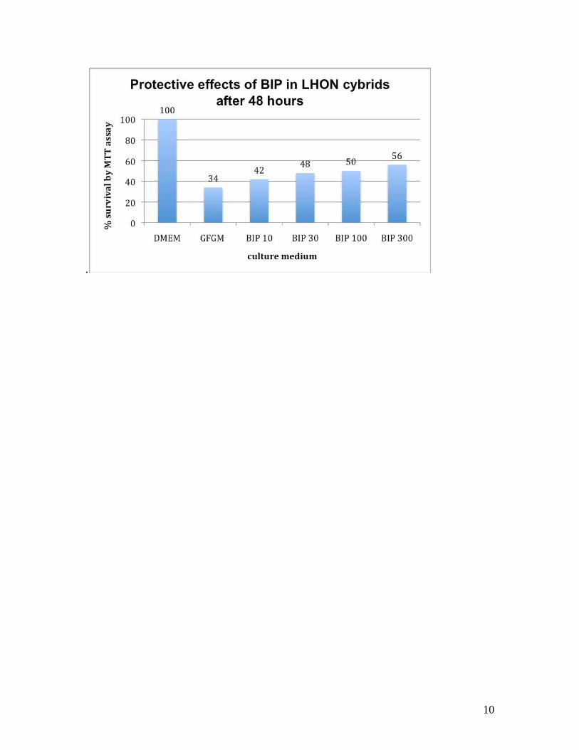
Rescue of cybrids containing Leber Hereditary Optic Neuropathy (LHON) mutation using Bax-inhibiting peptide DN Hu1, I Chan1, M Chen1, MJ Kupersmith1, J Guy2, J Walsh1
1The New York Eye and Ear Infirmary, New York NY 2Bascom Palmer Eye Institute, Miami FL
Purpose: To investigate the use of Bax-inhibiting peptide V5 (BIP) to block apoptosis in cybrids containing LHON mutation. BIP is derived from the amino acid sequence of Bax-binding domain of Ku70 and blocks the Bax-Bak oligomerization on outer mitochondria membrane. Methods: Homeoplasmic 143B osteosarcoma cybrids containing mtDNA (G11778A) mutation were grown in Dulbecco’s Modified Eagle Medium (DMEM) with high glucose supplemented with 10% fetal bovine serum. Cells were seeded into 96 well plates and cultured with DMEM, glucose-free galactose medium (GFGM) and GFGM with BIP at 10, 30, 100 and 300 μmol concentrations. After 48 hrs, 3-[4,5-dimethylthiazol-2-yl]-2,5-diphenyltetrazolium bromide (MTT, 50 μl of 1 mg/ml) was added, incubated for 4 hrs, and replaced by DMSO. A microplate spectrophotometer at 540 nm was used to evaluate for % cell survival. Results: Compared to cybrids in DMEM medium, cell viability from MTT assay showed that cybrids survival % dropped to 34% after incubation in GFGM after 48 hours. Adding BIP to GFGM at 10, 30, 100 and 300 μmol concentrations increased % survival at a dose dependent fashion to 42%, 48%, 50% and 56% respectively after 48 hrs. P-values for 2-tailed t-test of cell survival for BIP concentrations of 10, 30, 100 and 300 μmol with GFGM vs. GFGM alone were 0.02, 0.01, 0.0002 and 0.0001 respectively. Conclusion: BIP demonstrated statistically significant dose-dependent protective effects in the LHON cybrid model
10
.

11
Corneal Hysteresis and Beta-Zone Parapapillary Atrophy DD Hayes1, CC Teng1,2, CG de Moraes1,2, C Tello1,3, JM Liebmann1,2, R Ritch1,3 1Einhorn Clinical Research Center, New York Eye and Ear Infirmary, New York, NY; 2Ophthalmology, New York University School of Medicine, New York, NY; 3Ophthalmology, New York Medical College, Valhalla, NY. Purpose: Eyes with ß-zone parapapillary atrophy (βPPA) are at increased risk of glaucoma progression. Eyes with glaucoma have lower CH than eyes without, and in asymmetric glaucoma, lower CH is associated with the worse eye independently of its effect on IOP measurement. We sought a relationship between CH and βPPA. Methods: Consecutive, eligible glaucoma patients seen in a glaucoma referral practice were enrolled in this prospective study. We included patients aged 18-90 years with disc photographs within 12 months of the study visit. Exclusion criteria included history of ocular surgery other than clear corneal phacoemulsification, myopia >6 diopters, contact lens use and corneal abnormality. CH was measured using the Ocular Response Analyzer (Reichert, Inc., Buffalo, NY). Disc photos were evaluated by a single masked examiner for βPPA. Data was analyzed using Medcalc software (Mariakerke, Belgium). Results: We enrolled 99 patients (mean ± SD age 67.6 ± 13.8 years; 45 men, 54 women). Univariate analysis showed no statistically significant difference in CH between eyes with and without βPPA (8.70±1.57 vs. 8.10±1.95 mmHg, respectively, p=0.11). There were no differences in corneal resistance factor (CRF) (p=0.47), central corneal thickness (CCT) (p=0.11), ORA wave score (p=0.23), age (p=0.23), gender (p=0.40), IOP (p=0.86) or visual field mean deviation (VFMD) (p=0.45). Eyes with βPPA had greater myopia (-1.50±2.11 vs. -0.22±1.79 diopters, p=0.003). Multivariate analysis adjusting for age, spherical equivalent, VF MD, and wave score showed no significant difference in CH between eyes with and without βPPA (p=0.38). Eyes with asymmetric βPPA also showed no significant difference in CH (9.00±1.17 vs. 9.10±1.43 mmHg, p=0.69). Conclusions: There is no significant differrence in CH between eyes with and without βPPA or between fellow eyes with asymmetric βPPA.

12
Surgical Outcomes of the Boston Type 1 Keratoprosthesis
Author Block: Amar P Patel, MD, Elaine Wu, MD, John A Seedor, MD, David C Ritterband Department of Ophthalmology, New York Eye and Ear Infirmary, New York, NY, USA Purpose: The keratoprosthesis has had variable success rates in the past. However, significant modifications to design and management have led to recent successful outcomes. This study was undertaken to evaluate the outcomes of the Boston type 1 keratoprosthesis at our institution. Methods: Preoperative and postoperative data were collected from patients who received a Boston type 1 keratoprosthesis from December 2006 to August 2010. Outcome measures were visual acuity, retention rate, and complications. Results: 58 eyes of 51 patients were included. The most common indication was failed penetrating keratoplasty (PK) (81.0%; mean 2.4±1.3 PKs per eye). Glaucoma was the most common comorbidity (75.9%). Preoperative BCVA was <20/400 in 87.9% of eyes with no eye having BCVA ≥ 20/100. At the last follow-up (mean 21.5 ±11.4 months), 43.1% of eyes had BCVA ≥ 20/200. The retention rate was 87.9%. The most common complication was retroprosthetic membrane formation (50.0%). Conclusions: The Boston type 1 keratoprosthesis provides substantial visual recovery for eyes with multiple PK failures or with poor prognosis for primary PK, showing excellent retention rates with minimal serious complications.

13
Rates Of Visual Field (VF) Progression I/n Distinct Optic Disc Phenotypes Author: Kenneth S. Schor1, Carlos G. De Moraes1,2, Christopher C. Teng1,3, Celso Tello1,3, Jeffrey M. Liebmann1,2, Robert Ritch1,3. 1New York Eye and Ear Infirmary, Einhorn Clinical Research Center, New York, NY; 2Ophthalmology, New York University School of Medicine, New York, NY; 3New York Medical College, Valhalla, NY. Purpose: To assess clinical paramters and rates of visual field change in glaucoma patients with distinct optic disc phenotypes. Methods: Retrospective observational cohort. Glaucoma patients with 8 or more 24-2 SITA VF tests (n=392) with baseline and final follow-up stereo photographs between January 1999 and July 2009 were reviewed by 2 investigators masked to all clinical and perimetric data. Each disc was classified by criteria described by Drance & Nicolela: focal ischemic (FI), myopic (M), senile sclerotic (SS), and generalized enlargement (GE). Disagreements were adjudicated after consensus. Discs that could not be classified into one of these categories were excluded from analysis. VF progression (defined as at least 2 adjacent test points in the same hemifield progressing >1/0 dB/yr at p<0.01) was evaluated using automated pointwise linear regression (PLR). Also reviewed were age, gender, race, central corneal thickness (CCT), intraocular pressure (IOP, mean, peak, and fluctuation), disc hemorrhage (DH) and beta-zone PPA (βPPA), and PSD and MD data from baseline VFs. Results: 127 GE, 41 FI, 54 M, and 42 SS discs were reviewed. 128 eyes could not be classified into any of the categories and were excluded from analysis. Patients with SS discs were significantly older than the other categorized patients (68.5±1.9 yrs, p<0.01). There were more men in the M group (52%, p=0.12). SS group showed worse baseline VF MD (-8.49±0.64 dB, p<0.01). IOP mean, peak, and fluctuation were significantly greater in the GE group (16.0±0.2; 20.9±0.4; and 2.6±0.1 mmHg, respectively, p<0.01). CCT differed significantly among groups, even though FI tended to show decreased values (536.7±6.1 µm, p=0.78). More DH were detected in the FI (17%) and SS (19%) groups (p=0.01). The percentage of βPPA was greater in the M (96%) and SS (100%, definition criterion) groups (p<0.01). The SS group showed more rapid but not significant global rates of VF change than the other groups (-0.5±0.1 dB/yr, p=0.72). After adjusting for co-variates, the number of ey/es reaching a progression endpoint did not differ among groups (GE,30%; FI,29%; M,29%; SSE,28%, p=0.99). Conclusions: Glaucomatous eyes with different optic disc phenotypes showed different clinical features. However, the 4 groups showed similar VF outcomes despite these differences. Optic disc phenotypes should not be considered independent risk factors for rapid VF progression. :

14
SD-OCT Analysis of Subretinal Deposits/ in Acute Central Serous Retinopathy
Gennady Landa 1,2,MD, Jonathan Barnett 1, Patricia MT Garcia1,2, MD, Katy Tai 1,BA, Richard B Rosen1,2, MD
1RetinaCenter, Department of Ophthalmology, New York Eye and Ear Infirmary, New York, NY, USA
2Department of Ophthalmology, New York Medical College, Valhalla, NY, USA Purpose: To perform qualitative and quantitative analysis of subretinal protein deposits, seen in acute central serous retinopathy (CSR) patients, using high resolution spectral domain optical coherence tomography (SD-OCT), in order to investigate whether protein deposits present have any significant impact on best corrected visual acuity (BCVA). The secondary goal was to evaluate whether the presence of pigment epithelium detachment (PED) at baseline has any impact on final BCVA. Methods: Patients presenting to the Retina Center of the New York Eye and Ear Infirmary between July 2009 and July 2010 with evidence of acute CSR were included. SD-OCT was performed and multiple parameters were measured: central total retinal thickness, central neurosensory retinal thickness, maximum vertical and horizontal lengths of sub foveal subretinal fluid and thickness of any subfoveal protein deposit (PD) layer, if present. Additionally, height and width of any PED present subfoveally, was measured. Results: Thirty eight patients with acute CSR were included. Four types of PD, based on their shape and appearance (hanging, integrated, scattered, and massive), were noted. At least one type of PDs was seen at baseline SD-OCT exam in 84.2% (32/38) eyes. In 60.5% (23/38) eyes more than one type of PD were present. PD subfoveal thickness could be measured in 20 (52.6%) out of 38 eyes. A significant correlation was found between the subfoveal thickness of PD layer and baseline/final visual acuities in the eyes: (r = 0.60, p = <0.001 and r = 0.45, p = 0.008, respectively). Those eyes with protein deposits which demonstrated a resolution of CSR (resolved subgroup, n = 11) at 3 (± 1) months of follow-up, had a significantly thinner (p = 0.003) subfoveal thickness of the PD layer at baseline exam (28.8 ± 12.5 µm), than the unresolved group of eyes (n = 21)(57.0± 27.2 µm). The mean BCVA (LogMAR) at the baseline did not differ significantly between resolved and unresolved subgroups (0.43 ± 0.29 and 0.40 ± 0.22, respectively, p=0.375), however, at fi/nal follow-up, a significant difference(p=0.012) in BCVA was found between the resolved and unresolved subgroups (0.18± 0.20 and 0.33 ± 0.18, respectively), The final BCVA in eyes with and without PED at baseline was not significantly different (p=0.19). Conclusions: The thickness of subfoveal protein deposits at baseline appears to be an important parameter related to the BCVA and time of CSR resolution, whereas the presence of PED at baseline didn’t have an impact on the final BCVA.

15
Serum estrogen and progesterone level/s and intraocular inflammation Author Block: Olivia L. Lee, MD, Amar Patel, MD, Vicente Diaz, MD, MBA, Sanjay R. Kedhar, MD and C. Michael Samson, MD, MBA Abstract Purpose: Sex hormones are postulated to play a role in inflammatory mediation of autoimmune disease, as exemplified by changes in disease activity during pregnancy and the postpartum period. The present study aims to investigate the quantitative levels of serum estrogen and progesterone levels in patients with active uveitis. Methods: A prospective, observational case study including menstruating, non-pregnant women with clinically active autoimmune or idiopathic uveitis was conducted. Serum levels of estradiol and progesterone were measured at a single time point concurrent with clinically active disease, defined as at least trace anterior chamber cell in one or both eyes with previously diagnosed uveitis. Results: Serum samples from 10 female patients with average age 37+/-7.86 years were drawn 16.6+/-10.3 days after the last menstrual period. Diagnoses included anterior uveitis in 70.0% (7/10 patients) and 10% (1/10 patients) each with posterior, intermediate and panuveitis. 55.6% of patients displayed active uveitis in one eye, while the remainders were bilateral. Visual acuity in the involved eye at the time of hormone sampling was 20/40 or better in 60% of eyes, 40% 20/50 to 20/80 and no eye worse than 20/80. When adjusted for menstrual cycle phase, 50% of patients were found to progesterone level within the normal range while 37% of patients had low progesterone levels. Conclusions: Female sex hormones may play a role in the modulation of inflammatory disease activity in autoimmune uveitis. In some female patients, the onset of intraocular inflammation may be related to the withdrawl of anti-inflammatory effects of estrogen and progesterone during the menstrual cycle.

16
The Expanded Spectrum of Focal Choroidal Excavation Abstract
Sri Krishna Mukkamala, MD,2 Ron Margolis, MD,1 Lee M Jampol, MD, 3 Richard F Spaide, MD, 4 Michael D Ober, MD, 5 John A Sorenson, MD, 4 Ronald C Gentile, MD, 2
Joel A Miller, MD, 5 Jerome Sherman, OD, 6 K Bailey Freund, MD 4 Purpose: To describe the clinical and imaging findings in patients with a focal choroidal excavation (FCE). Methods: Retrospective observational case series. Medical records of 12 patients (13 eyes) with FCE were reviewed. Clinical histories and imaging including color photography, fundus autofluorescence (FAF), fluorescein angiography (FA), indocyanine green angiography (ICGA), spectral domain optical coherence tomography (SD-OCT) and enhanced depth imaging SD-OCT (EDI-OCT) were analyzed. Results: Mean patient age was 45 years (range 22-62 years). Five patients were Asian. Mean visual acuity was 20/31 (range 20/20-20/100). Mean refractive error was -3.54 diopters (range +6.00 to -8.00 diopters). One patient had bilateral involvement. All manifested varying degrees of foveal pigmentary changes that were usually hypoautofluorescent on FAF. FA findings varied with degree of retinal pigment epithelial (RPE) alterations. ICGA revealed relative hypofluorescence. In 7 eyes, SD-OCT showed outer retinal layers conforming to RPE within the excavation. In the other 6 eyes, SD-OCT demonstrated a separation between outer retina and RPE within the excavation. In 7 eyes studied with EDI-OCT, there was no evidence of scleral ectasia. Mean choroidal thickness of the uninvolved choroid was thicker than normal at 319 um (range 244-439 um). All lesions remained stable except for one eye which had findings of central serous chorioretinopathy (CSC) and secondary type 2 (subretinal) neovascularization. Conclusion: FCE is a newly described idiopathic entity in eyes having one or more focal areas of choroidal excavation. In some patients, there may be an association with CSC. Although most lesions remain stable, secondary choroidal neovascularization may occur

17
In Vivo Evaluation of Focal Lamina Cribrosa Defects in Glaucoma Authors: Saman Kiumehr,1 Sung Chul Park,1,2 Syril Dorairaj,1 Christopher C Teng,1,2 Celso Tello,1,2 Jeffrey M Liebmann,1,3 Robert Ritch1,2 Affiliations: 1Einhorn Clinical Research Center, New York Eye and Ear Infirmary, New York, NY; 2Dept. of Ophthalmology, New York Medical College, Valhalla, NY; 3Dept. of Ophthalmology, NYU School of Medicine, New York, NY Purpose: To assess focal lamina cribrosa (LC) defects in glaucoma using enhanced depth imaging optical coherence tomography (EDI OCT) and to investigate their spatial relationship(s) with neuroretinal rim and visual field (VF) loss. Methods: Serial horizontal and vertical EDI OCT images of the optic nerve head were obtained for normal and glaucoma subjects. Focal LC defects defined as localized LC deformation within a 45° sector of the optic disc were investigated regarding their configurations and locations. Spatial consistency was evaluated among focal LC defects, neuroretinal rim thinning/notching, and VF defects. Results: Twenty-five (50 eyes) normal and 31 (45 eyes) glaucoma subjects were included. Fifty-eight partial-thickness and 64 full-thickness focal LC defects were found in 38 glaucoma eyes, versus none in the normal eyes. The partial-thickness focal LC defects consisted of 9 smooth indentations, 18 moth-eaten appearance defects, 12 step like depressions and 19 partial disruptions of laminar insertion. The full-thickness focal LC defects consisted of 10 holes of various sizes and 54 directions and complete disruptions of laminar insertion. Eleven full-thickness LC defects appeared clinically as an acquired pit of the optic nerve (APON) and the others as neuroretinal rim thinning/notching. Most focal LC defects occurred in the inferior or inferotemporal far-periphery of the LC including its insertion. Eyes with focal LC defects limited to the inferior half of the optic disc had greater sensitivity loss in the superior visual hemifield, and vice versa. Conclusion: Mechanisms of LC deformation in glaucoma include focal melting/collapse of laminar beams and foc/al disruption of the LC insertion, both of which may cause a clinical APON in extreme cases. Focal LC defects occur in tandem with localized RNFL and VF defects.
18
Peripapillary blood flow and -Zone Per/ipapillary Atrophy in Primary Open-Angle Glaucoma Authors: Kasra Eliasieh, Nicole Scripsema, Syril Dorairaj, Gennady Landa, Kati Tai, Adrianne Monsef, Carmen Vasquez, Richard B. Rosen Purpose: To Evaluate peripapillary blood flow in patients with Primary Open-Angle Glaucoma (POAG) with and without β-Zone parapapillary atrophy (PPA) using the Retinal Functional Imager (RFI). Methods: Patients diagnosed with POAG as well as normal control subjects were prospectively imaged with the RFI. Arterial and venous blood flow velocities in the peripapillary area were evaluated in 73 eyes of 44 patients. Patients were divided into two groups based on the presence or absence of β-Zone PPA. β-Zone PPA was defined as a central zone of atrophy bordering the scleral ring characterized by retinal transparency with visible sclera and chorioretinal vessels. In order to normalize the severity of β-Zone PPA, patients were categorized into groups based on the size of PPA as a percentage of total disc area. Retinal nerve fiber layer (RNFL) thickness was measured using spectral domain optical coherence tomography (OCT). An ANOVA test was used for statistical analysis. Results: The difference in average arterial and venous blood flow was not statistically significant between eyes with and without PPA (p= 0.68 and 0.65 respectively). Although there was a small increase in arterial and venous blood velocity as β-Zone PPA increased (Graph 1), this difference was not statistically significant (p=.88, p=.47). POAG patients with PPA had significantly thinner peripapillary RNLF than those without PPA (p=0.002). Conclusions: Although the RNFL was thinner in patients with PPA, there was no significant difference in velocities among POAG patients with or without β-Zone PPA. While β-Zone PPA has been suggested as an indicator for a faster progression of glaucoma, we did not find an association between β-Zone PPA and retinal perfusion. There was a small increase in arterial and venous velocity as β-Zone PPA increased.

19
Optic Disc Progression and Rates of Visual Field Change in Treated Glaucoma
Carlos Gustavo De Moraes, M.D.; Jeffrey M. Liebmann, M.D.; Sung Chul Park, M.D. Christopher C. Teng, M.D.; Julia Nemiroff, M.D.; Celso Tello, M.D.; Robert Ritch, M.D.
ABSTRACT Purpose: To investigate the relationship between optic disc progression and rates of visual field (VF) change in patients with treated glaucoma. Methods: Glaucoma patients with repeatable VF loss, ≥8 SITA-Standard 24-2 VF tests, and good quality optic disc stereophotographs evaluated over a 10-year period were included. Optic disc photos were reviewed for signs of glaucoma progression (neuroretinal rim change, widening of retinal nerve fiber layer defect, disc hemorrhage, and enlargement of beta-zone parapapillary atrophy) by two glaucoma specialists masked to their temporal sequence. Disagreements were adjudicated by a third grader. VF progression was evaluated using automated pointwise linear regression (PLR) and defined as at least 2 adjacent test points progressing >1.0 dB/yr at p<0.01. VF progression outcomes were compared with adjudicated photograph review results. Results: 389 eyes (389 patients; mean age 64.9±13.0 yrs; mean baseline MD, -7.1±5.1 dB) were included. 82 eyes (21%) had confirmed optic disc progression and 115 eyes (29%) reached a VF PLR endpoint. Eyes with documented optic disc progression had more rapid rates of VF change (-0.66±0.7 vs. -0.36±0.7 dB/yr, p<0.01) and reached a VF PLR endpoint more often (univariate OR=1.85, p=0.02; multivariate OR=1.78, p=0.03) than eyes without optic disc progression. There was moderate spatial consistency between the location of the optic disc progression and the visual hemifield with more rapid progression (81%, kappa=0.40). Conclusions: Treated glaucomatous eyes with documented optic disc progression are at increased risk of diminished visual function over time and may require more aggressive therapy to prevent future vision loss.
20
Trends in Pediatric Corneal Ulcers at th/e New York Eye and Ear Infirmary Alice Hong MD, Dipika Joshi MS, Anna Djougarian MS, John Seedor MD, David Ritterband MD,
Department of Ophthalmology, The New York Eye and Ear Infirmary, New York, NY Department of Ophthalmology, New York Medical College, Valhalla, NY
Purpose: To study the microbiologic profile and clinical risk factors of pediatric corneal ulcers at the New York Eye and Ear Infirmary. Methods: All eligible patients with clinically diagnosed infectious keratitis aged 16 years and younger that presented to the New York Eye and Ear Infirmary during a five year period between January 1, 2005 to May 1, 2010 were retrospectively reviewed. The records were retrieved electronically by diagnosis codes. Microbial culture results and clinical characteristics were reviewed. Results: 75 eyes of 63 children (32 male and 31 female) aged 16 years or younger were analyzed. In the same 5 year time span over 2500 cases of infectious keratitis in all age groups were seen. 66 (88%) eyes were culture positive and 9 (12%) eyes were culture negative. Polymicrobial infections were seen in 22 eyes (29%). A total of 111 infectious agents were identified. 70/111 (63%) were Gram positive organisms and 27/111 (24%) were Gram negative organisms. The remaining organisms were fungal 6/111 (5%) , acanthamoeba 6/111 (5%) and herpes simplex virus 6/111 (5%) . The most common microorganisms in descending order were S. epidermidis (19), P. aeruginosa (17), P. acnes (15), S. aureus (12) and S. viridians (10). The predominant risk factor for infectious keratitis in children was contact lens wear (55%) followed by trauma (15%), ocular surface disease (11%), systemic disease (8%) and previous ocular surgery (4%). Conclusions: Infectious keratitis in the pediatric age grou/p is uncommon. Contact lens use is the predominant risk factor in the teenage years and trauma continues to be an important risk factor in the younger age group. Recognizing predisposing factors and the microbiologic trends is helpful in prompt and effective treatment in the pediatric population to prevent future complications that may lead vision threatening disease. Commercial Relationships: None

21
Decompression Retinopathy: A Review
Robert McGlynn, MD, Sri Krishna Mukkamala, MD, SyrilDorairaj, MD, Jade Rusoff, BS, Sunil Rao, MD, Joseph Nezgoda, MD, Paul A. Sidoti, MD, Ronald C. Gentile, MD Purpose To review the existing literature on decompression retinopathy to understand its pathogenesis, clinical features, management, and outcomes. Methods The literature was reviewed and We identified 30 articles, 15 case reports and 15 case series, from 1968 to 2009 that presented 62 eyes of 52 patients. Decompression retinopathy was defined as multifocal, hemorrhagic retinopathy that resulted from acute lowering of intraocular pressure (IOP) that could not be explained by another process. Results Hemorrhages were seen in all retinal layers and most patients (80%) were asymptomatic. The mean IOP change associated with DCPR was 33.2 ± 15.8 mmHg. Most patients (73%) were diagnosed with DCPR on the first post-operative day and all were diagnosed by day 14. It resolved in a mean of 13 ± 12.4 weeks (range 2-72 weeks). A vitrectomy was required in 15% of patients to remove a vitreous hemorrhage. Visual outcomes were generally good with 85% of eyes returning to baseline vision. Conclusion Although DCPR infrequently results in significant ocular morbidity requiring vitrectomy, gradual reduction in IOP might prevent this complication.

22
Specular Microscopy of the Contralater/al Eye in Unilateral Iridocorneal Endothelial Syndrome
L. Schneider MD,1 N. Harizman MD,1,3 D. Ritterband MD, 1,3J. Liebmann MD,1,2 R. Ritch MD,1,3J. Seedor,MD1,3
1Einhorn Clinical Research Center, New York Eye and Ear Infirmary, New York
2Manhattan Eye, Ear, and Throat Hospital, New York
3Department of Ophthalmology, New York Medical College, Valhalla, NY Background: The iridocorneal endothelial (ICE) syndrome is a condition involving the cornea, anterior chamber angle, and iris, and it encompasses the syndromes of progressive essential iris atrophy, Chandler’s syndrome, and the iris nevus syndrome. The corneal endothelium has been implicated as the primary site of pathogenesis in iridocorneal endothelial (ICE) syndrome, resulting in a number of anterior segment abnormalities. ICE syndrome has been traditionally regarded as a predominantly unilateral disease, yet the contralateral, presumably uninvolved eye, has been suspected of possessing subclinical manifestations of the disease. Purpose: To evaluate the contralateral clinically unaffected eye of ICE patients by specular microscopy, and analyze endothelial morphologic features including cell density, percentage hexagonal cells (pleomorphism), and coefficient of variation of mean cell area (polymegathism). Methods: The charts of all patients on the Cornea and Glaucoma Services at the New York Eye and Ear Infirmary with a clinical diagnosis of unilateral ICE syndrome from January 1999 through September 2006 were reviewed. Specular microscopic data was analyzed and compared to age- and sex-matched control subjects. The cell parameters were assessed using a semi-automated method of endothelial cell morphometric analysis. Results: Seventeen ICE syndrome patients (12 females, 5 males) and 17 control subjects (12 females, 5 males) were included in this study. The average number of cells analyzed per patient did not differ significantly between ICE patients and controls (88.5 ± 35.2 and 85.8 ± 17.9 respectively; p =0.78). Quantitative analysis of ICE patients compared to controls revealed a mean cell density of 2698.6 ± 777.5 and 2640 ± 268.9 (p=0.77), percent hexagonal cells of 61.1 ± 7.4 and 60.0 ± 10.0 (p=0.62), and coefficient of variation of mean cell area of 0.378 ± 0.204 and 0.318 ± 0.60 (p=0.25), respectively. Conclusions: There were no significant differences in the morphometric analyses of the endothelial cell layers in the contralateral eye of ICE patients compared to controls. Any differences are consistent with variability inherent to currently available methods of examining the corneal endothelium.

23
Title: Improving eccentric fixation using/ Jitterbug program Authors: Kevin Rosenberg, Richard Rosen, Allen Lubow, and William Seiple Purpose: Given the increased temporal sensitivity of the peripheral visual field and increased Troxler effect at peripheral eccentricities, we examined whether “jittering” text on a computer monitor increases acuity and decrease reaction times for identification. Methods: Three normally sighted subjects were tested monocular, with the non-tested eye patched. Letter stimuli were presented at an eccentricity of ten degrees in the temporal retina. Four “jitter” settings were tested: no jitter, frequency of 5 Hz with a distance of 2 pixels, frequency of 10 Hz with a distance of 5 pixels, and a frequency of 15 Hz with a distance of 10 pixels. In separate trials, 10 letter of each size (over a range from 20/50, to 20/170 equivalent Snellen) were presented. The computer program randomly generated a letter and the subject was required to identify the stimulus as quickly as possible. The accuracy and time required to identify each letter was recorded. Results: Two of the three subjects showed an improved in identification accuracy when the letters were “jittered” as compared to stationary letters. This improvement in accuracy was positively correlated with the frequency and the amplitude of jitter. Similar improvement was observed at all letter sizes Conclusions: In the future, a program that jitters text may serve as a useful tool in helping patients with damaged maculas, such as those with age-related macular degeneration. Reading using the peripheral retina might be improved by jittering the text. We plan to test this protocol on age-matched patients with AMD.

24
Review of Pediatric Uveitis Patients at a/ Tertiary Referral Center Author Block: Wendy Huang, MD, Olivia L. Lee, MD, Sanjay R. Kedhar, MD, C. Michael Samson, MD, MBA Abstract Purpose: To characterize a population of pediatric patients seen by Uveitis specialists at a tertiary referral eye care center. Clinical characteristics, visual outcomes, complications and treatment modalities associated with pediatric Uveitis are described. Methods: A retrospective review of pediatric patients seen by Uveitis specialists at New York Eye and Ear Infirmary. A random sampling of Uveitis patients age 21 years and younger seen between January 2006 and November 2010 were included. Results: Ninety-five pediatric patients diagnosed with Uveitis were identified, 56.8% of which were female. Caucasian (42.1%) and Hispanic (24.1%) race accounted for the majority of patients. Bilateral involvement was seen in 70.5% of patients. The average age at time of initial diagnosis was 12.2+/- 5.5years. Ocular inflammation was categorized most commonly as anterior Uveitis (57.9%), followed by panuveitis (15.8%), posterior Uveitis (6.3%)and intermediate Uveitis (8.3%). Idiopathic Uveitis was diagnosed in 43.2%, while infectious Uveitis accounted for 8.4% of cases. A systemic disease association was identified in 48.4% of patients, most commonly juvenile idiopathic arthritis (58.7%). Immunomodulatory therapy was employed in 75.8% of patients and 67.4% of patients were co-managed with a rheumatologist. 35.8% of patients underwent intraocular surgery. Visual acuity in the involved eye at the time of last clinical exam was 20/40 or better in 72.8% of eyes, and 20/200 or worse in 4.3% of eyes. Conclusions: Pediatric Uveitis may not be associated with poor prognosis in a subpopulation of patients cared for by Uveitis specialists in a tertiary care setting. These patients can be managed successfully, particularly with the use of immunomodulatory agents.

25
Title: Ocular Complications of NewColo/rIrisTM Cosmetic Iris Implants Authors: Ambika Hoguet, David Ritterband, Richard Koplin, Elaine Wu, John Aljiam, Tal Raviv, John Seedor Purpose: To describe complications in patients with NewColorIrisTM cosmetic iris implants and their management at the New York Eye and Ear Infirmary. Methods: The medical records of 14 eyes of 7 patients with NewColorIris TM cosmetic iris implants treated for complications were reviewed. Data collected included patient demographics, visual acuity (VA), intraocular pressure (IOP), endothelial cell count (ECC), and slit lamp examination findings at presentation. Medical and surgical interventions were also tabulated. Results: 14 eyes in 7 patients (ages 22-60; 71% male, 29% female) were identified. 64% (9/14) of eyes presented with decreased vision; 50% (7/14) with elevated IOP; 36% (5/14) with corneal edema; and 36% (5/14) with iritis. Average initial IOP was 25 (range 16-52) and the average ECC was 990 (range 266-1841). All eyes were eventually treated with explantion of the iris prosthesis (range 4-33 months after implant placement), although 2 patients (4 eyes) refused immediate explantion (explants were subsequently performed 4-15 months after presentation). The minimum follow-up after explantion for all eyes was 2 months (range 2-28 months). Intraoperative complication included one case of suprachoroidal hemorrhage during explantion. Post operative events included corneal decompensation (4/14), cataract formation (8/14), and increased IOP (&/14). Secondary surgeries included Descemet's Stripping Automated Endothelial Keratoplasty (5/14), cataract extraction with intraocular lens placement (4/14), trabeculectomy (3/14), tube placement (2/14), goniosynechiolysis (2/14), and penetrating keratoplasty (1/14). On most recent follow up, 12/14 showed improvement in IOP, 8/14 showed improved or stable VA, 6/14 had worse VA (including 3/4 who deferred immediate explantation). Conclusions: NewColorIris TM implants can cause serious complications including iritis, glaucoma, and corneal edema. Explantation may help stabilize symptoms but in most cases ocular morbidity is high, requiring additional surgical interventions to control IOP and corneal decompensation.
26
Clinical and Optical Coherence Tomography Findings in Patients Undergoing Focal Laser Treatment with the Navilas Laser System Author Block: Robert J. Lowe, Jasmine H. Francis, Patricia Garcia, Joseph Panarelli, Richard B. Rosen Purpose: To review the clinical and optical coherence tomography (OCT) findings in diabetic patients with clinically significant macular edema (CSME) or diabetic macula edema (DME), whom received a single episode of focal laser treatment with the Navilas laser (OD-OS GmbH, Teltow, Germany). The Navilas laser is equipped with a unique retinal navigation system, developed to improve safety and to be more predictable./ Methods: Our study was an interventional retrospective case series. All eyes underwent complete ophthalmic examination and OCT before and after receiving a single episode of focal laser treatment with the Navilas laser system. Results: Nine eyes of 9 diabetic patients (4 men, 5 women) with CSME or DME were included. Average age was 55 years (range: 27 to 63 years). Pre-laser mean visual acuity (Va) was logMAR 0.49 ± 0.36, Snellen equivalent of 20/62 (range: 20/25 to 20/400). All patients tolerated the procedure well. Of those who received additional treatment, 5 required further focal laser treatment, 1 had panretinal photocoagulation, and 2 received intravitreal bevacizumab. One patient required no further treatment. At 3 months post-laser, most patients had improvements in Va, with a statistically significant improved mean logMAR Va of 0.40 ± 0.39; p ≤ 0.03 (Snellen equivalent 20/50) compared to baseline. However, no statistically significant difference between the mean logMAR Va at 6 months and baseline was found. Post-laser OCTs at 3 months revealed increases in mean perifoveal thicknesses superiorly, temporally, and centrally, but these were not statistically significant. At 3 months patients did have statistically significant reductions in perifoveal thicknesses inferiorly (p ≤ 0.04) and nasally (p ≤ 0.005). All perifoveal areas demonstrated non-statistically significant decreased thicknesses at 6 months post-laser. Conclusions: The Navilas laser is a well-tolerated focal laser alternative with a unique retinal navigation system. At 3 months post-laser, patients had a mild but statistically significant improvement in Va. This was in addition to statistically significant reductions the perifoveal inferior and nasal thicknesses at 3 month post-laser. Gains in Va and reductions in perifoveal thicknesses were not found 6 months post-laser. In our study, one episode of laser treatment with the Navilas seemed to help improve vision in the intermediate term and stabilize vision more long term. Question: A statistically significant improvement in visual acuity and macular edema was found in patients with diabetic macular edema who were treated with one session of focal laser using the Navilas laser at whi/ch time point:

27
A) 1 month B) 3 months C) 6 months D) 9 months
/
28
Pars Plana Vitrectomy with Silicone Oil or Gas Tamponade in Eyes with Diabetic Tractional Retinal Detachment
David Warrow, M.D.,1 Robert J. Lowe, M.D.,1 Amar Patel, M.D.,1 Alberto DiStefano2, Vatsal Doshi, M.D.1 Purpose: To evaluate outcomes of vitreoretinal surgery for diabetic tractional retinal detachment in eyes that received silicone oil versus gas tamponade. More specifically, this study assesses the change in visual acuity and need for re-operation due to re-proliferation in these two groups. Methods: 34 patients were evaluated in this retrospective chart review. These eyes were diagnosed with diabetic tractional retinal detachment and were surgically repaired between January 2008 and September 2009 at the New York Eye and Ear Infirmary. Of the 44 eyes that were assessed in this study, 28 eyes received silicone oil, while 16 eyes received gas (C3F8 or SF6) tamponade. Clinical data, including patient age, preoperative and postoperative visual acuity, total follow-up, and re-operation, were collected from patient charts. Visual acuity was quantified on a logMAR scale. Statistical evaluation of the data was performed using the data analysis function of Microsoft Excel 2007. Results: The mean age of the 20 patients in the silicone oil group was 48.8 ± 10.7 years, and the mean age of the 14 patients in the gas tamponade group was 53.8 ± 9.1 years. The preoperative visual acuity for silicone oil eyes was 1.75 ± 0.97, and postoperatively the vision was 1.81 ± 1.20 (p ≤ 0.83). The preoperative visual acuity for the gas tamponade eyes was 1.73 ± 1.24, and postoperatively it was 1.45 ± 1.19 (p ≤ 0.52). Per the logMAR scale, the change in visual acuity for the silicone oil eyes was 0.06, while the change in visual acuity for the gas tamponade eyes was -0.28 (p = 0.50). Of the 28 eyes that received silicone oil, 5 (18%) required re-operation for re-proliferation, while 1 eye (6%) of the 16 that received gas required re-operation (p = 0.28). The mean postoperative follow-up time was 14.6 ± 6.7 months for the silicone oil group, while the gas group had a mean follow-up of 15.9 ± 5.0 months. Conclusions: The results of this study revealed a trend towards improved visual acuity in eyes that received gas tamponade relative to silicone oil in the surgical repair of diabetic tractional retinal detachment. The use of gas also led to a lower re-operation rate compared to eyes that received silicone oil.
29
Sympathizing Intraocular Pressure Response to Contralateral Steroid Drop Usage B. Bert, MD, N. Harizman, MD Department of Ophthalmology, The New York Eye and Ear Infirmary (NYEEI) Purpose: To evaluate intraocular pressure (IOP) response to topical steroid treatment in the contralateral eye of patients who underwent uncomplicated pterygium surgery. Methods: Retrospective chart review. Charts of patients who underwent pterygium excision at NYEEI, from January 2007 through October 2010 were reviewed. A minimum of 3 months follow up was required and no previous ocular history. Exclusion criteria included a suspected diagnosis of glaucoma, diabetic retinopathy, history of trauma or previous ocular surgery besides uncomplicated cataract extraction. A steroid response was defined as an IOP ≥ 24 mmHg and/or change of IOP from baseline of ≥11 mmHg. Results: 205 charts were analyzed with 56 being excluded. Ten developed a steroid response in their surgical eye (the highest pressures being seen in 1 patient at week 1, 7 patients at week 4, 1 patient at 3 months and 1 patient at 6 months). The maximal average IOP in the surgical eye was 27.1 ± 3.0 mmHg while the average IOP in the non-surgical eye on that visit was 14.8 ± 1.93 mmHg. The IOP change from baseline in the surgical eye was 13.0 ± 2.98 mmHg compared to 1.6 ± 2.67 in the contralateral eye (p<0.001). Conclusion: Topical steroid drops do not have a significant effect on the IOP of the contralateral, untreated eye.

30
Intravitreal Injection of Kenalog as an Adjunctive Treatment for Uveitis Patient Undergoing Cataract Surgery Authors: D. Yin, A. Ali, M.C. Samson Purpose: To evaluate the clinical outcome of intravitreal injection of kenalog as an adjunctive treatment for uveitis patients undergoing cataract surgery compare to cataract surgery without kenalog injection. Post operative best corrected visual acuity (BCVA), inflammation, IOP fluctuation, posterior capsule opacification (PCO) and complication of cystoid macular edema (CME) are assessed. Methods: The charts of 53 uveitis patients (56 eyes) who underwent cataract surgery were reviewed. We found a study group of 14 patients (15 eyes) that received 4mg of intravitreal injection and a control group of 39 patients (41 eyes) that did not receive intravitreal injection. The data were collected retrospectively for 4 months preoperatively and 6 months postoperatively. Analysis of the data was conducted by SAS statistic software. Results: In the intravitreal injection reviewed group, 57.2% patients improved more than 6-9 lines in BCVA after the cataract surgery comparing to 34.4% in the controlled group during postoperation follow-up. Improvement of BCVA (p<0.05) is greater in the study group compare to the control group. Patients with anterior uveitis and panuveitis responded to kenalog treatment better than intermediate and posterior uveitis. Statistically significant differences of BCVA improvement were seen postoperatively in anterior uveitis (p=0.0002) and in panuveitis (p=0.0022), but not in intermediate and posterior uveitis. In both the control and study groups, IOP was relatively stable postoperatively. In terms of posterior capsule opacification, 33% of the study group developed PCO compare to 27% in the control group. However, only 20% of the study group with PCO required Nd:YAG treatment compare to 64% in the control group that developed PCO. Moreover, patients that received kenalog treatment had higher AC cells pre-operatively as expected. However, by post operation month 3, the inflammation in the study group was lowered than the controlled group. As for postoperative complication, only 10% of the study group developed cystoid macular edema compare to 20% in the control group. No other complication emerged during the 6 months follow-up period. Conclusion: The results of this study suggest that intravitreal injection of 4mg of kenalog may of be benefit for improving visual acuity, decrease inflammation and lessen the severity of PCO development thus lessens the needs for Nd:YAG treatment in anterior uveitis and panuveitis intraocular. There were no significant fluctuations of IOP post intravitreal injection or any other side effects that may limit the therapeutic usefulness of this treatment.
31
Uveal Schwannoma with Extrascleral Extension: report of a case and review of literature Author: Jae Young (Jane) You, MD; Dr. Tatyana Milman, MD; Paul Finger, MD; Codrin Iacob, MD Kimberly Chin, MD Purpose To describe clinical and pathologic characteristics of uveal schwannoma with extrascleral extension and to review the literature on this topic. Methods Interventional clinical-pathologic case report. A literature search for all cases of uveal schwannoma was performed using PubMed database (keywords: schwannoma, neurilemmoma, neurinoma, choroid, ciliary body, iris, uvea, nerve sheath tumor, intraocular). Results A 23 year-old woman presented with an amelanotic posterior choroidal tumor with low-to-medium internal reflectivity and extrascleral extension. Enucleation was performed for suspected choroidal malignant melanoma. Pathologic evaluation of the enucleated eye revealed a benign schwannoma arising from the long posterior ciliary nerve. The tumor eroded through the equatorial sclera, extending onto the epibulbar surface. The patient was observed with no recurrence of the lesion 3 months after enucleation. Review of the literature revealed 40 cases of uveal schwannoma. The tumor was more common in females (69%) with mean age of diagnosis at 35 years. Three cases (7%) were associated with neurofibromatosis. Extrascleral extension was noted in 3 (7%) of 40 cases. 15 tumors were managed with local excision; 20 tumors were treated with primary enucleation; 4 tumors were initially under close observation, but all were eventually enucleated for intractable visual loss. The follow-up data (5 months to 15 years, mean 2.9 years) was available in 10 cases. There were no local recurrences or metastatic disease in the enucleated group (including the 2 eyes with extrascleral extension). One recurrence was noted following local excision, which was successfully managed with second local resection. Two cases following local excision underwent enucleation after 2.6 years and 2 years of follow-up, for painful eye and progressive visual deterioration, respectively. Conclusion Uveal schwannoma can be difficult to distinguish clinically from melanoma. The schwannomas of anterior uveal tract can be successfully treated with local resection. Enucleation can be reserved for those tumors which cause significant visual loss and/or are impossible do distinguish from melanoma. While extrascleral extension in intraocular tumors generally portends aggressive behavior, none of the choroidal schwannomas with extrascleral extension were histologically malignant or biologically aggressive. These findings argue for conservative treatment of such tumors.

32
Retinal Function Imaging for Evaluating of Changes in Retinal Blood Flow after Pan-retinal Laser Photocoagulation in Patients with Proliferative Diabetic Retinopathy Author Block: Chavakij BHOOMIBUNCHOO1, Nicole Scripsema1, Gennady Landa1, Richard B. Rosen1,2. 1Retina Center, The New York Eye &
Ear Infrimary, New York, NY; 2Dept. of Ophthalmology, New York Medical College, Valhalla, NY.
Keywords: 549 imaging/image analysis: clinical; 499 diabetic retinopathy; 577 laser
Purpose: Retinal function imaging for evaluating of changes in retinal blood flow following pan-retinal laser photocoagulation in patients with proliferative diabetic retinopathy. Methods: To evaluate the effect of pan-retinal laser photocoagulation (PRP) on retinal blood flow in eyes with proliferative diabetic retinopathy (PDR) using the Retinal Function Imager (RFI). Results: 20 eyes were enrolled. Mean age was 50.2 +/- 9.36 years (38 to 64 years). The mean venous velocity before PRP treatment was 2.82 +/- 0.61 mm/sec and after PRP treatment was 2.62 +/- 0.37 mm/sec. The results demonstrate a significant decrease in venous velocity following PRP treatment (p value =0.03). The venous velocity decreased by at least 14.04% in these patients. The mean arterial velocity before PRP treatment was 4.48 +/- 1.42 mm/sec and Abstract Print View Page 1 of 2 http://www.abstractsonline.com/plan/AbstractPrintView.aspx?mID=2684&sKey=de6227c... 5/12/2011 after PRP treatment was 4.46 +/- 1.57 mm/sec. Arterial velocity following PRP treatment showed inconsistent response to PRP treatment. Conclusions: The RFI is novel non-invasive imaging system which allows for quantitative analysis of retinal blood flow. The results of this study suggest that PRP treatment may effect retinal blood flow by reducing venous velocity in proliferative diabetic retinopathy patients,. Further analysis may help assess the utility of the RFI measurements of response to PRP as an indicator of suppression of PDR activity.

33
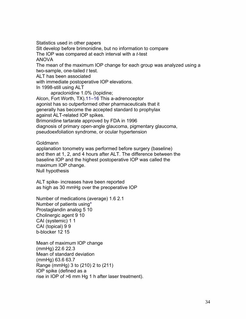
Post procedure pressure spike after ALT versus SLT with Iopidine Prophylaxis Timothy Sullivan, Ronald Hagadus Purpose: Argon laser trabeculoplasty (ALT) has been associated with higher risk of post-procedure intra-ocular pressure (IOP) spikes, commonly defined as an increase of >6 mm Hg, as compared to selective laser trabeculoplasty (SLT). However, little research has been performed to compare this effect after the initiation of alpha agonist prophylaxis. This study compares the change in pre and post-laser IOP between ALT and SLT patients when all subjects are provided iopidine prophylaxis. Methods: A retrospective review was performed of 50 consecutive ALT and 50 SLT procedures at the New York Eye and Ear Infirmary. Inclusion criteria included complete charts, patients diagnosed with primary open angle glaucoma, ocular hypertension, normotensive glaucoma, pigmentary dispersion or pseudo-exfoliative glaucoma, who required laser trabeculoplasty for the control of intraocular pressure. Exclusion criteria included previous laser trabeculoplasty. All patients receive one drop of iopidine 1% prior to treatment. Pre and post laser intraocular pressures were obtained by Goldmann tonometry. Results: 34 patients in the ALT treatment and 31 patients in SLT treatment met criteria for inclusion. The male to female ratio were 19/15 for the ALT arm and 15/16 for the SLT arm. No significant difference of pre-laser to post-laser intra-ocular pressure change was detected (p=0.931) between the ALT group (mean IOP decreased 1.50 mm) and SLT group (mean IOP decreased 1.39 mm). Regarding IOP spike, 0/34 ALT patients had IOP increase > 6 mm Hg, compared to 4/31 SLT patients. Conclusion: This limited retrospective chart review at a single institution suggests that patients who receive prophylaxis with iopidine prior to either ALT or SLT have no significant difference in IOP change after laser. Randomized clinical trials are needed to better assess any pressure difference. other factors pilcarpine versus non who measured the pressure Methods: From these charts were included complete charts of eyes that had no previous laser treatment.
34
Statistics used in other papers Slt develop before brimonidine, but no information to compare The IOP was compared at each interval with a t-test ANOVA The mean of the maximum IOP change for each group was analyzed using a two-sample, one-tailed t test. ALT has been associated with immediate postoperative IOP elevations. In 1998-still using ALT apraclonidine 1.0% (Iopidine; Alcon, Fort Worth, TX).11–16 This a-adrenoceptor agonist has so outperformed other pharmaceuticals that it generally has become the accepted standard to prophylax against ALT-related IOP spikes. Brimonidine tartarate approved by FDA in 1996 diagnosis of primary open-angle glaucoma, pigmentary glaucoma, pseudoexfoliation syndrome, or ocular hypertension Goldmann applanation tonometry was performed before surgery (baseline) and then at 1, 2, and 4 hours after ALT. The difference between the baseline IOP and the highest postoperative IOP was called the maximum IOP change. Null hypothesis ALT spike- increases have been reported as high as 30 mmHg over the preoperative IOP Number of medications (average) 1.6 2.1 Number of patients using* Prostaglandin analog 5 10 Cholinergic agent 9 10 CAI (systemic) 1 1 CAI (topical) 9 9 b-blocker 12 15 Mean of maximum IOP change (mmHg) 22.6 22.3 Mean of standard deviation (mmHg) 63.6 63.7 Range (mmHg) 3 to (210) 2 to (211) IOP spike (defined as a rise in IOP of >6 mm Hg 1 h after laser treatment).
35
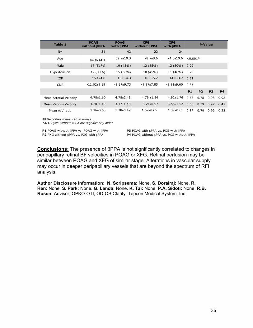
Effect of β-Zone Parapapillary Atrophy (βPPA) on Retinal Blood Flow Velocity in Primary Open-Angle Glaucoma (POAG) and Exfoliative Glaucoma (XFG) Nicole Scripsema1,2, Syril Dorairaj1, Ruojin Ren1, Sung Chul Park1,2, Gennady Landa1, Katy Tai1, Paul A. Sidoti1,2, Richard B. Rosen1,2. Affiliation: 1Einhorn Clinical Research Center, New York Eye and Ear Infirmary, New York, NY; 2Dept. of Ophthalmology, New York Medical College, Valhalla, NY.
Support: The Bendheim-Lowenstein Retina Fund of the New York Eye and Ear Infirmary; Ira and Judith Robinson Research Fund of the New York Glaucoma Research Institute, New York, NY. Purpose: Ocular blood flow (BF) is known to be reduced in glaucoma, but the relationship between reduced BF and βPPA is unclear. Using the Retinal Functional Imager (RFI), we evaluated the effect of βPPA on the peripapillary retinal BF velocities of secondary and tertiary vessels in POAG and XFG. Methods: Patients with POAG and XFG with no previous ocular surgery (with the exception of cataract extraction and laser iridotomy) were included in this prospective study. Peripapillary retinal arterial and venous BF velocities were evaluated with RFI. Color fundus photographs were reviewed by two glaucoma specialists, and patients were divided into one of four subgroups based on the presence or absence of βPPA. The four subgroups consisted of eyes with POAG without βPPA, POAG with βPPA, XFG without βPPA, and XFG with βPPA. Results: A total of 73 POAG and 46 XFG eyes were included. The demographic and clinical parameters for each group, including retinal BF velocities, are summarized in Table 1. Mean age was significantly greater in the XFG without βPPA than the other 3 groups (p<0.001). Before and after controlling for age, all 3 BF velocity parameters (mean arterial and venous BF velocities and mean A/V ratio) were not significantly different between the POAG groups with and without βPPA (p>0.6) or between the XFG groups with and without βPPA (p>0.3). BF velocity parameters were also similar between the POAG and XFG groups with (p>0.2) or without βPPA (p>0.9).
36
Conclusions: The presence of βPPA is not significantly correlated to changes in peripapillary retinal BF velocities in POAG or XFG. Retinal perfusion may be similar between POAG and XFG of similar stage. Alterations in vascular supply may occur in deeper peripapillary vessels that are beyond the spectrum of RFI analysis. Author Disclosure Information: N. Scripsema: None. S. Dorairaj: None. R. Ren: None. S. Park: None. G. Landa: None. K. Tai: None. P.A. Sidoti: None. R.B. Rosen: Advisor; OPKO-OTI, OD-OS Clarity, Topcon Medical System, Inc.

37
Title: Comparison of Retinal Blood Flow Velocities in Normal Tension Glaucoma (NTG) and High Tension Glaucoma (HTG) using the Retinal Functional Imager (RFI) Authors: Valerie Trubnik, RuojinRen, Nicole Scripsema, Syril K. Dorairaj, Gennady Landa, Richard B. Rosen Introduction:Glaucomatous optic neuropathy is usually characterized as a combination of retinal ganglion cell loss and remodeling of the lamina cribrosa.1,2 The etiology of glaucomatous optic nerve damage and its risk factors are only partially known.Ocular blood flow may be disturbed in glaucoma, and blood flow changes have been linked to progression of glaucomatous damage.3,4Numerous studies have attempted to evaluate ocular hemodynamics in patients with High Tension Glaucoma (HTG) and Normal Tension Glaucoma (NTG).Abnormal retinal autoregulation has been linked to an increase in intraocular pressure.4Previous research has suggested a decrease in blood flow in various ocular tissue beds in patients with both HTG and NTG and decreased choroidal blood flow velocities in NTG vs. HTG patients.4, 5,6The Retinal Functional Imager (RFI) (Optical Imaging, Rehovot, Israel) is a non-contrast, non-invasive angiographic device that provides direct qualitative and quantitative imaging of retinal blood flow velocities which can be used to evaluate patients with both HTG and NTG.1, 3,5We investigated differences in retinal blood flow velocities between NTG and HTG patients using the RFI. Methods: Age-matched eyes with NTG (n=22) and HTG (n= 72) were evaluated using the RFI at the New York Eye and Ear Infirmary. Glaucoma was defined by the presence of glaucomatous optic neuropathy and repeatable visual field abnormalities. All eyes had gonioscopically open angles and no secondary causes of glaucoma. Patients with the above characteristics and a mean untreated intraocular pressure (IOP) consistently ≤21 mmHg on diurnal testing were labeled NTG. Those patients with untreated IOP >21 mmHg were diagnosed as having HTG. Blood flow velocity measurements in the parapapillary retinal circulation were obtained by averaging three or more RFI session results from each eye, performed by two operators. Results: There were more women in the NTG group than in the HTG group (91% vs. 54%, p=0.002). Both groups had a similar mean age (62.3±11.5 vs. 62.7+15.4 yr, p=0.88), and the majority of patients in the NTG group were Caucasian (36%) compared to a Hispanic majority in the HTG group (53%) (p=0.003). Most patients were taking antiglaucoma medications (100% of NTG vs. 90% of HTG patients). The mean IOP in the NTG group was lower than that of the HTG group at the time of RFI analysis (15.5+4.6 vs. 12.0+2.8 mmHg p<0.001.) The NTG group had statistically significant higher average venous and arterial velocities than the HTG group (venous: 5.36+1.29 vs. 4.07+1.00 mm/sec, p=0.004, and arterial: 6.26+1.63 vs. 5.09+1.52 mm/sec, p=0.02), when measuring all vessels

38
(including the larger central vessels) in the parapapillary region.In a second analysis, the larger central vessels were excluded. The blood flow velocities were then measured only in secondary and tertiary vessels in the parapapillary region and the difference between the venous and arterial velocities of NTG and HTG patients was not found to be statistically significant (p=.86) Conclusion: Our study showed that treated NTG eyes show faster retinal blood flow velocities than treated HTG eyes when all vessels in the parapapillary region were evaluated.Data that included only the secondary and tertiary retinal vessels in the parapapillary region did not show a difference between the velocities of NTG and HTG patients.One of the disadvantages of the RFI is that it is designed to image the smaller vessels (4 – 100 microns) more accurately.5The measurements of the larger macular vessels are not as reliable secondary to faster luminal flow in the center of the vessel than in the periphery. Lower intraocular pressure values in NTG eyes could be related to increased retinal blood flow velocities. Our study, however, did not assess the blood flow velocity in the choroidal circulation, which may be more relevant to understand the genesis of glaucomatous optic neuropathies.Recent studies have suggested that NTG eyes have narrower central retinal vessels than normals.6The relationship between blood flow velocity and the caliber of the central, secondary and tertiary retinal vessels between HTG and NTG eyes warrants future investigation.

39
Comparison between Macular Pigment Optical Density Measurements Using Two-Wavelength Autofluorescence (SLO) and Macular Pigment Reflectometry (MPR) Author Block Christopher J. Seebruck1, Nicole Scripsema1,2, Katy W. Tai1, Gennady Landa1, Patricia Garcia1, Chavakij Bhoomibunchoo1, Leigh Spielberg1, Richard B. Rosen1. 1Ophthalmology, Einhorn Clinical Research Center, New York Eye & Ear Infirmary, New York, NY; 2Dept.
of Ophthalmology, New York Medical College, Valhalla, NY. Abstract: Purpose: Two-wavelength autofluoresence and fundus reflectometry are two objective techniques for measuring macular pigment (MP) concentration in the central retinal. The modified scanning laser ophthalmascope (SLO, Heidelberg Retinal Angiographer [HRA1]) uses two-wavelength autofluroesence to measure total optical density (MPOD). The MPR specifically measures the reflectance of two MPs, zeaxanthin (ZOD) and lutein (LOD). We compared MPOD to LOD and ZOD to understand the relationship between results obtained by these two techniques. Methods: Type 2 diabetic patients currently enrolled in an oral carotenoid supplementation trial were imaged with the HRA and MPR at each visit. Results from the initial visit were used for analysis. MPOD at 0.5° and 1° -diameter areas around the fovea were obtained from the HRA. MPR results included ZOD and LOD in a 1°-diameter around the fovea. We compared mean MPOD at 0.5° and 1°-diameters to mean ZOD and LOD using linear regression analysis. Results: A total of 11 eyes from 6 patients were included. The average age was 55±6.3 years. Males and females were not equally represented (80% and 20%, respectively). There was a not a strong correlation between LOD and MPOD at 0.5°or 1° -diameters (r=0.16 and 0.20, p=0.63 and 0.55, respectively). MPOD at 0.5°-diameter and ZOD were strongly correlated (r=-0.51, p=0.11). There is also strong correlation between MPOD at 1° diameter and ZOD (r=-0.48, p=0.13). Conclusions: There is a significant amount of variation in the distribution of macular pigment in the area of the fovea. In the present study, individual LOD and ZOD as measured by reflectometry did not demonstrate a strong positive correlation with two-wave autofluorescence MPOD at 0.5° and 1°-diameters. This may be a result of nonuniform distribution of macular pigment and comparing these techniques at a diameter greater than 1° may demonstrate a stronger correlation.
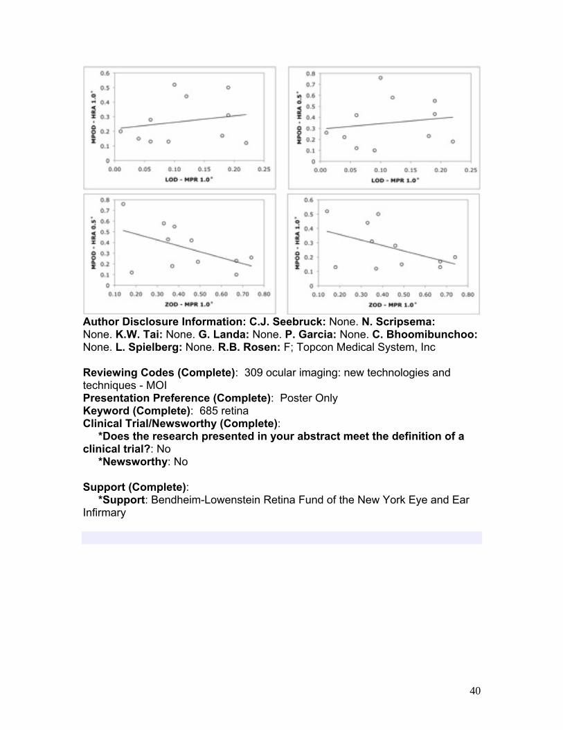
40
Author Disclosure Information: C.J. Seebruck: None. N. Scripsema: None. K.W. Tai: None. G. Landa: None. P. Garcia: None. C. Bhoomibunchoo: None. L. Spielberg: None. R.B. Rosen: F; Topcon Medical System, Inc Reviewing Codes (Complete): 309 ocular imaging: new technologies and techniques - MOI Presentation Preference (Complete): Poster Only Keyword (Complete): 685 retina Clinical Trial/Newsworthy (Complete): *Does the research presented in your abstract meet the definition of a clinical trial?: No *Newsworthy: No Support (Complete): *Support: Bendheim-Lowenstein Retina Fund of the New York Eye and Ear Infirmary
41
The Effect of Uncomplicated Cataract Extraction on Rates of Visual Field Sensitivity Change Measured with Automated Pointwise Linear Regression Analysis in Glaucoma Patients Author Block Nikola Ragusa1, Gustavo V. De Moraes2,1, Celso Tello1,3, Robert Ritch1,3, Jeffrey M. Liebmann2,1. 1Ophthalmology, Einhorn Clinical Research Center, New York Eye & Ear Infirmary, New York, NY; 2Ophthalmology, NYU School of Medicine, New York, NY; 3Ophthalmology, New York Medical College, Valhalla, NY. Abstract: Purpose: To investigate whether the improvement of visual field (VF) sensitivity observed following uncomplicated cataract extraction (CE) affects the ability to detect VF progression in glaucoma patients using pointwise linear regression analysis (PLR). Methods: Retrospective observational cohort. Treated, established glaucoma patients (optic disc damage and VF abnormalities) with ≥8 24-2 SITA VF tests seen between January 1999 and December 2009 were included. VF progression was evaluated using automated PLR (PROGRESSOR, Medisoft Ltd., Leeds, UK). A VF progression endpoint was defined as at least 2 adjacent test points in the same hemifield progressing >1.0 dB/yr at p<0.01. VF, MD and PSD values were compared using the VF tests performed before and after CE. Global rates of sensitivity change (dB/yr) and number of eyes reaching a progression endpoint were compared between eyes that underwent CE and no other surgery during the VF sequence (Group A) and eyes that did not undergo any surgery (Group B) during the same period. Results: 348 eyes of 348 patients (mean age, 64.8±12.8 yrs; 88.5% European descent; mean baseline MD, -6.4±4.7 dB) were evaluated. Group A had 71 eyes (20.4%) and Group B 277 (79.6%). Group A patients were older than those in Group B (72.1±9.7 vs. 62.9±13.0 yr, p<0.01). There was no significant difference between groups regarding baseline MD (p=0.75), CCT (p=0.72), or mean follow-up IOP (p=0.25). There was a non-significant change in mean MD and mean PSD following surgery (-7.6±4.6 to -6.8±4.2 dB, p=0.27; and 5.8±43.2 to 6.2±3.4 dB, p=0.50, respectively). Groups A and B showed similar velocities of sensitivity change (-0.35±0.4 vs. -0.35±0.6 dB/yr, p=0.98) even after adjusting for differences in age (p=0.27). Group A reached progression endpoints as often as Group B (30.9% vs. 21%, p=0.10). CE did not influence VF endpoint in the multivariate model after adjusting for differences in age (OR=1.40, p=0.26). Conclusions: VF global indices did not change significantly following uncomplicated CE. VF sensitivity changes observed following CE do not affect the ability of PLR to detect VF progression. Uncomplicated cataract surgery should not be considered an exclusion criterion when applying automated PLR to detect VF progression in glaucoma patients

42
Title: ASFDOCT Sequential Imaging of Schlemm’s Canal and the Collector Channels in Normal and POAG Eyes Authors: Alfredo R. Castillejos, MD1, Saman Kiumehr, MD1, Sung Chul Park, MD1,2, Andrea X. Arata MD1, Gustavo De Moraes, MD1,3, Jeffrey M. Liebmann, MD1,3, Robert Ritch, MD1,2
Abstract Purpose: To quantitatively and qualitatively evaluate and compare the microstructures comprising the trabecular outflow pathway, namely the trabecular meshwork, Schlemm’s canal and the collector channels in normal and POAG eyes using a novel technique of sequential anterior segment Fourier-domain OCT (ASFDOCT) imaging. Methods: 20 subjects with no eye disease (N group) and 20 subjects with POAG (G group) were imaged in the sitting position with the eye in adduction fixating on a target 15 cm away over 6 mm of the temporal or nasal limbus. The investigators first scanned the temporal limbus using the “live scan mode” searching for the presence of collector channels (CCs). If none was identified, the procedure was repeated over the nasal limbus. When a CC was identified, an initial set consisting of 5 radial and 5 transverse scans was performed over the area selected. One eye per subject was randomly selected for the study. One radial and one longitudinal image capturing the ostium of the selected CC were quantitatively analyzed for the following variables: Schlemm’s canal (SC) cross-sectional area, length and width (radial scan); CC lumen diameter and ostium diameter (radial scans); minimum and maximum longitudinal SC width, (longitudinal scan). The variables were compared between the two groups using an independent samples T test. Spearman’s correlation coefficients (r) were calculated to assess the correlation between the morphometry of the microstructures and the maximum recorded IOP, current IOP, cup to disc ratio, the pattern standard deviation (PSD) obtained from a 24-2 achromatic perimetry and the number of medications used for glaucoma control. With the purpose of describing the course and morphologic characteristics of SC and the CCs as well as the configuration of the trabecular meshwork, composite radial and longitudinal images were created using the sequential scans. A qualitative analysis of the composite images was performed by two investigators masked to the patients’ information. Results: The groups were comparable in age (N=64.9±10.5 G=65.3±6.9 years, p=0.88). SC cross-sectional area (N=1800 µm² G=1300 µm², p=0.49) and length (N=227 µm G=168 µm, p=0.35) as well as SC minimum longitudinal width (N=6.7 µm G=4.5 µm, p=0.41) were significantly reduced in the G group. No differences were observed in the CCs mean width (p=0.72) or ostium diameter (p=0.65). In the N group, a significant negative correlation was observed between age and SC cross-sectional area (r=-0.51 p=0.02) and SC width (r=-0.46 p=0.4); this age-
43
related reduction in SC dimensions was not observed in the G group. The mean number of medications positively correlated with SC cross sectional area (r=0.49 p=0.02), SC length (r=0.62 p=0.003) and SC minimum longitudinal width (r=0.57 p=0.008). Qualitatively, SC and the CCs appeared reduced in dimensions in both groups; however, in the G group, SC lumen showed increased tortuosity and had lower OCT signal intensity. Conclusions: SC and the CC morphology can be quantitatively and qualitatively assessed in the living eye using ASFDOCT. The combination of the sequential radial and longitudinal scans pathway provided a better assessment of the condition of the microstructures comprising the trabecular pathway. SC presents reduced dimensions in patients with advanced POAG as compared to age-matched normal individuals. Qualitatively, SC undergoes a change in configuration becoming increasingly tortuous possibly limiting aqueous flow around its circumference, processes that may be exacerbated in glaucomatous eyes.

44
Optical Coherence Tomography of Radiation Optic Neuropathy
Author: Yacoub A. Yousef, MD, Paul T. Finger, MD Objective: To study morphologic optic disc and retinal changes in patients with radiation optic neuropathy (RON) quantitatively using optical coherence tomography (OCT). Methods: A retrospective clinical case series of 10 patients with (RON). Each had been treated with palladium-103 ophthalmic plaque therapy for choroidal melanoma. OCT images taken at the time of initial RON diagnosis were collected. Using both Time Domain (n=6/10) and Spectral Domain (n=4/10), 10 OCT patients; thickness of optic disc and peripapillary retinal parameters were measured perpendicular to the Retinal Pigment Epithelium (RPE). Main outcome measurements included: cup depth, cup width, edema, vasculature diameter and peripapillary retinal thickness. Results: Radiation optic neuropathy was associated with morphological changes of the optic disc and peripapillary retina in all patients. They were characterized by optic disc edema, increased papillary retinal thickness and decreased optic cup depth. The average papillary retinal thickness was 341.6μm compared to 255μm in normal population (34% more than normal) and the average optic cup depth was 168μm Compared to 370μm in normal population (55% less than normal). Retinal arteries showed mean decrease in diameter of 32.5% and retinal veins showed mean increase in diameter of 29.4%. All RON patients in the study showed decrease in cumulative thickness of peripapillary retinal layers. Conclusion: Radiation optic neuropathy manifests characteristic morphologic retinal and optic disc changes (increased papillary thickness, decreased optic disc cup depth, retinal arteriolar narrowing, retinal vein engorgement and decreased thickness of peripapillary retinal layers) that can be quantitatively evaluated by optical coherence tomography.