department of histology and medical embriology phd...
TRANSCRIPT

Department of Histology and Medical Embriology
PhD in Cell Science and Morphogenesis
XVII CICLO
PHARMACOLOGICAL ACTIVATION AND REPROGRAMMING OF MUSCLE SATELLITE CELLS TO ACQUIRE AN EARLIER, PLURIPOTENT,
CIRCULATING “STEM-LIKE” BEHAVIOUR, SUITABLE FOR THERAPEUTICAL APPLICATIONS
PhD Student: Laura Castaldi
Tutor: Coordinator: Prof. Marina Bouchè Prof. Mario Molinaro

Acknowledgements
I would like to sincerely thank my supervisor prof. Marina Bouchè for her invaluable
advice and constant support throughout the last three years. I also wish to express my gratitude to prof. Mario Molinaro for the interest expressed in
my work and for giving me the opportunity to attend international meetings and courses.
I would like to thank all the people working in my department, especially my colleagues past and present in laboratory 24: Carlo, Alessandra, Lina, Macò, Massimiliano,
Renata, and a special mention to MariaPina.

2
INTRODUCTION __________________________________________________________ 3 Embryonic Myogenesis __________________________________________________________ 3 Adult Myogenesis _______________________________________________________________ 4 Skeletal Muscle Regeneration _____________________________________________________ 6 Adult Stem Cells ________________________________________________________________ 8 Muscle Diseases_________________________________________________________________ 9 Cell therapy___________________________________________________________________ 10 Reprogramming of committed cells _______________________________________________ 12
NFkB ______________________________________________________________________________ 14 AP-1 ______________________________________________________________________________ 15 Bis-peroxovanadium (BpV) ____________________________________________________________ 17
AIM OF THE WORK ______________________________________________________ 18
MATERIALS AND METHODS ______________________________________________ 19 Cell cultures __________________________________________________________________ 19 Cell transfections and luciferase assay _____________________________________________ 20 Western Blot analysis ___________________________________________________________ 20 Immunofluorescence analysis ____________________________________________________ 21 BrdU assay ___________________________________________________________________ 21 Preparation of RNA and RT-PCR analysis _________________________________________ 22 Flow Cytometry Analysis________________________________________________________ 24 Trans-differentiation assays _____________________________________________________ 25 Cell staining and histochemical analysis ___________________________________________ 26 In vivo transplantation__________________________________________________________ 26
RESULTS________________________________________________________________ 27 BpV treatment induces NF-kB and AP-1 trascriptional activities in C2C12 ______________ 27 BpV treatment induces morphological changes and reversibly inhibits myogenic differentiation. ________________________________________________________________ 30 BpV treatment induces cell cycle progression _______________________________________ 30 BpV modifies gene expression profile in C2C12 and primary satellite cells_______________ 31 BpV treatment induces SP phenotype in C2C12 cells_________________________________ 32 BpV induces stem and early hematopoietic markers in C2C12 and primary satellite cells __ 32 BpV treatment induces multi-lineage potentiality in C2C12 cells _______________________ 33 BpV treated muscle cells acquire the phenotype of a circulating progenitor ______________ 35
DISCUSSION ____________________________________________________________ 37

3
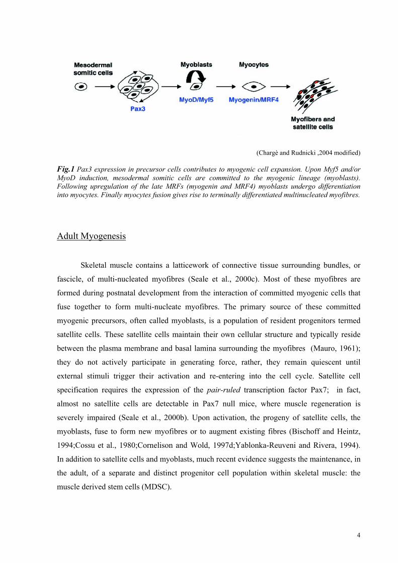
INTRODUCTION Embryonic Myogenesis
All vertebrate skeletal muscles (apart from head muscles) derive from mesodermal
precursor cells originating from the somites (Pourquie, 2001;Summerbell and Rigby, 2000).
During embryonic development, specification of mesodermal precursor cells to the myogenic
lineage is regulated by positive and negative signals from surrounding tissues. Specification
to the myogenic lineage requires the upregulation of MyoD and Myf5, basic helix-loop-helix
transcriptional activators of the myogenic regulatory factor family (MRF). Proliferating
MyoD and/or Myf5 positive myogenic cells are termed myoblasts. These cells withdraw from
the cell cycle to become terminally differentiated myocytes expressing the “late” MRFs,
Myogenin and MRF4, followed by the expression of muscle-specific genes, such as myosin
heavy chain (MHC) and muscle creatine kinase (MCK). Finally, mononucleated myocytes
specifically fuse to each other to form multinucleated syncytium, which eventually mature
into contracting muscle fibres (Fig. 1). The existence of this regulatory network mostly comes
from the phenotype observed in mutant mice. In fact, MyoD/Myf5 double knockout mice
totally lack skeletal muscle, and putative muscle progenitor cells remain multipotential
contributing to non muscle tissues in the trunk and the limbs of these mice (Rudnicki et al.,
1993;Kablar et al., 1999). By contrast, in myogenin-deficient mice MyoD and/or Myf5
positive cells are present, but the embryos die peri-natally due to a lack of myoblast
differentiation (Nabeshima et al., 1993;Hasty et al., 1993). Similarly, MRF4-deficient mice
display a range of phenotypes consistent with a late role for MRF4 in the myogenic pathway
(Braun and Arnold, 1995;Patapoutian et al., 1995). During the course of muscle development,
a distinct subpopulation of myoblasts fails to differentiate, but remains associated with the
surface of the developing myofiber as quiescent muscle satellite cells, contributing to the
post-natal skeletal muscle growth and remodelling.
4
(Chargè and Rudnicki ,2004 modified) Fig.1 Pax3 expression in precursor cells contributes to myogenic cell expansion. Upon Myf5 and/or MyoD induction, mesodermal somitic cells are committed to the myogenic lineage (myoblasts). Following upregulation of the late MRFs (myogenin and MRF4) myoblasts undergo differentiation into myocytes. Finally myocytes fusion gives rise to terminally differentiated multinucleated myofibres.
Adult Myogenesis
Skeletal muscle contains a latticework of connective tissue surrounding bundles, or
fascicle, of multi-nucleated myofibres (Seale et al., 2000c). Most of these myofibres are
formed during postnatal development from the interaction of committed myogenic cells that
fuse together to form multi-nucleate myofibres. The primary source of these committed
myogenic precursors, often called myoblasts, is a population of resident progenitors termed
satellite cells. These satellite cells maintain their own cellular structure and typically reside
between the plasma membrane and basal lamina surrounding the myofibres (Mauro, 1961);
they do not actively participate in generating force, rather, they remain quiescent until
external stimuli trigger their activation and re-entering into the cell cycle. Satellite cell
specification requires the expression of the pair-ruled transcription factor Pax7; in fact,
almost no satellite cells are detectable in Pax7 null mice, where muscle regeneration is
severely impaired (Seale et al., 2000b). Upon activation, the progeny of satellite cells, the
myoblasts, fuse to form new myofibres or to augment existing fibres (Bischoff and Heintz,
1994;Cossu et al., 1980;Cornelison and Wold, 1997d;Yablonka-Reuveni and Rivera, 1994).
In addition to satellite cells and myoblasts, much recent evidence suggests the maintenance, in
the adult, of a separate and distinct progenitor cell population within skeletal muscle: the
muscle derived stem cells (MDSC).
5
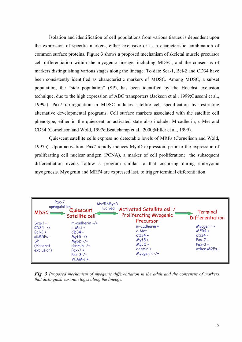
Isolation and identification of cell populations from various tissues is dependent upon
the expression of specific markers, either exclusive or as a characteristic combination of
common surface proteins. Figure 3 shows a proposed mechanism of skeletal muscle precursor
cell differentiation within the myogenic lineage, including MDSC, and the consensus of
markers distinguishing various stages along the lineage. To date Sca-1, Bcl-2 and CD34 have
been consistently identified as characteristic markers of MDSC. Among MDSC, a subset
population, the “side population” (SP), has been identified by the Hoechst exclusion
technique, due to the high expression of ABC transporters (Jackson et al., 1999;Gussoni et al.,
1999a). Pax7 up-regulation in MDSC induces satellite cell specification by restricting
alternative developmental programs. Cell surface markers associated with the satellite cell
phenotype, either in the quiescent or activated state also include: M-cadherin, c-Met and
CD34 (Cornelison and Wold, 1997c;Beauchamp et al., 2000;Miller et al., 1999).
Quiescent satellite cells express no detectable levels of MRFs (Cornelison and Wold,
1997b). Upon activation, Pax7 rapidly induces MyoD expression, prior to the expression of
proliferating cell nuclear antigen (PCNA), a marker of cell proliferation; the subsequent
differentiation events follow a program similar to that occurring during embryonic
myogenesis. Myogenin and MRF4 are expressed last, to trigger terminal differentiation.
Fig. 3 Proposed mechanism of myogenic differentiation in the adult and the consensus of markers that distinguish various stages along the lineage.
Sca-1 +CD34 -/+Bcl-2 +allMRFs -SP(Hoechstexclusion)
MDSC TerminalDifferentiation
Myogenin +MFR4 +CD34 -Pax-7 -Pax-3 -other MRFs +
Myf5/MyoDinvolved Activated Satellite cell /
Proliferating MyogenicPrecursorm-cadherin +c-Met +CD34 +Myf5 +MyoD +desmin +Myogenin -/+
QuiescentSatellite cell
m-cadherin -/+c-Met +CD34 +Myf5 -/+MyoD -/+desmin -/+Pax-7 +Pax-3-/+VCAM-1 +
Pax-7upregulation

6
Skeletal Muscle Regeneration
Adult mammalian skeletal muscle is a stable tissue with little turnover of nuclei, but
has the ability to complete a rapid and extensive regeneration in response to even severe
damage. Whether muscle injury is a result of a direct trauma or of genetic defects, muscle
regeneration is characterised by two phases: a degenerative phase followed by a regenerative
phase. The initial event of muscle degeneration is muscle fiber necrosis. This event is
generally triggered by disruption of the myofiber sarcolemma resulting in increased myofiber
permeability. The early phase of muscle injury is usually accompanied by the activation of
mononucleated cells, principally inflammatory cells and myogenic cells. Current reports
suggest that factors released by the injured muscle activate inflammatory cells residing within
the muscle, which in turn provide the chemotactic signals to circulating inflammatory cells:
neutrophilis and macrophages (Tidball, 1995).
Muscle degeneration is followed by the activation of the muscle repair process (Fig.
2). Satellite cells first exit their normal quiescent state to start proliferating. Cell proliferation
is an important event required for muscle regeneration. The expansion of myogenic cells
provides a sufficient source of new myonuclei for muscle repair (Grounds et al., 2002). After
several rounds of proliferation, the majority of satellite cells, often referred to as myogenic
precursor cells (mpc), differentiate and fuse to existing damaged fibres for repair or to one
another for new myofiber formation. On muscle sections, the fundamental morphological
characteristics of regenerating muscle are the presence of newly formed myofibres of small
caliber with centrally located myonuclei (Fig. 2).

7
(Chargè and Rudnicki, 2004)
Fig. 2 Schematic rapresentation of the molecular events regulating muscle satellite cell activation during skeletal muscle regeneration. Quiescent satellite cells are activated to enter the cell cycle and proliferate (A), allowing for expansion of the myogenic cell population (B). The proliferative phase is followed by terminal differentiation (C) and fusion of myoblasts to damaged myofibres for repair or to each other for new myofiber formation (D). Myoblast terminal differentiation is characterised by the upregulation of the MRFs Myogenin and MRF4. Finally, repaired or new myofibres grow to resemble original myofibres (E). During the course of muscle regeneration, a subset of myoblasts re-enters the quiescent state to replenish the satellite cell pool for subsequent muscle repair (F). The possible role of several growth factors is also highlighted, among which positive factors are in green while negative factors are in red. HGF, hepatocyte growth factor; FGF, fibroblast growth factor; IGF, insulin-like growth factor; IL-6, interleukin-6; LIF, leukemia inhibitory factor; TGFβ, transforming growth factor β family.
As mentioned before, the expression program followed during satellite cell activation,
proliferation, and differentiation is similar to that occuring during embryonic myogenesis. The
first phase is characterised by the rapid up-regulation of the two MRFs required for myogenic
specification, MyoD and Myf5 (Cooper et al., 1999a;Cornelison et al., 2000a;Zammit et al.,
2002). Proliferating myoblasts withdraw from the cell cycle to become differentiated
myocytes expressing the two “late” MRFs, Myogenin and MRF4 and then fuse to damaged
myofibres for repair or to each other for new myofiber formation.

8
In vitro and in vivo experiments have highlighted the possible role of several growth factors,
acting through a paracrine or autocrine mechanism to regulate specific events of muscle
regeneration. Among them FGF, HGF, IGF, IL6 and LIF may act as positive or negative
regulators depending on the specific phase (Fig. 2).
Adult Stem Cells
Adult stem cells (SC) are defined by two major functions: self renewal and
multilineage differentiation potentiality. Stem cells are functionally responsible for the
development and the regeneration of tissues and organs. Developmental signals, both
biochemical and biomechanical, trigger proliferation of stem cells in early and late
development. Adult stem cells have been found to reside in a variety of tissues including skin
(Watt, 1998), the central nervous system (Gage et al., 1995), muscle (Schultz and
McCormick, 1994), bone marrow (Weissman, 2000) the liver (Alison and Sarraf, 1998), and
many others. By definition, adult stem cells can self-renew and replenish multiple cell types
within the tissue in which they reside.
A central tenet of developmental biology is that, during embryogenesis, all cells
become committed to specific lineages, first via germ layer specification, and later through
additional levels of differentiation and specialisation. Stem cells that replenish adult tissues
are also set aside during development, and these cells were thought to be committed by
default to generate only a restricted lineage of cells. However, in the last few years, several
reports have suggested that the differentiation capability of adult stem cells might not be
restricted to exclusively producing cells specific to the tissue in which they reside, but it may
exert a much wider spectrum of potentiality, resulting in the so called “stem cell plasticity”. It
is now well known that Hematopoietic Stem Cells (HSC) and Mesenchimal Stem Cells
(MSC) are capable of metaplasia into various cell types (Priller et al., 2001b;Priller et al.,
2001a). Upon bone marrow transplantation, several laboratories have observed the
recruitment of donor-derived cells in several non-hematopoietic tissues, through the
expression of reporter genes such as LacZ or green fluorescent protein (GFP) (Ferrari et al.,
1998a;LaBarge and Blau, 2002c;Orlic et al., 2001). Similarly, transplantation of cells derived

9
from brain, muscle, skin and fat, has resulted in the detectable presence of these cells in
several lineages distinct from the tissue of origin (Jackson et al., 1999;Bjornson et al., 1999).
This behaviour was immediately attributed to a previously unexplored developmental
plasticity of stem cells residing in these tissues. It was thus postulated that, given a new
microenvironment, adult stem cells can trans-differentiate to produce cells according to the
new resident tissue. The notion of adult stem cell plasticity thus challenged the long-held
concepts of developmental biology.
Muscle Diseases
The term “myophaty” has come to apply to any disorder which can be attributed to
pathological, biochemical or electrical changes occurring in the muscle fibres or in the
interstitial tissues of the musculature; such disorders include many which are inflammatory,
metabolic or endocrine in nature. The term “muscular dystrophy” should be reserved for cases
of progressive, genetically determined, degenerative myopathy.
Muscular dystrophies (MDs) are a heterogeneous group of disorders caused by the
mutation of any one of a large number of genes. They are usually characterised by progressive
muscle weakness, which, depending on its severity, may result in immobilization and death.
Most of these diseases depend on mutation of gene coding for any protein involved in the
dystrophin-glycoprotein complex. This complex includes dystrophin, sarcoglycans and
dystroglycans, and interconnects cytoskeletal actin with the membrane and extracellular
matrix components. It maintains the mechanical stability of the muscle cell membrane by
anchoring and supporting the sarcolemma (Ervasti and Campbell, 1993) (Fig. 3). The most
common form of MD, Duchenne muscular dystrophy (DMD), is due to the lack of dystrophin
expression (Hoffman et al., 1992). Morphologically, dystrophic muscle appears to be in a
constant state of spontaneous necrosis and degeneration, with a concomitant gradual
replacement of muscle fibres by fibrous tissue. Thus, satellite cell-driven muscle regeneration
is not as efficient in balancing muscle degeneration.

10
Fig.3 Dystrophin and the dystrophin-glicoprotein complex in skeletal muscle.
Although various approaches to deliver dystrophin in dystrophic muscle have been
investigated extensively (e.g. cell and gene therapy), there is still no treatment that alleviates
the muscle weakness in this common inherited disease.
Cell therapy
There are currently three main experimental approaches to setting therapy for muscular
dystrophies. 1) Gene therapy focuses on the development of new vectors capable of efficiently
delivering the missing gene to the postmitotic nuclei of the muscle fibres in vivo. 2) The
pharmacological approach aims to restore the protein complex that is altered in many forms of
muscular dystrophy through different strategies. 3) The cell therapy approach aims to
functionally rescue the tissue by delivering “sane” cells, capable of reconstituting the tissue;
these may be satellite cells or pluripotent stem cells.
Third approach appears to be the more promising one, although its use still presents
many limitations. Re-appearance of dystrophin expression may result from the conversion of
native myofibres through donor-host fusion and from the formation of new myofibres from
both donor-donor and donor-host cell fusion. This process has been investigated extensively

11
in both animal and human clinical trials. Restoration of dystrophin expression can be achieved
through transplantation of donor-derived dystrophin-expressing myogenic cells into
dystrophic host skeletal muscle; however, only a minority of the grafted cells are responsible
for new muscle formation, while the majority of them quickly die (Beauchamp et al., 1999).
The recent identification of stem cells suitable for protocols of organ regeneration has
opened new perspectives in cell-mediated therapy. Some examples include muscle-derived
stem cells (MDSCs) (Torrente et al., 2001b) and the mesoangioblasts (Sampaolesi et al.,
2003a). Intra-muscular and intra-arterial injection of MDSCs into a dystrophic skeletal muscle
have demonstrated the ability of these muscle-derived stem cells to increase muscle
regeneration and improve the delivery of dystrophin (Torrente et al., 2001a;Lee et al., 2000),
although the efficiency of muscle engraftment was only 12%. A hallmark of any stem cell
therapy should be morphological and functional restoration of the target tissue. The recent
identification of a vessel-associated stem cell population, the mesoangioblasts, has opened
new perspectives. Under culture conditions, these clonally derived cells express CD34, c-Kit,
and Flk-1. It has recently been shown that transplanted donor-derived mesangioblasts into
α−sarcoglycan-/- (SCGα-/-) dystrophic mice were capable of restoring SCGα expression,
muscle architecture and function (Sampaolesi et al., 2003b). These results showed a
significant functional improvement in the pathology of this mouse strain using a wide variety
of physiological tests (Sampaolesi et al.2003). However, as these cells are derived from foetal
tissue, it may be difficult to identify and purify them on the basis of expression of specific
markers for use in adult humans.
In recent years cell therapy has even been taken into consideration for the treatment of
heart disease. Cellular cardiomyoplasty (cellular transplantation for cardiac repair into injured
cardiac regions) has recently become an area of intense research interest in the treatment of
cardiovascular disease. Stem or progenitor cells can be delivered into the site of cardiac
injury, restore blood flow and contractility to a previously infarcted, scarred or dysfunctional
heart (Taylor, 2004a). Preclinically, many types of cells have been transplanted into injured
myocardium, including cardiomyocytes, autologous myoblasts, smooth muscle cells,
fibroblasts and bone marrow-derived stem cells. Although several of these cell types may
hold future promise as a therapeutic option, to date only skeletal myoblasts, and bone
marrow-derived stem cells have been used in safety studies as a first step towards myocardial
repair (Carvalho et al., 2004;Taylor, 2004c;Horackova et al., 2004). The transplantation of
12
these two types of cells is still ongoing. Clinical studies show that skeletal myoblasts can be
engrafted into infarcted regions of myocardium, increase myocardiac wall thickening and
contractility; however, it is not clear if and how myoblasts electrically integrate into
surrounding myocardium and what impact integration may have on either function or
rhythmicity. In fact, early clinical data suggest that myoblast transplantation may be
associated with a transient period of electrical instability (Taylor, 2004b). The advantage of
using bone marrow-derived stem cells is that they contribute to the increase of cardiac
angiogenesis. The use of these cells in clinical trials shows an enhancement in left-ventricular
function and improvement in infarct tissue perfusion. Thus, transplantation of stem cells is
considered the most promising therapeutic approach to the damage caused by cardiovascular,
myogenic, neuro degenerative diseases and aging. However, the use of stem cells in cell
therapy applications remains problematic for a number of reasons: the difficulty in obtaining,
expanding and culturing ex vivo adult stem cells, the insufficient number of cells obtained
from a biopsy, the lack of robust methods for their propagation and efficient differentiation, as
well as host rejection to allogenic cells (Deasy et al., 2002); de Hann et al. 2003).
A desirable goal in cell therapy is to obtain a population of primary cells retaining
genetic and phenotypic flexibility to commit toward distinct lineages (pluripotent stem cells),
suitable for efficient in vitro expansion, for genetic engineering, and for successful
reintroduction in vivo as a gene-cell therapy protocol.
Reprogramming of committed cells
Terminally differentiated mammalian cells are thought to be incapable of undergoing
reversion from the differentiated state. Muscle cells represent a very suitable model for
terminal differentiation. These cells have permanently exited the cell cycle through the
expression of cyclin-dependent kinase (cdk) inhibitors, and are driven “irreversibly” to their
final morphology and function through the accumulation of proteins critical for cellular
specialisation. However, a number of recent studies indicate that ectopic expression of certain
genes can induce established cell lines and even terminally differentiated cells, to
dedifferentiate and/or transdifferentiate, indicating that some plasticity can be induced in
already committed/differentiated cells. This adds to the well established capability of both

13
HSC and MSC to transdifferentiate into various cell types (Ferrari et al., 1998b;Gussoni et al.,
1999b;LaBarge and Blau, 2002b;Camargo et al., 2003). In fact, several groups have recently
demonstrated that somatic cells can be induced to functionally reprogram. As an example,
fibroblasts can be induced to express T-cell specific genes and functions, when cultured in the
presence of nuclear and cytoplasmic extract derived from lymphocytes (Hakelien et al.,
2002d); myoblasts can also be converted to physiologically active neuronal phenotype
through the activation of genes of neuronal differentiation (Watanabe et al., 2004b);
surprisingly, even terminally differentiated myotubes can be induced to dedifferentiate when
stimulated with appropriate signals such as the expression of msx1 (Hu et al., 1995c;Hakelien
et al., 2002c;Odelberg et al., 2000;Kondo and Raff, 2000b).
These observations suggest that reprogramming of gene expression in committed cells in
order to obtain the reversion into an earlier pluri/multipotent “stem-like” cell, might be a
feasible option to develop innovative cell therapy strategies.
To this end, at first the differentiation program in committed cells should be reversibly
inhibited. In muscle cells it is well known that the expression of many oncogenes (such as c-
Myc or v-Src) reversibly inhibits differentiation in vitro due to reduced expression and/or
abnormal functioning of MRFs (Falcone et al., 2003;La Rocca et al., 1994). Although these
studies demonstrate that myogenic differentiation can be reversibly inhibited, the use of
oncogene expressing cells or retroviruses infection, even as a transient expression, cannot be
proposed for therapeutic strategies.
The possibility of pharmacologically inducing intracellular signalling pathways in
order to inhibit differentiation while maintaining cell proliferation, could be a reasonable
alternative; reversibility should be achieved just by removal of the drug. It is well known that
NFkB and AP1 transcriptional activity are quickly and strongly down-regulated when muscle
cells are transferred into differentiation medium (Lehtinen et al., 1996). Thus, the activity of
these factors is not compatible to the differentiation program. In fact, forced activity NF-kB
and/or AP-1 inhibits myogenesis through regulation of Cyclin D1, and/or Myogenin and
MyoD (Guttridge et al., 1999d;Guttridge et al., 2000). Thus, the use of drugs which activate
these pathways may represent a possible approach in reversibly inhibiting muscle
differentiation.

14
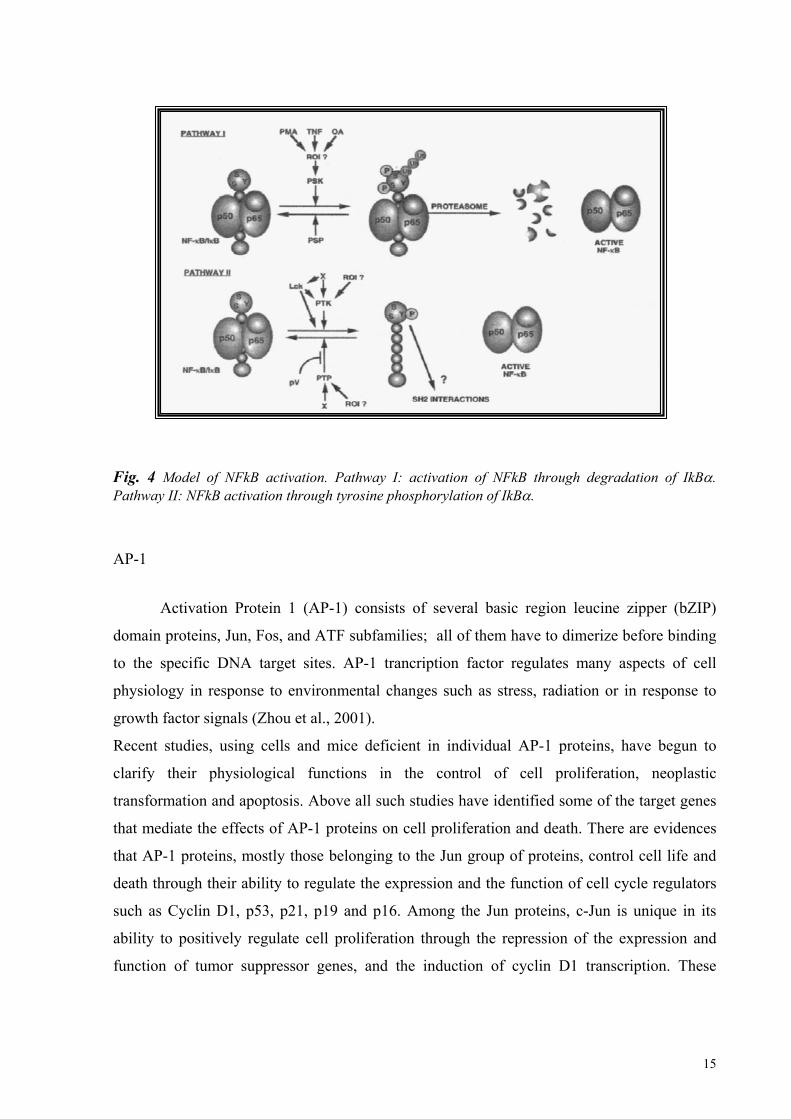
NFkB
The nuclear factor-κB (NF-κB) proteins are a small group of closely related
transcription factors, which in mammals consists of five members: Rel (also known as c-Rel),
RelA (also known as p65 or NF-κB3), RelB, NF-κB1 (also known as p50) and NF-κB2 (also
known as p52). All five proteins have a Rel homology domain (RHD), which serves as the
principal regulatory domain for dimerization and DNA-binding (Fig. 4). The C terminus of
RHD contains the nuclear-localization sequence (NLS) which is masked in non stimulated
cells through the binding of specific NF-κB inhibitors, known as the IκB proteins (Boone et
al., 2002b).
Nuclear transcription factor-kB plays a pivotal role in expression of various inducible
target genes related to immune and inflammatory responses, including the type I human
immunodeficiency virus. In non-stimulated cells, the heterodimer NF-kB complexes are
associated with its inhibitor, one of the IkB family members, thus retained in the cytoplasm.
In mammalian species, six structural homologs of IkB have been identified, but only one of
them, the IkBα form, has been extensively studied. In response to stimulation by various
agents, such as phorbol esters (e.g. phorbol 12-myristate 13-acetate), tumor necrosis factor
(TNF), interleukin-1a (IL-1a), γ-radiation, and lipopolysaccharide, IkBα undergoes
degradation, allowing the heterodimers to translocate to the nucleus (Fig.4). Upon stimulation
by TNF or IL-1, a protein kinase complex, containing the IkB kinase subunits α, β and γ (also
known as NEMO), phosphorylates IkBα in Ser-32 and Ser-36 residues; these
phosphorylation events are required for its degradation and consequent NF-kB nuclear import.
Before being degraded by 26 S proteasome, serine-phosphorylated IkBα is polyubiquitinated
at the Lys-21 and Lys-22 positions. Recently, it was also shown that hypoxia, reoxygenation,
and the PTPase inhibitor pervanadate (PV) induce IkBa Tyr42 phosphorylation and results in
NF-kB activation. Accordingly, protein tyrosine kinases (PTKs) and phosphatases (PTPase)
inhibitors suppress NF-kB activation. Thus, IkBα Tyr42 phosphorylation also triggers NF-kB
activation, but the PTK that phosphorylates IkBα is not known nor is the molecular
mechanism fully understood. Whether Tyr42 phosphorylation leads to IkBα degradation and
whether degradation is required for NF-kB activation is not known either.
15
Fig. 4 Model of NFkB activation. Pathway I: activation of NFkB through degradation of IkBα. Pathway II: NFkB activation through tyrosine phosphorylation of IkBα.
AP-1
Activation Protein 1 (AP-1) consists of several basic region leucine zipper (bZIP)
domain proteins, Jun, Fos, and ATF subfamilies; all of them have to dimerize before binding
to the specific DNA target sites. AP-1 trancription factor regulates many aspects of cell
physiology in response to environmental changes such as stress, radiation or in response to
growth factor signals (Zhou et al., 2001).
Recent studies, using cells and mice deficient in individual AP-1 proteins, have begun to
clarify their physiological functions in the control of cell proliferation, neoplastic
transformation and apoptosis. Above all such studies have identified some of the target genes
that mediate the effects of AP-1 proteins on cell proliferation and death. There are evidences
that AP-1 proteins, mostly those belonging to the Jun group of proteins, control cell life and
death through their ability to regulate the expression and the function of cell cycle regulators
such as Cyclin D1, p53, p21, p19 and p16. Among the Jun proteins, c-Jun is unique in its
ability to positively regulate cell proliferation through the repression of the expression and
function of tumor suppressor genes, and the induction of cyclin D1 transcription. These

16
actions are antagonized by JunB, which upregulates tumor suppressor genes and represses
cyclin D1 (Shaulian and Karin, 2002).

17
Bis-peroxovanadium (BpV)
In order to pharmacologically activate NFkB and AP1 pathways in muscle cells, we
treated myoblasts, cultured in differentiating conditions, with Bis-peroxovanadium (BpV), a
protein tyrosine phosphatase (PTP) inhibitor, known to be a strong activator of both
pathways. As expected, in these conditions, myogenesis is inhibited; moreover, the cells
continue to proliferate and acquire a gene expression profile and a plasticity compatible with
an “earlier” and/or “stem-like” phenotype. Removal of the drug rescues the muscle
differentiation capability. This may thus represent a valuable approach to easily obtaining
multi-potent cell populations from committed cells, suitable for gene-cell therapy
applications; moreover, it may be instrumental to study the molecular mechanisms
responsible for “stem cell” phenotype and genome reprogramming.
Phospho-Tyrosinephosphatases (PTP)
inhibitor

18
AIM OF THE WORK
It is well established that both HSC and MSC are capable of transdifferention into cell
types different from the tissue in which they originate (Ferrari et al., 1998c;Gussoni et al.,
1999c;LaBarge and Blau, 2002a;Camargo et al., 2003). However, given the difficulty to
obtain, expand and culture ex vivo adult stem cells, the physiological reservoir of precursors
for various tissues throughout life, and considering the ethical problems associated with the
use of human Embryonic Stem Cells, a desirable goal in cell therapy is to obtain a population
of primary cells retaining genetic and phenotypic flexibility to commit toward distinct
lineages (pluripotent stem cells), suitable for efficient in vitro expansion, genetic engineering,
and successful reintroduction in vivo as a gene-cell therapy protocol.
Terminal differentiation of mammalian cells was thought to be an irreversible process,
however, a number of recent results indicate that some “stem-like” plasticity can also be
induced in already committed cells (Hu et al., 1995b;Hakelien et al., 2002b;Odelberg et al.,
2000;Kondo and Raff, 2000a). These observations suggest that reprogramming of gene
expression in committed cells, in order to obtain their reversion into an earlier
pluri/multipotent “stem-like” cell, might be a feasible option to develop innovative cell
therapy strategies.
The aim of this work is to pharmacologically activate intracellular pathway(s) which
may induce reprogramming of gene expression in committed muscle satellite cells, and lead
the cells toward an earlier phenotype characterised by the ability to modulate self-
maintenance, activation and expansion. The pharmacological approach should give the
advantage of being a reversible and less invasive treatment in respect to a genetic-based
treatment. To this end, we tested and characterised the ability of Bis-peroxovanadium (BpV),
an activator of both NFkB and AP1 pathways, to induce the reversion of C2C12 or primary
satellite cells to an earlier, multipotent and circulating progenitor, suitable for therapeutical
applications.

19
MATERIALS AND METHODS
Cell cultures
C2C12 cells, a subclone of the C2 mouse myoblast cell line, were plated at 25.000
cells/35mm Petri dish and maintained in Dulbecco’s Modified Eagle medium (D-MEM)
supplemented with 10% Foetal Calf Serum (FCS) (Gibco) at 37°C in a humidified
atmosphere of 5% CO2 in air. To induce differentiation the medium was replaced with
differentiation medium, D-MEM supplemented with 2% Horse Serum (HS) (Gibco). BpV
(10µM) or TNFα (20 ng/ml) were added at the time of medium replacement.
Primary satellite cells (MSC) were obtained by enzymatic digestion of 6 old days mice leg
muscles. Muscles were minced and incubated in PBS containing 1mg/ml collagenase/dispase
(Roche) for 20 minutes at 37°C. The enzyme activity was inactivated by five times dilution
with PBS. Tissue fragments were removed by filtration through 70µm-pore-diameter nylon
mesh, while the cells were collected by centrifigation at 1200 rpm for 15 minutes. The cell
pellet was suspended in D-MEM containing 20% HS and 3% chick embryo extract (EE) and
cultured at 37°C in 10% CO2 for a pre-plating. After three hours, cell suspension was plated in
collagen-coated dishes. To induce differentiation the medium was replaced with D-MEM
containing 5% HS and 1,25% EE. BpV (10µM) or TNFα (20 ng/ml) were added at the time
of medium replacement.
Neonatal cardiac myocytes were obtained by enzymatic digestion of 1-2 day old mice hearts.
Atrial tissue was removed, and the ventricles were cut in four equal parts in a ADS buffer
(NaCl 116mM, KCl 5,36mM, NaH2PO4 0,9mM, MgSO4 0,4M, Glucose 5,5mM, Hepes
18mM) prior to enzymatic digesion. The ventricular tissue was subjected to multiple rounds
of enzymatic digestion of 12 minutes at 37°C using an Enzyme Solution (ES) containing 108
U/ml Collagenase Type 2 (Worthington) and 0,9mg/ml Pancreatin (Sigma); the digestion was
blocked by adding 1-2 ml of HS. Cells were collected by centrifugation, suspended in plating
medium (DMEM supplemented with 10% HS and 5% FCS) and plated for 2 hours in

20
uncoated cell culture dishes. The cardiomyocyte enriched suspension was then plated on
collagen-coated Petri dishes.
Cell transfections and luciferase assay
For transient transfection assay 1 x 10⁵ C2C12 cells were plated into 35 mm tissue culture
dishes. After 24 hours, proliferating myoblasts were transfected with 1,5 µg of plasmid
DNA/dish, using the lipid based Lipofectamine Reagent (Invitrogen), according to the
manufacturer’s instructions. After additional 32 hours the cells were incubated in DM
containing or not 10µM BpV or 20ng/ml TNFα. For reporter luciferase assay the cells were
harvested in Reporter Lysis Buffer 1x (Promega) and then centrifuged at 12.000xg for 15
seconds at room temperature. Supernatant was then analysed for luciferase assay reporter
activity with the employment of the AutoCliniLumat LB 952T/16 (Berthold), by using
Luciferase Assay Reagent containing the luciferin substrate (Promega).
The firefly luciferase, a monomeric 61 kDa protein, catalyses luciferin oxidation, a
bioluminescent reaction, using ATP-Mg2+ as a substrate. Light is produced by converting the
chemical energy of luciferin oxidation through an electron transition, forming the molecular
product oxyluciferin. Luciferase activity was expressed as the relative fold induction
compared to the luciferase activity assayed in untreated cells.
Western Blot analysis
C2C12 cells were harvested and lysed in ice cold RIPA extraction buffer (50mM Tris-HCl,
pH 7.4, 1% NP-40, 0.5% sodium deoxycholate, 0.1% sodium dodecyl sulfate, 5 mM EDTA,
150 mM NaCl, 50mM NaF) supplemented with protease inhibitors. Supernatant was collected
following high speed centrifugation for 20 minutes at 4°C. Equal amounts of protein extracts
were subjected to SDS-polyacrylamide gel electrophoresis and transferred to nitrocellulose
membranes. Blocking was performed by using 5% dry milk in TBST (25mM Tris-HCl pH 8,
125mM NaCl, 1.1% Tween20). Primary and secondary antibodies were diluted in 5% dry
milk in TBST, and the incubation proceeded for 30 minutes at room temperature. Washes

21
were performed in TBST for 5-10 minutes and repeated five minutes. Detection was
performed with HRP-conjugated antibodies (Bio-Rad Laboratories) and membrane-bound
immune complexes were visualised by CDP Star (PerkinElmer).
Western blots were probed with following antibodies: anti-p65 (Laboratories Tranduction),
ph-Tyr, IkBα, Pc-jun, actin (Santa Cruz).
Immunofluorescence analysis
Collected muscle samples were embedded in OCT compound (Tissue freezing medium,
Fluka) and frozen in liquid nitrogen cooled isopentane. Seven-micrometer cryosections were
fixed in 4% paraformaldehyde. Cells were fixed in Ethanol/Acetone (1:1 ratio) at -20°C for
20 min.
Fixed cells or tissue sections were then washed with Ca and Mg Free PBS (CMF) (Sigma),
incubated in CMF containing 1% BSA (bovine serum albumine) for 10 minutes at R.T.
followed by an additional incubation in CMF containing 1% goat serum for 30 minutes. The
cells or tissue sections were then incubated with primary antibody, at the appropriate
dilutions, overnight at 4°C in humidified conditions. After an additional incubation with
CMF/BSA 1% the cells were incubated for 1 hour with fluorochrome-conjugated secondary
antibody (Sigma) and Hoechst 33342 (Fluka) for nuclear staining.
After extensive washing with CMF, cell monolayers or tissue sections were mounted in 10
mM Tris-HCl, pH 9, containing 60% glycerol and examined under a Zeiss Axioskop 2 Plus
fluorescence microscope. Images were acquired by a digital camera using the Axiocam
software and then exported into Adobe Photoshop for processing.
Antibodies used were: MF20 provide by Fishman (Bader et al., 1982), anti Cardiac Troponin
I (Covance Research Products), anti GFP (Molecular Probes) and anti Laminin (Sigma).
BrdU assay
BrdU labelling medium (Roche) was added to BpV-treated or untreated C2C12 cells for 1 hr
at treatment different periods of time. BrdU incorporation was then detected following

22
manufacturer’s instructions (5-Bromo-2’-deoxy-uridine Labelling and Detection kit I, Roche),
and BrdU positive cells were visualised under an epifluorescence Zeiss Axioskop 2 Plus
microscope.
Preparation of RNA and RT-PCR analysis
Total RNA was extracted from cell cultures using a High Pure RNA Isolation Kit (Roche),
according to the manufacturer’s instructions. Total RNA was retrotranscribed into cDNA by
using the Superscript III system (Invitrogen), according to the supplier’s instructions. PCR
amplification was performed using Taq Polimerase (Taqara). To check for genomic
contamination, parallel aliquots of RNA were incubated without reverse transcriptase (RT)
and subjected to PCR amplification as well. The same reaction profile was used for all primer
sets: an initial denaturation at 95°C for 5 minutes, followed by 30 cycles of: 95°C for 1
minute; primer specific annealing temperature for 1 minute; 72°C for 2 minutes; a final
extension step of 7 minutes at 72°C. Amplification of a housekeeping transcript (beta-actin)
was used for normalisation. PCR products were run on 1% agarose gel in a TAE buffer
containing Etidium Bromide and digitised images were obtained using a CCD camera
Detection System (Diana II, Raytest).
Sequences of the primers, FW and RV, used for PCR were as follows:
TARGET cDNA primer Sequences (5’-3’) Annealing Temp. (°C) Product Size (bp)
β-actin 52 649
ATGCCTCTGGTCGTACCACAGGCATTG
TTGCTGATCCACATCTGCTGGAAGGTG
MyoD 59 330
GAGCAAAGTGAATGAGGCCTT
CACTGTAGTAGGCGGTGTCGT
Myogenin 59 450
AGTGAATGCAACTCCCACAG
TCAGAAGAGGATGCTCTCTGC
Myf-5 60 370
TGCCATCCGCTACATTGAGAG

23
CCGGGGTAGCAGGCTGTGAGTTG
Pax-3 60 450
AGGAGGCGGATCTAGAAAGGAAG
TGTGGAATAGACGTGGGCTGGTA
Pax-7 57 236
TACCAGGAGACCGGGTCCATC
TCCGAACTTGATTCTGAGC
PCNA 52 165
TCCTTGGTACAGCTTACT
TGCTAAGGTGTCTGCATT
Cyclin D1 52 362
GTGCCATCCATGCGGAA
GGATGGTCTGCTTGTTCTCA
Sca-1 56 200
GATTCTCAAACAAGGAAAGTA
GACTGAGCTCAGGCTGAACAG
Flt-1 57 300
CTCTGATGGTGATCGTGG
CATGCGTCTGGCCACTTG
c-Met FW 60 370
GAATGTCGTCCTACACGGCC
CACTACACAGTCAGGACACTGC
ALP 55 372
GCCCTCTCCAAGACATATA
CCATGATCACGTCGATATCC
Osteocalcin 60 292
AAGCAGGAGGGCAATAAGGT
AGCTGCTGTGACATCCATAC
Collagen 1A2 57 484
GCAATCGGGATCAGTACGAA
CTTTCACGCCTTTGAAGCCA
Cbfa 1 60 289
CCGCACGACAACCGCACCAT
CGCTCCGGCCCACAAATCTC
MITF 57 450
ACCATCAGCAACTCCTGTCC
TTCTTGCTTGATGATCCGATTC
MMP9 59 380
CCTGTGTGTTCCCGTTCATCT
CGCTGGAATGATCTAAGCCCA
IL3-Rα 60 428
TACCACATCCAGATGGAACC

24
TACCACATCCAGATGGAACC
GM-CSFRα 59 454
AACGTGACTGACAGGAAGG
TGTGTGTGCTGGCTGTTAAGG
Bcpr 1 57 330
CCATAGCCACAGGCCAAAGT
GGGCCACATGATTCTTCCAC
MDR 1a 55 500
CCCATCATTGCGATAGCTGG
TCCAACATATTCGGCTTTAGGC
MDR1b 55 430
TGCTTATGGATCCCAGAGTGAC
TTGGTGAGGATCTCTCCGGCT
Flow Cytometry Analysis
BpV-treated or untreated C2C12 or primary satellite cells were suspended by very rapid
trypsin digestion, washed in CMF/BSA 1% solution at 4°C and then incubated on ice with
1µg /106 cells fluorochrome-conjugated specific antibody. An aliquot of the cell suspension
was incubated with the corresponding labelled antibody isotype, to determine non-specific
fluorescent emission. Cells were then analysed by using a FACS FacsStar Plus. The
antibodies used were: Sca-1, CD45, c-kit, CD34, CD11b, Gr-1, Ter-119 and Mac-3 directly
conjugated to phycoerythrin, fluorescein or cy-chrome (all from Pharmingen),
For cell cycle analysis, BpV-treated or untreated C2C12 cells were suspended by trypsin
digestion and washed in 50% FCS in CMF; the cells were then suspended in the same
solution to which 70%ethyl alcohol was added. After an overnight incubation at 4°C the cells
were incubated in Propidium (Sigma) and the fluorescent DNA content was analysed by using
the FacsStar Plus.
For dye exclusion technique, the protocol by Goodell et al. (1996) was substantially followed.
In brief, BpV-treated and untreated C2C12 cells were suspended by trypsin digestion,
collected by centrifugation and suspended in pre-warmed DMEM containing 2% FCS and
10mM Hepes (Gibco) at a working concentration of 106 cells/ml. Hoechst was then added to a
final concentration of 10µg/ml and the cells were incubated in a 37°C water bath for exact 90

25
minutes. The cells were then collected by centrifugation at 4°C and suspended in ice cold
PBS-BSA 2% containing 2µg/ml propidium iodide (Sigma), and run on the FACS Vantage
SE cell sorter (BD) equipped with two lasers. Hoechst dye was excited at 475 nm and its
fluorescence was splitted through a 640/LP filter and detected at two wave-lengths using a
424/44 (Hoechst blue) and a 660/20 (Hoechst far red) filters.
Trans-differentiation assays
C2C12 cells were cultured in DM containing or not 10µM BpV for 24 hrs; the cells were
then cultured in the appropriate conditions in the presence or not of 2µM BpV for additional
5-7 days.
Hemopoietic conditions. BpV-treated or untreated cells (103 cells/35mm Petri dish) were
plated in complete methylcellulose medium for colony assay of murine cells (StemCell
Technologies). The appearance of clones was analysed after 5-7 days in methylcellulose
culture; the cells were then collected and either cyto-centrifuged for morphological analysis
or disrupted for RNA preparation.
To test their ability to acquire “osteoclast-like” phenotype BpV-treated or untreated C2C12
cells were cultured in DM supplemented with 100nM 1,25(OH)2 vitamin D3. After 5 days the
cells were analysed histochemically for the expression of the osteoclest marker acid
phosphatase (TRAP) or were disrupted for RNA preparation.
Osteoblast differentiation conditions. BpV-treated or untreated C2C12 cells were cultured in
DM supplemented with 50ng/ml of Bone Morphogenetic Protein 2 (BMP2) for 5 days; the
cells were then analysed histochemically for the expression of the osteoblast marker or
disrupted for RNA preparation.
Cardiomyocyte differentiation conditions. BpV-treated and untreated GFP-transduced C2C12
cells were cultured on a monolayer of neonatal cardiomyocytes for 4 additional days in DM.
The cells were then fixed in 5% paraphormaldehyde and GFP-expressing cells were analysed
for the expression of cardiac specific markers by immunofluorescence.

26
Cell staining and histochemical analysis
Wright staining. The cells were washed with PBS (Sigma) and incubated in Blu Wright
solution (Merck) for 5 minutes at RT. Two volumes of Wright buffer was then added. After
an additional incubation of 7 minutes the cells were washed with PBS.
TRAP histochemical analysis. For tartrate-resistant acid phosphatase activity (TRAP) cells
were washed with PBS, fixed with 3%Paraphormaldehyde diluted in 0,1M cacodilate tampon
for 15 minutes and incubated in a buffer containing: 0.4 M sodium acetate buffer, 2 mM
naphthol AS-BI phosphate, 100 mM sodium tartrate and 2 mM Fast Garnet. The incubation
was carried out for 90 minutes at 37°C, the reaction was stopped in distilled water and the
cells were observed with a light microscope.
ALP histochemical analysis. For the histochemical localization of alkaline phosphatase
activity (ALP), cells were washed with PBS and fixed with 3%Paraphormaldehyde diluted in
0,1M cacodilate tampon for 15 minutes; the cells were then incubated at 37°C for 30 minutes
in ALP substrate solution: 0,04 mg/ml naphthol AS-MX phosphate, 0,25 mg/ml Fast Violet B
Salt (Sigma). After washing in distilled water, the cells were observed with a light
microscope.
In vivo transplantation
BpV-treated and untreated GFP-transduced C2C12 cells were suspended by trypsin digestion,
washed in PBS and suspended in 25µl of PBS; 4x10⁵ cells were injected into the femoral
artery of SCG-/- dystrophic adult mice. The mice were then sacrificed at the appropriate time
points after transplantation and analysed by immunofluorescence.

27
RESULTS
BpV treatment induces NF-kB and AP-1 trascriptional activities in C2C12
In order to pharmacologically activate signalling pathways known to inhibit myogenic
differentiation, we treated C2C12 cells with BpV, a phosphotyrosine phosphatase inhibitor,
known to activate both NFkB and AP-1 transcriptional activities in different cell types (Barat
and Tremblay, 2003b). In fact, it is well known that myogenic differentiation requires down
regulation of both these pathways (Guttridge et al., 1999c;Lehtinen et al., 1996).
As first, we analysed the kinetics of NF-kB activation in BpV-treated C2C12 cells
using a gene reporter assay. C2C12 cells were transfected with a plasmid where Luciferase
expression is driven by a basic promoter element (TATA box) joined to tandem repeat of
NFkB binding elements. The medium was then replaced with DM and the cells were treated
with 10 µM BpV or 20ng/ml TNFα, another well known NFkB activator. Luciferase activity
was assayed at different periods of time, within the 24 hours of treatment. As shown in Figure
1, while BpV induces a slow 2-3 fold activation of luciferase activity, which is maintained
through 24 hours of treatment, TNFα treatment induces a transient 2-3 fold induction, which
decreases to below the basic activity within 12 hours.
Fig.1 NFkB activation in BpV-treated and untreated C2C12 cells. C2C12 cells were transfected with an NFkB-Luc reporter plasmid, treated with TNFα or BpV and analysed at different periods of time within 24h. Values were normalised to basal levels of promoter activity obtained by untreated NFkB-Luc plasmid transfected cells.
N F k B T ra n s a c tiv a tio n
0
1
2
3
4
0 5 10 15 2 0 2 5
Fold
indu
ctio
n
b p V
T N F a lp ha

28
Activation of NFkB is usually mediated by IkB kinase (IKK) complex-dependent
Ser32,36 phosphorylation of the NFkB inhibitor IkBα; these events are then followed by
ubiquitin-proteasome dependent degradation of IkBα. However, as shown in Figure 2a,
Ser32,36 IkBα phosphorylation is not affected by BpV treatment, nor the IkBα degradation.
Recently, an “alternative” NFkB activation pathway has been identified, which appears to be
mediated by Tyr42 phosphorylation of IkBα, followed or not (depending on the cell system)
by its proteolitic degradation. This pathway is activated by numerous stimuli including the
PTPase inhibitor pervanadate. Since BpV is a pervanadate derivative we evaluated whether it
might induce this “alternative” pathway in C2C12 cells. By immunoprecipitation and Western
Blot analysis we found that BpV induces, within 5 hrs of treatment, Tyr42 IkBα
phosphorylation Fig. 2a) followed by nuclear translocation (thus activation) of the NFkB p65
subunit (Fig.2b).
Taken together, these results demonstrate that BpV induces a “sustained” NFkB
transcriptional activity through the IKK-independent Tyr42 IkBα phosphorylation.
Fig.2 NFkB nuclear translocation through the IKK-independent Tyr42 IkBα phosphorylation induced by BpV treatment. A) Immunoprecipitation and Western Blot of Tyr42 IkBα phosphorylation. B) Western Blot analysis of p65 within 5 hrs of treatment.
ph-Tyr
IkBα
- + - + - + - +30’ 1h 2h 5h
IP a-IkBα
10 µM BpV
A
p65
- + - + - + - +
30’ 1h 2h 5h
30 µM BpV
B

29
Since, as mentioned before, BpV is known to also activate AP-1 pathway, we then
analysed AP-1 transcriptional activation in BpV-treated C2C12 cells. BpV-treated and
untreated cells were transfected with a plasmid were Luciferase gene was under control of
two human collagenase III sites. The medium was then replaced with DM and the cells were
treated with 10 µM BpV or 20ng/ml TNFα. Luciferase activity was assayed at different
periods of time, within the 8 hours of treatment. As shown in Figure 3a, while BpV induces a
transient fold activation of luciferase activity which peaks at 6 hours, TNFα does not exert
any induction. Moreover, c-jun Ser63 phosphorilation was observed by Western Blot analysis,
in BpV-treated C2C12 cells (Fig.3B).
Taken together, these results demonstrate that BpV activates both NFkB and AP-1 signalling
pathways, making it a good candidate to pharmacologically inhibit myogenic differentiation.
Fig.3 AP-1 activation in BpV-treated cells. A) C2C12 cells were transfected with an AP-1 reporter plasmid, treated with TNFα or BpV and anslysed at different periods of time within 24 hours. Values were normalised to basal levels of promoter activity obtained by untreated NFkB-Luc plasmid trasfected cells. B) c-jun Ser63 phosphorilation analysed by Western Blot within 24 hours.
A P - 1 a c ti v a ti o n
0 ,0 0
2 ,0 0
4 ,0 0
6 ,0 0
8 ,0 0
0 2 4 6 8
h o u rs
Fo
ld In
du
ctio
n
TN F
B p V
A
c-jun
- 30’ 1h 2h 5h 12h 24h
Actin
10 µM BpV
Ph-ser63-c-junB

30
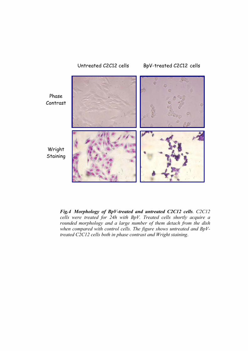
BpV treatment induces morphological changes and reversibly inhibits myogenic
differentiation.
The activation of NFkB and AP-1 signalling pathways is parallel to dramatic
morphological changes in C2C12 cells. In fact, when the cells were cultured in BpV-
containing DM, they promptly acquired a round shape morphology and many of them
detached from the dish, without showing any features of degeneration or apoptosis (Fig. 4).
This type of morphological change was evident even within 4-5 hours of treatment. Given the
already round shape morphology of primary muscle satellite cells (MSC), when MSC were
treated with BpV, as above, detachment from the dish was observed (not shown). As
expected, long term (72-96 hrs) BpV treatment inhibits the formation of multinucleated
myotubes (not shown). Treatment of C2C12 or MSC cells with TNFα did not induce the
short-term phenotypic changes observed with BpV treatment, although it inhibits long-term
myogenic differentiation (not shown).
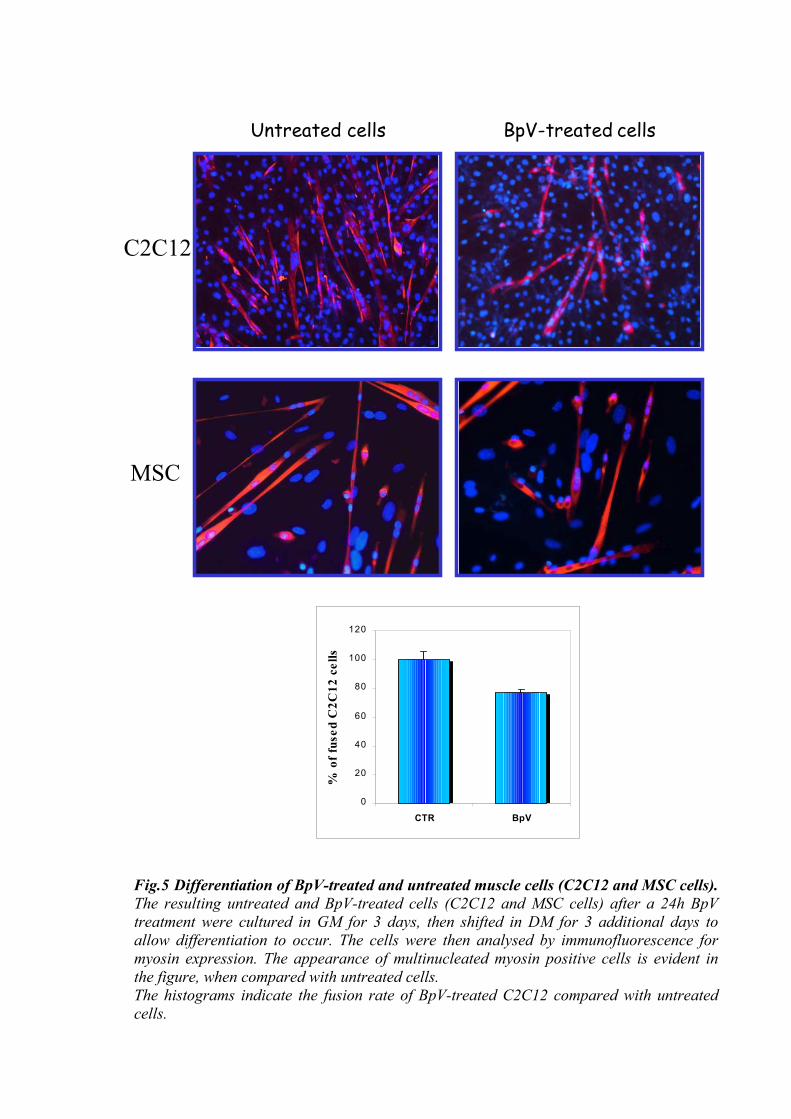
However, when the drug was removed and BpV pre-treated cells (C2C12 or MSC
cells) were cultured in DM for 3 additional days, the appearance of multi-nucleated myosin
positive fibres was observed, with a fusion rate comparable to control cells (Fig.5). Taken
together these results demonstrate that BpV-treated cells maintain their myogenic potentiality.
BpV treatment induces cell cycle progression
The morphological phenotype acquired by the BpV-treated cells prompted us to verify
the proliferation capability of treated cells. BpV-treated, untreated and proliferating C2C12
cells were FACS analysed for cell cycle at different time points within 48 hours of treatment.
As shown in Figure 6a C2C12 cells cultured in low serum containing medium in the presence
of BpV display a slower accumulation in G1 phase by the time, when compared to untreated
cells. The slower G1 accumulation is paralleled by a slower decrease in the fraction of cells
still in the S phase. This slow progressive accumulation in G1 was comparable to that
occurring when the cells were maintained in high serum containing medium due to cell
confluence being reached. The results are summarised in Table 1.
Untreated C2C12 cells BpV-treated C2C12 cells
PhaseContrast
WrightStaining
Fig.4 Morphology of BpV-treated and untreated C2C12 cells. C2C12 cells were treated for 24h with BpV. Treated cells shortly acquire a rounded morphology and a large number of them detach from the dishwhen compared with control cells. The figure shows untreated and BpV-treated C2C12 cells both in phase contrast and Wright staining.
BpV-treated cellsUntreated cells
C2C12
MSC
0
20
40
60
80
100
120
CTR BpV
% o
f fus
ed C
2C12
cel
ls
Fig.5 Differentiation of BpV-treated and untreated muscle cells (C2C12 and MSC cells).The resulting untreated and BpV-treated cells (C2C12 and MSC cells) after a 24h BpV treatment were cultured in GM for 3 days, then shifted in DM for 3 additional days to allow differentiation to occur. The cells were then analysed by immunofluorescence for myosin expression. The appearance of multinucleated myosin positive cells is evident inthe figure, when compared with untreated cells. The histograms indicate the fusion rate of BpV-treated C2C12 compared with untreatedcells.

A
12 hrs
2% HS + bpV 2% HS 10% FCS
24 hrs
48 hrs%G1 %S %G2 10% FCS 12h 52,3 ± 0,9 35,7 ± 0,8 ** 12,1 ± 1,8 ** 2% HS 64,2 ± 6,2 *** 27,5 ± 3,3 *** 8,5 ± 2,9
2% HS + BpV 50,3 ± 11,1 43,1 ± 8,7 6,7 ± 2,4
10% FCS 24h 65,2 ± 1,5 ** 27,9 ± 0,9 7,1 ± 0,5 *** 2% HS 75,8 ± 5,8 *** 18,1 ± 5,6 *** 6,2 ± 0,2 ***
2% HS + BpV 57,8 ± 10,1 30,1 ± 6,4 12,2 ± 3,7
10% FCS 48h 82,9 ± 0,9 *** 14,1 ± 0,2 *** 3,1 ± 0,7 * 2% HS 86,6 ± 1,9 *** 10,6 ± 3,4 *** 2,9 ± 1,5 *
2% HS + BpV 72,7 ± 3,5 22,9 ± 4,5 4,3 ± 0,9
C2C12 M SC
Table 1
*P<0.1 **P<0.02 ***P<0.001
β-actin
Cyclin D1
PCNA
Ctr BpV Ctr BpV
B
Fig.6 Cell cycle analysis in proliferating (10%FCS) untreated (2%HS) and BpV-treated (2%HS+ BpV) C2C12 cells. A) FACS analysis was performed on proliferating, untreated and BpV-treated C2C12 cells at different time points (12-24-48 hours). B) RT-PCR shows the upregulation of PCNA and CyclinD1 expression in untreated and BpV-treated cells (C2C12 and MSC).

31
Although cultured in differentiation medium, cell cycle progression of BpV-treated
C2C12 cells, as well as primary satellite cells, was also confirmed by RT-PCR analysis of the
expression of PCNA, a marker of the S phase, and Cyclin D1 which were both up-regulated
upon BpV treatment (Fig. 6b).
To further demonstrate the proliferative activity of BpV treated C2C12 cells, a BrdU
assay was performed on both untreated and BPV-treated C2C12 cells at different time points
within 24 hours. While the percentage of BrdU positive cells decreases progressively until a
significant decrement at 24 hours in untreated C2C12 cells, in BpV treated C2C12 the
percentage of BrdU positive cells is not significantly altered (Fig.7a). Figure 7b shows the
evident reduction, within 24 hours, of BrdU positive untreated cells; in contrast, upon BpV
treatment many BrdU positive cells were still present. Figure 7c shows that even in the cell
population that detached from the dish upon BpV treatment, many BrdU positive cells are
present.
BpV modifies gene expression profile in C2C12 and primary satellite cells
We then analysed the expression of markers defining muscle lineage in BpV-treated
cells (C2C12 and MSC cells) by RT-PCR. Figure 8 shows that the expression of the Muscle
Regulatory Factors (MRFs: MyoD, Myf5 and Myogenin) was down-regulated after 24 hours
of BpV treatment both in C2C12 cells and in MSC. By contrast the expression of markers
defining muscle precursor cells such as Pax-3, Pax-7 and c-Met was up-regulated in both
C2C12 and MSC cells, as well as Flt-1, whose expression is detectable in satellite cells and in
regenerating fibres.
Taken together these results demonstrate that BpV treatment induces reprogramming of gene
expression both in C2C12 and MSC cells.

0,00
15,00
30,00
45,00
60,00
% o
f cel
ls in
S p
hase
Ctr T0BpV T12hBpV T24h
A
0,00
15,00
30,00
45,00
60,00
% o
f ce
lls in
S p
hase
Ctr T0Ctr T12hCtr T24h
*
* P<0,02
CTR 0 CTR 12h
BpV 12h
BpV 12h detachedC detached BpV 24h
Fig.7 BrdU assay on BpV-treated and untreated C2C12 cells. BpV-treated and untreated cells were incubated with BrdU labelling, for 1 hour, at different time points within 24 hours. A) The percentage of cells in S phase was calculated by determining the number of BrdU-positive cells with respect to the total number of cells in given fields. B) Fluorescence of BrdU labelling of BpV-treated and untreated C2C12 cells at different time points. C) Fluorescence of BrdU labelling of BpV-treated cells detached from the dish.
24h BpV
24hCTR
B

CTR BpV CTR BpV
β-actin
MyoD
Myogenin
Myf-5
MyoD
Myf5
Myogenin
β-actin
Pax-7Pax-3
c-Met
Flt-1Flt-1
Pax-3
c-Met
Pax-7
C2C12 MSC
Fig.8 Gene expression profile in BpV-treated and untreated muscle cells(both C2C12 and MSC cells) by RT-PCR. RT-PCR analysis shows inuntreated and BpV-treated muscle cells the expression of MRFs, such asMyoD, Myf5 and Myogenin and of markers defining muscle precursor cellsas Pax-3, Pax-7 and c-Met.

32
BpV treatment induces SP phenotype in C2C12 cells
It is well known that the SP phenotype is a hallmark of stem cells in many tissues
(Bunting, 2002). The SP phenotype is characterised by the ability of this population of stem
cells to exert a “side” profile when analysed by FACS, and to exclude the Hoechst 33342 dye,
due to the expression of ABC transporters. Recently, it was reported that also C2C12 cell
population contains a subpopulation of cells exerting SP phenotype (Benchaouir et al.,
2004b). We then verified whether BpV-treatment could increase the percentage of this SP
population in C2C12 cells. FACS analysis of Hoechst 33342 stained C2C12 cells confirmed
the presence of a very small sub-population (0.02%) characterised by low staining (SP).
However, BpV treatment increases the percentage of this sub-population by 5 times (0.11%)
when compared to untreated cells (Fig. 9).
SP phenotype, and even the Stem Cell phenotype, was also defined by the expression
of two members of the ABC transporter superfamily: multidrug resistance-1 (MDR1) and
breast cancer resistance protein (Bcrp1) (Bunting, 2002). We thus analysed the expression of
both markers in both C2C12 and in MSC by RT-PCR analysis. Figure 9 shows that, in C2C12
cells, BpV treatment up-regulates both MDR1a and MDR1b isoforms but not Bcrp1, which is
already expressed in untreated cells. Conversely Bcrp1 upregulation was evident in BpV-
treated MSC, while the expression of MDR1a and MDR1b was fairly up-regulated slightly.
BpV induces stem and early hematopoietic markers in C2C12 and primary satellite cells
Untreated and BpV-treated C2C12 cells were then analysed for the expression of stem
and early hematopoietic markers by FACS analysis, using conjugated specific antibodies. The
results are summarised in Figure 10. Untreated cells already expressed low levels of the Stem
Cell markers c-Kit, Sca-1 and CD34; however, BpV treatment induced a significant up-
regulation of these markers. Among the early hematopoietic markers tested the expression of
both CD45 and CD11b was very low in untreated cells; BpV treatment induced a low but
detectable up-regulation of CD11b, while higher up-regulation of CD45 was evident;
moreover, a macrophage lineage specific marker, Mac-3, was already expressed in untreated

s
0.02%
s
Hoe
chst
blu
e
C2C12 MB CtBpVCtr
Bcrp1
MDR1a
MDR1b
-actinβ
BpV-treated C2C12 cell
Untreated C2C12 cell0.11%
d
Hoechst reA
SC
r BpV
Fig.9 SP phenotype in untreated and BpV-treated muscle cells (both C2C12 and MSC cells). A) FACS analysis of BpV-treated C2C12 cells using the fluorescence dye Hoechst 33342 staining. B) RT-PCR analisys of MDR1a, MDR1b and Bcrp1 expression in BpV-treated and untreated muscle cells (both in C2C12 and MSC cells).

Mean fluorescence intensity Untreated cells
BpV-treated cells
c-kit 13.1 69.2
Sca-1 83.2 96.0
CD34 28.9 77.8
Mac-3 19.7 56.2
CD11b 6.8 13.7
CD45 8.1 44.4
Gr-1 8.9 29.3
Fig.10 Expression ohematopoietic lineagecells. C2C12 were treexpression of specific m
Ter-119 16.2 36.3
f specific precursor markers for both muscle and s by FACS analysis of BpV-treated and untreated C2C12 ated with BpV for 24h and then analyzed by FACS for the arkers as indicated.

33
cells, but its expression was highly up-regulated after 24 hours of BpV treatment. Finally the
expression of Gr-1 and Ter119, granulocyte and erythrocyte lineage markers respectively, was
also up-regulated upon BpV treatment.
Preliminary results for the expression of stem and early hematopoietic markers on MSC cells
showed a similar profile.
Taken together these results suggest that BpV treatment promotes the exposed cells to
an earlier phenotypic stage, inducing the expression of markers specific for precursor of both
muscle and hematopoietic lineages.
BpV treatment induces multi-lineage potentiality in C2C12 cells
The gene expression profile acquired by the BpV-treated cells, prompted us to verify
their possible multi-lineage potential.
As first, to verify the potential to give rise to cells of different lineages, for example of
hematopoietic origin, C2C12 cells were cultured in the presence or in the absence of BpV for
24 hours; untreated and treated cells were then cultured for 5 additional days in a myeloid
colony assay conditions, in the presence of low BpV concentration (2µM). After 5 days, BpV-
treated cells formed hematopoietic-like colonies while untreated cells formed clones of
adherent cells (Fig.11a). The hematopoietic-like colonies were then cytocentrifuged and the
morphology of the obtained cells was analysed by Wright staining. Fig.11b shows that many
of the cells acquired morphological phenotype characteristic of myeloid lineages (such as
monocytes, macrophages or granulocytes). RT-PCR analysis performed on untreated and
BpV-treated C2C12 cells cultured in myeloid conditions shows that the expression of the
granulocytes macrophages colony stimulating factor receptor α (GM-CSFRα) and of the
interleukin 3 receptor α (IL-3Rα) was induced in BpV-treated cells; no expression of these
markers was detectable in untreated cells (Fig.11c). These two genes are expressed in both
hematopoietic stem cells (HSCs) and in common myeloid progenitors (CMPs) (Miyamoto et
al., 2002).
Osteoclasts are of myeloid origin, thus the potentiality to give rise to this lineage was
then analysed. Untreated and BpV-treated cells were cultured for 5 days in low serum

untreated C2C12 cells Bpv treated C2C12 cells
β-actin
GM-CSFRIL3-Rα
CTR BpV
A C
B
Fig.11 Methoculture of BpV-treated and untreated C2C12 cells. C2C12 cells weretreated with BpV (10µM) for 24h, and then cultured under myeloid conditions for 5 additional days; BpV was maintained at 2µM concentration through the whole period in culture. A) Hematopoietic-like clones are formed by BpV-treated cells, while untreated cells form clones of adherent cells. B) BpV-treated cells grown in methylcellulose culturewere collected, cytospinned on to glass slides and stained with Giemsa for morphologicalanalysis. The figure shows that the BpV-treated C2C12 cells show morphologicalcharacteristics of the myeloid lineages. C) RT-PCR analysis of the GM-CSFRα and IL-3Rα expression in untreated and BpV-treated C2C12 cells.

34
containing medium supplemented with 100nM 1,25(OH)2 vitaminD3 and 2µM BpV.
Conversion to osteoclast-like phenotype was then analysed by the histochemical assay
of the acid phosphatase activity (TRAP). As shown in Figure 12, while no TRAP positive
cells were detectable in untreated cells, many TRAP positive cells were present in BpV-
treated cells. The acquisition of the osteoclast-like phenotype by the BpV-treated cells was
also confirmed by RT-PCR analysis showing that BpV treatment up-regulates osteoclast
specific markers such as Microphthalmia Transcriptor Factor (MITF), known to regulate
transcription of TRAP (Luchin et al., 2001), and matrix metalloproteinase (MMP9), which
has a critical role in osteoclast migration and bone reabsorption (Delaisse et al., 2003). Thus,
BpV treatment can trigger C2C12 muscle cells towards an osteoclastic–like phenotype, which
however is of hematopioetic origin.
To verify whether BpV-treated cells were capable of differentiating into phenotype of
mesenchimal origin, rather than muscle, as osteoblast, untreated and BpV treated C2C12 cells
were cultured for 5 days in low serum containing medium, supplemented with 50ng/ml Bone
Morphogenetic Protein 2 BMP2 and 2µM BpV. Osteoblast conversion was then analysed by
the histochemical assay of the alkaline phosphatase activity (ALP). It is already known that
C2C12 cells, as well as primary satellite cells, can be converted to the osteoblast phenotype
by treatment with a high concentration of BMP2 (100-300 ng/ml) (Katagiri et al.,
1994a;Asakura et al., 2001b); however, at the lower concentration of 50ng/ml BMP2 used,
very few untreated cells became positive for ALP activity, while a three times increase in
ALP positive cells was observed in BpV-treated cells (Fig.13). Again, osteoblast conversion
was confirmed by RT-PCR analysis for the expression of osteoblast specific markers: ALP,
Osteocalcin, Collagen I and the transcriptor factor Cbfa1, which is considered the osteoblast
master gene for its essential role in osteogenesis and osteoblast differentiation (Komori et al.,
1997;Ducy et al., 1997). Thus BpV treatment enhances the BMP2 driven osteoblast
conversion of C2C12 cells.
We further analysed whether BpV treatment could convert C2C12 cells towards
cardio-myocyte phenotype. To this purpose GFP-labelled C2C12 cells were cultured in the
presence or not of BpV for 24 hours. Treated and untreated cells were then co-cultured for 4
additional days on a monolayer of cardiomyocytes, previously prepared from 1-2 day old

1,25(OH)2vitaminD3
TRAP
bpv treated cellscontrol
β-actin
MMP9
MITF
Ctr Bpv
50
Fig.12 TRAP activity and RT-PCR analysis on untreated or BpV-treated C2C12 cells. BpV-treated and untreated C2C12 cells were cultured in medium containing 1,25(OH)vitaminD3 and then analysed by the histochemical assay of the acid phosphatase activity (TRAP). The figure shows the evident induction of TRAP activity. RT-PCR shows the expression of osteoblastic specific markers such as MMP9 and MITF in both BpV-treated and untreated cells.
ng/ml BMP-2
ALP
Bpv treated cellscontrol
Osteocalcin
Collagen I
Ctr BpvALP
Cbfa 1
β-actin
Fig.13 ALP activity and RT-PCR analysis on untreated or BpV-treated C2C12 cells. BpV-treated and untreated C2C12 cells were cultured in medium containing BMP-2 and then analysed for ALP activity. The figure shows the evident induction of ALP activity. RT-PCR shows the expression of specific osteoblast markers such as Osteocalcin, Collagen I, Cbfa1, in BpV-treated and untreated C2C12 cells.

35
mice. The conversion towards cardiac phenotype was then evaluated by the
immunofluorescence analysis of cardiac troponin I (CTnI). The co-expression of cardiac
troponin I (red) and GFP labelling demonstrate the conversion to the cardiac phenotype. The
result shown in Figure 14 demonstrates that while no co-expression of GFP and CTnI was
detectable in the absence of BpV, some cells co-expressing GFP and CTnI were evident upon
BpV treatment. Thus, BpV treatment can trigger C2C12 muscle cells towards a cardiac
phenotype.
Taken together, these results suggest that BpV treatment imparts to muscle cells the
tendency towards different cell lineages (i.e. myeloid, osteoclasts, osteoblasts and cardiac
cells), though maintaining their myogenic potential.
BpV treated muscle cells acquire the phenotype of a circulating progenitor
In order to verify whether the gene expression profile and the ability to trans-
differentiate into different lineages may reflect the acquisition of the phenotype of circulating
progenitors capable of systemic delivery and diapedesi to colonize different tissues, untreated
and BpV-treated cells were transplanted into recipient mice via intra-arterial delivery. To this
purpose, C2C12 cells were previously infected with a lentivirus carrying the GFP cDNA. The
cells were subcultured for 2-3 times before use, to ensure the absence of free lentiviral DNA.
To test this, medium was used to feed uninfected cells where no GFP-labeled cells were
present. As first we investigated the possible BpV-induced homing into the skeletal muscle.
4x10⁵ GFP-labeled cells, untreated or treated with BpV for 24 hours, were injected into the
femoralis artery in vivo. As the mouse model SCGα-/- dystrophic mice were used. These mice
represent a model for muscular dystrophy due to the absence of SCGα, thus degenerating
muscle fibres need to be continuously replaced by muscle cell precursor. The capability of the
transplanted cells to reach skeletal muscle via circulation was thus evaluated after
transplantation, by the presence of GFP-cells in skeletal muscle. Five days after
transplantation the mice were sacrificed and leg muscles (quadriceps, tibialis, soleus and
gastrocnemius) were removed; GFP-labeled cells were visualised in cryosections under an
epifluorescence microscope, while muscle fibres were visualised by immunofluorscence

BpV treated C2C12-GFP cells Untreated C2C12-GFP cells
Fig. 14 Co-culture of GFP-C2C12 cells with neonatal cardiomyocytes. GFP-C2C12 cells were treated with BpV for 24 hours and co-cultured with cardiomyocytes for 4 additional days. The cells were then analysed by immunofluorescence for the expression of Cardiac Troponin I (red). The figure shows that BpV treatment induces the expression of Cardiac Troponin I in GFP-C2C12 cells.

Untreated GFP-C2C12 BpV treated GFP-C2C12
Fig.15 Intra-arterial injection of untreated and BpV-treated GFP-C2C12 cells inSCGα-/- dystrophic mice. GFP untreated and BpV-treated C2C12 cells weretransplanted via femoralis artery delivery into dystrophic mice. Mice were sacrificed 5days after transplantation and muscle cryosections were analysed byimmunofluorescence analysis. As showed in the figure, many more GFP-BpV treatedcells migrate from femoral artery to skeletal muscle when compared with control cells.

36
analysis using an antibody anti-laminin. As shown in Figure 15, very few GFP-labeled mono-
nucleated cells are present in very damaged areas of skeletal muscle of mice injected with
untreated cells; by contrast, many more GFP-labeled mono-nucleated cells were found even
in undamaged skeletal muscle when BpV-treated cells were transplanted. This result
demonstrates that BpV treatment induces C2C12 cells to acquire a phenotype of circulating
progenitor capable of overcoming the endothelial barrier and reaching the muscle tissue.

37
DISCUSSION
In this study we demonstrated the capability of committed muscle satellite cells to
acquire an earlier, pluripotent circulating “stem-like” behaviour, through a pharmacological
treatment.
A number of recent results indicate that some plasticity can be induced in already
committed cells (Hu et al., 1995a;Hakelien et al., 2002a;Odelberg et al., 2000;Kondo and
Raff, 2000c). Thus, reprogramming of gene expression in committed cells, in order to obtain
their reversion into earlier pluri/multipotent “stem-like” cells, might be a feasible option to
develop innovative cell therapy strategies.
Searching for signalling pathways that could modulate self-maintenance, activation
and expansion of muscle satellite cells we found that treatment of satellite muscle cells with
BpV, a protein tyrosine phosphatase (PTP) inhibitor, primarily known to activate NFkB and
AP-1 activities (Barat and Tremblay, 2003a), induces in both primary murine satellite cells
and C2C12 satellite cells the acquisition of a “stem-like” phenotype. When cultured in
appropriate conditions, BpV-treated satellite muscle cells are committed towards phenotypes
belonging to distinct lineages. Moreover, BpV-treated cells acquire the ability to home, via
circulation skeletal muscle.
At first we demonstrated that BpV induces both AP-1 and NFkB transcriptional
activity. While BpV induces a 2-3 fold activation of luciferase reporter activity which is
maintained through 24 hours of treatment, AP-1 is transiently activated within 8 hours. In
contrast, TNFα, another well known activator of both pathways, induces a transient 2-3 fold
induction which decreases below the basic activity within 12 hours. These results are in
contrast with other reports (Guttridge et al., 1999b;Ladner et al., 2003) which show that
TNFα induces, in the same cellular model, a biphasic activation of NF-kB which lasts for
nearly 2 days following initial TNFα treatment.
Moreover, we showed that BpV treatment, in contrast to TNFα treatment, does not
induce the well established NFkB activation pathway mediated by IkBα Ser32,36
phosphorylation (Boone et al., 2002a). It was previously shown that hypoxia, reoxygenation
and PTPase inhibitor pervanadate activate NFkB via the “alternative” Tyr42 phosphorylation
of IkBα, probably through the involvement of the catalytic subunits of PI3 kinase (Imbert et
al., 1996;Beraud et al., 1999;Mukhopadhyay et al., 2000;Waris et al., 2003). Since BpV is a

38
pervanadate derivative we demonstrated, by Western Blot analysis, that it actually induces
IkBα Tyr42 phosphorylation in satellite C2C12 cells which is not followed by its proteolitic
degradation, and results in p65 NFkB subunit nuclear traslocation, as demonstrated by the
immunoprecipitation analysis. These observations, including the different kinetics of NFkB
activation, suggest that the two stimuli, BpV and TNFα, activate a qualitatively different
NFkB response through different activation pathways.
Activation of these pathways is known to inhibit myogenesis; accordingly both BpV
and TNFα prevent the formation of multinucleated myosin positive myotubes. Inhibition of
differentiation is due to the inhibition of MRFs (MyoD, Myf5 and myogenin) expression, as
demonstrated by RT-PCR analysis. However, BpV-treated cells, but not TNFα ones, shortly
acquire a round shape morphology and continue to proliferate in low serum containing
medium. It is well known that myoblasts, when cultured in differentiation medium, through
MRFs activity, asynchronously and irreversibly exit the cell cycle and progress to the ultimate
compartment of the myogenic lineage (Walsh and Perlman, 1997;Wei and Paterson, 2001).
BpV treatment, on the other hand, maintains myoblasts proliferating, as shown by FACS
analysis as well as by BrdU incorporation and by up-regulation of PCNA and Cyclin D1
expression. It is worth noting that, while PCNA is a marker of the S phase, Cyclin D1 is up-
regulated during the G1/S transition, and that its expression is known to be induced by the
NFkB pathway (Guttridge et al., 1999a). Importantly, BpV-induced inhibition of
differentiation is reversible. In fact, when BpV-treated cells were cultured in absence of the
drug they were still capable of forming multinucleated, myosin positive cells, with a fusion
rate comparable to the untreated cells.
Thus, BpV appeared to exert the initial requisites required for the pharmacological
treatment we were looking for: reversible inhibition of muscle differentiation associated with
cell proliferation.
However, to determine whether BpV treatment may be a suitable approach to develop
innovative cell therapy strategies, it is a must that BpV induces re-programming of gene
expression. It is well known that quiescent and activated muscle satellite cells are
characterised by the specific expression of the transcription factor Pax-7 and c-met, whose
expression is down regulated upon myogenic differentiation (Seale et al., 2000a).
Subsequently, activation of satellite cells is characterised by the rapid up-regulation of MRFs
such as MyoD and Myf5, which then trigger myogenic differentiation (Cornelison and Wold,

39
1997a;Cornelison et al., 2000b;Cooper et al., 1999b). RT-PCR analysis, in both C2C12 and
MSC cells, demonstrated that while BpV down-regulates the expression of the MRFs (MyoD,
Myf5 and Myogenin), it up-regulates the expression of markers defining muscle precursor
cells, such as Pax-7, Pax-3 and c-Met. Moreover, BpV treatment induces, in both C2C12 and
MSC cells, up-regulation of the VEGF receptor Flt-1, whose expression has been shown to be
detectable in activated satellite cells, but decreases during the differentiation program
(Germani et al., 2003). Thus, the observed gene expression profile shows that BpV actually
induces re-programming of gene expression and that the acquired gene expression profile
resembles that of the muscle cell precursor.
Which is the real border line between a “cell precursor” and a “stem cell” and how
these stages are finely regulated, is not clear yet. It is well known that SP phenotype is a
hallmark of stem cells in many tissues; it has been recently reported that C2C12 cells
comprises a cell subpopulation actively excluding Hoechst 33342, through the activity
supported by ABC transporters (Benchaouir et al., 2004a). By FACS analysis we confirmed
this result, but a 5 times increase in the percentage of this population was also observed upon
BpV treatment. This result was also supported by the observation that BpV up-regulates
MDR1 and Bcrp1, markers characteristic of the SP population, as shown by RT-PCR
analysis.
The acquisition of a “stem cell-like” phenotype in BpV-treated satellite cells, was also
confirmed by the BpV-induced up-regulation of additional markers characteristic of adult
muscle stem cells, such as CD34, Sca-1 and c-kit. As shown by FACS analysis, some of these
markers are already expressed in satellite cells, however, BpV treatment significantly
enhances their expression. This result confirms that BpV treatment induces, on the exposed
cells, a reprogramming of gene expression, inducing the up-regulation of markers specific of
muscle cell precursors and triggers the cells towards a “stem-like” phenotype.
Moreover, FACS analysis shows that BpV treatment induces the expression of early
markers of the hematopoietic lineage, such as CD45, the leucocyte common antigen, CD11b,
an integrin also known to define early hematopoietic cells, Mac-3 and Gr-1, early markers of
the myeloid lineage and Ter-119, which defines the erythroid lineage.
The gene profile expression acquired prompted us to verify the full differentiation
potential of the BpV-treated cells. Previous works have demonstrated that myogenic cells,
such as C2C12 myoblasts (Teboul et al., 1995;Katagiri et al., 1994b) as well as muscle

40
satellite cells (Asakura et al., 2001a), display osteogenic or adipogenic differentiation
capability upon treatment with BMPs or adipogenic inducers respectively; recently, it was
also demonstrated that C2C12 cells may be converted to a neuronal phenotype (Watanabe et
al., 2004a); however, there is no evidence of their hematopoietic or myo-cardiac
differentiation capability. We presented direct evidence that BpV-treated C2C12 cells, when
cultured in the appropriate conditions, were able to differentiate into phenotypes of
hematopoietic and mesenchymal origin, other then muscle cells, while retaining their muscle
differentiation potentiality. In particular, we demonstrated that BpV-treated C2C12 cells,
when cultured in myeloid colony assay conditions, can form hematopoietic-like colonies,
whose cells display morphogical characteristics of the myeloid lineages; this result was also
confirmed by the observed upregulation of markers of myeloid cells such as IL3-Rα and GM-
CSFRα, as shown by the RTPCR analysis. The ability of BpV-treated cells to give rise to
myeloid-derived cells was further demonstrated by the BpV-induced up-regulation of the
activity and/or the expression of markers characteristic of the osteoclast phenotype, such as
TRAP, MMP9 and MITF, when the cells were cultured in medium containing 1,25(OH)2
vitamin D3.
Also the ability to differentiate into mesenchymal lineages different from muscle, such
as osteoblasts, is significantly enhanced by BpV treatment. Both C2C12 and MSC cells are
known to differentiate into osteoblasts when cultured in the presence of a high concentration
of BMP2 (100-300 ng/ml) (Katagiri et al., 1994c). At a lower BMP2 concentration (50ng/ml)
very few, if any, ALP positive cells were detected; however, if the cells were pre-treated with
BpV, a 5-10 times increase in ALP positive cells was observed, as the up-regulation of both
“early” (Cbfa1, considered as a “master gene” for the osteoblast lineage) and “late” (collagen
1 and osteocalcin) osteoblast-specific markers.
Importantly, when BpV-treated C2C12 cells were co-cultured over a monolayer of
cardiomyocytes, they express the cardiac specific isoform Troponin I. Although no beating
was observed in CTnI-positive C2C12 cells, this result may open new perspectives for
therapeutical approach in heart diseases; in fact, transplantation of skeletal muscle cells into
infarcted hearts is already used in experimental clinical approaches; however, early clinical
data suggest that myoblast transplantation may be associated with a transient period of
electrical instability (Taylor, 2004d). The ability of BpV-treated muscle cells to acquire a
cardiomyocyte-like phenotype may overcome the observed disadvantages.

41
It was recently shown that C2C12, HUVEC, fibroblasts and bone marrow cells express
cardiac markers when co-cultured with cardiomyocytes, but this expression is the result of
cell fusion (Reinecke et al., 2004;Matsuura et al., 2004). In our conditions no CTnI expressing
C2C12 cells were detected when untreated cells were co-cultured with cardiomyocytes.
However when BpV-treated cells were co-cultured with cardiomyocytes many of these
expressing cells were detected. Whether this is a result of a real trans-differentiation or a BpV
induced enhancement of fusion is still not known.
These results demonstrate that BpV treatment confers on C2C12 cells a multi-potential
plasticity, as a result of the acquisition of a “stem cell-like” phenotype and behaviour. An
additional requirement to determine whether BpV treatment may be a suitable approach to
develop innovative cell therapy strategies, is that the cells should acquire the behaviour of a
circulating progenitor, thus allowing transplantation through systemic delivery and home and
colonise different tissues. The BpV-induced re-programming of gene expression and plasticity
is already a strong starting point. However, the observation that, upon transplantation via
femoralis artery, BpV-treated C2C12 cells, but not untreated cells, are able to migrate outside
the vessels in the presence of inflammation, as in the case of dystrophic muscle, makes BpV a
very promising pharmacological tool for the establishment of cell therapy protocols. The
precise pathway used by transplanted cells to reach host muscle tissues is not known, however
the possible BpV-induced expression of specific inflammatory cytokines receptors should be
further investigated. Thus, BpV-treated C2C12 cells are able to circulate, migrate outside the
vessels and reach the muscle tissue, although more quantitative and statistical analyses are
required. Whether the cells are also capable of contributing to muscle regeneration is still
under investigation.
In conclusion, BpV treatment appears to respond to most of the requirements needed
for a pharmacologically-based cell mediated therapy, using already committed cells:
reversible inhibition of differentiation, maintenance of cell proliferation, re-programming of
gene expression towards an “earlier” “stem cell–like” phenotype, cell plasticity and systemic
delivery. Future work is obviously needed to determine the actual feasibility of this approach
for tissue repair in vivo, as well as to fine tune the treatment conditions (qualitative and/or
quantitative enhancement of the response by modifying culture conditions or by synergy with
other factors, or the efficacy of the pharmacological treatment in vivo).

42
Moreover, BpV treatment represents a good tool for studying the molecular
mechanism and chromatin remodelling responsible for genome reprogramming and for the
establishment of a “stem cell” phenotype.

43
REFERENCES
Alison, M. and C.Sarraf. 1998. Hepatic stem cells. J. Hepatol. 29:676-682.
Asakura, A., M.Komaki, and M.Rudnicki. 2001a. Muscle satellite cells are multipotential stem cells that exhibit myogenic, osteogenic, and adipogenic differentiation. Differentiation 68:245-253.
Bader, D., T.Masaki, and D.A.Fischman. 1982. Immunochemical analysis of myosin heavy chain during avian myogenesis in vivo and in vitro. J. Cell Biol. 95:763-770.
Barat, C. and M.J.Tremblay. 2003a. Treatment of human T cells with bisperoxovanadium phosphotyrosyl phosphatase inhibitors leads to activation of cyclooxygenase-2 gene. J. Biol. Chem. 278:6992-7000.
Beauchamp, J.R., L.Heslop, D.S.Yu, S.Tajbakhsh, R.G.Kelly, A.Wernig, M.E.Buckingham, T.A.Partridge, and P.S.Zammit. 2000. Expression of CD34 and Myf5 defines the majority of quiescent adult skeletal muscle satellite cells. J. Cell Biol. 151:1221-1234.
Beauchamp, J.R., J.E.Morgan, C.N.Pagel, and T.A.Partridge. 1999. Dynamics of myoblast transplantation reveal a discrete minority of precursors with stem cell-like properties as the myogenic source. J. Cell Biol. 144:1113-1122.
Benchaouir, R., P.Rameau, C.Decraene, P.Dreyfus, D.Israeli, G.Pietu, O.Danos, and L.Garcia. 2004a. Evidence for a resident subset of cells with SP phenotype in the C2C12 myogenic line: a tool to explore muscle stem cell biology. Exp. Cell Res. 294:254-268.
Beraud, C., W.J.Henzel, and P.A.Baeuerle. 1999. Involvement of regulatory and catalytic subunits of phosphoinositide 3-kinase in NF-kappaB activation. Proc. Natl. Acad. Sci. U. S. A 96:429-434.
Bischoff, R. and C.Heintz. 1994. Enhancement of skeletal muscle regeneration. Dev. Dyn. 201:41-54.
Bjornson, C.R., R.L.Rietze, B.A.Reynolds, M.C.Magli, and A.L.Vescovi. 1999. Turning brain into blood: a hematopoietic fate adopted by adult neural stem cells in vivo. Science 283:534-537.

44
Boone, D.L., E.G.Lee, S.Libby, P.J.Gibson, M.Chien, F.Chan, M.Madonia, P.R.Burkett, and A.Ma. 2002a. Recent advances in understanding NF-kappaB regulation. Inflamm. Bowel. Dis. 8:201-212.
Braun, T. and H.H.Arnold. 1995. Inactivation of Myf-6 and Myf-5 genes in mice leads to alterations in skeletal muscle development. EMBO J. 14:1176-1186.
Bunting, K.D. 2002. ABC transporters as phenotypic markers and functional regulators of stem cells. Stem Cells 20:11-20.
Camargo, F.D., R.Green, Y.Capetanaki, K.A.Jackson, M.A.Goodell, and Y.Capetenaki. 2003. Single hematopoietic stem cells generate skeletal muscle through myeloid intermediates. Nat. Med. 9:1520-1527.
Carvalho, K.A., L.C.Guarita-Souza, C.L.Rebelatto, A.C.Senegaglia, P.Hansen, J.G.Mendonca, C.C.Cury, J.C.Francisco, and P.R.Brofman. 2004. Could the coculture of skeletal myoblasts and mesenchymal stem cells be a solution for postinfarction myocardial scar? Transplant. Proc. 36:991-992.
Cooper, R.N., S.Tajbakhsh, V.Mouly, G.Cossu, M.Buckingham, and G.S.Butler-Browne. 1999a. In vivo satellite cell activation via Myf5 and MyoD in regenerating mouse skeletal muscle. J. Cell Sci. 112 ( Pt 17):2895-2901.
Cornelison, D.D., B.B.Olwin, M.A.Rudnicki, and B.J.Wold. 2000b. MyoD(-/-) satellite cells in single-fiber culture are differentiation defective and MRF4 deficient. Dev. Biol. 224:122-137.
Cornelison, D.D. and B.J.Wold. 1997. Single-cell analysis of regulatory gene expression in quiescent and activated mouse skeletal muscle satellite cells. Dev. Biol. 191:270-283.
Cossu, G., B.Zani, M.Coletta, M.Bouche, M.Pacifici, and M.Molinaro. 1980. In vitro differentiation of satellite cells isolated from normal and dystrophic mammalian muscles. A comparison with embryonic myogenic cells. Cell Differ. 9:357-368.
Deasy, B.M., Z.Qu-Peterson, J.S.Greenberger, and J.Huard. 2002. Mechanisms of muscle stem cell expansion with cytokines. Stem Cells 20:50-60.
Delaisse, J.M., T.L.Andersen, M.T.Engsig, K.Henriksen, T.Troen, and L.Blavier. 2003. Matrix metalloproteinases (MMP) and cathepsin K contribute differently to osteoclastic activities. Microsc. Res. Tech. 61:504-513.

45
Ducy, P., R.Zhang, V.Geoffroy, A.L.Ridall, and G.Karsenty. 1997. Osf2/Cbfa1: a transcriptional activator of osteoblast differentiation. Cell 89:747-754.
Ervasti, J.M. and K.P.Campbell. 1993. A role for the dystrophin-glycoprotein complex as a transmembrane linker between laminin and actin. J. Cell Biol. 122:809-823.
Falcone, G., L.Ciuffini, M.C.Gauzzi, C.Provenzano, S.Strano, R.Gallo, L.Castellani, and S.Alema. 2003. v-Src inhibits myogenic differentiation by interfering with the regulatory network of muscle-specific transcriptional activators at multiple levels. Oncogene 22:8302-8315.
Ferrari, G., G.Cusella-De Angelis, M.Coletta, E.Paolucci, A.Stornaiuolo, G.Cossu, and F.Mavilio. 1998a. Muscle regeneration by bone marrow-derived myogenic progenitors. Science 279:1528-1530.
Gage, F.H., J.Ray, and L.J.Fisher. 1995. Isolation, characterization, and use of stem cells from the CNS. Annu. Rev. Neurosci. 18:159-192.
Germani, A., A.Di Carlo, A.Mangoni, S.Straino, C.Giacinti, P.Turrini, P.Biglioli, and M.C.Capogrossi. 2003. Vascular endothelial growth factor modulates skeletal myoblast function. Am. J. Pathol. 163:1417-1428.
Grounds, M.D., J.D.White, N.Rosenthal, and M.A.Bogoyevitch. 2002. The role of stem cells in skeletal and cardiac muscle repair. J. Histochem. Cytochem. 50:589-610.
Gussoni, E., Y.Soneoka, C.D.Strickland, E.A.Buzney, M.K.Khan, A.F.Flint, L.M.Kunkel, and R.C.Mulligan. 1999c. Dystrophin expression in the mdx mouse restored by stem cell transplantation. Nature 401:390-394.
Guttridge, D.C., C.Albanese, J.Y.Reuther, R.G.Pestell, and A.S.Baldwin, Jr. 1999a. NF-kappaB controls cell growth and differentiation through transcriptional regulation of cyclin D1. Mol. Cell Biol. 19:5785-5799.
Guttridge, D.C., M.W.Mayo, L.V.Madrid, C.Y.Wang, and A.S.Baldwin, Jr. 2000. NF-kappaB-induced loss of MyoD messenger RNA: possible role in muscle decay and cachexia. Science 289:2363-2366.
Hakelien, A.M., H.B.Landsverk, J.M.Robl, B.S.Skalhegg, and P.Collas. 2002a. Reprogramming fibroblasts to express T-cell functions using cell extracts. Nat. Biotechnol. 20:460-466.

46
Hasty, P., A.Bradley, J.H.Morris, D.G.Edmondson, J.M.Venuti, E.N.Olson, and W.H.Klein. 1993. Muscle deficiency and neonatal death in mice with a targeted mutation in the myogenin gene. Nature 364:501-506.
Hoffman, E.P., R.H.Brown, and L.M.Kunkel. 1992. Dystrophin: the protein product of the Duchene muscular dystrophy locus. 1987. Biotechnology 24:457-466.
Horackova, M., R.Arora, R.Chen, J.A.Armour, P.A.Cattini, R.Livingston, and Z.Byczko. 2004. Cell transplantation for treatment of acute myocardial infarction: unique capacity for repair by skeletal muscle satellite cells. Am. J. Physiol Heart Circ. Physiol 287:H1599-H1608.
Hu, E., P.Tontonoz, and B.M.Spiegelman. 1995c. Transdifferentiation of myoblasts by the adipogenic transcription factors PPAR gamma and C/EBP alpha. Proc. Natl. Acad. Sci. U. S. A 92:9856-9860.
Imbert, V., R.A.Rupec, A.Livolsi, H.L.Pahl, E.B.Traenckner, C.Mueller-Dieckmann, D.Farahifar, B.Rossi, P.Auberger, P.A.Baeuerle, and J.F.Peyron. 1996. Tyrosine phosphorylation of I kappa B-alpha activates NF-kappa B without proteolytic degradation of I kappa B-alpha. Cell 86:787-798.
Jackson, K.A., T.Mi, and M.A.Goodell. 1999. Hematopoietic potential of stem cells isolated from murine skeletal muscle. Proc. Natl. Acad. Sci. U. S. A 96:14482-14486.
Kablar, B., K.Krastel, C.Ying, S.J.Tapscott, D.J.Goldhamer, and M.A.Rudnicki. 1999. Myogenic determination occurs independently in somites and limb buds. Dev. Biol. 206:219-231.
Katagiri, T., A.Yamaguchi, M.Komaki, E.Abe, N.Takahashi, T.Ikeda, V.Rosen, J.M.Wozney, A.Fujisawa-Sehara, and T.Suda. 1994c. Bone morphogenetic protein-2 converts the differentiation pathway of C2C12 myoblasts into the osteoblast lineage. J. Cell Biol. 127:1755-1766.
Komori, T., H.Yagi, S.Nomura, A.Yamaguchi, K.Sasaki, K.Deguchi, Y.Shimizu, R.T.Bronson, Y.H.Gao, M.Inada, M.Sato, R.Okamoto, Y.Kitamura, S.Yoshiki, and T.Kishimoto. 1997. Targeted disruption of Cbfa1 results in a complete lack of bone formation owing to maturational arrest of osteoblasts. Cell 89:755-764.
Kondo, T. and M.Raff. 2000a. Oligodendrocyte precursor cells reprogrammed to become multipotential CNS stem cells. Science 289:1754-1757.
47
La Rocca, S.A., D.H.Crouch, and D.A.Gillespie. 1994. c-Myc inhibits myogenic differentiation and myoD expression by a mechanism which can be dissociated from cell transformation. Oncogene 9:3499-3508.
LaBarge, M.A. and H.M.Blau. 2002. Biological progression from adult bone marrow to mononucleate muscle stem cell to multinucleate muscle fiber in response to injury. Cell 111:589-601.
Ladner, K.J., M.A.Caligiuri, and D.C.Guttridge. 2003. Tumor necrosis factor-regulated biphasic activation of NF-kappa B is required for cytokine-induced loss of skeletal muscle gene products. J. Biol. Chem. 278:2294-2303.
Lee, J.Y., Z.Qu-Petersen, B.Cao, S.Kimura, R.Jankowski, J.Cummins, A.Usas, C.Gates, P.Robbins, A.Wernig, and J.Huard. 2000. Clonal isolation of muscle-derived cells capable of enhancing muscle regeneration and bone healing. J. Cell Biol. 150:1085-1100.
Lehtinen, S.K., P.Rahkila, M.Helenius, P.Korhonen, and A.Salminen. 1996. Down-regulation of transcription factors AP-1, Sp-1, and NF-kappa B precedes myocyte differentiation. Biochem. Biophys. Res. Commun. 229:36-43.
Luchin, A., S.Suchting, T.Merson, T.J.Rosol, D.A.Hume, A.I.Cassady, and M.C.Ostrowski. 2001. Genetic and physical interactions between Microphthalmia transcription factor and PU.1 are necessary for osteoclast gene expression and differentiation. J. Biol. Chem. 276:36703-36710.
Matsuura, K., H.Wada, T.Nagai, Y.Iijima, T.Minamino, M.Sano, H.Akazawa, J.D.Molkentin, H.Kasanuki, and I.Komuro. 2004. Cardiomyocytes fuse with surrounding noncardiomyocytes and reenter the cell cycle. J. Cell Biol. 167:351-363.
Mauro, A. 1961. Satellite cell of skeletal muscle fibers. J. Biophys. Biochem. Cytol. 9:493-495.
Miller, J.B., L.Schaefer, and J.A.Dominov. 1999. Seeking muscle stem cells. Curr. Top. Dev. Biol. 43:191-219.
Miyamoto, T., H.Iwasaki, B.Reizis, M.Ye, T.Graf, I.L.Weissman, and K.Akashi. 2002. Myeloid or lymphoid promiscuity as a critical step in hematopoietic lineage commitment. Dev. Cell 3:137-147.

48
Mukhopadhyay, A., S.K.Manna, and B.B.Aggarwal. 2000. Pervanadate-induced nuclear factor-kappaB activation requires tyrosine phosphorylation and degradation of IkappaBalpha. Comparison with tumor necrosis factor-alpha. J. Biol. Chem. 275:8549-8555.
Nabeshima, Y., K.Hanaoka, M.Hayasaka, E.Esumi, S.Li, I.Nonaka, and Y.Nabeshima. 1993. Myogenin gene disruption results in perinatal lethality because of severe muscle defect. Nature 364:532-535.
Odelberg, S.J., A.Kollhoff, and M.T.Keating. 2000. Dedifferentiation of mammalian myotubes induced by msx1. Cell 103:1099-1109.
Orlic, D., J.Kajstura, S.Chimenti, F.Limana, I.Jakoniuk, F.Quaini, B.Nadal-Ginard, D.M.Bodine, A.Leri, and P.Anversa. 2001. Mobilized bone marrow cells repair the infarcted heart, improving function and survival. Proc. Natl. Acad. Sci. U. S. A 98:10344-10349.
Patapoutian, A., J.K.Yoon, J.H.Miner, S.Wang, K.Stark, and B.Wold. 1995. Disruption of the mouse MRF4 gene identifies multiple waves of myogenesis in the myotome. Development 121:3347-3358.
Pourquie, O. 2001. Vertebrate somitogenesis. Annu. Rev. Cell Dev. Biol. 17:311-350.
Priller, J., A.Flugel, T.Wehner, M.Boentert, C.A.Haas, M.Prinz, F.Fernandez-Klett, K.Prass, I.Bechmann, B.A.de Boer, M.Frotscher, G.W.Kreutzberg, D.A.Persons, and U.Dirnagl. 2001a. Targeting gene-modified hematopoietic cells to the central nervous system: use of green fluorescent protein uncovers microglial engraftment. Nat. Med. 7:1356-1361.
Priller, J., D.A.Persons, F.F.Klett, G.Kempermann, G.W.Kreutzberg, and U.Dirnagl. 2001b. Neogenesis of cerebellar Purkinje neurons from gene-marked bone marrow cells in vivo. J. Cell Biol. 155:733-738.
Reinecke, H., E.Minami, V.Poppa, and C.E.Murry. 2004. Evidence for fusion between cardiac and skeletal muscle cells. Circ. Res. 94:e56-e60.
Rudnicki, M.A., P.N.Schnegelsberg, R.H.Stead, T.Braun, H.H.Arnold, and R.Jaenisch. 1993. MyoD or Myf-5 is required for the formation of skeletal muscle. Cell 75:1351-1359.
Sampaolesi, M., Y.Torrente, A.Innocenzi, R.Tonlorenzi, G.D'Antona, M.A.Pellegrino, R.Barresi, N.Bresolin, M.G.De Angelis, K.P.Campbell, R.Bottinelli, and G.Cossu. 2003a. Cell therapy of alpha-sarcoglycan null dystrophic mice through intra-arterial delivery of mesoangioblasts. Science 301:487-492.

49
Schultz, E. and K.M.McCormick. 1994. Skeletal muscle satellite cells. Rev. Physiol Biochem. Pharmacol. 123:213-257.
Seale, P., L.A.Sabourin, A.Girgis-Gabardo, A.Mansouri, P.Gruss, and M.A.Rudnicki. 2000a. Pax7 is required for the specification of myogenic satellite cells. Cell 102:777-786.
Shaulian, E. and M.Karin. 2002. AP-1 as a regulator of cell life and death. Nat. Cell Biol. 4:E131-E136.
Summerbell, D. and P.W.Rigby. 2000. Transcriptional regulation during somitogenesis. Curr. Top. Dev. Biol. 48:301-318.
Taylor, D.A. 2004b. Cell-based myocardial repair: how should we proceed? Int. J. Cardiol. 95 Suppl 1:S8-12.
Teboul, L., D.Gaillard, L.Staccini, H.Inadera, E.Z.Amri, and P.A.Grimaldi. 1995. Thiazolidinediones and fatty acids convert myogenic cells into adipose-like cells. J. Biol. Chem. 270:28183-28187.
Tidball, J.G. 1995. Inflammatory cell response to acute muscle injury. Med. Sci. Sports Exerc. 27:1022-1032.
Torrente, Y., J.P.Tremblay, F.Pisati, M.Belicchi, B.Rossi, M.Sironi, F.Fortunato, M.El Fahime, M.G.D'Angelo, N.J.Caron, G.Constantin, D.Paulin, G.Scarlato, and N.Bresolin. 2001a. Intraarterial injection of muscle-derived CD34(+)Sca-1(+) stem cells restores dystrophin in mdx mice. J. Cell Biol. 152:335-348.
Walsh, K. and H.Perlman. 1997. Cell cycle exit upon myogenic differentiation. Curr. Opin. Genet. Dev. 7:597-602.
Waris, G., A.Livolsi, V.Imbert, J.F.Peyron, and A.Siddiqui. 2003. Hepatitis C virus NS5A and subgenomic replicon activate NF-kappaB via tyrosine phosphorylation of IkappaBalpha and its degradation by calpain protease. J. Biol. Chem. 278:40778-40787.
Watanabe, Y., S.Kameoka, V.Gopalakrishnan, K.D.Aldape, Z.Z.Pan, F.F.Lang, and S.Majumder. 2004a. Conversion of myoblasts to physiologically active neuronal phenotype. Genes Dev. 18:889-900.
Watt, F.M. 1998. Epidermal stem cells: markers, patterning and the control of stem cell fate. Philos. Trans. R. Soc. Lond B Biol. Sci. 353:831-837.

50
Wei, Q. and B.M.Paterson. 2001. Regulation of MyoD function in the dividing myoblast. FEBS Lett. 490:171-178.
Weissman, I.L. 2000. Translating stem and progenitor cell biology to the clinic: barriers and opportunities. Science 287:1442-1446.
Yablonka-Reuveni, Z. and A.J.Rivera. 1994. Temporal expression of regulatory and structural muscle proteins during myogenesis of satellite cells on isolated adult rat fibers. Dev. Biol. 164:588-603.
Zammit, P.S., L.Heslop, V.Hudon, J.D.Rosenblatt, S.Tajbakhsh, M.E.Buckingham, J.R.Beauchamp, and T.A.Partridge. 2002. Kinetics of myoblast proliferation show that resident satellite cells are competent to fully regenerate skeletal muscle fibers. Exp. Cell Res. 281:39-49.
Zhou, L.Z., A.P.Johnson, and T.A.Rando. 2001. NF kappa B and AP-1 mediate transcriptional responses to oxidative stress in skeletal muscle cells. Free Radic. Biol. Med. 31:1405-1416.

Manuscript in preparation
PHARMACOLOGICAL ACTIVATION AND REPROGRAMMING OF MUSCLE SATELLITE CELLS
TO ACQUIRE AN EARLIER, PLURIPOTENT, CIRCULATING “STEM-LIKE” BEHAVIOUR, SUITABLE FOR THERAPEUTICAL APPLICATIONS
L. Castaldi1, Carlo Serra1,*, M. Sampaolesi2, F. Moretti3, F. Padula1, G. Cossu1,2, M. Levrero3, M. Molinaro1 and M. Bouché1
1Dept. of Histology and Medical Embryology, University of Rome “La Sapienza”, 00161 Rome, Italy; 2Stem Cell Research Institute, DIBIT, HSR, Milan, Italy; 3Laboratory of Gene Expression, Fondazione Andrea Cesalpino, University of Rome "La Sapienza," Rome, Italy;
*Present address: DTI at Cisalpino Foundation, IRE, Rome, Italy Corresponding author: Marina Bouché
Dept. of Histology and Med. Embr. Univ. of Rome “La Sapienza” Via A. Scarpa 14 00161 Rome, Italy ph +39 06 49766755; fax +39 06 4462854 e-mail: [email protected]

During my PhD I have been also collaborating to a different project run in the
laboratory aimed to study the possible role of the PKC-theta in regulating skeletal
muscle homeostasis.
The results of this study are included in a manuscript to be submitted.

Manuscript to be submitted
PKC-THETA SYNERGIZE WITH CALCINEURIN FOR THE ACTIVATION OF MUSCLE-SPECIFIC PROMOTERS
M.P. D’Andrea, C. Serra, A. Pisaniello, L. Castaldi, M. Molinaro, and M. Bouchè
Dept. of Histology and Medical Embryology, University of Rome “La Sapienza”, 00161 Rome, Italy
Corresponding author: Marina Bouché Dept. of Histology and Med. Embr. Univ. of Rome “La Sapienza” Via A. Scarpa 14 00161 Rome, Italy ph +39 06 49766755; fax +39 06 4462854 e-mail: [email protected]