dentmagazine_r0
TRANSCRIPT
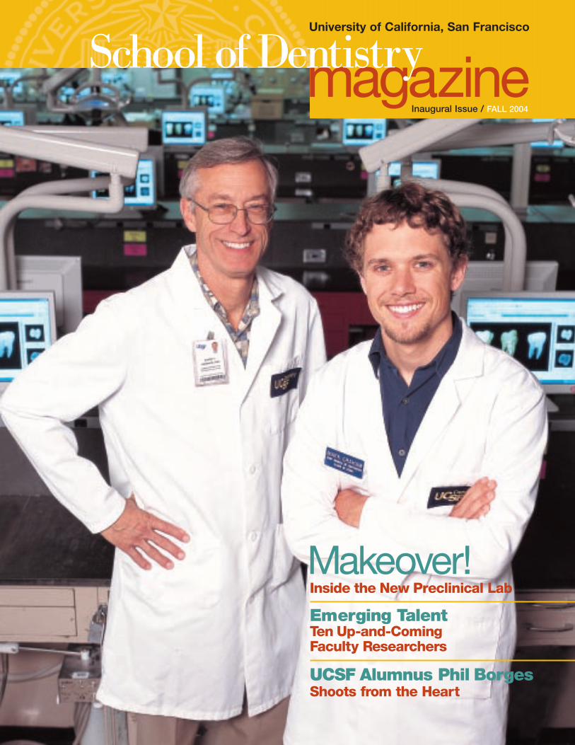
Makeover!Inside the New Preclinical Lab
Emerging TalentTen Up-and-Coming Faculty Researchers
UCSF Alumnus Phil Borges Shoots from the Heart
Inaugural Issue / FALL 2004
University of California, San Francisco
magazineSchool of Dentistry

http://dentistry.ucsf.edu
profile BACK COVER
Oral and Maxillofacial Surgery Clinical Professor Newton Gordongives to the School to widen access to oral health care.
ON THE COVER: Clinical Professor DaveGraham (D’71) and son Mark Graham (D3).
magazineSchool of DentistryUniversity of California, San Francisco
University of CaliforniaSan Francisco
School of Dentistry
FALL 2004 / VOLUME 1
Editorial Advisory Board:
Charles J. Alexander, Associate Dean for Student Affairs
James A. Anderson, Assistant Dean for Community Relations and Continuing Education
Charles N. Bertolami, Professor and Dean
Barbara Gerbert, Professor and Chair, Division of Behavioral Sciences
David Graham, Clinical Professor
John S. Greenspan, Professor, Dean for Research
Mark Kirkland, Assistant Dean, Clinic Administration
Richard McKenzie, Director of Development & Alumni Relations
Dorothy A. Perry, Assistant Dean for Curricular Affairs
Jane Weintraub, Lee Hysan Professor and Chair, Division of Oral Epidemiology and Dental Public Health
Mission Statement
The UCSF School of Dentistry seeks to improvepublic health through excellence in teaching,research, patient care, and public service in the dental and craniofacial sciences. We foster an inspired environment where individuals identify themselves as scholars and realize their scholarship through service as clinicians, educators, and scientists.
The goal of the School of Dentistry Magazine is toadvance the mission of the School of Dentistry. The magazine is published annually for the alumni,students, faculty, staff and friends of the UCSFSchool of Dentistry.
The School of Dentistry Magazine is produced by:
School of Dentistry CommunicationsUniversity of California, San Francisco513 Parnassus Avenue San Francisco, CA 94143-0430
Editor/Writer: Cameron Heffernan
Contributing Writers: Charles Bertolami, Phil Borges, Richard McKenzie
Design: Laura Myers Design
Photography: David M. Allen, Chris T. Anderson,Phil Borges, Margot Hartford, Cameron Heffernan,Getty Images, Richard McKenzie
© 2004 UC Regents
news
> RWJ FOUNDATION GRANT AFFECTS
SCHOOL OF DENTISTRY — A $1.4million grant from the Robert WoodJohnson Foundation improves theway the school recruits, teachesand cares for patients.
PAGE 2
> ACCREDITATION: WHY WE
TAKE IT SO SERIOUSLY — Everyseven years, the School must beaccredited by the Commission onDental Accreditation. This process of self-examination has a positiveeffect on everything we do.
PAGE 4
> SPECIALTY SPOTLIGHT: ORAL
AND MAXILLOFACIAL SURGERY —
Dedication of the Khosla SurgicalSuite; naming of the ScottLambert Memorial Oral andMaxillofacial Surgery Library.
PAGE 26
> CLASS OF 2004 STATISTICS —
Postgraduate educational and practice plans; educationaldebt rankings.
PAGE 27
updates

Greetings,
It is my pleasure to welcome you
to this inaugural issue of the UCSF
School of Dentistry Magazine.
This annual publication will update
and inform the friends of the
School — faculty, staff, students, alumni, parents —
about the many outstanding programs taking place at
our institution.
We hope that by sharing news and information
about the components of our mission — education,
scientific exploration and service to the community —
we can further enhance our relationship with our
internal and external constituents, and continue to
make a difference in the lives of the patients and
students we serve, while promoting the advancement
of the dental and craniofacial arts and sciences.
Aspiring to excellence in every aspect of our mission
is a continual challenge. And yet, excellence is
precisely what has been achieved — in part because
of the judicious allocation of the funds we have. We
have leveraged resources by collaborating with the
other schools on campus, other dental schools, the
UCSF Medical Center, the National Institutes of Health
(NIH), and other friends and affiliated organizations.
We have tried to ferret out every entrepreneurial
opportunity open to us. Most importantly, we have
asked for, and consistently received, the financial help
of constituencies that care about what happens in
the School — especially our alumni, faculty and
grateful patients. We have relied heavily on the
extraordinary generosity of those who have entered
the dental and oral health care professions.
This publication is one way we can thank you, our
generous benefactors, for all that you have given and
are continuing to give.
Best regards,
Charles N. Bertolami, DDS, DMedSc
Professor, Dean
1
TEACHING
Renovated Preclinical Lab Changes DDS ExperienceThe School of Dentistry is in the midst of a modern-ization of its preclinical lab that, when completed, willtake more than three years and cost nearly $3 million.Generous alumni have made the initial equipmentenhancements possible, but more funds are requiredto complete the job.
PAGE 5
features
COMMUNITY
In Focus: Phil Borges, UCSF Medal WinnerOrthodontist Phil Borges (D’69) left a lucrative practice in Northern California to pursue his dream of becoming a professional photographer. We talked with him to find out why, and to see what he’s up to next.
PAGE 12
RESEARCH
Emerging TalentTen up-and-coming, young faculty members discuss their research, motivations and background.They’re just part of the reason that the School ofDentistry has earned the most NIH awards of any dental school annually for the past 13 years.
PAGE 18
Dean Charles Bertolami
Welcome

The grant allows the School to increase the number of students (from 10 to 15)accepted into the one-yearPost Bac Program, the first ofits kind nationwide in dentistry.
The goal of the Post BacProgram is to better prepareeconomically and educationallydisadvantaged students, who had initially failed to gainadmission, for re-application to a U.S. dental school.Participants receive mentor-ship, preparation for the Dental Admissions test, housing, and a stipend forstudy, allowing them toenhance their academic competitiveness for admissionto dental school.
Strong Track Record
Over the six years of the program’s existence, 99% ofparticipants (69 of 70) in theprogram have successfullygained admission to at leastone U.S. dental school. By the end of the academicyear in 2004, 22 students
newsThe School of Dentistry is currently in the second year ofa $1.4 million, five-year grantfrom the Robert Wood Johnson(RWJ) Foundation to recruit disadvantaged students to oralhealth professions, enhance theamount of cultural competency
training in the curriculum, andincrease access to care formembers of underserved populations.
With respect to recruitment,funding from the RWJ grantallows the School greater contact with prospective dentalstudents, according to CharlesAlexander, Associate Dean for Student Affairs. Visits byprospective students to theUCSF campus and recruitmenttrips by School of Dentistrystaff to undergraduate campuses have both beenincreased due to the funding.
Unique RecruitmentProgram
Money from the RWJ grant isalso being used to expand asupplementary recruitmentendeavor — UCSF’s successfulPost Baccalaureate (Post Bac)Program, which assists collegegraduates in gaining accept-ance into a U.S. dental school.
who had completed the Post Bac program went on to graduate from a U.S. dentalschool. According to Alexander, the majority of those graduates are practicingin dentally underserved areasof California.
“The Post Bac programgives students from disadvan-taged backgrounds the sameopportunity that students frommore privileged backgroundshave,” Alexander said. “It hopefully opens doors tocareers for students who previously would not have beenextended this opportunity.”
Multi-Cultural Curriculum
Another component of the RWJ grant involves preparingdental students and faculty toprovide culturally sensitivehealth care for the patients they will be treating, both at theParnassus Student DentalClinics (at 707 Parnassus) andon externships in communityclinics that treat underservedcommunities.
Grant Improves Student Recruitment, Cultural Awareness and Patient Treatment
2

3
Students will learn about many differentdimensions of culture, such as verbal and non-verbal greetings, health and illness beliefs,traditional healing, attitudes toward prevention,barriers to care, and guidelines for working withinterpreters. “We assist students in developinga culturally informed approach to patient care,” said Linda Centore, Associate ClinicalProfessor, and curriculum advisor for the grant.
Putting it into Practice
The final component of the RWJ grant involvesexpanding the number of community clinicswith which the school has affiliations from fiveto 19 (see “Where in California is the UCSFSchool of Dentistry?” at right). The first year of the expanded curriculum commenced withthe 2003-2004 academic year. All fourth-year DDS students contributed 15 days of dentalcare at one of the 19 externship clinics. Nextyear, the number of days of community-basedcare will increase to 25. By the end of the 2004 academic year in June, UCSF studentshad completed 9,113 patient visits at theexternship sites, and provided $748,070 worth of dental care, based on the UCSF pre-doctoral fee schedule.
“The partnering community clinics havebeen very pleased with the UCSF relationshipin its first year,” said Bill Bird, Clinical Professor,Associate Dean for Clinic Administration, and Principal Investigator on the RWJ grant.“The externships prepare students to improve treatment for patients in underservedareas, including those on public assistance and fixed incomes and the working poor. A long-term goal is for some students to locate their practices in these underservedareas after graduation,” Bird concluded. ■
Where in California is the UCSF School of Dentistry?Last year, all fourth-year dental students contributed 15 days of dental care at community externship clinics, completing 9,113patient visits and providing $748,070 worth of dental care — primar-ily for people on limited incomes. In 2003, 47 percent of Denti-Cal procedures done at all California dental schools were done at UCSF.
Starting with the 2004-2005 academic year, D4 students will work 25 days at 19 clinics scattered around California. From theMountain Valley Health Center in Alpine County to the San YsidroHealth Center in San Diego County, School of Dentistry studentscover much of the state.
1. Shasta Community Health Center / Redding2. Anderson Dental Clinic / Anderson3. San Ysidro Health Center / San Ysidro 4. Mountain Valley Health Center / Bieber5. St. Joseph Foundation Dental Clinic / Santa Rosa6. Chico Family Dentistry / Chico7. Asian Health Services, Inc. / Oakland8. Orland Family Dentistry / Orland9. Native American Health Center / Oakland
10. Oroville Family Dentistry / Oroville11. Fruitvale Dental / Oakland12. La Clinica De La Raza / Oakland13. Dientes! Community Dental Clinic / Santa Cruz14. Transit Village / Oakland15. Native American Health Center / San Francisco16. Marin County Dental Clinic / San Rafael17. Sonrisas Community Dental Clinic / Half Moon Bay18. Dr. Yolanda Mangrum’s office / Sonoma19. Red Bluff Dental / Red Bluff
15
1
16
5
18
13
17
4
6
2
7,9,1112,14
10
198
3

news Dental Accreditation:
Why We Take It So SeriouslyAt this writing, the School of Dentistry’s accreditation site visit — scheduled for April12-14, 2005 — is fast approaching. DorothyPerry, Assistant Dean for Curricular Affairs, isleading the school’s accreditation preparation.She heads the Accreditation SteeringCommittee, which is preparing the self-studydocuments. Members of the committee willmeet with a pair of consultants this coming fallto finalize those documents. The consultantswill also tour school facilities, review patientcare services, analyze goals and outcomemeasures, and help the committee strategizeon the best approach for the final few monthsleading up to the site visit.
CODA’s Charge
Every seven years, the Commission on DentalAccreditation (CODA) visits each U.S. dentalschool to ensure that it is meeting establishedstandards for dental education institutions.According to the website of the AmericanDental Association (ADA), CODA’s mission “...is to serve the public by establishing, maintaining and applying standards that ensurethe quality and continuous improvement of
dental and dental-related education and reflectthe evolving practice of dentistry.”
The site visit allows the CODA-appointedteam to assess the school’s compliance withboth the accreditation standards as well aswith our own stated goals and objectives. Thesite visit complements the information that wewill present to CODA in the comprehensiveself-study document.
Positive Outcomes of the Accreditation Process
The entire process of preparing for accredita-tion has a positive impact on the school. This is a time for self-examination, duringwhich we review our approaches to teaching,patient care, outcomes and facilities, andensure that we are maintaining fidelity to ourmission. For this reason, we take the accreditation process very seriously, and view the current phase of self-study as one inwhich we can honestly review our performanceand strategize for the future.
Benefits of a Positive Site Visit
Areas resulting in a commendation from CODAhave a positive impact on the school for years.There are several fortuitous results that canarise from a favorable site visit:
© Positive public perception. The AmericanDental Association website lists the currentaccreditation status of all dental schools. Being listed as an accredited institution engenders confidence among not only currentfaculty, staff and students, but also prospectivefaculty, students, patients and alumni.
© Morale boost. Commendations are a positive reflection on the school, and should be celebrated by all faculty, staff, students andalumni.
© Positive effects on alumni/donors. A successfully accredited institution sends asignal to alumni and other donors that they arein full compliance with nationwide standards,and that they are doing a wonderful job of educating the next generation of oral healthcare practitioners.
I truly feel that the accreditation processshould be viewed as a positive exercise. It gives us the opportunity to reflect on ouraccomplishments and promote change and growth. If you have questions aboutaccreditation, please contact Dr. Perry or me.
— Dean Charles Bertolami
4

Second-year dental student* Mark Graham sits among more than 75 of his Class of 2006 classmates in the Preclinical Lab in the Dental Clinics Building (at 707 ParnassusAvenue) for their Removable Partial DentureDesign class, RD 125.10c. The course is
led by Associate Clinical Professor Mark Dellinges (D’81) with help from six other professors. Over the next four hours,Mark Graham and the other D2s will survey and design twocasts of partially edentulous arches requiring removable partial dentures. > > >
MakeoverExtensive equipment upgrades comprising the first two phases of the Preclinical LabModernization have changed the DDS learning experience. But the modifications are
not complete, and the School must raise an additional $2 million to finish the job.
*Mark Graham (inset above) was still a D2 student when this article was researched. He, along with the rest of the Class of 2006, has since moved on to his third year as a DDS student.
5
feature > > > TEACHING

6
The goal of the course is to pre-pare each student to diagnosepatients, develop a treatmentplan and prescribe removable
partial dentures that will be fabricatedby the lab. Before they start theirdesign work today, Dellinges will present an introductory lecture andinstructions on the procedure.
Up until 2003 in the preclinical lab,students had to jockey for positionnear one of several monitors displayingDellinges’ presentation, and strain tohear the lecture through an old, poor-quality audio system. But thanksto equipment upgrades that are a partof the ongoing modernization of thepreclinical facilities, the entire learningand teaching experience in the lab has changed. The overall renovation(see pages 10-11) is estimated to takemore than three years altogether, cost-ing between $2.77 million and $2.98million. The work will be completed in four phases, with funding and construction of the first two phasesalready finished, at a cost of $869,200.
Phases 1 and 2: Equipment Modernization
The first two phases of the moderniza-tion entailed completely overhaulingand upgrading the lab’s audio andvideo equipment and installing newdental lights for each student (Phases3 and 4 entail asbestos removal andthe installation of new workstations, as detailed on page 9). Each studentstation was equipped with full-color,15-inch, flat-panel Sharp monitors.The monitors allow each student tofollow the professor’s instructions far better than they were able to previously. The other significant difference is that the instructors nowhave an elevated presenter station atone side of the room from which they deliver lectures to students. Thisstage-like station (pictured at right) isequipped with a presentation camera
— similar to an overhead projector, but with the images being transmitted to the student monitors — and a touchscreen annotationmonitor that allows the professor to highlight components s/hewants to emphasize. The professor uses the presentation camera toillustrate specific techniques to students. “If a student asks me aquestion about a procedure, I can hold the typodont up to the presenter camera, and walk them through an explanation of whatthey should be doing,” Dellinges said. “That way, I can make thesame point to all 80 students, instead of just to one at a time.”
High-quality audio and video are also integrated into the centralteaching station. According to Dellinges, the presenter station greatly enhances the quality of his lectures. “I’ve been in situations[prior to the renovation] in which an instructor doesn’t hear something and circulates contradictory information amongst thestudents,” Dellinges said. “Now, with the central stage, professors all speak from the same podium, and this enhances our ability togive the same information to students. It makes for more consistentteaching with less confusion,” Dellinges concluded.
State-of-the-art A-dec lights at each student’s workstation and adedicated lecture computer, built into the presenter station, werealso installed in the first two phases of the renovation. “The use of apersonal computer during lectures brings a great deal of consistency
The monitors allow each

7
to teaching,” Dellinges said. “All of my lectures are readily accessi-ble because they are right on the hard drive. I think it illustrateshow the School is making better use of resources available on campus. I can switch from the PC monitor to the presenter cameraor annotation screen during a lecture. I can’t do that in a a lecturehall. I can display websites that students can see on their screens,and access old lectures for their reference. It’s invaluable,”Dellinges said.
Before or after class, students can access previously deliveredlectures on their own home PC thanks to WebCT, UCSF’s virtualonline learning environment.
Leading by Example
After Dellinges completes his lecture in today’s Removable PartialDenture Design class, the students are ready to start their designwork. They refer to their printed syllabi, which include pictures ofthe design work in various stages, or refer to the completed den-ture model that Dellinges leaves displayed on the student monitors.
Dellinges and the other six facultymembers teaching the course circulateamong the students, answering questions and assisting students. “Don’tforget to cut rest seat preparations into the designed casts and fill out the treatment planning forms,” Dellingesreminds the students from the professor’s station. The students haveapproximately two hours to prepare the designs. Many ask questions of theinstructors during their design work.“How wide should we make the mid-palatal strap for this RPD?” one studentasks from the middle of the room.“Eight to twelve millimeters,” Dellingesresponds over the microphone, afterrepeating the question.
student to follow the professor’s instructions far better than they were able to previously.
Clinical Professor Dave Graham (at left) with new presentation camera in the preclinical lab.Up to seven classes per week use the lab and its facilities (above).

8
Both first- and second-year DDS students take classes in the preclinical lab before moving on to the clinical phase of their
education. International students alsostudy in the lab to refresh and improvetheir technical skills prior to seeingpatients in the clinic. With the first two phases of the preclinical lab modernization completed between the first and second years of their education, the Class of 2006 wasuniquely positioned to witness the dramatic difference that the new
“We have to givestudents experiencewith tools and
they will see in the real world technology brought to the classroom.
“With the new equipment in thelab, things are much clearer, both inprocedural techniques and the visualaspect of learning dentistry,” MarkGraham said. “Last year, as D1 stu-dents, we sat at our chairs and lookedat a few monitors for the whole class.We had to move around to get goodviews of the old, run-down screensmounted from the ceiling. Now we sitat our individual workstation and each of us has a very clear shot of theimage. The clarity and ease of viewing professors’ presentations was greatlyenhanced,” he added.
Jin Kim, a fellow member of theclass of 2006, appreciates the range of options that the new technologyprovides. “This year, preclinical labprofessors were able to use differentmethods of presenting classroom materials using the new equipment,”Kim said. “Different professors madeuse of the multimedia technology indifferent ways. That made class thatmuch more engaging. For example, Dr. Dellinges used his DVD to show

9
us his demonstration. Dr. Lloyd usedPowerPoint and a morphology CD very efficiently in endodontics lectures.And Dr. Mendoza occasionally used avideo camera to demonstrate how todo intra- and extraoral exams. I saw adramatic improvement between myfirst and second year because of thistechnology,” Kim concluded.
Effect on Teaching
Mark Dellinges graduated from UCSF in 1981 and practiced generaldentistry for four years before returning to the School of Dentistryand participating in the prosthodontics residency certificate program. Like the students, he finds the impact of the new equipment remarkable.
information to the students in so many different ways, whichincreases our students’ ability to retain it.”
Renovation Not Yet Complete
Yet for all of the amazing differences the new technology hasbrought to the preclinical lab, professors, students and visitors can’thelp but notice how outdated the rest of the room and the studentworkstations appear. The floors and workstations contain asbestosand must be replaced. Neither have been upgraded since the room was originally built in 1979.
The third and fourth phases of the modernization, estimated to cost between $1.9 and $2.11 million, will remove all asbestosmaterial in the lab, and will also include the installation of modern workstations for each student. The new workstations will be designed to better simulate work done at a dental chair, in comparison to the current workstations, which are comprised of a mannequin attached to a lab bench (see “PreclinicalModernization: Phases 3 and 4,” page 11). The campaign for these remaining two phases is currently underway, according toSchool of Dentistry Director of Development & Alumni RelationsRichard McKenzie. “Fundraising for the preclinical lab has gonefaster than I expected,” McKenzie commented. “In fact, donationsfor the first two phases exceeded the goal, putting us ten percent toward our goal of $2 million for the third and fourth phases. Alumni really care about this project because they know how critical it is to the success of dental students, and what a lasting impact it can have on their entire dental education.”
According to Bill Bird, Clinical Professor and Associate Dean for Clinic Administration, the remaining construction work is justas important as the installation of the equipment from the first two phases. “It’s essential that we complete the final two phases of the renovation in order to give our DDS students a truly moderndental education experience,” Bird said. “Furthermore, much ofthe equipment has rusted out or is so old that parts are no longeravailable; casting machines, for example, are not functioning properly. We have to give students experience with tools andequipment that they will see in the real world when they graduate.Finally, some of the facilities in the lab are obsolete, and repairsrequire expensive custom-made parts. By upgrading the entirefacility, we save money in the long run,” Bird continued.
Grateful Students
Although fundraising for the third and fourth phases of the renovation is still ongoing, students we spoke with for this articleexpressed gratitude for donations already made by generous alumni.
“I’d like to let all of those who gave toward the restoration ofour lab know how much we appreciate their support,” said Helen Kim, also a member of the Class of 2006. “The new labgreatly enhances the level of education that we receive, and betterprepares us for the clinical work still to come. Thank you.” ■
equipment that when they graduate.”
“I feel lucky to be able to teach this course with all of the wonderfultechnology now at our disposal, andthe consistency within it,” Dellingessaid. “Compared to when I took labback in 1979, the difference is nightand day. It is a pleasure to work in thenew environment, and see studentslearning these procedures that muchmore quickly,” he said.
Dave Graham (D’71), has been aClinical Professor at the School for 31 years. He teaches restorative, andcrown and bridge components in fiveD1 and D2 courses in the preclinicallab, as well as clinical general dentistryto the third- and fourth-year students.Dave Graham agrees that the equip-ment upgrades have revolutionized theteaching experience. “There is somuch information and terminology toabsorb, especially in the first four tofive weeks of the year,” Dave Grahamsaid. “The range of multimedia equipment that we now have access togives us the ability to present that

10
UCSF School of Dentistry Completed Construction Projects and Renovations,1995-2004
P R O J E C T N A M E C O N S T R U C T E D C O S T I M P R O V E M E N T S
Preclinical Lab Modernization 2003 $869,200 Complete overhaul planned in three phases. (Phases 1 and 2) Phases 1 and 2: installation of individual student monitors; touch-screens for
faculty; fully integrated audio-video system; teaching presentation cameras and dedicated lecture PC; modern dental unit lights, dentist operating stools and handpiece controls.
Khosla Surgical Suite (Phase I) 2004 $50,000 Installed new sink, surgical lights, fiber optic headlight — Oral and Maxillofacial Surgery with built-in video camera, surgical drills, cabinetry.
Axium Information System Installation 2000-04 $920,000 Integrated patient management system. Goal is for paperless/chartless patient records by 2006; clinics being phased in over six-year period.
Stomatology Lab Renovation (HSW-6) 2002-04 $6,183,111 Upgrading of faculty research labs in craniofacial, cell and molecular biology; (pictured below) addition of multidisciplinary conference room; 20% increase in space.
Faculty Group Practice Renovation1 2003 $413,481 Installed 31 patient chairs (including five multimedia special operatory units); replaced all 62 doctor/assistant stools (including high and low-speed handpieces).
Scott Lambert Memorial Oral 2002-03 $80,000 Installed new carpeting, A/V system with plasma TV, Internet/Intranet access; and Maxillofacial Surgery Library integrated with UCSF Medical Center network.
Buchanan Dental Center Upgrade 2003 $237,000 Dental equipment upgrades and clinical facility modernization.
Periodontic Clinic Renovation 2003 $239,073 Replaced dental units and chairs, including privacy screen risers.
Orthodontics/Periodontics Research 1998-2001 $983,022 Installed special tissue culture facilities, new lab benches; Labs and Clinics Renovation renovation of imaging equipment and imaging rooms.
Faculty Incubation Lab Modernization2 1999-2000 $873,642 Complete renovation of the suite. Old lab was razed, and new lab was designed and built. New service rooms, offices and lab facilities installed.
Predoctoral Clinics Modernization 1997-99 $2,867,819 Complete clinic modernization: new roof for Clinics Building; extensive remodelling; (Clinics A&B, 707 Parnassus) lighting retrofit; upgrading of facilities, chairs, equipment and waiting rooms.
Oral and Maxillofacial Surgery Clinic 1997-99 $180,372 Brought existing clinic up to standard for Accreditation Association for (D1201) Ambulatory Health Care; installed changing room for patients, restroom,
sinks, surgical sinks, upgraded facilities.
PRDS Research Laboratory Upgrade 1995-96 $123,338 Replaced and upgraded lab benches and equipment.
Growth & Development Research 1995-96 $549,550 Complete overhaul of former pediatric dental clinic into lab facility for Lab Renovation (MSB 704) pediatric dentistry.
Craniofacial Anomalies Clinic 1996 $42,663 Expanded waiting room, expanded/enhanced consultation rooms; Upgrades (S747) created more efficient appointment desk configuration.
Prosthodontic Resident Room 1995 $129,453 Renovated study carrels, including A/V presentation equipment and computer Renovation (D4307) terminals for residents.
Grand Total $14,741,724
1 includes Graduate Prosthodontics and University of California Prosthodontics Faculty Practice2 C-415 (aka Bertolami Lab)
Source of Funds For completed construction projects and renovations, 1995-2004.
School of Dentistry 6%
Federal Government 14%
Campus 29%
Alumni / Friends 51%

Future Projects FUNDRAISING REQUIRED
Preclinical Lab Modernization (Phase 3 and 4)— details at right
Pediatric Dental Clinic Renovation$225,000 — $1 million$225,000 for equipment replacement; $1 million forinfrastructure remodeling, including reception area.
Implant Center$1,000,000Creation of Implant Center in Dental Clinics Building.
Oral and Maxillofacial Surgery Suite (Phase 2)$23,000For renovation of second surgical suite.
Endodontic Graduate Clinic $400,000 (est.)Clinic reconfiguration, equipment upgrade, including addition of endodontic microscopes.
Health Sciences East, 15th Floor$6.8 million — $7.1 millionConstruction of labs, offices, research area for Craniofacial and Mesenchymal Biology Program.
To make a donation to the
preclinical lab, phone the School of Dentistry
Director of Development & Alumni Relations at
(415) 476-3645, or use the donor envelope
inserted in this magazine.
Preclinical Modernization:Phases 3 and 4
The preclinical lab renovation is taking placein phases. Construction and fundraising for the first two phases are complete. The School is currently raising funds for the construction of phases three and four.
PHASE 3Estimated Cost: $1.4 million
ENHANCEMENTS / REPLACEMENTS:© Replacement of the current lab benches with modern
laboratory workstations (benches). Workstations will beequipped with internal vacuum suction.
© Removal of current asbestos flooring and replacementwith new floor.
© Updating of simulation lab handpiece controls (both air-driven and electric) to modern equipment.
© Replacement of current dust collectors and vacuum systems, which are not functioning optimally.
BENEFITS:© Currently any repairs to drawer modules have to be
custom made, because the parts are obsolete. Thus, the renovated workstations will save the school in thelong run.
© Modern equipment for students.© Safe, asbestos-free work area.
PHASE 4Estimated Cost: $500,000-600,000
ENHANCEMENTS / REPLACEMENTS:© Complete renovation of the wet lab/plaster lab.
Replacement of burnout ovens, casting lab equipment,ceramic ovens and workstations.
BENEFITS:© Current equipment rusting out and parts are unavailable.
Casting machines not functioning.© Gives students up-to-date equipment.
11
In the preclinical lab, existing student workstationsand the floor contain asbestos and must be replaced.
> > >

In Focus
feature > > > COMMUNITY
12
Phil Borges graduated from the UCSF School
of Dentistry as a Regents Scholar in 1969 with a
specialization in Orthodontics. He practiced
dentistry in Northern California for 18 years before
deciding, in 1989, to leave his lucrative practice
and become a professional photographer.
He and his family moved to Seattle, where he
still lives with his wife and 18-year-old son, who
will attend college this fall and aspires to a career
in digital media studies. In a 15-year career as a
photographer, Borges has published five books
documenting indigenous and tribal people
worldwide, contributed to television documentaries
for the Discovery Channel and National Geographic,
and strives to promote and preserve the cultural
diversity of the many different peoples and tribal
groups he portrays. > > >
School of Dentistry Graduate Phil Borges Wins UCSF Medal
UCSF Chancellor J. MichaelBishop (left) presents the UCSFMedal to Phil Borges (D’69).

13
In 1996, he published the award-winning book Tibetan Portrait: the Power of Compassion, that included a photograph of, and text by, the Dalai Lama (pictured below). In 1998 he published Enduring Spirit for Amnesty International in
observance of the 50th anniversary of the UniversalDeclaration of Human Rights. In 2000, he publishedThe Gift, a book that documents the work ofInterplast, a non-profit organization that partnerswith physiciansto provide freereconstructivesurgery to needy childrenand adultsthroughout thethird world.
By his ownestimation,Borges has traveled to more than 35countries. He is currentlyworking on abook on alterna-tive medicinesand shamanismphotographedduring visits toMongolia, thePhilippines andthe Amazonbasin. In 2000,he founded anon-profit organization,BRIDGES to Understanding (see “Building Bridges,” page 17),to which he currently devotes a great deal of time,energy and personal funds.
In April 2004, Borges was presented with theUCSF Medal, the University’s highest honor, byChancellor J. Michael Bishop at the annual FoundersDay banquet. This honor is bestowed on individualswho have made outstanding personal contributions to the health sciences and whose efforts mirror thegoals and values of the University.
We caught up with Borges recently by phone from Seattle, on a rare respite between trips.
SCHOOL OF DENTISTRY: How long have you hadan interest in photography?
PHIL BORGES: I fell in love with photography when I was in dental school at UCSF from 1965-69.I lived in the Haight-Ashbury district while going to school. I had a work-study job for a UCSF sociologist, and they asked me to go down to Haight Street and survey people as to why they were sharing needles. Eventually, I started taking
pictures of my interviewsubjects becausethey were sowild looking. It was a veryunusual andinteresting timeto be there, so there wereplenty of thingsto photographaround the city. I would do the inter-views and photography,then take the N-Judah to a Parks andRecreationdepartmentdarkroom todevelop the pictures. What was yourfirst camera?A Minolta SRT-101.
Were there components to your training as anorthodontist that prepared you for a successfulcareer in photography?The type of photography I do is very people oriented; I don’t just do landscapes. What you do as a dentist is get people to feel comfortable in an awkward situation. As an orthodontist, you have to get kids to do terrible things like wear headgear or put rubber bands in their mouths to get the job done. Winning people over and establishing rapport quickly is very useful whengoing into a tribe.

14
How did your orthodontist career lead to your current career as a photographer and philanthropist?It enabled me to make a change. It took me fouryears to get started in photography. I had built up a savings account that allowed me to get into photography. I found when I would take a breakfrom my practice I would travel to remote areas. I did some volunteer work in Mexico with some traveling doctors, and I would also do some photography while I was there.
individual. I would advise that it’s important to havesomething [that you’re passionate about]. In my case,I didn’t do photography while I was practicing den-tistry. My renewed interest in photography cameabout due to the birth of our son. I took a bunch ofblack and white pictures after he was born — this was before the advent of those one-hour photodevelopers — and got access to a community collegedarkroom to develop them. I took a photographyclass and reestablished my love for photography. I wanted to do it more, and realized that I couldn’t
World-renowned photographer Phil Borges’ work has taken him to remote destinations all over the world, including Irian Jaya (above, at left) and Thailand.
A lot of the alumni of our school struggle withwanting to change careers in mid-life. Whatprompted your decision to leave orthodontics, and what advice would you give to someone indentistry who is contemplating a career change?In my case, I had this whole side of myself that wasn’t being fulfilled. I wanted to travel and to create something that communicated. It’s somethingthat went a long way to making me whole, the communication arts. The desire to change varies by
do justice to the dental practice because I was in love with something else. If something like that grabs me, I have to make the change. I’m a one-trackperson. If I love something, everything else suffers. I had a practice with a partner in Benicia and Vallejothat would have suffered due to my newfound love of photography.Can you touch briefly on what changes you had to make in your lifestyle to pursue your dreamsoutside of dentistry?

15
I needed to move to a city with others alreadyinvolved in the business of photography to learn it. I was living in Sonoma at the time, and SanFrancisco was too expensive, so we moved to Seattle,bought an inexpensive house in a lower middle-classneighborhood. I cut down on my expenses as muchas possible and started a new career.How did you first get interested in photographingindigenous peoples? My interest comes from when I was a kid. I grew upin San Lorenzo, and in the summers, my mother
How did you get into that?Well, I would get on my bike and ride to Chinatownin Oakland and buy fireworks and then bring them back to the neighborhood and sell them at aconsiderable markup.That sounds very enterprising.But when the kids got caught with them, it obviouslycame back to me. So it wasn’t long before I had a record.What have you learned from the indigenous peoples you photographed?How strong and tight their community is. They have
to share when they farm fields, build homes andraise kids. It’s all done communally. Mothers taketurns nursing each other’s children. Men put up newhouses with each other. There is less emphasis on theindividual and more emphasis on the community.They have time to sit and be with each other, share a joke. I remember when I was visiting IrianJaya, there was a great-grandmother of a family who was dying. She died during the three days I wasthere. The extended family took turns staying in her hut, holding her, rubbing her legs, visiting her.
would send me to a ranch that some relatives ownedin Utah in the middle of nowhere. People living on the ranch were highly subsistent; they grew their own food and lived a life close to the land. I worked the ranch and fell in love with the lifestyle.Living close to the land was always attractive to me.It was very different from my life in California oftract developments. [In Utah] I put my energy intoworking on the ranch. That was certainly better than in California where I put my energy into selling firecrackers.

16
they had done in Vietnam. I opened the packet and that film was in there and it was very moving.Also, I had had a lot of cleft palate patients when I practiced and had seen a lot of untreated cleft lipsin the developing world. In the third world this kindof affliction goes untreated, unless there is a teamlike Interplast that takes care of it. So I decided todonate my services to them.For your 1996 book Tibetan Portrait, you photographed the Dalai Lama. What was it like,meeting him?
Well on thatshoot, it was justlike any otherportrait because I had limited time and was losing my light;the sun was goingdown. I was ontask to get theimage. But before,I had spent threehours standingnext to him whenhe was meetingnew arrivals fleeing to Indiafrom Tibet. It waslike a weddingreceiving line. As people cameby he wouldshake their hands.Since he’s like a god to them,some fainted andfell to the ground.
He would kneel to the ground and speak to each ofthem individually to encourage them, even thoughhis handlers were nervously trying to have him keep the line moving. I noticed how he put his totalattention and care into each person he met. I had anadmiration seeing how he treated other people.How difficult was it to travel to remote places inIrian Jaya, Ethiopia, Kenya, Peru and Siberia?
I thought it would be a wonderful way to make thattransition, in contrast to how it happens here manytimes, in a nursing home. What sort of challenges do these tribes live withevery day? While the tribes that I visited are tight and connected, they are also very distrusting of neighbor-ing tribes. For example, there are 14 tribes in theLower Omo Valley in Ethiopia, and they’re all at war. You can’t raise your voice to anyone in thetribe, or you’ll be ostracized. But if you kill someonein a neighboringtribe, you’re a hero.They’re very loyal to their own group,but very distrustful of other groups. Plus everything thattechnology hasbrought us — medicines, labor-saving devices and niceties we have — they don’thave. They have to live with a cleft palate or goblind from easilycurable infections.They lack access that we take forgranted.Interplast has a lotof support in theBay Area. How didyou get involvedwith them?I had a photographyexhibit in San Francisco around 1998, and their PR Director came to the show and gave me a packetof information on them, and asked me to do a bookfor them. I was very busy at that time, and didn’thave time to review the packet. A couple weeks later, I was watching the Academy Awards and theyannounced that the winner of the short documentarycategory was a film about Interplast and the work
Lan (above, this page and facing page) is from Tinh Chau, a tiny village near My Lai, Vietnam, the site of one of the war’s most infamous massacres.Born with a cleft lip, Lan was ostracized from her community. Interplast visited Lan when she was 22, performing the surgery that is routine in the developed world, giving Lan a new face and outlook on life (facing page, bottom). Borges photographed Interplast’s work for his book The Gift.

Did you have people helping you,translators? It was easy to get to the little townsoutside of the areas where I wasshooting. There I would find theguides who would lead me into thejungles or more remote areas andinterpret for me. I generally wantto get [a guide] from the sametribe that I’ll be photographing.I’m more concerned with theirpeople skills than their commandof English. They have to knowsome English, but it can be broken. I go into the jungle for up to four weeks, and stay in townonly three or four days hiringguides and porters for my gear. On the shoot, I usually start withthe kids. I give them Polaroids andsoon they become a production
For more information on BRIDGES workshops, call 206-275-3247or visit the website at www.bridgesworkshops.org.
crew: holding the lights, openingand closing my pack as I movearound. They get involved and itintegrates me into the community.I saw in your book EnduringSpirit that you had eaten beetlesin Thailand. What are some of the other strange things you’ve eaten while traveling?How were the beetles?They were great — crunchy andsalty. I hosted a couple of showsfor the Discovery Channel, and in one there is a tribe in theEcuadorian Amazon where thewomen take a root vegetable, likea potato, and cook it then grind it and mix it around in a pot. They then take a handful [of thefood], put it in their mouth, chew it and spit it back into thepot and it ferments. It tasted prettygood. Generally, I’m not eating too exotically. In most places Ibring my own food because theyhave so little. Sometimes I bringfood for them as well. Have you ever encountered anydanger on a shoot?Once on a visit to Kenya, there was tribal violence between theTurkana and the Samburu tribes. This fighting is traditionally basedon cattle raiding, and in the pastthey used to do it with spears. But now, with so much warringgoing on in these regions, they’re equipped with RussianKalashnikov rifles and it can bevery dangerous. I was in a villagewhere a rumor was going aroundthat there was going to be a raidthe next morning. So I got out ofthere and spent the night in thehills. The raid never happened. ■
Editor’s Note: Phil Borges started the nonprofit BRIDGES to Understandingin 2000. While the organization has since expanded to 12 sitesworldwide, the goal remains thesame — facilitating interaction andthe sharing of knowledge betweenchildren of different cultures andnations. Borges, a humanitarian and world-renowned photographer,encourages others to becomeambassadors to indigenous culturesthrough travel and collaboration.
Photography is an extraordinary medium for communication and artistic expression; it is an equally powerful tool for
advocacy. I would like to invite my fellow photographers — amateur andprofessional alike — to join me oninternational digital storytelling workshops to sites in Kenya, India,Peru and Nepal. With instruction fromme and the BRIDGES team, partici-pants are able to connect one-on-onewith students, helping them createdocumentaries about their cultures,while developing meaningful relation-ships with the communities they visit.BRIDGES sites are located in some of the most interesting, scenic andout-of-the-way corners of the world.
There’s something about volunteer-ing, something about being able to say to yourself, ‘I’m here to give, notto get.’ This is what I want to give toBRIDGES workshop attendees — to allow them to have a vehicle to connect with people in the culture they visit, and to make a real impact,instead of just quickly passing through a place with their cameras,clicking away.
— Phil Borges, DDS
Building Bridges
17

1818
Emerging TalentThey come
from different
countries
with diverse
scientific
interests
and unique
backgrounds.
But each
of these ten
young faculty
researchers
contributes
to the UCSF
School of
Dentistry’s
reputation
as a premier
scientific
research
institution.
Recently the UCSF News Service disseminated
a press release* proclaiming that the UCSF
School of Dentistry had again distinguished
itself as the largest recipient of total National
Institutes of Health research funds among
U.S. dental schools, having received 51 awards
totaling $28 million for the 2003 fiscal year.
The School has maintained this top
distinction over the past 13 years, thanks to the
hard work of faculty investigators and their
staffs in diverse areas of oral health care and
craniofacial sciences. Some of the School’s
most recognized researchers have been here for
years, and their work is well known on the
UCSF campus and among dental educators
and researchers worldwide.
For this article, we chose instead to profile
ten individuals with divergent interests who are
at relatively early phases in their careers, and
who therefore don’t have the same level of
name recognition among the alumni, members
of the UCSF community, or other oral health
researchers. Here, then, are some of our new
up-and-coming faculty researchers. > > >*To see the press release, go to http://pub.ucsf.edu/newsservices
Researchers (from top left, top to bottom):Tamara Alliston,Stefan Habelitz, Brian Schmidt, Umo Isong, Richard Jordan,Janice Lee, Daniel Fried, John Huang, Caroline Shiboski,Dusko Ilic.
feature > > > RESEARCH

19
Daniel FriedBiomedical Optics, Imaging andLaser-tissue Interactions
Daniel Fried earned his PhD in physical chemistry with aminor in physics from WayneState University in Michigan. An associate professor in theDepartment of Preventive andRestorative Dental Sciences(PRDS), Fried did postdoctoralwork in biomedical optics andcariology in Rochester, N.Y.
What is your research specialty?It focuses on the diagnosis, detection and treatment of dentalcaries. A spectroscopist by training, I am interested in the development of spectroscopic methods and toolsfor use in medicine and dentistry.Spectroscopy is the physics thatdeals with the theory and interpreta-tion of interactions between matterand radiation.
What are some of your recentresearch accomplishments?
Recent findings with clinical implica-tions by my group in our OpticalDiagnostic and Therapeutic LaserLaboratories include: © the development of a low-cost multipurpose laser for dentistry thatcan be used for both hard- and soft-tissue applications; © the development of a selective,nondestructive laser-based methodfor removal of composite resinsealants, adhesives and restorationswithout peripheral damage to toothstructure;© and the development of near-infrared-based imaging systems forthe early detection and diagnosis ofdental caries. Such systems offerenhanced sensitivity for early cariesdetection, and do not require the useof ionizing radiation.
What are your future researchplans?I hope to eventually explore otherareas of oral medicine in which recenttechnological developments in opticaltechnology may provide significantadvantages, such as the optical biopsy of oral lesions and the laserprocessing of bone for dentalimplants. Fluorescence, with either
single or multiphoton excitation, can be used for optical biopsy, identification and detection ofmicroorganisms, as well as the detection of dental caries. It is likelythat these new spectroscopic methods can be coupled with novelmethods of optical imaging and laser surgery for the development ofminimally invasive diagnostic toolsthat will revolutionize the way dentistry is practiced in the future.
Tamara AllistonMesenchymal Stem Cells and Bone Biology
Tamara Alliston earned her PhD from Baylor College ofMedicine. She is an AssistantAdjunct Professor in the forthcoming department of Cell and Tissue Biology1.Alliston is also the mother oftwo sons, ages 3 and 1.
What is your research specialty?I study the control of osteoblast (bone cell) differentiation. I explore
1 The School of Dentistry is currently undergoing a departmental re-organization and consolidation that will result in abasic sciences department and a clinical/translational sciences department. The proposed name for the former department is Cell and Tissue Biology; Orofacial Sciences for the latter. Both proposed department names awaitapproval from the Chancellor’s Office. These newly named departments join the existing departments of Preventive and Restorative Dental Sciences, and Oral and Maxillofacial Surgery.
Daniel Fried with staff research associate Patara Ngaotheppitak.
Tamara Alliston and her three-year old son, Gabe, at the Lucia Child Care Center on Parnassus campus.

20
how extracellular signals turnosteoblast genes on and off, and theimpact of these cellular decisions onbone tissue formation and turnover.
What specific research questions are you attempting toanswer in your current work? I’m examining the ability of growthfactor signaling to improve bonestrength and ability to resist fracture. I am also studying the role of bonequality in hearing loss.
How did you become drawn toyour field of research? In addition to the scientific interest of studying the role of bone in eardevelopment and function, I amundoubtedly motivated by my brother-in-law and my graduate mentor, both of whom struggle withsevere hearing loss.
Who do you see benefiting fromyour research? I am hopeful that by better understanding bone formation andcomposition, therapies may bedesigned to prevent fractures causedby osteoporosis, or to better repairtissues damaged by osteoarthritis orbone fracture.
What is it like balancing ademanding scientific career with raising two young boys?Challenging but possible, thanks to excellent childcare, strong professional support at UCSF, andgreat personal support at home.
Caroline ShiboskiOral Medicine/Epidemiology
Caroline Shiboski, an associateprofessor in the forthcomingdepartment of OrofacialSciences2, earned her DDSdegree at Université RenéDescartes in Paris, France, followed by a Masters in PublicHealth from the University ofCalifornia, Berkeley, and
then a PhD in Epidemiology,also at Cal Berkeley. Shiboski completed residency programsat UCSF in AdvancedEducation in General Dentistry,Dental Public Health and OralMedicine. Currently Director ofthe Stomatology Clinical Center at UCSF, Shiboski bothpractices and teaches oralmedicine. Shiboski has beenmarried to her husband, Steve,an associate professor ofEpidemiology and Biostatisticsat the UCSF School ofMedicine, for 19 years. Theyhave two sons, ages 9 and 11.
Tell us about your research andscientific specialty.As an epidemiologist and oral medi-cine specialist I conduct clinicalresearch on oral diseases that affectpatients with immunosuppression dueto various causes (HIV disease, solidorgan transplantation, hematopoieticstem cell transplantation). Part of my current work focuses on oral cancer epidemiology, in particular onexploring trends in oral cancer inci-dence among younger populationsthrough national cancer registry data(the SEER Program).
What does your work with theSEER Program entail? Analyses of the SEER Program dataprovide the opportunity to identifysite-specific and age-specific trendsin incidence rate and relative survivalrate among people with cancer. Fromthese analyses, I identified a recentincrease in the incidence of tongueand tonsil squamous cell carcinomaamong young adults (20-44 yearsold). Ultimately, my goal is to design astudy to identify specific risk factorsthat may explain this recent increase.
Tell us about your work in Sub-Saharan Africa.As a co-investigator in the UCSF Oral AIDS Center, I designed a collab-orative project with Zimbabwean colleagues to explore the feasibility oftraining nurses in the diagnosis ofHIV-related oral lesions and of usingthese lesions as surrogate markers for HIV disease progression. As themonitoring of HIV disease progressionthrough CD4 count testing is prohibi-tively expensive in sub-SaharanAfrica, a visual inspection of themouth may be an affordable way ofdetermining when anti-retroviral therapy should begin — assumingthat such drugs will one day becomeboth available and affordable in thatpart of the world.
Caroline Shiboski directs the Stomatology Clinical Center.
2 See footnote 1, page 19.

21
Do you and your husband worktogether frequently? We had never really worked togetheruntil two years ago when we wereinvited to participate, as the statistician and epidemiologist team,in the writing of a proposal aimed atdeveloping a registry to studySjögren’s syndrome. The project wasfunded last year, and it has beenexciting to work on it together. It hasn’t caused any marital tension yet!We commute to campus together, but we try to restrict our “job-talk” toonly the first ten minutes of the drive.
Stefan HabelitzBiomaterials and Bioengineering
Stefan Habelitz, an AssistantProfessor in Residence in thePRDS department, earned hisPhD in Materials Chemistry at the University of Jena inGermany.
What does your research intoDental Materials entail?I am working to synthesize and
regenerate dental tissues. Like my other research projects, the common goal is to reconstitute damaged or lost dental tissue byregeneration using nature’s principlesof engineering.
So how does this help in therestoration of teeth?We would like to be able to remineralize enamel in carious lesions,or to provide a natural solution forbuilding a dental restoration by usingartificial human enamel. To do this, we are attempting to understand how organic molecules interact with inorganic minerals and how a biological system is able to designremarkably organized structures in tissues at the molecular level. Onecomponent of this involves examiningthe importance of recombinant proteins of the enamel matrix, andtheir potential to control the growth of calcium phosphate crystals, which make up about 98 percent ofdental enamel.
We hope to understand the mechanisms of protein-guided crystalgrowth, and apply that knowledgetoward efforts at growing an enamel-like material in vitro.
How is that work proceeding?So far we have succeeded in growingthin layers of about one micrometerthat resemble the composition, properties and structure of humanenamel. The growth rates in this artificial system, however, are so slowthat it would take up to 40 years torecreate the enamel crown from acaries lesion. Hence we are currentlyadjusting the physical, chemical, andbiological parameters to speed up theprocess significantly.
What impact do you anticipateyour research having?I am very excited about the possibilityof generating artificial dental tissuesor regenerating lost teeth. Not onlycould people with genetically impairedteeth recover their full dentition, but this research may lead to the discovery of important principles andmechanisms of biomineralization andtissue engineering that may beapplied to other fields of regenerativemedicine.
John HuangOrthodontics
John Huang earned his DMDdegree at Tufts University, andhis DMedSc research degreeand orthodontics specialty certificate from the HarvardSchool of Dental Medicine. He is currently an assistant professor in the Division ofOrthodontics, within the forth-coming Orofacial Sciencesdepartment3, at UCSF.
Please describe your researchand scientific specialty.At the clinical level, I have been integrally involved in setting up andimplementing the 3-D volumetricimaging system in the Division ofOrthodontics. Through several projects, we are examining and evaluating the reliability of 3-D meas-urements compared to conventional
Assistant Professor Stefan Habelitz with fiancé Nil Yucel, an assistant adjunct professor in PRDS.
3 See footnote 1, page 19.

22
2-D radiographic images. Currentstudies on volumetric quantification ofairway space in mouth-breathingpatients and analysis of airway spacein pre- versus post-surgical patientswill also answer many existing clinicalquestions.
How reliable is 3-D volumetricimaging? It is the technology of the future.Once a clinician has seen the capabilities of the 3-D scans and thetremendous amount of informationthat it provides, it is unlikely thathe/she will revert back to convention-al 2-D radiographs. The measure-ments obtained from the 3-D systemand its full potential in cephalometryare still being validated. Clinical stud-ies of airway space and growthparameters in, for example, hemifacialmicrosomia patients become muchmore realistic with this new system.The overall goal is to be able to provide a large database of patientinformation for researchers to be ableto use and analyze. We have alreadyscanned numerous patients with
impacted canines, cleft lip and palatecases, and other oral pathologies, and found that the technique greatlyfacilitates accurate diagnosis.
How did you become drawn toorthodontics? I have always been interested inbending wires, although there is a lotmore to orthodontics than just themechanical component. I enjoy thediagnosis and treatment planningaspect, of having the opportunity totry different techniques and bio-mechanics in each individual case. Itis always refreshing to interact with adolescents who seek treatment, andthe long-term social interaction withthe patient during the course of ortho-dontic treatment is a characteristicrelatively unique to orthodontics.
Dusko IlicSkin and Mesenchymal Biology
Dusko Ilic earned his MD fromthe University of Belgrade inYugoslavia, followed by aMasters of Science degree in Oncology from the sameinstitution and a PhD from theUniversity of Tokyo. He is an
Associate Adjunct Professor inthe forthcoming department of Cell and Tissue Biology4.
Were you interested in scientificresearch from a young age?I always enjoyed solving puzzles. As a medical student I earned pocketmoney by making crosswords formagazines. I see research as the very same game of solving puzzles.Of course, the puzzles that I play withnow are much more complex andchallenging — they are made bynature, not by people.
Tell us about your research inmesenchymal biology.Cells in virtually all tissues are in contact with organized complexes of structural molecules collectivelycalled the extracellular matrix (ECM).The ECM is an important source ofsignals that regulate the adhesive andmigratory behavior of cells related tofundamental physiological processessuch as morphogenesis, tissue-specific differentiation, wound healingand immunological defense. The long-term goal of my research is toincrease understanding of how signalsfrom the ECM are transduced intocells, and how cells interpret andrespond to signals from the ECM.
Orthodontist John Huang.
Dusko Ilic in the newly renovated Stomatology Lab (HSW-6).
4 See footnote 1, page 19.

23
What is the nature of yourresearch? My research focuses on oral healthdisparities. Disparities research tries to understand why certain subgroups of the population, such asthe uninsured, poor, and racial andethnic minorities, are at higher risk fordental disease than others. Diseaserisk is not uniform even within thesesubgroups. My research attempts tounderstand the reasons for theseinequities using risk assessmentmethods. Risk assessment is used toidentify factors that are associatedwith increased risk for disease, sothat targeted efforts may be directedtoward high-risk individuals. Such factors may include genetics, culture,health beliefs, health behaviors andothers. Dental diseases are largelypreventable. If researchers can identify children who are at high riskfor future dental disease, then targeted efforts could be directedtoward these children, thereby potentially preventing future pain anddiscomfort.
How did you become drawn tooral health disparities? As a dental public health resident, Ihad the opportunity to work on tworesearch projects as part of the resi-
dency requirements. These were thefirst research projects in which I hadever been involved. One project wasto determine the success of thesealant program of the New YorkState school system, while the otherwas to evaluate the state’s migrantdental program. From working onthese projects, I discovered that I had a natural analytic flair that led me to the field of epidemiology. Whiletraining to be an epidemiologist, mymentor was Jim Beck, a pioneer inthe field of dental risk assessment.These combined experiences drewme to my current field. I have enjoyedworking in public health so much thatI’ve recruited nearly every member ofmy family into the field!
Richard JordanOral Pathology
Richard Jordan received hisDDS degree from the Universityof Toronto, where he alsoearned a diploma in OralPathology and Oral Medicine,and a Master’s degree inPathology. Jordan later earneda PhD from the University ofLondon in ExperimentalPathology. Now an AssociateProfessor in the forthcomingdepartment of Cell and TissueBiology5, as well as chair of theDivision of Oral Medicine, OralPathology and Oral Radiology,Jordan joined the School ofDentistry in 2000 after six yearson the faculty at the Universityof Toronto.
What components of your professional background influenced the nature of your current research? In the course of a 15-year career as aspecialist who sees patients withmouth lesions, I have been the initialclinician or pathologist who diagnoses
Such insights should provide novelideas and targets for cancer therapy,and promote effective tissue repair.
How did you become drawn tothis field? What is the most profound way inwhich we can influence the physiolo-gy and behavior of animals andhuman beings? The answer is verysimple — by changing their environ-ment. Thus, I am trying to understandhow we can manipulate basic cellularbehavior — such as cell migration,proliferation and differentiation — bymanipulating the cellular environment.
Who do you see benefiting fromyour current research? As a physician, I believe that the ultimate goal of research has to bethe improvement of human life.However, to be able to address themolecular basis of disease, we haveto understand the biology behind disease. Even though the results ofmy work may not be immediatelyapplicable in clinics, the findings are important for therapy of skin diseases, wounds and cancer.
Umo IsongDental Public Health/OralEpidemiology
Umo Isong, an AssistantProfessor in Residence in thePRDS department, earned herDDS degree from the Universityof Lagos, Nigeria. She also hasa Masters in Public Healthdegree from the University ofAlabama at Birmingham Schoolof Public Health, and a PhD inOral Epidemiology from theUniversity of North Carolina atChapel Hill School of PublicHealth. Isong did a GeneralPractice Residency at HubbardHospital in Nashville, Tenn., anda residency in Dental PublicHealth at the New York StateDepartment of Health in Albany.
Umo Isong studies oral health disparities.
5 See footnote 1, page 19.

24
many diseases, including cancer ofthe mouth. I have seen many of thesepatients die of this terrible diseaseand this has provided me the impetusto identify the causes of the disease,and develop molecular markers thatwill impact diagnosis and therapy.
Tell us a bit about yourresearch. My research focuses on understand-ing the biology of oral precancer and cancer using modern molecularmethods, specifically the applicationof molecular tests to routine diagnos-tic patient material. We are trying toidentify molecular biomarkers thatmay allow clinicians to predict whichpatients are at risk of developing oral cancer, and will allow for earlieridentification and treatment. We’realso working on the development ofmolecular tests that will better detectmicroscopic cancer cells in regionallymph nodes.
How does your research fit intoyour longer-term career plans? I work as a pathologist and seepatient biopsy material daily. It wouldbe nice, one day, not to have to everdiagnose cancer again.
You might be one of the fewfaculty members who knowshow to disassemble a carengine. That’s right. I financed my universityeducation as a motor-home mechanic. This means that not onlycan I take apart a motor-home engine,I actually have first-hand experienceof what the word “double wide” really means.
Janice LeeOral and Maxillofacial Surgery
Janice Lee, an AssistantProfessor of Clinical Oral andMaxillofacial Surgery, earnedher DDS degree at UCLA, herMD at Harvard and then addedan MS at UCLA. She did a resi-dency in oral and maxillofacial
surgery at MassachusettsGeneral Hospital and Harvard,followed by a two-year fellowship in Craniofacial and Skeletal Diseases at the NIH in Bethesda, Md.
What does your oral surgeryresearch entail?I’m working on bone regeneration. It’s exciting because not many peopleare doing it. No one else has studiedrib regeneration to see if we can influence other parts of the body toregenerate like the rib, where thebone actually grows back. I am looking at the characteristics of stem cells from the ribs of humansand comparing them to the regeneration we see on quantitativeCT (computerized tomography), to figure out what it is about the ribthat allows it to grow, a characteristicthat doesn’t typically happen in anyother bone in the body.
What is the benefit of regeneration over traditionalbone-grafting approaches?With large bone irregularities or deficiencies in the head and neck —those caused by a congenital anomaly, such as hemifacial microsomia, or bone loss throughtrauma or tumor removal — no
matter how long you wait, the human body won’t fill in that space.Surgeons have to use bone fromanother area of the body, most com-monly the hip and rib, to reconstructdefects in the craniofacial skeleton.You’re effectively robbing from Peter to pay Paul in that case. Noone has yet found a way to fix thedefects other than to take from thesesites. When you take bones fromother areas of the body, you create asecond surgical site and, with it, anew defect. This can be particularlyproblematic in the elderly, who oftennaturally develop complications in thehip area later in life or in children whonaturally have smaller bones andfewer harvesting sites. Through thisresearch, I am studying areas of thebody that regenerate on their ownwithout any assistance — the rib and,occasionally, the pediatric mandible.I’m looking at what we can do to themandible to make it “think” that it toocan form bone. Though growing amandible from a few stem cellsseems far into the future, it is anexciting area of research and one thatcan make a big difference in patientcare and the way we will practice inthe future. I’m also interested indeveloping an animal model to furtherstudy this ability to grow bone.
Richard Jordan, Associate Professor and Chair, division of Oral Medicine, Oral Pathologyand Oral Radiology.

25
Is there anything from yourbackground that made you think you would become an oral surgeon?I am the second of four girls. My parents encouraged us to be professionals. My other sisters are apediatrician, an occupational therapistand a teacher. My mom, who was aneonatal nurse, taught us to workhard and play hard. Originally, I wastorn between being a physician and adentist. I think I’ve found the perfectcombination in oral and maxillofacialsurgery. Being a surgeon, you reallyhave to be creative, see beyond what is presented to you, understandthree-dimensional treatment, and beconceptual and artistic. You need tobe able to visualize a product you’recreating in your head, like an architect. The face is your productand everyone can see it. Anytime you can correct a dentofacial deformity orhelp someone go from asymmetric tosymmetric, it is extremely rewarding.
Brian SchmidtOral and Maxillofacial Surgery
Brian Schmidt earned his DDSdegree from the UCSF Schoolof Dentistry before beingaccepted into the UCSF Oraland Maxillofacial Surgery(OMFS) Residency Program.Schmidt obtained an MDdegree from the UCSF Schoolof Medicine, and then a PhD inOral Biology from the UCSFSchool of Dentistry. He thencompleted a head and neckoncology fellowship at LegacyEmanuel Hospital and HealthCenter in Portland, Ore., and iscurrently an assistant professorin the OMFS department.
What is the nature of your oralcancer research?Outside of survival, oral cancerpatients’ primary concern is pain
mitigation. At the moment we have noidea why oral cancer produces somuch discomfort in these patients.Traditional analgesics, including opiates, have very low efficacy withoral cancer pain. Therefore, myresearch attempts to address not onlyoral cancer patients’ survival, but alsothe pain they face. My other area ofresearch involves early identificationof recurrent, or second, oral cancers.Once a patient has an oral cancer,they have a five percent chance eachyear of developing a second oral cancer. Once a second oral cancerdevelops, a patient’s five-year survivalrate is just 25 percent. If we couldbetter understand why and how thesepatients develop second oral cancers,we could significantly improve survivalrates and enhance their quality of life.
What research initiatives areyou exploring in the area ofimproved oral cancer control?We’re working on the use of molecu-lar techniques to identify cells in the
Oral and Maxillofacial Surgeons Janice Lee and Brian Schmidt.
oral cavity that are at risk for trans-formation. I collaborate with DonnaAlbertson at the UCSF CancerResearch Institute and Richard Jordan here at the School of Dentistry on thisproject. My hope is that by identifyingnormal-appearing cells in the oralcavity that have the propensity todevelop into oral cancer, we would be able to treat oral cancer patientsmore effectively at an earlier time.
What does your work in thearea of oral pain entail?I have had a longstanding interest inthe neurological basis and treatmentof cancer pain. We are currentlyinvestigating mediators produced byoral cancer that could be sensitizingnerve fibers in the region of the cancer and producing the excruciating pain that these patientsexperience. Our work aims at identifying how cancers can producepain, and targeting our pain manage-ment at those mechanisms. ■

updatesceremony attended by the Dean, faculty andstaff of the Department of OMFS. (See “UCSFSchool of Dentistry Completed ConstructionProjects and Renovations, 1995-2004,” page10.) The Khosla donation funded constructionof an initial surgical suite. Pogrel hopes that thefinal phase of this project can proceed withinthe next year, allowing a second surgical suiteto be built. (See “Future Projects,” page 11.)
“The new Khosla suite is extremely benefi-cial because it brings us up to standard for anaccredited surgery center,” Pogrel commented.“That is a tremendous differentiator amongstdental schools, and we are again grateful to theKhoslas for their generosity.”
Scott Lambert Memorial Library DedicatedThis past April, friends and family of the lateScott A. Lambert (D’79, GPR’80, OMFS’83)gathered to honor his memory at the dedicationof the new OMFS Library, now officiallyrenamed by Chancellor Bishop as the ScottLambert Memorial Oral and MaxillofacialSurgery Library. A dozen close friends and colleagues contributed $80,000 for the renovation, including Scott’s wife, Amie, andher two children, Eric Morita-Lambert andAshley Lambert. Amie was the personal assistant to former OMFS department chairWilliam W. Ware. During Scott’s residency,Amie and Scott fell in love and were married.
A native of Castro Valley, where he playedbasketball and tennis, Scott Lambert latergraduated from UC Berkeley. After graduationfrom UCSF, Scott and Amie moved to theSacramento area, where he set up oral surgeryoffices in Citrus Heights and Sacramento. He supported the Sacramento Music Circusand was a big fan UC Berkeley’s sports teams.He also enjoyed scuba diving and snorkeling.
Scott died tragically in July 2002 during anannual bicycling event in Alpine County. Tohonor his memory, the following OMFS alumni,faculty and friends provided funds for thelibrary renovation: Steve Brown, Robert Byers, Bill Chan, Jeff Fujimoto, Newt Gordon, Jeff Graves, Jim Jensvold, Calvin Lee,Ebenezer Scipio, Amie Lambert, Rick McBride,Chuck McNeill, Tony Pogrel, Richard Smith and Bill Ware. — Richard McKenzie
The Department of Oral and MaxillofacialSurgery (OMFS) at the School of Dentistryengenders a strong commitment to giving backamong its alumni, faculty and residents.
In 1996, department Chair Tony Pogrellaunched the OMFS Resident EducationalSupport Fund. The fund pays for the Journal of Oral and Maxillofacial Surgery for all residents, provides a small textbook allowance,and partially funds continuing education opportunities for residents (all of these aboveand beyond the normal residency experience).Last year, OMFS alumni donated $36,000toward the Educational Support Fund andother departmental initiatives.
“I typically ask our oral surgery graduates to commit to $1,000 a year for five years,”Pogrel said. “Of course, I’m grateful for whatever we can generate, because thatmoney provides a margin of excellence for the residency program at UCSF. We hope that,as this fund grows, it will be able to pay foradditional residency-related activities.”
“Tony Pogrel is one of the School’s mosteffective and tireless fundraisers,” said DeanCharles Bertolami. “The fact is that significantgifts from OMFS specialty graduates help set the pace for many of our capital campaigns, as well as for the Parnassus Annual Fund.”
Khosla Surgical SuiteDedicatedProfessor Emeritus Ved Khosla stands out forhis visionary philanthropy at UCSF. In 1998,Ved Khosla, former chief of Oral andMaxillofacial Surgery at San Francisco GeneralHospital, and his wife, Santosh, established the Khosla Family Endowed Fund, with giftstotaling $100,000. The Khosla endowmentenables OMFS residents to spend a period oftime training abroad. The Khoslas — who areoriginally from Tanzania, and lived in Edinburgh,Scotland and Canada before settling in the Bay Area — appreciate the enrichment provided by international experience.
Last year, the Khoslas again stepped forward with a $50,000 donation to help thedepartment establish a state-of-the-art oralsurgery suite in the Clinical Sciences Building(513 Parnassus). This past July, the KhoslaSurgical Suite was launched at a special
Specialty Spotlight: Oral and Maxillofacial Surgery
26
Santosh and Ved Khosla
> To donate to OMFS upgradeprojects or formore informationon fundraising initiatives in thedepartment, phoneTony Pogrel at415/476-8225.

27
Postgraduate Education Plans(Admitted to programs)
Dental Public Health ............................. 1
Oral and Maxillofacial Surgery ............. 2
Orthodontics .......................................... 7
Pediatric Dentistry................................. 1
Periodontology ...................................... 2
General Practice Residency ..................8
Advanced Educationin General Dentistry............................ 6
Other....................................................... 2
2004 GRADUATING CLASS POSTGRADUATE PLANSResults of an online exit survey completed by 88 of 90 graduating DDS students, including international program students.
Pictured above is the graduatingclass of 2004 (from left): Dental Hygiene,DDS, andInternationalDentistry Program.
EDUCATIONAL DEBT COMPARISONSAverage debt of dental students upon graduation (2003 graduates) — according to statistics from the UCSF Director of Student Financial Services.
All U.S. DentalSchools
Public DentalSchools
Private/Private(state related)
UCSF School of Dentistry
$118,720$105,350
$152,525
$57,330
16 (18%) Rural
39 (44%) Urban
48 (55%) Suburban
33 (38%) Underserved area
28 (32%) accepted to
postgraduate education programs
58 (66%) going into general practice
2 (2%) no response
Intended Location of Practice(Some graduates plan multiple locations — therefore percentages total more than 100%.)
Congratulations Class of 2004
Seminars at Sea
Office of Community Relations and Continuing Education
The Office of Community Relations and Continuing Education presents a unique opportunity for dental professionals to earn continuing dental education units. We offer two cruise programs a year that combine travel to outstanding locations on premier cruise lines. The lectures are offered while the ship is at sea so you are free to enjoy the exciting ports of call. This relaxed, informal atmosphere provides numerous opportunities for networking with both the faculty and the other attendees.
Mark your calendars and make your plans to come cruise with us on one of the following itineraries:
Western Caribbean Cruise Aboard Royal Caribbean’s “Grandeur of the Seas”March 26-April 2, 2005Departure: New Orleans
Ports of Call: Cozumel, Grand Cayman, Costa Maya
New England/Canada Cruise Aboard Royal Caribbean’s “Enchantment of the Seas”September 11-18, 2005Departure: Boston
Ports of Call: Martha’s Vineyard, Bar Harbor, Portland, Halifax and Saint John
Please visit our website at http://dentistry.ucsf.edu.

We regularly offer travel programs to Hawaii for
general dentists and specialists. These offerings
are designed for all members of the dental
team and feature outstanding speakers who are
leaders in their respective fields.
In these one-week continuing education programs,
lectures are scheduled for each morning,
leaving your afternoon free for enjoyment of the
island’s attractions and activities.
We hope you join us.
Travel with usto Hawaii
Call us at 415/476-1101.
State-of-the-Art Endodontic PracticeOctober 25-29, 2004Island of MauiSheraton Kaanapali Beach Resort
12th InternationalSymposium in Oral and MaxillofacialSurgeryJanuary 31-February 4, 2005Big Island of HawaiiThe Fairmont Orchid at Mauna Lani
11th Annual UCSF/Pacific Island DentalColloquiumFebruary 21-25, 2005Island of MauiSheraton Kaanapali Beach Resort

magazineSchool of Dentistry
whyI giveDr. Newton Gordon has a way
of doing the right thing…and doing things
right. During his 31 years on the faculty
at UCSF, he has been a doer and a giver.
Gordon is a member of the Dean’s Associates
and has supported a variety of programs and
campaigns, including the School of Dentistry
Annual Fund, the Clinics Modernization
Campaign, the William W. Ware Endowed
Lectureship and the John C. Greene
Endowed Chair.
Currently, Gordon is president of the
Dental Board of California, whose mission is
to protect California consumers through
the enforcement of licensing standards.
Gordon is credited with helping change the
Board of Dental Examiners test to eliminate
race and sex bias.> For more information on ways to support the UCSF School of Dentistry, phone the Director ofDevelopment & Alumni Relations at415/476-3645.
Nonprofit Org.
U.S. Postage
PA I DSacramento, CA
Permit No. 333
http
://d
entis
try.
ucsf
.edu
“It’s very gratifying to know thatthe money I give goes to improvethe learning environment of thestudents,” Gordon said. “The profession of dentistry has done so much for me, and this is just asmall way that I can give back.With costs rising at unprecedentedrates and the state cutting support for education, it is nowmore important than ever to support the School of Dentistry.The majority of students andtrainees in public institutions must accept higher education as a privilege and not a right. Such an acceptance should presage alumni participation and, foremost, volunteerism andfinancial contribution to theirrespective institution.”
Newton GordonClinical Professor; President, Dental Board of California
University of CaliforniaSan Francisco
School of DentistryUCSF Box 0248San Francisco, CA 94143-0248
ADDRESS SERVICE REQUESTED