dentale, taking the hassle out of implant training · lower dentures, was referred to me by her...
TRANSCRIPT
BEYOND IMPLANTSAdopting 3-D technology can expand
Fig. 1
Fig. 2
For clinicians who place implants, 3-D imaging can be an asset that helps improve the way they diagnose and treat implant patients, as well
as those with other dental conditions. During my residency, 3-D imaging was
an important part of learning to evaluate and plan every aspect of implant surgery. So when I purchased my practice a year and a half ago, I knew that to build an implant practice, I needed to add CBCT technology; the extra information it provides helps build my reputation and my practice’s potential. (I had experience with several brands, but ultimately opted to purchase an i-CAT system.)
With 3-D imaging, the process for preparing a patient for an implant is simpli-fied and more precise. After confirming candidacy for the procedure, I take a 3-D scan and use it as a visual aid while I discuss anatomy, jaw, soft tissue and possible implant location. If the patient is on board with the treatment, then the clinical process begins.
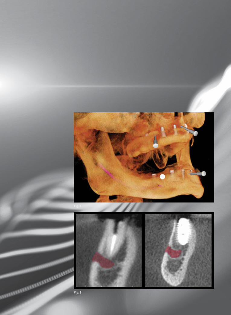
Again, I review the scan and decide on the size of the implant, based on the ana-tomical dimensions. I choose the diameter and the length of the implant and, using 3-D planning software, virtually “place” it in the proper position for optimal occlusal forces. I then export this information and place an order for a surgical guide; within a few days, it’s ready for approval.
It takes a week or less from finalizing
the implant plan to receiving the surgical guide—a quick turnaround time. With this planning capability (Fig. 1), my implant cases have been well-placed, and postop outcomes have been good.
Informed views lead to informed decisions
Having 3-D imaging not only provides me with accurate and viable diagnostic information, but I’ve also noted that allowing patients to see their condition motivates them to make the right choices. Patients are more apt to schedule treatment when they see the condition of their teeth. This has also led to word-of-mouth referrals and an improved reputation as a technology practice.
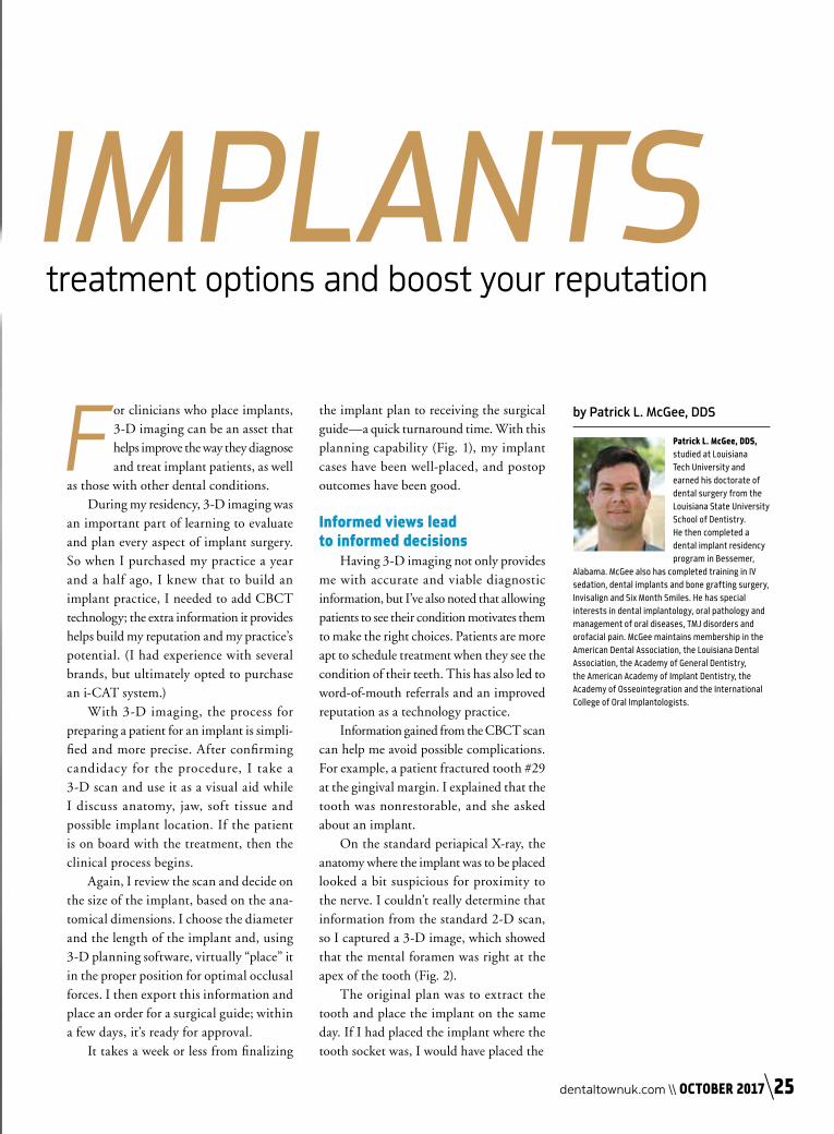
Information gained from the CBCT scan can help me avoid possible complications. For example, a patient fractured tooth #29 at the gingival margin. I explained that the tooth was nonrestorable, and she asked about an implant.
On the standard periapical X-ray, the anatomy where the implant was to be placed looked a bit suspicious for proximity to the nerve. I couldn’t really determine that information from the standard 2-D scan, so I captured a 3-D image, which showed that the mental foramen was right at the apex of the tooth (Fig. 2).
The original plan was to extract the tooth and place the implant on the same day. If I had placed the implant where the tooth socket was, I would have placed the
by Patrick L. McGee, DDS
Patrick L. McGee, DDS, studied at Louisiana Tech University and earned his doctorate of dental surgery from the Louisiana State University School of Dentistry.He then completed a dental implant residency program in Bessemer,
Alabama. McGee also has completed training in IV sedation, dental implants and bone grafting surgery, Invisalign and Six Month Smiles. He has special interests in dental implantology, oral pathology and management of oral diseases, TMJ disorders and orofacial pain. McGee maintains membership in the American Dental Association, the Louisiana Dental Association, the Academy of General Dentistry, the American Academy of Implant Dentistry, the Academy of Osseointegration and the International College of Oral Implantologists.
IMPLANTStreatment options and boost your reputation
dentaltownuk.com \\ OCTOBER 2017 25

implant into the nerve canal. Thanks to the scan, however, I knew exactly how to angle the implant into an anatomically safe position. The implant looked well-placed and had a positive prognosis (Fig. 3). The patient was pleased with how the technology helped her overall outcome and the lack of complications.
More detail, more applicationsThree-dimensional imaging has opened
new treatment options for other procedures beyond implants as well.
For instance, CBCT provides extremely useful data in airway cases. One patient diagnosed with sleep apnea recently scheduled a consultation and brought his sleep study
results with him. The CBCT scan I performed revealed a narrow airway, so I told him that based on his sleep study results, his airway was still occluded while he slept, even with the appliance in place.
He couldn’t believe his airway when he saw the scan. “That’s why I can’t breathe and why I’m always tired,” he said. “I need to have this treated.” While he had been resistant to the idea of using a CPAP device while sleeping, once he saw the scan, he scheduled an appointment with his medical doctor to be evaluated for one.
My reputation among the medical com-munity has also grown since I’ve included 3-D imaging in my practice. For example, a new patient, edentulous with upper and
Fig. 3
CBCT provides extremely useful data in airway cases.
The scan I showed a recent patient diagnosed with sleep apnea
revealed a narrow airway, so I told him that based on his sleep
study results, his airway was still occluded while he slept,
even with the appliance in place.
Dentale, taking the hassle out of implant trainingJust arrive, and learn from some of the UK’s most trusted dental experts
We offer successful and effective training
courses designed to help you achieve your goals.
All of our courses are ‘hassle free’ so you’re able to focus on your training journey. We provide the patients, you care for them. We will also provide you with a Skills Portfolio which we will help you complete as your patient’s treatment progresses, saving you hours of your own time.
Simply book your course, arrive and learn;
we’ll take care of the rest.
HANDS-ON SURGERY UK BASED TRAINING
LATEST TECHNIQUES COMPLETE PATIENT CARE
WE SUPPLY PATIENTS
View our course schedule online - week long and 10 day courses availableTel: 08450 178899 | [email protected] | www.dentaletraining.co.uk
FULL‘HANDS ON’
CASES FROMCONSULTATION TO
RESTORATION
26 OCTOBER 2017 // dentaltownuk.com
lower dentures, was referred to me by her oncologist. She had stage 4 breast cancer, and her oncologist wanted a head and neck oral exam before starting chemotherapy to make sure there were no lesions in her mouth that might cause complications during treatment.
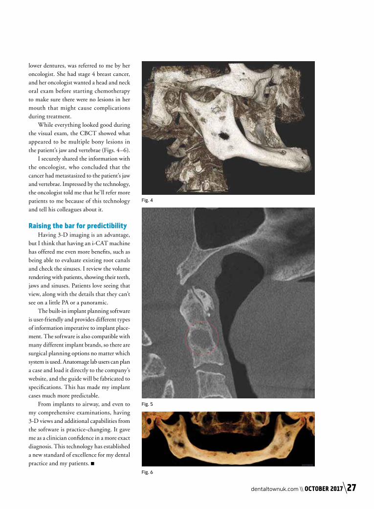
While everything looked good during the visual exam, the CBCT showed what appeared to be multiple bony lesions in the patient’s jaw and vertebrae (Figs. 4–6).
I securely shared the information with the oncologist, who concluded that the cancer had metastasized to the patient’s jaw and vertebrae. Impressed by the technology, the oncologist told me that he’ll refer more patients to me because of this technology and tell his colleagues about it.
Raising the bar for predictibilityHaving 3-D imaging is an advantage,
but I think that having an i-CAT machine has offered me even more benefits, such as being able to evaluate existing root canals and check the sinuses. I review the volume rendering with patients, showing their teeth, jaws and sinuses. Patients love seeing that view, along with the details that they can’t see on a little PA or a panoramic.
The built-in implant planning software is user-friendly and provides different types of information imperative to implant place-ment. The software is also compatible with many different implant brands, so there are surgical planning options no matter which system is used. Anatomage lab users can plan a case and load it directly to the company’s website, and the guide will be fabricated to specifications. This has made my implant cases much more predictable.
From implants to airway, and even to my comprehensive examinations, having 3-D views and additional capabilities from the software is practice-changing. It gave me as a clinician confidence in a more exact diagnosis. This technology has established a new standard of excellence for my dental practice and my patients. ■
Fig. 4
Fig. 5
Fig. 6
dentaltownuk.com \\ OCTOBER 2017 27