dental - multimedia.3m.com · dental magazine | content 3 04 young talent full-mouth...
TRANSCRIPT

no. 3
2 |
nov
embe
r 20
18dental
3MSM Health Care Academy
magazine

3dental magazine | content
04
young talentFull-mouth rehabilitation: Direct or indirect?
Successful use of an esthetic bulk fill material
10
product newsNew super quick polyether impression material in three viscosities
Extended portfolio of zirconia materials
Webinar with Dr. Giuseppe Chiodera
16
ask the expertAbout the development of the first super quick polyether
22 clinical excellenceThe next generation polyether: Superfast. Super detailed.
Esthetic restoration of an endodontically treated premolar
Simplified cementation of glass ceramic restorations
Increased efficiency in post-endodontic restorative treatment
42
3M Science. Applied to Life. “Science is just science …”
News on glass-ionomer restoratives
54
scientific activitiesPre-launch event at ACTA: About a superfast polyether
The post-amalgam revolution has begun!
Train the trainer: Young Asian dentists meeting in Seefeld
Sofia Dental Meeting 2017: Identifying future-oriented treatment techniques
A faster path to posterior esthetics
Talent Award 2017: Introducing young professionals with talent and ambition
80 outside the boxProfessional smartphone photography – a contradiction in itself?
86
3M around the world 3M Customer Innovation Center in St. Paul: Exploring new ways of thinking and learning
Cost-efficiency is an important success factor for every dental office. In order to ensure that clinical procedures can be carried out in an efficient way, different strategies may be adopted. One such strategy is the use of easy-to-use high-quality dental materials that support the user in getting e. g. the impression, restoration or cementation procedures right the first time. By virtually eliminating the need for retakes, remakes and recementations, treatment time and cost can be reduced and the patient’s level of satisfaction is increased. Simplified and standardized operative techniques also help dentists to become more efficient and successful.
3M offers a whole range of innovative products that support efficient procedures and great outcomes, like the new 3M™ Impregum™ Super Quick Polyether Impression Material. Detailed information about their benefits and clinical use is compiled in this issue of the 3MSM Health Care Academy dental magazine, which comes in a new layout. Starting with the next issue to be published in early 2019, the magazine will also have a new editor: Our Global Scientific Affairs Manager Mitsuko O’Neill from Saint Paul, Minnesota is looking forward to taking on this new and exciting task. I am sure she will be able to serve our increasingly global leadership in the best possible way!
Enjoy reading!
Frédéric van Vliet
dear
read
er,
Frédéric van Vliet 3M Oral Care
Mitsuko O’Neill 3M Oral Care
Cover: Reaction tank in the preparative laboratory used for the production of (polyether) impression materials at 3M in Seefeld, Germany.
dental magazine | editorial
young
Tommaso Brunelli, Italy
Successful use of an esthetic bulk fill material
8 – 9
Asmaa Al-Taie, United Kingdom
Full-mouth rehabilitation: Direct or indirect?
6 – 7
talent

6 7dental magazine | young talentdental magazine | young talent
Asmaa Al-Taie, United Kingdom
Full-mouth rehabilitation: Direct or indirect?
Dr. Asmaa Al-Taie is a clinical lecturerin restorative dentistry at Leeds University, where she completed her MSc and PhD. For her PhD she developed novel nano-hybrid dental composite with anticaries properties. Previously she has worked in both General Practice and as a Senior House Officer in Maxillofacial Surgery and Restorative Dentistry. She has also been awarded the Membership Diploma from the Faculty of Dental Surgeons, (Royal College of Surgeons of Edinburgh). Her research interest is in the field of resin composites, light curing and amalgam alternatives.
For a minimally invasive treatment of patients with severe tooth wear, an antagonist-friendly material with a high modulus of elasticity is usually preferred. Appropriate options are a direct build-up with resin composite or the placement of indirect restorations made of resin-based or hybrid ceramic materials. The latter option is particularly suitable in cases with a high amount of tooth substance loss, where multiple layers of composite would be needed.
In order to compare both procedures with respect to the effort and time involved, a 59-year-old patient with severe wear and sensitivity problems was treated in a split mouth design. In this case, wear was mainly caused by Naswar (acidic powdered snuff) consumed three times per day.
The upper and lower right quadrants showed a limited amount of wear so that it was decided to restore them – as well as all anterior teeth – with direct composite material (ceram.x® universal, Dentsply Sirona). Due to a significant amount of substance loss in the upper and lower left quadrants, the placement of direct restorations would have been challenging here. Hence, indirect restorations (made of 3M™ Lava™ Ultimate CAD/CAM Restorative) were planned in this area.
Treatment was carried out in two stages. In the first stage, impressions, a jaw registration at a 2 mm increase of the OVD and a facebow record were taken.
Both restorative treatments illustrated in this case seem to be suitable for patients with severe tooth wear. Chair time may be reduced when indirect restorations are chosen.
Fig. 1: Initial situation: 59-year old patient in retruded contact position (centric relation) with erosive tooth wear caused by Naswar.
Fig. 2: Frontal view of the teeth in intercuspal position (centric occlusion).
Fig. 3: Occlusal view of the upper jaw with a severely worn dentition.
Fig. 4: Occlusal view of the lower jaw. The lower left quadrant is more severely affected by wear than the right.
Fig. 5: Wax-up on the upper jaw model for planning of both, the direct and the indirect restorations.
Fig. 6: Wax-up on the lower jaw model for planning of all restorations.
Fig. 7: Frontal view of the wax-up on the model.
Fig. 8: Occlusal view of the maxillary teeth after placement of direct composite restorations and resin nano ceramic onlays.
Fig. 9: Occlusal view of the mandibular teeth after placement of direct composite restorations and resin nano ceramic onlays.
Fig. 10: Frontal view of the treatment result with a slightly increased OVD. The patient was given a soft splint for the mandible as a protective measure.
After the production of models and a wax-up, the composite build-ups were created. In the second stage, impressions were taken and models casted as well as scanned in order to design and produce the resin nano ceramic onlays with the CEREC System (Dentsply Sirona). The restorations were cemented with 3M™ Scotchbond™ Universal Adhesive and 3M™ RelyX™ Ultimate Adhesive Resin Cement after selective enamel etching.
The case shows that the placement of the onlays is a suitable treatment option for patients with severe tooth wear. The outcome was similar to that of the direct restorative treatment, while the procedure required less effort and time.
2 6
3 7
4 8
5 9
10
1
“”Asmaa Al-Taie, Leeds, United Kingdom
Dr. Asmaa Al-Taie BDS, MSc, MFDS RCS(Ed)[email protected]
cont
act

8 9dental magazine | young talentdental magazine | young talent
Fig. 2: Isolation with rubber dam, which is difficult to
achieve due to incomplete eruption of the second molar.
In order to hold the rubber dam in place, the rubber dam clamp
is stabilized …
Fig. 3: … with wings created using flowable composite.
This figure shows the result of caries excavation and cavity
preparation on the first molar as well as cleaning of the
cavity.
Fig. 4: Selective enamel etching with 3M™ Scotchbond™
Universal Etchant. The second molar has already
received a restoration made of 3M™ Filtek™ One Bulk Fill
Restorative in the shade A2 using the essential lines
technique including bulk filling (one-step modeling technique).
Fig. 5: Situation after the application of
3M™ Scotchbond™ Universal Adhesive, build-up of the mesial wall, and filling of
the cavity on the first molar with 3M™ Filtek™ One Bulk
Fill Restorative in the shade A2. The occlusal anatomy was created and fissures
were added with specific instruments.
2
3
4
5
Tommaso Brunelli graduated in Dentistry from the University of Brescia in October 2016. Since then, he has been working in different private practices in Bergamo and Brescia, with a focus on restorative dentistry and endodontics. In June 2018, he became the first prize winner of the OneAndOnlyBulkContest powered by StyleItaliano and 3M.
This case was also published on Facebook and on www.styleitaliano.org.
cont
act
Tommaso [email protected]
Tommaso Brunelli, Italy
Successful use of an esthetic bulk fill materialIn 2016, three weeks before I graduated in Dentistry at the University of Brescia, I had the pleasure to attend an intensive StyleItaliano workshop in Santa Margherita Ligure in Italy. The direct restoration techniques taught and practiced there and the materials used were so convincingly easy and promising that I immediately decided to implement them in my daily work procedures.
In order to continuously improve my skills, I followed StyleItaliano on Facebook and regularly experimented with new techniques and materials promoted by the group. This is where I learned about the launch of 3M™ Filtek™ One Bulk Fill Restorative, which I instantly wanted to test. Using it in the clinical environment, I discovered that it is capable of simplifying posterior restoration procedures – especially when combined with the essential lines technique developed by StyleItaliano.
It is this technique that also played a decisive role in my clinical case that won the OneAndOnlyBulkContest powered by StyleItaliano and 3M. After having decided to participate in this European contest that invited young dental practitioners to post a nice case on Facebook, I first focused on case selection. A case that caught my special attention was that of a young patient whose mandibular left first and second molar were in need of a direct restorative treatment. The specific challenge was caused by the fact that the second molar had not completely erupted so that isolation was difficult. The problem was solved by use of a steel rubber dam clamp stabilized with flowable wings, as shown in the following. In this context, the use of a simple and time-efficient one-step modelling technique was particularly beneficial.
Fig. 6: Treatment result one week after the
treatment completed by finishing, polishing and
checking the occlusion.
Fig. 1: Pre-operative view: Both molars show carious lesions and need to be restored with resin composite.

productnews
Extended portfolio of zirconia materials
14
New super quick polyether impression material in three viscosities
12 – 13
Webinar with Dr. Giuseppe Chiodera
15

12 13dental magazine | product news dental magazine | product news
New super quick polyether impression material in three viscositiesExtremely quick, extremely accurate: In order to provide dental practitioners with an impression material that offers precisely these beneficial features, 3M™ Impregum™ Super Quick Polyether Impression Material was launched. The material is available in light body, medium body and heavy body viscosities.
Perfectly suitable for the monophase technique:
3M™ Impregum™ Super Quick Medium Body Polyether
Impression Material.
Quick set, better tasteAll three material options were specifically developed for small cases. With a working time of 45 seconds and an intra-oral setting time of only two minutes, the first true superfast polyether is predestined for crown, inlay/onlay, implant and short-span bridge impressions (up to three units). Using it, the dental practitioner will benefit from a more efficient procedure and from happier patients, as the tray may be removed from the mouth very quickly and the brand-new formulation offers an improved material taste.
Developed for the 1-step technique: 3M™ Impregum™ Super Quick Heavy Body Polyether Impression Material in combination with 3M™ Impregum™ Super Quick Light Body Polyether Impression Material.
Proven polyether propertiesDespite the changes in the chemistry, Impregum Super Quick materials still offer the well-known and trusted properties of a polyether impression material. They are highly moisture tolerant, will reliably flow into every spot and show a consistent flow behavior throughout the whole working time. These properties support the user by capturing every detail in a highly accurate way. More information about the new materials and the complete portfolio of materials and devices for a precise and simplified impression taking procedure from 3M is available at 3M.com.

14 15dental magazine | product news dental magazine | product news
Three material options for all kinds of zirconia restorations.
Additional pre-shaded optionsLava Plus zirconia is predominantly used for the fabrication of large- span bridge frameworks and implant-supported structures. It offers bending strength that meets ISO 6872:2015 type 2 class 5 requirements and a high trans-lucency. The expanded portfolio not only includes white material for custom shading in the dental laboratory, but also a new pre- shaded variant offered in the shades A1, A2, A3 and A3.5 that allows for increased efficiency. Blanks are available in three different heights (14 mm, 18 mm and 25 mm). In addition, for the custom-shaded material, a blank with 30 mm height was recently launched to facilitate the production of complex implant-supported substructures.
Seventeen shadesLava Esthetic zirconia is popular among dentists and dental technicians due to its extraordinarily high esthetic potential. It offers optimized translucency, a color gradient designed for excellent shade match to VITA classical shades and built-in fluorescence*. The portfolio was just expanded to include 17 shades – all 16 VITA classical shades and one bleach shade. This contributes to an even more efficient manufacturing procedure since the availability of virtually all tooth shades reduces manual color adjustments to a min-imum. Offering a flexural strength of 800 MPa**, Lava Esthetic zirconia is perfectly suited for the production of monolithic single tooth restorations and three-unit bridges with up to one pontic between two crowns.
Extended portfolio of zirconia materials
Over the years, dental zirconia has developed into a versatile restorative solution. Depending on the indication and the time available for manual steps in the dental laboratory, material variants with specific properties are needed. The new portfolio from 3M includes all suitable options. It is comprised of 3M™ Lava™ Esthetic Fluorescent Full-Contour Zirconia for cases with high esthetic demands and 3M™ Lava™ Plus High Translucency Zirconia for the production of restorations that require a high flexural strength. The portfolio extension relates to the shade options and blank sizes offered.
* Fluorescence determined with light sources simulating natural UV light.** Three-point bending strength according to ISO 6872:2015; qualified for Type II, class 4; indications: crowns, bridges with a maximum of
one pontic that must be supported on each side by a crown (prosthesis not to include more than three units) inlays, onlays and veneers.
3mrestorativesolution.com
How to simplify direct restoration procedures in the posterior region? Dr. Giuseppe Chiodera, member of the StyleItaliano group, uses 3M™ Filtek™ One Bulk Fill Restorative in combination with a simplified modelling technique to reach this goal. The technique called essential lines eliminates the need for a cusp-by-cusp build-up and creates a three-dimensional optical effect on a relatively flat occlusal surface by
Webinar with Dr. Giuseppe Chiodera
drawing fissures with a specific instrument. The lecturer and developer of the technique explains every detail needed for its successful implementation in this on-demand webinar which is available in several different languages. Additional webinars created by 3M and StyleItaliano are accessible here as well.

ask theexpert
Elke Kopp, Germany
About the development of the first super quick polyether
18 – 21

18 19dental magazine | ask the expert dental magazine | ask the expert
Elke Kopp, Germany
About the development ofthe first super quick polyether
With the complexity of an indirect restorative procedure, the requirements concerning impression accuracy tend to increase. An error in an impression used to produce a crown may lead to a poor fit, but manual adjustments are usually sufficient to produce a satisfactory result. If a similar error occurs in an impression involving multiple prepared teeth or implants, a remake of the prosthetic work is often necessary.
Does that mean that it is sufficient to use a less accurate impression material for small cases? It does not, as optimizing impression quality means minimizing the risk that impressions need to be retaken and prosthetic work needs to be adjusted. This, in turn, it will save valuable time and lead to a more efficient and economic treatment procedure.
For this reason, many dentists rely on 3M™ Impregum™ Polyether Impression Materials even for their small cases. So far, however, they had to accept a draw-back – the comparably long setting time of polyether materials. It slowed down polyether users in comparison to those using fast-setting VPS materials and reduced the potential time savings. This is no longer the case – due to the launch of 3M™ Impregum™ Super Quick Polyether Impression Material that is currently avail-able in two viscosities (medium and light body). We talked about the development of this innovation with Dr. Joachim Zech, Head of the R&D Team for Impression Materials at 3M in Seefeld, Germany.
Interview withDr. Joachim Zech, Head of Research & Development of Dental Impression Materials, 3M Oral Care, Seefeld, Germany.
Decanting of the base paste of 3M™ Impregum™ Super Quick Polyether Impression Material into a storage container.

20 21dental magazine | ask the expert dental magazine | ask the expert
Elke Kopp graduated from ESB Business School in Reutlingen and CESEM in Reims. She worked several years in large pharmaceutical and health care companies before joining 3M ESPE in 1995. Since then she held various positions in Global Marketing and is currently New Procedure Marketer in the Indirect Business Team based in Seefeld, Germany.
Dr. Zech, why did you decide to develop a new superfast polyether impression material?The main aim of the project was developed based on market research and user feedback. This gave us the insight that the general interest in conventional impression materials is still high – despite the availability and evolution of high- performance intraoral scanners. In addition, we found that polyether users are generally very happy with the existing polyether properties, especially the great flow behavior and reliable performance in the presence of moisture. At the same time, we identified a growing de-mand for a material that offers these benefits combined with a setting time ideal for small cases. This may be related to the fact that the number of single tooth restorations placed increases continuously and dentists simply want to obtain the best possible results in the shortest possible time.
How did you manage to reduce the setting time?The reduction of the setting time was challenging, as we needed to develop a new initiator compound. It is always possible to adjust the setting times to a certain extent without fundamental changes, but we had exhausted this potential for the existing initiator. Thus, there was no other way to reach our goal than by altering the basic chemistry. We were able to successfully accom-plish this task within a reasonable time span thanks to the possibility of collaborating closely with our skilled colleagues from the chemical synthesis plant and from production in Seefeld. In this way, we were able to streamline the complete process from raw material development to production.
You said that the basic chemistry was altered. Is 3M™ Impregum™ Super Quick Polyether Impression Material still a true polyether?Yes, the new material is a true polyether. The new base paste also contains the aziridino-polyether – the beating heart of every polyether impression material. Hence, the reactive groups and the curing mechanism in this paste are still the same. In addition, the newly devel-oped initiator compound is made of a molecule that is similar to the existing one. The small, but decisive difference lies in the substituents, which are larger and exhibit a higher reactivity. The result is a faster setting reaction and – as a beneficial side effect – a more neutral taste of the impression material. Exchanging the initiator also required us to ex-change or add a few other compo-nents mainly in the catalyst paste, including plasticizers and pigments.
How is a high product quality ensured?Extensive testing of the basic raw materials and the pastes in the development phase was carried out to ensure that the proven polyether properties are still offered. The raw materials undergo comprehensive chemical-physical analysis and physical-mechanical tests are usu-ally used for characterization of the pastes. In the first step, the test results are used to identify the most promising formulations and to adjust and fine-tune the components for final product development. Later, they are needed to ensure that the internal quality standards are met, while some of the tests are needed e. g. for FDA approval and CE certification of the final product. As a matter of course, every batch of polyether impression material produced in Seefeld is subjected to strict quality controls.
Is the high product quality the main argument for dentists to test the new material?It is the well-balanced and proven material properties combined with the short setting reaction that make the material worth testing in the practice environment. Many dental practitioners prefer polyether impression materials whenever intraoral moisture control is difficult. For those who are not familiar with the typical intrinsic hydrophilicity of polyethers and the clinical behav-ior related to this feature, it might be a perfect occasion to find out more about it now. For existing Impregum us-ers, it might be interesting to start using a true polyether for their small cases as well – or to increase their productivity by replacing a slower setting polyether in these situation. Many of those who have already tested the innovative addition to the Impregum family are enthusiastic about it and would recom-mend it to their colleagues*.
Dr. Zech, thank you very much.
* Source: 3M field evaluation with 447 participants from Europe and the U.S., 2017.
cont
act
Elke Kopp, Germany [email protected]
Employees at the chemical synthesis plant of 3M in Seefeld.
View into the preparative laboratory.
Reaction tank in this laboratory. Automatic packaging of the cartridges.

clinicalexcellence
case 3
Paulo Monteiro, Portugal
Simplified cementation of glass ceramic restorations
30 – 35
case 4
Walter Devoto and Daniele Rondoni, Italy
Increased efficiency in post-endodontic restorative treatment
36 – 41
case 1
Gunnar Reich, Germany
The next generation polyether: Superfast. Super detailed.
24 – 25
case 2
Giuseppe Chiodera, Italy
Esthetic restoration of an endodontically treated premolar
26 – 29

24 25dental magazine | clinical excellencedental magazine | clinical excellence
The next generation polyether: Superfast. Super detailed.
It is thus as fast as or even faster than many quick-setting VPS-based impression materials and particularly suited for impression taking in the context of producing single-unit restorations or small bridges. In addition to the increased speed, it offers all proven polyether benefits that lead to a reliable clinical performance and highly accurate results. These include a great flow behavior and an intrinsic hydrophilicity, i.e. high affinity to water, which ensure that the material flows deeply into the sulcus and captures every detail. In addition, polyethers maintain their flowability consistently throughout the whole working time, meaning that a user does not need to be afraid of any premature setting reaction that may have a negative effect on the quality of the final impression.
The use of the new material developed for the monophase technique – 3M™ Impregum™ Penta™ Super Quick Medium Body Polyether Impression Material – is demonstrated showing two different patient cases.
The first patient had a fractured composite restoration on her lower first molar that needed to be replaced. The second patient had previously received an implant in the region of the upper first premolar. After the healing phase, the final prosthetic work needed to be produced and placed. A closed tray impression technique was used in this case.
Taking outstandingly precise impressions in an efficient procedure – this
is feasible for everyone using the new 3M™ Impregum™ Penta™ Super Quick
Polyether Impression Material launched by 3M in April 2018. The material offers
a working time of 45 seconds and an intraoral setting time of only two minutes.
Gunnar Reich, Germany
Fig. 1: Initial situation of case 1: Fractured old composite restoration on the lower first molar.
Fig. 2: Deep distal preparation with bleeding from inflamed gingival tissue.
Fig. 3: Challenging moisture control and bleeding managed by using a soaked retraction cord.
Fig. 4: Impression taken with the monophase technique. Syringing of 3M™ Impregum™ Penta™ Super Quick Polyether Impression Material (Medium Body) around the preparation with the 3M™ Penta™ Elastomer Syringe.
Fig. 5: Final monophase precision impression made of 3M™ Impregum™ Penta™ Super Quick Polyether Impression Material (Medium Body).
Fig. 6: Final situation: 3M™ Lava™ Esthetic Fluorescent Full-Contour Zirconia restoration cemented with 3M™ RelyX™ Unicem 2 Self-Adhesive Resin Cement.
Fig. 7: Initial situation of case 2: Implant with healing cap six months after implant placement.
Fig. 8: Syringing of 3M™ Impregum™ Penta™ Super Quick Medium Body Polyether Impression Material around the impression coping with the 3M™ Penta™ Elastomer Syringe.
Fig. 9: Impression coping securely fixed in the impression that was taken using the monophase technique and a closed tray.
Fig. 10: Final veneered all-ceramic crown cemented on an implant abutment.
63
1 4
2 5
Case 1
Dr. Gunnar Reich attended the Universities of Munich and Berlin and obtained his Dr. med. dent. (DDS) degree in 1986. Ever since, he has been practicing dentistry in the South of Germany. Today, he is the owner of a private practice in Munich.
97
8 10
Case 2
cont
act
Dr. med. dent. Gunnar [email protected]

26 27dental magazine | clinical excellencedental magazine | clinical excellence
In April 2018, 3M launched a new, faster setting polyether impression material: 3M™ Impregum™ Super Quick Polyether Impression Material, which is now available in three different viscosities for the 1-step and monophase techniques. Its development was based on the insight that dental practitioners would like to use a polyether material that accurately reproduces the important details not only in complex cases, but also in everyday procedures – without sacrificing their efficiency.
Being convinced that this new material might offer several clinical benefits, I decided to test it in my dental office. The idea was that – with an impression taking procedure that is highly accurate and fast – it would be possible to spend more time on the preparation.
The case below illustrates the use of 3M™ Impregum™ Super Quick Heavy Body and Light Body Polyether Impression Materials for restorative treatment of a 45-year-old male patient.
Fig. 2: Situation after root canal treatment, try-in of two 3M™ RelyX™
Fiber Post 3D Glass Fiber Posts size 0 and determination as well as marking of
their final length
Fig. 1: Initial situation: Upper first premolar in need of a final restoration after endodontic treatment. The aim was to achieve an esthetic result and to protect the weakened cusps of the premolar, which were at high risk of fracturing.
1
2
Fig. 3: Proximal box elevation with 3M™ Filtek™ One Bulk Fill Restorative and try-in of the extraorally shortened 3M™ RelyX™ Fiber Posts 3D.
Fig. 4: Application of 3M™ RelyX™ Unicem 2 Aplicap™ Self-Adhesive Resin Cement into the root canal using the elongation tip.
Fig. 5: 3M™ RelyX™ Fiber Posts 3D after cementation and core build-up accomplished with 3M™ Filtek™ One Bulk Fill Restorative in the shade A2.
Fig. 6: After final preparation, a capsule containing 3M™ Astringent Retraction Paste is inserted into the sulcus and applied around the preparation for retraction and moisture control before impression taking.
Giuseppe Chiodera, Italy
Esthetic restoration of an endodontically treated premolar
4
3
5
6

28 29dental magazine | clinical excellencedental magazine | clinical excellence
Fig. 7: Complete removal of the 3M™ Astringent Retraction Paste with water
after two minutes.
Fig. 8: Clean and clearly visible preparation margin with pilot cord in the sulcus prior to
impression taking.
Fig. 9: Syringing of 3M™ Impregum™ Super Quick Light Body Polyether Impression Material around the preparation for the
1-step impression technique.
Fig. 10: Close-up view of the final impression taken with 3M™ Impregum™
Super Quick Heavy Body and Light Body Polyether Impression Materials.
Fig. 11: Selective etching of the enamel with 3M™ Scotchbond™ Universal Etchant for 30
seconds.
Fig. 12: Etching of the all-ceramic restoration’s intaglio surface
(IPS e.max® CAD, Ivoclar Vivadent) with 5 % hydrofluoric acid for 20 seconds.
Dr. Giuseppe Chiodera graduated with a degree in Dentistry from the University of Brescia in 2004. In the same year, he received a scholarship and followed post-graduate training at the King’s College in London. Since 2004, he is also the owner of a private practice in Brescia which is dedicated primarily to conservative dentistry, adhesive esthetic restorations, prevention, endodontics and new technologies. He is an honorary member of StyleItaliano and loves developing new treatment techniques that simplify diagnostic and restorative procedures.
Fig. 13: Application of 3M™ Scotchbond™ Universal Adhesive to the prepared tooth surface.
Fig. 14: Following air-drying for approximately 5 seconds, the universal adhesive is light cured for 10 seconds.
Fig. 15: Application of 3M™ Scotchbond™ Universal Adhesive to the all-ceramic restoration for 20 seconds.
Fig. 16: Esthetic treatment outcome: perfectly fitting all-ceramic overlay restoration immediately after placement.
7 9
11
8 10
12
13
16
15
14
cont
act
Dr. Giuseppe Chiodera, [email protected]

30 31dental magazine | clinical excellencedental magazine | clinical excellence
Paulo Monteiro, Portugal
Simplified cementation of glass ceramic restorationsIs it possible to simplify indirect anterior restoration procedures without compromising restoration longevity and esthetics? According to my personal experience and scientific studies, it is – provided that suitable materials are used. A material combination that has proven its worth in the context of placing glass ceramic restorations in a simplified way is 3M™ Scotchbond™ Universal Adhesive and 3M™ RelyX™ Ultimate Adhesive Resin Cement1-3. It is well-suited for both, retentive and non-retentive preparation designs.
Efficiency and simplicity are increased as only two components are required to obtain a strong and durable bond: the single-bottle universal adhesive and the adhesive resin cement. The adhesive works well with selective enamel etching preferred by me, but also in the total-etch and self-etch modes. Containing active and stable silane, it also works as a glass ceramic primer. In addition, it incorporates a dark cure activator for RelyX Ultimate resin cement. The following clinical case is used to illustrate the simplified procedure and provide evidence for the great long-term performance of the selected materials.
Fig. 2: The patient expressed the desire to receive restorations that are stable
over time and highly esthetic. The clinical examination revealed that both teeth
were vital. After additional assessment of the situation on X-rays, it was decided
to remove the old restorations and see what kind of and how much of the tooth
structure would be left.
Fig. 1: A 21-year old female patient (dental student) presented in our dental office telling us that she was unhappy with the existing composite restorations on her maxillary central incisors. The restorations had already been replaced several times due to fractures and discoloration.
1
2
Fig. 3: The old restorations were removed and, as large amounts of enamel were still available, it was decided to opt for a conservative preparation and place a crown and a veneer made of feldspathic porcelain.
Fig. 4: Situation after tooth preparation: Most of the structure is still covered by sound enamel favorable for adhesive restoration procedures.
Fig. 5: Feldspathic crown and veneer …
Fig. 6: … on the model.
4
3
5
6

11
32 33dental magazine | clinical excellencedental magazine | clinical excellence
Fig. 7: The restorations have a very high translucency.
Fig. 8: Crown ready for try-in.
Fig. 9: Veneer ready for try-in.
Fig. 10: Application of 3M™ RelyX™ Ultimate Try-In Paste (Translucent) into the veneer.
Fig. 11: Try-in of the crown and veneer: Both restorations are highly esthetic.
Fig. 12: Conditioning of the restorations’ intaglio surface with hydrofluoric acid gel
(PORCELAIN ETCHANT, 9.5% HF, Bisco) for 90 seconds. The etchant was then
suctioned, thouroughly rinsed off with water and the surface dried with
water and oil-free air.
8 10 14
7
12
9 13
16
Fig. 13: Use of 3M™ Scotchbond™ Universal Adhesive on the etched ceramic surface. The universal adhesive contains active silane, eliminating the need for additional silane application. It should be allowed to react with the substrate for 20 seconds and treated with a gentle stream of air to ensure that the solvent evaporates completely. Visual control is possible: the adhesive layer will no longer move when the solvent is gone. Light curing is not required.
Fig. 14: Isolation of the working field with rubber dam for the adhesive cementation procedure.
Fig. 15: Assessment of the available space between the restoration margin and the rubber dam.
Fig. 16: Products used for the adhesive cementation procedure. Use of the phosphoric etching gel is optional when a universal adhesive is used. However, studies have shown that selective enamel etching may lead to an enhanced bond strength.
15

34 35dental magazine | clinical excellencedental magazine | clinical excellence
References1 Fasbinder DJ, Neiva GF, Dennison JB, Heys D, Heys R: Clinical Evaluation of CAD/CAM resin nano ceramic and leucite-reinforced glass-ceramic onlays. AADR 2016, Los Angeles, Abstract No. 254.
2 Vogl V, Hiller KA, Buchalla W, Federlin M, Schmalz G. Controlled, prospective, randomized, clinical split-mouth evaluation of partial ceramic crowns luted with a new, universal adhesive system/resin cement: results after 18 months. Clin Oral Investig. 2016 Dec;20(9):2481-2492.
3 3M RelyX Ultimate Adhesive Resin Cement: 5-year clinical performance. THE DENAL ADVISOR, Volume 34, Number 2, March-April 2017.
Fig. 17: Etching of the enamel on the maxillary left central incisor with
phosphoric acid (3M™ Scotchbond™ Universal Etchant) for 30 seconds.
Fig. 18: Application of 3M™ Scotchbond™ Universal Adhesive onto the tooth surface after rinsing and drying. According to the
manufacturer’s instructions, the adhesive is rubbed into the tooth surface for
20 seconds and air-dried until it no longer shows any movement.
Fig. 19: Situation after pre-treatment of the maxillary right central incisor in the
same way, application of 3M™ RelyX™ Ultimate Adhesive Resin Cement into the
restorations, restoration placement, excess removal with a sponge pellet and covering
of the margins with glycerin gel to avoid oxygen inhibition. The cement was finally
light-cured for 20 seconds per surface.
Fig. 20: Esthetic treatment result one week after placement of the restorations.
Fig. 21: Perfect optical integration of the indirect restorations is obtained.
Fig. 22: Beauty shot stressing the natural appearance of the glass ceramic
restorations.
Professor Paulo Monteiro obtained his degree as a Doctor of Dental Medicine at the Instituto Superior de Ciências da Saúde in Caparica (ISCSEM), Portugal. Here, he started to develop a passion for esthetic dentistry. In 2005, the author completed post-graduation programs in Esthetic and Restorative Dentistry at the ISCSEM. He also obtained a Master’s degree in Dental Medicine at the same Institute. He is a Coordinator and Professor of the Restorative post-graduation program Aesthetic and Restorative Dentistry at Instituto Universitário Egas Moniz and has an exclusive dental practice in Lisbon that focuses on esthetic and cosmetic dental treatments.
Fig. 23: Comparison of the situation at baseline …
Fig. 24: … and six years after placement of the restorations: The patient is still happy with the functional and esthetic result and clinically, there are no signs of marginal degradation or discoloration.
18
19
20
17 23
24
21
22
cont
act
Paulo Monteiro DMD, [email protected]

36 37dental magazine | clinical excellencedental magazine | clinical excellence
Walter Devoto and Daniele Rondoni, Italy
Increased efficiency in post-endodontic restorative treatmentRestorative procedures following endodontic treatment are usually highly complex and time-consuming. Often, a post is needed to stabilize the remaining tooth structure and this post needs to be cemented properly, which is not always easy given the lack of space in the root canals. Afterwards, the tooth needs to be built up layer by layer with composite. In this context, the establishing of a long-lasting bond between the resin cement, the tooth structure and the build-up material is a decisive factor. Finally, a crown needs to be produced and placed, and this crown should fulfill high demands regarding function and esthetics.
With so many steps in the process, the use of feasible treatment techniques and innovative materials that enable the user in the dental practice and laboratory to streamline the procedures is particularly beneficial. They can be implemented easily, increase the work efficiency by reducing the number of potentially error-prone work steps and lead to predictable results. An example of a streamlined treatment approach in the context of an endodontic revision is described using this case example.
Fig. 2: Removal of the gutta-percha filling with rotating instruments.
Apically, 4 mm of the root filling should remain in place.
Fig. 1: Clinical situation after endodontic retreatment of the mandibular second premolar which had originally been restored with a metal screw post. The tooth is ready for the restorative procedure.
Fig. 3: Preparation of the root canal for the placement of a 3M™ RelyX™ Fiber Post 3D Glass Fiber Post. The RelyX Fiber Post system includes drills that match the post diameters. Afterwards, the canal needs to be carefully rinsed with water.
Fig. 4: Try-in of a 3M™ RelyX™ Fiber Post 3D Glass Fiber Post size 2 (1.6 mm diameter), which should fit exactly and still needs to be easily removable, and marking of the desired final length of the post.
Fig. 5: Shortening of the fiber post with a diamond disc. It is necessary to use a suction system and to wear a mask.
Fig. 6: Checking of the obtained post length in the root canal.
1
2
4
3
5
6

38 39dental magazine | clinical excellencedental magazine | clinical excellence
7 9 13
8 10 14
Fig. 7: Thorough rinsing of the root canal with water after disinfection with a sodium
hypochlorite solution (NaOCl).
Fig. 8: Careful drying of the disinfected root canal with paper tips.
Fig. 9: Application of 3M™ RelyX™ Unicem 2 Automix Self-Adhesive Resin Cement into
the root canal with an endo tip.
Fig. 10: Situation after insertion of the fiber post into the root canal and excess removal. The post has been shortened extraorally with
a diamond disk and cleaned with alcohol.
Fig. 11: Tooth build-up using 3M™ Scotchbond™ Universal Adhesive and
3M™ Filtek™ One Bulk Fill Restorative.
Fig. 12: Tooth stump after build-up and preparation. As the preparation margin
is in a subgingival position, soft tissue management is particularly important.
11
12 16
Fig. 13: Situation after soft tissue management with the 3M™ Astringent Retraction Paste which was used according to the manufacturer’s instructions for use. The precision impression was taken with polyether impression material.
Fig. 14: Temporary restoration in place. At the same appointment, the adjacent molar has received a new direct restoration made of 3M™ Filtek™ One Bulk Fill Restorative. This material offers a tooth-like opacity and may be placed in layers of up to 5 mm height (provided that a specific light-curing protocol is respected).
Fig. 15: Computer-aided full-contour design of the crown to be produced using 3M™ Lava™ Esthetic Fluorescent Full-Contour Zirconia. The transparent view of the planned restoration enables the technician to visually check the wall thickness. In addition, virtual tools are used to ensure the required material thickness (0.8 mm in this case) as well as a suitable crown shape and ideal occlusal contacts.
Fig. 16: Crown after milling, removal from the blank and optimization of the restoration surface with finishing instruments (left). It is possible to elaborate the morphological details including fissures prior to sintering. The crown on the right shows the result after sintering.
15

40 41dental magazine | clinical excellencedental magazine | clinical excellence
Fig. 17: Sintered crown made of 3M™ Lava™ Esthetic Fluorescent Full-Contour Zirconia on the model. It fits precisely and has a natural surface texture and color thanks to gradient shading technology.
Fig. 18: Crown after the application of low-temperature (< 900 °C) firing stains used to increase the chroma in the cervical body area and in the non-functional areas of the occlusal surface, glazing and glaze firing.
Fig. 19: Sandblasting of the crown’s inner surface with 40 µm aluminum oxide. This procedure was followed by cleaning with alcohol and air-drying.
Fig. 20: Cleaning of the roughened stump surface with ethanol before it is air-dried as well.
Fig. 21: Application of 3M™ RelyX™ Unicem 2 Automix Self-Adhesive Resin Cement into the restoration.
Fig. 22: Crown placement with excess cement visible at the margin.
Dr. Walter Devoto graduated with honors in Dentistry and Dental Prosthesis in 1991 at the University of Genoa, Italy. He is particularly interested in the fields of Conservative Dentistry and Esthetic Dentistry and runs his own private practices in Sestri Levante and Portofino. In addition, he is collaborating with diverse prestigious dental offices throughout Europe that specialize in Esthetic Dentistry. He has worked as a teacher and demonstrator at the University of Genoa and as a lecturer at the universities of Siena and Madrid. At the moment, he is lecturer at the International University of Catalonia, Barcelona, and visiting professor at the Universitè de la Mediterranee, Marseille.
Daniele Rondoni obtained his degree as a Dental Technician in 1979 at “P. Gaslini” Professional Institute in Genoa. Upon request, he helped establishing the first Savona-based School for Dental Technicians in 1981 and has worked in the field of professional training as a teacher and counsellor ever since. In 1982, he opened his own dental laboratory (Daniele Rondoni Dental Lab) in Savona. His career features numerous international professional experiences in Italy, Switzerland, Germany and Japan. Particularly devoted to the study of morphology and dental esthetics, he is actively involved in the development of esthetic restorative materials.
Fig. 23: Removal of the excess material with a spatula after brief light-curing for 2 seconds. It is easy to remove the cement in this gel state.
Fig. 24: Buccal view of the treatment outcome immediately after cementation of the monolithic crown, light curing of each restoration surface for 20 seconds and a final checking of the occlusal contacts.
Fig. 25: Occlusal view of the final clinical situation with the crown in place next to the new direct restoration on the first molar.
Fig. 26: Radiographs of the initial and the final situation.
Daniele Rondoni, MDT [email protected]
Dr. Walter Devoto, [email protected]
cont
act
1917 23
18 20 24
21 25
22 26

Compressive strength Edge stability
Barbara Cerny, Germany
“Science is just science …”
44 – 48
Christiane Stein, Germany
News on glass-ionomer restoratives
50 – 53
3M Science.
to Life.Applied

44 45dental magazine | 3M Science. Applied to Life. dental magazine | 3M Science. Applied to Life.
Barbara Cerny, Germany
“ Science is just science …
… until you make it do something, change something – improve something.” At 3M, this is what we are striving for every day as we develop materials with enhanced features that improve the lives of the dental professionals using them. In this context, scientific knowledge is not only used when new formulations and basic chemistries are invented, but also when specific product properties need to be ensured and tested.
The sheer number of in-vitro investigations usually carried out during dental product development is impressive. For 3M™ Impregum™ Super Quick Polyether Impression Material for example, the raw materials and the paste formulations underwent extensive testing. With the final material, the focus is on physical-mechanical tests which are needed not only to control the new properties. They are also used to ensure that the proven polyether benefits – like hydrophilicity, flowability throughout the working time and snap setting – remain unchanged.
HydrophilicityHydrophilicity describes a material’s affinity for water: Intrinsically hydrophilic materials tend to maximize contact with water, which allows them to flow across wet surfaces. This is clinically relevant due to the presence of oral fluids such as blood or saliva in the patient’s mouth. If a material flows reliably across moist teeth and soft tissues, void formation is prevented, which provides the conditions for an exact reproduction of details.
Essentially, two test methods are used at 3M in Seefeld to determine the hydrophilicity of impression materials: Contact angle measurement1 and the two-phase contact test2. In the first test, the initial contact angle of a water droplet applied to a layer of unset light-body impression material is determined with a specific drop shape analyzer.
Water droplet applied to a layer of unset impression material. With the aid of video analysis, the contact angle is determined.
Measurement of the initial water contact angle.
In this test, polyethers including 3M™ Impregum™ Super Quick Light Body Polyether Impression Material showed the lowest contact angles of all materials tested. This outcome was obtained independent of the point in time the water droplet was applied. Based on this study, it is possible to conclude that polyether materials show a significantly higher initial hydrophilicity than the VPS materials tested. This fact is confirmed by the results of the two-phase contact test. Here, a water droplet is applied at the interface of two unset impression materials placed in a side-by-side setup with a clear interface line. The applied water droplet is always attracted by the more hydrophilic impression material and shows a larger horizontal spread on its surface. In this experiment, Impregum Super Quick Light Body Impression Material attracts water more strongly than any VPS material tested.
A water droplet is applied to the interface of two unset impression materials – a VPS material (left, Aquasil® Ultra LV Fast Set) and 3M™ Impregum™ Super Quick Light Body Polyether Material (right). The water droplet is attracted to the material with the higher hydrophilicity, moves immediately toward that material and shows a larger horizontal spread on its surface.

46 47dental magazine | 3M Science. Applied to Life. dental magazine | 3M Science. Applied to Life.
Constant flow behaviorAnother important property of an impression material is its flowability not only in the beginning, but throughout the whole working time, while elastic properties are required after setting. In many VPS impression materials, the transition from plastic to elastic starts too early, which causes limited flowability often resulting in inaccurate impressions.
Polyether impression materials usually show a constant flow behavior throughout the working time. This is mainly due to their snap-set property: It ensures the material will not start setting before the working time ends and when it does set, it does so immediately. This puts the user in control as the indicated working time can be used to its full capacity and problems associated with premature setting are avoided.
Two test setups are used by 3M to evaluate the flowability. The first one is the shark fin test, the second the consistency test. The shark fin test is conducted with a test device consisting of a receptacle and a weighted mold with a slit. When the receptacle is filled with impression material and the stamp released to sink into it, a shark-fin like layer of impression material is produced. The taller the fin, the higher the flowability of the material. By repeating this experiment at different predefined moments in time between mixing and the end of the working time, shark fins of different heights can be obtained and compared so that the changes in flowability are easily detected.
There are virtually no differences visible for fins produced with Impregum Super Quick Light Body material 25 seconds after mixing and at the end of the working time (green fins at the very top). All tested VPS materials show significantly decreasing fin heights over time, which is an indicator for premature setting that may compromise a detailed reproduction of relevant intraoral structures.3
Weighted Mold
Receptacle
Shark fins produced with different materials throughout their working time. 3M™ Impregum™ Super Quick Light Body Polyether Impression Material is shown in the back (light green color). Image courtesy of Dr. Carlos Sabrosa, Brazil.
Test Apparatus Outcome of the consistency test.4
50
45
40
35
30
25
20
15
10
5
0
Con
sist
ency
/mm
3M™
Impr
egum
™
Supe
r Qui
ck
Ligh
t Bod
y M
ater
ial
EXA
fast
™ N
DS
Inje
ctio
n-Ty
pe
Flex
itim
e® L
ight
Fl
ow
Aqu
asil®
Ult
ra X
LVFa
st S
et
Aqu
asil®
Ult
ra L
VFa
st S
et
Take
1® A
dvan
ced
Ligh
t Bod
y Fa
st S
et
Hon
igum
-Pro
- Li
ght-
Fast
Start of working time End of working time
Consistency of light body impression materials during working time
The consistency test leads to similar results. In this test setup (described in ISO 4823:2015), 0.5 ml of impression material is applied to a plate. The material is then loaded with a weight of 1.5 kg. The larger the diameter of the resulting material disc, the lower the viscosity of the material used. Again, the disc diameters produced with Impregum Super Quick Light Body Impression Material 25 seconds after mixing and at the end of the material’s working time show no significant difference.4
48 dental magazine | 3M Science. Applied to Life.
Dr. Barbara Cerny graduated from dental school in 2000. Afterwards, she followed post-graduate training in pediatric dentistry and implantology while she was employed as an assistant dentist in various dental practices in Germany. In 2007, Dr. Cerny started working for 3M in Seefeld, Germany, where she holds the position of a Global Scientific Affairs Manager since 2014.
Dr. Barbara Cerny Global Scientific Affairs Manager, Indirect Solutions [email protected]
cont
act
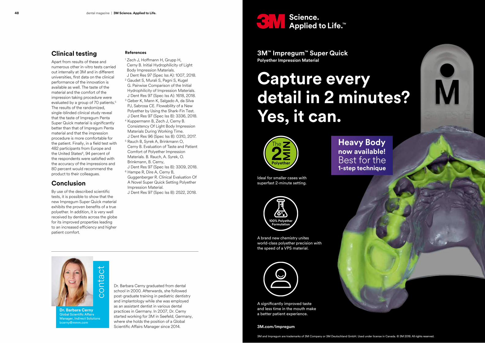
Clinical testingApart from results of these and numerous other in-vitro tests carried out internally at 3M and in different universities, first data on the clinical performance of the innovation is available as well. The taste of the material and the comfort of the impression taking procedure were evaluated by a group of 70 patients.5 The results of the randomized, single-blinded clinical study reveal that the taste of Impregum Penta Super Quick material is significantly better than that of Impregum Penta material and that the impression procedure is more comfortable for the patient. Finally, in a field test with 482 participants from Europe and the United States6, 94 percent of the respondents were satisfied with the accuracy of the impressions and 80 percent would recommend the product to their colleagues.
ConclusionBy use of the described scientific tests, it is possible to show that the new Impregum Super Quick material exhibits the proven benefits of a true polyether. In addition, it is very well received by dentists across the globe for its improved properties leading to an increased efficiency and higher patient comfort.
References1 Zech J, Hoffmann H, Grupp H, Cerny B. Initial Hydrophilicity of Light Body Impression Materials. J Dent Res 97 (Spec Iss A): 1007, 2018.
2 Gaudet S, Murali S, Pagni S, Kugel G. Pairwise Comparison of the Initial Hydrophilicity of Impression Materials. J Dent Res 97 (Spec Iss A): 1618, 2018.
3 Geber K, Mann K, Salgado A, da Silva PJ, Sabrosa CE. Flowability of a New Polyether by Using the Shark-Fin Test. J Dent Res 97 (Spec Iss B): 3336, 2018.
4 Kuppermann B, Zech J, Cerny B. Consistency Of Light Body Impression Materials During Working Time. J Dent Res 96 (Spec Iss B): 0310, 2017.
5 Rauch B, Syrek A, Brinkmann O, Cerny B. Evaluation of Taste and Patient Comfort of Polyether Impression Materials. B. Rauch, A. Syrek, O. Brinkmann, B. Cerny, J Dent Res 97 (Spec Iss B): 3309, 2018.
6 Hampe R, Dire A, Cerny B, Guggenberger R. Clinical Evaluation Of A Novel Super Quick Setting Polyether Impression Material. J Dent Res 97 (Spec Iss B): 2522, 2018.
Capture every detail in 2 minutes? Yes, it can.
3M™ Impregum™ Super QuickPolyether Impression Material
Ideal for smaller cases with superfast 2-minute setting.
A brand new chemistry unites world-class polyether precision with the speed of a VPS material.
A significantly improved taste and less time in the mouth make a better patient experience.
3M.com/Impregum
3M and Impregum are trademarks of 3M Company or 3M Deutschland GmbH. Used under license in Canada. © 3M 2018. All rights reserved.
Heavy Bodynow available!Best for the1-step technique

50 51dental magazine | 3M Science. Applied to Life. dental magazine | 3M Science. Applied to Life.
News on glass-ionomer restoratives
Glass ionomers have been introduced to dentistry more than 40 years ago. Since then, they have proven their worth as luting cements and restorative materials. Different properties make them clinically attractive: they are biocompatible materials that adhere to moist enamel and dentin without the need for a conditioner and they have long-term fluoride release.
Drawbacks of the classical glass ionomer cements are inferior esthetics compared to composite resins, and their limited mechanical properties including a low strength and wear resistance. Thus, it comes to no surprise that diverse dental manufacturers intensified their effort to develop new glass-ionomer materials with improved mechanical properties.
Christiane Stein, Germany
Compressive strength
Test setup for the determination of the compressive strength (left) versus edge stability (right).
Edge stability
Improved formulationAt 3M, this effort led to the introduction of 3M™ Ketac™ Universal Glass Ionomer Restorative in 2015. The product offers an accelerated setting performance and can be applied without pre-conditioning the cavity and without a protective coating. The latter is possible due to an improved filler composition that leads to a high compressive and flexural strength and a high surface hardness obtained within ten minutes after the beginning of the setting reaction. These improved properties make Ketac Universal restorative suitable for an extended range of indications: apart from the classical applications, it is approved for long-term use in stress-bearing Class I and Class II cavities under certain conditions.* When the material was launched, 3M was already able to present laboratory data that made the improved properties perceptible. The compressive strength and surface hardness obtained with the new material used without a coating are higher than those delivered by several other clinically proven glass ionomers with a coating.
* Stress-bearing Class I restorations with at least one additional support outside of the filling area; stress-bearing Class II restorations when the isthmus is less than half of the intercuspal distance and with at least one additional support outside of the filling area.
Edge stabilityMeanwhile, a number of additional in-vitro studies have been carried out to shed light on several other clinically relevant parameters. One of them is the edge stability. Due to their brittleness, glass ionomer restoratives tend to fracture not in the center, but in the marginal areas of a restoration when exposed to chewing forces. Corresponding to this clinical observation, a test method was developed for determining the edge stability of glass-ionomer restoratives. The figure above demonstrates how the test is carried out: Instead of loading the whole specimen under compression (compressive strength testing), only an elliptical area at the edge of the specimen is loaded. This method has been used in different laboratory studies. One of them was conducted at the University of Munich. The obtained results were presented by Prof. Dr. Nicoletta Ilie at the CED-IADR Congress in Vienna in 20171.

52 53dental magazine | 3M Science. Applied to Life. dental magazine | 3M Science. Applied to Life.
In this study, both compressive strength and edge stability of different glass ionomer restoratives were determined before and after a 24-hour or three-month maturation process. The correlation between the two values was moderate. Ketac Universal Restorative achieved very good results in both tests prior to and after maturation. Like in most other products tested, compressive strength and edge stability were higher after maturation.
The results of another laboratory investigation that focuses on the compressive strength and edge stability were presented at the IADR/PER General Session in London in 20182. In this study that was conducted at King's College London Dental Institute, two types of specimens were used: The first group consisted of usual single-layer specimens, while the specimens in the second group were composed of a glass-ionomer restorative base plus an additional layer of material applied after one month of artificial aging. This latter setup was developed to simulate repair of the restorations.
Compressive strength and edge stability were determined in both groups of specimens after one day, one week, one month and three months of water storage. Of all materials tested, Ketac Universal Restorative showed the best compressive
Peter Braun during the poster presentation.Prof. Dr. Nicoletta Ilie presenting the results of her in-vitro study in Vienna.
strength and edge stability in the classical and the repaired specimens after short-term aging. This indicates that the material may be expected to be well-suited for intraoral repair of partly failed glass ionomer restorations.
HomogeneityThe great results obtained with Ketac Universal restorative in these tests might be related to another factor investigated at 3M – the low amount of air bubbles incorporated after mixing.3 In general, air inclusions present in a cured restoration are defects that act as sources of stress contraction. They have a negative impact on the mechanical as well as optical properties of the restorative material in use and may limit the longevity of a restoration. In order to measure the amount of air bubbles present in restorations made of different glass-ionomer restoratives, samples were created, cross-cut and colored using a black marker. Subsequently, the surfaces were cleaned with ethanol, so that the black color was left in the voids only. This enabled the researchers to determine the amount of black areas – i.e. air bubbles in the samples. In this investigation, Ketac Universal restorative showed the best performance of all restoratives tested.
Christiane Stein is educated in Pharmaceutical Sciences. She worked at the chair of pharmaceutical technology, Ludwig Maximilians University Munich for several years. Afterwards, she was employed in a pharmacy in Munich and in the drug regulatory department of a pharmaceutical company before joining the company 3M ESPE in Seefeld as a Regulatory Affairs Manager – Drug Registration in 1998. Since 2004, she holds the position of a Scientific Affairs Manager, currently responsible for direct restoratives.
Researchers of King's College London Dental Institute and their poster.
Wear resistanceFinally, the fact that Ketac Universal restorative exhibits an improved wear resistance was confirmed in a study conducted at the University of Regensburg, Germany and presented by Dr. Verena Preis at the CED-IADR Congress in Vienna.4 In this laboratory investigation, the sliding wear resistance of different glass-ionomer cements and one composite was determined. The obtained results were good for Ketac Universal restorative, which turned out to deliver a high restoration quality independent of the fabrication temperature (tested at 25 and 37° C).
ConclusionThe scientific results show that with Ketac Universal restorative, a glass-ionomer material is available that offers the proven benefits of this material group combined with improved mechanical properties. This beneficial combination of characteristics makes it worthwhile to test the product in the clinical environment.
References1 Ilie N. Edge and Compressive Strengths variation in Modern Glass Ionomer Cements. J Dent Res 96 (Spec Iss B): 0396, 2017.
2 Zhang J, Braun P, Pilecki P. In-vitro Compressive Strength and Edge Stability of Glass Ionomer Cements (GICs). J Dent Res 97 (Spec Iss B): 2478, 2018.
3 Braun P, Claussen K, Stein C, Mikulla M, Thalacker C. Determination of the Amount of Air Bubbles in Different Glass Ionomer Restoratives. J Dent Res 96 (Spec Iss B): 0093, 2017.
4 Preis V, Hahnel S, Behr M, Rosentritt M. Sliding wear of Glass-Ionomer-Cements. J Dent Res 96 (Spec Iss B): 0092, 2017.
Christiane SteinScientific Affairs Manager 3M Oral Care [email protected]
cont
act

scientificactivities
Joanne Lipman, UK
A faster path to posterior esthetics
72 – 75
Armin Bock, Germany
Talent Award 2017: Introducing young professionals with talent and ambition
76 – 79
Sigrid Hader and Christiane Stein, Germany
The post-amalgam revolution has begun!
60 – 63
Rodica Zerguine, Germany
Sofia Dental Meeting 2017: Identifying future-oriented treatment techniques
68 – 70
Frédéric van Vliet, Germany
Pre-launch event at ACTA: About a superfast polyether
56 – 59
Sigrid Hader, Germany
Train the trainer: Young Asian dentists meeting in Seefeld
64 – 67

56 57dental magazine | scientific activities dental magazine | scientific activities
The introduction of 3M™ Impregum™ Super Quick Polyether Impression Material – the first superfast polyether – was celebrated by 3M in an exclusive pre-launch event that took place in February 2018 at the Academic Center for Dentistry Amsterdam (ACTA). A group of 25 dental practitioners seized the opportunity to meet 3M experts including chief developer Dr. Joachim Zech and receive first-hand information about the new product.
Attendees of the pre-launch event in Amsterdam …
Pre-launch event at ACTA: About a superfast polyether
Frédéric van Vliet, Germany

58 59dental magazine | scientific activities dental magazine | scientific activities
Shark fin test carried out by Dr. Joachim Zech.
… moderated by Leticia Cárcamo Wucherpfennig, Western Europe Marketing Supervisor at 3M Oral Care.
Dr. Barbara Cerny, Global Scientific Affairs Manager, Indirect Solutions at 3M Oral Care …
Richard van Hooijdonk lecturing about future healthcare opportunities and challenges.
… and developer Dr. Joachim Zech introducing the new polyether impression material and its properties along with the results of scientific studies its true polyether performance.
Dr. Marcus Engelschalk counting down 45 seconds to give the participants a feeling of the material’s working time.
Being prepared for the futureThe new generation polyetherApart from product-related information, the group also received inspiration on how to change their way of practicing dentistry. The futurist and trendwatcher Richard van Hooijdonk provided some food for thought by revealing that computed technologies are already transforming the world.While bioprinting and robotics have
already become reality, future options will allow us to deeply interfere with natural processes that determine our health, physical fitness and mental capability. For example, health care professionals will make use of smart pills to monitor our health, inject proteins to rejuvenate our bodies and manipulate character traits or
our outer appearance by changing the DNA of an unborn child. In his presentation, the speaker stressed that dental practitioners, like any human being, need to develop the ability to cope with change in order to lay the foundations for personal and professional success in the near future.
They learned how the accelerated working and setting times and the improved taste were achieved and were informed about study results which show that Impregum Super Quick material offers the beneficial properties of a true polyether. This fact was confirmed by pilot user Dr. Marcus Engelschalk from Munich. While an intraoral scanner is usually
used in his dental office for impression taking, he still relies on conventional impression materials in specific situations. Talking about his initial clinical experience with Impregum Super Quick material, he stressed that he was particularly impressed by its highly accurate reproduction of all relevant details and the overall precision of the impressions. For him,
the material is a suitable supplement to his digital impression system. Some of the typical polyether properties – like its constant flow behavior throughout the working time – were demonstrated live using the so-called shark fin test. Finally, the participants were invited to test Impregum Super Quick material on models or in their own mouths.
Frédéric van Vliet studied Dentistry at the Academic Center for Dentistry in Amsterdam and worked as a dental, orthodontic and implantology assistant in a dental office in Amsterdam for several years before joining the Dental Division of 3M in the Netherlands in 2005. From the beginning, he has been responsible for the consolidation and the building of a network of dental academics. In 2011, he came to Seefeld, Germany, where he soon took over his position as a Scientific Affairs and Education Manager for Western Europe. His tasks included the organization of lectures with international speakers, the realization of trainings for dental professionals on one side and for internal staff and dealers on the other and the carrying out of scientific studies. Since May 2018, he is responsible for the support of group practices in Western Europe.
cont
act
Frédéric van Vliet [email protected]

60 61dental magazine | scientific activities dental magazine | scientific activities
Sigrid Hader and Christiane Stein, Germany
The post-amalgam revolution has begun!
With the decision to phase out dental amalgam in the European Union, the search for alternative restorative materials and treatment concepts has been initiated. An evaluation of the current situation reveals that many suitable approaches are already available, while some work still needs to be done. This is the essence of the three lectures held during the 3M Symposium titled “The post-amalgam revolution” that took place on Friday, July 27, 2018 during the IADR/PER General Session in London.
IADR Symposium with approximately 200 participants.
Prof. Avijit Banerjee lecturing about minimally-invasive operative techniques.
Prof. Timothy Watson unveiling the truth about future restorative materials.
Prof. Ivo Krejci during his passionate plea for the placement of onlays instead of crowns.
Minimally-invasive caries removalThe first speaker, Prof. Avijit Banerjee (King’s College London Dental Institute), focused on contemporary operative concepts for carious lesion management. Leveraging a huge body of scientific evidence, he stressed that it is time to move away from the classical concept of cavity preparation towards less invasive procedures. Study results are already available to show that it is no longer necessary to excavate all dentin bacterial biomass (i.e. infected and affected dentin) to prevent further disease progression. Instead, only the superficial layer of infected necrotic dentin needs to be removed. In addition, peripheral caries removal must be ensured in order to be able to seal the cavity and to arrest the lesion under the lining. Arrest is only possible if a complete seal can be obtained and maintained, which is the case whenever starting and finishing in healthy enamel.
For long-term success, the speaker recommended that a balance must
be achieved using minimally invasive operative concepts, between the quality of the tooth substrate retained (affected dentin or healthy enamel at the cavity periphery), the chemistry of the restorative material used and the clinical handling of both the material and tooth structure. It is imperative long-term success revolves around the maintenance of pulp sensibility and tooth structure, as opposed to assessing the quality of the restoration itself – consider the complete tooth-restoration complex.
Future restorative materialsThe fact that there is still a need for improvement of direct restorative materials was highlighted by Prof. Timothy Watson (King’s College London Dental Institute). He provided an insight into what is happening to the weakened carious substrate in the context of restoration and what is going on at the interface between the tooth tissue and restorative materials.
The available evidence shows that, using resin-based adhesive systems for bonding, a hybrid layer is formed also on caries-affected dentin, but that this layer is prone to hydrolytic degradation, meaning that the interface is going to break down over time. According to the speaker, the use of materials that can reinforce the hybrid layer and encapsulate the collagen fibrils might be a solution.
Some of the available bioactive materials already have this ability, as revealed using different methods of optical analysis including fluorescence and Raman spectra. These materials are able to induce changes in the mineral content of affected and infected dentin, so that a rebuilding of the dentin structure seems possible. However, the commercially available products are usually used for temporary restoration only and have unfavorable optical properties. Hence, Prof. Watson expressed the need for stronger, more esthetic and simpler-to-use materials that offer this desired effect.

62 63dental magazine | scientific activities dental magazine | scientific activities
Dr. Sigrid Hader obtained her Doctor of Medicine (MD) and PhD degrees at the University of Cologne in Germany as well as certificates in Emergency Medicine and Immuno-Allergology from the Université Paris V in France. She worked as a medical consultant and clinical research manager for large medical and pharmaceutical companies before joining the dental products division of 3M in 2005. Since then, she is a Global Scientific Affairs Manager responsible for prosthodontic products based in Seefeld, Germany.
Christiane Stein is educated in Pharmaceutical Sciences. She worked at the chair of pharmaceutical technology, Ludwig Maximilians University Munich for several years. Afterwards, she was employed in a pharmacy in Munich and in the drug regulatory department of a pharmaceutical company before joining the company 3M ESPE in Seefeld as a Regulatory Affairs Manager – Drug Registration in 1998. Since 2004, she holds the position of a Scientific Affairs Manager, currently responsible for direct restoratives.
Speakers and organizers of the 3M Symposium.
3M employees at the company’s booth during the IADR/PER General Session.
A resin-based, self-adhesive hybrid ionomer material is currently being developed: Initial laboratory studies have revealed that it is not losing its retentive properties on the tooth over time. The speaker presented images from fluorescence analysis which confirm that, unlike composite material, the experimental material is clearly able to improve the properties of the carious dentin. This hints to the fact that the experimental formulation might contribute to simpler and clinically successful esthetic restorative procedures.
Onlays instead of crownsFinally, Prof. Ivo Krejci (University of Geneva) presented his concept of restoration replacement or re-dentistry. In line with scientific study results, he never places ceramic crowns and opts for composite onlays instead. Even endodontically treated teeth do not receive a post and core restoration and a crown, but an endocrown which is very similar to an onlay. These types of restorations
are possible today due to the availability of high-performance dental adhesives. What is important for the success of endocrowns is the availability of a sufficiently large surface for bonding, which might be calculated in the future during treatment planning with the aid of an intraoral scanner and finite element analysis.
In order to avoid catastrophic fractures after the replacement of amalgam restorations, the speaker promotes a reinforcement of the tooth. His hypothesis is that the fractures originate from an existing fissure or fracture underneath the amalgam restoration. Especially high-copper amalgams (introduced after the 1970s) seem to induce fissures that lead to tooth fractures.
Hence, these restorations should be replaced as soon as possible. In order to reinforce the tooth, the speaker includes a glass-fiber network in the adhesive layer on the prepared tooth surface. On top of this layer, he applies light-curing composite instead of a resin cement to lute the composite onlay. One of the benefits of this type of restoration is that there is no need for a perfect fit, as a
monoblock is created and slight inaccuracies are compensated by the composite filling material. This makes the procedure very simple and leads to good clinical results.
OutlookIn the end, all speakers agreed that minimally invasive operative techniques and materials supporting these procedures are the way forward in restorative dentistry. The concepts presented by them have already proven their worth in the clinical environment. Now, it is time that general dental practitioners start implementing them in their dental offices.
Successful eventNot only the symposium, but the whole congress in London was a huge success for 3M. More than 20 employees from Seefeld, St. Paul and Loughborough (UK) were present to showcase their research results in poster presentations and oral sessions and to network with visitors at the 3M booth as well as in the presentation area.
Dr. med. Sigrid Hader Scientific Affairs Manager 3M Oral Care [email protected]
Christiane SteinScientific Affairs Manager 3M Oral Care [email protected]
cont
act

64 65dental magazine | scientific activities dental magazine | scientific activities
Sigrid Hader, Germany
Train the trainer: Young Asian dentists meeting in Seefeld
Simplified direct restorationsDuring the first day of the meeting, the group focused on direct restorative procedures. The 3M team shared the latest information about the restorative procedure including 3M™ Scotchbond™ Universal Adhesive, 3M™ Filtek™ Bulk Fill Posterior Restorative and 3M™ Filtek™ One Bulk Fill Restorative, 3M™ Filtek™ Z350XT Universal Restorative and other composite filling materials that allow for simplified use and thus support the implementation of efficient procedures. Afterwards, Dr. Walter Devoto, co-founder of StyleItaliano, presented feasible, teachable and repeatable techniques for direct restoration procedures in the anterior and posterior regions realized using the proven 3M materials.
In this context, the lecturer emphasized that it is extremely important to respect the desires of the patient, which leads to the fact that most treatments cannot follow the ideal treatment plan. Instead, it is necessary to offer a solution depending on the esthetic needs of the patient, time he is willing to invest in the treatment and financial investment he is willing to make. For this purpose, StyleItaliano developed a menu with different levels of treatment and corresponding treatment recommendations. The core technique that was also practiced by the participants in a workshop is a two-shade technique based on a universal idea of layering: A dentin core is combined with an enamel layer of 0.5 mm thickness.
The technique was tested with 3M™ Filtek™ Supreme XTE Universal Restorative using recipes developed by StyleItaliano specifically for this product. In the first step, an individual shade guide was created according to recipes shared by Dr. Devoto. In the second step, an anterior tooth was restored on a model. In this context, more than 20 clinical tips from the speaker helped the participants to obtain beautiful results.
In the spirit of building dental material knowledge and developing well-trained and informed external lecturers in every country and region across the globe, 3M invited a group of 12 young Key Opinion Leaders from the APAC region to visit 3M Oral Care in Seefeld, Germany. The participants enjoyed an intensive and interactive four-day training. In this context, they learned a lot about 3M products and procedures from the company’s and the dental practitioner’s perspectives, participated in a presentation skills training and leveraged the opportunity to develop and exchange their own ideas for collaboration.
Dr. Walter Devoto sharing his knowledge on feasible, teachable and repeatable treatment techniques.
Modelling of an anterior restoration.
Participants during the workshop.
For the posterior region, the “essential lines” concept developed by Dr. Giuseppe Chiodera (Member of the StyleItaliano group) was presented. It enables the dentist to avoid a cusp-by-cusp build-up and to create the occlusal anatomy in a quick and repeatable way. After bulk filling, the occlusal anatomy is simply shaped following the essential lines template. The technique was also practiced by the attendees.

66 67dental magazine | scientific activities dental magazine | scientific activities
PD Dr. Jan-Frederik Güth focusing on
indirect procedures.
Simplified indirect procedures Products that may be used to simplify indirect restorative procedures took center stage during the second day of the group’s visit. The participants were informed about 3M innovations in impression materials, products for a simplified and more efficient post-and-core procedure and different kinds of cement.
Subsequently, PD Dr. Jan-Frederik Güth from the University of Munich shared his experience and presented his way of working with the products in daily clinical practice. He concentrated on each step in indirect procedures, from conservative tooth preparation to gingiva management and impression taking and from temporization to cementation.
His personal recommendations were e. g. to define the treatment aim first by creating a mock-up, which is then not only used for functional and esthetic evaluation, but also as a prep-guide to obtain the desired space for the indirect restoration. In order to decrease the risk of gingival recession, the speaker avoids sub-gingival preparation margins whenever possible. Regarding impression taking, Dr. Güth either opts for an intraoral scanner (e. g. the 3M™ True Definition Scanner) or a medium body polyether impression material that is capable of displaying the margin in an astonishingly precise way. In his opinion, the major benefit of intraoral scanners lies in the possibility of checking the scanning result and the preparation immediately with the option of correcting and rescanning, which might lead to higher-quality prosthetic works. Regarding cementation, he elaborated on the principles of adhesive luting and emphasized the differences in pretreatment of glass ceramics and oxide ceramics like zirconia: While
the former needs to be etched with hydrofluoric acid, gentle sandblasting is recommended for the latter.
In hands-on sessions, the participants focused on temporization with 3M™ Protemp™ 4 Temporization Material and on cementation. In this context, they seized the opportunity to test e. g. the tack-cure function of 3M™ Ketac™ Cem Plus Resin-Modified Glass-Ionomer Cement and practiced post cementation with 3M™ RelyX™ Unicem 2 Automix Self-Adhesive Resin Cement.
Developing and improving presentation skillsThe third day was devoted to the topic of lecturing.
Overwhelming feedbackThe feedback received from the group was overwhelming, with an average score of 4.85 out of 5 points. One participant stated: “At first, I thought the schedule is too packed but later because of activities together and encouragement from each other, it has become the best session. The best experience of my life! We are colleagues forever!” It seems that they simply cannot wait implementing what they learned: “Thank you for sharing your valuable time together. I am back to the real world. It was like a dream, but I clearly know what I have to do for better dentistry. Thank you, 3M!” The 3M team would also like to thank the group for their commitment and passion.
Dr. Gerhard Kultermann (Freiburg) shared his experience on how to give a good presentation. Afterwards, the participants each had the opportunity to practice their own skills in a five-minute lecture. Their performance was evaluated by the group and tips for improvement were given. Finally, on day four, they met the developers of different 3M materials and gained insight into current development projects.
After three and a half days full of intensive work, the group visited Linderhof Palace in Upper Bavaria. The team work and passion built among the KOLs and 3Mers during this Train the Trainer session is the strong foundation to work together for better dentistry and better oral health. In this context, first plans were created for future collaboration – not only with 3M, but also among each other.
Tack-curing of 3M™ Ketac™ Cem Plus Resin-Modified Glass-Ionomer Cement in a workshop.
Participants, lecturers and organizers of the successful event.
Dr. Gerhard Kultermann supporting the group in improving their presentation skills.
Dr. Sigrid Hader obtained her Doctor of Medicine (MD) and PhD degrees at the University of Cologne in Germany as well as certificates in Emergency Medicine and Immuno-Allergology from the Université Paris V in France. She worked as a medical consultant and clinical research manager for large medical and pharmaceutical companies before joining the dental products division of 3M in 2005. Since then, she is a Global Scientific Affairs Manager responsible for prosthodontic products based in Seefeld, Germany.
cont
act
cont
act
Dr. med. Sigrid Hader Scientific Affairs Manager 3M Oral Care [email protected]

68 69dental magazine | scientific activities dental magazine | scientific activities
Participants at the Sofia Dental Meeting.
Looking at the past – building the future: This is what people at 3M do every day to help simplify and improve procedures in dental practices all over the world. They have a look at what is already available and successful, assess the unmet needs of dental professionals and collaborate with innovative dental communities to develop materials and devices that really make a difference.
The same motto “Looking at the past – building the future” was chosen by the organizers of the 10th Sofia Dental Meeting that took place in the capital of Bulgaria from October 12 to 15, 2017. They invited 101 experts from various countries to share their professional knowledge and clinical experience in numerous lectures and workshops. Together, they would identify the developments that are worth advancing and following.
Rodica Zerguine, Germany
Sofia Dental Meeting 2017: Identifying future-oriented treatment techniques
Prof. Angelo Putignano …
… and Dr. Walter Devoto elaborating on when to do what.
Feasible, teachable, repeatableSeveral of them were representatives of the popular community named StyleItaliano, whose members pursue the objective of developing feasible, teachable and repeatable dental treatment techniques. Many of the clinical protocols recommended by them include the use of 3M products, which are selected due to their reliable performance and ease of use that lead to great clinical outcomes. For direct restorative procedures in the anterior region, 3M™ Filtek™ Supreme XTE Universal Restorative is the composite of choice, and in the posterior area, 3M™ Filtek™ One Bulk Fill Restorative or 3M™ Filtek™ Bulk Fill Posterior Restorative are used. Reasons for selecting these materials are their great polishability and polish retention as well as their sculptability. 3M™ Scotchbond™ Universal Adhesive completes the procedures by offering reliable adhesion coupled with ease of use.
Direct or indirect?At the Sofia Dental Meeting, the two founders of StyleItaliano, Prof. Angelo Putignano and Dr. Walter Devoto, shared their clinical tips of when to do what in restorative dentistry. As patients have different expectations regarding the quality, cost and duration of their dental treatment, it is decisive to opt for a treatment that suits their individual needs. This choice is simplified by defining three different levels of treatment, like different kinds of restaurants: street food, trattoria and gourmet. The street food or economic approach should involve minimum time and minimum cost to achieve the best possible quality. Usually, patients that are identified as individuals requiring a street food treatment receive direct restorations made of composite resin. In the anterior region, a single shade technique with Filtek Supreme XTE restorative is often most appropriate. It works well and leads to a natural appearance if the anatomical shape of the tooth is properly restored. In the posterior region, a single layer of bulk fill restorative may be indicated.
The next level, the trattoria or efficient approach, is best suited whenever a treatment needs to be carried out within a restricted time span at moderate cost leading to an excellent quality. Direct and indirect options are available for this level: Anterior teeth may be restored with a two-layer approach using StyleItaliano recipes and an enamel layer thickness of 0.5 mm or with composite veneers. A bulk fill concept or CAD/CAM restorations are recommended for the posterior region.The gourmet level (excellent quality approach) is selected whenever esthetic demands are extraordinarily high and time or money are not an issue. Here, indirect is usually the way to go according to Dr. Devoto: Ceramic veneers, crowns and onlays will be the best possible option in most cases.
In many situations, the speakers prefer composite resin as a restorative material. In their opinion, it is best suited e. g. for young patients where some teeth might continue to erupt and for patients with a high caries risk, as repair is always possible. Another advantage is that the material often involves a minimally invasive preparation procedure.

70 dental magazine | scientific activities
Dr. Rodica Zerguine, DDS, MBAGlobal Category Manager – Core Orthodontics [email protected]
cont
act
Workshop participants imprinting essential lines into a composite surface.
Dr. Giuseppe Chiodera instructing a workshop participant.
Dr. Giuseppe Chiodera lecturing on direct posterior restorations.
Posteriors: Tips & tricksAs shown by Dr. Giuseppe Chiodera in his lecture, preservation of healthy tooth structure is also supported by the use of transillumination. A small-diameter, high-intensity light placed on the buccal side of the teeth reveals everything that needs to be visible for proper caries diagnosis and the following treatment. It enables the user to read the anatomy, identify cracks, assess the depth of white spots or the dimension of a carious lesion and differentiate between a discoloration, a lesion and a crack e. g. on the margin of an amalgam restoration. Based on this detailed information, it is easy to select the most appropriate treatment strategy. He finished his lecture with several tips and tricks for simplification in restoring posterior teeth, like the use of copying techniques employed to record the occlusal anatomy.
The speaker delved into this subject more deeply in his four-hour workshop. Here, he demonstrated different occlusal anatomy copying strategies and showed how the occlusal anatomy may be restored using the instruments Misura Posterior and Condensa (LM Arte). According to Dr. Chiodera, however, fissures need to be added as an occlusal anatomy without fissures is like a face without eyes. Simplification in this procedure is offered by the speaker’s concept of essential lines: It starts from the assumption that every posterior tooth has position-specific lines that
were identified by Dr. Chiodera. By implementing these lines on the occlusal surface of a restoration, a natural, three-dimensional appearance is created. The workshop participants learned how to design these lines on a dab of Filtek Supreme XTE restorative placed on a flat surface and, later, on a restoration they created on a model with Filtek Bulk Fill Posterior Restorative. It turned out that, once they knew how to work with the provided instruments, this strategy was much easier than a three-dimensional cusp-by-cusp build-up. One important trick is to design the fissures not by pulling the instrument through the uncured composite material like a pen, but by putting it in and out again, like creating footprints in the snow.
From photography to the full mock-up conceptOther lecturers from StyleItaliano were Prof. Louis Hardan, who revealed how case documentation becomes more simple, efficient and economic using MobileDentalPhotography (MDP) (Smile Line) and a smartphone, and Prof. Stefen Koubi. The latter focused on the treatment of a severely worn dentition using the full mock-up concept and a combination of glass ceramic and hybrid material veneers and onlays.
The dental professionals who attended the lectures and workshops were thankful for the wealth of recommendations and ideas that will support them in taking the right treatment decisions. They also look forward to implementing some of the promising strategies that will help simplify procedures in their dental offices.
™
Dental RestorativesFiltek
A billion smiles around the world.
As the global leader in restorative and esthetic technology, 3M™ Filtek™ Dental Restoratives has been put to the test in over 1 billion restorations worldwide.
We pioneered the nano-composite category and continue to raise the performance bar to meet restorative challenges around the world.
Simplify your procedures, your inventory and your practice with a single brand – 3M Filtek Dental Restoratives.
www.3M.com/Filtek3M Science. Applied to Life.™3M, 3M Science. Applied to Life. and Filtek are trademarks of 3M or 3M Deutschland GmbH. Used under license in Canada. © 3M 2018. All rights reserved.
Dr. Rodica Zerguine graduated with a degree in Dentistry from University of Cluj-Napoca, Romania in 2004 , followed by an MBA from EM Lyon, France. She joined 3M in 2008 as Scientific Affairs Manager for France and in 2011 relocated to Germany where she held a number of global marketing roles within the Oral Care Division. In 2018 she joined the Core Orthodontics group as Global Business Manager for the Digital Orthodontics, Ortho Adhesives as well as 3M’s proprietary APC platform.

72 73dental magazine | scientific activities dental magazine | scientific activities
Joanne Lipman, UK
The speakers: Dr. Jordi Manauta and Dr. Anna Salat.
Pizza techniqueWhen the first generation bulk fill materials were launched, they allowed for some simplification in posterior restorative procedures: With the option of applying the material in 4 mm instead of 2 mm layers, it became possible to fill many cavities in one go. On top, a layer of universal composite had to be applied to provide for the required mechanical strength. In order to ensure esthetic results, this final layer is usually modelled cusp by cusp (pizza technique). Great outcomes can be achieved with this technique, however, for everyday clinical procedures, it is still too time-consuming.
Essential linesWith this in mind, StyleItaliano developed a new technique called essential lines. The team started from the assumption that anatomy is the most important factor in the posterior area, and that the fissures are an essential part of it: “A posterior tooth without fissures is like a face without eyes. If we put lines into the right place, we are even able to fool the eye and create a 3D effect on an almost flat surface”, explains Dr. Anna Salat. Hence, given that the fissures are imitated correctly, a complex occlusal anatomy is no longer required. To ensure that each tooth is given the right lines (fissures), different teeth were compared and the essential lines which are found in every tooth of a certain position de-termined. For the first practical exer-cise, the participants received a card with those lines. They were asked to
A faster path to posterior esthetics
extrude the content of one capsule of 3M™ Filtek™ Supreme XTE Universal Restorative on the card, and condense it to create a flat surface. “When you start drawing the lines, you should proceed with the instruments like you do with your feet when walking through deep snow”, said Dr. Jordi Manauta. The participants defined the cen-tral point of the tooth first with the Fissura instrument (LM Arte) and then proceeded as demonstrated. After opening the fissures, they were closed again carefully using the instrument and a brush.
Dr. Anna Salat assisting the participants during the hands-on session.
Workshop participant finalizing his essential lines with a brush.
Increasing efficiency in direct posterior restoration procedures is something that can save lots of time every day, as the treatment is carried out quite frequently in virtually every dental office. Who would not love to skip the laborious cusp-by-cusp build-up procedure and still obtain esthetically pleasing outcomes? Most probably, every dental practitioner does.
The good news: It is possible today, thanks to simplified techniques and innovative materials, as shown by Dr. Jordi Manauta and Dr. Anna Salat from StyleItaliano™ in a 3M-sponsored workshop held at the BACD 14th Annual Conference in London.

74 75dental magazine | scientific activities dental magazine | scientific activities
Clinical workflowSubsequently, a clinical procedure was demonstrated – with lots of tips and tricks from the workshop instructors. According to them, diagnostics in the posterior region should start with transillumination – a strong light source used to screen teeth with possible lesions. When the required information about the type and size of the defect is avail-able, further steps can be planned more accurately.
If a box preparation needs to be carried out, it may be useful to copy the available anatomy with a custom ring. It is produced by applying a small amount of light-curing block-out resin or flowable composite in the proximal areas. After light curing, the ring is placed in the desired posi-tion and more resin is applied, so that the ends of the ring are completely embedded in the material. “In case of a short crown, you will have to struggle with a lack of space: rubber
dam, wedge, matrix and ring may not remain in the desired position. This problem is solved by cutting the wedge so that its ends are out of the way”, recommends Dr. Jordi Manauta.
After caries excavation, cavity preparation and cleaning of the cavity, rubber dam is placed as are the wedge, a preferably dark sec-tional matrix band and the custom ring. Then, a universal adhesive (e. g. 3M™ Scotchbond™ Universal Adhesive) is applied according to the manufacturer’s instructions. The proximal wall of the tooth is built up e. g. with a sculptable bulk fill ma-terial like 3M™ Filtek™ One Bulk Fill Restorative. “I was not fond of bulk fill materials until 3M launched Filtek One restorative. While other bulk fills are too translucent and difficult to polish, the 3M material offers an almost body-like opacity and an amazing surface quality, which is
stable over time. Moreover, the consistency is just right for sculpting. Since its introduction, I use the material for virtually every posterior restoration”, said Dr. Jordi Manauta.
When the wall is finished, the cavity is filled with bulk-fill material. Ideally, 1.5 mm of space are left for the cre-ation of the final anatomy with Filtek One restorative and the Condensa instrument (LM Arte). “First, use the thick side of the instrument to con-dense the composite. Then, move it over the surface to remove excess material, thereby following the incli-nation of the remaining cusps. Repeat the procedure with the other side of the instrument and about 90 percent of the anatomy is done”, Dr. Manauta explains. Finally, the fissures are cre-ated using the essential lines concept.
In the second hands-on session, the participants produced a custom ring and a posterior restoration using
Joanne Lipman graduated from Loughborough University with a BSc First Class (Hons) in Management Science in 2001. She spent a placement year at 3M during her Degree, and then joined on the Graduate scheme in 2002. She has held a number of marketing and sales roles within the 3M Health Care division. In 2016 she became a Western European Marketing Supervisor in the Oral Care Solutions Division, currently responsible for the direct product portfolio.
Joanne Lipman [email protected]
the demonstrated techniques. Their feedback was positive: “I registered because I hoped to be given a formula that enables me to simplify, yet improve, my day-to-day dentistry. What I actually learned exceeded my expectations: The essential lines concept is completely new to me, and combined with the use of bulk
fill materials, it saves a lot of time compared to the pizza technique I have used so far. I will definitely implement it in my dental office”, said Dr. Sameer Shah from The Newmarket Road Dental Clinic in Norwich.
Maybe it is time to implement the concept, too!
Dr. Jordi Manauta explaining how to produce a custom ring.
Production of the custom rings in the workshop.
Creation of the occlusal anatomy on the model.
cont
act

76 77dental magazine | scientific activities dental magazine | scientific activities
Armin Bock, Germany
Talent Award 2017: Introducing young professionals with talent and ambitionYoung oral care professionals with talent and ambition is what we need: With their fresh ideas and different ways of thinking, they have the potential to develop new approaches and techniques that help increase the simplicity, predictability and efficiency of treatment procedures. In order to identify these talents and be able to support their further development, 3M organizes a European Talent Award every two years.
In 2017, 21 young practitioners and researchers seized the opportunity to show what they are made of: At the 3M site in Seefeld, Germany, they gave a 15-minute presentation that focused on a research project or clinical procedure of their choice. Winners were selected by an expert jury in three different categories this time: research, clinical and orthodontic.
3M Oral Care European Talent Award 2017: Participants, jury and organizers of the event.
Winner of the research award: Maria Bruhnke during her lecture.
The winner of the clinical award, Jotautas Kaktys.
Research awardThe winner of the first category is Maria Bruhnke (Charité Berlin, Germany). She presented the results of an in-vitro study that was initiated to work out how to successfully restore endodontically treated maxillary central incisors. In order to determine the best possible treatment option for teeth with different amounts of remaining tooth structure, four cavity designs were selected and realized on extracted human maxillary central incisors (24 teeth per cavity design). The teeth either received a direct resin composite restoration or a glass fiber post plus composite resin composite restoration. Aging was simulated by exposure to thermo-mechanical loading. The results of the study revealed that post placement is beneficial whenever the tooth to be restored has no remaining cavity wall. In these cases, the post led to a significantly increased load capability and fewer early failures. Central incisors with more coronal substance available (the most invasive preparation being a biproximal Class III cavity and the least invasive an access cavity only) do not benefit from the placement of a fiber post – a composite restoration is sufficient. Thanks to her clearly structured lecture, the high relevance of the selected topic and the excellent quality of her research, Maria Bruhnke won a financial sponsorship for a three-month stay at a foreign university.
Clinical awardJotautas Kaktys (Kaunas, Lithuania) won the prize for the best clinical presentation – a voucher for attendance of a 3M educational course with a leading international expert. He focused on full mouth rehabilitations and showed how digital technologies may be employed to facilitate planning of highly complex treatment procedures. With the use of the latest software and hardware, it is possible to take into account all required functional and esthetic aspects in the planning phase. Based on data about the planned outcome, the different treatment steps are carried out and the restorations are produced with the required accuracy. In this way, the whole treatment becomes easier, more efficient and highly predictable.

78 79dental magazine | scientific activities dental magazine | scientific activities
Dr. Armin Bock graduated from Ludwig-Maximilians University Munich as Biologist where he worked as post graduated scientist at the Departments for Electron Microscopy and Pharmaceutical Biology. He joined ESPE in 1999 as a Market Researcher. At 3M Dr. Bock held various positions of increasing responsibility in Oral Care and is currently responsible for Professional Service and Training & Education in the DACH Region.
Dr. Armin Bock Professional Service Manager & General Manager CIC [email protected]
cont
act
Orthodontic awardIn the new orthodontic category, Dr. Carlos González Serrano (University of Madrid, Spain) was announced as the winner. The speaker shared information about a study that was carried out to evaluate the bond strength of an innovative flash-free adhesive. This adhesive is incorporated in a fiber mat on the bracket base and eliminates the need for flash removal, which leads to a simplified clinical procedure. The study results presented by the young orthodontist show that the bond strength obtained with the new adhesive system is similar to that of a conventional bonding agent even after artificial aging. It was also confirmed that less adhesive is left on the enamel surface, so that the efficiency of the bonding procedure is increased. As a reward for his excellent performance, Dr. Serrano is invited to attend an educational course with the renowned orthodontic specialist Dr. Juan Carlos Pérez Varela.
Second and third prizeThe second and third places were awarded across the categories, the prizes being vouchers for participation in a photography course or a presentation skills training. The second prize went to the participant who travelled farthest – Dr. Riaan Mulder (University of the Western Cape, South Africa). The topic of his lecture was ion exchange between different glass ionomer restoratives and tooth structure. In order to assess if the level of ion exchange and, with it, the potential to protect the tooth from caries, is similar in different materials, an in-vitro study was initiated by the speaker. In this investigation, the ion movement from three different glass ionomer materials with similar gradient potentials to the tooth structure (inter-diffusion zone) was measured. According to the results, an inter-diffusion zone was formed with all restoratives, but the percentage of ion movement was different. It may therefore be advantageous to take the level of ion movement into account when selecting a glass ionomer restorative.
Finally, Allegra Comba (University of Bologna, Italy) was selected as the third-prize winner. She presented patient cases showing specific clinical situations – e. g. a patient with a post-traumatic fracture or a diastema – and suggested individual direct restorative treatment protocols. These protocols are designed to enable the dental practitioner to save the maximum possible amount of sound tooth structure, fulfill the esthetic demands of the patient and provide for long-lasting results.
Networking opportunitiesThe feedback of the participants was overwhelming after this exciting and productive day: They were inspired by the work of their colleagues, welcomed the chance to learn more about diverse fields of dentistry and leveraged the networking opportunities. The next day, they were given qualitative feedback on their lectures by Prof. Albert Waning, who also provided them with useful advice of how to further improve their own presentation skills.
Dr. Carlos González Serrano lecturing.
Dr. Riaan Mulder sheds light on ion movement in GICs.
Allegra Comba during her clinical case presentation.

outsidethe box
Louis Hardan, Lebanon
Professional smartphone photography – a contradiction in itself?
82 – 85

82 83dental magazine | outside the box dental magazine | outside the box
Professional smartphone photography – a contradiction in itself? With DSLR (digital single lens reflex) cameras, it is possible to
take high-quality intraoral pictures. However, too few dental practitioners make use of these cameras on a regular basis. Surveys reveal that even in a country like the United Kingdom, only about 50 percent of the dentists properly document their patient cases, and not even half of them use a DSLR camera for this purpose.
The participants of the Multi Media Masterclass 2018 learned how to take professional photos with their DSLR …
Louis Hardan, Lebanon
… and with a smartphone.
∙ treatment planning
∙ communication with the laboratory
∙ communication with existing patients
∙ acquisition of new patients
∙ self-evaluation and self-improvement
∙ lecturing and publishing
∙ communication with insurance companies and for medico legal reasons, etc.
Still, many practitioners decide against making use of this tool. Probably, the main reasons are the enormous amount of time needed and the training required to be able to manage the equipment. Last but not least, expenses are high even for those with a camera body already at hand: They need a specific macro lens, one or two flash systems, a number of diffusors and filters. Thanks to advances in technology
development, however, the use of a professional DSLR camera is no longer a strict requirement for high-quality dental photography. Instead, a smartphone may be utilized – provided that it is operated with the right lighting equipment. This is what I demonstrate during my own mobile dental photography courses, and every time, the participants are astonished by the results and enthusiastic about implementing
smartphone photography in their own dental office. This was also the case during the Multi Media Masterclass organized by 3M in March 2018, where Prof. Paulo Monteiro and I performed a battle of the cameras. In the end, we were able to show that very good results can be obtained with a DSLR camera and with many of the modern smartphones, if light is used properly.
This is a pity, since professional documentation is very useful for many purposes such as
Smartphone as an alternative

5
84 85dental magazine | outside the box dental magazine | outside the box
This can be easier with a smartphone than with a DSLR. The latter requires the use of a ring flash for photography of posterior teeth and a twin flash for photography of anterior teeth. For smartphones, a single device is available that provides the required light for the anterior and the posterior region: It is called SMILE LITE MDP (Mobile Dental Photography) by Prof. Louis Hardan. I started to develop this device four years ago as a mini photo studio that may be used with any smartphone. It has an integrated ring flash (central LEDs) and a twin flash (lateral LEDs) that can be used separately or simultaneously at different light intensity levels. The system also comprises a diffusor and a polarizing filter.
The mini photo studio developed specifically for mobile dental photography.
How to get startedFor taking intraoral photos, the distance between the camera and the front teeth should be about 15 to 20 cm, however, it may vary slightly depending on the smartphone in use. In order to produce high-quality images, it is also essential that no retractors, lips and gloves are visible on them. Instead of cropping the photos afterwards, the user should use the zoom function of the camera until he can see only the teeth and gums and then take the picture.
Images of anterior teethIn DSLR photography, it is recommended to use a twin flash for pictures of anterior teeth. Correspondingly, the lateral LEDs of the MDP device are turned on at full power and the central LEDs are turned off. This shows more details of the teeth than a central light source, since the light is coming for the mesial and distal sides; the differences in translucency and color of the teeth will be revealed. For a natural light-and-shadow effect and a better representation of the surface structures, the diffusors should always be used when pictures of anterior teeth are taken. In addition, the use of a lip retractor and – if only the upper teeth are shown – a contrastor placed on the lingual side of the teeth is highly recommended. The best position for the operator is behind the patient.
For evaluation of internal translucencies and colors and for choosing the recipes of shades while doing a direct composite restoration, polarized imaging may be useful. With the MDP, the best effect is obtained with the polarized filter and the central LEDs at full power, while the lateral LEDs are turned off. For those who would like to combine diffusors and the polarizing filter, it is recommended to turn on all LEDs.
Images of posterior teethIn the posterior region, proper illumination of the area to be captured poses the greatest challenge. In DSLR photography, it is recommended to use a ring flash. With the MDP device, the central and lateral light sources may be combined, so that the illumination effect is maximized. For this purpose, all LEDs need to be set to full power. Diffusors are not required, but proper retraction of the cheeks and lips is particularly important here to ensure an unobstructed view. An occlusal mirror supports the user in that it facilitates the exposition of the teeth to be captured. The operator should stand next to the patient.
Portraits, videos and moreApart from intraoral photography, the smartphone can also be successfully used for everyday procedures, e. g. for the digitization of old X-rays, for teaching purposes to show the smallest details, for video documentation of clinical procedures and even for skype lecturing. While capturing video data of a treatment procedure, it is possible to use the screen of the phone for any visual control, and to leverage the zoom function to have a more detailed view e. g. of the margin during tooth preparation. Finally, portrait photography is feasible with smartphones and Smile Lite MDP or with additional light sources (soft boxes).
Portrait taken with a mobile phone for a smile design project.
Fig. 1: Image taken with the lateral LEDs at full power with diffusors, the central LEDs turned off.
Fig. 2: Image taken with the lateral LEDs at full power, with diffusers. The central LEDs turned off and a black background is used. This image is used for shade matching and communication with the lab technician.
Fig. 3: Image taken with the central LEDs at full power, with a polarizing filter.
Fig. 4: Polarized picture with composite buttons to confirm the recipe of composite shades chosen to restore the tooth, A1 Dentine and A2 Enamel.
Fig. 5: Image of posterior teeth taken with all LEDs at full power.
ConclusionToday, it is possible to use a smartphone for professional documentation in dentistry. The quality of the images is strongly dependent on the camera sensor and on the light conditions, the latter being easily controlled with the MDP device. I am convinced that, due to the availability of this tool, dental photography will become accessible to a great number of dentists who do not use a DSLR camera for different reasons including time, economic considerations and complexity of the procedure.
Prof. Hardan graduated in Dentistry in 1989 and continued his post-doctoral education at Saint Joseph University in Beirut; he obtained a certificate for basic science in 1993, and completed his specialization in restorative and esthetic dentistry in 1995. Finally, he completed his PhD in oral biology and materials in 2009. At the present, he is an associate professor at Saint Joseph University and owns a private practice in his home town Byblos.
He is an honorary and active international StyleItaliano member and the inventor of Smile Lite MDP (Smile Line, Switzerland).
Prof. Louis Hardan, DDS, CEA, DEA, PhD Head of the Department of Restorative and Esthetic Dentistry Saint Joseph University, Beirut, [email protected]
cont
act
1
2
4
3

3M aroundthe world
Karen L. Sullivan, USA
3M Customer Innovation Center in St. Paul: Exploring new ways of thinking and learning
88 – 90

88 89dental magazine | 3M around the world dental magazine | 3M around the world
Karen L. Sullivan, USA
3M Customer Innovation Center in St. Paul: Exploring new ways of thinking and learning
3M technology advancing every company, 3M products enhancing every home, 3M innovation improving every life: This company vision can only be achieved through close collaboration with our customers. One of the places where 3M employees and dental professionals can meet to learn from each other and develop ideas together is the 3M Customer Innovation Center in St. Paul, Minnesota. This Innovation Center located at the company’s global headquarters is one of many 3M Global Technical Centers located in more than 30 countries worldwide. Designed to showcase the breadth and depth of the rich and diverse technologies‚ processes and products 3M has created‚ these centers bring to life the spirit of 3M Science. Applied to Life.
Welcome to the 3M Customer Innovation Center in St. Paul!
What’s behind this door?
Innovation experienceAt the very heart of the Innovation Center is the World of 3M Innovation. Through dynamic displays and interactive‚ hands-on demonstrations‚ 3M customers have the opportunity to collaborate with 3M to explore potential solutions to help their global businesses thrive and grow. From the moment a customer enters the Center‚ they enter a world where anything is possible – they will see previously unknown technologies and fascinating‚ practical real life applications. A team of highly trained‚ informative hosts from a variety of 3M technical and professional backgrounds guide visitors on their journey of discovery. Color‚ sound‚ design – all play a significant part in creating the 3M Customer Experience.
Working together very well: The US-based Education and Training Team after successful com-pletion of a team building activity. (From Left to Right: Shannon Myers, Bethany Vanyo, Karen Sullivan, Lorayne Etoll, Kathleen Randolph, Bob Devine, Ashley Haslund, and Steve Johnson)
This is what customers see before …

90
… entering the World of 3M Innovation.
dental magazine | 3M around the world
Karen L. Sullivan started working for 3M in 1989, and for much of her career, she was a Marketing Communications Manager working in the areas of Oral Care, Food Safety, Medical Devices, and Industrial Converting. After completing her Master’s Degree in Business Management and Marketing, she took on a global marketing role for 3M Oral Care’s Global Business Team and was a Certified Six Sigma Black Belt consulting and leading business programs to improve processes and accelerate growth. She currently leads the Global Education and Training Team.
cont
act
Karen L. Sullivan Global Education and Training Manager 3M Oral Care
Continuing educationApart from showcasing innovations, the Customer Innovation Center is a place where 3M hosts a wide range of internal and external trainings as well as other key partnership meetings. Supporting 3M Oral Care Solutions Division, I lead the US-based Global Education and Training Team responsible for the development and implementation of these of programs designed for both dental professionals and the development of our new employees. I am truly fortunate to work with such a talented and highly enthusiastic team.
Programs for dental professionals include onboarding, training and assistance provided to new customers on site and in the digital space as well as impactful continuing education courses for experienced users of 3M dental products. In addition, we help create programs and tools to facilitate our business teams’ relationships with amazing key opinion leaders from around the world. Our 3M colleagues meet with these leaders in St. Paul and in Innovation Centers across the globe on a regular basis to obtain information about new product developments, support 3M in development projects and further develop their own skills in using simplified techniques and procedures offered by 3M. 3M is a trademark of 3M Company or 3M Deutschland GmbH. Used under license in Canada. © 3M 2018. All rights reserved.
Education and Training for Dental Professionals3M.com/DentalEducation
3MSM Health Care Academy

3M Oral CareESPE Platz82229 Seefeld · [email protected]
3M, Aplicap, Filtek, Impregum, Ketac, Lava, Penta, Protemp, RelyX and Scotchbond are trademarks of 3M Company or 3M Deutschland GmbH. Used under license in Canada. All other trademarks are owned by other companies.
© 3M 2018. All rights reserved.
edito
rial i
nfor
mat
ion
Please note that not all products are available in all countries. Product name and package may vary from country to country. For more information, please contact your local 3M Representative.
3M.com/Dental
editorFrédéric van Vliet
editorial teamBarbara CernySigrid HaderYoung-Chul KwonJoanne LipmanAurelio LopezMitsuko O'NeillChristiane SteinKaren L. SullivanRodica Zerguine
project managementOlivia Besten
designeleven eyes GmbH · www.eleveneyes.de
productionEberl Print GmbH · www.eberl.de
We accept no liability for unsolicited manuscripts or photographs. Court of Jurisdiction: Munich
published by3M Deutschland GmbH ESPE Platz 82229 Seefeld · Germany [email protected] www.3mespe.com