dental aspects of hyperparathyroidism · sol silverman, jr., m.a., d.d.s., william h. ware, ... the...
TRANSCRIPT
DENTAL ASPECTS OF HYPERPARATHYROIDISM
SOL SILVERMAN, JR., M.A., D.D.S. WILLIAM H. WARE, D.D.S., M.D.S.
and CHARLES GILLOOLY, JR., B.A., D.D.S.
San Francisco, Calif. Division of Oral Biology, School of Dentistry,
University of California
Reprinted from
ORAL SURGERY, ORAL MEDICINE AND ORAL PATHOLOGY
St. Louis
Vol. 26, No.2, Pages 184·189, August, 1968
(Copyright© 1968 by The C. V. Mosby Company) (Printed In the U. S. A.)
Dental aspects of hyperparathyroidism
Sol Silverman, Jr., M.A., D.D.S., William H. Ware, D.D.S., M.D.S., and Charles Gillooly, Jr., B.A., D.D.S., San Francisco, Calif.
DIVISION OF ORAL BIOLOGY, SCHOOL OF DENTISTRY,
UNIVERSITY OF CALIFORNIA
A previous study1 of forty-two consecutive dentulous patients with surgically proved primary hyperparathyroidism indicated that bone disease is a late manifestation of primary hyperparathyroidism and that dental changes are, in turn, late manifestations of hyperparathyroid bone disease. Only three patients showed the pathognomonic triad of dental changes for hyperparathyroidism, consisting of loss of lamina dura, giant-cell lesions, and demineralization of bone. Two other patients showed a partial loss of lamina dura and demineralization, while in thirty-seven patients dental roentogenograms were either completely normal or revealed nonspecific atypical changes in the trabeculation pattern.
The purpose of this article is to add thirteen cases to the original series, to di~cuss the significance of jawbone changes in the diagnosis of hyperparathyroidism, and to present the case history of the youngest patient to be seen at this institution with primary hyperparathyroidism in whom investigation of dental changes facilitated the diagnosis.
MATERIAL AND METHODS
Thirteen dentulous patients with histologically proved hyperparathyroidism were seen at the University of California Medical Center between 1961 and 1963. Their ages ranged from 15 to 65 years, with an average of 44 years. Data obtained for all patients included serum calcium, phosphorus, and alkaline phosphatase; skeletal surveys; full-mouth dental x-rays; renal tubular reabsorption of phosphate; and the surgical specimen of the parathyroid adenoma.
FINDINGS
Table I summarizes the findings on the thirteen patients in this study. Kidney stones, which are the most frequent clinical finding in hyperparathyroidism, occurred in only four patients. In the earlier report on forty-two
184

Volume26 Number2
Dental aspects of hyperparathyroidism 185
Table I. Thirteen dentulous patients with surgically proved primary hyperparathyroidism
Increased Hypo- alkaline
Hyper- phospha- phos-Stones as caloemia temia phatase Lowered W eight
Dental chief (above (below (above 7 TRP of x-ray oom- 5.5 S.5 SJR (below Skeletal adenoma
findings M F plaint mEq/L.) mg.%) wnits) 75%) lesions (Gm.)
Partial loSB of lamina. dura, de-mineral-ization, giant-cell lesion 0 1 0 1 1 1 1 1 3.2
Deminer-alization 0 1 0 1 0 1 1 8.0
Normal 7 4 4 11 7 2 4 0 0.3 to 5.7 (mean 2.3)
Totals 7 6 4 13 8 4 5 2
cases, 79 per cent of the patients had stones. All patients were hypercalcemic, which is the essential laboratory finding in this disease. The values ranged from 5.5 to 7.7 mEq. per liter, with a mean of 6.4 (normal for our laboratory is 4.6 to 5.4). Hypophosphatemia, increased alkaline phosphatase, and lowered tubular reabsorption of phosphate were less frequently and erratically found. Abnormality of all chemical findings was found in only one of the thirteen patients, a 15-year-old girl who is the youngest such patient ever diagnosed at this medical center.
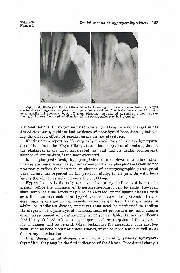
This youngest patient was referred because of gradual loosening and drifting of the lower anterior teeth. Roentgenograms revealed a radiolucency around the teeth in question, but the cause was not obvious. A biopsy was performed and led to a diagnosis of fibrous dysplasia (Fig. 1, left). On the basis of the histologic report, no treatment was advised and the patient was placed on periodic recall. Some recalcification with increased firmness of the teeth occurred following the biopsy. One year later the teeth were again quite loose. Roentgenograms suggested an increase in osteolytic activity (Fig. 2A). Another biopsy was performed and a diagnosis of giant-cell reparative granuloma was made (Fig. 1, right) . The serum calcium level was found to be 7.7 mEq. per liter. Additional laboratory tests revealed the following: serum phosphorus, 2.2 mg. per cent; tubular reabsorption of phosphate, 69 per cent; and alkaline phosphatase, 10 Bodansky units. A skeletal survey showed the tyical subperiosteal reabsorption of the phalanges of the index and middle fingers. All laboratory findings were consistent with a diagnosis of primary hyperparathyroidism, and surgical intervention was recommended. A 3.2 gram parathyroid adenoma was removed. The patient's blood chemistry returned to normal within one month. By the end of 6 months the lower anterior teeth were again firm and calcification of the radiolucency had occurred (Fig. 2B).

186 Silverman, Ware, and Gillooly O.S., O.M. & O.P. August, 1968
Fig. 1. Left, Biopsy specimen from mandibular bone lesion associated with gradual loosening of lower anter ior teeth. Note bone trabeculae (B), fibrous connective tissue (F), and dilated capillaries (C). The microscopic diagnosis was fibrous dysplasia. (Magnification, x140.) Right, One year later, loosening of teeth and roentgenolucency have increased (see F ig. 2, A ) . Bone biopsy at t his time revealed fibrous connective t issue interspersed wi th giant cells (arrows) and red blood cells. The microscopic diagnosis was giant-cell reparative granuloma. (Magnification, x140. )
DISCUSSION
As indicated in our previous repol't on forty-two patients, loss of lamina dura, giant-cell tumors, and demineralization are insensitive indicators of primary hyperparathyroidism. In addition, other skeletal lesions are also late manifestations, as evidenced by the finding of bone lesions in only two of thirteen patients in this series and eight of forty-two patients in our previous series.
Rosenberg and Guralnick2 examined the pretreatment x-ray films of 116 patients with surgically proved hyperparathyroidism and reported that the lamina dura was missing in varying degrees in forty-seven of these patients. Forty-two of these patients had hyperparathyroid bone disease, indicating that these were probably advanced and long-standing cases. In the remaining five patients in whom the lamina dura was absent, no evidence o.£ other bone disease was reported. However, the common bone lesion of subperiosteal reabsorption of the phalanges apparently was not assessed by hand films. In ten of their patients, giant-cell tumors of the jawbone were the presenting features. This further confirms the importance of the differential diagnosis in
Volume26 Number2
Dental aspects of hyperparathyroidism 187
Fig. S. A, Osteolytic lesion associated with loosening of lower anterior teeth. A. biopsy specimen was diagnosed as giant·cell reparative granuloma. The lesion was a manifestation of a patlathyroid adenoma. B, A. 3.2 gram adenoma was removed surgically; 6 months later the teef)h became firm, and calcification of the roentgenolucency had occurred.
giant-cell lesions. Of sixty-nine persons in whom there were no changes in the dental structures, eighteen had evidence of parathyroid bone disease, indicating the delayed effects of parathormone on jaw structures.
Keating,3 in a report on 395 surgically proved cases of primary hyperparathyroidism from the Mayo Clinic, states that subperiosteal reabsorption of the phalanges is the most underrated test and that its dental counterpart, absence of lamina dura, is the most overrated.
Renal phosphate leak, hypophosphatemia, and elevated alkaline phosphatase are found irregularly. Furthermore, alkaline phosphatase levels do not necessarily reflect the presence or absence of roentgenographic parathyroid bone disease. As reported in the previous study, in all patients with bone lesions the adenomas weighed more than 1,000 mg.
Hypercalcemia is the only consistent laboratory finding, and it must be present before the diagnosis of hyperparathyroidism can be made. However, since serum calcium levels may also be elevated by malignant diseases with or without osseous metatases, hyperthyroidism, sarcoidosis, vitamin D overdose, milk alkali syndrome, immobilization in children, Paget's disease in adults, or Addison's disease, numerous tests must be performed to confirm the diagnosis of a parathyroid adenoma. Indirect procedures are used, since a direct measurement of parathormone is not yet available. Our series indicates that if any skeletal lesions occur, subperiosteal reabsorption of the cortex of the phalanges will be present. Other techniques for measuring bone involvement, such as bone biopsy or tracer studies, might be more sensitive indicators than x-ray examination.
Even though dental changes are infrequent in early primary hyperparathyroidism, they may be the first indication of the disease. Once dental changes

188 Silverman, Ware, and Gillooly O.S., O.M. & O.P. August, 196R
are observed, however, the differential diagnosis may still be complex. Loss of lamina dura and demineralization may be found in fibrous dysplasia, osteomalacia, and Paget's disease, and giant-cell tumors, giant-cell reparative granulomas, and parathyroid bone tumors may be confused histologically. • Therefore, since hypercalcemia is a common finding and is mandatory in the diagnosis of hyperparathyroidism, serum calcium assays must be performed when these roentgenographic and histologic features are found. Since serum calcium levels may be elevated by other diseases, hypercalcemia demands further investigation.
Of special interest are the histologic alterations in the jaw lesion observed in the patient reported here. The initial biopsy was typical of a fibrous dysplasia. A biopsy of the same lesion one year later was suggestive of a parathyroid tumor. The longitudinal study of this lesion suggests that the roentgenographic bone lesion is present long before (1 year in this patient) development of the characteristic giant-cell lesion. This finding further suggests the advisability of careful follow-up where bone lesions are diagnosed as fibrous dysplasia.
Skeletal lesions, including the giant-cell tumors, heal spontaneously following removal of the parathyroid tumor. This observation demonstrates a cause-and-effect relationship between the adenoma and metabolic bone disease. Surgical treatment of a giant-cell lesion should be delayed until parathyroid disease has been investigated. If hyperparathyroidism is established, surgical treatment of giant-cell lesions is unnecessary.
Early detection of a parathyroid adenoma is important, since advanced hyperparathyroidism may cause irreversible kidney damage, hypertension, and death.
SUMMARY
This report is based upon the study of thirteen dentulous patients with histologically proved hyperparathyroidism. The patients are in addition to the forty-two consecutive dentulous patients with surgically proved hyperparathyroidism previously reported. Four of the total of fifty-five patients in both studies revealed dental-structure changes of loss of lamina dura, giant-cell tumors, and demineralization. Involvement of the jawbones appears to be a late change and the least sensitive indicator of parathyroid overactivity, as compared to hypercalcemia, renal phosphate leak, hypophosphatemia, elevated alkaline phosphatase levels, and other bony lesions.
It is concluded that the loss of lamina dura and the apearance of giantcell tumors are late signs of hyperparathyroid bone disease, which is itself a late complication of primary hyperparathyroidism.
However, even though infrequently found, changes in the dental structures may be the first sign of primary hyperparathyroidism. Roentgenographic jawbone alterations must be differentiated from other diseases which may have the same roentgenographic manifestations. Giant-cell lesions may reflect local neoplasia, an inflammatory process, or a parathyroid adenoma. Therefore, correlation of microscopic, clinical, roentgenographic, and laboratory findings is necessary before one can arrive at a definitive diagnosis.

Volume26 Number2
REFERENCES
Dental aspects of hyperparathyroidism 189
1. Silverman, B., Gordan, G., Grant, T., Steinbach, H., Eisenberg, E., and Manson, R.: The Dental Structures in Primary Hyperparathyroidism, ORAL SURG., ORAL MED. & ORAL . PATH. 15: 4261 1962.
2. Rosenberg, E. H., and Guralnick, W. C.: Hyperparathyroidism: A Review of 220 Proven Cases, With Special Emphasis on Findings in the Jaws, ORAL SURG., ORAL MEn. & ORAL PATH. 15: Supp. II, p. 84, 1962.
3. Keating, F. R., Jr.: Clinical and Laboratory Aspects of the Diagn-osis of Primary Hyperparathyroidism, J. A. M. A. 178: 547, 1961.
4. Silverman, B., Ware, W., and Dimas, L.: Biologic Variations in Giant-Cell Lesions of the Mouth, ORAL SURG., ORAL MED. & ORAL PATH. 18: 3461 1964.