demonstration of cell-specific phosphorylation of ltc4 synthase
TRANSCRIPT
Demonstration of cell-speci¢c phosphorylation of LTC4 synthase
Namrata Guptaa;b;*, Donald W. Nicholsona, Anthony W. Ford-Hutchinsona
aMerck Frosst Centre for Therapeutic Research, P.O. Box 1005, Pointe Claire-Dorval, Que. H9R 4P8, CanadabDepartment of Pharmacology and Therapeutics, McGill University, 3655 Drummond, Montreal, Que. H3G 1Y6, Canada
Received 14 February 1999
Abstract PMA-induced leukotriene C4 synthase (LTC4S)phosphorylation was investigated over a period of 8 h in amonocytic cell line (THP-1). The level of LTC4S phosphoryla-tion was increased 3^5 fold over a 4 h period decreasing to basallevels after 8 h. This phosphorylation event was found to bespecific to THP-1 cells as there was a lack of LTC4Sphosphorylation in both COS-7 and K-562 cells, and was alsofound to be dependent on the cellular confluency. In the presenceof specific protein kinase C (PKC) inhibitors, a dose-dependentinhibition of the phosphorylation of LTC4S became evident, aneffect not seen with PKA and tyrosine kinase inhibitors. Thisrepresents the first direct demonstration of LTC4S phosphoryla-tion in whole cells.z 1999 Federation of European Biochemical Societies.
Key words: Leukotriene C4 synthase; Protein kinase C;Phorbol ester; THP-1 cell ; Protein phosphorylation
1. Introduction
Cysteinyl leukotrienes (LTs), LTC4, LTD4, and LTE4 com-prise the slow-reacting substance of anaphylaxis (SRS-A) andare smooth muscle contractile lipid mediators with importantbiological e¡ects elicited through G-protein coupled receptors[1^4]. The formation of cysteinyl LTs is initiated by the acti-vation of cytosolic phospholipase A2 (cPLA2) which liberatesarachidonic acid (AA) from phospholipids [5,6]. In the lipoxy-genase pathway, AA binds to an integral membrane protein,5-lipoxygenase-activating protein (FLAP), which is then pre-sented to the soluble enzyme 5-lipoxygenase (5-LO). Via theaction of 5-LO, AA is converted to the unstable epoxide,LTA4 [7]. At this point, LTA4 can follow one of two diver-gent pathways within the cell of synthesis. Hydrolysis ofLTA4 through the action of cytosolic LTA4 hydrolase yieldsthe pro-in£ammatory mediator LTB4 [8]. Conjugation withreduced glutathione (GSH) converts LTA4 to biologically ac-tive LTC4, a stereospeci¢c reaction catalyzed by leukotrieneC4 synthase (LTC4S), an integral perinuclear membrane pro-tein active as a homodimer [9]. LTC4 is then either exportedinto the extracellular milieu directly, or is sequentially metab-olized by the successive cleavage of glutamic acid and glycineto generate LTD4 and LTE4 via a transpeptidase and a pep-tidase, respectively [10,11].
Proteins in the leukotriene biosynthetic pathway are tightlyregulated and many require various co-factors for functionand full activity. Cytosolic PLA2 requires increased Ca2� con-centrations in the WM range in conjunction with a serinephosphorylation by MAP kinase at position 505 of cPLA2
for activation and translocation [12]. The reaction, catalyzed
by 5-LO, requires not only increases in Ca2� levels but alsoATP [13]. In whole cells, FLAP is an essential component for5-LO activity [14^17]. Cytosolic LTA4 hydrolase has recentlybeen shown to be regulated by phosphorylation in endothelialcells where under basal conditions the protein is phosphoryl-ated at serine-415, leading to an inactive state of the enzyme.Incubation with phosphatases reverts the protein to an activestate possessing epoxide hydrolase activity, thereby showingthat LTA4 hydrolase may be regulated by a kinase/phospha-tase cycle [18].
The alternative pathway taken by LTA4 through the actionof LTC4S is its conjugation to GSH. LTC4S is an 18-kDaprotein which is neither dependent on any co-factors for ac-tivity, requires post-translational modi¢cation by glycosyla-tion nor formed as a pro-enzyme. Of interest are two putativeprotein kinase C (PKC) consensus sites, Ser-Ala-Arg, foundon the protein at positions 28 and 111 [9,19]. These are sug-gestive of LTC4S being subject to regulation by phosphoryla-tion. Studies on the activity of LTC4S have been conducted inour laboratory [20,21], and by other investigators [22], whichhave also led to the same conclusions showing that the activ-ity of LTC4S was attenuated upon activation of PKC by thephorbol ester, PMA. This suggests the possibility of LTC4Senzymatic activity being directly regulated by phosphorylationof the enzyme.
2. Materials and methods
2.1. Cell cultureCells from the human monocytic leukemic cell line THP-1 (ATCC),
and K-562 (ATCC), a lymphoid cell line, were both cultured in asimilar manner. Cells were maintained in sterile RPMI 1640 mediasupplemented with 10% (v/v) heat-inactivated fetal bovine serum, 50U/ml penicillin, 50 Wg/ml streptomycin, 0.05 mM 2-mercaptoethanol,1 mM sodium pyruvate, 2 mM glutamine and 25 mM HEPES bu¡erat pH 7.4. COS-7 cells (ATCC), a ¢broblast-like cell line, was main-tained in Dulbecco's modi¢ed Eagle's medium supplemented with10% (v/v) heat-inactivated fetal bovine serum, 50 U/ml penicillin,and 50 Wg/ml streptomycin. Cells were grown at 37³C in a humidi¢edatmosphere of 94% air and 6% CO2.
2.2. Creation of LTC4S-£agHuman LTC4S cDNA (in pBluescript II KS�, Invitrogen) was used
as a template for the introduction of a 5P Kozak translation initiationconsensus sequence (CCACC) and a £ag epitope (DYKDDDDK) atthe carboxyl terminus of LTC4S via PCR with the following oligonu-cleotides: 5P CCG CTC GAG CGG CCG CGG ATC CAC CATGAA GGA CGA GGT AGC TCT ACT GGC TGC 3P and 5PGCT CTA GAG CGG CCG CGG ATC CTA TTA TTT ATCATC ATC ATC TTT ATA ATC GGC CCA CGG CAG CAGCGT CCG GAG CCG TC 3P.
A 25-cycle PCR reaction was run containing deoxynucleotides(250 WM), cDNA template (1/100 of mini-prep), sense and antisenseprimers (5 WM each), GC melt (Clontech) (1:5 by volume), and PwoDNA polymerase (2.5 units) in PCR bu¡er (10 Wl of 100 mM Tris-HCl, pH 8.85 (20³C), 250 mM KCl, 50 mM (NH4)2SO4, and 20 mMMgSO4). The initial denaturing step was 2 min at 94³C followed by
FEBS 21865 12-4-99
0014-5793/99/$20.00 ß 1999 Federation of European Biochemical Societies. All rights reserved.PII: S 0 0 1 4 - 5 7 9 3 ( 9 9 ) 0 0 3 9 7 - X
*Corresponding author. Fax: (1) (514) 428 4900.E-mail: [email protected]
FEBS 21865FEBS Letters 449 (1999) 66^70

25 cycles (94³C for 1 min, 60³C for 1 min, 72³C for 1 min). The PCRproduct was PCR puri¢ed (Qiagen), digested with XhoI and BamHI,gel puri¢ed (Qiagen), ligated into pBluescript II KS� (Rapid DNALigation Kit from Boehringer Mannheim), and transformed into Epi-curian Coli XL2 Blue Ultracompetent cells (Stratagene). Transform-ants were selected on LB plates containing X-galactosidase (40 Wg/ml).Several clones were picked, grown in culture, and the plasmid DNAwas prepared by miniprep (Qiagen). The orientation of the insert(LTC4S-£ag) was con¢rmed by restriction digestion of the plasmid,and the lack of any inadvertently incorporated mutations was ensuredwith bi-directional sequencing using the ABI automated sequencer.LTC4S-£ag/pBluescript was then subcloned into pcDNA3.1(+) (Invi-trogen) for transfections into cell lines.
2.3. Transient transfection of COS-7 and K-562 cellsCOS-7 cells were seeded a day prior to reach a con£uency of 50^
75% for transfections in 60-mm plates. Transfections of COS-7 cellswere performed using the Lipofectamine Plus method (Life Technol-ogies). 10 Wg of LTC4S DNA was incubated for 15 min with 300 Wl ofOpti-MEM media and 5 Wl of Plus reagent followed by the addition ofLipofectamine-Opti-MEM media (12 Wl Lipofectamine+300 Wl Opti-MEM) which was further incubated for another 15 min. Culture me-dia was removed from the plates and in place 2.4 ml of Opti-MEMwas added to the cells. DNA-Lipofectamine solution was then addedto these plates. Transfections were performed for 3 h at 37³C in a 6%CO2 atmosphere. DNA-Lipofectamine-Opti-MEM solution was thenaspirated and replaced with complete DMEM media and the cellswere then cultured at 37³C with 6% CO2. Cells were then eitherharvested after 40^48 h of incubation, assayed for protein concentra-tion as per Bio-Rad, and subject to Western Blot analysis to detecttransfection e¤ciency, or transfected cells were directly subjected to32P-metabolic labeling followed by protein determination and Westernblot analysis.
Transfection into K-562 cells followed the same protocol as forCOS-7 cells with the following exceptions. DMEM media was re-placed with RPMI 1640 and Opti-MEM was used to wash the cellsprior to transfection. The number of K-562 cells used per transfectionwas 1U106 cells.
2.4. Phosphorylation of LTC4S into transfected cellsAfter transfections were complete, cells were washed twice with
phosphate-bu¡ered saline and then starved for 1 h at 37³C, 6%CO2 in 2 ml of phosphate-free DMEM medium. Cells were thenlabeled with 0.5 mCi/ml of aqueous [32P]orthophosphate (10 mCi/mlfrom Amersham) for 4 h and treated with or without 50 nM PMA forappropriate activation times. Cells were subsequently harvested by awash with phosphate-bu¡ered saline followed by a spin at 4³C. Thepellets were then resuspended in 1 ml of solubilization bu¡er (50 mMTris-HCl, pH 7.7, 1% (v/v) Triton X-100, 10% (v/v) glycerol, 100 mMNaCl, 2.5 mM EGTA, 10 mM NaF, 0.2 mM vanadate and a cocktailof protease inhibitors from Boehringer Mannheim) and vortexed toensure lysis. After a 10-min incubation on ice, the lysates were spundown at 4³C and the supernatant either frozen at 380³C or usedimmediately for Western blot analysis and immunoprecipitation ofLTC4S.
2.5. Endogenous phosphorylation of LTC4S in THP-1 cellsTHP-1 cells grown to con£uency were washed once with phosphate-
bu¡ered saline and then resuspended at 2U106 cells/ml in phosphate-free RPMI 1640, supplemented with 10% (v/v) dialyzed fetal bovineserum for a 0.5 h starvation period at 37³C and 6% CO2. Aqueous[32P]orthophosphate (10 mCi/ml) was added to 1 mCi/ml, and treatedwith or without 50 nM PMA for appropriate activation times in thepresence or absence of kinase inhibitors, and the cells were labeled fora period of 4 h. Cells were subsequently harvested by centrifugation,washed in phosphate-bu¡ered saline and lysed by vigorous vortexingin 1 ml of solubilization bu¡er (components already described). Thelysates were then spun down and the supernatants were either storedat 380³C or processed immediately for level of LTC4S phosphoryla-tion by immunoprecipitation and protein determination by Westernblot analysis.
2.6. Western blot analysis of cell lysates using anti-LTC4S antibodySDS-containing Laemmli sample bu¡er was added to 100 Wg of
protein as measured by the Bio-Rad assay and the volume was ad-
justed equally with dH2O. The samples were subsequently heated for5 min at 95³C, electrophoresed through SDS-polyacrylamide gels (No-vex), and electroblotted onto nitrocellulose membranes (Novex). Pon-ceau S staining was used to visualize the e¤ciency of transfer. Mem-branes were then soaked for 1 h at 25³C in Tris-bu¡ered salinecontaining 0.1% (v/v) Tween 20 (0.1% T TBS; 20 mM Tris-HCl,pH 7.5, 0.5 M NaCl) containing 5% (w/v) Bio-Rad Blotting GradeBlocker non-fat dry milk. Blots were then washed twice for 5 mineach with 0.1% T TBS and subsequently treated for 1 h at 25³C withanti-LTC4S antibody (dilution 1:2000) in 0.05% T TBS containing 5%milk. After washing the blots three times for 5 min each with 0.1% TTBS the membranes were incubated for 1 h at 25³C with a horse-radish peroxidase-linked donkey anti-rabbit antibody (Amersham)(dilution 1:3000) in 0.05% T TBS containing 1% milk. The blotswere washed three times for 5 min each with 0.3% T TBS and thenthree times for 5 min each with 0.1% T TBS and subsequently devel-oped using enhanced chemiluminescence (Renaissance Western BlotChemiluminescence Reagent, DuPont NEN), according to the manu-facturer's instructions.
2.7. Immunoprecipitation of LTC4S from labeled cellsEquivalent amounts of protein as measured by the Bio-Rad assay
were mixed with 500 Wl of immunoprecipitation bu¡er (10 mM Tris-HCl pH 7.2, 1% (v/v) Triton X-100 and 300 mM NaCl) and incubatedon a rotator for 0.5 h at 4³C followed by a 15 min spin at 14 000Ug inan Eppendorf microfuge. LTC4S was then isolated from the super-natants by immunoprecipitation using 20 Wl of anti-LTC4S antibodyprebound to protein A-Sepharose (Pharmacia). The immune complexwas then further incubated on a rotator for 1 h at 4³C. The immunecomplexes were collected by centrifugation for 5 min at 14 000Ug inan Eppendorf microfuge, and the pellets were washed twice with 0.5 mlof solubilization bu¡er (components described above) and twice with0.5 ml of a more stringent wash bu¡er (50 mM NaF, 30 mM Na-pyrophosphate, 0.2 mM vanadate). After the ¢nal wash the pellet wasaspirated to dryness and LTC4S was then eluted with 40 Wl of2ULaemmli's bu¡er. Samples were denatured by boiling for 5 min,and following centrifugation the eluants were subject to SDS-PAGE.
2.8. SDS-PAGE and analysis of LTC4SAliquots of immunoprecipitated proteins were separated by SDS-
PAGE on 10^20% polyacrylamide gels. For autoradiography, gelswere ¢xed in 40% (v/v) methanol, 10% (v/v) acetic acid for 15 minat room temperature, soaked in Enlightening for 20 min at roomtemperature, dried at 80³C at a ramp setting for a period of 2 hand exposed to Kodak MR ¢lm for 2^3 days at room temperature.Quantitation of proteins was done using a PhosphorImager (Molec-ular Dynamics) with the phosphorscreen scanned after 1^2 days ex-posure. For each experiment the area under the peak of the scanswhich corresponded to the LTC4S band was used as a measure ofthe amount of LTC4S in the sample. Although this determination isnot a measure of the absolute quantity of the protein in the samples,these data provide a reliable estimate of relative di¡erences of levels ofLTC4S among the various samples.
3. Results
3.1. Evidence for phosphorylation of endogenous LTC4S and itsspeci¢city to THP-1 cells
Examination of the deduced amino acid sequence of LTC4Sshowed that the enzyme contains two PKC serine phospho-rylation consensus sites at positions 28 and 111 [9,19]. It hasalso been shown that activation of PKC by PMA leads to anattenuation of LTC4S enzymatic activity [20^22]. To deter-mine whether the inactivity of the enzyme might be due to apost-translational modi¢cation by a protein kinase, we labeledTHP-1 cells with [32P]orthophosphate for a period of 4 h andactivated the cells with 50 nM PMA. The cells were then lysedin a bu¡er containing protease and phosphatase inhibitors,and endogenous LTC4S was recovered by immunoprecipita-tion using a speci¢c polyclonal antibody raised against pep-tides of LTC4S. The precipitate was then separated by SDS-
FEBS 21865 12-4-99
N. Gupta et al./FEBS Letters 449 (1999) 66^70 67
PAGE, dried down, and exposed to X-ray ¢lm. Under theseconditions, incorporation of radioactive phosphate would in-dicate that the native protein is phosphorylated.
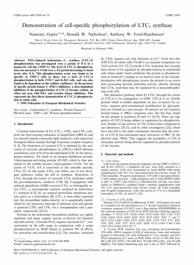
LTC4S was found to be directly phosphorylated in THP-1cells. The level of phosphorylation in THP-1 cells was seen toincrease by a factor of 3^5 fold as a result of PMA activationfor 4 h in comparison to the DMSO control, as measured byautoradiography (Fig. 1).
In an attempt to determine whether the observed phospho-rylation of LTC4S is a post-translational modi¢cation com-mon to other cell types other than the human monocytic-likeTHP-1 cell type, recombinant LTC4S was transfected into twoother cell types, namely COS-7, and K-562. Transfected cellswere then subjected to metabolic labeling with[32P]orthophosphate, activation by PMA, followed by immu-noprecipitation with anti-LTC4S antibody and detection bySDS-PAGE-autoradiography. Transfections into both celllines were successful in view of the Western blotting resultsin the presence of LTC4S DNA (Fig. 1). Both of these celllines were found to lack endogenous LTC4S as detected byWestern blotting of the mock transfections (data not shown).As can be seen by the autoradiography results, LTC4S wasnot phosphorylated in COS-7 cells nor in K-562 cells althoughLTC4S protein was present. In contrast, endogenous LTC4Sin THP-1 cells was detected at the protein level and was foundto be phosphorylated (Fig. 1). This is indicative of the factthat phorbol-ester induced PKC activity requires downstreame¡ectors leading to the phosphorylation of LTC4S in THP-1cells, which are absent in COS-7 and K-562 cell lines butpresent in THP-1 cells. Therefore, phosphorylation of LTC4Swas observed only in THP-1 cells and thus was found to becell-speci¢c, requiring speci¢c e¡ector(s) (kinases and phos-phatases) which impart the cell-speci¢city. Since phosphoryla-tion of LTC4S was only evident in THP-1 cells, all furtherstudies were conducted using this system.
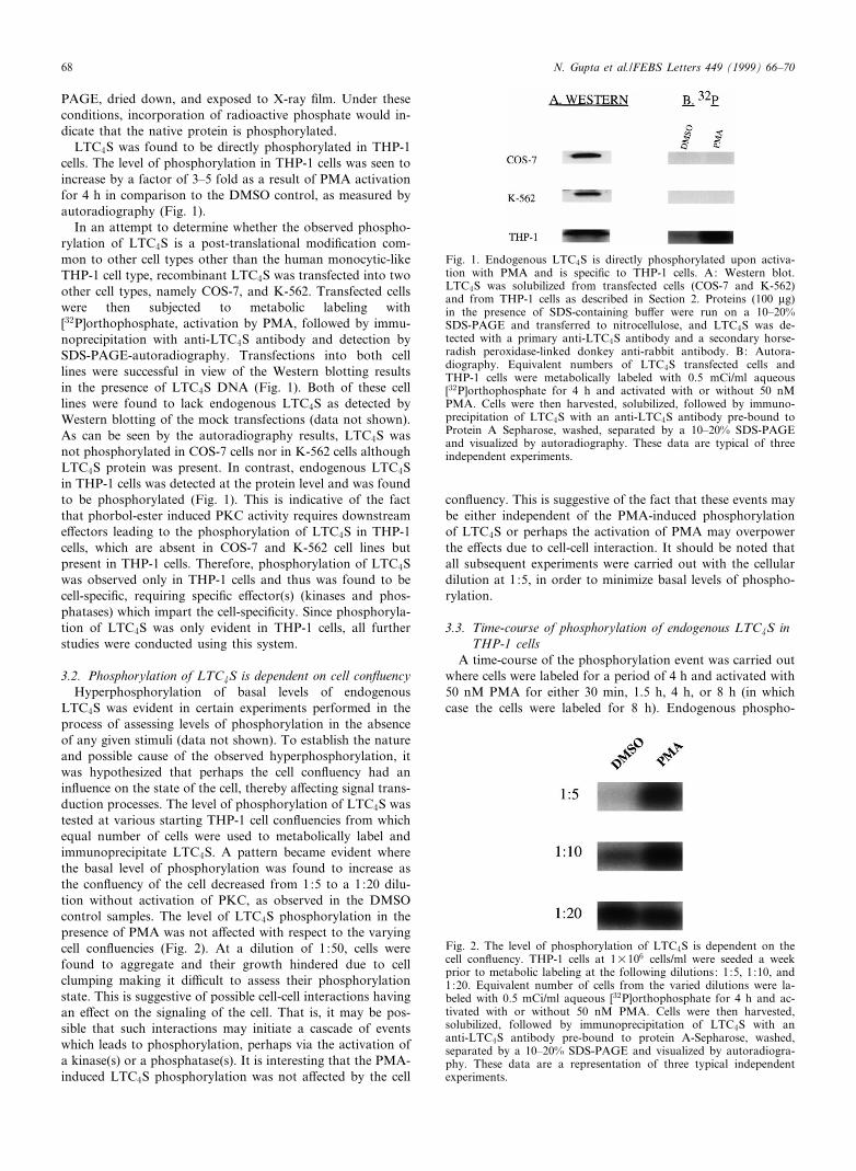
3.2. Phosphorylation of LTC4S is dependent on cell con£uencyHyperphosphorylation of basal levels of endogenous
LTC4S was evident in certain experiments performed in theprocess of assessing levels of phosphorylation in the absenceof any given stimuli (data not shown). To establish the natureand possible cause of the observed hyperphosphorylation, itwas hypothesized that perhaps the cell con£uency had anin£uence on the state of the cell, thereby a¡ecting signal trans-duction processes. The level of phosphorylation of LTC4S wastested at various starting THP-1 cell con£uencies from whichequal number of cells were used to metabolically label andimmunoprecipitate LTC4S. A pattern became evident wherethe basal level of phosphorylation was found to increase asthe con£uency of the cell decreased from 1:5 to a 1:20 dilu-tion without activation of PKC, as observed in the DMSOcontrol samples. The level of LTC4S phosphorylation in thepresence of PMA was not a¡ected with respect to the varyingcell con£uencies (Fig. 2). At a dilution of 1:50, cells werefound to aggregate and their growth hindered due to cellclumping making it di¤cult to assess their phosphorylationstate. This is suggestive of possible cell-cell interactions havingan e¡ect on the signaling of the cell. That is, it may be pos-sible that such interactions may initiate a cascade of eventswhich leads to phosphorylation, perhaps via the activation ofa kinase(s) or a phosphatase(s). It is interesting that the PMA-induced LTC4S phosphorylation was not a¡ected by the cell
con£uency. This is suggestive of the fact that these events maybe either independent of the PMA-induced phosphorylationof LTC4S or perhaps the activation of PMA may overpowerthe e¡ects due to cell-cell interaction. It should be noted thatall subsequent experiments were carried out with the cellulardilution at 1:5, in order to minimize basal levels of phospho-rylation.
3.3. Time-course of phosphorylation of endogenous LTC4S inTHP-1 cells
A time-course of the phosphorylation event was carried outwhere cells were labeled for a period of 4 h and activated with50 nM PMA for either 30 min, 1.5 h, 4 h, or 8 h (in whichcase the cells were labeled for 8 h). Endogenous phospho-
FEBS 21865 12-4-99
Fig. 2. The level of phosphorylation of LTC4S is dependent on thecell con£uency. THP-1 cells at 1U106 cells/ml were seeded a weekprior to metabolic labeling at the following dilutions: 1:5, 1:10, and1:20. Equivalent number of cells from the varied dilutions were la-beled with 0.5 mCi/ml aqueous [32P]orthophosphate for 4 h and ac-tivated with or without 50 nM PMA. Cells were then harvested,solubilized, followed by immunoprecipitation of LTC4S with ananti-LTC4S antibody pre-bound to protein A-Sepharose, washed,separated by a 10^20% SDS-PAGE and visualized by autoradiogra-phy. These data are a representation of three typical independentexperiments.
Fig. 1. Endogenous LTC4S is directly phosphorylated upon activa-tion with PMA and is speci¢c to THP-1 cells. A: Western blot.LTC4S was solubilized from transfected cells (COS-7 and K-562)and from THP-1 cells as described in Section 2. Proteins (100 Wg)in the presence of SDS-containing bu¡er were run on a 10^20%SDS-PAGE and transferred to nitrocellulose, and LTC4S was de-tected with a primary anti-LTC4S antibody and a secondary horse-radish peroxidase-linked donkey anti-rabbit antibody. B: Autora-diography. Equivalent numbers of LTC4S transfected cells andTHP-1 cells were metabolically labeled with 0.5 mCi/ml aqueous[32P]orthophosphate for 4 h and activated with or without 50 nMPMA. Cells were then harvested, solubilized, followed by immuno-precipitation of LTC4S with an anti-LTC4S antibody pre-bound toProtein A Sepharose, washed, separated by a 10^20% SDS-PAGEand visualized by autoradiography. These data are typical of threeindependent experiments.
N. Gupta et al./FEBS Letters 449 (1999) 66^7068

rylation was found to peak at 4 h and then decrease to basallevels for both PMA activated cells and DMSO controls (Fig.3).
Of interest was the trend observed with the DMSO vehiclecontrols where the level of phosphorylation was also seen toincrease up to 4 h and then return to basal levels at 8 h. Thiscon¢rms that the observed phosphorylation event was notonly a consequence of induction by PMA but is also signi¢-cant physiologically under no stimulus and follows a partic-ular kinetic pro¢le. The same trend was evident in the absenceof DMSO thereby con¢rming that the trend was not an arti-fact due to DMSO (data not shown).
3.4. Inhibition of LTC4S phosphorylation by PKC-speci¢cinhibitors
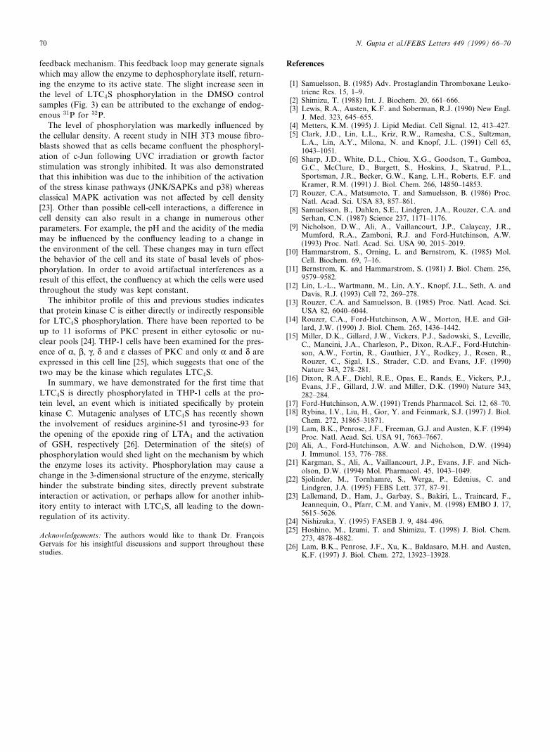
In order to shed light on the protein kinase responsible forthe phosphorylation of LTC4S, inhibitors of PKC, PKA, andof tyrosine kinase were tested for their ability to block theobserved PMA-mediated phosphorylation. Staurosporine, bis-indolylmaleimide, and GF109203X, all speci¢c inhibitors ofPKC, dose-dependently attenuated the phosphorylation ofLTC4S evoked by PMA (Fig. 4). The e¡ects of staurosporinewere apparent at a concentration of 0.1 WM, bisindolylmale-imide was e¡ective at 1 WM, and GF109203X at 7.5 WM.These PKC inhibitors were found to be capable of attenuatingthe level of phosphorylation to basal levels and below at high-er inhibitor concentrations. In contrast, inhibitors of tyrosinekinase, namely, herbimycin and tyrophostin and of PKA,KT5270, were found to produce no inhibition. This evidencesuggests a PKC-mediated cascade of events either directly orvia other proteins, leading to the phosphorylation of LTC4S,and is in accordance with previous studies on PMA-mediatedattenuation of LTC4S activity being restored upon treatmentwith inhibitors of PKC.
4. Discussion
Several independent studies have demonstrated downregu-lation of LTC4S enzymatic activity by phosphorylation, ini-tiated by a cascade of kinases, in particular PKC [20^22].LTC4S activity measured in neutrophilic [21], and eosinophilic[20] cell lines and in human granulocytes [22], challenged withPMA have demonstrated that the activity was inhibited result-ing in an attenuation of cysteinyl LTs without a¡ecting non-cysteinyl LT biosynthesis. Moreover, the attenuation of theactivity was completely prevented in the presence of inhibitors
of PKC but not by inhibitors of tyrosine kinase nor proteinkinase A. The presence of two putative PKC consensus siteson LTC4S was another indicator for the potential for LTC4Sphosphoregulation [9,19]. Prior to the present study, there wasno evidence of a direct phosphorylation event upon LTC4S.This study clearly indicates that LTC4S is phosphorylated atthe protein level in THP-1 cells, not only in response to PMAactivation, but also at a lower basal level which may be at-tributed to a pool of LTC4S which consists of both phos-phorylated and non-phosphorylated forms of the enzyme.Therefore in our study, the levels of phosphorylation werecompared with basal phosphorylation levels.
The lack of LTC4S phosphorylation in COS-7 and K-562cells indicates THP-1 cell-speci¢city. Numerous reasons mayexplain this observed speci¢city, including the lack of an ap-propriate target for PMA, downstream e¡ectors, and the lo-calization of LTC4S in these cells as compared to THP-1 cells.
An interesting observation was made from the time-courseof activation which revealed that the phosphorylation eventfollowed a particular kinetic pro¢le. The level of LTC4S phos-phorylation was seen to increase with time of PMA activationup to 4 h, after which the enzyme became dephosphorylatedto basal levels after 8 h of activation, suggesting a negative
FEBS 21865 12-4-99
Fig. 4. Phosphorylation of LTC4S is a PKC-mediated event. THP-1cells were grown to con£uency and labeled with 0.5 mCi/ml aqueous[32P]orthophosphate for a period of 4 h. During labeling, cells weretreated with or without 50 nM PMA in the presence or absence ofa range of kinase inhibitor concentrations. Staurosporine, 0.05^10 WM; bisindolylmaleimide, 0.5^10 WM; GF109203X, 0.5^10 WM;tyrophostin, 10 WM; herbimycin, 10 WM; and KT5720, 1 WM. Cellswere then harvested, solubilized, followed by immunoprecipitationof LTC4S with an anti-LTC4S antibody pre-bound to proteinA-Sepharose, washed, separated by a 10^20% SDS-PAGE and vis-ualized by autoradiography. Represented data is one of three typicalindependent experiments.
Fig. 3. Time course of the phosphorylation of endogenous LTC4Sin THP-1 cells. Con£uent THP-1 cells were labeled for a period of4 h with 0.5 mCi/ml aqueous [32P]orthophosphate and activatedwith or without 50 nM PMA for either 30 min, 1.5 h, 4 h, or 8 h(in which case the cells were labeled for 8 h). Cells were then har-vested, solubilized, followed by immunoprecipitation of LTC4S withan anti-LTC4S antibody pre-bound to protein A-Sepharose, washed,separated by a 10^20% SDS-PAGE and visualized by autoradiogra-phy. Four independent experiments were performed and representedhere is one typical one.
N. Gupta et al./FEBS Letters 449 (1999) 66^70 69
feedback mechanism. This feedback loop may generate signalswhich may allow the enzyme to dephosphorylate itself, return-ing the enzyme to its active state. The slight increase seen inthe level of LTC4S phosphorylation in the DMSO controlsamples (Fig. 3) can be attributed to the exchange of endog-enous 31P for 32P.
The level of phosphorylation was markedly in£uenced bythe cellular density. A recent study in NIH 3T3 mouse ¢bro-blasts showed that as cells became con£uent the phosphoryl-ation of c-Jun following UVC irradiation or growth factorstimulation was strongly inhibited. It was also demonstratedthat this inhibition was due to the inhibition of the activationof the stress kinase pathways (JNK/SAPKs and p38) whereasclassical MAPK activation was not a¡ected by cell density[23]. Other than possible cell-cell interactions, a di¡erence incell density can also result in a change in numerous otherparameters. For example, the pH and the acidity of the mediamay be in£uenced by the con£uency leading to a change inthe environment of the cell. These changes may in turn e¡ectthe behavior of the cell and its state of basal levels of phos-phorylation. In order to avoid artifactual interferences as aresult of this e¡ect, the con£uency at which the cells were usedthroughout the study was kept constant.
The inhibitor pro¢le of this and previous studies indicatesthat protein kinase C is either directly or indirectly responsiblefor LTC4S phosphorylation. There have been reported to beup to 11 isoforms of PKC present in either cytosolic or nu-clear pools [24]. THP-1 cells have been examined for the pres-ence of K, L, Q, N and O classes of PKC and only K and N areexpressed in this cell line [25], which suggests that one of thetwo may be the kinase which regulates LTC4S.
In summary, we have demonstrated for the ¢rst time thatLTC4S is directly phosphorylated in THP-1 cells at the pro-tein level, an event which is initiated speci¢cally by proteinkinase C. Mutagenic analyses of LTC4S has recently shownthe involvement of residues arginine-51 and tyrosine-93 forthe opening of the epoxide ring of LTA4 and the activationof GSH, respectively [26]. Determination of the site(s) ofphosphorylation would shed light on the mechanism by whichthe enzyme loses its activity. Phosphorylation may cause achange in the 3-dimensional structure of the enzyme, stericallyhinder the substrate binding sites, directly prevent substrateinteraction or activation, or perhaps allow for another inhib-itory entity to interact with LTC4S, all leading to the down-regulation of its activity.
Acknowledgements: The authors would like to thank Dr. Franc°oisGervais for his insightful discussions and support throughout thesestudies.
References
[1] Samuelsson, B. (1985) Adv. Prostaglandin Thromboxane Leuko-triene Res. 15, 1^9.
[2] Shimizu, T. (1988) Int. J. Biochem. 20, 661^666.[3] Lewis, R.A., Austen, K.F. and Soberman, R.J. (1990) New Engl.
J. Med. 323, 645^655.[4] Metters, K.M. (1995) J. Lipid Mediat. Cell Signal. 12, 413^427.[5] Clark, J.D., Lin, L.L., Kriz, R.W., Ramesha, C.S., Sultzman,
L.A., Lin, A.Y., Milona, N. and Knopf, J.L. (1991) Cell 65,1043^1051.
[6] Sharp, J.D., White, D.L., Chiou, X.G., Goodson, T., Gamboa,G.C., McClure, D., Burgett, S., Hoskins, J., Skatrud, P.L.,Sportsman, J.R., Becker, G.W., Kang, L.H., Roberts, E.F. andKramer, R.M. (1991) J. Biol. Chem. 266, 14850^14853.
[7] Rouzer, C.A., Matsumoto, T. and Samuelsson, B. (1986) Proc.Natl. Acad. Sci. USA 83, 857^861.
[8] Samuelsson, B., Dahlen, S.E., Lindgren, J.A., Rouzer, C.A. andSerhan, C.N. (1987) Science 237, 1171^1176.
[9] Nicholson, D.W., Ali, A., Vaillancourt, J.P., Calaycay, J.R.,Mumford, R.A., Zamboni, R.J. and Ford-Hutchinson, A.W.(1993) Proc. Natl. Acad. Sci. USA 90, 2015^2019.
[10] Hammarstrom, S., Orning, L. and Bernstrom, K. (1985) Mol.Cell. Biochem. 69, 7^16.
[11] Bernstrom, K. and Hammarstrom, S. (1981) J. Biol. Chem. 256,9579^9582.
[12] Lin, L.-L., Wartmann, M., Lin, A.Y., Knopf, J.L., Seth, A. andDavis, R.J. (1993) Cell 72, 269^278.
[13] Rouzer, C.A. and Samuelsson, B. (1985) Proc. Natl. Acad. Sci.USA 82, 6040^6044.
[14] Rouzer, C.A., Ford-Hutchinson, A.W., Morton, H.E. and Gil-lard, J.W. (1990) J. Biol. Chem. 265, 1436^1442.
[15] Miller, D.K., Gillard, J.W., Vickers, P.J., Sadowski, S., Leveille,C., Mancini, J.A., Charleson, P., Dixon, R.A.F., Ford-Hutchin-son, A.W., Fortin, R., Gauthier, J.Y., Rodkey, J., Rosen, R.,Rouzer, C., Sigal, I.S., Strader, C.D. and Evans, J.F. (1990)Nature 343, 278^281.
[16] Dixon, R.A.F., Diehl, R.E., Opas, E., Rands, E., Vickers, P.J.,Evans, J.F., Gillard, J.W. and Miller, D.K. (1990) Nature 343,282^284.
[17] Ford-Hutchinson, A.W. (1991) Trends Pharmacol. Sci. 12, 68^70.[18] Rybina, I.V., Liu, H., Gor, Y. and Feinmark, S.J. (1997) J. Biol.
Chem. 272, 31865^31871.[19] Lam, B.K., Penrose, J.F., Freeman, G.J. and Austen, K.F. (1994)
Proc. Natl. Acad. Sci. USA 91, 7663^7667.[20] Ali, A., Ford-Hutchinson, A.W. and Nicholson, D.W. (1994)
J. Immunol. 153, 776^788.[21] Kargman, S., Ali, A., Vaillancourt, J.P., Evans, J.F. and Nich-
olson, D.W. (1994) Mol. Pharmacol. 45, 1043^1049.[22] Sjolinder, M., Tornhamre, S., Werga, P., Edenius, C. and
Lindgren, J.A. (1995) FEBS Lett. 377, 87^91.[23] Lallemand, D., Ham, J., Garbay, S., Bakiri, L., Traincard, F.,
Jeannequin, O., Pfarr, C.M. and Yaniv, M. (1998) EMBO J. 17,5615^5626.
[24] Nishizuka, Y. (1995) FASEB J. 9, 484^496.[25] Hoshino, M., Izumi, T. and Shimizu, T. (1998) J. Biol. Chem.
273, 4878^4882.[26] Lam, B.K., Penrose, J.F., Xu, K., Baldasaro, M.H. and Austen,
K.F. (1997) J. Biol. Chem. 272, 13923^13928.
FEBS 21865 12-4-99
N. Gupta et al./FEBS Letters 449 (1999) 66^7070