delayed acute capture myopathy in three roe deer …...capítulo 8 142 capture myopathy is a...
TRANSCRIPT

Capítulo 8
Delayed acute capture myopathy in three roe deer (Capreolus capreolus)
Capítulo basado en el artículo: Delayed acute capture myopathy in three roe deer (Capreolus capreolus). (2002). Montané, J., Marco, I., Manteca, X., López, J. and Lavín, S. Journal of Veterinary Medicine. Series A, 49: 93-98.

Capítulo 8
140
Abstract Delayed-acute capture myopathy is the term used to describe the clinical syndrome observed in three roe deer (Capreolus capreolus) captured by drive-nets and transported to an enclosure for scientific purposes. The animals died 48 hours, 60 hours and 8 days after being captured. The simultaneous deaths coincided with a previous episode of deliberate human disturbance. The histopathological findings were indicative of acute myopathy and myoglobinemic nephrosis. These could be related to an ataxic myoglobinuric syndrome brought on by capture and transport operations. The lack of clinical signs and negative prognosis indicators in the period between capture and just before death, the absence of gross muscular lesions in the animal that died after 8 days post-capture, the simultaneous deaths of animals captured at different times and the evidence of deliberate human disturbance in the enclosure were suggestive of death triggered by a second stress episode.
Capture myopathy in roe deer
141
Introduction Capture myopathy is a syndrome that occurs in wild (free-ranging and captive) mammals and birds. It is associated with the stress of capture, restraint and transportation. In ungulates the syndrome is characterised clinically by depression, muscular stiffness, lack of coordination, paralysis, metabolic acidosis and death. Pathologically, capture myopathy resembles the myodegenerative disorders of domestic cattle, sheep, horse and swine (Chalmers and Barrett, 1982). The classification of capture myopathy into peracute, acute, subacute and chronic is somewhat arbitrary and different authors have adopted different classification systems (Harthoorn, 1976; Chalmers and Barrett, 1982; Shepherd 1984). Spraker (1993) describes four clinical syndromes in animals: capture shock, ataxic myoglobinuric, ruptured muscle and delayed-peracute syndrome. The ataxic myoglobinuric syndrome probably occurs most commonly. It can be seen several hours to several days post-capture. Clinical signs include ataxia, torticollis and myoglobinuria, and vary from mild to severe. At necropsy there are renal and skeletal muscle lesions. The kidneys are swollen and dark. The urinary bladder is empty or contains a small amount of brownish urine. Multifocal, pale, soft, dry areas, accentuated by small white foci in a linear pattern are usually found within the cervical and lumbar muscles and in the flexor and extensor muscles of the limbs. These lesions are subtle in animals that die one to two days post-capture but pronounced in animals that survive longer. The delayed-peracute syndrome is usually seen in animals that have been in captivity for at least 24 hours. These animals appear normal while undisturbed, but if recaptured or suddenly stressed they die within several minutes. The syndrome pathogenesis is the consequence of a surge of epinephrine, which acts on altered membranes (due to hyperkalemia) of heart and skeletal muscle cells, resulting in ventricular fibrillation. Usually, there are no lesions or only a few pale foci are found within the skeletal muscle at necropsy. Histological lesions are characterised by a mild to moderate rhabdomyolysis throughout the skeletal muscle, especially in the hind limbs.

Capítulo 8
142
Capture myopathy is a well-known pathologic condition in wild ungulates, but only a Letter to the Editor (Fairlie, 1964) has been published in relation to a myopathy suggestive of capture myopathy in a roe deer (Capreolus capreolus).
Materials and Methods In March 2000, three free-ranging female roe deer were captured by means of drive-nets in the National Game Reserve of Alt Pallars Aran (47º22’ N 3º48’ E, north-eastern Spain), translocated into transport boxes and then placed in captivity for scientific purposes. Drive-trapping was conducted by a line of beaters, each one within sight of the next, and went on for 30 minutes. The animals were initially restrained by using the net to wrap them in, blindfolded, their legs restrained and finally introduced in a transport net sack (Ziboni Ornitecnica, Bergamo, Italy), where they were maintained for approximately one hour before being introduced into the transport boxes. The enclosure where the animals were kept in captivity was located in the Cadí-Moixeró Natural Park (42º20’ N 1º50’ E, northeastern Spain) with an area of 1500m2 and had a forest zone. The behaviour of the animals was monitored every day at dawn and dusk for two-hour periods during captivity. No clinical signs of capture myopathy or abnormal behaviour were observed during the captivity period before death. The animals died 48 hours (animal no.1), 72 hours (animal no.2) and eight days (animal no.3) after being captured. Evidence of deliberate human disturbance was found in the enclosure, indicative of a second stress episode. It probably consisted of an intense pursuit inside the enclosure and the capture of one animal that disappeared and thus it is not included in this report (initially there were four animals in the enclosure). The time-to-death from this disturbance was probably of 15, 41 and 21 hours respectively. Two blood samples had been taken 6 hours (2 animals) and 9 hours (1 animal, no. 3) apart, at capture and after a three-hour road journey, as part of a research study. The blood was collected from the jugular vein and placed in a tube with EDTA K3 as an anticoagulant and used for haematological analyses. The remainder was placed in a plain tube, and used for biochemical purposes, after being allowed to clot at room temperature. Serum was kept at –18ºC until biochemical analyses were carried out. Haematological examinations were performed by means of a semiautomatic analyser
(Sysmex F-800, Toa Medical Electronics Co. Ltd., Japan), except for packed cell

Capture myopathy in roe deer
143
volume (PCV), which was determined with a haematocrit centrifuge (Micro-Haematocrit Centrifuge, Hawksley, Lancing, UK). Biochemical analyses were
performed by means of an automated analyser (COBAS MIRA, Roche, Nutley, NJ, USA), except for sodium and potassium concentrations, which were measured by
flame photometry (Corning 410C, Corning Medical Medfield, USA), and serum cortisol, which was determined using an ELISA commercial kit (DRG Cortisol EIA-1887, DRG Diagnostics, Germany). Statistical analyses were performed using a
statistics software package (SPSS-PC, SPSS Inc., Chicago, Illinois, USA). A Wilcoxon test was used to compare haematological and biochemical values at capture and after transport. Body temperature was recorded every minute after capture and during transportation
using Mätman datalogger temperature probes (Chipsobits Eltex AB, Sweden). Heart
rate was continuously recorded using Polar Vantage NV non-invasive heart rate monitors (Polar Electro Oy, Finland). These measurements were also taken as part of the research study. A necropsy of the roe deer was performed. Samples were taken from the heart, kidneys and skeletal muscles and were fixed in 10% buffered formalin for routine histopathological studies.
Results Clinical and clinicopathological findings No clinical signs of capture myopathy or abnormal behaviour were observed during the captivity period before death. Clinical signs just before death included depression, inability to rise or stand and unresponsiveness to human presence. Haematological (Table 8.1) and biochemical (Table 8.2) data did not show any statistical difference at capture and after transport. Haematological values (except for elevated eosinophils), serum glucose, cholesterol and sodium concentrations were within the reference values reported by Jaouen (1981) for female roe deer captured by drive-nets. Serum urea was almost two-fold that reported by Jaouen (1981) (6.97

Capítulo 8
144
mmol/L ± SD 1.22 mmol/L) and the concentration of potassium was also higher than
that reported by the same author (5.27 mmol/L ± SD 0.81 mmol/L).
Table 8.1. Haematological values of the three roe deer at capture and after transport. No statistical differences were found between samples obtained at capture and after transport.
SEM, standard error of the mean; RBC, red blood cells; PCV, packed cell volume; MCV, mean cell volume; MCHC, mean cell haemoglobin concentration; MCH, mean cell haemoglobin; WBC, white blood cells.
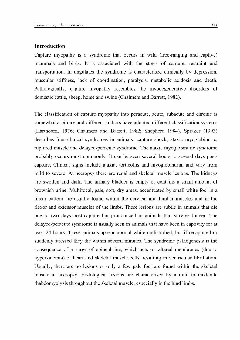
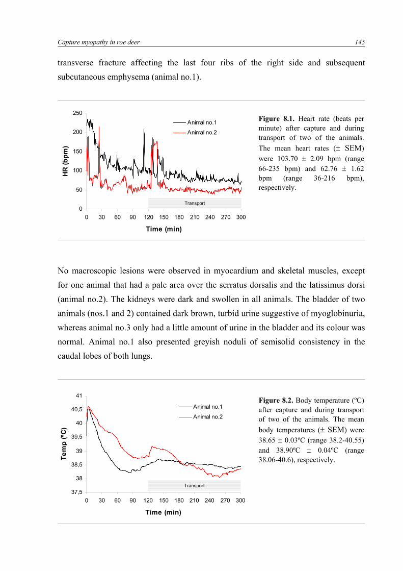
Heart rate and body temperature after capture and during transportation of two of the animals are shown in Figures 8.1 and 8.2, respectively. The heart rate decreased to the normal physiological values (70-80 beats per minute -bpm-) reported by Nielsen (1999) for deer in three hours (animal no.1) and less than 30 minutes (animal no. 2). Schober and Wagner (1993) reported heart rate values obtained by telemetry of below 90 bpm in roe deer not submitted to exertion. Rectal temperature reached the normal physiological value (38.4ºC) (Nielsen, 1999) after one and three hours, respectively. Gross pathological findings The roe deer were in good condition. Small superficial lacerations were found in the skin of their faces, on the upper ciliar arc, due to them pressing their head hard against the enclosure fence as an attempt to escape when disturbed. One animal presented a
At capture After transport
Animal no. 1 2 3 Mean (± SEM) 1 2 3 Mean (± SEM)
RBC (x 1012/L) 10.19 9.13 11.16 10.16 (± 0.59) 8.45 12.75 9.72 10.31 (± 1.28)
Haemoglobin (g/L) 145.0 196.0 164.0 168.30 (± 14.90) 140.0 184.0 140.0 154.70 (± 14.70)
PCV (L/L) 0.41 0.57 0.48 0.47 (± 0.05) 0.41 0.53 0.40 0.45 (± 0.42)
MCV (fl) 40.20 62.40 41.40 48.00 (± 7.21) 48.50 41.60 41.10 43.73 (± 2.39)
MCHC (g/dL) 35.40 34.40 34.20 34.67 (± 0.37) 34.20 34.70 35.00 34.63 (± 0.23)
MCH (pg) 14.20 21.50 14.70 16.80 (± 2.35) 16.60 14.40 14.40 15.13 (± 0.73)
WBC (x 109/L) 3.20 4.00 2.30 3.17 (± 0.49) 5.00 9.40 6.00 6.80 (± 1.33)
Lymphocytes (x 109/L) 1.50 1.68 1.15 1.44 (± 0.16) 1.10 0.19 0.48 0.59 (± 0.27)
Neutrophils (x 109/L) 1.34 1.80 0.83 1.32 (± 0.28) 3.80 9.02 5.34 6.05 (± 1.55)
Monocytes (x 109/L) 0.03 0.00 0.02 0.02 (± 0.01) 0.10 0.00 0.12 0.07 (± 0.04)
Eosinophils (x 109/L) 0.32 0.52 0.30 0.38 (± 0.07) 3.80 0.19 0.06 1.35 (± 1.22)
Basophils (x 109/L) 0.00 0.00 0.00 0.00 (± 0.00) 0.00 0.00 0.00 0.00 (± 0.00)
Capture myopathy in roe deer
145
transverse fracture affecting the last four ribs of the right side and subsequent subcutaneous emphysema (animal no.1).
No macroscopic lesions were observed in myocardium and skeletal muscles, except for one animal that had a pale area over the serratus dorsalis and the latissimus dorsi (animal no.2). The kidneys were dark and swollen in all animals. The bladder of two animals (nos.1 and 2) contained dark brown, turbid urine suggestive of myoglobinuria, whereas animal no.3 only had a little amount of urine in the bladder and its colour was normal. Animal no.1 also presented greyish noduli of semisolid consistency in the caudal lobes of both lungs.
0
50
100
150
200
250
0 30 60 90 120 150 180 210 240 270 300
Time (min)
HR (b
pm)
Animal no.1
Animal no.2
Figure 8.1. Heart rate (beats perminute) after capture and duringtransport of two of the animals.The mean heart rates (± SEM)were 103.70 ± 2.09 bpm (range66-235 bpm) and 62.76 ± 1.62bpm (range 36-216 bpm),respectively.
Transport
37,5
38
38,5
39
39,5
40
40,5
41
0 30 60 90 120 150 180 210 240 270 300
Time (min)
Tem
p (ºC
)
Animal no.1
Animal no.2
Figure 8.2. Body temperature (ºC)after capture and during transportof two of the animals. The meanbody temperatures (± SEM) were38.65 ± 0.03ºC (range 38.2-40.55)and 38.90ºC ± 0.04ºC (range38.06-40.6), respectively.
Transport

Capítulo 8
146
Histopathological findings The muscles that appeared grossly normal showed microscopic evidence of mild (animal no.3) to severe (2 animals, those with myoglobinuria) degeneration and fragmentation of skeletal muscle fibres (Figure 8.3). There were fibres with central nuclei, marked exudation of eosinophilic material, probably proteinaceous, into the interstitial space, and the presence of some macrophages and polymorphonuclear neutrophils between the degenerated muscular fibres. All three animals also presented parasitic cysts in some muscular fibres compatible with parasites of the genus Sarcocystis. Those animals with myoglobinuria had myocardic fibres that showed hyalinisation and pycnotic nuclei multifocally. In the kidneys of all three animals, some tubular cells in the cortex contained different amounts of a yellow-brown intracytoplasmic pigment (Figure 8.4). Multifocal renal tubular necrosis in the cortex and the presence of degenerated tubular cells in the medulla and the cortex were observed. Animal no.1 presented large amounts of parasitic structures in pulmonary alveoli and bronchioli associated with fibrosis of septa and large amounts of macrophages, lymphocytes and some multinucleated giant cells. A diagnosis of chronic parasitic interstitial pneumonia was made.
Discussion Meneguz et al. (1996) reported hyperthermia (> 41ºC), tachycardia (> 200 beats per minute), hypoglycaemia (< 3.64 mmol/L), persistent relative polycythaemia (red blood cells > 13 x1012/L, haemoglobin > 200 g/L; packed cell volume > 0.55 L/L), leucopenia (< 2.5 x10⁹ leukocytes/L) and neutropenia (< 1.0 x10⁹ neutrophils/L) as negative prognosis indicators in roe deer at 3-4 hours post-capture. In this case, all three animals had a heart rate below 200 beats per minute at 25 minutes post-capture and their rectal temperature never reached 41ºC. Neither haematological values nor serum glucose at capture and after transport were indicative of a negative evolution of the animals.

Capture myopathy in roe deer
147
Figure 8.3. Photomicrograph of a muscle specimen, showing hyalinisation and fragmentation of muscular fibres. Note the inflammatory infiltrate. Haematoxylin and eosin. Bar = 72 µm.
Figure 8.4. Photomicrograph of a kidney specimen, showing proximal tubularcells containing an intracytoplasmic pigment (probably myoglobin, see arrows)and necrosis of tubular cells. Haematoxylin and eosin. Bar = 29 µm.

Capítulo 8
148
Table 8.2. Serum biochemical values of the three animals at capture and after transport. No statistical differences were found between samples obtained at capture and after transport.
SEM, standard error of the mean; ALT, alanine aminotransferase; LDH, lactate dehydrogenase; CK, creatine kinase; AST, aspartate aminotransferase; ALP, alkaline phosphatase. The differences in heart rate observed in Figure 8.1 could be due to the chronic parasitic interstitial pneumonia and pneumothorax that animal no.1 was suffering from, which could increase its mean heart rate and delay the return to heart rate basal values. Alternatively, it could only reflect the inter-individual differences existing in heart rate (Hopster, 1998). Raised heart rates after capture are due to physical activity and the acute stress response (Broom and Johnson, 1993). Kock et al. (1987a) reported that temperature in bighorn sheep (Ovis canadensis) classified as being 'normal' (40.5ºC) after capture was significantly lower than that of bighorn sheep that died of capture myopathy (41.9ºC). In our study, all animals were
At capture After transport
Animal no. 1 2 3 Mean (± SEM) 1 2 3 Mean (± SEM)
Glucose (mmol/L) 12.77 17.58 9.52 13.29 (± 2.34) 6.83 11.31 4.59 7.57 (± 1.97)
Urea (mmol/L) 11.39 12.24 16.32 13.32 (± 1.52) 11.90 12.41 14.79 13.03 (± 0.89)
Creatinine (µmol/L) 173.3 186.5 244.0 201.55 (± 22.1) 172.4 141.4 181.2 165.30 (±12.38)
Lactate (mmol/L) 19.69 23.81 20.24 21.25 (± 1.29) 4.72 4.29 3.49 4.17 (± 0.36)
Total bilirubin (µmol/L) 5.13 1.71 0.00 2.22 (± 1.54) 11.97 15.39 23.94 17.10 (± 3.60)
Cholesterol (mmol/L) 1.33 1.35 1.46 1.38 (± 0.04) 1.38 1.07 1.43 1.29 (± 0.11)
Triglycerides (mmol/L) 0.46 1.18 0.89 0.84 (± 0.21) 0.36 0.22 0.21 0.26 (± 0.05)
ALT (IU/L) 63.0 72.0 63.0 66.00 (± 3.00) 111.0 84.0 103.0 99.33 (± 8.01)
LDH (IU/L) 2040 1833 2402 2091.67 (± 166.28) 5350 1975 5071 4132 (± 1081.50)
CK (IU/L) 6165 1573 2939 3559 (± 1361.36) 50393 13918 29841 31384 (± 10557.65)
AST (IU/L) 571 181 527 426.33 (± 123.32) 2310 573 1348 1410.33 (± 502.40)
ALP (IU/L) 225 165 225 205.00 (± 20.00) 137 68 116 107.00 (± 20.42)
Chloride (mmol/L) 93.80 89.40 91.70 91.63 (± 1.27) 102.9 101.8 100.3 101.67 (± 0.75)
Sodium (mmol/L) 146 151 149 148.67 (± 1.45) 156 167 156 159.67 (± 3.67)
Potassium (mmol/L) 7.10 8.20 7.70 7.67 (± 0.32) 5.50 7.40 7.10 6.67 (± 0.59)
Total Protein (g/L) 82.0 82.0 62.0 75.30 (± 6.7) 82.0 96.0 79.0 85.70 (± 5.2)
Cortisol (nmol/L) 143.8 151.5 207.0 167.45 (± 19.90) 164.2 181.3 103.5 149.68 (± 23.62)

Capture myopathy in roe deer
149
below 40.5ºC for the entire period of monitoring, except for five minutes during which animals 1 and 2 were between 40.5ºC and 40.6ºC. Increases in serum creatine kinase (CK), aspartate aminotransferase (AST) and lactate dehydrogenase (LDH) are usually due to an increase in muscular cell permeability and muscular cell damage resulting from physical stress (Duncan and Prasse, 1986). Some authors think that alanine aminotransferase (ALT) measurement could be useful in cases of capture myopathy (Vassart et al., 1992). Reference values of these parameters have not been found for roe deer, but in comparison with those obtained from other roe deer that were also submitted to physical capture and road transport (see Chapter 6), no differences were found. However, the elevation of serum CK activity indicated that muscular damage had occurred during capture and transportation. Increased serum urea values were probably caused by prolonged exertion resulting from drive-trapping. Severe, prolonged exercise in man causes an increase in serum urea and creatinine of about 60% (Finco, 1997). Sealander et al. (1975) also found increased serum urea values in white-tailed deer (Odocoileus virginianus) physically restrained. McAllum (1985) found that blood urea levels increased in myophatic red deer (Cervus elaphus). However, in our study no differences were found between samples collected at capture and after transport. High serum lactate concentrations at capture were probably due to the previous intense physical exercise (Kaneko, 1997a) and to the activation of the sympathetic-adrenomedullary system (Verde and Gascón, 1987). When anaerobic metabolism changes to aerobic, the lactate concentration decrease, as occurred in the 'after transport' blood sample. Vigorous short-term exercise at high intensity and muscle necrosis result in hyperkalemia (Carlson, 1997). In this case, the increases in potassium concentrations were also caused by exertion, as it tended to decrease when the animals were at rest. McAllum (1985) recorded a potassium level of nearly four times normal in myopathic red deer in the first 30 minutes after capture. This was not the case comparing present data with those of Jaouen (1981).

Capítulo 8
150
The use of blood cortisol levels as indicators of stress has been controversial, although some investigators feel that it is potentially the best single indicator of stress in different wildlife species (Franzmann et al., 1975; Spraker, 1982; Morton et al., 1995). However, Kock et al. (1987a) did not find any statistical difference in cortisol concentrations between normal bighorn sheep and those that suffered from capture myopathy. No serum cortisol concentrations in roe deer have been reported in the literature for comparison with our results. Gross and histopathological findings were indicative of acute myopathy and myoglobinemic nephrosis. These could be related to an ataxic myoglobinuric syndrome resulting from capture and transport operations. However, the lack of clinical signs and negative prognosis indicators in the period between capture and just before death, the absence of gross muscular lesions in the animal that died 8 days post-capture, the simultaneous deaths of animals captured at different times and the evidence of deliberate human disturbance in the enclosure suggest that death was triggered by a second stress episode. Unlike the delayed peracute syndrome, these animals did not die within several minutes of the second stress episode and this is why it seems improbable that they shared the same pathogenesis. These are the main reasons for using the term 'delayed acute capture myopathy syndrome'. Although, it is not found in the literature, it seems appropriate for describing the clinical syndrome observed.