deep convolutional neural networks for pan-specific
TRANSCRIPT
METHODOLOGY ARTICLE Open Access
Deep convolutional neural networks forpan-specific peptide-MHC class I bindingpredictionYoungmahn Han1,2 and Dongsup Kim1*
Abstract
Background: Computational scanning of peptide candidates that bind to a specific major histocompatibilitycomplex (MHC) can speed up the peptide-based vaccine development process and therefore various methods arebeing actively developed. Recently, machine-learning-based methods have generated successful results by traininglarge amounts of experimental data. However, many machine learning-based methods are generally less sensitivein recognizing locally-clustered interactions, which can synergistically stabilize peptide binding. Deep convolutionalneural network (DCNN) is a deep learning method inspired by visual recognition process of animal brain and it isknown to be able to capture meaningful local patterns from 2D images. Once the peptide-MHC interactions can beencoded into image-like array(ILA) data, DCNN can be employed to build a predictive model for peptide-MHCbinding prediction. In this study, we demonstrated that DCNN is able to not only reliably predict peptide-MHCbinding, but also sensitively detect locally-clustered interactions.
Results: Nonapeptide-HLA-A and -B binding data were encoded into ILA data. A DCNN, as a pan-specific predictionmodel, was trained on the ILA data. The DCNN showed higher performance than other prediction tools for the latestbenchmark datasets, which consist of 43 datasets for 15 HLA-A alleles and 25 datasets for 10 HLA-B alleles. In particular,the DCNN outperformed other tools for alleles belonging to the HLA-A3 supertype. The F1 scores of the DCNN were 0.86, 0.94, and 0.67 for HLA-A*31:01, HLA-A*03:01, and HLA-A*68:01 alleles, respectively, which were significantly higherthan those of other tools. We found that the DCNN was able to recognize locally-clustered interactions that couldsynergistically stabilize peptide binding. We developed ConvMHC, a web server to provide user-friendly web interfacesfor peptide-MHC class I binding predictions using the DCNN. ConvMHC web server can be accessible via http://jumong.kaist.ac.kr:8080/convmhc.
Conclusions: We developed a novel method for peptide-HLA-I binding predictions using DCNN trained on ILA datathat encode peptide binding data and demonstrated the reliable performance of the DCNN in nonapeptide bindingpredictions through the independent evaluation on the latest IEDB benchmark datasets. Our approaches can beapplied to characterize locally-clustered patterns in molecular interactions, such as protein/DNA, protein/RNA, anddrug/protein interactions.
Keywords: T cell epitope prediction, Peptide-based vaccine development, Peptide-MHC class I binding prediction,Deep learning, Convolutional neural network
* Correspondence: [email protected] of Bio and Brain Engineering, Korea Advanced Institute ofScience and Technology, Daejeon, Republic of KoreaFull list of author information is available at the end of the article
© The Author(s). 2017 Open Access This article is distributed under the terms of the Creative Commons Attribution 4.0International License (http://creativecommons.org/licenses/by/4.0/), which permits unrestricted use, distribution, andreproduction in any medium, provided you give appropriate credit to the original author(s) and the source, provide a link tothe Creative Commons license, and indicate if changes were made. The Creative Commons Public Domain Dedication waiver(http://creativecommons.org/publicdomain/zero/1.0/) applies to the data made available in this article, unless otherwise stated.
Han and Kim BMC Bioinformatics (2017) 18:585 DOI 10.1186/s12859-017-1997-x

BackgroundCytotoxic T lymphocytes (CTLs) play a key role in elimin-ating infections caused by intracellular pathogens. Sincethe CTL T-cell receptor recognizes foreign peptides incomplex with major histocompatibility complex (MHC)molecules on the infected cell surface, the response of thehost immune system to pathogens can be activated bypeptide binding of MHC molecules. Determining peptidesthat bind specific MHC molecules is important for identi-fying T cell epitopes and can facilitate the development ofpeptide-based vaccines and design of immunotherapies.However, experimental identification of peptide-MHC istime-consuming and laborious; computer-assisted bindingpredictions can be a cost-effective and practical alternativeand various methods have been developed [1].Sette and Sidney grouped HLA class I (HLA-I) mole-
cules into HLA supertypes using binding specificities char-acterized by the binding motifs of peptides [2]. Earlypeptide binding prediction methods were based on search-ing for allele-specific peptide binding motifs [3, 4]. As moreexperimental data became available, statistical methodshave been developed using positional scoring matrixes thatutilize amino acid occurrence frequencies at each position[5, 6]. Recently, more sophisticated machine learningmethods [7–9] have generated the most successful resultsby training large amount of experimental data derived frompublic databases, such as the Immune Epitope Database[10]. Allele-specific machine learning methods generallyachieve more accurate predictions as more data are learnedfor each HLA-I allele. A significant portion of currentlyavailable data was biased towards a limited number ofcommon alleles [11], and this makes it difficult to predictpeptide bindings for rare alleles. Sequence-based pan-specific methods have been proposed to overcome thisproblem and transfer the knowledge of other peptide-MHC binding information to improve the predictions forrare and even new alleles [12–14].The pan-specific methods utilize information on not only
the peptide sequence but also the MHC residues inpeptide-MHC contact sites derived from the crystal struc-tures of peptide-MHC complexes. The contact sites areclustered around the peptide anchor positions and thebinding pockets of MHC molecules [14–16]. The aminoacids of a peptide interact with MHC molecules in com-pensatory and synergistic manner rather than independ-ently [17–19]. A large-scale structural simulation study ofthe peptide-MHC binding landscapes revealed statisticallysignificant pairwise correlations in amino acid preferencesat different positions of a peptide [15]. Many machinelearning-based methods have a risk of learning the featuresassociated with amino acids of peptide and the HLA-I mol-ecule independently. Therefore, they could be less sensitivein recognizing the locally-clustered interactions, whichcould synergistically produce peptide-HLA-I binding.
Deep convolutional neural network (DCNN) is a branchof deep learning methods that extract and learn high-levelrepresentations (features or patterns) from the low-levelraw data through nonlinear transformations of multiplelayers. It was originally designed to process the spatial andtemporal data, particularly two-dimensional images withmultiple color channels. DCNNs are inspired by the ani-mal visual cortex and imitate cognitive functions of thecortex using three key concepts: capturing local motifs ofhighly connected pixels, invariance to the motif location,and hierarchical composition of the local motifs [20].DCNNs have achieved successful results in many objectrecognition and detection tasks [21–23]. Recent studieshave proposed bioinformatics applications of DCNNs in-cluding protein contact predictions [24] and small mol-ecule bioactivity predictions [25, 26].In this study, we propose a novel method for pan-
specific peptide-HLA-I binding prediction using DCNN.The peptide-HLA-I binding structure can be encoded intotwo-dimensional image-like array (ILA) data. A contactsite between the peptide and MHC molecule is corre-sponded to a “pixel” of the ILA data. For each “pixel”,physicochemical property values of the amino acid pair atthe contact site are assigned to its channels. The locally-clustered contact sites at peptide anchor positions andbinding pockets of the HLA-I molecule form local motifson the ILA data, which can be captured by DCNN. Theresultant multi-channel ILA data were used to train theDCNN for peptide-HLA-I binding prediction. The DCNNshowed a reliable performance for the independent bench-mark datasets. In particular, we report that the DCNN sig-nificantly outperformed other tools in peptide bindingpredictions for alleles belonging to the HLA-A3 supertype.We also highlight the ability of DCNN to recognize thelocally-clustered interactions in three peptides that bind toHLA-I molecules in synergistic manner.
MethodsFigure 1 shows the schematic representation of overalltraining process of our DCNN. Each peptide binding in-formation was encoded into ILA. The DCNN extractslow-level features from the ILA and combines them intohigh-level features(motifs) through multiple convolu-tional and pooling layers. The DCNN learns these high-level features to be used for classifying the ILA intobinder or non-binder through fully connected layers.
Training datasetsFor benchmark with other tools, including NetMHCPan[14], SMM [5], ANN [7], and PickPocket [6], we usedthe same training dataset used in these tools. The data-set was compiled from three sources (the IEDB and theSette and Buus laboratories) contained BD2009 andBD2013 data from [27] and additional binding data,
Han and Kim BMC Bioinformatics (2017) 18:585 Page 2 of 9

which can be downloaded from the IEDB website(http://tools.iedb.org/mhci/download/). We used nona-peptide binding data for HLA-A and -B to generate apan-specific prediction model. For the binary classifica-tion of peptide binding affinities, peptides with a half-maximal inhibitory concentration (IC50) value of lessthan 500 nM were designated as binders. In total, thetraining dataset consisted of 118,174 binding data cover-ing 76 alleles: 37 HLA-A (72,551) and 39 HLA-B
(45,623). Additional file 1: Table S1 shows the detaileddescription of the training dataset.
Encoding peptide binding data into ILA dataAs depicted in Fig. 2, a peptide binding structure can beencoded into a width (W) × height (H) ILA with C chan-nels. The ILA width and height were the number of con-tact residue of the HLA molecule and the number ofamino acids of the peptide, respectively. A contact site
Fig. 1 Schematic representation of overall training process of the DCNN. An ILA is converted from peptide binding information oftraining dataset. The DCNN extracts low-level features from the ILA and combines them into high-level features(motifs) through multipleconvolutional and pooling layers. The DCNN learns these high-level features to be used for classifying the input ILA into binder ornon-binder through fully connected layers
Fig. 2 Encoding a peptide binding structure into an ILA. The left panel shows the nonapeptide (green)-HLA-A*02:01 (magenta) complex (PDBentry 1qsf). HLA residues at contact sites are depicted in cyan. The right panel shows the ILA data. The ILA width and height were the number ofcontact residue of the HLA molecule and the number of amino acids of the peptide, respectively. A contact site between the peptide and MHCmolecule is corresponded to a “pixel” of the ILA. For each “pixel”, physicochemical property values of the amino acid pair at the contact site areassigned to its channels
Han and Kim BMC Bioinformatics (2017) 18:585 Page 3 of 9
between the peptide and MHC molecule is corre-sponded to a “pixel” of the ILA. For each “pixel”, physi-cochemical property values of the amino acid pair at thecontact site are assigned to its channels. We used 9physicochemical scores out of 11 physicochemical scoressuggested by [28] excluding two highly correlated scores(pairwise correlation, R2 > 0.8) as the physicochemicalproperty values of an amino acid; the channel size C is18, the sum of the number of physiochemical scores ofthe amino acid pair at the contact site.We used 34 HLA-I contact residues proposed by
NetMHCPan [14]. Consequently, the nonapeptide-HLA-I binding data were encoded into ILA data with the di-mension of 34 (width) × 9 (height) with 18 channels.
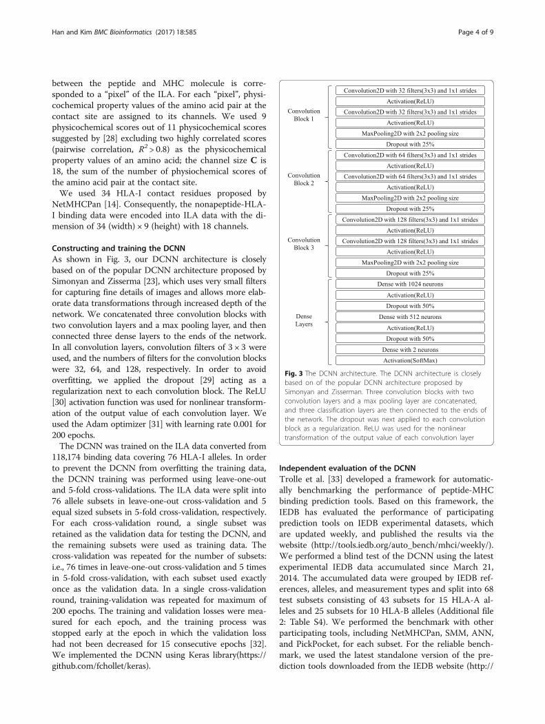
Constructing and training the DCNNAs shown in Fig. 3, our DCNN architecture is closelybased on of the popular DCNN architecture proposed bySimonyan and Zisserma [23], which uses very small filtersfor capturing fine details of images and allows more elab-orate data transformations through increased depth of thenetwork. We concatenated three convolution blocks withtwo convolution layers and a max pooling layer, and thenconnected three dense layers to the ends of the network.In all convolution layers, convolution filters of 3 × 3 wereused, and the numbers of filters for the convolution blockswere 32, 64, and 128, respectively. In order to avoidoverfitting, we applied the dropout [29] acting as aregularization next to each convolution block. The ReLU[30] activation function was used for nonlinear transform-ation of the output value of each convolution layer. Weused the Adam optimizer [31] with learning rate 0.001 for200 epochs.The DCNN was trained on the ILA data converted from
118,174 binding data covering 76 HLA-I alleles. In orderto prevent the DCNN from overfitting the training data,the DCNN training was performed using leave-one-outand 5-fold cross-validations. The ILA data were split into76 allele subsets in leave-one-out cross-validation and 5equal sized subsets in 5-fold cross-validation, respectively.For each cross-validation round, a single subset wasretained as the validation data for testing the DCNN, andthe remaining subsets were used as training data. Thecross-validation was repeated for the number of subsets:i.e., 76 times in leave-one-out cross-validation and 5 timesin 5-fold cross-validation, with each subset used exactlyonce as the validation data. In a single cross-validationround, training-validation was repeated for maximum of200 epochs. The training and validation losses were mea-sured for each epoch, and the training process wasstopped early at the epoch in which the validation losshad not been decreased for 15 consecutive epochs [32].We implemented the DCNN using Keras library(https://github.com/fchollet/keras).
Independent evaluation of the DCNNTrolle et al. [33] developed a framework for automatic-ally benchmarking the performance of peptide-MHCbinding prediction tools. Based on this framework, theIEDB has evaluated the performance of participatingprediction tools on IEDB experimental datasets, whichare updated weekly, and published the results via thewebsite (http://tools.iedb.org/auto_bench/mhci/weekly/).We performed a blind test of the DCNN using the latestexperimental IEDB data accumulated since March 21,2014. The accumulated data were grouped by IEDB ref-erences, alleles, and measurement types and split into 68test subsets consisting of 43 subsets for 15 HLA-A al-leles and 25 subsets for 10 HLA-B alleles (Additional file2: Table S4). We performed the benchmark with otherparticipating tools, including NetMHCPan, SMM, ANN,and PickPocket, for each subset. For the reliable bench-mark, we used the latest standalone version of the pre-diction tools downloaded from the IEDB website (http://
Fig. 3 The DCNN architecture. The DCNN architecture is closelybased on of the popular DCNN architecture proposed bySimonyan and Zisserman. Three convolution blocks with twoconvolution layers and a max pooling layer are concatenated,and three classification layers are then connected to the ends ofthe network. The dropout was next applied to each convolutionblock as a regularization. ReLU was used for the nonlineartransformation of the output value of each convolution layer
Han and Kim BMC Bioinformatics (2017) 18:585 Page 4 of 9

tools.iedb.org/mhci/download/), which were trained onthe same training data as that of our DCNN. The F1score, the harmonic mean of precision and recall, wasused to quantify the prediction performance, where anF1 score reaches its best value at 1 and worst value at 0.The F1 score is defined as:
F1 ¼ 2� precision� recallprecisionþ recall
;
precision ¼ TPTP þ FP
;
recall ¼ TPTP þ FN
;
where TP, FP, and FN are the numbers of true positives,false positives, and false negatives, respectively.
Identifying informative pixels recognized by the DCNNIn order to find locally-clustered interactions, informativepixels captured by the DCNN on the ILA classified as abinder were investigated. This was enabled due to thedevelopment of several recent methods that identifyinformative pixels of DCNN inputs, including Deconvnet[34], guided backpropagation [35], and DeepLIFT [36].The informative pixels were found by using high-resolution DeepLIFT method in this study.
Results and discussionTraining resultsIn order to compare the prediction performance of theDCNN and other prediction methods, the DCNN wastrained on the dataset that was used in other tools. The118,174 nonapeptide-HLA-I binding data for 76 HLA-Aalleles (72,551) and 37 HLA-B alleles (45,623) wereencoded into the two-dimensional ILA data. The pre-dictive performance was evaluated with leave-one-outand 5-fold cross-validation approaches. DCNN modelswere trained up to 200 epochs with early stopping con-dition. The mean validation losses were 0.318 in leave-one-out and 0.254 in 5-fold cross-validation, and themean validation accuracies were 0.855 and 0.892, re-spectively (Table 1), and this indicate that our DCNNwas able to be generally trained on the ILA data withoutmuch overfitting problems. Additional file 3: Table S2and Additional file 4: Table S3 show the detailed cross-validation results.
Independent evaluation of the DCNNWe performed a blind test of the DCNN using the latestIEDB experimental data accumulated since March 21,2014. The data were grouped by IEDB references, alleles,and measurement types and split into 68 test subsets con-sisting of 43 subsets for 15 HLA-A alleles and 25 subsetsfor 10 HLA-B alleles. For each subset, the prediction per-formances of other prediction tools, including NetMHC-Pan, SMM, ANN, and PickPocket, were measured. The F1scores were used to quantify their predictive perfor-mances. Table 2A and 2B summarize the prediction re-sults for HLA-A and HLA-B test subsets, respectively,and Additional file 2: Table S4 shows the detailed predic-tion results. The mean and median of the F1 scores of theDCNN were 0.638 and 0.696, respectively; these valueswere slightly higher than those of other tools, suggestingthat the DCNN was more reliable in nonapeptide-HLA-Abinding predictions (Table 2A). The mean of the F1 scoresof the DCNN was 0.593, which was almost the same asthose of other tools; however, the median was 0.667,which was higher than that of the other tools, indicatingthat the DCNN was also reliable in nonapeptide-HLA-Bbinding predictions (Table 2B).In particular, our DCNN showed significantly higher pre-
diction performance than other prediction tools for thesubsets for HLA-A*31:01, HLA-A*03:01, and HLA-A*68:01alleles belonging to the HLA-A3 supertype (Table 3).The HLA-A3 supertype were known to have import-
ant locally-clustered interactions that synergistically sta-bilizes the peptide-MHC complexes [26]. We thusinvestigated whether the trained DCNN was capturedthis features by inspecting its informative sites or pixelsfor three peptide-MHC complex pairs that were cor-rectly predicted by our method but were failed in other
Table 1 Summary of cross-validation results
Average accuracy Average loss
Leave-one-out 0.855 0.318
5-fold 0.892 0.254
Table 2 Prediction results for HLA-I test subsets
(A) Summary of prediction results for 43 HLA-A test subsets
DCNN NetMHCPan SMM ANN PickPocket
Mean 0.638 0.608 0.601 0.579 0.561
Median 0.696 0.667 0.667 0.667 0.625
Standard Deviation 0.230 0.267 0.250 0.286 0.318
(B) Summary of prediction results for 25 HLA-B test subsets
DCNN NetMHCPan SMM ANN PickPocket
Mean 0.593 0.606 0.578 0.606 0.560
Median 0.667 0.625 0.615 0.643 0.593
Standard Deviation 0.286 0.286 0.302 0.290 0.277
Han and Kim BMC Bioinformatics (2017) 18:585 Page 5 of 9

methods: KVFGPIHEL for HLA-A*31:01, RAAPPPPPRfor HLA-A*03:01, and LPQWLSANR for HLA-A*68:01.In KVFGPIHEL-HLA-A*31:01, the amino acids K, V,
and F of the peptide were preferred at the primary andsecond anchor positions 1, 2, and 3, respectively, but thenonpolar and hydrophobic L was deleterious at the pri-mary anchor position 9, and the charged H was toleratedat the secondary anchor position 7. We investigated theinformative pixels on the transformed ILA data captured
by the DCNN to identify the locally-clustered motifs atpositions 1, 2, and 3. Fig. 4a shows that the informativepixels with higher red intensities (red and blue intensitiesindicated the degree of contribution to the binder andnon-binder, respectively) were dominant and locally-clustered at the positions 1, 2, and 3, whereas the inform-ative pixels with higher blue intensities were located atposition 9. These findings were consistent with the factthat the locally-clustered patterns recognized by the
Table 3 Prediction results for HLA-A*31:01, HLA-A*03:01, and HLA-A*68:01 alleles
IEDB ID Allele Meas.Type
DCNN NetMHCPan SMM ANN PickPocket
315312 HLA-A*31:01 binary 0.857 0.667 0.571 0.667 0.400
1031253 HLA-A*03:01 ic50 0.941 0.875 0.667 0.875 0.941
1026840 HLA-A*68:01 binary 0.340 0.275 0.456 0.208 0.045
1026840 HLA-A*68:01 ic50 0.667 0.600 0.583 0.444 0.143
Mean 0.701 0.604 0.569 0.549 0.382
Fig. 4 Informative pixels on the ILA data. (a) In KVFGPIHEL-HLA-A*31:01, the informative pixels with higher red intensities (red and blue intensitiesindicated the degree of contribution to the binder and non-binder, respectively) were dominant and locally-clustered at the positions 1, 2, and 3(b) In RAAPPPPPR-HLA-A*03:01, informative pixels with higher red intensities were dominant and locally-clustered at the peptide positions 1 and2 (c) In LPQWLSANR-HLA-A*68:01, informative pixels with red intensities were slightly dominant at positions 4, 5, and 6 and at the primary anchorposition 9, with clustering at position 9
Han and Kim BMC Bioinformatics (2017) 18:585 Page 6 of 9

DCNN were informative when the KVFGPIHEL was clas-sified as a binder.In RAAPPPPPR-HLA-A*03:01, the positively charged
amino acid R of the peptide was preferred at the secondaryanchor position 1, but the amino acids A, and R at the pri-mary and secondary anchor positions 2, 3, and 9, respect-ively, were tolerated. Considering binding contributions ofthe individual amino acids at the primary and secondaryanchor positions, the peptide could not be a binder. Fig. 4bshows that the informative pixels with higher red intensitieswere dominant and locally-clustered at the peptide posi-tions 1 and 2, thus suggesting that the locally-clustered in-teractions between the amino acids at the peptide positionscould produce stable binding together.In LPQWLSANR-HLA-A*68:01, the positively charged
R of the peptide was preferred at the primary anchor pos-ition 9, but the L, P, and Q were not preferred at the pri-mary and secondary anchor positions 1, 2, and 3,respectively. The amino acids at positions 4, 5, 6, and 7were tolerated. As shown in Fig. 4c, informative pixels withred intensities were slightly dominant at positions 4, 5, and6 and at the primary anchor position 9, with clustering atposition 9, thus indicating that amino acids at positions 4,5, 6, and 9 synergistically induced stable binding.We found that our DCNN was able to correctly predict
the three binder peptides KVFGPIHEL, RAAPPPPPR,and LPQWLSANR with preferred amino acids only atsome primary and secondary anchor positions but with
amino acids that could synergistically induce stable bind-ing. This small number of cases are insufficient to supportthe general higher prediction performance of DCNN ap-proach for the HLA-A3 supertype, but these cases providethe possibilities that the DCNN can capture the locally-clustered interaction patterns in the peptide-HLA-A3binding structures, which cannot be easily captured byother methods.
Web serverWe developed ConvMHC(http://jumong.kaist.ac.kr:8080/convmhc), a web server to provide user-friendly web inter-faces for peptide-MHC class I binding predictions usingour DCNN. The main web interface consists of the inputform panel (left) and the result list panel (right) as shownin Fig. 5. Users can submit multiple peptide sequencesand a HLA-I allele in the input form panel. Once the pre-diction process is completed, the user can see the predic-tion results of the input peptides in the result list panel.For each prediction result, the user can also identify theinformative pixels captured by the DCNN on the ILA datathrough a pop-up panel.
ConclusionsIn this study, we developed a novel method for pan-specificpeptide-HLA-I binding prediction using DCNN trained onILA data that were converted from experimental bindingdata and demonstrated the reliable performance of the
Fig. 5 ConMHC Web Server. The main web interface of ConvMHC consists of the input form panel (left) and the result list panel (right). Users cansubmit multiple peptide sequences and a HLA-I allele in the input form panel. Once the prediction process is completed, the user can see theprediction results of the input peptides in the result list panel. For each prediction result, the user can also identify the informative pixels capturedby the DCNN on the transformed binding ILA data through a pop-up panel
Han and Kim BMC Bioinformatics (2017) 18:585 Page 7 of 9

DCNN in nonapeptide binding predictions through the in-dependent evaluation on IEDB external datasets. In particu-lar, the DCNN significantly outperformed other tools inpeptide binding predictions for alleles belonging to theHLA-A3 supertype. By investigating the informative pixelscaptured by the DCNN on the ILA data converted from thebinder nonapeptides that were predicted correctly by theDCNN but were failed in other methods, we found that theDCNN was better able to capture locally-clustered interac-tions that could synergistically produce stable binding in thepeptide-HLA-A3 complexes: KVFGPIHEL-HLA-A*31:01,RAAPPPPPR-HLA-A*03:01, and LPQWLSANR-HLA-A*68:01.We anticipate that our DCNN would become more reli-
able in peptide binding predictions for HLA-A3 allelesthrough further training and evaluations on more experi-mental data. DCNNs for MHC class II will be generatedand evaluated in further studies. Moreover, our approachesdescribed herein will be useful for identifying locally-clustered patterns in molecular binding structures, such asprotein/DNA, protein/RNA, and drug/protein interactions.However, it is not easy to build a reliable prediction modelusing DCNNs because deep learning tasks require largeamounts of training data to extract high-level and general-ized representations from the data. Currently, in order toovercome the limited training data, state-of-the-art learningtechnologies, such as generative adversarial nets [37] andtransfer learning [38] are attracting attentions. These tech-nologies can be effectively applied to generate more reliablebinding prediction models.
Additional files
Additional file 1: Table S1. Detailed description of the training dataset.(XLSX 16 kb)
Additional file 2: Table S4. Detailed prediction results for the IEDBHLA-I benchmark datasets. (XLSX 18 kb)
Additional file 3: Table S2. Detailed results for leave-one-out cross-validation. (XLSX 16 kb)
Additional file 4: Table S3. Detailed results for 5-fold cross-validation.(XLSX 11 kb)
AbbreviationsDCNN: Deep Convolutional Neural Network; HLA: Human Leukocyte Antigen,the human version of MHC; ILA: Image-Like Array; MHC: MajorHistocompatibility Complex
AcknowledgementsThe authors would like to thank Dr. S. Hong for helpful discussions andcomments.
FundingThis work was supported by the Bio & Medical TechnologyDevelopment Program of the NRF funded by the Korean government,MSIP(2016M3A9B6915714), the National Research Council of Science &Technology (NST) grant by the Korea government (MSIP) (No. CRC-16-01-KRICT) and the KAIST Future Systems Healthcare Project funded bythe Korea government(MSIP).
Availability of data and materialsConvMHC web server can be accessible via http://jumong.kaist.ac.kr:8080/convmhc. Python source codes and all the datasets supporting this work canbe downloaded from https://github.com/ihansyou/convmhc.
Authors’ contributionsYH designed the method, conducted the experiments, and wrote themanuscript. DK gave research ideas and supervised this project. All authorsread and approved the final manuscript.
Ethics approval and consent to participateNot applicable.
Consent for publicationNot applicable.
Competing interestsThe authors declare that they have no competing interests.
Publisher’s NoteSpringer Nature remains neutral with regard to jurisdictional claims inpublished maps and institutional affiliations.
Author details1Department of Bio and Brain Engineering, Korea Advanced Institute ofScience and Technology, Daejeon, Republic of Korea. 2Department ofConvergence Technology Research, Korea Institute of Science andTechnology Information, Daejeon, Republic of Korea.
Received: 17 September 2017 Accepted: 12 December 2017
References1. Lundegaard C, Hoof I, Lund O. State of the art and challenges in sequence
based T-cell epitope prediction. Immunome Research. 2010;6(Suppl 2):S3.2. Sette A, Sidney J. Nine major HLA class I supertypes account for the vast
preponderance of HLA-A and-B polymorphism. Immunogenetics. 1999;50:3–4.3. Rötzschke O, Falk K, Stevanović S. Exact prediction of a natural T cell
epitope. Eur J Immunol. 1991;21:2891–4.4. Sette A, et al. Prediction of major histocompatibility complex binding
regions of protein antigens by sequence pattern analysis. Proc Natl Acad SciU S A. 1989;86:3296–300.
5. Peters B, Tong W, Sidney J, Sette A, Weng Z. Generating quantitativemodels describing the sequence specificity of biological processes with thestabilized matrix method. BMC Bioinformatics. 2005;6:132.
6. Zhang H, Lund O, Nielsen M. The PickPocket method for predicting bindingspecificities for receptors based on receptor pocket similarities: applicationto MHC-peptide binding. Bioinformatics. 2009;25:1293–9.
7. Nielsen M, Lundegaard C, Worning P, Lauemøller SL, Lamberth K, Buus S, etal. Reliable prediction of T-cell epitopes using neural networks with novelsequence representations. Protein Sci. 2003;12(5):1007–17.
8. Andreatta M, Nielsen M. Gapped sequence alignment using artificial neuralnetworks: application to the MHC class I system. Bioinformatics. 2016;32:511–7.
9. Saethang T, Hirose O, Kimkong I, Tran VA, Dang XT, Nguyen L, et al.EpicCapo: epitope prediction using combined information of amino acidpairwise contact potentials and HLA-peptide contact site information. BMCbioinformatics. 2012;13(1):313.
10. Vita R, Overton JA, Greenbaum JA, Ponomarenko J, Clark JD, Cantrell JR, etal. The immune epitope database (IEDB) 3.0. Nucleic Acids Res. 2014;43(D1):405–12.
11. Sette A, Fleri W, Peters B, Sathiamurthy M, Bui HH, Wilson S. A roadmap forthe immunomics of category A–C pathogens. Immunity. 2005;22(2):155–61.
12. Zhang GL, Khan AM, Srinivasan KN, August JT, Brusic V. MULTIPRED: acomputational system for prediction of promiscuous HLA binding peptides.Nucleic Acids Research. 2005;33(suppl_2):172–9.
13. Jojic N, Reyes-Gomez M, Heckerman D, Kadie C, Schueler-Furman O.Learning MHC I—peptide binding. Bioinformatics. 2006;22(14):227–35.
14. Nielsen M, Lundegaard C, Blicher T, Lamberth K, Harndahl M, JustesenS, et al. NetMHCpan, a method for quantitative predictions of peptidebinding to any HLA-A and-B locus protein of known sequence. PLoSOne. 2007;2(8):e796.
Han and Kim BMC Bioinformatics (2017) 18:585 Page 8 of 9

15. Yanover C, Bradley P. Large-scale characterization of peptide-MHC bindinglandscapes with structural simulations. Proc Natl Acad Sci. 2011;108(17):6981–6.
16. Ehrenmann F, Kaas Q, Lefranc MP. IMGT/3Dstructure-DB and IMGT/DomainGapAlign: a database and a tool for immunoglobulins or antibodies,T cell receptors, MHC, IgSF and MhcSF. Nucleic Acids Research. 2009;38(suppl_1):301–7.
17. Guan P, Doytchinova IA, Flower DR. HLA-A3 supermotif defined by quantitativestructure–activity relationship analysis. Protein Eng. 2003;16(1):11–8.
18. DiBrino M, Parker KC, Shiloach J, Knierman M, Lukszo J, Turner, et al.Endogenous peptides bound to HLA-A3 possess a specific combination ofanchor residues that permit identification of potential antigenic peptides.Proc. Natl. Acad. Sci. 1993;90(4):1508–12.
19. Sidney J, Grey HM, Southwood S, Celis E, Wentworth PA, del Guercio MF, etal. Definition of an HLA-A3-like supermotif demonstrates the overlappingpeptide-binding repertoires of common HLA molecules. Hum Immunol.1996;45(2):79–93.
20. LeCun Y, Bengio Y, Hinton G. Deep learning. Nature. 2015;521(7553):436–44.21. Krizhevsky A, Sutskever I, Hinton GE. Imagenet classification with deep
convolutional neural networks. In: Advances in neural informationprocessing systems; 2012. p. 1097-105
22. Szegedy C, Liu W, Jia Y, Sermanet P, Reed S, Anguelov D, et al. Goingdeeper with convolutions. In: Proceedings of the IEEE conference oncomputer vision and pattern recognition; 2015. p. 1–9.
23. Simonyan, K., & Zisserman, A. Very deep convolutional networks for large-scale image recognition. arXiv preprint arXiv:1409.1556, 2014.
24. Wang S, Sun S, Li Z, Zhang R, Xu J. Accurate de novo prediction ofprotein contact map by ultra-deep learning model. PLoS Comput Biol.2017;13(1):e1005324.
25. Wallach, I., Dzamba, M., & Heifets, A. AtomNet: a deep convolutional neuralnetwork for bioactivity prediction in structure-based drug discovery. arXivpreprint arXiv:1510.02855, 2015.
26. Ragoza M, Hochuli J, Idrobo E, Sunseri J, Koes DR. Protein–Ligand scoringwith Convolutional neural networks. J Chem Inf Model. 2017;57(4):942–57.
27. Kim Y, Sidney J, Buus S, Sette A, Nielsen M, Peters B. Dataset size andcomposition impact the reliability of performance benchmarks for peptide-MHC binding predictions. BMC Bioinformatics. 2014;15(1):241.
28. Liu W, Meng X, Xu Q, Flower DR, Li T. Quantitative prediction of mouseclass I MHC peptide binding affinity using support vector machineregression (SVR) models. BMC Bioinformatics. 2006;7(1):182.
29. Srivastava N, Hinton GE, Krizhevsky A, Sutskever I, Salakhutdinov R. Dropout:a simple way to prevent neural networks from overfitting. J Mach Learn Res.2014;15(1):1929–58.
30. Nair V, Hinton GE. Rectified linear units improve restricted boltzmannmachines. In: Proceedings of the 27th international conference on machinelearning (ICML-10); 2010. p. 807–14.
31. Kingma, D., & Ba, J. Adam: A method for stochastic optimization.arXivpreprint arXiv:1412.6980. 2014.
32. Prechelt L. Early stopping—but when? In: Neural networks: tricks of thetrade. Berlin Heidelberg: Springer; 2012. p. 53–67.
33. Trolle T, Metushi IG, Greenbaum JA, Kim Y, Sidney J, et al. Automatedbenchmarking of peptide-MHC class I binding predictions. Bioinformatics.2015;31(13):2174–81.
34. Zeiler MD, Fergus R. Visualizing and understanding convolutional networks.In: European conference on computer vision; 2014. p. 818-33.
35. Springenberg, J. T., Dosovitskiy, A., Brox, T., & Riedmiller, M. Striving forsimplicity: The all convolutional net. arXiv preprint arXiv:1412.6806, 2014.
36. Shrikumar, A., Greenside, P., Shcherbina, A., & Kundaje, A. Not just a blackbox: Learning important features through propagating activationdifferences. arXiv preprint arXiv:1605.01713, 2016.
37. Goodfellow I, Pouget-Abadie J, Mirza M, et al. Generative adversarial nets. In:Advances in neural information processing systems; 2014. p. 2672–80.
38. Greenspan H, van Ginneken B, Summers RM. Guest editorial deep learningin medical imaging: overview and future promise of an exciting newtechnique. IEEE Trans Med Imaging. 2016l;35(5):1153–9.
• We accept pre-submission inquiries
• Our selector tool helps you to find the most relevant journal
• We provide round the clock customer support
• Convenient online submission
• Thorough peer review
• Inclusion in PubMed and all major indexing services
• Maximum visibility for your research
Submit your manuscript atwww.biomedcentral.com/submit
Submit your next manuscript to BioMed Central and we will help you at every step:
Han and Kim BMC Bioinformatics (2017) 18:585 Page 9 of 9