darier’s disease
TRANSCRIPT

DARIER’S DISEASE
facebook.com/groups/dermatologycourseonline

DARIER’S DISEASE
previously also known as
Keratosis folliculiris

DARIER’S DISEASE
rare genetic disorder that is
manifested predominantly
by skin changes

Like benign chronic familial
pemphigus (Hailey-Hailey
disease), Darier’s disease is
classified as a hereditary
acantholytic dermatosis.

AD with complete
penetrance & variable
expressivity

Onset 6-20y.


Pathogenesis

ATP2A2 on12q

This gene codes for the SERCA2 enzyme or pump (Sarco-Endoplasmic Reticulum Calcium-ATPase) that is required to transport calcium within the cell.


mutation in ATP2A2 gene that affects the activity of SERCA2, pumps which causes a complete loss of Ca++ transport activity into ER cytosolic Ca++

normal extra cellular calcium level is important in maintaining structure and function of desmosomalproteins as well as cell cycle check points otherwise;

acantholysis

apoptosis

X- ray crystallography of the pump


Clinical Picture


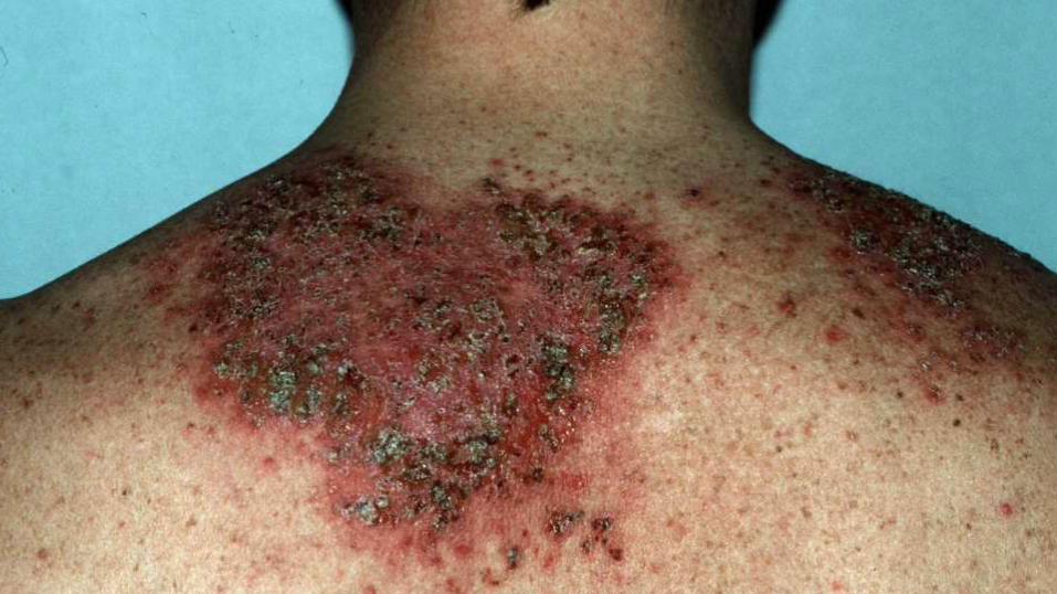
Darier disease on
the mid-back


Darier’s Disease: Warty papules on
dorsum of hands & feet

Palmar
pits


acrokeratosis
verruciformis

Segmental Darier
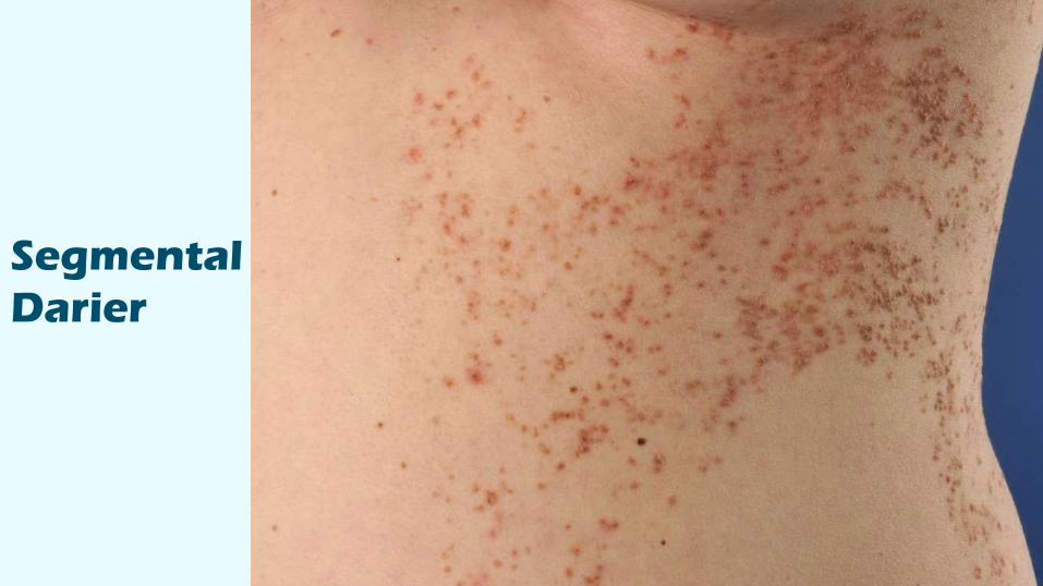
Segmental
Darier


Facial
cysts

ExacerbationsOften by sunlight
Heat
Sweat
Occlusion
Occasionally by corticosteroid use (although this may be useful for other patients).
Infections: Bacterial or viral Widespread infection with the herpes simplex virus is a well recognized complication called eczema herpeticum.

The symptoms and signs of Darier’s disease vary markedly between individuals. Some have very subtle signs that are asymptomatic and found only on careful inspection. Others have extensive lesions which can cause considerable distress to the affected individual.Moderate itch Malodor In an affected person the severity of the disease can fluctuate over time.

The skin lesions

persistent, greasy, scaly papules which tend to occur over the "seborrhoeic" areas of the face (scalp margins, forehead, ears, around the nostrils and sides of nose, eyebrows, and beard area), neck, and central chest and back. The flexures and skin under breasts and between buttocks are also commonly affected.

The papules have a firm, harsh feel like coarse sandpaper and may be skin-colored, yellow-brown or brown in color.
If several of the small papules grow together they may form larger warty lesions which can become quite smelly within skin folds.
The scalp is often affected with a heavily crusted rash which can be similar to seborrhoeic dermatitis but is usually harsher to the touch.

Lesions onthe hands & nails

Small pits (tiny indentations) on the palms and soles may occur and are very characteristic of Darier disease.
Small warty lesions or areas of bleeding under the skin can also be seen on the palms and soles as well as the dorsum of the hands and feet. These are known as acrokeratosisverruciformis.

Most patients with Darier’sdisease will have longitudinal broad stripes of white and reddish color on the nails (erytheronychia).
V-shaped notch at the free edge of the nail “pathognomonic”

Lesions affecting the mucous membranes

Raised
papular
lesions on
the
palate.

Patients with Darier‘s disease may uncommonly have a white cobblestone pattern or small papules affecting the mucous membranes.
Overgrowth of the gums is also seen.

Other presentationsSome patients develop a linear pattern of lesions, often following the lines of embryonal development of the skin segmental Darier

Other presentations
Comedones & N.C. acne

Other presentations
Vesiculo-bullous type


Histopathology
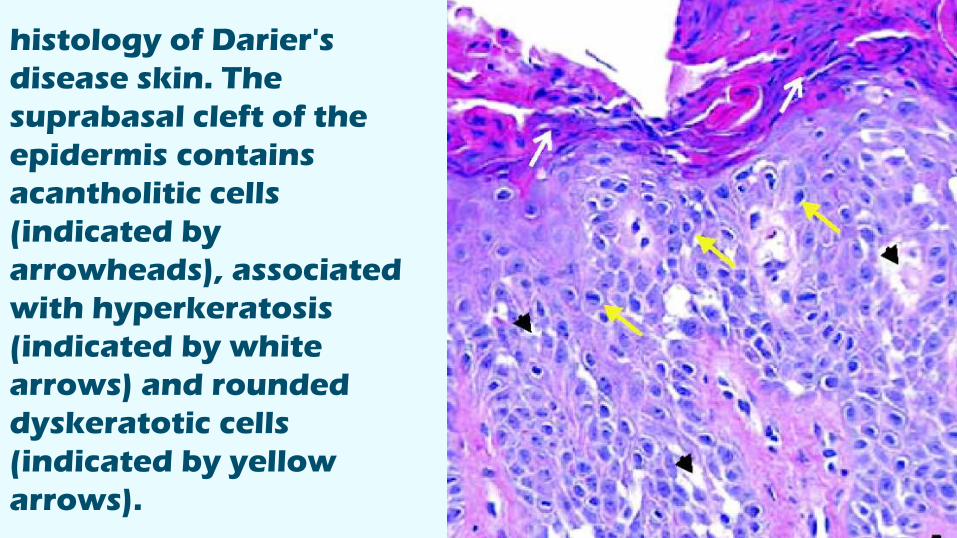
histology of Darier's
disease skin. The
suprabasal cleft of the
epidermis contains
acantholitic cells
(indicated by
arrowheads), associated
with hyperkeratosis
(indicated by white
arrows) and rounded
dyskeratotic cells
(indicated by yellow
arrows).


HistopathologyThe histology is characteristic, known as focal acantholytic dyskeratosisassociated with varying degrees of papillomatosis

Irregular acanthosis, papillomatosis, hyperkeratosis.
Suprabasal acantholysis clefts and lacunae containing dyskeratotic cells.
Irregular upward proliferation of one layer of basal keratinocytes (villi) into the lacunae.
CORPS RONDS rounded cells with dark pyknoticnuclei and perinuclear halo encircled by eosinophilic cytoplasm in the granular layer.
GRAINS elongated cells with shrunken parakeratotic nuclei in corneal layer.
Dermal chronic inflammation


Treatment

TreatmentLocalized disease
Mild cases
Sever cases
2ry infection

Localized disease

Dermabrasion, laser,
surgical excision & PDT
may be effective; may also
be treated successfully
with topical retinoids.

Milddisease

simple moisturizers, sun
protection and selection of
the right clothing to avoid
heat and sweating are
usually sufficient.

Severdisease

a trial of an oral retinoid
medication such as
acitretin or isotretinoin
may be effective

Result of one month's acitretin

Ciclosporin has been
reported to be effective in
a few patients.

Rx of 2ry
infections

2ry bacterial infection (usually
due to S. aureus) should be
treated with antibiotics, and
herpes simplex with antiviral
agents.


References
google images
Bolognia 3rd ed.
Dermntnz.org

YOU
Thank