daniel l. nickrent and lytton j. musselman
TRANSCRIPT

277
17 Parasitic PlantsDaniel L. Nickrent and Lytton J. Musselman
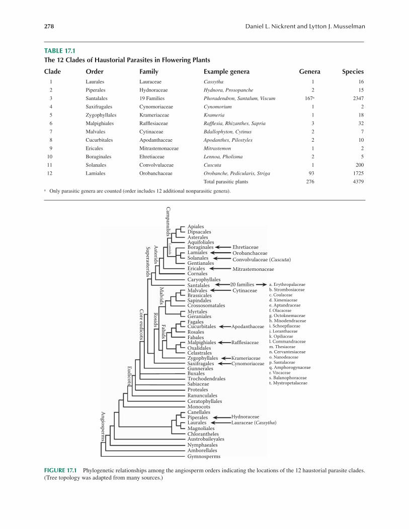
Parasitic organisms represent a significant proportion of the earth’s biodiversity, and flowering plants are no exception. Parasitic plants attach to their hosts by means of a hausto-rium, and this feeding mode has evolved independently in angiosperms 12 times (Table 17.1). There are no parasitic monocots, but haustorial parasites occur in the magnoliid and eudicot clades on the angiosperm phylogenetic tree (Figure 17.1). In an analogous fashion, a type of parasitism involving a mycorrhizal fungus has repeatedly evolved, and these plants are called mycoheterotrophs. The hall-mark of green plant evolution is photosynthesis; how-ever, this process has been lost repeatedly in all but two lineages of haustorial parasites (and many mycohetero-trophs). These plants are termed holoparasites, whereas those that retain photosynthesis are called hemiparasites. Approximately 4400 species in 276 genera of parasitic plants exist (Table 17.1; Parasitic Plant Connection web-site), but among these relatively few are pathogens, which cause diseases in plants of economic importance used by humans. About 11% of the genera are pathogens, and spe-cies in the following five genera inflict the most damage: Arceuthobium, Cuscuta, Orobanche, Striga, and Cuscuta (Figures 17.2 and 17.3).
Species of the genus Cuscuta, known by the English common name of dodder, are common throughout much of the world and are easily recognized by their twining, tangled masses of yellow or orange stems often blanket-ing their host plants (Figure 17.2). Some, like Cuscuta campestris, are serious pathogens of crops such as alfalfa (Medicago sativa), carrots (Daucus carota), lentils (Lens esculentus), and a diversity of other crops, weeds, and
native plants. Except for onion (Allium cepa), monocots are not attacked. The only plants that can be confused with Cuscuta are species of Cassytha (Lauraceae), a genus of tropical parasites bearing a stunning resem-blance to dodder. In addition to differences in morphol-ogy of their tiny flowers and the kind of fruit, the two genera differ in stem pubescence: dodder has glabrous stems, whereas Cassytha has pubescent stems. These two parasitic plants are good examples of convergent evolu-tion, not only in overall morphology, but also in other aspects of their biology such as seed germination and host impact.
Parasitic plants have evolved three times on the asterid I clade (Figure 17.1). In addition to Cuscuta in Convolvulaceae, the order Lamiales contains Orobanchaceae, one of the most economically impor-tant parasitic plant families. Although the vast majority of Orobanchaceae are benign root parasites, two gen-era, Striga (witchweeds) and Orobanche (broomrapes) together cause more damage to crop plants than all other parasitic plants combined. Striga asiatica (Figure 17.3), a pathogen of maize (Zea mays), is native to Africa, but was found in the eastern United States in the 1950s. Since then, a governmental program has been successful in eradicat-ing witchweed from the affected regions. Several species of Orobanche are agricultural pathogens throughout the Middle East and Europe, and some species have been accidentally introduced to North America. Like Striga, Orobanche seeds are tiny and require specific molecules exuded by host roots to stimulate germination. The final group of economically important parasitic plants that will
Concept Box
• The haustorium of dodder is a complex organ that effects the morphological and physiological connection between parasite and host.
• Unlike some other parasitic plants, Cuscuta seed germination does not depend on host molecular signaling.
• Dodder seedling undergoes circumnutation and responds to the presence of host plants through various cues including light and volatile chemicals. These cues help the plant orient toward and attach to the host.
• Dodder is a powerful physiological sink, outcompeting host sinks such as fruits that normally receive the lion’s share of photosynthates.
278 Daniel L. Nickrent and Lytton J. Musselman
TABLE 17.1The 12 Clades of Haustorial Parasites in Flowering Plants
Clade Order Family Example genera Genera Species1 Laurales Lauraceae Cassytha 1 16
2 Piperales Hydnoraceae Hydnora, Prosopanche 2 15
3 Santalales 19 Families Phoradendron, Santalum, Viscum 167a 2347
4 Saxifragales Cynomoriaceae Cynomorium 1 2
5 Zygophyllales Krameriaceae Krameria 1 18
6 Malpighiales Rafflesiaceae Rafflesia, Rhizanthes, Sapria 3 32
7 Malvales Cytinaceae Bdallophyton, Cytinus 2 7
8 Cucurbitales Apodanthaceae Apodanthes, Pilostyles 2 10
9 Ericales Mitrastemonaceae Mitrastemon 1 2
10 Boraginales Ehretiaceae Lennoa, Pholisma 2 5
11 Solanales Convolvulaceae Cuscuta 1 200
12 Lamiales Orobanchaceae Orobanche, Pedicularis, Striga 93 1725
Total parasitic plants 276 4379
a Only parasitic genera are counted (order includes 12 additional nonparasitic genera).
ApialesDipsacalesAsteralesAquifolialesBoraginalesLamialesSolanales
Ericales
EhretiaceaeOrobanchaceae
Convolvulaceae (Cuscuta)
Mitrastemonaceae
20 familiesCytinaceae
CornalesCaryophyllalesSantalalesMalvalesBrassicalesSapindalesCrossosomatalesMyrtalesGeranialesFagales
ApodanthaceaeCucurbitalesRosalesFabalesMalpighiales RafflesiaceaeOxalidalesCelastrales
KrameriaceaeCynomoriaceaeSaxifragales
Zygophyllales
GunneralesBuxalesTrochodendralesSabiaceaeProtealesRanunculalesCeratophyllalesMonocotsCanellales
Hydnoraceae
An
gio
sperm
s
Eu
dico
ts
Co
re eud
icots
Malvid
s
Su
perasterid
s
Asterid
s
Cam
pan
ulid
sL
amiid
s
Ro
sids Fab
ids
Lauraceae (Cassytha)MagnolialesChloranthelesAustrobaileyalesNymphaealesAmborellalesGymnosperms
PiperalesLaurales
Gentianales
a. Erythropalaceaeb. Strombosiaceaec. Coulaceaed. Ximeniaceaee. Aptandraceaef. Olacaceaeg. Octoknemaceaeh. Misodendraceaei. Schoepfiaceaej. Loranthaceaek. Opiliaceael. Commandraceaem. Thesiaceaen. Cervantesiaceaeo. Nanodeaceaep. Santalaceaeq. Amphorogynaceaer. Viscaceaes. Balanophoraceaet. Mystropetalaceae
FIGURE 17.1 Phylogenetic relationships among the angiosperm orders indicating the locations of the 12 haustorial parasite clades. (Tree topology was adapted from many sources.)

279Parasitic Plants
be considered here are the mistletoes (Mathiasen et al., 2008). Taxonomically, mistletoes can be found in five dif-ferent families within the sandalwood order (Santalales), but in North America, only Arceuthobium (Figure 17.4) and Phoradendron (Figure 17.5) (both Viscaceae) occur. As parasites of conifers in the pine family in North and
Central America, the dwarf mistletoes (Arceuthobium) cause billions of dollars of damage on commercial timber trees. One example is A. douglasii, which causes signifi-cant losses to Douglas fir (Figure 17.4).
LABORATORY EXERCISES
The purposes of these exercises are (1) to familiarize you with the morphology and anatomy of mature Cuscuta plants parasitizing a host; (2) to observe seed germination and seedling behavior; (3) to document the growth, twin-ing, and attachment of dodder stems on different hosts; and (4) to demonstrate how this parasitic plant acts as a physiological sink for host nutrients. You (or your instruc-tor) will first need to locate a population of dodder. During summer and fall in North America, several Cuscuta species are commonly seen, particularly C. campestris and C. gronovii. The first species is often seen grow-ing on clover and alfalfa along roadsides, whereas the latter favors edges of ponds and lakes, often grow-ing on water willow (Justicia americana), buttonbush (Cephalanthus occidentalis), and jewelweed (Impatiens capensis). Although commonly occurring on these hosts in nature, both the species will parasitize a wide range of other hosts when cultivated. Bear in mind that labora-tory experiments taking place in the spring may not have mature Cuscuta plants available from nature; however, with planning, flowering and fruiting plants can be made available by cultivating them under greenhouse condi-tions. Fruits (capsules) are greenish when immature, but become brown and translucent when mature. Only these capsules should be used when collecting seeds for cul-tivation and experimentation. Fortunately, these seeds remain viable for years when kept cool and dry. If a good crop of fruits is found, it is worthwhile to harvest many, remove the seeds, and store these in glass jars in the refrigerator. The germination percentages are never 100%, and the rate drops off in successive years.
EXPERIMENT 1. MORPHOLOGY OF MATURE CUSCUTA
When a population of Cuscuta is located, cuttings of the host with attached dodder can be brought into the lab for further observation. Make sure the host cutting is about 20 cm long and has some well-developed leaves. Place the cutting immediately in water in a large plastic cup. If the plants are not to be studied the same day, the cup with the cutting can be enclosed in a plastic bag and kept at room temperature for a few days or at 4°C for up to a week. This infected host plant cutting can be used to infect other host plants in the greenhouse. If you have grown Cuscuta in the greenhouse, you can bring the entire potted host plant with parasite into the laboratory for observation and dissection. If the dodder has flowers and/or fruits are present, these can be dissected and studied. It is easy to grow C. campestris
FIGURE 17.2 Field of sunflowers in Turkey heavily par-asitized by dodder (Cuscuta campestris). (Courtesy of L. Musselman.)
(a)
(b)
FIGURE 17.3 Striga asiatica (Asiatic witchweed). (a) S. asiatica emerging from soil next to its host plant, maize (Zea mays). (b) Although the flowers are showy, they self-pollinate prior to opening and upon fruiting, and produce thou-sands of tiny seeds. (Courtesy of L. Musselman.)

280 Daniel L. Nickrent and Lytton J. Musselman
to flowering, but this will require an adequate supply of hosts as well as care to ensure that the parasite growth does not spread to unintended plants. Flowering usually begins 3 weeks after host attachment. The induction of flowering in Cuscuta depends upon environmental cues such as short days (technically, long night of 14 h) and flowering of the host upon which it is attached (Baldev, 1962; Fratianne, 1965).
Observe the dodder plant attached to the host. The mature stems are yellow to orange, indicating that this plant has very little chlorophyll and is generally referred to as a holoparasite (Figure 17.6a). However, seedlings and other parts of the plant can be greenish, and some photosynthetic activity has been detected (reviewed in
Dawson et al., 1994). If desired, the chlorophyll extrac-tion method outlined in Procedure 21.1 can be tried with dodder and compared with a typical green plant. You will find that the amount of chlorophyll present is very low and, in fact, insufficient to provide sufficient sugars to maintain the plant. For this reason, Cuscuta is considered an obligate parasite because it ultimately depends upon a host for survival.
Notice that the plant lacks expanded leaves, but does have small-scale leaves at the nodes where branching occurs. Also notice that not all the stems are the same—some are coiled and some are not. Young stems are 1 mm or less in diameter, whereas well-nourished, attached stems can be 2 mm in diameter. The distal parts of the stems that are not coiled undergo circumnutation, a circular move-ment in a counterclockwise direction. If this stem encoun-ters a support, it will coil around it. Such a twining stem can also be called a tendril. In C. campestris, the main stems do not twine, whereas the tendrils, formed from the axils do. In C. gronovii, when a stem branches, either branch can twine around the host (Costea and Tardif, 2006).
When the tendril has coiled, it will form small bumps called prehaustoria on the inner surface of the curved stem (Figure 17.6a). Coiling and prehaustorium forma-tion respond to mechanical stimulation and the type of light striking the plant (Lane and Kasperbauer, 1965; Orr et al., 1996; Haidar et al., 1997; Li et al., 2010). Prehaustoria have specialized elastic cells that allow them to closely adhere and attach to the host (Vaughn, 2002). At this point, cells inside the prehaustorium dif-ferentiate from haustorial initials and begin growing through the prehaustorium tissue and then into the host tissue. These are called hyphal cells. Find some dodder
(a) (b)
FIGURE 17.4 Arceuthobium douglasii (Douglas-fir dwarf mistletoe). (a) Parasitized Douglas fir (Pseudotsuga menziesii) showing a massive witches’ broom in its lower half. The mistletoe endophyte induces the host to branch profusely. (b) Close-up of Douglas-fir dwarf mistletoe with young fruits. The mature fruit explosively dehisces the seed, thus no animal agents are required for dispersal to new host branches. (Courtesy of D. Nickrent.)
(a) (b)
FIGURE 17.5 Phoradendron leucarpum (leafy mistletoe). (a) Leafy mistletoe infesting a tree. (b) Close-up of P. leucarpum. (Courtesy of D. Nickrent.)

281Parasitic Plants
stems that have just begun to attach to the host. Pull on the stems with prehaustoria and notice that you feel the resistance. Continue pulling until you have removed the parasite stem from the host. Examine the prehaustorium with the stereo microscope and you will see that that they are oval in shape and that the tissue in the center appears different. The central part represents the hyphal cells of the haustorium that have torn away from the host.
Further development of the haustorium requires examination of sectioned or cleared parasite and host tis-sues (Figure 17.6b). As the hyphal cells of the haustoria enter the host, they will grow through the cortex until they reach vascular tissue. If haustoria are on the leaf, they will be attached to the veins, whereas if haustoria are on the stem, they will force their way through the cortical tissue and be connected to the host vascular cyl-inder. Although more details can be seen from slides pre-pared by sectioning the tissue with a microtome and then staining, free-hand sections are quick and easy and can show the overall structure of the haustorium. Procedure 17.1 gives the protocol for examining the Cuscuta haus-torium. You should be able to see the haustorium in the host cortex. This portion of the haustorium that is inside the host is called the endophyte. If the haustorium is suf-ficiently mature, you can also see the tip of the endophyte branching. These cells are called searching hyphae. When they reach host vascular tissue, they connect with xylem and phloem cells. The Cuscuta vascular system originates at the parasite stele, traverses the haustorium and endophyte, and connects to the host vascular system. This appears as a thin strand composed of tracheids in the haustorium, but in the vicinity of the host stele, it appears as several branched strands. The host–parasite vascular system can be seen in great detail using free-hand sections as well as clearings (see Procedure 17.1).
MaterialsEach student or team of students will need the following items:
• Cuscuta plants attached to the host• Stereo and compound microscopes• Glass slides and coverslips• Double-edged razor blades snapped in half
(these are sharper than single-edged razor blades)
• Forceps (fine tipped ones are best)• Toluidine blue stain (1% in 50% ethanol/water,
in dropper bottle)• Wash bottle with distilled water• 5% sodium hydroxide solution• Safranin G stain (1% in 50% ethanol/water)• Paper towels
Follow the protocol in Procedure 17.1 to complete this exercise.
Anticipated ResultsObservations made in this experiment document the detailed structure and development of the haustorium of a parasitic flowering plant. The processes of haustorial ini-tiation, host attachment, host penetration, and the estab-lishment of xylem to xylem link-up between parasite and host can all be seen. Attachment of the searching hyphae to host phloem cannot be seen with free-hand sections; this requires light or electron microscopic methods. The genus Cuscuta obtains the contents of host xylem (water and minerals) and phloem (sugars, amino acids, etc.), thus it is a holoparasite. The morphological and physiological link between the two organisms is extremely intimate, and in effect, the two function as one organism.
(a) (b)
50.00 μm
FIGURE 17.6 (a) Cuscuta gronovii (scaldweed) parasitizing the stem and leaf of its host, Eupatorium rugosum. (Courtesy of D. Nickrent.) (b) Cross section through the haustorium of C. campestris showing host tissue penetration. (Courtesy of M. Costea.)

282 Daniel L. Nickrent and Lytton J. Musselman
Questions• Do prehaustoria form only on living hosts? You
can test this by placing various objects such as glass rods, wooden dowels, dead tree branches, and so on in the path of the tendril. You may notice that the dodder will coil around vertical objects, but not horizontal ones? Why do you think this might be a useful adaptation?
• You probably noticed that Cuscuta forms attach-ments to its own stems. Do you think these haus-toria are structurally and functionally the same as those attached to a host plant? Section some of these and compare to help develop your answer.
• The term used to describe the Cuscuta endo-phyte cells is “hyphae.” This is the same term used to refer to the growing strands of fungi. Are these homologous structures? Why?
• The manner in which the haustorium grows through the prehaustorial tissue from the para-site stele is reminiscent of the development of branch roots in nonparasitic plants. What fea-tures are the same and what features are dif-ferent between the two? Do you think Cuscuta haustoria are modified roots?
• The formation of a haustorial endophyte (from inside the prehaustorium) is apparently con-trolled by the presence of the plant growth regu-lator cytokinin (Tsivion, 1978). How would you design an experiment to test this?
EXPERIMENT 2. SEED GERMINATION AND SEEDLING BEHAVIOR
Dodder produces numerous flowers in dense cymose clus-ters (Figure 17.7a and b). These flowers have four to five sepals, petals, and stamens. The gynoecium is composed of two fused carpels. One of the most distinguishing fea-tures of the Cuscuta flower is the presence of fringed or cleft scales alternating with the stamens. The fruit is a capsule (Figure 17.7c), but it may be either dehiscent or indehiscent, depending upon the species. Each capsule contains up to four seeds that have very hard seed coats (Figure 17.7d). Because of this, dodder seeds need scari-fication to germinate (Hutchison and Ashton, 1979). This can be accomplished using chemical or mechanical means.
When the seed of C. gronovii germinates, a primary root emerges first from the seed followed by the stem (Truscott, 1966). This vestigial root does not have a root cap nor does it elongate or participate in providing water
Procedure 17.1Morphology of the Cuscuta Haustorium
Step Instructions and Comments
1 Locate a portion of the host plant that has coiled Cuscuta stems and attached prehaustoria. It is useful to find early (young) attachments as well as mature (older) attachments for comparative purposes. The next steps are easier if you use smaller host stems and leaf petioles (less than 4 mm in diameter).
2 With the razor blade, remove a 5-mm long section of host stem infected with dodder and place on a micro-scope slide. Add a drop of water.
3 Working under the stereo microscope, hold the host stem with haustoria with the forceps and carefully make a transverse section (also called a cross section). From the cut surface, move inward along the host stem and make another cut producing as thin a section as possible. Try to make a slice that tapers in thickness. Let this section drop into the water on the microscope slide. Take care that sections do not dry. If your first attempts are not very good, keep going to get a variety of sections to examine.
4 Remove all plant materials that are not a section. Add a drop of toluidine blue stain. Place a coverslip over the sections. Place the slide on a paper towel and, using the wash bottle, gently add water to one side of the coverslip and absorb it out from the other. This will wash away excess stain.
5 Place the slide on the stage of the compound microscope and view your sections at various magnifications. 6 To visualize just the vascular tissue of the host and parasite, a technique called clearing is used. The tissue is
placed in a capped vial and covered with 5% NaOH in a 50°C–60°C oven overnight (or at room temperature for 2–3 days). The length of time in NaOH varies with different tissues; do not let the process proceed too far or else the plant material will dissolve! As the NaOH solution becomes discolored, change it with fresh NaOH. Rinse the tissue with water several times. Replace water with 50% ethanol. Stain with Safranin G (1% in 50% ethanol) for 2–24 h. Destain with 70% ethanol. The vascular tissue and other lignified cells should be red and other tissues light pink. The material can now be viewed using the compound microscope.

283Parasitic Plants
and nutrients as one sees in nonparasitic plants. In fact, this root and the base of the seedling will wither and will die as the shoot system grows and eventually attaches to a host. At this stage the dodder is not rooted in the soil and is an aerial parasite.
In C. campestris, maximum seed germination occurs between 30°C and 33°C, whereas no germination occurs above 39°C or below 15°C (Hutchison and Ashton, 1979). Germination does not appear to be dependent upon light; however, further development of the seedling is strongly influenced by light. Cuscuta seedlings respond to red light by the straightening of the hypocotyl hook. Once this hook straightens, the seedling stem grows actively and circumnutates. Seedlings that encounter no host will die, whereas those that make a haustorial connection will continue growing and developing. The growth rate (length increase per unit of time) and circumnutation rate (number of revolutions or arcs per unit of time) can be calculated from a time-lapse video.
MaterialsEach student or team of students will need the following items:
Germination
• Cuscuta seeds• Gloves
• Fume hood• 50-mL beaker• Concentrated sulfuric acid• Petri dishes• Filter paper circles• Wood blocks (2) with attached fine-grade
sandpaper
Cultivation of dodder seeds on greenhouse plants
• Germinated dodder seedlings• 1.7-mL microfuge tubes• Microfuge tube racks• Pipettes and tips• Potted coleus (Solenostemon scutellarioides)
host plants
Time-lapse photography of circumnutation
• Germinated dodder seedlings in tubes in racks.• Macintosh iPhone or iPad.• Support stand for iPhone or iPad (many versions
sold on the Internet).• OSnap, free application. Excellent tutorial on
how to use it here: http://www.osnapphotoapp .com/tutorials.php. This substitutes for a more expensive intervalometer, plus camera setup to obtain time-lapse video.
(a) (b)
(d)(c)
5.0 mm
5.0 mm
1.0 mm
1.0 mm
FIGURE 17.7 (a) Cuscuta campestris (scaldweed) flowering stem twining around its host stem. (b) Inflorescence (cyme) removed to show flower details. (c) Fruits (capsules). (d) Seeds. (Courtesy of D. Nickrent.)

284 Daniel L. Nickrent and Lytton J. Musselman
• Fluorescent lamps to provide supplemental lighting.
• Centimeter ruler.
Follow the instructions in Procedure 17.2 to complete this experiment.
Anticipated ResultsIt is expected that some, but not all, of the dodder seeds will germinate. With their hard seed coats, dodder can remain viable, but dormant in the soil for many years (Dawson et al., 1994). It is this viability and longevity that makes some species of Cuscuta formidable pests in agricultural situations. The time-lapse video should doc-ument that dodder seedlings (and actually all plants) are
not stationary, but in fact are in constant motion, albeit very slow. If you conducted your time-lapse experiment in the laboratory without any host plants nearby, the cir-cumnutation of the seedlings will show no directionality. Another study you can do is a time lapse, but this time put a potential host plant (e.g., coleus) near the seedling. Recent work has shown that C. campestris senses volatile compounds given-off by host plants and that its growth is then oriented in that direction (Runyon et al., 2006).
Questions• The seeds of most Cuscuta are dormant and
require scarification for germination to occur. An exception seems to be seen in C. pedicellata where no dormancy is documented (Lyshede,
Procedure 17.2Seed Germination and Seedling Behavior in Cuscuta
Step Instructions and Comments
1 Obtain about 40 dodder seeds. Two methods of scarification of the seed coat can be tried: chemical and mechanical.
2 While working in the fume hood, wear gloves, place 20 seeds in a beaker, and cover with concentrated sulfu-ric acid which will quickly turn dark. Allow to scarify for 15 min.
3 Decant the acid and thoroughly wash the seeds with distilled water. Do not let the seeds dry. Place 10 scarified seeds in each of two Petri dishes with moistened filter paper. Keep both the dishes at room temperature (about 25°C), but put one in the dark (a drawer) and the other in light on the benchtop. Within 5 days, the seeds will germinate producing the characteristic hook-shaped seedling.
4 Place the other 20 seeds between the two wood blocks with sandpaper. Rub the blocks against each other to scarify the seeds. Place the seeds in a beaker, rinse well with water, and place in Petri dishes as described in Step 3.
5 After 5 days, record the percent germination and appearance of the light and dark grown seedlings. 6 All germinated seeds (now seedlings) can be used in the next part of the experiment. Pipette 100 μL of water
into as many microfuge tubes as there are germinated seeds. Place the seedlings, root end down, into the tubes. Note that the roots will remain undeveloped; the stem of the seedling will grow and circumnutate, “searching” for a host. Only by forming a haustorial connection to the host can this seedling survive.
7 Place half the seedlings (tubes in racks) in the greenhouse adjacent to a potted coleus host plant. Seedlings in nature may initially attach to small plants near the soil surface, and only later they are sufficiently robust to parasitize larger hosts. For this reason, it might be best to start the seedlings on small coleus plants (rooted from cuttings). At these early stages, it is also important to not allow the seedlings to dry out. Once the para-site is attached, it will become more vigorous and the tendrils can attach to larger plants.
8 The other half of the seedlings can be kept at ambient conditions (typical temperature, moisture, and light). For these, the circumnutation rate of the seedling stem can be documented using time-lapse photography. The instructions for using the OSnap application with your iPhone™ or iPad™ are clear and straightforward. Adjusting the timer interval, that is, the time between each photo that is taken, is the critical factor for record-ing movement in the seedlings. Start with one photo per minute and record the seedling’s movement over at least 2 h. Be sure to plug in the power cord to your iPhone™ or iPad™ so that it does not run out of power during the experiment. A solid, neutral color background with a centimeter ruler should be placed behind the seedling. Illuminate the seedling with a cool light source (not incandescent bulbs that will cause heating).

285Parasitic Plants
1984). What are some possible reasons for this difference? Could there be an adaptive advan-tage to dormancy versus nondormancy in seeds from different species?
• It appears that in dodder, seed germination is entirely independent of any influence from the host plant. Do you know of any parasitic plants where the opposite is the case? A bit of Internet inquiry should help you answer this question.
• In C. pedicellata, seedlings undergo circumnu-tation for a few days, but after this they become prostrate and creep along the soil (Lyshede, 1985). Why do you think this happens? Could such behavior be an adaptation?
• Many dodder species have honeycombed seed coats when they are dry. When water is absorbed, the seed coat cells bulge out in fin-gerlike extensions. What possible role can you propose for the function of such structures?
EXPERIMENT 3. GROWTH AND DEVELOPMENT OF DODDER ON DIFFERENT HOSTS
One of the most common misconceptions about parasitic flowering plants is that most of them only grow on par-ticular hosts, that is, they are host specific. Indeed, spe-cialist parasites have evolved in all the major haustorial parasite clades (Table 17.1), but the majority of species are generalists. The two species of Cuscuta that are used in these exercises, C. campestris and C. gronovii, are generalists. That said, it is important to point out that the range of hosts parasitized in nature may differ from what is seen when the parasite is presented with hosts under controlled experimental conditions. Frequently, plants in which the parasite would never encounter in nature make perfectly suitable hosts. An example of this is the coleus plant (S. scutellarioides), which is native to Southeast Asia and Malaysia, outside the distributional range of C. gronovii.
Dodder is an excellent experimental organism because it is relatively easy to grow, plus it can be cloned. Having genetically identical individuals allows control in experiments where otherwise differences between indi-viduals might interfere with the effects being measured. Moreover, hosts such as coleus are easy to propagate by cuttings, thus host clones are genetically uniform as well. A single dodder plant attached to one plant will spread to any available neighbors. Once attachment to the next plant is established, the dodder stem can be severed and the attached parasite will continue growing even though separated from the original host plant.
Although the dodder species can grow on many different host species, given a “choice,” they will pre-fer some hosts over others. The research to date has not
fully determined the cause for such host choice, but it has been suggested that dodder will preferentially parasitize hosts with higher nutritional quality. As mentioned in Experiment 2, Cuscuta seedlings can detect and discrim-inate among volatile compounds emanating from various host plants (Runyon et al., 2006).
The purpose of this exercise is to demonstrate how dodder can differentiate among different potential hosts and preferentially parasitize certain species. This experi-ment could be carried out with dodder seedlings such as the ones produced in Experiment 2, but as mentioned, the initial haustorial connection of the seedling is critical and dependent upon a number of environmental and host factors. For this reason, host preference studies will be conducted using dodder shoots already established on a host plant.
MaterialsEach student or team of students will need the following items:
• A coleus plant that has previously been infected with dodder
• At least 10 species of plants representing a range of taxonomic groups and/or life forms
• Paper tags with string, permanent markers• Single-edged razor blades
Follow the protocols in Procedure 17.3 to complete this experiment.
Anticipated ResultsIt is expected that the host range of C. campestris and C. gronovii will be quite large, parasitizing a wide taxonomic diversity of plants. In contrast, it might be expected that plants with dense pubescence or a very thick cuticle will present greater obstacles to parasitism. Another observation that is quite amazing is that a rela-tively small segment of dodder, excised from the mother plant, can survive for days under fairly severe (desiccat-ing) conditions.
Questions• In nature, dodder is often simultaneously
attached to many different host plant species at one time. Each of these species has different cell surface features, different vascular anatomy, different defense chemistry, and so on, yet the single dodder individual can successfully form haustorial connections to all of them. What sorts of adaptations do you think dodder has evolved to allow it to be such a generalist parasite?
• Dodder stems are frequently seen attached to themselves; that is, they are self-parasitic. However, studies using one Cuscuta reflexa plant

286 Daniel L. Nickrent and Lytton J. Musselman
attached to another injected with radioactively labeled sucrose showed that no labeled sugar moved to the parasitizing plant. Do you think there is any adaptive function to such haustoria?
• Dodder can detect volatile compounds coming from different host species and thus discrimi-nate among hosts prior to forming haustorial connections. How do you think such a complex detection system has evolved?
EXPERIMENT 4. DODDER IS A PHYSIOLOGICAL SINK
In green plants, the photosynthates (sugars) are pro-duced in leaf mesophyll cells, loaded into the phloem, translocated to other parts of the plant where the sugar is unloaded and used for metabolism (e.g., actively growing shoots) or stored (e.g., roots and fruits). This is accom-plished by a process called mass flow, which explains the movement from source cells to sink cells. This experi-ment will show that Cuscuta attached to a host plant rep-resents a very powerful physiological sink.
In some ways, the haustorium of Cuscuta functions like a combination of a mature leaf and a root. Like a root, it is involved in taking up water and minerals from the host xylem, moving these materials through the hausto-rium into the parasite vascular system. This water move-ment takes place within the tracheids, part of the apoplast.
Inside the dodder haustorium, when a searching hypha contacts a host sieve tube element, it grasps, but does not penetrate, it like a hand with fingers. The sieve tube ele-ment is part of the symplast. Later, the contact hypha dif-ferentiates into an absorbing hypha which is functionally a specialized sieve tube element (with no companion cell). The ultrastructure of the parasite sieve element shows many features typical of transfer cells (Pate and Gunning, 1972), such as highly convoluted portions of the cell walls that are in contact with the host cell. Early radiolabeling and ultrastructural studies (Israel et al., 1980) showed the continuity between host and parasite phloem, and more recently interspecific plasmodesmata have been docu-mented (Birschwilks et al., 2007). Thus, in terms of move-ment of sugars and amino acids in the phloem, the dodder haustorium functions like a leaf, moving these materials to parasite sinks (meristems, fruits, etc.).
MaterialsEach student or team of students will need the following supplies:
• Arabidopsis thaliana (e.g., cv. Columbia)• Coleus plants grown as described in Procedure 17.3
with a well-developed growth of dodder• Paper envelopes
Procedure 17.3Growth and Development of Cuscuta on Different Host Plants
Step Instructions and Comments
1 This exercise requires a coleus plant, grown to a fairly large size that has also been previously infected with dodder. Under most greenhouse conditions, a coleus cutting will be 0.5 m tall in 5 months. There should be sufficient numbers of dodder stems to allow segments to be removed and placed on other host plants. It should be pointed out that in C. campestris seedling attachment and normal twining will not occur if fluorescent light under greenhouse conditions in winter is given (Dawson et al., 1994); hence, natural sunlight supplemented with incandescent light should be used.
2 The number of potential hosts used here depends upon the diversity of plants available in your greenhouse. We suggest you present the dodder with both taxonomically diverse plants and plants with various growth forms. For the former, try ferns, gymnosperms, monocots, and dicots. For the latter, try glabrous versus pubescent plants, succulents, plants with milky latex, and so on.
3 The methodology for transferring dodder from one plant to another is straightforward. Using a razor blade, cut a segment of dodder stem approximately 5 cm long and secure it to another plant. Tags with strings are useful in that the plant number can be recorded and the strings are used to gently tie the dodder to the host plant. Be sure to make your greenhouse personnel aware of this experiment so that the dodder is not purposely or inadvertently removed (e.g., during watering).
4 Monitor the dodder cuttings at least once a day. Record any dodder plants that have died, been lost, are pres-ent, but not attached, and attached by haustoria. After about 2 weeks (or more), make your final observations, including information on which dodder cuttings have done well and which have not. Measurements of the size (length) of the dodder stems on these different hosts could be made.
287Parasitic Plants
• Drying oven• Analytical balance
Follow the instructions provided in Procedure 17.4 to complete this exercise.
Anticipated ResultsIn studies with faba bean (Vicia faba), C. campes-tris was such an efficient parasite that it could divert the movement of nearly all phloem contents away from fruits (the typical sinks) and into its own tissues (Wolswinkel, 1974). This experiment should document the same phenomenon in Arabidopsis. It is expected that fruit number and fruit weight in the plants inocu-lated with dodder will be lower than those in the con-trols with no dodder.
Questions• In green plants, the mass flow hypothesis
requires the active transport of sucrose into the phloem at the source and active transport of sugars out of the phloem at the sink. The differ-ence in sucrose concentration at the source and sink locations sets up an osmotic gradient and hydrostatic pressure, thus water (and sucrose) movement from source to sink. In Cuscuta, excising the parasite leaving only its coiled stem with haustoria attached to the host does not stop sucrose movement into the haustorium. How is this system different than what is seen in a green plant?
• Lateral buds that have actively growing meri-stems are considered sinks in typical green plants. As you may know, removing the apical meristem in a plant (decapitation) will change the concentration of auxin arriving at lateral meristems below and “release them” to elon-gate. Design an experiment using coleus plants to test which sink is stronger, lateral meristems or dodder.
• Until recently, it was not clear whether the sym-plast of dodder is connected to the symplast of its host. Past work has shown that host DNA and even viruses can be translocated from host to the dodder. More recent work has shown that dyes can move from host to parasite, but not a green fluorescent protein (GFP)—ubiquitin fusion (molecular size of 36 kDa). How are the two symplasts connected? What features of this connective structure might be involved in limit-ing the types of molecules moving between the two organisms?
REFERENCES
Baldev, B. 1962. In vitro studies of floral induction on stem api-ces of Cuscuta reflexa Roxb, a short day plant. Ann. Bot. 26:173–180.
Birschwilks, M., N. Sauer, D. Scheel, and S. Neumann. 2007. Arabidopsis thaliana is a susceptible host plant for the holoparasite Cuscuta spec. Planta 226:1231–1241.
Procedure 17.4Dodder as a Physiological Sink
Step Instructions and Comments
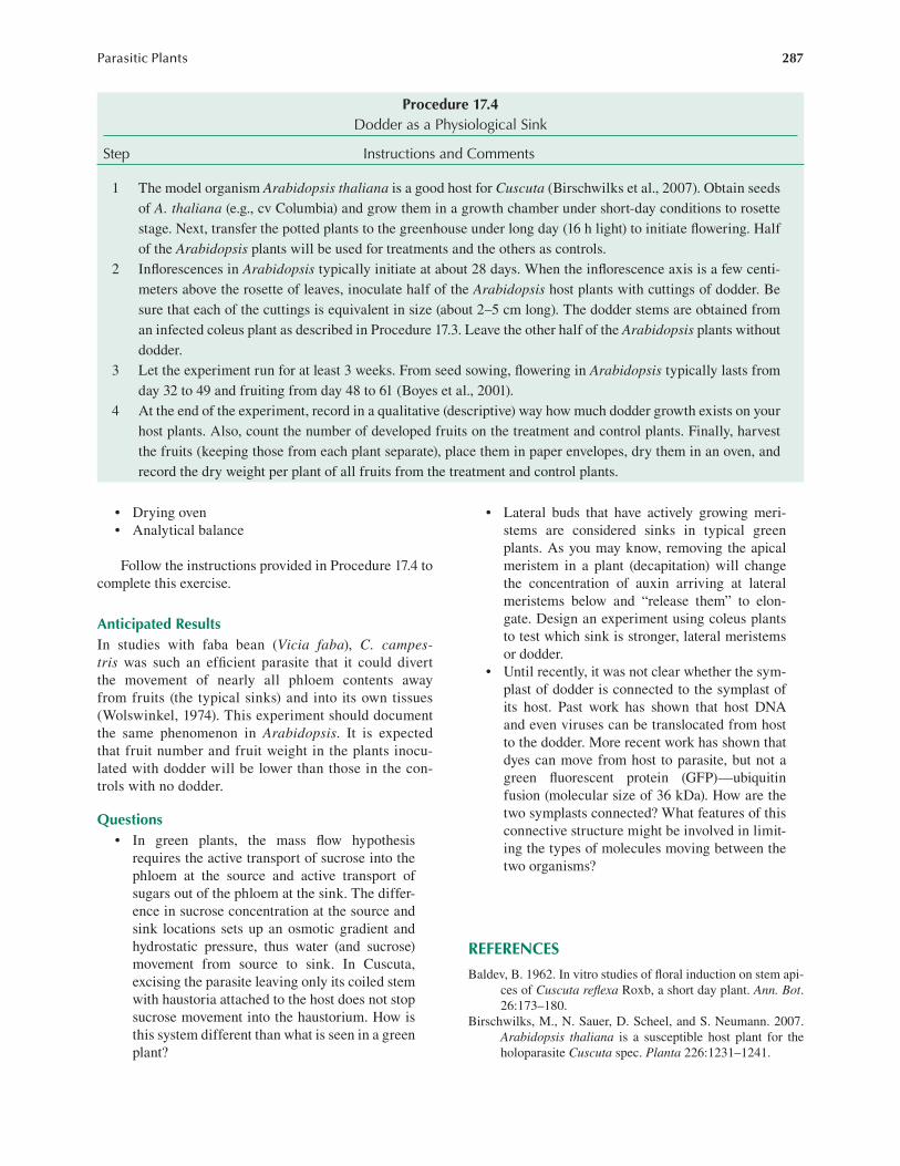
1 The model organism Arabidopsis thaliana is a good host for Cuscuta (Birschwilks et al., 2007). Obtain seeds of A. thaliana (e.g., cv Columbia) and grow them in a growth chamber under short-day conditions to rosette stage. Next, transfer the potted plants to the greenhouse under long day (16 h light) to initiate flowering. Half of the Arabidopsis plants will be used for treatments and the others as controls.
2 Inflorescences in Arabidopsis typically initiate at about 28 days. When the inflorescence axis is a few centi-meters above the rosette of leaves, inoculate half of the Arabidopsis host plants with cuttings of dodder. Be sure that each of the cuttings is equivalent in size (about 2–5 cm long). The dodder stems are obtained from an infected coleus plant as described in Procedure 17.3. Leave the other half of the Arabidopsis plants without dodder.
3 Let the experiment run for at least 3 weeks. From seed sowing, flowering in Arabidopsis typically lasts from day 32 to 49 and fruiting from day 48 to 61 (Boyes et al., 2001).
4 At the end of the experiment, record in a qualitative (descriptive) way how much dodder growth exists on your host plants. Also, count the number of developed fruits on the treatment and control plants. Finally, harvest the fruits (keeping those from each plant separate), place them in paper envelopes, dry them in an oven, and record the dry weight per plant of all fruits from the treatment and control plants.

288 Daniel L. Nickrent and Lytton J. Musselman
Boyes, D., A. Zayed, R. Ascenzi, A. McCaskill, N. Hoffman, K. Davis, and J. Görach. 2001. Growth stage-based phenotypic analysis of Arabidopsis: A model for high throughput functional genomics in plants. The Plant Cell 13:1499–1510.
Costea, M. and F. J. Tardif. 2006. The biology of Canadian weeds. 133. Cuscuta campestris Yuncker, C. gronovii Willd. ex Schult., C. umbrosa Beyr. ex Hook., C. epithy-mum (L.) L. and C. epilinum Weihe. Can. J. Plant Sci. 86:293–316.
Dawson, J. H., L. J. Musselman, P. Wolswinkel, and I. Dörr. 1994. Biology and control of Cuscuta. Rev. Weed Sci. 6:265–317.
Fratianne, D. G. 1965. The interrelationship between the flow-ering of dodder and the flowering of some long and short day plants. Amer. J. Bot. 52:556–562.
Haidar, M. A., G. L. Orr, and P. Westra. 1997. Effects of light and mechanical stimulation on coiling and prehaus-toria formation in Cuscuta spp. Weed Res. (Oxford) 37:219–228.
Hutchison, J. M., and F. M. Ashton. 1979. Effect of desicca-tion and scarification on the permeability and structure of the seed coat of Cuscuta campestris. Amer. J. Bot. 66:40–46.
Israel, S., I. Dörr, and R. Kollmann. 1980. Das Phloem der Haustorien von Cuscuta. Protoplasma 103:309–321.
Lane, H. C. and M. J. Kasperbauer. 1965. Photomorphogenic responses of dodder seedlings. Plant Physiol. 40:109–116.
Li, D. X., L. J. Wang, X. Yang, G. Zhang, and L. Chen. 2010. Proteomic analysis of blue light induced twining response in Cuscuta australis. Plant Molec. Biol. 72: 205–213.
Lyshede, O. B. 1984. Seed structure and germination in Cuscuta pedicellata with some notes on C. campestris. Nord. J. Bot. 4:669–674.
Lyshede, O. B. 1985. Morphological and anatomical features of Cuscuta pedicellata and C. campestris. Nord. J. Bot. 5:65–77.
Mathiasen, R. M., D. L. Nickrent, D. C. Shaw, and D. M. Watson. 2008. Mistletoes: Pathology, systematics, ecology, and management. Plant Dis. 92:988–1006.
Orr, G. L., M. A. Haidar, and D. A. Orr. 1996. Smallseed dodder (Cuscuta planiflora) phototropism toward far-red when in white light. Weed Sci. 44:233–240.
Pate, J. S. and E. S. Gunning. 1972. Transfer cells. Annu. Rev. Plant Physiol. 23:173–196.
Runyon, J. B., M. C. Mescher, and C. M. De Moraes. 2006. Volatile chemical cues guide host location and host selec-tion by parasitic plants. Science 313:1964–1967.
Truscott, F. H. 1966. Some aspects of morphogenesis in Cuscuta gronovii. Amer. J. Bot. 53:739–750.
Tsivion, Y. 1978. Possible role of cytokinins in nonspecific rec-ognition of a host and in early growth of haustoria in the parasitic plant, Cuscuta campestris. Bot. Gaz. 139:27–31.
Vaughn, K. C. 2002. Attachment of the parasitic weed dodder to the host. Protoplasma 219:227–237.
Wolswinkel, P. 1974. Complete inhibition of setting and growth of fruits of Vicia faba L., resulting from the draining of the phloem system by Cuscuta species. Acta Bot. Neerl. 23:48–60.
ONLINE RESOURCES
The Parasitic Plant Connection: http://www.parasiticplants.siu .edu/ (accessed May 9, 2014).
The Digital Atlas of Cuscuta: http://www.wlu.ca/page.php?grp _id=2147&p=8968 (accessed May 9, 2014).
The Angiosperm Phylogeny Website, Version 13: http://www .mobot.org/MOBOT/research/APweb/ (accessed May 9, 2014).