“danger” conditions increase sulfamethoxazole-protein...
TRANSCRIPT
“Danger” Conditions Increase Sulfamethoxazole-ProteinAdduct Formation in Human Antigen-Presenting Cells□S
S. N. Lavergne,1 H. Wang, H. E. Callan, B. K. Park, and D. J. NaisbittCentre for Drug Safety Science, Department of Pharmacology, University of Liverpool, Liverpool, United Kingdom
Received April 20, 2009; accepted August 6, 2009
ABSTRACTAntigen-presenting cells (APC) are thought to play an importantrole in the pathogenesis of drug-induced immune reactions.Various pathological factors can activate APC and thereforeinfluence the immune equilibrium. It is interesting that severaldiseases have been associated with an increased rate of drugallergy. The aim of this project was to evaluate the impact ofsuch “danger signals” on sulfamethoxazole (SMX) metabolismin human APC (peripheral blood mononuclear cells, Epstein-Barr virus-modified B lymphocytes, monocyte-derived dendriticcells, and two cell lines). APC were incubated with SMX (100�M–2 mM; 5 min–24 h), in the presence of pathological factors:bacterial endotoxins (lipopolysaccharide and staphylococcalenterotoxin B), flu viral proteins, cytokines [interleukin (IL)-1�,IL-6, IL-10; tumor necrosis factor-�; interferon-�; and trans-forming growth factor-�], inflammatory molecules (prostaglan-din E2, human serum complement, and activated protein C),oxidants (buthionine sulfoximine and H2O2), and hyperthermia
(37.5–39.5°C). Adduct formation was evaluated by enzyme-linked immunosorbent assay and confocal microscopy. SMX-protein adduct formation was time- and concentration-dependent for each cell type tested, in both physiological anddanger conditions. A danger environment significantly in-creased the formation of SMX-protein adducts and significantlyshortened the delay for their detection. An additive effect wasobserved with a combination of danger signals. Dimedone(chemical selectively binding cysteine sulfenic acid) and anti-oxidants decreased both baseline and danger-enhanced SMX-adduct formation. Various enzyme inhibitors were associatedwith a significant decrease in SMX-adduct levels, with a patternvarying depending on the cell type and the culture conditions.These results illustrate that danger signals enhance the forma-tion of intracellular SMX-protein adducts in human APC. Thesefindings might be relevant to the increased frequency of drugallergy in certain disease states.
Drug hypersensitivity reactions are thought to result froman abnormal immune reaction triggered by a drug or itsmetabolites. According to the hapten hypothesis, drugs aretoo small to stimulate the immune system, and effectiveimmune activity is directly related to drug-protein complexformation. For most drugs, metabolism is required to gener-ate an electrophilic intermediate that can attack nucleophilicresidues on proteins. These drug-protein adducts provide
antigenic determinants for the immune response, whereasadditional signals, often referred to as “danger signals,” de-termine the outcome between immunological tolerance andimmune reaction (Matzinger, 1998). Modifications of criticalproteins through drug haptenation, drug-associated oxida-tive stress, and drug-induced cell death are drug-dependentevents associated with danger signaling. Non–drug-depen-dent factors such as disease-induced oxidative stress or bac-terial and viral infections have also been identified as poten-tial danger signals (Gallucci and Matzinger, 2001).
Antigen-presenting cells (APC) take up and process drug-protein adducts for presentation to specific T lymphocytes.APC also seem to play an important role in the balancebetween immune tolerance and immune reactivity throughmodulation of the expression of costimulatory or coinhibitorymolecules (e.g., CD expression and cytokine secretion) afterdanger signaling (Turley, 2002). Dendritic cells are powerful
This work was supported by the Wellcome Trust [Grant 078598/Z/05/Z] (aspart of the Centre for Drug Safety Science supported by the Medical ResearchCouncil [Grant G0700654]).
1 Current affiliation: Veterinary College of Illinois, Urbana-Champaign,Illinois.
Article, publication date, and citation information can be found athttp://jpet.aspetjournals.org.
doi:10.1124/jpet.109.155374.□S The online version of this article (available at http://jpet.aspetjournals.org)
contains supplemental material.
ABBREVIATIONS: APC, antigen-presenting cells; SMX, sulfamethoxazole; HIV, human immunodeficiency virus; SMX-HA, sulfamethoxazole-hydroxylamine; SMX-NO, sulfamethoxazole-nitroso; Mo-DC, monocyte-derived dendritic cells; LPS, lipopolysaccharide; PMA, phorbol 12-myristate 13-acetate; PBMC, peripheral blood mononuclear cells; IL, interleukin; EBV, Epstein-Barr virus; ELISA, enzyme-linked immunosorbentassay; SEB, staphylococcal enterotoxin B; TNF, tumor necrosis factor; TGF, transforming growth factor; IFN, interferon; PGE, prostaglandin E;BSO, buthionine sulfoximine; GSH, glutathione; ABT, aminobenzotriazole; MTZ, methimazole; COX, cyclooxygenase; ABH, aminobenzoichydrazide; MPO, myeloperoxidase; OD, optical density; AA, ascorbic acid; DMSO, dimethyl sulfoxide.
0022-3565/09/3312-372–381$20.00THE JOURNAL OF PHARMACOLOGY AND EXPERIMENTAL THERAPEUTICS Vol. 331, No. 2Copyright © 2009 by The American Society for Pharmacology and Experimental Therapeutics 155374/3522883JPET 331:372–381, 2009 Printed in U.S.A.
372
http://jpet.aspetjournals.org/content/suppl/2009/08/07/jpet.109.155374.DC1Supplemental material to this article can be found at:
at ASPE
T Journals on M
ay 29, 2018jpet.aspetjournals.org
Dow
nloaded from
APC that are efficient at antigen uptake and processing intheir immature state, whereas costimulatory signals triggertheir maturation associated with functions essential for ef-fective antigen presentation.
Sulfamethoxazole (SMX) is an inexpensive sulfonamideantimicrobial that has a broad spectrum of action and a widetissue distribution. Sulfonamides are used to treat bacterialand protozoal infections and to prevent opportunistic infec-tions in immunocompromised patients, such as HIV-positiveindividuals or transplanted patients. The use of sulfonamides,however, has been limited by the occurrence of potentially life-threatening hypersensitivity reactions. It is important to re-member that most drugs are given to a patient because of adisease state in the first place, implying that drugs are usu-ally not exposed to physiological conditions, especially not inthe case of antibiotics. Moreover, the incidence of certaindrug allergies, such as SMX allergy, seems increased in somedisease states, such as viral infections like HIV (Slatore andTilles, 2004), or cystic fibrosis (Wills et al., 1998).
SMX is normally metabolized to an inert N-acetyl metab-olite, but a small fraction can be oxidized to a hydroxylamine-metabolite (SMX-HA) that redox cycles with a toxic nitroso-intermediate (SMX-NO) (Vyas et al., 2005; Lavergne et al.,2006) (see Fig. 6). SMX-NO has been shown to be directlytoxic (Naisbitt et al., 2002; Lavergne et al., 2006), to bindproteins covalently (Summan and Cribb, 2002; Callan et al.,2009), to activate dendritic cells (Sanderson et al., 2007), andto be immunogenic in animal models (Naisbitt et al., 2001;Farrell et al., 2003). SMX-protein adducts are thought to playa role in the pathogenesis of SMX hypersensitivity (Naisbittet al., 2002; Cheng et al., 2008). It is interesting that humanAPC, such as monocytes (Cribb et al., 1990) and dendritic cells(Sieben et al., 1999; Sanderson et al., 2007), metabolize SMX toa nitroso intermediate that forms T-cell-stimulating intracellu-lar SMX-protein adducts (S. N. Lavergne, B. K. Park, and D. J.Naisbitt, unpublished observation).
Certain factors, such as lipopolysaccharide (LPS) (Yadav etal., 2006), phorbol 12-myristate 13-acetate (PMA) (Asseffa etal., 1993), cytokines (Chomarat et al., 2003), and oxidativestress (Rutault et al., 1999) have been shown to modify thephenotype and functions of dendritic cells and other APC.Such danger signals have also been showed to modify theoxidation status of cysteine-containing proteins (Carballal etal., 2003). Patients treated with SMX are, by definition,carriers of such pathological factors. Thus, the aim of thisstudy was to evaluate the impact of danger signal on theformation of intracellular SMX-protein adduct by humanAPC.
Materials and MethodsHuman Antigen-Presenting Cells
Peripheral Blood Mononuclear Cells. PBMC were isolatedfrom fresh blood of healthy volunteers collected in heparinized tubesusing a Ficoll gradient separation protocol. They were then sus-pended in F1 media (RPMI 1640 medium, 10% fetal bovine serum, 2mM L-glutamine, 100 U/ml penicillin, 100 �g/ml streptomycin, and25 mM HEPES).
Monocyte-Derived Dendritic Cells. Monocyte-derived den-dritic cells were grown from monocyte using an established methodin DC media (F1 media complemented with 0.8 U/ml human gran-ulocyte macrophage–colony-stimulating factor and human IL-4)(Sanderson et al., 2007).
Dendritic Cell-Like Cell Lines. Commercially available THP1and HL60 cell lines (European Collection of Cell Cultures, Salisbury,UK) were cultured in F1 media.
Epstein-Barr Virus-Modified B Lymphocytes. EBV-modifiedB lymphocytes were developed using PBMC from three healthyvolunteers and three SMX allergic patients and cultured in F1media.
Drug Exposure
Nonadherent cell suspension (106 cells/ml) or monocyte-deriveddendritic cells (grown from a PBMC suspension of 2 � 106 cells/ml)were incubated with SMX (0.05–2 mM), for 5 min to 24 h. Cells werethen washed three times with phosphate-buffered saline before pro-cessing for confocal microscopy or before freezing for ELISA.
Generation of a Danger Environment for Human APC
Cells were exposed to danger signals aiming to mimic variouspathological conditions encountered by patients treated with SMX.LPS (100 ng/ml) and SEB (2 �g/ml) were used as bacterial patho-genic factors, for Gram-negative and Gram-positive bacteria, respec-tively, whereas inactivated flu virus (1 �g/ml; A/Jap/305/57 virus[H2N2]) served as a model of viral infection.
In certain experiments, cells were coincubated with cytokines suchas IL-1� (10 ng/ml), IL-6 (0.1 �g/ml), IL-10 (1 ng/ml), TGF-� (2 ng/ml),TNF-� (25 ng/ml), and IFN-� (1 �g/ml). In addition, cells werecoincubated with markers of inflammatory conditions, such as PGE2(10 �M), human serum complement (1 mg/ml), and activated proteinC (10 �g/ml).
Buthionine sulfoximine (BSO; 1 mM), a suicide inhibitor of �-glutamylcysteine synthetase, the rate-limiting enzyme in glutathi-one (GSH) biosynthesis, and H2O2 (18 �M), a powerful oxidant, werealso added to certain incubations.
Finally, experiments were performed in parallel at various tem-peratures (37–39.5°C) to evaluate the effect of hyperthermia associ-ated with fever. PMA (5 ng/ml) was used as a nonspecific immuneactivator. All pathological factors were used at concentrations that
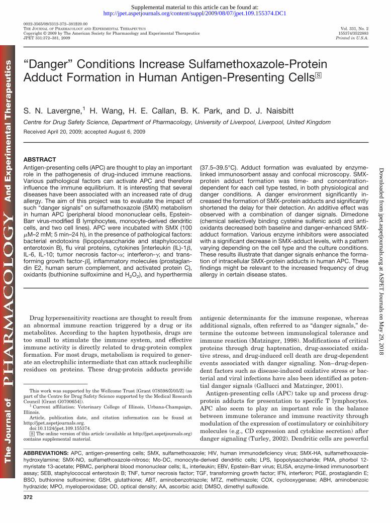
Fig. 1. Formation of intracellular SMX-protein adducts in human PBMCis enhanced by LPS. Human PBMC were incubated with SMX (500 �Mand 2 mM) in the presence or absence of LPS (100 ng/ml) for 24 h, beforebeing processed for confocal microscopy. LPS-stimulated PBMC showed ahigher level of SMX-protein adducts than PBMC incubated in physiolog-ical conditions.
“Danger Signals” and SMX Adducts in Human APC 373
at ASPE
T Journals on M
ay 29, 2018jpet.aspetjournals.org
Dow
nloaded from
have been shown to have an in vitro effect on immune cells (activa-tion, maturation, proliferation, or stress) without significant celltoxicity.
Enzyme Inhibition Assays
To confirm that SMX-protein adduct formation is due to the en-zymatic oxidation of SMX, experiments were repeated in the pres-ence of various inhibitors. Cells were incubated for 1 h with 1 mM ofthe following enzyme inhibitors before SMX was added with orwithout danger signal. Aminobenzotriazole (ABT) was used as aP450 inhibitor, methimazole (MTZ) was used as an inhibitor ofperoxidases and flavin-monooxygenases, aspirin and indomethacinwere used as cyclooxygenase (COX) 1 inhibitors, ketoprofen andibuprofen were used as nonselective COX1/2 inhibitors, and 4-ami-nobenzoic hydrazide (ABH) and salicylhydroxamic acid were used asmyeloperoxidase (MPO) inhibitors. Results are presented as percent-age of inhibition [100 � (inhibited sample blanked OD � baselineblanked OD)].
Antioxidant Assay
To evaluate the effect of antioxidants on the formation of SMX-protein adducts in human APC, experiments were performed withcells preincubated for 1 h with GSH, ascorbic acid (AA), or tocopherol(vitamin E) at 250 �M to 4 mM concentrations, before the addition ofSMX (1 mM) with or without danger signal.
Dimedone Assay
Dimedone is a reactive chemical that binds to oxidized cysteine(sulfenic acid) residues on proteins (Saurin et al., 2004), reducing
levels of in vitro SMX adduct formation (Callan et al., 2009). Cellswere incubated for 1 h with dimedone (5 mM) before adding SMX (2mM) to evaluate the existence of a sulfenic acid intermediate in APC.
Generation of a Specific Rabbit Anti-SMX Antiserum
The oxidative metabolites of SMX (SMX-HA and SMX-NO) weresynthesized according to a method published previously (Naisbitt etal., 1996). SMX-conjugated keyhole limpet hemocyanin was synthe-sized according to a protocol described previously. This conjugatewas used to raise rabbit anti-SMX antiserum according to an estab-lished method (Lavergne et al., 2006). Its specificity was testedagainst human serum albumin and SMX-human serum albuminconjugate (generated with the same protocol than SMX-keyhole lim-pet hemocyanin), using a specific rabbit anti-SMX antiserum kindlyprovided by Dr. Michael Rieder (London, ON, Canada) as a positivecontrol. The specificity of this antiserum was further demonstratedusing hapten inhibition (SMX at 2 mM) in the ELISA detectionsystem described below (Supplemental Fig. 1).
Detection of Sulfamethoxazole-Protein Adducts by ELISA
Wells were coated overnight with cell lysate (25 �g) in the refrig-erator. After phosphate-buffered saline/0.01% Tween washes, andblocking with 2.5% milk, samples were incubated overnight in therefrigerator with rabbit anti-SMX antisera (1:2000). Samples werethen incubated for an additional 2 h with alkaline phosphatase-conjugated anti-rabbit IgG (1:1000) at room temperature. Finally,the plate was read at 405 nm, after a 30-min incubation with alka-line phosphatase substrate (Sigma-Aldrich, Gillingham, UK).
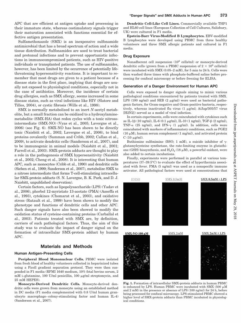
Fig. 2. PMA and LPS significantly increase the formation of SMX-protein adducts in human dendritic cells. Human monocyte-derived dendritic cellswere coincubated with SMX (0.01–2 mM) and PMA (5 ng/ml) or LPS (100 ng/ml). Both PMA and LPS significantly increased the formation ofSMX-protein adducts in human PBMC from SMX 100 �M (n � 3; A). Over a period of 24 h, SMX-protein adducts increased gradually in cells incubatedwith SMX (2 mM), with significant levels detected as early as 5 min with LPS-stimulated cells (n � 4; B). OD in DMSO control was 0.29 � 0.05. Similarresults were observed with PBMC dendritic cells lines, and EBV-modified lymphocytes from volunteers and SMX allergic patients (data not shown).
374 Lavergne et al.
at ASPE
T Journals on M
ay 29, 2018jpet.aspetjournals.org
Dow
nloaded from
Detection of Sulfamethoxazole-Protein Adducts byConfocal Microscopy
APC were fixed with 4% paraformaldehyde for 30 min, permeabil-ized with NH4Cl buffer (0.16 M) for 10 min, followed by 0.1% TritonX-0.1% bovine serum albumin for 30 min, and finally blocked with F1media for 1 h. Samples were then incubated overnight with rabbitanti-SMX antibody (1:500), before incubation with fluorescein iso-thiocyanate-anti-rabbit IgG for 2 h. Slides were finally mounted inVectashield H-1200 (Vector Laboratories, Peterborough, UK).
Detection of Sulfamethoxazole-Protein Adducts byImmunoblotting
APC (1.5 � 106) were incubated with DMSO (as a negative con-trol), SMX-NO (20 �M) (as a positive control), and SMX (2 mM) withor without LPS (100 ng/ml). After electrophoresis on a 12% SDS gelusing a protocol described previously (Callen et al., 2009), sampleswere transferred onto a polyvinylidene difluoride membrane thatwas then blocked with 5% milk. The membrane then was incubatedovernight with rabbit anti-SMX antiserum (up to 1:50), washed, andfinally incubated with a peroxidase-conjugated anti-rabbit secondaryantibody.
Statistical Analysis
Data were analyzed with a Student’s t test. Each cell experimentwas conducted three to seven times. Each of these experiments led toan ELISA in which samples were analyzed in duplicate. DuplicateOD readings were first averaged for each sample. For each ELISA,the average OD was compared with the average OD of the DMSOcontrol with a paired t test to ensure that the sample readings weresignificantly different from the ELISA background signal. Further-more, the average OD of the DMSO control from each experimentwas subtracted from the average OD of each sample, leading to“blanked” OD values. An average of blanked OD was calculated foreach sample from the different ELISA. Finally, average blanked ODvalues were compared with the SMX baseline sample of the corre-sponding assay using a paired t test. To ensure a more stringentanalysis of the inhibition, each Student’s t test was performed onblanked OD values (on paired conditions), and on the percentage ofinhibition and the percentage of remaining signal. A similar controlof the statistical analysis was performed with percentage of increasein the activation assays. In all cases, p 0.05 was considered asstatistically significant.
ResultsPMA and LPS Significantly Increase Intracellular
Sulfamethoxazole-Protein Adduct Formation in Hu-man APC. Using confocal microscopy, LPS treatment wasfound to increase SMX-protein adduct levels detected in hu-man PBMC exposed to SMX (500 �M and 2 mM; Fig. 1).
Using ELISA, PMA (5 ng/ml) and LPS (100 ng/ml) werefound to significantly increase SMX-protein adduct forma-tion in human monocyte-derived dendritic cells (Fig. 2A).Similar results were found in human PBMC, dendriticcell-like cell lines, and volunteer and allergic patient EBV-modified B lymphocytes exposed to titrated concentrationsof SMX for 24 h (data not shown). Over a SMX concentra-tion range of 10 �M to 2 mM, statistical significance wasreached at 100 �M (with the exception of PMA-stimulatedmonocyte-derived dendritic cells for which the significancewas only reached at 500 �M). Basal- and activator-inducedSMX adduct formation in EBV-modified B lymphocyteswas not statistically different between volunteer and al-lergic patients.
Over a period of 24 h, PMA- and LPS-enhanced SMX-TA
BL
E1
Eff
ect
ofva
riou
spa
thol
ogic
alfa
ctor
son
SM
X-p
rote
inad
duct
form
atio
nin
hu
man
den
drit
icce
lls
Dat
aar
epr
esen
ted
asth
eav
erag
epe
rcen
tage
ofin
crea
sein
addu
ctfo
rmat
ion
�S
.D.(
n�
5fo
rM
o-D
Can
dn
�4
for
TH
P1
and
HL
60):
100
�(�
OD
path
o�
�O
Db
ase
lin
e)�
100�
.DM
SO
con
trol
OD
aver
age:
0.34
�0.
06in
Mo-
DC
,0.
29�
0.03
inH
L60
,an
d0.
28�
0.03
inT
HP
1.
SE
BF
luV
iru
sIL
-1�
PG
E2
IFN
-�IL
-6T
NF
-�T
GF
-�IL
-10
CR
PC
3B
SO
H2O
2
Mo-
DC
47.1
�26
.427
.3�
8.2
68.1
�39
.150
.4�
30.4
96.5
�13
.452
.3�
20.8
45.1
�33
.036
.5�
18.5
24.4
�7.
525
.6�
10.1
24.3
�14
.294
.0�
62.1
96.5
�30
.2(p
�0.
008)
(p�
0.01
4)(p
�0.
004)
(p�
0.00
1)(p
�0.
0004
)(p
�0.
0001
)(p
�0.
006)
(p�
0.03
8)(p
�0.
015)
(p�
0.00
03)
(p�
0.00
4)(p
�0.
004)
(p�
0.00
4)H
L60
NE
NE
48.8
�21
.455
.2�
35.2
38.2
�26
.852
.0�
41.4
70.4
�49
.420
.6�
13.4
38.5
�31
.489
.3�
34.4
48.9
�21
.271
.8�
18.1
NE
(p�
0.01
0)(p
�0.
026)
(p�
0.03
3)(p
�0.
020)
(p�
0.01
7)(p
�0.
020)
(p�
0.04
6)(p
�0.
002)
(p�
0.02
9)(p
�0.
002)
TH
P1
NE
NE
39.4
�29
.137
.2�
5.0
45.0
�25
.5N
E26
.7�
7.3
22.2
�9.
945
.4�
24.3
53.1
�29
.938
.5�
22.5
NE
NE
(p�
0.03
7)(p
�0.
003)
(p�
0.01
9)(p
�0.
003)
(p�
0.03
0)(p
�0.
017)
(p�
0.01
9)(p
�0.
021)
C3,
hu
man
seru
mco
mpl
emen
t;N
E,
not
exam
ined
;C
RP
,C
reac
tive
prot
ein
.
“Danger Signals” and SMX Adducts in Human APC 375
at ASPE
T Journals on M
ay 29, 2018jpet.aspetjournals.org
Dow
nloaded from
adduct formation was statistically significant at 5 min inmonocyte-derived dendritic cells incubated with SMX (2mM), compared with 15 min in cells in physiological condi-tions (Fig. 2B). Similar results were obtained with otherhuman APC (data not shown).
Blanked OD values observed with cells incubated with SMX(with or without danger signal) were in the range of values forwhich the ELISA signal increased linearly with levels of ad-ducts obtained with the protein reactive SMX-NO (used as apositive control). Adduct formation was not detected by immu-noblotting when APC were incubated with SMX in the presenceor absence of LPS (Supplemental Figure S2).
Other Danger Signals Increase Sulfamethoxazole-Adduct Formation in Human APC. SEB (1 �g/ml), amajor component of Gram-positive Staphylococcus bacte-ria, and flu virus (JAP strain; 1 �g/ml) also significantlyincreased levels of SMX-protein adducts formed by humanmonocyte-derived dendritic cells exposed to SMX (2 mM)(p � 0.0008 and p � 0.014, respectively; Table 1). Likewise,cytokines (IL-1�, IL-6, TNF-�, IFN-�, IL-10, and TGF-�),inflammatory molecules (PGE2, human serum comple-ment, and activated protein C), and oxidative stress induc-ers (BSO and H2O2) were associated with a statisticallysignificant increase in levels of SMX-protein adducts in
human monocyte-derived dendritic cells (Table 1). A sim-ilar pattern of results was obtained with dendritic celllines (HL60 and THP1; Table 1) and EBV-modified B lym-phocytes (data not shown).
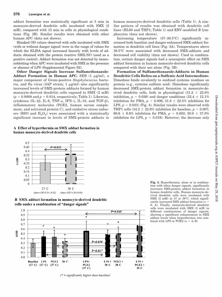
Increasing temperature (37–38.5°C) significantly in-creased both baseline and danger-enhanced SMX-adduct for-mation in dendritic cell lines (Fig. 3A). Temperatures above38.5°C were associated with decreased SMX-adducts anddecreased cell viability (data not shown). Used in combina-tion, certain danger signals had a synergistic effect on SMXadduct formation in human monocyte-derived dendritic cellscompared with their use alone (Fig. 3B).
Formation of Sulfamethoxazole-Adducts in HumanDendritic Cells Relies on a Sulfenic Acid Intermediate.Dimedone binds covalently to oxidized cysteine residues onprotein (e.g., cysteine sulfenic acid). Dimedone significantlydecreased SMX-protein adduct formation in monocyte-de-rived dendritic cells, both in physiological (31.2 � 22.8%inhibition, p � 0.036) and danger conditions (32.4 � 12.1%inhibition for PMA, p � 0.006; 31.8 � 22.5% inhibition forLPS, p � 0.033) (Fig. 4). Similar results were observed withTHP1 cells (34.2 � 22.6% inhibition for baseline, p � 0.007;60.6 � 6.6% inhibition for PMA, p � 0.002; 50.0 � 37.0%inhibition for LPS, p � 0.019). However, the decrease only
Fig. 3. Hyperthermia, alone or in combina-tion with other danger signals, significantlyincreases SMX-protein adduct formation inhuman dendritic cells. Human monocyte-de-rived dendritic cells were incubated withSMX (2 mM) at 37 or 38°C, which signifi-cantly increased SMX adduct formation (n �6; A). Finally, monocyte-derived dendriticcells were incubated with SMX (2 mM) indifferent combinations of danger signals,showing a significant enhancement in SMXadduct levels when hyperthermia was com-bined with LPS or PGE2 (n � 4; B).
376 Lavergne et al.
at ASPE
T Journals on M
ay 29, 2018jpet.aspetjournals.org
Dow
nloaded from
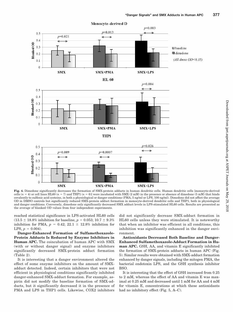
reached statistical significance in LPS-activated HL60 cells(13.5 � 18.8% inhibition for baseline, p � 0.053; 10.7 � 9.3%inhibition for PMA, p � 0.42; 22.1 � 12.8% inhibition forLPS, p � 0.004).
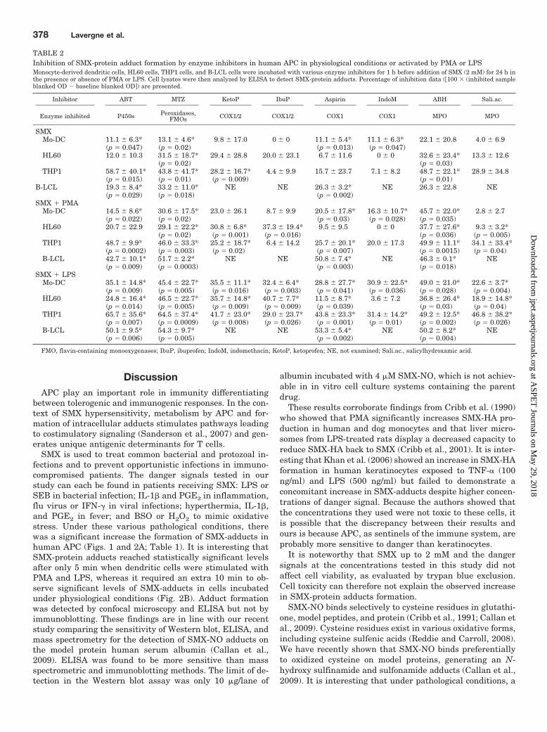
Danger-Enhanced Formation of Sulfamethoxazole-Protein Adducts Is Reduced by Enzyme Inhibitors inHuman APC. The coincubation of human APC with SMX(with or without danger signal) and enzyme inhibitorssignificantly decreased SMX-protein adduct formation(Table 2).
It is interesting that a danger environment altered theeffect of some enzyme inhibitors on the amount of SMX-adduct detected. Indeed, certain inhibitors that were notefficient in physiological conditions significantly inhibiteddanger-enhanced SMX-adduct formation. For example, as-pirin did not modify the baseline formation of SMX-ad-ducts, but it significantly decreased it in the presence ofPMA and LPS in THP1 cells. Likewise, COX2 inhibitors
did not significantly decrease SMX-adduct formation inHL60 cells unless they were stimulated. It is noteworthythat when an inhibitor was efficient in all conditions, thisinhibition was significantly enhanced in the danger envi-ronment.
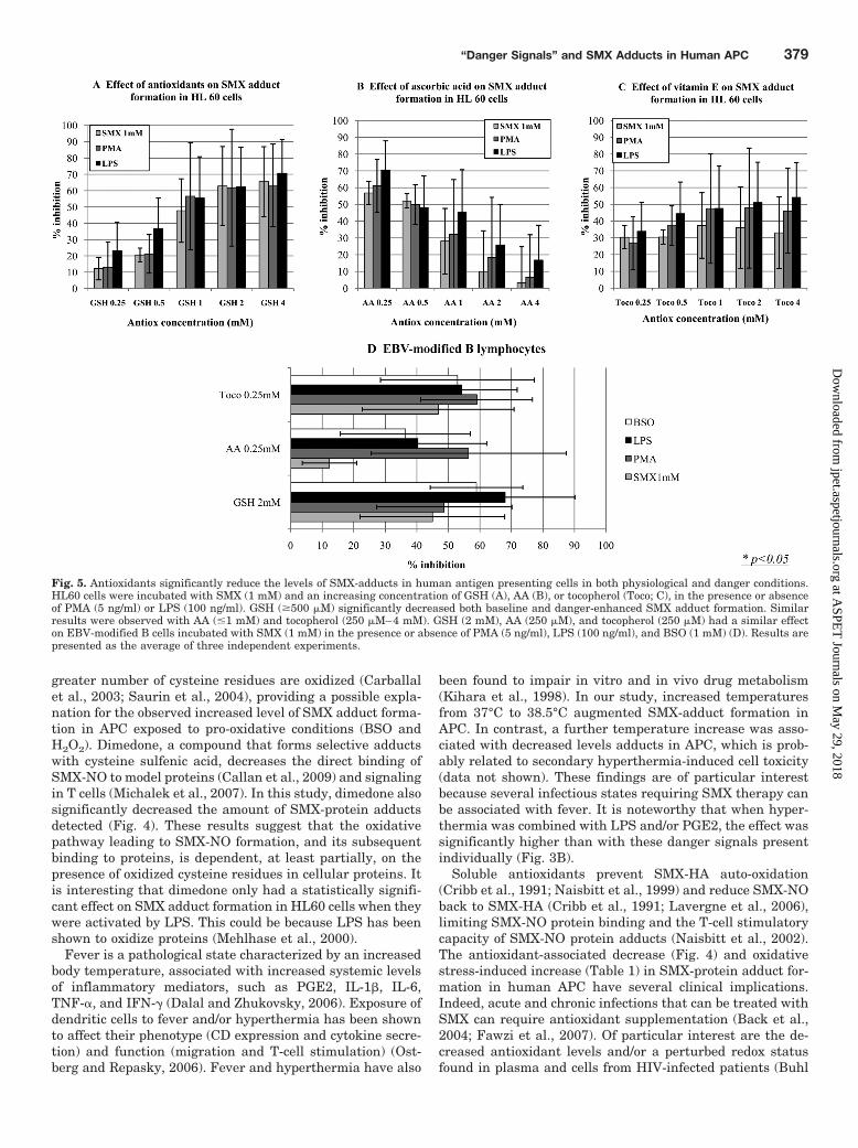
Antioxidants Decreased Both Baseline and Danger-Enhanced Sulfamethoxazole-Adduct Formation in Hu-man APC. GSH, AA, and vitamin E significantly inhibitedthe formation of SMX-protein adducts in human APC (Fig.5). Similar results were obtained with SMX-adduct formationenhanced by danger signals, including the mitogen PMA, thebacterial endotoxin LPS, and the GSH synthesis inhibitorBSO.
It is interesting that the effect of GSH increased from 0.25to 2 mM, whereas the effect of AA and vitamin E was max-imal at 0.25 mM and decreased until 1 mM for AA and 4 mMfor vitamin E, concentrations at which these antioxidantshad no inhibitory effect (Fig. 5, A–C).
Fig. 4. Dimedone significantly decreases the formation of SMX-protein adducts in human dendritic cells. Human dendritic cells [monocyte-derivedcells (n � 4) or cell lines HL60 (n � 7) and THP1 (n � 6)] were incubated with SMX (2 mM) in the presence or absence of dimedone (5 mM) that bindscovalently to sulfenic acid cysteine, in both a physiological or danger conditions (PMA, 5 ng/ml or LPS, 100 ng/ml). Dimedone did not affect the averageOD in DMSO controls but significantly reduced SMX-protein adduct formation in monocyte-derived dendritic cells and THP1, both in physiologicaland danger conditions. Conversely, dimedone only significantly decreased SMX adduct levels in LPS-stimulated HL60 cells. Results are presented asthe average of blanked OD values from four independent experiments.
“Danger Signals” and SMX Adducts in Human APC 377
at ASPE
T Journals on M
ay 29, 2018jpet.aspetjournals.org
Dow
nloaded from
DiscussionAPC play an important role in immunity differentiating
between tolerogenic and immunogenic responses. In the con-text of SMX hypersensitivity, metabolism by APC and for-mation of intracellular adducts stimulates pathways leadingto costimulatory signaling (Sanderson et al., 2007) and gen-erates unique antigenic determinants for T cells.
SMX is used to treat common bacterial and protozoal in-fections and to prevent opportunistic infections in immuno-compromised patients. The danger signals tested in ourstudy can each be found in patients receiving SMX: LPS orSEB in bacterial infection; IL-1� and PGE2 in inflammation,flu virus or IFN-� in viral infections; hyperthermia, IL-1�,and PGE2 in fever; and BSO or H2O2 to mimic oxidativestress. Under these various pathological conditions, therewas a significant increase the formation of SMX-adducts inhuman APC (Figs. 1 and 2A; Table 1). It is interesting thatSMX-protein adducts reached statistically significant levelsafter only 5 min when dendritic cells were stimulated withPMA and LPS, whereas it required an extra 10 min to ob-serve significant levels of SMX-adducts in cells incubatedunder physiological conditions (Fig. 2B). Adduct formationwas detected by confocal microscopy and ELISA but not byimmunoblotting. These findings are in line with our recentstudy comparing the sensitivity of Western blot, ELISA, andmass spectrometry for the detection of SMX-NO adducts onthe model protein human serum albumin (Callan et al.,2009). ELISA was found to be more sensitive than massspectrometric and immunoblotting methods. The limit of de-tection in the Western blot assay was only 10 �g/lane of
albumin incubated with 4 �M SMX-NO, which is not achiev-able in in vitro cell culture systems containing the parentdrug.
These results corroborate findings from Cribb et al. (1990)who showed that PMA significantly increases SMX-HA pro-duction in human and dog monocytes and that liver micro-somes from LPS-treated rats display a decreased capacity toreduce SMX-HA back to SMX (Cribb et al., 2001). It is inter-esting that Khan et al. (2006) showed an increase in SMX-HAformation in human keratinocytes exposed to TNF-� (100ng/ml) and LPS (500 ng/ml) but failed to demonstrate aconcomitant increase in SMX-adducts despite higher concen-trations of danger signal. Because the authors showed thatthe concentrations they used were not toxic to these cells, itis possible that the discrepancy between their results andours is because APC, as sentinels of the immune system, areprobably more sensitive to danger than keratinocytes.
It is noteworthy that SMX up to 2 mM and the dangersignals at the concentrations tested in this study did notaffect cell viability, as evaluated by trypan blue exclusion.Cell toxicity can therefore not explain the observed increasein SMX-protein adducts formation.
SMX-NO binds selectively to cysteine residues in glutathi-one, model peptides, and protein (Cribb et al., 1991; Callan etal., 2009). Cysteine residues exist in various oxidative forms,including cysteine sulfenic acids (Reddie and Carroll, 2008).We have recently shown that SMX-NO binds preferentiallyto oxidized cysteine on model proteins, generating an N-hydroxy sulfinamide and sulfonamide adducts (Callan et al.,2009). It is interesting that under pathological conditions, a
TABLE 2Inhibition of SMX-protein adduct formation by enzyme inhibitors in human APC in physiological conditions or activated by PMA or LPSMonocyte-derived dendritic cells, HL60 cells, THP1 cells, and B-LCL cells were incubated with various enzyme inhibitors for 1 h before addition of SMX (2 mM) for 24 h inthe presence or absence of PMA or LPS. Cell lysates were then analyzed by ELISA to detect SMX-protein adducts. Percentage of inhibition data (100 � (inhibited sampleblanked OD � baseline blanked OD�) are presented.
Inhibitor ABT MTZ KetoP IbuP Aspirin IndoM ABH Sali.ac.
Enzyme inhibited P450s Peroxidases,FMOs COX1/2 COX1/2 COX1 COX1 MPO MPO
SMXMo-DC 11.1 � 6.3* 13.1 � 4.6* 9.8 � 17.0 0 � 0 11.1 � 5.4* 11.1 � 6.3* 22.1 � 20.8 4.0 � 6.9
(p � 0.047) (p � 0.02) (p � 0.013) (p � 0.047)HL60 12.0 � 10.3 31.5 � 18.7* 29.4 � 28.8 20.0 � 23.1 6.7 � 11.6 0 � 0 32.6 � 23.4* 13.3 � 12.6
(p � 0.02) (p � 0.03)THP1 58.7 � 40.1* 43.8 � 41.7* 28.2 � 16.7* 4.4 � 9.9 15.7 � 23.7 7.1 � 8.2 48.7 � 22.1* 28.9 � 34.8
(p � 0.015) (p � 0.01) (p � 0.009) (p � 0.01)B-LCL 19.3 � 8.4* 33.2 � 11.0* NE NE 26.3 � 3.2* NE 26.3 � 22.8 NE
(p � 0.029) (p � 0.018) (p � 0.002)SMX PMA
Mo-DC 14.5 � 8.6* 30.6 � 17.5* 23.0 � 26.1 8.7 � 9.9 20.5 � 17.8* 16.3 � 10.7* 45.7 � 22.0* 2.8 � 2.7(p � 0.022) (p � 0.02) (p � 0.03) (p � 0.028) (p � 0.035)
HL60 20.7 � 22.9 29.1 � 22.2* 30.8 � 6.8* 37.3 � 19.4* 9.5 � 9.5 0 � 0 37.7 � 27.6* 9.3 � 3.2*(p � 0.02) (p � 0.001) (p � 0.016) (p � 0.036) (p � 0.005)
THP1 48.7 � 9.9* 46.0 � 33.3* 25.2 � 18.7* 6.4 � 14.2 25.7 � 20.1* 20.0 � 17.3 49.9 � 11.1* 34.1 � 33.4*(p � 0.0002) (p � 0.003) (p � 0.02) (p � 0.007) (p � 0.0015) (p � 0.04)
B-LCL 42.7 � 10.1* 51.7 � 2.2* NE NE 50.8 � 7.4* NE 46.3 � 0.1* NE(p � 0.009) (p � 0.0003) (p � 0.003) (p � 0.018)
SMX LPSMo-DC 35.1 � 14.8* 45.4 � 22.7* 35.5 � 11.1* 32.4 � 6.4* 28.8 � 27.7* 30.9 � 22.5* 49.0 � 21.0* 22.6 � 3.7*
(p � 0.009) (p � 0.005) (p � 0.016) (p � 0.003) (p � 0.041) (p � 0.036) (p � 0.028) (p � 0.004)HL60 24.8 � 16.4* 46.5 � 22.7* 35.7 � 14.8* 40.7 � 7.7* 11.5 � 8.7* 3.6 � 7.2 36.8 � 26.4* 18.9 � 14.8*
(p � 0.014) (p � 0.005) (p � 0.009) (p � 0.009) (p � 0.039) (p � 0.03) (p � 0.04)THP1 65.7 � 35.6* 64.5 � 37.4* 41.7 � 23.0* 29.0 � 23.7* 43.8 � 23.3* 31.4 � 14.2* 49.2 � 12.5* 46.8 � 38.2*
(p � 0.007) (p � 0.0009) (p � 0.008) (p � 0.026) (p � 0.001) (p � 0.01) (p � 0.002) (p � 0.026)B-LCL 50.1 � 9.5* 54.3 � 9.7* NE NE 53.3 � 5.4* NE 50.2 � 8.2* NE
(p � 0.006) (p � 0.005) (p � 0.002) (p � 0.004)
FMO, flavin-containing monooxygenases; IbuP, ibuprofen; IndoM, indomethacin; KetoP, ketoprofen; NE, not examined; Sali.ac., salicylhydroxamic acid.
378 Lavergne et al.
at ASPE
T Journals on M
ay 29, 2018jpet.aspetjournals.org
Dow
nloaded from
greater number of cysteine residues are oxidized (Carballalet al., 2003; Saurin et al., 2004), providing a possible expla-nation for the observed increased level of SMX adduct forma-tion in APC exposed to pro-oxidative conditions (BSO andH2O2). Dimedone, a compound that forms selective adductswith cysteine sulfenic acid, decreases the direct binding ofSMX-NO to model proteins (Callan et al., 2009) and signalingin T cells (Michalek et al., 2007). In this study, dimedone alsosignificantly decreased the amount of SMX-protein adductsdetected (Fig. 4). These results suggest that the oxidativepathway leading to SMX-NO formation, and its subsequentbinding to proteins, is dependent, at least partially, on thepresence of oxidized cysteine residues in cellular proteins. Itis interesting that dimedone only had a statistically signifi-cant effect on SMX adduct formation in HL60 cells when theywere activated by LPS. This could be because LPS has beenshown to oxidize proteins (Mehlhase et al., 2000).
Fever is a pathological state characterized by an increasedbody temperature, associated with increased systemic levelsof inflammatory mediators, such as PGE2, IL-1�, IL-6,TNF-�, and IFN-� (Dalal and Zhukovsky, 2006). Exposure ofdendritic cells to fever and/or hyperthermia has been shownto affect their phenotype (CD expression and cytokine secre-tion) and function (migration and T-cell stimulation) (Ost-berg and Repasky, 2006). Fever and hyperthermia have also
been found to impair in vitro and in vivo drug metabolism(Kihara et al., 1998). In our study, increased temperaturesfrom 37°C to 38.5°C augmented SMX-adduct formation inAPC. In contrast, a further temperature increase was asso-ciated with decreased levels adducts in APC, which is prob-ably related to secondary hyperthermia-induced cell toxicity(data not shown). These findings are of particular interestbecause several infectious states requiring SMX therapy canbe associated with fever. It is noteworthy that when hyper-thermia was combined with LPS and/or PGE2, the effect wassignificantly higher than with these danger signals presentindividually (Fig. 3B).
Soluble antioxidants prevent SMX-HA auto-oxidation(Cribb et al., 1991; Naisbitt et al., 1999) and reduce SMX-NOback to SMX-HA (Cribb et al., 1991; Lavergne et al., 2006),limiting SMX-NO protein binding and the T-cell stimulatorycapacity of SMX-NO protein adducts (Naisbitt et al., 2002).The antioxidant-associated decrease (Fig. 4) and oxidativestress-induced increase (Table 1) in SMX-protein adduct for-mation in human APC have several clinical implications.Indeed, acute and chronic infections that can be treated withSMX can require antioxidant supplementation (Back et al.,2004; Fawzi et al., 2007). Of particular interest are the de-creased antioxidant levels and/or a perturbed redox statusfound in plasma and cells from HIV-infected patients (Buhl
Fig. 5. Antioxidants significantly reduce the levels of SMX-adducts in human antigen presenting cells in both physiological and danger conditions.HL60 cells were incubated with SMX (1 mM) and an increasing concentration of GSH (A), AA (B), or tocopherol (Toco; C), in the presence or absenceof PMA (5 ng/ml) or LPS (100 ng/ml). GSH (�500 �M) significantly decreased both baseline and danger-enhanced SMX adduct formation. Similarresults were observed with AA (�1 mM) and tocopherol (250 �M–4 mM). GSH (2 mM), AA (250 �M), and tocopherol (250 �M) had a similar effecton EBV-modified B cells incubated with SMX (1 mM) in the presence or absence of PMA (5 ng/ml), LPS (100 ng/ml), and BSO (1 mM) (D). Results arepresented as the average of three independent experiments.
“Danger Signals” and SMX Adducts in Human APC 379
at ASPE
T Journals on M
ay 29, 2018jpet.aspetjournals.org
Dow
nloaded from
et al., 1989) who have develop a higher number of hypersen-sitivity reactions after SMX exposure. Collectively, our datasupport the theory that localized oxidative stress within APCmay be a risk factor for SMX hypersensitivity.
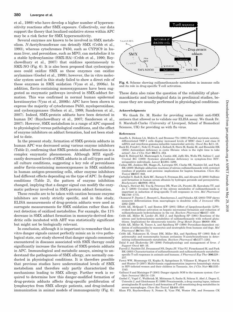
Several enzymes are known to be involved in SMX metab-olism. N-Acetyltransferase can detoxify SMX (Cribb et al.,1993), whereas cytochromes P450, such as CYP2C9 in hu-man liver, and peroxidase, such as MPO, can metabolize it toa stable hydroxylamine (SMX-HA) (Cribb et al., 1990; Roy-chowdhury et al., 2007) that oxidizes spontaneously toSMX-NO (Fig. 6). It is also been proposed that cyclooxygen-ases could oxidize SMX as these enzymes can oxidizearylamines (Goebel et al., 1999); however, the in vitro molec-ular system used in this study failed to show a direct role ofthese enzymes in SMX oxidation (Vyas et al., 2006a). Inaddition, flavin-containing monooxygenases have been sug-gested as enzymatic pathways involved in SMX-adduct for-mation. This was confirmed in normal human epidermalkeratinocytes (Vyas et al., 2006b). APC have been shown toexpress the majority of cytochromes P450, myeloperoxidase,and cyclooxygenases (Sieben et al., 1999; Sanderson et al.,2007). Indeed, SMX-protein adducts have been detected inhuman DC (Roychowdhury et al., 2007; Sanderson et al.,2007). However, SMX metabolism in a range of APC exposedto physiological versus pathological conditions, and the effectof enzyme inhibitors on adduct formation, had not been stud-ied to date.
In the present study, formation of SMX-protein adducts inhuman APC was decreased using various enzyme inhibitors(Table 2), confirming that SMX-protein adduct formation is acomplex enzymatic phenomenon. Although MTZ signifi-cantly decreased levels of SMX adducts in all cell types and inall culture conditions, suggesting a key role of peroxidasesand/or flavin-containing monooxygenases in SMX oxidationin human antigen-presenting cells, other enzyme inhibitorshad different effects depending on the type of APC. In dangerconditions (Table 2), the pattern of enzyme inhibitionchanged, implying that a danger signal can modify the enzy-matic pathway involved in SMX-protein adduct formation.
These results are to be taken with caution because enzymeinhibitors are rarely strictly specific, and in this study,ELISA measurements of drug-protein adducts were used assurrogate measurements for SMX oxidation rather than di-rect detection of oxidized metabolites. For example, the 11%decrease in SMX adduct formation in monocyte-derived den-dritic cells incubated with ABT was statistically significantbut might not be biologically relevant.
In conclusion, although it is important to remember that invitro danger signals cannot perfectly mimic an in vivo patho-logical state, our study showed that danger signals commonlyencountered in diseases associated with SMX therapy couldsignificantly increase the formation of SMX-protein adductsin APC. Immunological and metabolic assays, aiming to un-derstand the pathogenesis of SMX allergy, are normally con-ducted in physiological conditions. It is therefore possiblethat previous studies have underestimated levels of SMXmetabolism and therefore only partly characterized themechanisms leading to SMX allergy. Further work is re-quired to determine how this danger-modified formation ofdrug-protein adducts affects drug-specific proliferation oflymphocytes from SMX allergic patients, and drug-inducedimmunization in animal models of immunogenicity (Fig. 6).
These data also raise the question of the reliability of phar-macokinetic and toxicological data in preclinical studies, be-cause they are usually performed in physiological conditions.
Acknowledgments
We thank Dr. M. Rieder for providing some rabbit anti-SMXantisera that allowed us to validate our ELISA assay. We thank Dr.S. Marshall-Clarke (University of Liverpool, School of BiomedicalSciences, UK) for providing us with flu virus.
ReferencesAsseffa A, Dickson LA, Mohla S, and Bremner TA (1993) Phorbol myristate acetate-
differentiated THP-1 cells display increased levels of MHC class I and class IImRNA and interferon-gamma-inducible tumoricidal activity. Oncol Res 5:11–18.
Back EI, Frindt C, Nohr D, Frank J, Ziebach R, Stern M, Ranke M, and Biesalski HK(2004) Antioxidant deficiency in cystic fibrosis: when is the right time to takeaction?. Am J Clin Nutr 80:374–384.
Buhl R, Holroyd KJ, Mastrangeli A, Cantin AM, Jaffe HA, Wells FB, Saltini C, andCrystal RG (1989) Systemic glutathione deficiency in symptom-free HIV-seropositive individuals. Lancet 2:1294–1298.
Callan HE, Jenkins RE, Maggs JL, Lavergne SN, Clarke SE, Naisbitt DJ, and ParkBK (2009) Multiple adduction reactions of nitroso sulfamethoxazole with cysteinylresidues of peptides and proteins: implications for hapten formation. Chem ResToxicol 22:937–948.
Carballal S, Radi R, Kirk MC, Barnes S, Freeman BA, and Alvarez B (2003) Sulfenicacid formation in human serum albumin by hydrogen peroxide and peroxynitrite.Biochemistry 42:9906–9914.
Cheng L, Stewart BJ, You Q, Petersen DR, Ware JA, Piccotti JR, Kawabata TT, andJu C (2008) Covalent binding of the nitroso metabolite of sulfamethoxazole isimportant in induction of drug-specific T-cell responses in vivo. Mol Pharmacol73:1769–1775.
Chomarat P, Dantin C, Bennett L, Banchereau J, and Palucka AK (2003) TNF skewsmonocyte differentiation from macrophages to dendritic cells. J Immunol 171:2262–2269.
Cribb AE, McQuaid T, and Renton KW (2001) Effect of lipopolysaccharide (LPS)-evoked host defense activation on hepatic microsomal formation and reduction ofsulfamethoxazole hydroxylamine in the rat. Biochem Pharmacol 62:457–459.
Cribb AE, Miller M, Leeder JS, Hill J, and Spielberg SP (1991) Reactions of thenitroso and hydroxylamine metabolites of sulfamethoxazole with reduced gluta-thione. Implications for idiosyncratic toxicity. Drug Metab Dispos 19:900–906.
Cribb AE, Miller M, Tesoro A, and Spielberg SP (1990) Peroxidase-dependent oxi-dation of sulfonamides by monocytes and neutrophils from humans and dogs. MolPharmacol 38:744–751.
Cribb AE, Nakamura H, Grant DM, Miller MA, and Spielberg SP (1993) Role ofpolymorphic and monomorphic human arylamine N-acetyltransferases in deter-mining sulfamethoxazole metabolism. Biochem Pharmacol 45:1277–1282.
Dalal S and Zhukovsky DS (2006) Pathophysiology and management of fever. JSupport Oncol 4:9–16.
Farrell J, Naisbitt DJ, Drummond NS, Depta JP, Vilar FJ, Pirmohamed M, and ParkBK (2003) Characterization of sulfamethoxazole and sulfamethoxazole metabolite-specific T-cell responses in animals and humans. J Pharmacol Exp Ther 306:229–237.
Fawzi WW, Msamanga GI, Kupka R, Spiegelman D, Villamor E, Mugusi F, Wei R,and Hunter D (2007) Multivitamin supplementation improves hematologic statusin HIV-infected women and their children in Tanzania. Am J Clin Nutr 85:1335–1343.
Gallucci S and Matzinger P (2001) Danger signals: SOS to the immune system. CurrOpin Immunol 13:114–119.
Goebel C, Vogel C, Wulferink M, Mittmann S, Sachs B, Schraa S, Abel J, Degen G,Uetrecht J, and Gleichmann E (1999) Procainamide, a drug causing lupus, inducesprostaglandin H synthase-2 and formation of T cell-sensitizing drug metabolites inmouse macrophages. Chem Res Toxicol 12:488–500.
Khan FD, Roychowdhury S, Nemes R, Vyas PM, Woster PM, and Svensson CK
Fig. 6. Scheme showing sulfamethoxazole metabolism in immune cellsand its role in drug-specific T-cell activation.
380 Lavergne et al.
at ASPE
T Journals on M
ay 29, 2018jpet.aspetjournals.org
Dow
nloaded from
(2006) Effect of pro-inflammatory cytokines on the toxicity of the arylhydroxy-lamine metabolites of sulphamethoxazole and dapsone in normal human keratin-ocytes. Toxicology 218:90–99.
Kihara T, Toda A, Umesue I, Ono N, Shigematsu H, Soeda S, and Shimeno H (1998)Effect of interleukin 1 beta-induced fever on hepatic drug metabolism in rat.Xenobiotica 28:559–569.
Lavergne SN, Kurian JR, Bajad SU, Maki JE, Yoder AR, Guzinski MV, GrazianoFM, and Trepanier LA (2006) Roles of endogenous ascorbate and glutathione inthe cellular reduction and cytotoxicity of sulfamethoxazole-nitroso. Toxicology222:25–36.
Matzinger P (1998) An innate sense of danger. Semin Immunol 10:399–415.Mehlhase J, Gieche J, Ullrich O, Sitte N, and Grune T (2000) LPS-induced protein
oxidation and proteolysis in BV-2 microglial cells. IUBMB Life 50:331–335.Michalek RD, Nelson KJ, Holbrook BC, Yi JS, Stridiron D, Daniel LW, Fetrow JS,
King SB, Poole LB, and Grayson JM (2007) The requirement of reversible cysteinesulfenic acid formation for T cell activation and function. J Immunol 179:6456–6467.
Naisbitt DJ, Farrell J, Gordon SF, Maggs JL, Burkhart C, Pichler WJ, PirmohamedM, and Park BK (2002) Covalent binding of the nitroso metabolite of sulfamethox-azole leads to toxicity and major histocompatibility complex-restricted antigenpresentation. Mol Pharmacol 62:628–637.
Naisbitt DJ, Gordon SF, Pirmohamed M, Burkhart C, Cribb AE, Pichler WJ, andPark BK (2001) Antigenicity and immunogenicity of sulphamethoxazole: demon-stration of metabolism-dependent haptenation and T-cell proliferation in vivo.Br J Pharmacol 133:295–305.
Naisbitt DJ, Hough SJ, Gill HJ, Pirmohamed M, Kitteringham NR, and Park BK(1999) Cellular disposition of sulphamethoxazole and its metabolites: implicationsfor hypersensitivity. Br J Pharmacol 126:1393–1407.
Naisbitt DJ, O’Neill PM, Pirmohamed M, and Park BK (1996) Synthesis and reac-tions of nitroso sulphamethoxazole with biological nucleophiles: implications forimmune-mediated toxicity. Bioorg Med Chem Lett 6:1511–1516.
Ostberg JR and Repasky EA (2006) Emerging evidence indicates that physiologicallyrelevant thermal stress regulates dendritic cell function. Cancer Immunol Immu-nother 55:292–298.
Reddie KG and Carroll KS (2008) Expanding the functional diversity of proteinsthrough cysteine oxidation. Curr Opin Chem Biol 12:746–754.
Roychowdhury S, Vyas PM, and Svensson CK (2007) Formation and uptake ofarylhydroxylamine-haptenated proteins in human dendritic cells. Drug MetabDispos 35:676–681.
Rutault K, Alderman C, Chain BM, and Katz DR (1999) Reactive oxygen speciesactivate human peripheral blood dendritic cells. Free Radic Biol Med 26:232–238.
Sanderson JP, Naisbitt DJ, Farrell J, Ashby CA, Tucker MJ, Rieder MJ, Pirmo-hamed M, Clarke SE, and Park BK (2007) Sulfamethoxazole and its metabolitenitroso sulfamethoxazole stimulate dendritic cell costimulatory signaling. J Im-munol 178:5533–5542.
Saurin AT, Neubert H, Brennan JP, and Eaton P (2004) Widespread sulfenic acidformation in tissues in response to hydrogen peroxide. Proc Natl Acad Sci U S A101:17982–17987.
Sieben S, Baron JM, Blomeke B, and Merk HF (1999) Multiple cytochrome P450-isoenzymes mRNA are expressed in dendritic cells. Int Arch Allergy Immunol118:358–361.
Slatore CG and Tilles SA (2004) Sulfonamide hypersensitivity. Immunol Allergy ClinNorth Am 24:477–490, vii.
Summan M and Cribb AE (2002) Novel non-labile covalent binding of sulfamethox-azole reactive metabolites to cultured human lymphoid cells. Chem Biol Interact142:155–173.
Turley SJ (2002) Dendritic cells: inciting and inhibiting autoimmunity. Curr OpinImmunol 14:765–770.
Vyas PM, Roychowdhury S, and Svensson CK (2006a) Role of human cyclooxygen-ase-2 in the bioactivation of dapsone and sulfamethoxazole. Drug Metab Dispos34:16–18.
Vyas PM, Roychowdhury S, Koukouritaki SB, Hines RN, Krueger SK, Williams DE,Nauseef WM, and Svensson CK (2006b) Enzyme-mediated protein haptenation ofdapsone and sulfamethoxazole in human keratinocytes: II. Expression and role offlavin-containing monooxygenases and peroxidases. J Pharmacol Exp Ther 319:497–505.
Vyas PM, Roychowdhury S, Woster PM, and Svensson CK (2005) Reactive oxygenspecies generation and its role in the differential cytotoxicity of the arylhydroxy-lamine metabolites of sulfamethoxazole and dapsone in normal human epidermalkeratinocytes. Biochem Pharmacol 70:275–286.
Wills R, Henry RL, and Francis JL (1998) Antibiotic hypersensitivity reactions incystic fibrosis. J Paediatr Child Health 34:325–329.
Yadav R, Zammit DJ, Lefrancois L, and Vella AT (2006) Effects of LPS-mediatedbystander activation in the innate immune system. J Leukoc Biol 80:1251–1261.
Address correspondence to: Dr. Dean John Naisbitt, Centre for DrugSafety Science, Department of Pharmacology, Sherrington Bldg., Ashton St.,The University of Liverpool, Liverpool L69 3GE, UK. E-mail: [email protected]
“Danger Signals” and SMX Adducts in Human APC 381
at ASPE
T Journals on M
ay 29, 2018jpet.aspetjournals.org
Dow
nloaded from