daftar pustaka - diponegoro...
TRANSCRIPT

46
DAFTAR PUSTAKA
1. World Health Organization. Diabetes. http://www.who.int/topics/diabetes_
mellitus/en/ (accessed 4 February 2013).
2. World Health Organization. Diabetes. http://www.who.int/mediacentre/
factsheets/fs312/en/index.html (accessed 4 February 2013).
3. International Diabetes Federation. IDF Diabetes Atlas 5th
edition.
http://idfnews.cmail1.com/t/r-i-kullnk-l-o/ (accessed 4 February 2013).
4. Wild S, Roglic G, Green A, Sicree R, King H. Global Prevalence of
Diabetes: Estimates for the year 2000 and projections for 2030. Diabetes
Care.2004;27(5):(1047-1053)
5. American Diabetes Association. Diabetes Basics. http://www.diabetes.org/
diabetes-basics/?loc=GlobalNavDB (accessed 4 February 2013).
6. Tripathi BK, Srivastava AK. Diabetes mellitus: Complications and
therapeutics. Med Sci Monit. 2006;12(7):(130-147)
7. Boudina S, Abel ED. Diabetic cardiomyopathy, causes and effects. Rev
Endocr Metab Disord. 2010;11(1): 31-39.
8. Rubler S, Dlugash J, Yuceoglu YZ, Kumral T, Branwood AW, and
Grishman, A. New type of cardiomyopathy associated with diabetic
glomerulosclerosis. Am. J. Cardiol. 1972;30:(595–602)
9. Kannel, W B, Hjortland, M, and Castelli WP. Role of diabetes in
congestive heart failure: The Framingham Study. Am. J. Cardiol. 1976;34:
(29–34).
10. Stratton IM, Adler AI, Neil HA, et al. Association of glycemia with
macrovascular and microvascular complications of type 2 diabetes (United
Kingdom Prospective Diabetes Study 35): prospective observational study.
BMJ. 2000;321:405-412.
11. Gottdiener JS, Arnold AM, Aurigemma GP, et al. Predictors of congestive
heart failure in the elderly: the Cardiovascular Health Study. J Am Coll
Cardiol. 2000;35:1628-1637.
12. Follath F. University Hospital Zürich, Switzerland: ESC Congress 2007
Press Release. September 2, 2007.
13. Anand Preetha, et al. Biological activities of curcumin and its analogues
(congeners) made by man and mother nature. Biochemichal
Pharmacology. 2008:76:1590-1611.
40

47
14. World Health Organization (WHO). Definition, Diagnosis and
classification of diabetes mellitus and its complications. Part 1: Diagnosis
and classifi cations of diabetes mellitus. Geneva: Department of Non-
communicable Disease Surveillance; 1999.
15. Spector KS. Diabetic cardiomyopathy. Clin Cardiol. 1998;21:885– 887.
16. Tziakas DN, Chalikias GK, Kaski JC. Epidemiology of the diabetic heart.
Coron Artery Dis. 2005;16(suppl 1):S3–S10.
17. Avendano GF, Agarwal RK, Bashey RI, Lyons MM, Soni BJ, Jyothirmayi
GN, Regan TJ. Effects of glucose intolerance on myocardial function and
collagen-linked glycation. Diabetes. 1999;48:1443–1447.
18. Capasso JM, Robinson TF, Anversa P. Alterations in collagen crosslinking
impair myocardial contractility in the mouse heart. Circ Res.
1989;65:1657–1664.
19. Berg TJ, Snorgaard O, Faber J, Torjesen PA, Hildebrandt P, Mehlsen J,
Hanssen KF. Serum levels of advanced glycation end products are
associated with left ventricular diastolic function in patients with type 1
diabetes. Diabetes Care. 1999;22:1186 –1190.
20. Cesario DA, Brar R, Shivkumar K. Alterations in ion channel physiology
in diabetic cardiomyopathy. Endocrinol Metab Clin North Am.
2006;35:601– 610, ix-x.
21. Zhao XY, Hu SJ, Li J, Mou Y, Chen BP, Xia Q. Decreased cardiac
sarcoplasmic reticulum Ca2-ATPase activity contributes to cardiac
dysfunction in streptozotocin-induced diabetic rats. J Physiol Biochem.
2006;62:1– 8.
22. Lopaschuk GD, Tahiliani AG, Vadlamudi RV, Katz S, McNeill JH.
Cardiac sarcoplasmic reticulum function in insulin- or carnitine-treated
diabetic rats. Am J Physiol Heart Circ Physiol. 1983;245:H969–H976.
23. Pierce GN, Dhalla NS. Cardiac myofibrillar ATPase activity in diabetic
rats. J Mol Cell Cardiol. 1981;13:1063–1069.
24. Fang ZY, Prins JB, Marwick TH. Diabetic cardiomyopathy: evidence,
mechanisms, and therapeutic implications. Endocr Rev. 2004;25: 543–567.
25. Dhalla NS, Liu X, Panagia V, Takeda N. Subcellular remodeling and heart
dysfunction in chronic diabetes. Cardiovasc Res. 1998;40:239–247.
26. Fiordaliso F, Li B, Latini R, Sonnenblick EH, Anversa P, Leri A, Kajstura
J. Myocyte death in streptozotocin-induced diabetes in rats in angiotensin
II- dependent. Lab Invest J Tech Methods Pathol. 2000;80:513–527.

48
27. Khatter JC, Sadri P, Zhang M, Hoeschen RJ. Myocardial angiotensin II
(Ang II) receptors in diabetic rats. Ann N York Acad Sci. 1996;793:466–
472.
28. Christlieb AR, Long R, Underwood RH. Renin-angiotensin-aldosterone
system, electrolyte homeostasis and blood pressure in alloxan diabetes. Am
J Med Sci. 1979;277:295–303.
29. Frustaci A, Kajstura J, Chimenti C, Jakoniuk I, Leri A, Maseri A, Nadal-
Ginard B, Anversa P. Myocardial cell death in human diabetes. Circ Res.
2000;87:1123–1132.
30. Liu X, Suzuki H, Sethi R, Tappia PS, Takeda N, Dhalla NS. Blockade of
the renin-angiotensin system attenuates sarcolemma and sarcoplasmic
reticulum remodeling in chronic diabetes. Ann N Y Acad Sci.
2006;1084:141–154.
31. Cai L, Wang Y, Zhou G, Chen T, Song Y, Li X, Kang YJ. Attenuation by
metallothionein of early cardiac cell death via suppression of
mitochondrial oxidative stress results in a prevention of diabetic
cardiomyopathy. J Am Coll Cardiol. 2006;48:1688 –1697.
32. Cai L. Suppression of nitrative damage by metallothionein in diabetic
heart contributes to the prevention of cardiomyopathy. Free Radic Biol
Med. 2006;41:851– 861.
33. Barouch LA, Berkowitz DE, Harrison RW, O’Donnell CP, Hare JM.
Disruption of leptin signaling contributes to cardiac hypertrophy
independently of body weight in mice. Circulation. 2003;108:754 –759.
34. Zhou YT, Grayburn P, Karim A, Shimabukuro M, Higa M, Baetens D,
Orci L, Unger RH. Lipotoxic heart disease in obese rats: implications for
human obesity. Proc Natl Acad Sci U S A. 2000;97:1784 –1789.
35. Wold LE, Ren J. Streptozotocin directly impairs cardiac contractile
function in isolated ventricular myocytes via a p38 map kinasedependent
oxidative stress mechanism. Biochem Biophys Res Commun.
2004;318:1066 –1071.
36. Cai L, Li W, Wang G, Guo L, Jiang Y, Kang YJ. Hyperglycemiainduced
apoptosis in mouse myocardium: mitochondrial cytochrome C-mediated
caspase-3 activation pathway. Diabetes. 2002;51:1938–1948.
37. Lopaschuk GD. Metabolic abnormalities in the diabetic heart. Heart Fail
Rev. 2002;7:149 –159.

49
38. Taegtmeyer H, McNulty P, Young ME. Adaptation and maladaptation of
the heart in diabetes, part I: general concepts. Circulation. 2002;105:1727–
1733.
39. Stanley WC, Lopaschuk GD, McCormack JG. Regulation of energy
substrate metabolism in the diabetic heart. Cardiovasc Res. 1997;34:25–
33.
40. Carley AN, Severson DL. Fatty acid metabolism is enhanced in type 2
diabetic hearts. Biochim Biophys Acta. 2005;1734:112–126.
41. McGavock JM, Victor RG, Unger RH, Szczepaniak LS. Adiposity of the
heart, revisited. Ann Intern Med. 2006;144:517–524.
42. Sharma S, Adrogue JV, Golfman L, Uray I, Lemm J, Youker K, Noon GP,
Frazier OH, Taegtmeyer H. Intramyocardial lipid accumulation in the
failing human heart resembles the lipotoxic rat heart. FASEB J.
2004;18:1692–1700.
43. Szczepaniak LS, Dobbins RL, Metzger GJ, Sartoni-D’Ambrosia G,
Arbique D, Vongpatanasin W, Unger R, Victor RG. Myocardial
triglycerides and systolic function in humans: in vivo evaluation by
localized proton spectroscopy and cardiac imaging. Magn Reson Med.
2003;49:417–423.
44. Zhou YT, Grayburn P, Karim A, Shimabukuro M, Higa M, Baetens D,
Orci L, Unger RH. Lipotoxic heart disease in obese rats: implications for
human obesity. Proc Natl Acad Sci U S A. 2000;97:1784 –1789.
45. Boudina S, Abel ED. Mitochondrial uncoupling: a key contributor to
reduced cardiac efficiency in diabetes. Physiology (Bethesda).
2006;21:250–258.
46. Russell LK, Mansfield CM, Lehman JJ, Kovacs A, Courtois M, Saffitz JE,
Medeiros DM, Valencik ML, McDonald JA, Kelly DP. Cardiacspecific
induction of the transcriptional coactivator peroxisome proliferator-
activated receptor gamma coactivator-1alpha promotes mitochondrial
biogenesis and reversible cardiomyopathy in a developmental stage-
dependent manner. Circ Res. 2004;94:525–533.
47. An D, Rodrigues B. Role of changes in cardiac metabolism in
development of diabetic cardiomyopathy. Am J Physiol Heart Circ
Physiol. 2006;291:H1489–H1506.
48. Kuo TH, Moore KH, Giacomelli F, Wiener J. Defective oxidative
metabolism of heart mitochondria from genetically diabetic mice.
Diabetes. 1983;32:781–787.

50
49. Pierce GN, Dhalla NS. Heart mitochondrial function in chronic
experimental diabetes in rats. Can J Cardiol. 1985;1:48 –54.
50. Tanaka Y, Konno N, Kako KJ. Mitochondrial dysfunction observed in situ
in cardiomyocytes of rats in experimental diabetes. Cardiovasc Res.
1992;26:409–414.
51. Lashin O, Romani A. Hyperglycemia does not alter state 3 respiration in
cardiac mitochondria from type-I diabetic rats. Mol Cell Biochem.
2004;267:31–37.
52. Shen X, Zheng S, Thongboonkerd V, Xu M, Pierce WM Jr, Klein JB,
Epstein PN. Cardiac mitochondrial damage and biogenesis in a chronic
model of type 1 diabetes. Am J Physiol Endocrinol Metab.
2004;287:E896–E905.
53. 1. Herr, R. R., H. K. Jahnke, and A. D. Argoudelis. The structure of
streptozotocin. J. Am. Chewi. Soc. 1967:89: 4808-4809.
54. Schein, P. S., D. A. Cooney, and M. L. Vernon. The use of nicotinamide to
modify the toxicity of streptozotocin diabetes without loss of antitumor
activity. Cancer Res. 1967:27: 2324-2332.
55. Schein, P. S., R. A. DeLellis, C. R. Kahn, P. Gorden, and A. R. Kraft. Islet
cell tumors: current concepts and management. Ann. Intern. Med.
1973:79: 239-257.
56. Schein, P. S., D. A. Cooney. M. G. McMenamin, and T. Anderson.
Streptozotocin diabetes: further studies on the mechanisms of depression
of nicotinamide adenine dinucleotide concentrations in mouse pancreatic
islets and liver. Biochein. Pharmacol. 1973:22: 2625-2631.
57. Lenzen S. The mechanisms of alloxan- and streptozotocin-induced
diabetes. Diabetologia. 2008:51: 216-226.
58. Arora S, Ojha SK, Vohora D. Characterisation of Streptozotocin Induced
Diabetes Mellitus in Swiss Albino Mice. Global Journal of Pharmacology.
2009:3(2):81-84.
59. Chueng Samarn S, Rattanamongkolgul S, Luechapudiporn R, Philasapong
C, Jirawatnotai S. Curcumin Extract for Prevention of Type 2 Diabetes.
Diabetes Care. 2012:35(11):2121-2127.
60. Yu W, Wu J, Xiang J, Zha W, et al. Curcumin Alleviates Diabetic
Cardiomyopathy in Experimental Diabetic Rats. PLos ONE. 2012:7:12:
e52013.

51
61. Sigal RJ, Kenny GP, Wasserman DH, Castadena-Sceffa C, White RD.
Physical Activity/Exercise and Type 2 Diabetes: A consensus statement
from the American Diabetes Association. Diabetes Care.
2006:29(6):1433-1438.
62. Stein R, Goldberg N, Kalman F, Chelster R. Exercise and the patient with
Type 1 diabetes mellitus. Pediatr Clin North Am. 1984:31:665-667.
63. Charlton GA, Crawford MH. Physiologic consequences of training.
Cardiol Clin. 1997:15:345-354.
64. Ades PA, Green NM, Coello CE. Effects of exercise and cardiac
rehabilitation of cardiovascular outcomes. Cardiol Clin. 2003:21:435-448.
65. Baldwin KM. Effects of chronic exercise on biochemical and functional
properties of heart. Med Sci Sport Exerc. 1985:17: 522-528.
66. Bidasee, Keshore R., et al. Exercise training initiated after the onset of
diabetes preserves myocardial function: effects on expression of β-
adrenoceptors. J Appl Physiol. 2008:105(6): 907-914.
67. Soetikno V, et al. Curcumin prevents diabetic cardiomyopathy in
streptozotocin-induced diabetic rats: Possible involvement of PKC-MAPK
signaling pathway. European Journal of Pharmaceutical Sciences.
2012:47:604-614.
68. Irawan AM. Metabolisme Energi Tubuh & Olahraga [Internet]. No date
[cited 2013 Feb 2]. Available from: http://www.pssplab.com/
journal/07.pdf
69. Botolin, S dan L.R. McCabe. Bone loss and increased bone adiposity in
spontaneous and pharmacologically induced diabetic mice. Endocrinol.
2007:148(1):198-205.
70. He-Lin Tian et al. Correlation Between Blood Glucose Level and Diabetes
Signs in Streptozotocin-Induced Diabetic Mice. Global Journal of
Pharmacol. 2010:4(3):111–116.
71. Jovanovic SV, Boone CW, Steenken S, Trinoga M, Kaskey RB. How
curcumin works preferentially with water soluble antioxidants. J Am Chem
Soc. 2001:123: 3064–3068.
72. Ruby AJ, Kuttan G, Babu KD, Rajasekharan KN, Kuttan R. Anti-tumour
and antioxidant activity of natural curcuminoids. Cancer Lett. 1995:94:
79–83
73. Rajesh M, Mukhopadhyay P, Batkai S, Patel V, Saito K, et al. Cannabidiol
attenuates cardiac dysfunction, oxidative stress, fibrosis, and inflammatory
52
and cell death signaling pathways in diabetic cardiomyopathy. J Am Coll
Cardiol. 2010:56: 2115–2125
74. Kenneth Walsh. Akt Signaling and Growth of the Heart. Circulation.
2006:113:2032-2034
75. McMullen JR, et al. Protective effects of exercise and phosphoinositide 3-
kinase(p110α) signaling in dilated and hypertrophic cardiomyopathy.
PNAS. 2007:104:612-617
76. World Health Organization (WHO). General Guidelines for
Methodologies on Research and Evaluation of Traditional Medicine
[Internet]. Geneva: WHO; 2001 [cited 2013 Feb 1].

53
LAMPIRAN 1. ETHICAL CLEARANCE

54
LAMPIRAN 2. SURAT PERSETUJUAN PENGGUNAAN
LABORATORIUM

55
LAMPIRAN 3. DOKUMENTASI PENELITIAN
Persiapan injeksi STZ.
Perlakuan ExT. Terminasi Mencit

56
LAMPIRAN 4. HASIL UJI LABORATORIUM
Data sebelum injeksi streptozotocin
Kode Sampel Berat Badan (g) Glukosa Darah (mg/dl)
K1 - 1 28.3 98
K1 - 2 32.4 112
K1 - 3 30.9 137
K1 - 4 31.7 145
K1 - 5 30.3 100
K1 - 6 34.3 108
K1 - 7 27.7 121
K2 - 1 29.4 97
K2 - 2 31.3 120
K2 - 3 29.8 110
K2 - 4 32.8 105
K2 - 5 31.4 128
K2 - 6 34.4 95
K2 - 7 28.7 143
P1 - 1 30.4 113
P1 - 2 30.3 120
P1 - 3 28.8 93
P1 - 4 31.5 124
P1 - 5 26.9 136
P1 - 6 32.8 127
P1 - 7 29.6 96
P2 - 1 31.3 137
P2 - 2 30.3 99
P2 - 3 28.8 87
P2 - 4 33.8 121
P2 - 5 30.4 94
P2 - 6 32.8 130
P2 - 7 29.6 128
P3 - 1 34.2 79
P3 - 2 28.9 123
P3 - 3 27.8 131
P3 - 4 34.5 98
P3 - 5 30.2 119
P3 - 6 31.6 134
P3 - 7 32.2 126

57
Data Saat Terminaasi
Kode
Sampel Berat Badan
(g)
Glukosa Darah
(mg/dl)
Luas
Penampang Sel
(µm2)
K1 1 38 120 217.05
K1 2 37.6 85 163.6045
K1 3 40.4 143 173.78
K1 4 43.5 155 152.0044
K1 5 41.9 143 181.6515
K2 - 1 32.7 507 178.6983
K2 - 2 33.1 463 149.6182
K2 - 3 30.6 507 215.3854
K2 - 4 36.4 540 231.4534
K2 - 5 33.9 434 166.7281
P1 - 1 37.4 427 203.806
P1 - 2 36.4 505 225.4005
P1 - 3 39.1 347 151.1196
P1 - 4 36.2 361 237.0641
P1 - 5 40.8 352 121.6672
P2 - 1 35.4 392 282.6368
P2 - 2 34.3 592 204.5078
P2 - 3 32.7 339 203.877
P2 - 4 44.7 323 204.7616
P2 - 5 41.3 396 232.2478
P3 - 1 35.4 390 181.2264
P3 - 2 37.6 361 168.4738
P3 - 3 35 416 163.2012
P3 - 4 43.4 428 194.7827
P3 - 5 37.2 403 195.866

46
LAMPIRAN 5. HASIL ANALISIS (OUTPUT PROGRAM STATISTIK)
1. Ujinormalitas data sebeluminjeksistreptozotocin
Tests of Normality
.147 7 .200* .971 7 .908
.172 7 .200* .953 7 .759
.140 7 .200* .986 7 .982
.204 7 .200* .949 7 .721
.155 7 .200* .948 7 .716
.187 7 .200* .916 7 .442
.162 7 .200* .943 7 .663
.180 7 .200* .926 7 .519
.214 7 .200* .894 7 .295
.280 7 .105 .854 7 .133
Berat badan K1
Berat badan K2
Berat badan P1
Berat badan P2
Berat badan P3
Glukosa darah K1
Glukosa darah K2
Glukosa darah P1
Glukosa darah P2
Glukosa darah P3
Statistic df Sig. Statistic df Sig.
Kolmogorov-Smirnova
Shapiro-Wilk
This is a lower bound of the true significance.*.
Lilliefors Significance Correctiona.
47
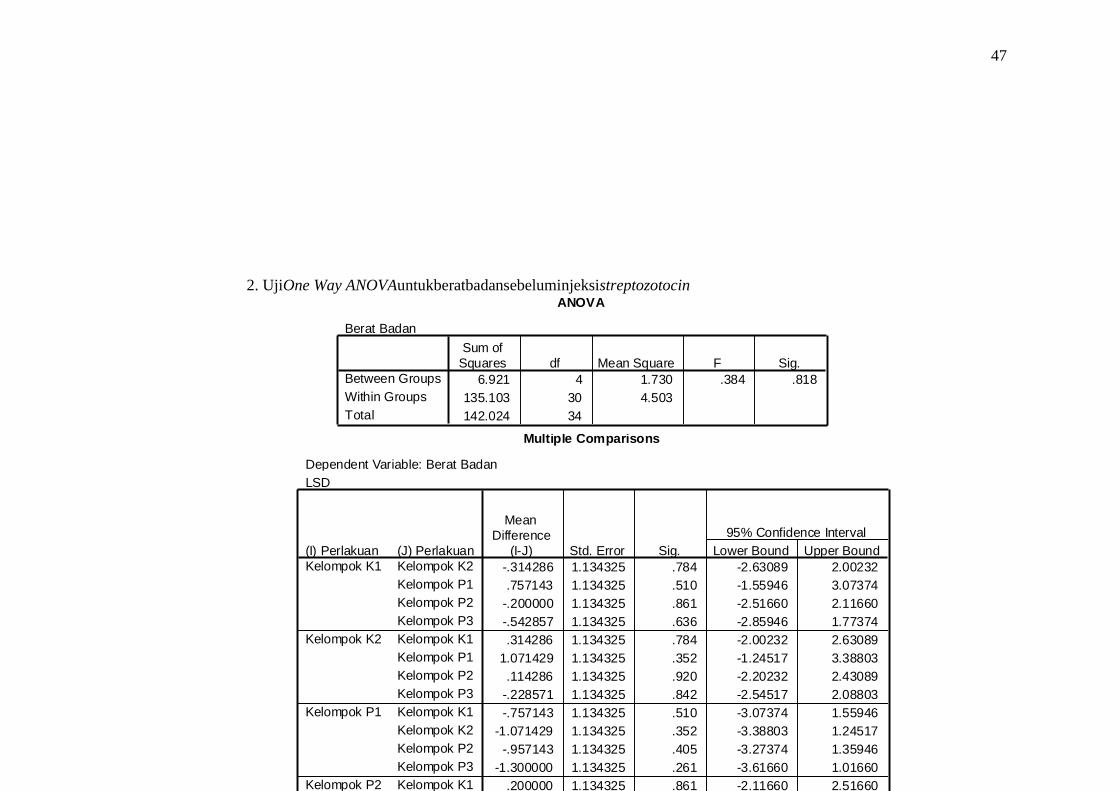
Multiple Comparisons
Dependent Variable: Berat Badan
LSD
-.314286 1.134325 .784 -2.63089 2.00232
.757143 1.134325 .510 -1.55946 3.07374
-.200000 1.134325 .861 -2.51660 2.11660
-.542857 1.134325 .636 -2.85946 1.77374
.314286 1.134325 .784 -2.00232 2.63089
1.071429 1.134325 .352 -1.24517 3.38803
.114286 1.134325 .920 -2.20232 2.43089
-.228571 1.134325 .842 -2.54517 2.08803
-.757143 1.134325 .510 -3.07374 1.55946
-1.071429 1.134325 .352 -3.38803 1.24517
-.957143 1.134325 .405 -3.27374 1.35946
-1.300000 1.134325 .261 -3.61660 1.01660
.200000 1.134325 .861 -2.11660 2.51660
-.114286 1.134325 .920 -2.43089 2.20232
.957143 1.134325 .405 -1.35946 3.27374
-.342857 1.134325 .765 -2.65946 1.97374
.542857 1.134325 .636 -1.77374 2.85946
.228571 1.134325 .842 -2.08803 2.54517
1.300000 1.134325 .261 -1.01660 3.61660
.342857 1.134325 .765 -1.97374 2.65946
(J) Perlakuan
Kelompok K2
Kelompok P1
Kelompok P2
Kelompok P3
Kelompok K1
Kelompok P1
Kelompok P2
Kelompok P3
Kelompok K1
Kelompok K2
Kelompok P2
Kelompok P3
Kelompok K1
Kelompok K2
Kelompok P1
Kelompok P3
Kelompok K1
Kelompok K2
Kelompok P1
Kelompok P2
(I) Perlakuan
Kelompok K1
Kelompok K2
Kelompok P1
Kelompok P2
Kelompok P3
Mean
Difference
(I-J) Std. Error Sig. Lower Bound Upper Bound
95% Confidence Interval
ANOVA
Berat Badan
6.921 4 1.730 .384 .818
135.103 30 4.503
142.024 34
Between Groups
Within Groups
Total
Sum of
Squares df Mean Square F Sig.
2. UjiOne Way ANOVAuntukberatbadansebeluminjeksistreptozotocin

48
Multiple Comparisons
Dependent Variable: Glukosa Darah
LSD
3.285714 9.805732 .740 -16.74026 23.31169
1.714286 9.805732 .862 -18.31169 21.74026
3.571429 9.805732 .718 -16.45455 23.59741
1.571429 9.805732 .874 -18.45455 21.59741
-3.285714 9.805732 .740 -23.31169 16.74026
-1.571429 9.805732 .874 -21.59741 18.45455
.285714 9.805732 .977 -19.74026 20.31169
-1.714286 9.805732 .862 -21.74026 18.31169
-1.714286 9.805732 .862 -21.74026 18.31169
1.571429 9.805732 .874 -18.45455 21.59741
1.857143 9.805732 .851 -18.16883 21.88312
-.142857 9.805732 .988 -20.16883 19.88312
-3.571429 9.805732 .718 -23.59741 16.45455
-.285714 9.805732 .977 -20.31169 19.74026
-1.857143 9.805732 .851 -21.88312 18.16883
-2.000000 9.805732 .840 -22.02598 18.02598
-1.571429 9.805732 .874 -21.59741 18.45455
1.714286 9.805732 .862 -18.31169 21.74026
.142857 9.805732 .988 -19.88312 20.16883
2.000000 9.805732 .840 -18.02598 22.02598
(J) Perlakuan
Kelompok K2
Kelompok P1
Kelompok P2
Kelompok P3
Kelompok K1
Kelompok P1
Kelompok P2
Kelompok P3
Kelompok K1
Kelompok K2
Kelompok P2
Kelompok P3
Kelompok K1
Kelompok K2
Kelompok P1
Kelompok P3
Kelompok K1
Kelompok K2
Kelompok P1
Kelompok P2
(I) Perlakuan
Kelompok K1
Kelompok K2
Kelompok P1
Kelompok P2
Kelompok P3
Mean
Difference
(I-J) Std. Error Sig. Lower Bound Upper Bound
95% Confidence Interval
ANOVA
Glukosa Darah
58.686 4 14.671 .044 .996
10096.000 30 336.533
10154.686 34
Between Groups
Within Groups
Total
Sum of
Squares df Mean Square F Sig.
3. Uji One Way ANOVA untuk glukosa darah sebelum injeksi streptozotocin

49
4. Uji normalitas data saat terminasi

50
Tests of Normality
.217 5 .200* .931 5 .606
.195 5 .200* .973 5 .895
.217 5 .200* .905 5 .436
.273 5 .200* .900 5 .409
.314 5 .120 .825 5 .127
.290 5 .195 .879 5 .305
.257 5 .200* .947 5 .713
.309 5 .133 .825 5 .127
.346 5 .050 .802 5 .084
.155 5 .200* .967 5 .856
.216 5 .200* .934 5 .623
.113 5 .200* .999 5 1.000
.262 5 .200* .934 5 .621
.184 5 .200* .955 5 .774
.233 5 .200* .894 5 .378
Berat badan K1
Berat badan K2
Berat badan P1
Berat badan P2
Berat badan P3
Glukosa darah K1
Glukosa darah K2
Glukosa darah P1
Glukosa darah P2
Glukosa darah P3
Fibrosis K1
Fibrosis K2
Fibrosis P1
Fibrosis P2
Fibrosis P3
Statistic df Sig. Statistic df Sig.
Kolmogorov-Smirnova
Shapiro-Wilk
This is a lower bound of the true significance.*.
Lilliefors Significance Correctiona.

51
Multiple Comparisons
Dependent Variable: Berat badan
LSD
6.940000* 2.036585 .003 2.69176 11.18824
2.300000 2.036585 .272 -1.94824 6.54824
2.600000 2.036585 .216 -1.64824 6.84824
2.560000 2.036585 .223 -1.68824 6.80824
-6.940000* 2.036585 .003 -11.18824 -2.69176
-4.640000* 2.036585 .034 -8.88824 -.39176
-4.340000* 2.036585 .046 -8.58824 -.09176
-4.380000* 2.036585 .044 -8.62824 -.13176
-2.300000 2.036585 .272 -6.54824 1.94824
4.640000* 2.036585 .034 .39176 8.88824
.300000 2.036585 .884 -3.94824 4.54824
.260000 2.036585 .900 -3.98824 4.50824
-2.600000 2.036585 .216 -6.84824 1.64824
4.340000* 2.036585 .046 .09176 8.58824
-.300000 2.036585 .884 -4.54824 3.94824
-.040000 2.036585 .985 -4.28824 4.20824
-2.560000 2.036585 .223 -6.80824 1.68824
4.380000* 2.036585 .044 .13176 8.62824
-.260000 2.036585 .900 -4.50824 3.98824
.040000 2.036585 .985 -4.20824 4.28824
(J) Perlakuan
Kelompok K2
Kelompok P1
Kelompok P2
Kelompok P3
Kelompok K1
Kelompok P1
Kelompok P2
Kelompok P3
Kelompok K1
Kelompok K2
Kelompok P2
Kelompok P3
Kelompok K1
Kelompok K2
Kelompok P1
Kelompok P3
Kelompok K1
Kelompok K2
Kelompok P1
Kelompok P2
(I) Perlakuan
Kelompok K1
Kelompok K2
Kelompok P1
Kelompok P2
Kelompok P3
Mean
Difference
(I-J) Std. Error Sig. Lower Bound Upper Bound
95% Confidence Interval
The mean difference is significant at the .05 level.*.
ANOVA
Berat badan
126.476 4 31.619 3.049 .041
207.384 20 10.369
333.860 24
Between Groups
Within Groups
Total
Sum of
Squares df Mean Square F Sig.
5. Uji One Way ANOVA untuk berat badan saat terminasi

52
Multiple Comparisons
Dependent Variable: Glukosa darah
LSD
-361.00000* 39.322563 .000 -443.02543 -278.97457
-269.20000* 39.322563 .000 -351.22543 -187.17457
-279.20000* 39.322563 .000 -361.22543 -197.17457
-270.40000* 39.322563 .000 -352.42543 -188.37457
361.000000* 39.322563 .000 278.97457 443.02543
91.800000* 39.322563 .030 9.77457 173.82543
81.800000 39.322563 .051 -.22543 163.82543
90.600000* 39.322563 .032 8.57457 172.62543
269.200000* 39.322563 .000 187.17457 351.22543
-91.800000* 39.322563 .030 -173.82543 -9.77457
-10.000000 39.322563 .802 -92.02543 72.02543
-1.200000 39.322563 .976 -83.22543 80.82543
279.200000* 39.322563 .000 197.17457 361.22543
-81.800000 39.322563 .051 -163.82543 .22543
10.000000 39.322563 .802 -72.02543 92.02543
8.800000 39.322563 .825 -73.22543 90.82543
270.400000* 39.322563 .000 188.37457 352.42543
-90.600000* 39.322563 .032 -172.62543 -8.57457
1.200000 39.322563 .976 -80.82543 83.22543
-8.800000 39.322563 .825 -90.82543 73.22543
(J) Perlakuan
Kelompok K2
Kelompok P1
Kelompok P2
Kelompok P3
Kelompok K1
Kelompok P1
Kelompok P2
Kelompok P3
Kelompok K1
Kelompok K2
Kelompok P2
Kelompok P3
Kelompok K1
Kelompok K2
Kelompok P1
Kelompok P3
Kelompok K1
Kelompok K2
Kelompok P1
Kelompok P2
(I) Perlakuan
Kelompok K1
Kelompok K2
Kelompok P1
Kelompok P2
Kelompok P3
Mean
Difference
(I-J) Std. Error Sig. Lower Bound Upper Bound
95% Confidence Interval
The mean difference is significant at the .05 level.*.
ANOVA
Glukosa darah
377364.2 4 94341.040 24.405 .000
77313.200 20 3865.660
454677.4 24
Between Groups
Within Groups
Total
Sum of
Squares df Mean Square F Sig.
6. Uji One Way ANOVA untuk glukosa darah saat terminasi

53
7. 4. UjiOne Way ANOVA untuk luas penampang sel/hipertrofi
ANOVA
Hipertrofi (Luas Penampang)
Sum of Squares df Mean Square F Sig.
Between Groups 7471.755 4 1867.939 2.073 .122
Within Groups 18021.214 20 901.061
Total 25492.969 24
Multiple Comparisons
Dependent Variable: Hipertrofi (Luas Penampang)
LSD
(I) Kelompok Perlakuan (J) Kelompok Perlakuan Mean Difference Std. Error Sig. 95% Confidence Interval
54
(I-J) Lower Bound Upper Bound
K1
K 2 -10.758589 18.984843 .577 -50.36028 28.84310
P 1 -10.193361 18.984843 .597 -49.79505 29.40833
P 2 -47.988136* 18.984843 .020 -87.58983 -8.38645
P 3 -3.091924 18.984843 .872 -42.69361 36.50977
K2
K 1 10.758589 18.984843 .577 -28.84310 50.36028
P 1 .565228 18.984843 .977 -39.03646 40.16692
P 2 -37.229548 18.984843 .064 -76.83124 2.37214
P 3 7.666665 18.984843 .691 -31.93502 47.26835
P 1
K 1 10.193361 18.984843 .597 -29.40833 49.79505
K 2 -.565228 18.984843 .977 -40.16692 39.03646
P 2 -37.794776 18.984843 .060 -77.39647 1.80691
P 3 7.101437 18.984843 .712 -32.50025 46.70313
P 2
K 1 47.988136* 18.984843 .020 8.38645 87.58983
K 2 37.229548 18.984843 .064 -2.37214 76.83124
P 1 37.794776 18.984843 .060 -1.80691 77.39647
P 3 44.896212* 18.984843 .028 5.29452 84.49790
P 3
K 1 3.091924 18.984843 .872 -36.50977 42.69361
K 2 -7.666665 18.984843 .691 -47.26835 31.93502
P 1 -7.101437 18.984843 .712 -46.70313 32.50025
P 2 -44.896212* 18.984843 .028 -84.49790 -5.29452
*. The mean difference is significant at the 0.05 level.

59
LAMPIRAN 6. LEMBAR PERUBAHAN KTI
LEMBAR PERUBAHAN KTI
Yang bertanda tangan di bawah ini:
Nama : dr. Mochamad Ali Sobirin
NIP : 197806132008121002
Bagian : Farmako
telah menyetujui perubahan KTI untuk mahasiswa
Nama : Muhammad Avicenna Abdul Syukur
NIM : G2A 009 036
Dari semula judul
PENGARUH KOMBINASI PEMBERIAN EXERCISE TRAINING DAN CURCUMIN TERHADAP
APOPOTOSIS MIOSIT PADA MENCIT DENGAN DIABETES MELLITUS YANG DIINDUKSIKAN
DENGAN STREPTOZOTOCIN
Berganti variabel apoptosis menjadi hipertrofi dan curcumin menjadi ekstrak kunyit, dengan
desain penelitian yang serupa, sehingga judul menjadi
PENGARUH KOMBINASI EXERCISE TRAINING DAN EKSTRAK KUNYIT (Curcuma domestica)
TERHADAP HIPERTROFI MIOSIT MENCIT DENGAN DIABETES MELLITUS YANG DIINDUKSIKAN
DENGAN STREPTOZOTOCIN
Semarang, Agustus 2013
Pembimbing
dr. Mochamad Ali Sobirin NIP. 197806132008121002

60
LAMPIRAN 7. DAFTAR RIWAYAT HIDUP
Identitas
Nama : Muhammad Avicenna Abdul Syukur
NIM : G2A 009 036
Tempat/tanggal lahir : Jakarta, 7 Agustus 1990
Jenis kelamin : Laki - laki
Alamat : Jalan Lempongsari Gang II No. 507A
Nomor Telepon/HP : +62857 2586 8439
Alamat email : [email protected]
Riwayat Pendidikan Formal
1. SD : SDN 02 Salatiga, lulus tahun : 2003
2. SMP : SMPN 1 Salatiga, lulus tahun : 2006
3. SMA : SMA Taruna Nusantara, lulus tahun : 2009
4. FK UNDIP : Masuk tahun : 2009
Keanggotaan organisasi
1. Komandan Tingkat Angaktan 2009 (2009-Sekarang)
2. Rohis KU Undip (2009-2011)
3. Staf Ahli Bagian Keuangan BEM FKKM (2012)
4. KRESNA FK Undip (2011-Sekarang)
5. Ketua Ikatan Alumni SMA TN Cabang Semarang (2011)
Pengalaman mengikuti lomba karya ilmiah
1. Finalis Lomba Poster Ilmiah Scientific Fair FK Undip (2011)