d. pp. 779--804, 1992 printed in great britain. all rights ... · steroid end product is listed...
TRANSCRIPT

d. Steroid Biochem. Molec. Biol. Vol. 43, No. 8, pp. 779--804, 1992 09600760/92 $5.00+0.00 Printed in Great Britain. All rights reserved Copyright © 1992 Pcrgamon Press Ltd
S T E R O I D O G E N I C E N Z Y M E S :
S T R U C T U R E , F U N C T I O N , A N D R O L E I N R E G U L A T I O N
O F S T E R O I D H O R M O N E B I O S Y N T H E S I S
ISRAEL HANUKOGLU Department of Hormone Research, Weizmann Imtitute of Science, Rehovot 76100, Israel
(Fax: 972-8-344116)
S-mmmT--In the pathways of steroid hormone biosynthesis there are two major types of enzymes: cytochromes P450 and other steroid oxidoreductases. This review presents an over- view of the function and expression of both types of enzymes with emphasis on steroidogenic P450s. The final part of the review on regulation of steroidogenesis includes a description of the normal physiological fluctuations in the steroid output of adrenal cortex and gonads, and provides an analysis of the relative role of enzyme levels in the determination of these fluctu- ations. The repertoire of enzymes expressed in a steroidogenic cell matches the cell's capacity for the biosynthesis of specific steroids. Thus, steroidogenic capacity is regulated mainly by tissue and cell specific expression of enzymes, and not by selective activation or inhibition of enzymes from a larger repertoire. The quantitative capacity of steroidogenic cells for the biosynthesis of specific steroids is determined by the levels of steroidogenlc enzymes. The major physiological variations in enzyme levels, are generally associated with parallel changes in gene expression. The level of expression of each steroidogenlc enzyme varies in three characteristics: (a) tissue- and cell-specific expression, determined during tissue and cell differentiation; (b) basal expression, in the absence of trophic hormonal stimulation; and (c) hormonal signal regulated expression. Each of these three types of expression probably represent the function- ing of distinct gene regulatory elements. In adult steroidogenic tissues, the levels of most of the ceil- and tissue-specific steroidogenic enzymes depend mainly on trophic hormonal stimulation mediated by a complex network of signal transduction systems.
OUTLINE
1. Introduction 2. Enzymes in the Initial Pathways of Steroido-
genesis 2.1. P450scc 2.2. 3~-HSD 2.3. P450c17 2.4. P450c21 2.5. P450cll and P450c18 2.6. P450arom 2.7. 17~-HSD 2.8. Enzymes that further metabolize steroids
3. Biochemistry and Molecular Biology of Steroidogenic P450 Systems 3.1. Enzymology of P450 systems 3.2. Intracellular targeting of the P450
system enzymes 3.3. Sequence and structural similarities
4. Role of Enzyme Expression in Regulation of Steroidogenesis
Proceedings of the First International Symposium on A Molecular View of Steroid Biosynthesis and Metabolism, Jerusalem, Israel, 14-17 October 1991.
4.1. Enzyme expression as a determinant of steroidogenic capacity
4.2. Methodological issues 4.3. Adrenal cortex 4.4. Testis 4.5. Ovary 4.6. Mechanisms of regulation of steroido-
genic enzyme levels 4.7. Intracellular regulators of steroidogcnic
enzyme expression
1. INTRODUCTION
The concentrations of steroid hormones in blood fluctuate with a specific periodicity, or in response to physiological or pathological changes, to regulate diverse processes in the body. In mammals there are three endocrine organs that specialize in steroid hormone production: adrenal cortex, ovary, and testis. During pregnancy, placenta develops as an additional major source of steroid hormones. The steroid hormone output of these organs is regulated by specific extracellular hormones and
779

780 ISRAEL HANUKOGLU
._ ii~ii
c ~ iiii!ili!iiii
¢ ii!~iiiiiiiii Pregnenolone ~i~i~ -- 17-OH-Pmgnenolone
Proou~rone i~i~i~i - 17-OH-Progeaterone .....
Codicosterone Corlisol
18-OH-Cortlcosterone
Aldoeterone
! iiL il
, DHEA ii ~i~::::
, Andromce4 ione N~i: FJtrone
1 1 1 1 END PRODUCT: MINERALOCORTICOID GLUCOCORTICOID ANDROGEN ESTROGEN
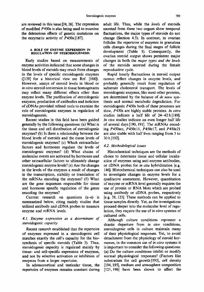
Fig. 1. Pathways of initial steps of steroid hormone biosynthesis in the adrenal cortex and the gonadal cells. The arrows mark substrate conversion to product by the enzyme listed in the shaded area. The enzymes in the shaded area with borders (P450sec, P450cl I and P450c18) are located inside the mito- chondria. The other enzymes are located in the endoplasmic reticulum. The physiological function of each steroid end product is listed below the frame. The steroids that appear in this figure may be further metabolized within the steroidogenic tissues or in other organs by the enzyme listed in Table 2. DHEA:
3p-hydroxy-5-androsten- 17-one (dehydroepiandosterone).
J
factors that activate a series of intracellular processes that ultimately change the steroid output.
In our current understanding, steroid hormone output depends on the rate of steroid metabol- ism, i.e. biosynthesis and catabolism[l]. The pathways of steroid hormone biosynthesis start with cholesterol (Fig. 1). Cholesterol is stored in esterified form inside lipid vesicles. Trophic hormones activate a chain of reactions that lead to the hydrolysis of cholesterol esters into free cholesterol, and the transport of cholesterol into mitochondria where it is converted to pregnenol- one by P450scc [2-4]. After this rate-limiting step, subsequent biosynthetic steps proceed with the flow of substrates through the enzyme systems located in the endoplasmic reticulum and mitochondria. Steroid hormones are hydro- phobic molecules that can penetrate biological membranes, and flow into the blood stream after their synthesis without being stored in intracellular vesicles. An increase in the blood levels of steroid hormones is dependent on the continual synthesis and secretion of steroids. Tissue levels of steroids rise transiently after
hormonal stimulation but even these high concentrations are not sufficient to supply enough steroids to increase blood levels [36, 37]. This contrasts with the mode of secretion of peptide hormones and neurotransmitters which accumulate in intracellular vesicles and are secreted in response to specific stimuli.
Thus, steroid hormone output is mainly regulated by events that ultimately affect steroid production through four parameters or processes:
(1) Steroidogenic enzyme level, determined by transcription, stability and translation of the mRNAs encoding the enzymes.
(2) Steroidogenic enzyme activity, determined by the conditions of the intracellular milieu, cofactor availability, or the post- translational modification of the enzymes.
(3) Substrate availability, generally deter- mined by cholesterol mobilization and transport to the mitochondrial P450scc which catalyzes the first step in the pathways of steroid biosynthesis.
(4) Tissue growth, determined by cell division

Steroidogenic enzymes 781
Table 1. Enzymes that catalyze the initial reaction in the pathways of steroid hormone biosynthesis (see Fig. I)
Nomenclature
Enzyme Gene Reaction Reviews
P450 type P 4 5 0 ~ C Y P l lA
P450cl I C Y P I I B I
P450cl8 C Y P I I B 2
P450c17 C Y P I 7
P450c21 CYP21 P450arom C Y P I 9
Oxidoreductase (dehydrogenase) 3#-HSD HSD3B
17p-HSD
Cholesterol side chain cleavage 2-4, (5) (3 successive monooxygenations) C-I 1 hydroxylation 4, 6, (7-9) C- 18 hydroxylation C- 18 hydroxylation 6, (7-10) C-I 8 oxidation 17,, -Hydroxylation 11, (I 2) C 17---C20 bond cleavage C-21 hydroxylation ! 1, (13, 14) Steroid ring-A aromatization 15, 16 (3 successive monooxygenations)
type 3p-Hydroxysteroid dehydrogenation 17 (5) 5-Ene-4-ene isomerization 17/I-Hydroxysteroid dehydrogenation 18-20, (21) 17-Keto-steroid reduction
The references in parentheses include description of inborn deficiencies of enzyme. The table includes at most only two reactions for each enzyme, based on the highest amount of products observed in in vitro incubations with the enzyme. All the enzymes listed catalyze additional reactions with different stereoselectivities, but at lower yields than the major reactions listed above.
and multipScation, as in the corpus luteum formation.
Several of these processes may operate con- currently to determine the final steroid hormone output. For example, growth of corpus luteum is accompanied by changes in the expression of steroidogenic enzymes (Section 4.5). Trophic hormonesmay also activate several of these four processes simultaneously. Whereas, cholesterol supply can be increased within minutes, the induction of enzyme synthesis may require many hours. Hence, these actions of stimulatory hormones have been referred to as 'rapid' vs 'delayed' or 'trophic'. However, the period of initiation of these responses may overlap. For example, brief (30 min) stimulation of adreno- cortical cells with ACTH leads to both a rapid increase in steroid secretion and a delayed increase in enzyme synthesis that peaks 36 h later [38].
Among the 4 processes mentioned, this review concentrates on the role of steroidogenic enzyme expression and activity as a determinant of steroidogenic capacity. Studies on the tran- scriptional regulatory sequences in genes that encode the enzymes are the subject of other reviews in this issue[16,39-42]. Cholesterol transport is also reviewed in another section of this issue [2, 43].
In the pathways of steroid hormone biosyn- thesis there are two major types of enzymes: cytochromes P450 and other steroid oxido- reductases (Fig. 1 and Table 1). Initially this review presents an overview of the function and
expression of both types of enzymes and then expounds on the structure and regulation of steroidogenic P450s. Steroid dehydrogenases [17], and P450s involved in the biosynthesis of vitamin D and sterols are reviewed in other sections of this issue [44].
The final part of this review on the regulation of steroidogenesis includes a description of the normal physiological fluctuations in the steroid output of adrenal cortex and gonads and provides an analysis of the relative role of the enzyme levels in the determination of these fluc- tuations (for reviews on placental steroidogenesis see Refs [45, 46]).
2. E N Z Y M E S IN T H E INITIAL P A T H W A Y S OF S T E R O I D O G E N E S I S
The initial steps of steroid biosynthesis in steroidogenic tissues are shown in Fig. 1. Activity and expression of the enzymes in different tissues are listed in Tables 1-3. During the past decade, cDNAs for nearly all the
Table 2. A partial list of enzymm that further metabolize steroids
Itefe~nc~
P450 type P450s that hydroxylate steroids at positions 22-29 2% 2p, 6~, 7,,, 15,,, 15p, and 16~
Oxidoreductaae (dehydrogenase) type 3,, -OH-steroid dehydrogenase 30 5~-Rednctase 31 5F-Rednctase I 1 # -OH.~teroid dehydrogenaae 8 20~-OH-steroid dehydrogenue 32 20# -OH-steroid dehydrogenase
Conjugating enzyme type Sulfotrnosfera~ 33 UDP glncuronosyltrnn~erase 34, 35

782 ISRAEL HANUKOGLU
Table 3. Specificity o f expression o f steroidogenic enzymes in different ceil types in the adrenal cortex, ovary and testis
Adrenal cortex Ovary Test i s
Luteinized Luteinized Granulosa Granulosa thcca granulosa Leydig
Zona Zona Theca (preantral (preovulatory (corpus (corpus (interstitial glomerulosa faseiculata interna follicle) follicle) luteum) luteum) tissue)
P450-~c 3 p - H S D P450cl 1 P450cl 8 P450cl 7 P450c2 I P450arom 17p-HSD
Major steroid product
+ + + -- + + + " + + + + + + + + + + + + + - - + . . . . . .
+ _{_b + _[_ .{_
+ + . . . . . . . . . . + -- + c -- ? + + . . . . +
Miueralo- Glucocorticoid Androgen Estrogen Progestins Progesterone Prosesterone Androgen corticoid Androgen progesterone Androgen Estrogen
• + + , indicates much higher levels only relative to ovarian granulosa ceils and not other ceils. bNot expressed in rat, hamster and rabbit adrenals. CExpression dependent on species.
major steroidogenic enzymes have been isolated (Table 4). One of the most important findings that emerge from molecular studies is that almost every steroidogenie enzyme is encoded by only one gene (Table 4). For key enzymes, e.g. P450see, different genes that are specifically expressed in different tissues do not exist. Thus,
a single gene for each enzyme must carry all the signals for its specific regulation in different tissues and cells. A second most important find- ing is that some of the enzymes, e.g. P450c17, P450c18, catalyze more than one step in the pathway (Fig. 1). Therefore, it is recommended that the enzymes be referred to with a single
Table 4. Characteristics o f the genes and m R N A s o f cytochromes P450 and their electron transfer proteins
Gene m R N A Protein Gene length length length
Species (n) Chromosome (kb) (kb) (aa) (preseq.)" References
Mitochondrial Adrenodoxin reductase Man I 17 12 2 459 (491) 47-51
Cow I - 6-12 2 460 (492) 47, 52, 53
Andrenodoxin Man 2 (2) b 11 (20 , 21) >20 I, 1.4, 1.7 124 (184) 51, 54, 55 Cow I - > 27 I, 1.4, 1.7 128 (186) 56-59
P4508¢c Man 1 15 >20 2 482(521) 51 ,60-62 Cow 1 - - 2 481 (520) 63-65 Pig . . . . 481 (520) 66 Rat 1 - - 2 490 (526) 67, 68
P450cl I Man 1 8 - - 479 (503) 7, 8, 69, 70 Cow 2 (2) - - 1.8, 4.1 479 (503) 71-74 Rat . . . . 475 (499) 6, 75 Mouse I 15 7 - (500) 76
P450cl 8 Man I 8 - - 479 (503) 7, 8, 69, 77 Rat - (7) - - - 476 (510) 6, 78 Mouse 1 15 7 2.6 (500) 76
Microsomal P 4 50red.
P450cl7
P450c21
P450arom
H u m a n 1 - - - 676 79, 80 Porcine . . . . 678 81 Rabbit - - - 2.4 679 82 Rat 1 - 20 2.7 678 79, 83-85 Mouse I 6 - - - - - - 79
Man I 10 6.6 1.9 508 51, 86--88 Cow 1 - 6.6 1.9 509 89, 90 Rat 1 - - 1.9 507 91, 92 Mouse 1 19 - - 507 93 Chicken - - - 1.9 508 94
Man 1 (1 ) 6 3 .3 2 494-495 13, 95, 96 Cow 1 (1) - 3.4 2.2, 2.4 496 97-101 Mouse I ( I ) 17 3.1 2.2 487 102, 103
Man I 15 > 70 2 .9 , 3.4 503 16, 104-108 R a t . . . . 508 109 Mouse 1 9 - 2 .1 , 2.5 503 I i0 Chicken - - - 4 507 I ! 1 Trout - - - 2.6 522 ! 12
"The p r e s e q u e n c e o f the protein includes amino terminal extension signal peptide. el 'he number o f l~endogeues are listed in parentheses.

Steroidogenic enzymes 783
P450 nomenclature and not with a commonly used "hydroxylase" name that would require the use of two or more hydroxylase names for a single enzyme molecule [25].
The first two major steps in the biosynthesis of steroids are common to all steroidogenic organs: (1) conversion of cholesterol to preg- nenolone by P450scc inside the mitochondria; and (2) conversion of pregnenolone to pro- gesterone by 3fl-hydroxysteroid dehydrogenase (3fl-HSD) in the endoplasmic reticulum (Fig. 1). The following steps of steroidogenesis are catalyzed by enzymes expressed only in some steroidogenic cell types (Table 3). The cell- specific expression of these enzymes determines the capacity of each cell to synthesize different steroid hormones (Table 3). The major functions of these enzymes and their sites of expression are listed below.
2.1. P450scc
This enzyme catalyzes the first and rate- limiting step in the biosynthesis of steroid hormones [2-4, 113, 114]. It converts cholesterol to pregnenolone in three successive monooxy- genations (hydroxylations at C-22, followed by C-20, and finally cleavage of the C-20,22 bond). Hydroxylated intermediates of cholesterol bind very tightly to P450scc and do not show significant dissociation from the enzyme [4]. In contrast, the final product pregnenolone has a dissociation constant 40 to 600-fold higher than those of intermediates, facilitating its release from the enzyme [4]. Purified P450scc can effici- ently catalyze side chain cleavage of cholesterol sulfate, and 6fl-hydroxylation of deoxycortico- sterone [115, 116]. Yet, it is not known whether these reactions represent a major activity in vivo.
P450scc is expressed in all three zones of the adrenal cortex [117, 118]. In the testis it is found in the steroidogenic Leydig cells[ll9]. In the ovary it is expressed in the theca interna; its expression in the granulosa cells depends on the stage of growth of the follicle as discussed in Section 4.5. In addition to steroidogenic tissues the expression of P450scc has been detected in the brain [120], but at levels that are more than an order of magnitude lower (Table 5).
2.2. 3fl-HSD
This enzyme has two major catalytic activities which in concert convert 3fl-hydroxy-5-ene steroids into 3-keto-4-ene (Table 1). In contrast to steroidogenic P450s each of which is encoded by a single gene, in the human, rat and mouse
Table 5. The concentration of mitochondrial P450scc in steroido- genie tissues and brain
Concentration Tissue (prnol/m s protein) ° Reference
Adrenal cortex 400 121 Corpus luteum 80-400 121,122 Ovary < 5 121,122 Placenta < 50 123 Testis Leydig cells - 119 Brain < 10 124
'The values shown are per total tissue pro~'in. The concentrations for placenta and brain were calculated assuming that the mito- cbondrial fraction represents 20% of the total cellular protein.
genomes there are at least 2-3 homologous genes encoding 3fl-HSDs that share 80-94% sequence identity within each species [17]. Two types of 3fl-HSDs from human and rat can use either pregnenolone, 17-OH-pregnenolone or dehydroepiandosterone (DHEA) as substrates [17]. The different types have different Km values for the same substrates. However, each type of enzyme can use pregnenolone or DHEA as sub- strates with similar K,, values [17]. Rat type I 3fl-HSD also shows 17fl-HSD activity with 5u-androstane steroids but not with estradiol, estrone, androstenedione, or testosterone [125].
In the human adrenal cortex and gonads, but not placenta, only type II 3~-HSD is expressed, whereas in the rat both type I and II are found in these tissues [17]. Immunohistochemical studies reveal localization of 3fl-HSD in the same steroidogenic cells as P450scc in all three zones of the adrenal cortex, in the interstitial Leydig cells of the testis, but not in seminiferous tubules, in the theca interna of the ovary and in corpora lutea [126]. Immunostaining failed to detect 3fl-HSD in rat granulosa cells [126] even though these cells display high 3fl-HSD activity converting pregnenolone to progesterone even before the induction of P450scc [127]. In con- trast to stcroidogenic P450s, 3fl-HSD activity is present in a wide range of tissues [17].
2.3. P450c17
This enzyme catalyzes two key reactions: (a) 17u-hydroxylation of C21 steroids; and (b) cleavage of the C17---C20 bond of C21 steroids. The 170t-hydroxylation is a required step in cortisol biosynthesis, whereas the C17---C20 bond side chain cleavage is essential for the biosynthesis of androgens (Fig. 1). Genetic deficiencies affecting either one or both activities have been characterized [12].
Immunohistochemical analysis shows that P450c17 is present in the zona fasciculata and reticularis but not in zona glomerulosa of the porcine adrenal [128]. This is consistent with the

784 ISRAEL HANUKOGLU
role of zona glomerulosa as the site of mineralo- corticoid biosynthesis where 17~-hydroxylation is not required (Fig. 1). P450c17 is not expressed in the rat adrenal cortex; consequently cortico- sterone, and not cortisol, is the major glucocor- ticoid in this species and in some other rodents [129]. However, P450c17 is expressed in guinea pig adrenal cortex [130-134].
In the ovary, P450c17 is expressed in theca interna cells[135, 136]. Antibody or cDNA probes show very low expression of P450c17 in granulosa cells of humans and rats[135- 137]. Thus, theca interna cells can synthesize androgens, but granulosa cells which produce estrogens are dependent on androgen precursor supply from theca interna[138]. This process is called the two cell hypothesis of follicular estrogen production [138].
The fact that P450cl 7 catalyzes two reactions, poses a conundrum: What determines the relative flow of steroids through glucocorticoid and androgen pathways (see Fig. 1)? Adrenal cortex capacity for androgen synthesis changes during puberty[139]. These pathways are probably developmentally regulated by changes in the function of the zona reticularis. In Leydig cells, in the absence of P450c21 there is no alternative pathway but to androgen production (see Fig. 1).
The C17--C20 bond cleavage activity of P450c17 depends on the concentration of elec- tron transfer protein P450 reductase, and it can be increased to the level of the 17~-hydroxyl- ation activity [11]. Thus, changing levels of this reductase or cytochrome b5 may affect the ratio of the two activities [11,140]. Different steroido- genic tissues appear to have different levels of these proteins[l 1]. The two activities of P450c17 are also strongly dependent on steroid concentrations [141,142]. Thus, the intracellular concentrations of substrates and products prob- ably also play a role in regulating the relative rates of these activities [141,142].
The substrate specificity of P450c17 varies with species[129]. P450c17 from all species examined can hydroxylate C-17 of both preg- nenolone and progesterone, although the Km for these two substrates can differ by 10-fold in some species [11, 91,143-145]. Whereas, the rat P450cl 7 can convert both 17-OH-pregnenolone and 17-OH-progesterone into DHEA and andro- stenedione, respectively [91,143], the human and bovine P450c17 can cleave the C17--C20 bond of 17-OH-pregnenolone but not of 17-OH- progesterone [91,144, 145]. Thus, the substrate
specificity of P450c17 determines whether androgen biosynthesis proceeds mainly through pregnenolone or progesterone as shown in Fig. 1. The formation of androgen may be catalyzed without the 17~-hydroxylated substrate leaving the enzyme [141].
2.4. P450c21
This enzyme catalyzes an essential step in the synthesis of gluco- and mineralocorticoids (Fig. 1). Congenital adrenal hyperplasia which results from an inborn deficiency of this enzyme is a common genetic disorder [5, 13, 14]. Defici- ency in the P450c21 hydroxylation step, channels the steroid biosynthetic pathway in the direction of androgen production, resulting in gluco- and mineralocorticoid deficiency and excessive virilization [5, 14].
P450c21 is expressed in all three zones of the adrenal cortex [146]. Apart from the adrenal cor- tex, P450s with steroid 21-hydroxylation activity have been found in other tissues, e.g. liver [147]. However, these P450s show little similarity to the steroidogenic P450c21 and represent products of other genes [25, 147]. Immunohisto- chemical studies using anti-P450c21 antibody revealed a cross reacting protein only in the distal tubules of the bovine kidney, which is the site of the mineralocorticoid action in the kidney [146]. However, it is not known whether this protein is the product of the same gene that encodes the adrenal P450c21. Examination of the kidney by in situ hybridization using a specific P450c21 probe could resolve this question.
2.5. P450c l i and P450c18
These two enzymes are also uniquely expressed in the adrenal cortex [6, 148]. In the human and rat adrenal, P450cl 1 is expressed in the zona fasciculata which specializes in glucocorticoid production under mainly ACTH regulation. Whereas P450c18 is expressed in the zona glomerulosa and is regulated by the renin- angiotensin system [6-8, 149-151]. The human and rat P450cl 8 can catalyze aldosterone form- ation, but P450cll cannot [6-8]. Two bovine isozymes of P450cl I expressed in COS-7 cells can catalyze both corticosterone and aldosterone production[152]. However, it is not known whether these enzymes function in both zones, or whether there is a bovine P450c18 gene that is specifically expressed in zona glomerulosa.
With 11-deoxy substrates, both P450cl 1 and P450c18 also catalyze 19-hydroxylation but at

Steroidogenic enzymes 785
less than 10% of 1 lp-hydroxylation activity [7]. P450cl 1 can also hydroxylate androstenedione resulting in the formation of 11/~-hydroxylated androgens [e.g. 134]. Steroid 11/~-hydroxylation activity has been detected in the gonads of some species [153], but it is not known whether this reflects a low expression of P450cl 1 in these tissues or the activity of another enzyme.
2.6. P450arom
P450arom catalyzes the conversion of testosterone into the 17/~-estradiol (Fig. 1 and Table 1). In the ovary P450arom is expressed in granulosa cells which is the major site of estrogen production in females [136, 154] (see Section 4.5). However, this enzyme is widely expressed in many tissues besides gonads, e.g. adipocytes, breast, central nervous system, skin and placenta[15, 16]. The gene encoding P450arom is the longest amongst steroidogenic P450 genes (Table 4). This gene is also unique among P450 genes in having alternative promoters that are utilized in a tissue-specific manner [16].
2.7. 17[$-HSD
This enzyme is also referred as 17-keto- steroid reductase. It catalyzes the reversible conversion of the 17-keto and 17/~-hydroxy groups in androgens and estrogens, including androstenedione, DHEA, and 17/~-estradiol. The direction of the reaction depends on the sub- strate and cofactor [18-20]. For the conversion between androstenedione and testosterone, the porcine testicular 17fl-HSD prefers NADPH rather than NADH (Kin = 11 vs 177 #M) [155]. There are multiple 17fl-HSD isozymes with androgen or estrogen specificity [18-20]. The iso- zyme from the porcine testis also displays 200t- HSD activity at about a tenth of the 17fl-HSD activity [155].
A 17fl-HSD has been purified from placenta, and a eDNA and two in tandem homologous genes have been cloned and sequenced [19, 156]. This enzyme catalyzes the interconversion of 17/~-estradiol and estrone, and can also use androgen substrates [19]. Its Km for 17/~-estradiol is 10#M vs 250#M for testosterone[19]. Thus, this enzyme is specific for estrogens and not androgens. Its cDNA hybridizes relatively weakly to a few bands on a testis RNA blot [156]. One of these may represent a homologous mRNA that encodes a different enzyme with specificity for androgens rather than estrogens. Testicular 17/~-HSD deficiency is a rare genetic
disorder, but is observed frequently in an inbred Arab population[21]. In affected individuals androstenedione to testosterone conversion is markedly impaired [21]. The cloning of the gene that encodes the androgenic 17/~-HSD may be useful in elucidating the molecular defect in this disease.
Immunohistochemical studies using an antibody against a testicular 17//-HSD indicate that the enzyme is expressed in the testis in Leydig cells of the interstitial tissue, and in the ovary, in the theca interna, but not in granulosa ceils or corpus luteum [157]. This is consistent with androgen synthesis in testicular Leydig cells and in ovarian theca interna. Both andro- genic and estrogenic 17p-HSD activities have been observed in a wide range of tissues [20]. These activities probably do not represent the androgenic isozyme. In testicular 17I/-HSD deficiency, 17/~-HSD activity is defective only in the testis but appears normal in other tissues [21], thus supporting other findings that there are multiple isozymes with 17f/-HSD activity [18-20].
2.8. Enzymes that further metabolize steroids
The enzymes noted above catalyze the initial steps in steroidogenesis. The end products of these reactions can be further metabolized by other enzymes (Table 2) within the cell of production or after transport to cells in the same or other organs. Some properties of these enzymes are outlined below:
(1) Steroid metabolism by some of these enzymes may result in the formation of a steroid product with potent biological activity, as in the case of conversion of testosterone to 5~-dihydrotestosterone by 5~t-reductase, or inactivation of the steroid, as in the case of liver hydroxyl- ation and conjugation of steroids, or inactivation of cortisol by 1 lfl-HSD (see refs in Table 2).
(2) In contrast to the initial steroidogenic enzymes which are encoded by a single gene (Table 4), the cloning of cDNAs for some of these enzymes revealed large families of genes with many members (see refs in Table 2). The enzymes encoded by these genes have similar sequences, but different substrate specificities.
(3) While most steroidogenic enzymes have strict substrate specificities, the enzymes that further metabolize steroids generally

786 ISRAEL HANUKOGLU
(4)
have broader substrate specificities. For example, 30t-HSD and 20~-HSD can metabolize both C19 (androstane) and C21 (pregnane) steroids. Similarly, some conjugating enzymes can accommodate a broad range of steroid structures. The stereoselectivities of some enzymes may also be broader: different P450s can hydroxylate a steroid at several positions [28] and 20at-HSD also shows 3ct-HSD activity [32]. Whereas the expression of most enzymes in the initial pathways (Fig. 1) is limited to steroidogenic tissues, enzymes of further metabolism (Table 2) are expressed in a broad range of tissues besides steroido- genic organs. For example, 3ct-HSD, and 5~t-reductase activities are present in many peripheral tissues as well as all areas of the central nervous system including white matter [158, 159].
3. BIOCHEMISTRY AND MOLECULAR BIOLOGY OF STEROIDOGENIC P450 SYSTEMS
3.1. Enzymology of P450 systems
As noted above, most of the reactions in the pathways of steroid biosynthesis are catalyzed by P450 type enzymes. P450s are found in nearly all tissues of the vertebrates where they catalyze different metabolic or biosynthetic reactions generally involving small hydrophobic molecules. The steroidogenic P450s resemble other P450s in many biochemical properties. All cytochromes P450 are b type cytochromes wherein the environment of the heme forms the active site of the enzyme that binds the substrate and 02[22, 23]. CO competes with 02 for binding to this site and thus can inhibit enzymatic activity. The name P450 derives from the distinct absorption maximum at 450 nm of the reduced and CO bound form of the enzyme.
The reactions catalyzed by steroidogenic P450s are either a single hydroxylation (mono- oxygenation) at a specific position on the steroid molecule, or a series of consecutive monooxy- genations which result in C---C bond cleavage or aromatization of the steroid ring A (Table 1). Each P450-catalyzed hydroxylation has the following stoichiometry:
Steroid-H + NADPH + H ÷ + 02
Steroid-OH + NADP ÷ + n 2 0
This reaction consumes two electrons, a H +, and 02, one atom of which is incorporated into the substrate, while the second is reduced to H20 (hence mono-oxygenation). The electrons are transferred from NADPH to cytochrome P450 by specific electron cartier proteins. In the mitochondrial P450 systems this function is performed by two proteins: adrenodoxin reduct- ase, which is an FAD containing flavoprotein, and adrenodoxin, which is a ferredoxin type iron-sulfur protein [3, 4, 47, 113, 114, 160]. The microsomal P450 systems are dependent on P450 reductase which is a flavoprotein with FAD and FMN as cofactors [22, 23, 47, 161]. In a catalytic cycle, the mitochondrial P450 accepts one electron at a time from adrenodoxin, and the microsomal P450 from the FMN of P450 reductase. Thus, the cofactor FMN fulfills a role similar to that of adrenodoxin in the mitochon- drial P450 systems as a single electron acceptor and donator[161] (Fig. 2). Some microsomal P450s can also accept the second electron from cytochrome bs [11, 22, 140].
All eukaryotic P450s are membrane associ- ated enzymes. The amino termini of both the microsomal P450s and P450 reductase invari- ably contain a 20-30 residue long highly hydro- phobic segment which anchors these enzymes to the lipid bilayer of the endoplasmic reticulum, while their main body remains in the cytoplasmic side[162, 163,163a] (Fig. 2). Mitochondrial P450s are located on the matrix side of the inner mitochondrial membrane [117, 118, 164]. The mitochondrial P450s behave as highly hydro- phobic proteins, but contrary to the microsomal P450s they lack a hydrophobic amino terminal sequence that could be predicted as a membrane spanning segment [71,165, 166]. Thus, the region that anchors the mitochondrial P450s to the membrane remains unidentified. Both adrenodoxin reductase and adrenodoxin are easily solubilized proteins, and do not contain a highly hydrophobic segment[47]. However, immunocytochemical studies show that these proteins are also associated with the inner mito- chondrial membrane [167], most likely mainly by ionic interactions.
Generally the reductase component of P450 systems is present at much lower concentrations than the P450s inside cells [121,168]. This low molar ratio of reductase to P450, and kinetic studies of enzyme--enzyme interactions indicate that the P450 system enzymes do not form static complexes on the membrane and are independently mobile [22, 113, 160].

NAI
Adr. reductase
Steroidogenic enzymes
Adrenodoxin P450
787
Inner mitochondrial membrane
N A F -
P450-Reductase P450
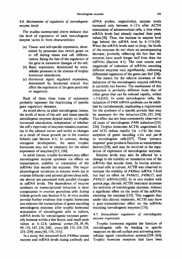
Endoplasmic reticulum membrane Fig. 2. Models for the membrane organization of the mitochondrial and microsomal P450 systems. The mitochondrial system faces the matrix (inner) side of the membrane. The microsomal system enzymes face the cytosol and both are anchored to the membrane by hydrophobic amino terminal domains. The membrane binding region of the mitochondrial P450s is not known and is depicted in analogy to
the microsomal P450s.
3.2. Intracellular targeting of the P450 system enzymes
The mitochondrial P450s, adrenodoxin reductase and adrenodoxin are synthesized in precursor forms with an amino terminal exten- sion of 30-60 residues that is absent in the mature enzymes (Table 4). These amino terminal signal peptides are necessary for the specific entry of the enzymes into mitochondria[169-171], and are cleaved by proteases upon entry of the precursor into the mitochondria[172-175]. In bovine adrenal cortex, P450scc and adrenodoxin a r e processed by two distinct proteases [173, 175]. The sequences of the extensions of these proteins share little or no similarity, are not hydrophobic, and invariably contain several basic residues (Arg and Lys) which are essential for the transfer of at least some of these proteins into mitochon- dria [169-171]. In these respects, these proteins resemble some other mitochondrial proteins [176]. The specific association of the mitochon-
drial precursor proteins with the mitochondrial membrane may be mediated by the polar heads of cardiolipin, a phospholipid that is specifically present in the inner mitochondrial membrane [177]. The bovine mitochondrial P450s can be imported only into mitochondria isolated from steroidogenic tissues, whereas the precursor of adrenodoxin which has a wider tissue distri- bution, does not show this tissue specificity [175-178]. The sequence or structural features responsible for this tissue specificity of transfer have not yet been identified.
The role of the mitochondrial targeting signals has also been examined using expression plasmids containing cDNAs with truncated or modified signal peptide sequences. Mature bovine adrenodoxin reductase and adrenodoxin sequences expressed in yeast (heterologous expression) were found in the cytosolic fraction because of the lack of the mitochondrial signal peptide[179]. Whereas, with a spliced yeast mitochondrial cytochrome c oxidase subunit

788 ISRAEL HANUKOGLU
signal peptide sequence, the expressed proteins were directed into the mitochondrial fraction [179].
Unlike the mitochondrial P450s, the micro- somal P450s and P450 reductase do not have an extension peptide or basic residues in the amino termini (Table 4). The hydrophobic amino terminus of the microsomal P450s deter- mines the membrane entry and association of these proteins [162, 163a]. Wild type P450 re- ductase expressed in yeast is normally incorpor- ated into the microsomes[180]. However, truncation of its hydrophobic amino terminus produces an enzyme that remains soluble in the cytosolic fraction [180]. Thus, this segment ap- pears to be the only segment that anchors the protein to the microsomal membrane.
3.3. Sequence and structural similarities
The biochemical similarities between cyto- chromes P450 noted above indicated some degree of structural similarity between these enzymes. The precise sequence similarities were established after the sequencing of the cDNAs for each of these enzymes. Comparison of the sequences of these enzymes revealed the following relationships [25]:
(1) The microsomal steroidogenic P450s share <40% sequence similarity with each other. Thus each P450 represents a distinct family with generally a single member. In contrast, the liver microsomal P450s, which are involved in the metab- olism of endo- and xenobiotics, belong to large gene families which include many members with > 50% sequence similarity [25].
(2) The three mitochondrial P450s, namely P450scc, P450cll and P450c18, share sequence similarity along their entire length [7, 8, 71].
(3) The mitochondrial and microsomal P450s show sequence similarity only around a limited region in the heme binding seg- ment close to the carboxy termini of the enzymes [181].
(4) The microsomal P450¢17 and P450¢21 share sequence similarity in their amino terminal regions and carboxy terminal halves.
(5) P450c17 and P450c21 share <30% over- all sequence identity with the liver micro- somal P450s. Thus, the liver microsomal and steroidogenic P450s represent distinct
but evolutionarily related families of genes.
(6) The cDNA sequences of most of the human and bovine steroidogenic P450 system enzymes have been determined (Table 4). The homologous enzymes in these two species share >70% sequence similarity.
(7) The flavoprotein reductase components of the mitochondrial and microsomal P450 systems show no sequence similarity and belong to different families of oxidoreductases [47, 52].
(8) The analysis of the sequence of the mitochondrial adrenodoxin reductase led to the definition of a new large class of oxidoreductases that share an NADP binding site consensus sequence [47, 52]. These analyses also led to the discovery of a sequence difference between NAD and NADP binding sites [52, 182].
Determination of the 3-D structure of eukary- otic P450s has been hindered by the difficulties inherent in the crystallization of hydrophobic membrane bound proteins. However, the crystal structure of a bacterial P450BM-3 which shares significant sequence homology with eukaryotic microsomal P450s has been determined[183]. This structure can serve as a model to under- stand the structure of other P450s. The 3-D structure of P450cam from Pseudomonas putida was elucidated earlier [184]. Although the struc- tures of P450BM-3 and P450cam share similar structural domains, the only significant sequence similarity between P450cam and the eukaryotic P450s is observed around the heme binding region. There were many attempts to align and match the full sequence of P450cam with eukary- otic P450 sequences. However, the elucidation of the structure of P450BM-3 showed that these previous alignments did not correctly match different structural domains of the molecules (J. A. Peterson and S. S. Boddupalli, personal communication).
Currently the structure of many P450s that metabolize steroids is being investigated by expressing modified cDNAs, chimeric cDNAs that include parts of different P450s [e.g. 185], and fused cDNAs that include both P450 and reductase [e.g. 186], in bacteria and yeasts. These studies are defining the role of specific regions and residues involved in substrate bind- ing, enzymatic activity and interaction with membrane and other molecules. Some of these
Steroidogenic enzymes 789
are reviewed in this issue [24, 26]. The expression of modified P450s is also being used to examine the deleterious effects of genetic mutations on the enzymatic activity of P450s [187].
4. ROLE OF ENZYME EXPRESSION IN REGULATION OF STEROIDOGENESIS
Early studies based on measurements of enzyme activities indicated that some changes in blood levels of steroids may result from changes in the levels of specific steroidogenic enzymes ([129] for a historical view see Ref. [188]). However, assays of steroid levels in blood or in vitro steroid conversion in tissue homogenates may reflect many different effects other than enzyme levels. The purification of steroidogenic enzymes, production of antibodies and isolation of cDNAs provided refined tools to examine the role of steroidogenic enzymes in regulation of steroidogenesis.
Recent studies in this field have been guided generally by the following questions: (a) What is the tissue and cell distribution of steroidogenic enzymes? (b) Is there a relationship between the blood levels of steroids and the tissue levels of steroidogenic enzymes? (c) Which extraceUular factors and hormones regulate the levels of steroidogenic enzymes? (d) What chains of molecular events are activated by hormones and other extracellular factors to ultimately change steroidogenic enzyme levels? (e) Are the changes in the levels of the enzymes a result of changes in the transcription, stability or translation of the mRNAs encoding the enzymes? (f) What are the gene sequences responsible for tissue and hormone specific regulation of the genes encoding the enzymes?
Current research on questions (a-e) is summarized below, citing mainly studies that utilized antibody and eDNA probes to measure enzyme and mRNA levels.
4.1. Enzyme expression as a determinant of steroidogenic capacity
Recent research established that the repertoire of enzymes expressed in a steroidogenic cell matches exactly the cell's capacity for the bio- synthesis of specific steroids (Table 3). Thus, steroidogenic capacity is regulated mainly by tissue- and cell-specific expression of enzymes, and not by selective activation or inhibition of enzymes from a larger repertoire.
In adrenocortical and testicular tissue, the repertoire of enzymes remains constant during
adult life. Thus, while the levels of steroids secreted from these two organs show temporal fluctuations, the major types of steroids do not change (Section 4.3). In contrast, in ovarian follicles the repertoire of enzymes in granulosa cells changes during the final stages of follicle development (Table 3). Consequently, the ovarian steroid output shows persistent major changes in both the major types and the levels of the steroids secreted during the female reproductive cycle.
Rapid hourly fluctuations in steroid output cannot reflect changes in enzyme levels, and probably generally result from regulation of substrate cholesterol transport. The levels of steroidogenic enzymes, like most other proteins, are determined by the balance of de novo syn- thesis and normal metabolic degradation. For steroidogenic P450s both of these processes are slow, P450s are highly stable proteins. In vitro studies indicate a half life of 24-42h[189]. In vivo studies indicate an even longer half life of several days [190, 191]. The mRNAs encod- ing P450scc, P450c11, P450c17, and P450c21 are also stable with half lives ranging from 5 to 30 h [192].
4.2. Methodological issues
Histochemical techniques are the methods of choice to determine tissue and cellular localiz- ation of enzymes using anti-enzyme antibodies, or eDNA probes for in-situ hybridization [e.g. 146]. Histochemical techniques can also be used to investigate changes in enzyme levels for a qualitative assessment. However, quantitation of enzyme or mRNA level generally requires the use of protein or RNA blots which are probed using antibody or eDNA probes, respectively [e.g. 38, 121]. These methods can be applied to tissue samples directly. Yet, as the investigations proceed deeper into the molecular level of regu- lation, they require the use of in vitro systems of cultured cells.
Although culture conditions represent a drastic departure from in vivo conditions, steroidogenic cells in culture maintain many of their physiological responses. Yet, to avoid detachment from the physiology of steroid hor- mones, in the common use of in vitro systems it is important to consider the following questions: (a) Do the culture conditions inhibit or modify normal physiological responses? (Factors like substratum for cell growth[193], cell density [194, 195], medium and atmosphere composition [121,196] have been shown to affect the

790 ISRAEL HANUKOGLU
responses of steroidogenic cells.) (b) Does the blood or local level of the examined factor show changes that parallel the physiological variation in the steroid levels? (c) Is the concentration of the factor within physiological or pharmaco- logical range? (d) Do the in vitro changes in steroidogenic capacity and enzyme levels reflect physiological changes in steroid and enzyme levels?
4.3. Adrenal cortex
The blood levels of the major steroid products of all three zones of adrenal cortex, vary in a circadian pattern in response to trophic hormones specific for each tissue[197-200]. Steroidogenic enzyme levels cannot vary drastic- ally within 12 h (see Section 4.1). Thus, circadian variations cannot result from changes in the levels of the enzymes, and most probably reflect hormonal stimulation of steroid biosynthesis via increased cholesterol transport to mitochondria. This assumption is supported by the observations that in rats the diurnal increase in plasma corticosterone is associated with a diurnal decrease in adrenal cholesteryl esters that serve as the precursor of adrenal steroids [200]. In man, the patterns of circadian variation of some adrenal androgens are different from that of cortisol, indicating that these may be regulated mainly by factors different from ACTH [199].
The blood levels of adrenocortical steroids rise rapidly in response to stress conditions that stimulate ACTH secretion [201]. After ACTH injection to human subjects, both plasma cortisol and aldosterone levels increase several fold within 1 h [139, 202]. However, the plasma level of ACTH, after injection, highly correlates with cortisol, but not aldosterone levels, indicating that while cortisol secretion is mainly determined by ACTH, aldosterone secretion is affected by multiple factors secondary to ACTH stimulation [202]. The hypothalmus-pituitary-adrenal sys- tem can also be stimulated by cytokines, e.g. interleukin-l, produced by the cells of the immune system [203]. Rapid increases in steroid secretion in response to A C T H or other factors, probably as a rule, result from stimulation of steroidogenesis via increased cholesterol mobil- ization and transport into mitochondria [2].
The activation of the hypothalarnus-pituitary- adrenal axis is vital to cope with stress and disease [201]. Under continued stress, the constant simulation of the adrenal cortex can enhance its capacity to secrete glucocorticoids by increasing the levels of key steroidogenic
enzymes. Blood glucocorticoid levels are elevated many fold in certain disease states [204, 205], while adrenal androgen levels may be suppressed [205]. Moreover, during acute illness, the responsiveness of the adrenal cortex to ACTH stimulation increases, indicating enhanced steroidogenic capacity probably due to increased steroidogenic enzyme levels (Hanukoglu A., Fried D. and Hanukoglu I., unpublished observ- ations). ACTH treatment increases the activities of certain adrenal steroidogenic enzymes in rodents [133, 206], and changes the cellular fine morphology [207]. Extensive ACTH exposure initially stimulates cellular hypertrophy of adrenocortical cells without causing cell prolifer- ation [208]. Thus, elevations in blood gluco- corticoid levels during acute disease probably do not represent adrenal tissue growth.
Plasma levels of glucocorticoids do not differ between males and females[139]. In bovine adrenal cortex, the levels of P450scc, P450cl 1, and their electron transfer proteins are similar in different animals and show no sex difference [121]. In guinea pig adrenal cortex the levels of P450c17 and P450c21 are also similar in both sexes[131]. In inbred mice, strain differences have been observed in the levels of steroidogenic enzymes in both adrenal cortex and testis [209]. Some P450s in the guinea pig adrenal cortex that are immunologically related to the liver P450s, and 5~t-reductase in the rat adrenal cortex, show sexual dimorphism and are affected by sex steroids [131, 210]. These findings indicate that sex steroids do not have a major influence on the initial steroidogenic pathway of the adrenal cortex, but determine later steps of steroid metabolism in these species. Certain steroid metabolizing enzymes in the liver are also expressed in a sexually dimorphic pattern [28, 29]. This dimorphism may have developed to deal with the exposure of each sex to a different steroid spectrum.
Daily fluctuations in the levels of ACTH are not immediately associated with changes in enzyme levels; yet, this daily intermittent stimu- lation of the adrenal cortex is essential for the maintenance of the normal levels of the steroidogenic enzyme in these tissues. In hypo- physectomized animals, the activities of many steroidogenic enzymes decrease greatly [17, 129, 190, 191], in addition to many other cellular changes [e.g. 208]. If the duration of trophic hormone deprivation is not prolonged, these changes can be reversed by the administration of trophic hormones [190, 191].

Steroidogenic enzymes 791
The secretion of aldosterone from zona glomerulosa is regulated mainly by the renin- angiotensin system and blood sodium and potassium levels [6-8, 149-151]. Angiotensin II can rapidly stimulate aldosterone secretion from glomerulosa cells by increasing cholesterol mobilization similar to ACTH stimulation of the zona fasciculata [2]. However, the intra- cellular transduction systems involved in this rapid stimulation are different from that of ACTH [151, 211].
In rats fed a low sodium or high potassium diet, plasma aldosterone levels increase to main- tain a normal level of plasma electrolytes [150]. The maintenance of this diet for several days, enhances the activity, the protein level, and the mRNA level of P450scc and P450c18 while the same parameters for P450cl 1 remain with- out a significant change [149, 150]. These results establish that constant stimulation of zona glomerulosa activity enhances its steroidogenic capacity by increasing specific enzyme levels in zona glomerulosa and not in zona fasciculata [149, 150].
4.4. Testis
Similar to adrenocortical steroids, the plasma levels of the testicular steroids fluctuate in re- sponse to diurnal variations in trophic hormone gonadotropin secretion [197]. Studies on hypo- physectomized animals indicate that this daily intermittent stimulation by gonadotropins is necessary for the expression of steroidogenic enzymes [190, 191,212]. Yet, the degree of this dependence varies for different enzymes [212]. In different strains of inbred mice the steroido- genie capacity of the Leydig cells is highly correlated with the levels of P450scc [209].
In hypogonadal mice with a defective gonado- tropin releasing hormone gene, and consequently undetectable levels of LH and FSH, the activities of most steroidogenic enzymes, except 3fl-HSD, are very low or undetectable in the testis [213]. LH treatment of these mice increases the testieular enzyme activities within 10 days. These results provide additional evidence for the dependence of steroidogenic enzyme expression on gonadotropin stimulation, and reveal that exposure to gonadotropins during ontogeny is not essential for the development of this capacity [213].
In contrast to adrenal cortex, stress and patho- logical conditions often disrupt the function of the hypothalamic-pituitary-testicular axis by suppressing LH [214]. However, there may also
be effects directly on the testicular function [214]. In vivo immune activation of macrophages that neighbor Leydig cells in the interstitial tissue was found to decrease the level of P450c17 in Leydig cells of mice to 10% of control values [215]. In vitro treatment of Leydig cells with interleukin-1 similarly decreased P450c17 expression [215]. Interferon-), also inhibits Leydig cell steroidogenesis and hCG induced enzyme expression[216]. Thus, cytokines and other factors released from immune cells may affect testicular steroidogenic output by suppressing the expression of enzymes essential for androgen synthesis.
4.5. Ovary
In contrast to the adrenal cortex and testis, the blood levels of ovarian steroid hormones change greatly during the female reproductive cycle, which can last many days. The estrous cycles of rodents and ruminants, and the menstrual cycles of primates are characterized by stages of ovarian follicle development, ovula- tion, corpus luteum formation and subsequent luteolysis and regression in the absence of preg- nancy. The lengths of these stages vary greatly among species, yet, the pattern of steroid hor- mone levels during the reproductive cycle of most mammalian species shows certain com- mon characteristics: (1) a sharp rise in estradiol levels prior to the ovulatory surge of LH. (2) An increase in progesterone levels after ovulation that is sustained during the luteal phase or ensuing pregnancy (human [217], monkeys [218-220], cow[221], rat[222]). The studies, summarized below, indicate that these changes result from major changes in the levels of the steroidogenic enzymes during the development of the ovarian follicle.
The rise in estradiol levels prior to ovulation is dependent on LH/FSH stimulation of the final stages of preovulatory follicle development [134]. As noted above, estradiol biosynthesis by P450arom in granulosa cells is dependent on androgen precursor from the theca interna (Table 3). Thus, to increase estradiol biosynthe- sis, the following changes would be expected to occur in the follicle: a rise in theca cell enzymes responsible for androgen biosynthesis, e.g. P450scc and P450c17, and a rise in granulosa cell P450arom. Studies on rats indeed showed that gonadotropin stimulation greatly enhances P450scc levels in thecal and interstitial tissue within 24 h, while granulosa cells remain with- out detectable P450scc[223]. The level of
SBMB 43/&--E
792 ISRAEL HANUKOGLU
P450c17 is correlated with the developmental stage/size of preovulatory follicles [135]. In vitro studies with human and rat granulosa cells showed that gonadotropin stimulation greatly increases P450arom mRNA, protein level and activity [109, 224]. These studies establish that the preovulatory rise of estradiol is a result of enhancement of expression of specific steroido- genic enzymes in a defined population of cells in the preovulatory follicle.
The increase in blood progesterone levels after ovulation, results from two concomitant processes: growth of the corpus luteum, and an increase in steroidogenic enzymes. Progesterone biosynthesis is dependent on two enzymes P450scc, and 3fl-HSD (Fig. 1). The expression of these enzymes is greatly enhanced in luteal cells (cow [121,225-227], pig [122], rat [17, 135,228, 229]). Expression of P450scc in rat granulosa cells is initiated with a delay after expression in theca cells, and takes place close to the time of LH surge [223,229]. After ovula- tion, the theca and granulosa cells become the progenitors of small and large luteal cells of the corpus luteum, while this cell lineage continues to be reflected in the functioning of the two types of luteal cells [230-232, 291]. The ovula- tory surge of LH is associated with the disap- pearance of P450c17 expression in bovine [225] and rat [135], but not in human corpora lutea. The factor(s) that mediate this suppression may be different from LH, because in cultured luteinizing rat follicles the activity of P450c17 is maintained with or without LH [135, 154]. The continued expression of P450arom for estrogen synthesis in the corpus luteum varies among species [109, 138, 230].
In the corpus luteum, the total amount of P450scc can increase about 100-fold over the levels found in the ovary (Table 5). This is accompanied by increases in the levels of the electron carriers adrenodoxin reductase and adrenodoxin [121,122]. In contrast, the level of the microsomal P450 reductase shows no significant change [225]. The increases in the levels of P450scc and 3fl-HSD are correlated with increases in their mRNAs, indicating enhanced transcription of the respective genes [226, 227]. The correlation of the levels of the three mitochondrial P450scc system proteins[121] indicate that the genes of these enzymes may share certain common regulatory sequences which remain to be ident- ified. The change in the levels of these enzymes is probably directly initiated by gonadotropins
which have been shown to also enhance their expression in granulosa cells in culture, ([154]; human [137, 233, 234], bovine [235], porcine [236, 237], rat [229, 238], chicken [238a]. However, after this initial turning-on, the expression of these genes can continue in luteal cells in the absence of gonadotropin stimulation [68, 154, 229].
One interesting aspect of steroidogenic enzyme expression in the developing follicle is the vectorial change that is initiated by the gonadotropins. During the final stages of maturation of the rat preovulatory follicle, P450scc is expressed first at the periphery of the follicle in theca interna cells and then in granulosa cells and finally, just before ovula- tion, in cumulus cells [223, 241]. The sequential expression of P450scc in the different follicular cells could result from a gradient of gonado- tropin concentration or responsiveness in the follicle. In the rat preovulatory follicle, the concentration of LH receptors shows a steep gradient decreasing from the periphery of the follicle to its center [242, 243]. Immunohisto- chemical and in situ hybridization studies re- vealed that LH receptors are expressed only in granulosa cells of preovulatory follicles, but not in small follicles [291,292]. Thus, the expression of P450scc in granulosa cells is closely associ- ated with the induction of LH receptors in these cells.
The steroidogenic response of follicular cells to gonadotropins may be modulated by other endocrine and paracrine factors, e.g. ovarian steroids, inhibin, activin, and various peptide factors [138, 244, 245]. Among these, estradiol and insulin-like growth factors (IGF) have been shown to have a strong synergistic effect with gonadotropins on the induction of P450scc in granulosa cells from some species [67, 236, 246]. In human granulosa cells, IGF-II and P450scc mRNAs are coordinately regulated [233]. IGFs probably function mainly as paracrine factors, mediating some actions of trophic hormones, as they are secreted by ovarian cells, and can act locally on granulosa cells that have receptors for IGF [245]. Vasoactive intestinal peptide was shown to induce P450scc in rat granulosa cells though to a very minor degree as compared to FSH [247].
Ovarian cells possess a system of cholesterol mobilization similar to adrenocortical cells [237, 248, 249]. Thus, gonadotropin stimulation of cholesterol mobilization is required in addition to effects on enzyme expression.
Steroidogenic enzymes 793
4.6. Mechanisms of regulation of steroidogenic enzyme levels
The studies summarized above indicate that the level of expression of each steroidogenic enzyme varies in three characteristics:
(a) Tissue- and cell-specific expression, deter- mined by processes that switch genes on or off during tissue and cell differen- tiation, fixing the fate of the regulation of the gene in successive lineages of the cell.
(b) Basal expression, determined by intra- cellular processes in the absence of trophic hormonal stimulation.
(c) Hormonal signal regulated expression, determined by hormonal stimuli that affect the expression of the gene positively or negatively.
Each of these three types of expression probably represent the functioning of specific gene regulatory elements.
As noted above, in adult steroidogenic tissues, the levels of most of the cell- and tissue-specific steroidogenic enzymes depend mainly on trophic hormonal stimulation, independent of whether the cell's repertoire of enzymes remains constant (as in the adrenal cortex and testis) or changes as a result of tissue growth (as in the ovarian follicle) (see Section 4.1). In contrast, during ontogenic development, the same trophic hormones may not be necessary for the initial expression of enzymes [213, 250].
In adult tissues, trophic hormones may affect steroidogenic enzyme synthesis via effects on transcription, stability or translation of the mRNAs that encode the enzymes. The major physiological variations in enzyme levels (as in ovarian follicular and adrenal glomerulosa cells, see above) are associated with parallel changes in mRNA levels. The dependence of enzyme synthesis on transcriptional induction is most conspicuous in ovarian granulosa cells during follicle growth (see Section 4.5). In vitro studies provide further evidence that trophic hormones can enhance the transcription of genes encoding steroidogenic enzymes: after trophic hormonal stimulation of steroidogenic cells in culture, mRNA levels for steroidogenic enzymes gener- ally increase within a few hours, and reach peak values at 6-12 h (adrenal cortex [38, 39, 90, 99, 132, 147, 234, 248], ovary [68, 153, 224, 229, 235-239] testis [40, 119, 251]).
In a study that measured both P450scc system enzyme and mRNA levels (using antibody and
cDNA probes, respectively), enzyme levels increased only between 6-12h after ACTH stimulation of adrenocortical cells, a time when mRNA levels had already reached their peak values [38]. Thus, the increase in enzyme level lags behind the mRNA level by 6-12h[38]. When the mRNA levels start to drop, the levels of the enzymes do not show an accompanying decrease, probably reflecting the fact that the enzymes have much longer half lives than the mRNAs (Section 4.1). The time course and magnitude of induction of mRNAs encoding different enzymes vary significantly, indicating differential regulation of the genes (see Ref. [38]).
The reason for the relative slowness of the induction of the steroidogenic enzyme mRNAs is currently not known. The mechanism of this induction is probably different from that of other genes that can be induced rapidly, within < 1 h [252]. In some steroidogenic cells, the induction of P450 mRNA synthesis can be inhib- ited by cycloheximide, implicating a requirement for the synthesis of a specific protein that may be necessary for the induction [250, 253, 254]. This effect has not been consistently observed in all cases of steroidogenic P450 induction [255, 256]. Trophic hormones ACTH, angiotensin II, and hCG induce rapidly (in < 1 h) the tran- scription of genes encoding c-fos and jun-B in steroidogenic cells[257]. These 'primary response' gene products function as transcription factors [258], and may be involved in the regu- lation of expression of steroidogenic enzymes.
Enzyme levels may also be affected by a change in the stability or translation rate of the mRNAs that encode them. In bovine adreno- cortical cells in culture, ACTH was observed to increase the stability of P450scc mRNA 5-fold but had no effect on P450c11, P450c17, and P450c21 mRNAs [192]. In in vivo studies with guinea pigs, chronic ACTH treatment increases the activities of steroidogenic enzymes, without a significant effect on the levels of the mRNAs encoding the enzymes [133]. This suggests that under this chronic treatment, ACTH may have a post-transcriptional effect on the mRNAs encoding steroidogenic enzymes [133].
4.Z Intracellular regulators of steroidogenic enzyme expression
Trophic hormones regulate the function of steroidogenic cells by binding to specific receptors on the cell surface and activating intra- cellular signal transduction systems [259-263]. Trophic hormone receptors that have been

794 ISRAEL HANUKOGLU
characterized to-date belong to the superfamily of G-protein linked receptors with seven hydro- phobic segments embedded in the cell membrane [264, 265]. The interaction of hormone bound receptor with specific G-proteins [266] activates some membrane bound enzymes, e.g. adenylate cyclase and phospholipases, causing a transient increase in intracellular levels of second messen- gers such as cAMP, cGMP, inositol triphos- phate, Ca 2+, and diacylglycerol. These small regulatory molecules transmit the hormonal sig- nal inside the cell by activating protein kinases, phospholipases, or by directly interacting with other proteins. For example, cAMP activates protein kinase A, and Ca ~+ and diacylgycerol activate protein kinase C [259-262]. An addi- tional signal transduction system involves tyro- sine kinases, which are generally activated by growth factors, but also may be involved in the actions of some hormones [263]. Some of these different signaling systems may be activated in concert to modulate the cellular response by a complex crosstalk network [259-262]. The ulti- mate cellular response to hormones is generally effected by protein kinases, which phosphorylate proteins and affect their function [259-262].
In examining possible molecules that may mediate the effects of trophic hormones, the fol- lowing questions are posed: (1) Does the agent have the same effect as the hormone? (2) Does the trophic hormone increase the levels of the agent at doses similar to those required for the cellular response? (3) Are the time courses and durations of action of the agent and the hormone similar? (4) Does inhibiting the action of the agent inhibit the action of the hormone? Based on some of these criteria, the following agents have been suggested as mediators of rapid hormonal activation of steroidogenesis via cholesterol mobilization: cAMP, cGMP, inositol triphosphate, Ca 2+, diacylglycerol and arachidonic acid [211,267-281].
In both adrenocortical and gonadal cells, cAMP has been viewed as the main mediator of trophic hormone (ACTH and LH/FSH, respect- ively) induction of steroidogenic enzymes, based on the following observations: (a) these hor- mones stimulate adenylate cyclase and increase intracellular cAMP (refs in previous paragraph); (b) treatment of cultured cells for > 24 h with cAMP analogs, or other agents (e.g. forskolin, cholera toxin) that increase intracellular cAMP, induces expression of steroidogenic enzyme mRNAs to the same levels as hormonal stimu- lation (refs in Sections 4.3-4.5); (c) a mutation
in cAMP activated protein kinase A alters enzyme inducibility in a mouse adrenocortical cell line [282].
The criterion of similar duration of action (see above) was examined only in a recent study that compared the effects of ACTH and cAMP [38]. Normally, ACTH is secreted in pulses with a circadian pattern and the blood level of ACTH shows a peak that lasts only a few hours [198]. The intracellular levels of cAMP peak even more rapidly, within 5-10 min after hormonal stimulation, and then gradually decrease [38, 283]. In an experiment designed to mimic this physiological pattern, adrenocortical cells were exposed to ACTH, cAMP analogs, or forskolin, for only short periods, and the cells were harvested after 12-36h to assay enzyme and mRNA levels. Whereas, a short exposure (30- 60 min) of cells to ACTH induced the enzymes and their mRNAs, cAMP induction required treatment of cells continuously for nearly 24 h [38]. These findings question the physiological relevance of experiments using prolonged (24 h) stimulation with cAMP analogs, and suggest that trophic hormone action is not mediated solely through cAMP but uses additional signal transduction systems.
The cAMP pathway of enzyme induction may be modified by the activation of other signaling systems. As noted in Section 4.5, IGF-I has a synergistic effect with gonadotropins on the in- duction of P450scc in granulosa cells. Similarly, in secondary cultures of adrenocortical cells the induction of lift- and 21-hydroxylases by cholera toxin, but not of 17~-hydroxylase, is dependent on IGF-I [284, 285]. A tyrosine kinase inhibitor, tryphostin, arrests FSH or cAMP induced synthesis of P450scc in granulosa cells, suggesting that a tyrosine kinase may be involved in the pathway of signal transduction downstream from cAMP [286].
Besides protein kinase A, both adrenocortical and gonadal cells possess protein kinase C [287, 288]. The concurrent activation of protein kinase A and C pathways, (using agents that increase cAMP, and phorbol ester, TPA, respec- tively) generally has been observed to suppress expression of steroidogenic enzymes in both adrenocortical cells [61,284, 289] and a granu- losa cell line where P450scc can be induced by cAMP analogs [167, 290]. In the granulosa cells, TPA shows no effect by itself, but severely inhibits induction of steroidogenesis when added with forskolin [287]. TPA does not inhibit fors- kolin elevation of cAMP levels, indicating that

Steroidogenic enzymes 795
TPA prevents the action, but not the synthesis of cAMP [287]. The effect of TPA on P450e21 expression in adrenocortical cells depends on the mitotic activity of the cells: in non-dividing cultures it is without effect, whereas in cultures with high mitotic activity it induces P450c21 at low concentrations (0.3-3 nM TPA), and inhibits expression at a higher concentration [285]. Even extensive exposure to ACTH does not stimulate adrenocortical cell proliferation in vivo [208]. Thus, effects in cultures undergoing mitosis probably do not reflect the normal function of the adult adrenal cortex. The inhibitory effect of protein kinase C activation may be part of a homeostatic mechanism.
To summarize the current results it can be concluded that while cAMP pathway may be part of the mechanism of steroidogenic enzyme gene induction by trophic hormones, many parts of the puzzle are still missing. There are lines of evidence indicating that the same pathway is not mediating both rapid cholesterol mobilization and slow enzyme induction. The pathways for these distinct processes may have a common origin but we do not yet know where and how they diverge. Elucidation of the mechanism of enzyme induction requires an understanding of how brief pulses of trophic hormones can exert long lasting effects [e.g. 38]. These actions may be mediated by a cascade of short-lived medi- ators (e.g. cAMP ---, protein kinase A ---, protein phosphorylation --.---, primary response gene induction--, cellular response), or alternatively by as yet unidentified long-lasting mediators that after initial hormonal stimulation maintain a metabolic memory in the cell. Future research coming from two directions, investigating events from the cell membrane towards the cell nucleus on the one hand, and from gene regulatory elements towards the cytoplasmic regulatory factors on the other, gives hope to provide the answers.
Acknowledgements--I am most grateful to Drs A. Hanukoglu, A. M. Kaye, J. Orly, M. Raikhinstein and A. Rosier and Ms M. Zohar for their valuable comments on the manuscript. Our research reviewed here was supported by grants from the U.S. National Institutes of Health, The Leo and Julia Forchheimer Center for Molecular Genetics, The Joseph and Ceil Mazer Center for Structural Biology, and The Dr Josef Cohn Center for Biomembrane Research at the Weizmann Institute of Science. The author is the incumbent of the Delta Research Career Development Chair.
REFERENCES
1. O'Malley B. W. and Strott, C. A.: Steroid hormones. In Reproductive Endocrinology (Edited by S. S. C. Yen
and R. B. Jaffe). W. B. Saunders, Philadelphia, 3rd Edn (1991) pp. 156-180.
2. Jefcoate C. R., McNamara B. C., Artemenko I. and Yamazaki T.: Regulation of cholesterol movement to mitochondrial cytochrome P4508cc in steroid hormone synthesis. J. Steroid Bioehem. Molec. Biol. 43 (1992) 751-767.
3. Lambeth J. D. and Stevens V. L.: Cytochrome P-450sec: Enzymology, and the regulation of intra- mitochondrial delivery to the enzyme. Endocrine Res. 10 (1985) 283-309.
4. Orme-Johnson N. R.: Distinctive properties of adrenal cortex mitochondria. Biochim. Biophys. Acta 1020 (1990) 213-231.
5. Miller W. L. and Levine L. S.: Molecular and clinical advances in congenital adrenal hyperplasia. J. Pediat. 111 (1987) 1-17.
6. Okamoto M. and Nonaka Y.: Molecular biology of rat steroid 1 l~-hydroxylase [P450(11/~)] arid aldosterone synthase [P450(llfl, ALDO)]. J. Steroid Biochem. Molec. Biol. 41 (1992) 3-8.
7. Shizuta Y., Kawamoto T., Mitsuuchi Y., Toda K., Miyahara K., Ichikawa Y., Imura H. and Ulick S.: Molecular genetic studies on the biosynthesis of aldo- sterone in humans. J. Steroid Biochem. Molec. Biol. 43 (1992) 981-987.
8. White P. C., Pascoe L., Curnow K, M., Tannin G. and R6sler A.: Molecular biology of 1 lfl-hydroxylase and 1 lfl-hydroxysteroid dehydrogenase enzymes. J. Steroid Biochem. Molec. Biol. 43 (1992) 827-835.
9. Rosier A. and E. Leiberman: Enzymatic defects of steroidogenesis: 1 lfl-hydroxylase deficiency congenital adrenal hyperplasia. In Pediatric and Adolescent Endocrinology (Edited by Z. Laron). S. Karger, Basel, Vol. 13 (1984) pp. 47-71.
10. Pascoc L., Curnow K. M., Slutsker L., Rosier A. and White P. C.: Mutations in the human CYPi IB2 (aidosterone synthase) gene causing corticosterone methyloxidase II deficiency. Proc. Natn. Acad. Sci. U.S.A. 89 (1992) 4996-5000.
11. Takemori S. and Kominami S.: Adrenal microsomal cytochrome P-450 dependent reactions in steroido- genesis and biochemical properties of the enzymes involved therein. In Frontiers in Biotransformation (Edited by K. Ruckpaul and H. Rein). Akademie, Verlag Berlin. 3 (1991) pp. 153-203.
12. Yanase T., Imai T., Simpson E. R. and Waterman M. R.: Molecular basis of 17,,-hydroxylase/17,20-1yase deficiency. J. Steroid Biochem. Molec. Biol. 43 (1992) 973-979.
13. Miller W. L., Gitelman S. E., Bristow J. and Morel Y.: Analysis of the duplicated human C4/P450c21/X gene cluster. J. Steroid Biochem. Molec. Biol. 43 (1992) 961-971.
14. White P. C. and New M. 1.: Genetic basis of Endocrine disease 2: congenital adrenal hyperplasia due to 21- hydroxylase deficiency. J. Clin. Endocr. Metab. 74 (1992) 6-11.
15. Santen R. J.: Aromatase: future perspectives. Steroids 50 (1987).
16. Simpson E. R., Kilgure M. W., Mahendroo M. S., Means G. D., Corbin C. J. and Mendelson C. R.: Regulation of human aromatase cytochrome P450 gene expression. 3'. Steroid Biochem. Molec. Biol. 43 (1992) 923-930.
17. Labrie F., Simard J., Luu-The V., B~langer A. and Pelletier G.: Structure, function and tissne-specific gene expression of 3fl-hydroxysteroid dehydrogenase/5-ene- 4-ene isomerase enzymes in classical and peripheral intracrine steroidogenic tissues. J. Steroid Biochem. Molec. Biol. 43 (1992) 805-826.
18. Inano H., Ishii-Ohba H., Sugimoto Y., Ohta Y., Morikawa T., Yoshida M. and Tamaoki B.:

796 ISRAEL HANUKOGLU
Purification and properties of enzymes related to ster- oid hormone synthesis. Ann. N.Y. Acad. Sci. 595 (1990) 17-25.
19. Luu-The V., Labile C., Zhao H.-F., Couet J., Lachance Y., Simard J., Cote J., Leblanc G., Lagace L., Berube D., Gagne R. and Labile F.: Purification, cloning, complementary DNA structure, and predicted amino acid sequence of human estradiol 17p-dehydro- genase. Ann. N.Y. Acad. Sci. 595 (1990) 40-52.
20. Martel C., Rheaume E., Takahashi M., Trudel C., Couet J., Luu-The V., Simard J. and Labile F.: Distribution of 17//-hydroxysteroid dehydrogenase gene expression and activity in rat and human tissues. J. Steroid Biochem. Molec. Biol. 41 (1992) 597- 603.
21. R6sler A.: Steroid 17fl-hydroxysteroid dehydrogenase deficiency in man: an inherited form of male pseudo- hermaphroditism. J. Steroid Biochem. Molec. Biol. 43 (1992) 989-1002.
22. Archakov A. I. and Bachmanova G. I.: Cytochrome P-450 and Active Oxygen. Taylor & Francis, U.K. (1990).
23. Guengerich F. P. (Ed.): Mammalian Cytochromes P-450. CRC Press, Boca Raton, FL. Vols 1 & 2 (1987).
24. Hanioka N., Gonzalez F. J., Lindberg N. A., Liu G. and Korzekwa K. R.: Chimeric eDNA expression and site directed mutagenesis studies of cytochrome P450s CYP2A1 and CYP2A2. J. Steroid Biochem. Melee. Biol. 43 (1992) 1037-1043.
25. Nebert D., Nelson D. R., Coon M. J., Estabrook R. W., Feyereisen R., Fujii-Kuriyama I., Gonzalez F. J., Guengerich F., Gunsalus I. C., Johnson E. F., Loper J. C., Sate R., Waterman M. R. and Waxman D. J.: The P450 superfamily: update on new sequences gene mapping, and recommended nomenclature. DNA Cell Biol. 10 (1991) 1-14.
26. Negishi M., Iwasaki M., Juvonen R. O. and Aida K.: Alteration of the substrate specificity of mouse 2A P450s by the identity of residue-209: steroid-binding site and orientation. J. Steroid Biochem. Melee. Biol. 43 (1992) 1031-1036.
27. Schenkman J. B.: Steroid metabolism by constitutive cytochromes P450. J. Steroid Biochem. Melee. Biol. 43 (1992) 1023-1030.
28. Waxman D. J.: Regulation of liver-specific steroid metabolizing cytochromes P450: cholesterol 7~- hydroxylase, bile acid 6fl-hydroxylase, and growth hormone-responsive steroid hormone hydroxylases. J. Steroid Biochem. Melee. Biol. 43 (1992) 1055-1072.
29. Westin S., Toilet P., Str6m A., Mode A. and Gustafsson J. A.: The role and mechanism of growth hormone in the regulation of sexually dimorphic P450 enzymes in rat liver. J. Steroid Biochem. Melee. Biol. 43 (1992) 1045-1053.
30. Cheng K.-C.: Molecular cloning of rat liver 3a-hydroxy- steroid dehydrogenase and identification of structurally related proteins from rat lung and kidney. J. Steroid Biochem. Molec. Biol. 43 (1992) 1083-1088.
31. Jenkins E. P., Andersson S., Imperato-McGinley J., Wilson J. D. and Russell D. W.: Genetic and pharmaco- logical evidence for more than one human steroid 5a-reductase. J. Clin. Invest. 89 (1992) 293-300.
32. Nakajin S., Fujii S., Ohno S. and Shinoda M.: 3a-Hydroxysteroid dehydrogenase activity catalyzed by purified pig adrenal 20a-hydroxysteroid dehydro- genase. J. Steroid Biochem. Melee. Biol. 41 (1992) 179-184.
33. Demyan W. F., Song C. S., Kim D. S., Her S., Gallwitz W., Rao T. R., Slomczynska M., Chatterjee B. and Roy A. K.: Estrogen sulfotransferase of the rat liver: Complementary DNA cloning and age- and sex-specific regulation of messenger RNA. Molec. Endocr. 6 (1992) 589-597.
34. Burchell B. and Coughtrie W. H.: UDP-glucuronosyl- transferases. Pharmac. Ther. 43 (1989) 261-289.
35. Mackenzie P. I., Rodbourne L. and Stranks S.: Steroid UDP giucuronosyltransferases. J. Steroid Biochem. Melee. Biol. 43 (1992) 1099-1105.
36. Henry F. J. and Bassett J. R.: Corticosterone storage within the adrenal cortex: Evidence for a sulphate conjugate. J. Endocr. 104 (1985) 381-386.
37. Bclanger B., Couture J., Caron S., Bodou, P., Fiet J. and Belanger A.: Production and secretion of C19 steroids by rat and guinea pig adrenals. Steroids 55 (1990) 360-365.
38. Hanukoglu I., Feuchtwanger R. and Hanukoglu A.: Mechanism of ACTH and cAMP induction of mito- chondrial cytochrome P450 system enzymes in adrenal cortex cells. J. Biol. Chem. 265 (1990) 20602-20608.
39. Hornsby P. J., Cheng C. Y., Lala D. S., Maghsoudlou S. S., Raju S. G. and Yang L.: Changes in gene expression during senescence of adrenocortical cells in culture. J. Steroid Biochem. Molec. Biol. 43 (1992) 951-960.
40. Payne A. H., Youngblood G. L., Sha L., Burgos- Trinidad M. and Hammond S. H.: Hormonal regula- tion of steroidogenic enzyme gene expression in Leydig cells. J. Steroid Biochem. Melee. Biol. 43 (1992) 895-906.
41. Schimmer B. P. and Parker K. L.: Promoter elements of the mouse 21-hydroxylase (Cyp-21) gene involved in cell-selective and cAMP-dependent gene expression. J. Steroid Biochem. Melee. Biol. 43 (1992) 937-950.
42. Waterman M. R., Kagawa N., Zanger U. M., Momoi K., Land J. and Simpson E. R.: Comparison of cAMP- responsive DNA sequences and their binding proteins associated with expression of the bovine CYP17 and CYPl lA and human CYP21B genes. J. Steroid Biochem. Molec. Biol. 43 (1992) 931-935.
43. Hall P. F. and Almahbobi G.: The role of cytoskeleton in the regulation of steroidogenesis. J. Steroid Biochem. Melee. Biol. 43 (1992) 769-777.
44. Armbrecht H. J., Okuda K., Wongsurawat N., Nemani R. K., Chen M. L. and Boltz M. A.: Charac- terization and regulation of the vitamin D hydroxyl- ases. J. Steroid Biochem. Molec. Biol. 43 (1992) 1073-1081.
45. Shemesh M.: Production and regulation of progester- one in bovine corpus luteum and placenta in mid and late gestation: a personal review. Reprod. Fert. Devel. 2 (1990) 129-135.
46. Simpson E. R. and MacDonald P. C.: Endocrine physiology of the placenta. A. Rev. Physiol. 43 (1981) 163-188.
47. Hanukoglu I.: Reductases of the bacterial, mito- chondrial and microsomal cytochrome P450 systems: structural comparisons. In Cytochrome P-450: Bio- chemistry and Biophysics (Edited by A. I. Archakov and G. I. Bachmanova). INCO-TNC, Moscow (1992) pp. 339-344.
48. Hanukoglu I., Gutfinger T., Haniu M. and Shively J. E.: Isolation of a eDNA for adrenodoxin reductase (ferredoxin--NADP + reductase): implications for mitochondrial cytochrome P-450 systems. Eur. J. Biochem. 169 (1987) 449-455.
49. Lin D., Shi Y. and Miller W. L.: Cloning and sequence of the human adrenodoxin reductase gene. Prec. Nam. Acad. Sci. U.S.A. 87 (1990) 8516-8520.
50. Solish S. B., Picado-Leonard J., Morel Y., Kuhn R. W., Mohandas T. K., Hanukogiu I. and Miller W. L.: Human adrenodoxin reductase--Two mRNAs encoded by a single gene on chromosome 17cen-q25 are expressed in steroidogenic tissues. Prec. Natn. Acad. Sci. U.S.A. 85 (1988) 7107-7108.
51. Sparkes R. S., Klisak I. and Miller M. L.: Regional mapping of genes encoding human steroidogenic

Steroidogenic enzymes 797
enzymes: P450scc 15q23-q24 adrenodoxin to 11q22; Adrenodoxin reductase to 17q24-q25; and P450c17 to 10q24-q25. DNA Cell Biol. I0 (1991) 359-365.
52. Hanukoglu I. and Gutfinger T.: eDNA sequence of adrenodoxin reductase: Identification of NADP binding sites in oxidoreduetases. Eur. J. Biochem. 180 (1989) 479-484.
53. Sagara Y., Takata Y., Miyata T., Hara T. and Horiuchi T.: Cloning and sequence analysis of adreno- doxin reductase eDNA from bovine adrenal cortex. J. Biochem. 102 (1987) 1333-1336.
54. Chang C. Y., Wu D. A., Mohandas T. K. and Chung B. C.: Structure, sequence, chromosomal location, and evolution of the human ferredoxin gene family. DNA Cell Biol. 9 (1990) 205-212.
55. Picado-Leonard J., Voutilainen R., Kao L., Chung B., Strauss III J. F. and Miller W. L.: Human adreno- doxin: Cloning of three cDNAs and cycloheximide enhancement in JEG-3 cells. J. Biol. Chem. 263 (1988) 3240-3244 (correction on p. 11016).
56. Hiwatashi A., Sakihama N., Shin M. and Ichikawa Y.: Heterogeneity of adrenocortical ferredoxin. FEBS Left. 209 (1986) 311-315
57. Okamura T., John M. E., Zuber M. X., Simpson E. R. and Waterman M. R.: Molecular cloning and amino acid sequence of the precursor form of bovine adreno- doxin: Evidence for a previously unidentified COOH- terminal peptide. Proc. Natn. Acad. Sci. U.S.A. 82 (1985) 5705-5709.
58. Okumura T., Kagimoto M., Simpson E. R. and Waterman M. R.: Multiple species of bovine adreno- doxin mRNA. Occurrence of two different mito- chondrial precursor sequences associated with the same mature sequence. J. Biol. Chem. 262 (1987) 10335- 10338.
59. Sagara Y., Sawae H., Kimura A., Sagara-Nakano Y., Morohashi K., Miyoshi K. and Horiuchi T.: Struc- tural organization of the bovine adrenodoxin gene. J. Biochem. 107 (1990) 77-83.
60. Chung B., Matteson K. J., Voutilainen R., Mohandas T. K. and Miller W. L.: Human cholesterol side-chain cleavage enzyme, P450scc: eDNA cloning, assignment of the gene to chromosome 15, and expression in the placenta. Proc. Natn. Acad. Sci. U.S.A. 83 (1986) 8962-8966.
61. Moore C. C. D., Brentano S. T. and Miller W. L.: Human P450~6c gene transcription is induced by cyclic AMP and repressed by 12-O-tetradecanoylphorbol-13- acetate and A23187 through independent cis elements. Molec. Cell. Biol. 10 (1990) 6013-6023.
62. Morohashi K., Sogawa K., Omura T. and Fujii- Kuriyama Y.: G-erie structure of human cytochrome P-450(SCC), cholesterol desmolase. J. Biochem. 101 (1987) 879--887.
63. John M. E., John M. C., Ashley P., MacDonald R. J., Simpson E. R. and Waterman M. R.: Identification and characterization of eDNA clones specific for cholesterol side-chain cleavage cytochrome P-450. Proc. Natn. Aead. Sci. U.S.A. 81 (1984) 5628-5632.
64. Morohashi K., Fujii-Kuriyama Y., Okada Y., Sogawa K., Hirose T., Inayama S. and Omura T.: Molecular cloning and nucieotide sequence of eDNA for mRNA of mitochondrial cytochrome P-450(SCC) of bovine adrenal cortex. Proc. Nam. Aead. Sci. U.S.A. gl (1984) 4647-4651.
65. Ahlgren R., Simpson E. R., Waterman M. R. and Lurid J.: Characterization of the promoter regulatory region of the bovine CYPI 1A (P-450scc) gene. Basal and cAMP-dependent expression. J. Biol. Chem. 265 (1990) 3313-3319.
66. Mulheron G. W., Stone R. T., Miller W. L. and Wise T.: Nucleotide sequence of cytochrome P-450 chol-
esterol side-chain cleavage eDNA isolated from porcine testis. Nucleic Acids ICes. 17 (1989) 1773.
67. McMasters K. M., Dickson L. A., Shamy R. V., Robischon K., Macdonald G. J. and Moyle W. R.: Rat cholesterol side-chain cleavage enzyme (P-450scc): use of a cDNA probe to study the hormonal regulation of P-450scc mRNA levels in ovarian granulosa cells. Gene 57 (1987) 1-9.
68. Oonk R. B., Krasnow J. S., Beattie W. G. and Richards J. S.: Cyclic AMP-dependent and inde- pendent regulation of cholesterol side chain cleavage cytochrome P-450 in rat ovarian granulosa cells and corpora lutea, eDNA and deduced amino acid sequence of rat P-450see. J. Biol. Chem. 264 (1989) 21934- 21942.
69. Mornet E., Dupont J., Vitek A. and White P. C.: Characterization of two genes encoding human steroid 1 l~-hydroxylase (P-45011~0). J. Biol. Chem. 264 (1986) 20961-20967.
70. Kawamoto T., Mitsuuchi Y., Toda K., Miyahara K., Yokoyama Y., Nakao K., Hosoda K., Yamamoto Y., Imura H. and Shizuta Y.: Cloning of eDNA and genomic DNA for human cytochrome P-45011/L FEBS Lett. 269 (1990) 345-349.
71. Morohashi K., Yoshioka H., Gotoh O., Okada T., Yamamoto K., Miyata T., Sogawa K., Fujii-Kuriyama Y. and Omura T.: Molecular cloning and nucleotide sequence of DNA of mitochondrial cytochrome P- 450(11/1) of bovine adrenal cortex. J. Biochem. 102 (1987) 559-568.
72. Hashimoto T., Morohashi T. and Omura T.: Cloning and characterization of bovine cytochrome P-450(1 l/I) genes. J. Biochem. 105 (1989) 676-679.
73. Kirita S., Morohashi K., Hashimoto T., Yoshioka H., Fujii-Kuriyama Y. and Omura T.: Expression of two kinds of cytochrome P-450(I I/]) mRNA in bovine adrenal cortex. J. Biochem. 104 (1988) 683-686.
74. Kirita S., Hashimoto T., Kitajima M., Honda S., Morohashi T. and Omura T,: Structural analysis of multiple bovine P-450(I 1/]) genes and their promoter activities. J. Biochem. 108 (1990) 1030-1041.
75. Nonaka Y., Matsukawa N., Morohashi K., Omura T., Ogihara T., Teraoka H. and Okamoto M.: Molecular cloning and sequence analysis of eDNA encoding rat adrenal cytochrome P-450u~. FEBS Left. 255 (1989) 21-26.
76. Domalik L. J., Chaplin D. D., Kirkman M. S., Wu R. C., Liu W., Howard T. A., Seldin M. F. and Parker K. L.: Different isozymes of mouse 1 l~-hydroxylase produce mineralocorticoids and glucocorticoids. Molec. Endocr. $ (1991) 1853-1862.
77. Kawamoto T., Mitsuuchi Y., Ohnishi T., Ichikawa Y., Yokoyama Y., Sumimoto H., Toda K., Miyahara K., Kuribayashi I., Nakao K., Hosoda K., Yamamoto Y., Imura H. and Shizuta Y.: Cloning and expression of a eDNA for human cytochrome P-450aldo as related to primary aldosteronism. Biochem. Biophys. Res. Commun. 173 (1990) 309-316.
78. Matsukawa N., Nonaka Y., Ying Z., Higaki J., Ogihara T. and Okamoto M.: Molecular cloning and expression of cDNAs encoding rat aldosterone synthase: variants of eytochrome P-45011~. Biochem. Biophys. Res. Commun. 169 (1990) 242-252.
79. Simmons D. L., Lalley P. A. and Kasper C. B.: Chromosomal assignments of genes coding for com- ponents of the mixed function oxidase system in mice. J. Biol. Chem. 260 (1985) 515-521.
80. Haniu M., McManus M. E., Birkett D. J., Lee T. D. and Shively J. E.: Structural and functional analysis of NADPH--cytochrome P-450 reductase from human liver: complete sequence of human enzyme and NADPH binding sites. Biochemistry 28 (1989) 8639- 8645.

798 ISRAEL HANUKOGLU
81. Haniu M., Iyanagi T., Miller P., Lee T. D. and Shively J. E.: Complete amino acid sequence of NADPH--cyto- chrome P-450 reductase in porcine hepatic microsomes. Biochemistry 25 (1986) 7906-7911.
82. Katagiri M., Murakami H., Yabusaki Y., Sugiyama T., Okamoto M., Yamano T. and Ohkawa H.: Molecular cloning and sequence analysis of full-length eDNA for rabbit liver NADPH-cytochrome P-450 reductase mRNA. J. Biochem. 100 (1986) 945-954.
83. Gonzalez F. J. and Kasper C. B.: Cloning of DNA complementary to rat liver NADPH-cytochrome c (P- 450) oxidoreductase and cytochrome P-450b mRNAs. Evidence that phenobarbital augments transcription of specific genes. J. Biol. Chem. 257 (1982) 5962-5968.
84. Porter T. D. and Kasper C. B.: Coding nucleotide sequence of rat NADPH-cytochrome P-450 oxido- reductase eDNA and identification of flavin-binding domains. Proc. Natn. Acad. Sci. U.S.A. 82 (1985) 973-977.
85. Porter T. D., Beck T. W. and Kasper C. B.: NADPH-cytochrome P-450 oxidoreductase gene organization correlates with structural domains of the protein. Biochemistry 29 (1990) 9814-9818.
86. Bradshaw K. D., Waterman M. R., Couch R. T., Simpson E. R. and Zuber M. X.: Characterization of complementary deoxyribonucleic acid for human adrenocortical 17~-hydroxylase: A probe for analysis of 17~-hydroxylase deficiency. Molec. Endocr. 1 (1987) 348-354.
87. Chung B., Picado-Leonard J., Haniu M., Bienkowski M., Hall P. F., Shively J. E. and Miller W. L.: Cytochrome P450c17 (steroid 17~-hydroxylase/17,20 lyase): Cloning of human adrenal and testis cDNAs indicates the same gene is expressed in both tissues. Proc. Natn. Acad. Sci. U.S.A. 84 (1987) 407-411.
88. Picado-Leonard J. and Miller W. L.: Cloning and sequence of the human gene for P450c17 (steroid 17~-hydroxylase/17,20 lyase): Similarity with the gene for P450c21. DNA 6 (1987) 439-448.
89. Bhasker C. R., Alder B. S., Dee A., John M. E., Kagimoto M., Zuber M. X., Ahlgren R., Wang X., Simpson E. R. and Waterman M. R.: Structural characterization of the bovine CYP17 (17cc-hydroxyl- ase) gene. Archs Biochem. Biophys. 271 (1989) 479-487.
90. Zuber M. X., John M. E., Okamura T., Simpson E. R. and Waterman M. R.: Bovine adrenocortical cyto- chrome P-45017a. Regulation of gene expression by ACTH and elucidation of primary sequence. J. Biol. Chem. 261 (1986) 2475-2482.
91. Fevold H. R., Lorence M. C., McCarthy J. L., Trant J. M., Kagimoto M., Waterman M. R. and Mason J. I.: Rat P45017~ from testis: characterization of a full length eDNA encoding a unique steroid hydroxylase capable of catalyzing both A 4- and AS-steroid-17,20 - lyase reactions. Molec. Endocr. 3 (1989) 968-975.
92. Namiki M., Kitamura M., Buczko E. and Dufau M. L.: Rat testis P-45017~ eDNA: The deduced amino acid sequence expression and secondary structural configuration. Biochem. Biophys. Res. Commun. 157 (1988) 705-712.
93. Youngblood G. L., Sartorius C., Taylor B. A. and Payne A. H.: Isolation characterization, and chromo- somal mapping of mouse P450 17~-hydroxylase/ C17-20 lyase. Genomics 10 (1991) 270-275.
94. Ono H., Iwasaki M., Sakamoto N. and Mizuno S.: eDNA cloning and sequence analysis of a chicken gene expressed during the gonadal development and homo- logous to mammalian cytochrome P-450cl 7. Gene 66 (1988) 77-85.
95. Higashi Y., Yoshioka H., Yamane M., Gotoh O. and Fujii-Kuriyama Y.: Complete nucleotide sequence two steroid 21-hydroxylase genes tandemly arranged in human chromosome: a pseudogene and a genuine
gene. Proc. Natn. Acad. Sci. U.S.A. 83 (1986) 2841 2845.
96. White P. C., New M. I. and Dupont B.: Structure of human steroid 21-hydroxylase genes. Proc. Natn. Acad. Sci. U.S.A. 83 (1986) 5111-5115.
97. Chung B., Matteson K. J. and Miller W. L.: Cloning and characterization of the bovine gene for steroid 21-hydroxylase (P-450c21). DNA 4 (1985) 211-219.
98. Chung B., Matteson K. J. and Miller W. L.: Structure of a bovine gene for P-450e21 (steroid 2 i-hydroxylase) defines a novel cytochrome P-450 gene family. Proc. Natn. Acad. Sci. U.S.A. 83 (1986) 4243-4247.
99. John M. E., Okamura T., Dee A., Adler B., John M. C., White P. C., Simpson E. R. and Waterman M. R.: Bovine steroid 21-hydroxylase: Regulation of biosynthesis. Biochemistry 25 (1986) 2846-2853.
I00. Skow L. C., Womack J. E., Petresh J. M. and Miller W. L.: Synteny mapping of the genes for 21 steroid hydroxylase, alpha A crystallin, and class I bovine leukocyte antigen in cattle. DNA 7 (1988) 143-149.
101. Yoshioka H., Morohashi K., Sogawa K., Yamane M., Kominami S., Takemori S., Okada Y., Omura T. and Fujii-Kuriyama Y.: Structural analysis of cloned eDNA for mRNA of microsomal cytochrome P-450 (C21) which catalyzes steroid 21-hydroxylation in bovine adrenal cortex. J. Biol. Chem. 261 (1986) 4106-4109.
102. White P. C., Chaplin D. D., Weis J. H., Dupont B., New M. I. and Seidman J. G.: Two steroid 21- hydroxylase genes are located in the murine S region. Nature 312 (1984) 465-467.
103. Chaplin D. D., Galbraith L. J., Seidman J. G., White P. C. and Parker K. L.: Nucleotide sequence analysis of murine 21-hydroxylase genes: Mutations affecting gene expression. Proc. Natn Acad. Sci. U.S.A. 83 (1986) 9601-9605.
104. Chen S., Besman M. J., Sparkes R. S., Zollman S., Klisak I., Mohandas T., Hall P. F. and Shively J. E.: Human aromatase: cDNA cloning, southern blot analysis and assignment of the gene to chromosome 15. DNA 7 (1988) 27 38.
105. Harada N.: Cloning of a complete eDNA encoding human aromatase: Immunochemical identification and sequence analysis. Biochem. Biophys. Res. Commun. 156 (1988) 725-732.
106. Simpson E. R., Evans C. T., Corbin C. J., Powell F. E., Ledesma D. B. and Mendelson C. R.: Sequencing of eDNA inserts encoding aromatase cytochrome P-450 (P-450AROM). Molec. Cell. Endocr. 52 (1987) 267- 272.
107. Toda K., Terashima M., Mitsuuchi Y., Yamasaka Y., Yokoyama Y., Nojima S., Ushiro H., Maeda T., Yamamoto Y., Sagara Y. and Shizuta Y.: Alternative usage of different poly(A) addition signals for two major species of mRNA encoding human aromatase P-450. FEBS Lett. 24"/ (1989) 371-376.
108. Toda K., Terashima M., Kawamoto T., Sumimoto H., Yokoyama Y., Kuribayashi I., Mitsuuchi Y., Maeda T., Yamamoto Y., Sagara Y., Ikeda H. and Shizuta Y.: Structural and functional characterization of human aromatase P-450 gene. Eur. J. Biochem. 193 (1990) 559-565.
109. Hickey G. J., Chen S., Besman M. J., Shively J. E., Hall P. F., Gaddy-Kurten D. and Richards J. S.: Hormonal regulation, tissue distribution and content of aromatase cytochrome P450 messenger ribonucleic acid and enzyme in rat ovarian follicles and corpora lutea: Relationship to estradiol biosynthesis. Endocrinology 122 (1988) 1426-1436.
110. Terashima M., Toda K., Kawamoto T., Kuribayashi I., Ogawa Y., Maeda T. and Shizuta Y.: Isolation of a full-length cDNA encoding mouse aromatase P450. Archs Biochem. Biophys. 285 (1991) 231-237.

Steroidogenic enzymes 799
111. McPhaul M. J., Noble J. F., Simpson E. R., Mendelson C. R. and Wilson J. D.: The expression of a functional eDNA encoding the chicken cytochrome P-450arom (aromatase) that catalyzes the formation of estrogen from androgen. J. Biol. Chem. 263 (1988) 16358-16363.
112. Tanaka M., Telecky T. M., Fukada S., Adachi S., Chen S. and Nagarna Y.: Cloning and sequence analysis of the eDNA encoding P-450 aromatase (P-450arom) from a rainbow trout (Oncorhynchus mykiss) ovary; relationship between the amount of P450arom mRNA and the production of oestradiol-17/] in the ovary. J. Molec. Endocr. g (1992) 53-61.
113. Hanukoglu I. and Jefcoate C. R.: Mitochondilal cyto- chrome P-450scc: Mechanism of electron transport by adrenodoxin. J. Biol. Chem. 255 (1980) 3057-3061.
114. Hanukoglu I., Spitsberg V., Bumpus J. A., Dus K. M. and Jefcoate C. R.: Adrenal mitochondrial cytochrome P-450scc: Cholesterol and adrenodoxin interactions at equilibrium and during turnover. J. Biol. Chem. 256 (1981) 4321-4328.
115. Greenfield N. J. and Parsons R.: The side-chain cleav- age of cholesterol sulfate-III. The effect of adrenodoxin, membrane phospholipids and tween 80 on the kinetics of oxidation of the sterol sulfate by a reconstituted cholesterol desmolase system. J. Steroid Biochem. 24 (1986) 909-916.
116. Mochizuki H., Suhara K. and Katagiri M.: Adrenal P-450scc catalyzes deoxycorticosterone 6~-hydroxylase reaction. J. Steroid Biochem. Molec. Biol. 37 (1990) 133-136.
117. Mitani F., Shimizu T., Ueno R., Ishimura Y., Izumi S., Komatsu N. and Watanabe K.: Cytochrome P-45011~ and P-450scc in adrenal cortex: Zonal distribution and intramitochondrial localization by the horseradish peroxidase-labeled antibody method. J. Histochem. Cytoehem. 30 (1982) 1066-1074.
118. Geuze H. J., Slot J. W., Yanagibashi K., McCracken J. A., Schwartz A. L. and Hall P. F.: Immunogold cytoehemistry of cytochromes P-450 in porcine adrenal cortex. Histochemistry 86 (1987) 551-557.
119. Payne A. H.: Hormonal regulation of cytochrome P450 enzymes, cholesterol side-chain cleavage and 17,,- hydroxylase/C 17-20 lyase in Leydig cells. Biol. Reprod. 42 (1990) 399-404.
120. Akwa Y., Young J., Kabbadi K., Sancho M. J., Zucman D., Vourc'h C., Jung-Testas I., Hu Z. Y., Le Goascogne C., Jo D. H., Corpechot C., Simon P., Baulieu E. E. and Robel P.: Neurosteroids: Bio- synthesis metabolism and function of pregnenolone and dehydroepiendrosterone in the brain. J. Steroid Biochem. Molec. Biol. 40 (1991) 71-81.
121. Hanukoglu I. and Hanukoglu Z.: Stoichiometry of mitochondrial cytochromes P-450, adrenodoxin and adrenodoxin reductase in adrenal cortex and corpus luteurn: Implications for membrane organization and gene regulation. Eur. J. Biochem. 157 (1986) 27-31.
122. Tuckey R. C., Kostadinovic Z. and Stevenson P. M.: Ferredoxin and cytochrome P-450scc concentrations in granulosa cells of porcine ovaries during follicular cell growth and luteinization. J. Steroid Biochem. 31 (1988) 201-205.
123. Simpson E. R. and Miller D. A.: Cholesterol side chain cleavage cytochrome P450, and iron-sulfur protein in human placental mitochondria. Archs Biochem. Biophys. 190 (1978) 800-808.
124. Walther B., Ghersi-Egea J.-F., Minn A. and Siest G.: Brain mitochondrial cytochrome P-450scc: spectral and catalytic properties. Archs Biochem. Biophys. 254 (1987) 592-596.
125. de Launoit Y., Simard J., Durocher F. and Labile F.: Androgenic 17ff-hydroxysteroid dehydrogenase activity of expressed rat type I 3]/-hydroxysteroid dehydro-
genase/A5-A4 isomerase. Endocrinology 130 (1992) 553-555.
126. Dupont E., Zhao H.-F., Rheaume E., Simard J., Luu-The V., Labrie F. and Pelletier G.: Localization of 3fl-hydroxysteroid dehydrogenase/AS-A4-isomerase in rat gonads and adrenal glands by immunocytochemistry and /n situ hybridization. Endocrinology 127 (1990) 1394-1403.
127. Goldring N. B. and Ody J.: Concerted metabolism of steroid hormones produced by cocultured ovarian cell types. J. Biol. Chem. 260 (1985) 913-921.
128. Sasano H., Mason J. I. and Sasano N.: Immunohisto- chemical analysis of cytochrome P-450 17,,-hydroxylase in pig adrenal cortex, testis and ovary. Molec. Cell. Endocr. 62 (1989) 197-202.
129. Fevold R. H.: Regulation of the adrenal and gonadal microsomal mixed function oxygenases of steroid hormone biosynthesis. A. Rev. Physiol. 45 (1983) 19-36.
130. Brody R. I. and Black V. H.: Differential ACTH response of immanodetectable HMG coenzyme A reductase and cytochromes P450-17~ and P450-21 in guinea pig adrenal outer zone cell types, zona glomeru- losa and zona fasciculata. Endocrine Res. 17 (1991) 195-208.
131. Black V. H.: Cytochrome P450s in the guinea pig adrenal that are immunologically similar to liver forms: Estrogen suppression explains male-female differences. J. Steroid Biochem. Molec. Biol. 43 (1992) 863-868.
132. Provencher P. H., Tremblay Y., Fiet J. and Belanger A.: Effect of ACTH on steroidogenic enzymes in guinea pig fasciculata-glomerulosa cells: changes in activity and mRNA levels. J. Steroid Biochem. Molec. Biol. 41 (1992) 59-67.
133. Provencber P. H., Tremblay Y., Caron S. and Belanger A.: Effect of chronic ACTH treatment on guinea-pig adrenal steroidogenesis: steroid plasma levels, steroid adrenal levels, activity of steroidogenic enzymes and their steady-state mRNA levels. J. Steroid Biochem. Molec. Biol. 41 (1992) 69-78.
134. Provencher P. H., Tremblay Y., Belanger B. and B~langer A.: Steroidogenesis in guinea pig adrenal cortex: effects of ACTH on steroid secretion and steroidogenic enzyme activities and expression. J. Steroid Biochem. Molec. Biol. 43 (1992) 855-862.
135. Hedin L., Rodgers R. J., Simpson E. R. and Richards J. S.: Changes in content of cytochrome P45017~, cytochrome P450scc, and 3-hydroxy-3-methylglutaryl CoA reductase in developing rat ovarian follicles and corpora lutea: Correlation with theca cell steroido- genesis. Biol. Reprod. 37 (1987) 211-223.
136. Sasano H., Okamoto M., Mason J. I., Simpson E. R., Mendelson C. R., Sasano N. and Silverberg S. G.: Immunolocalization of aromatase, 17~-hydroxylase and side-chain cleavages cytochromes P450 in the human ovary. J. Reprod. Fert. 85 (1989) 163-169.
137. Voutilainen R., Tapanainen J., Chung B.-C., Matteson K. J. and Miller W. L.: Hormonal regulation of P450scc (20,22-desmolase) and P450c17 (17,,-hydroxylase/ 17,20-1yase) in cultured human granulosa cells. J. Clin. Endocr. Metab. 63 (1986) 202-207.
138. Adashi E.: The ovarian life cycle. In Reproductive Endocrinology (Edited by S. S. C. Yen and R. B. Jaffe). W. B. Saunders, Philadelphia, 4rd Ed (1991) pp. 181-237.
139. Lashansky G., Saenger P., Fishman K., Gautier T., Mayes D., Berg G., Di Martino-Nardi J. and Reiter E.: Normative data for adrenal steroidogenesis in a healthy pediatric population: Age- and sex-related changes after adrenocorticotropin stimulation. J. Clin. Endocr. Metab. 73 (1991) 674-686.
140. Kominami S., Ogawa N., Morimune R., De-Ying H. and Takemori S.: The role ofcytochrome b5 in adrenal

800 ISRAEL HANUKOGLU
microsomal steroidogenesis. J. Steroid Biochem. Molec. Biol. 42 (1992) 57-64.
141. Higuchi A., Kominami S. and Takemori S.: Kinetic control of steroidogenesis by steroid concentration in guinea pig adrenal mierosomes. Biochim. Biophys Acta 1084 (1991) 240-246.
142. Kuhn-Velten W. N., Bunse T. and Forster M. E. C.: Enzyme kinetic and inhibition analyses of cytochrome P450XVII, a protein with a bifunctional catalytic site. Quantification of effective substrate concentrations at the active site and their significance for intrinsic control of the hydroxylase/lyase reaction sequence. J. Biol. Chem. 266 (1991) 6291-6301.
143. Kitamura M., Buczko E. and Dufau M. L.: Dissoci- ation of hydroxylase and lyase activities by site-directed mutagenesis of the rat P45017a, Molec. Endocr. 5 (1991) 1373-1380.
144. Barnes H. J., Aflotto M. P. and Waterman M. R.: Expression and enzymatic activity of recombinant cyto- chrome P450 17a-liydroxylase in Escherichia coli. Proc. Natn. Acad. Sci. U.S.A. 88 (1991) 5597-5601.
145. Perrin A., Defaye G., Guidicelli C. and Chambaz E. M.: Purification and properties of steroid 17~- hydroxylase from calf testis. Jr. Steroid Biochem. Molec. Biol. 40 (1991) 431-435.
146. Sasano H., White P. C., New M. I. and Sasano N.: Immunohistochemistry of cytochrome P-450 21- hydroxylase: Microscopic examination of the enzyme in the bovine adrenal cortex and kidney. Endocrinology 122 (1988) 291-295.
147. Tukey R. H., Okino S., Barnes H., Gril~n K. J. and Johnson E. F.: Multiple gene-like sequences related to the rabbit-hepatic progesterone 21-hydroxylase cyto- chrome P-450 1. J'. Biol. Chem. 260 (1985) 13347-13354.
148. John M. E., John M. C., Simpson E. R. and Waterman M. R.: Regulation of cytochrome P-45011fl gene expression by adrenocorticotropin. J. Biol. Chem. 260 (1985) 5760-5767.
149. LeHoux J.-G. and Tremblay A.: In vivo regulation of gene expression of enzymes controlling aldosterone synthesis in rat adrenal. J. Steroid Biochem. Molec. Biol. 43 (1992) 837-846.
150. Shibata H., Ogishima T., Mitani F., Suzuki H., Murakami M., Saruta T. and Ishimura Y.: Regulation of aldosterone synthase cytochrome P-450 in rat adrenals by angiotensin II and potassium. Endocrinology 128 (1991) 2534-2539.
151. Rainey W. E., Bird I. M. and Mason J. I.: Angiotensin- II-directed glomerulosa cell function in fetal adrenal cells. J. Steroid Biochem. Molec. Biol. 43 (1992) 847-854.
152. Morohashi K., Nonaka Y., Kiilta S., Hatano O., Takakusu A., Okamoto M. and Omura T.: Enzymatic activities of P-450(1 lfl)s expressed by two cDNAs in COS-7 cells. J. Biochem. 107 (1990) 635-640.
153. Raeside J. I., Renaud R. L. and Friendship R. M.: Isolation of I lfl-liydroxylated androgens from testicular blood of the mature boar. Biochem. Biophys. Res. Comraun. 162 (1989) 1194-1199.
154. Richards J. S. and Hedin L.: Molecular aspects of hormone action in ovarian follicular development, ovulation, and luteinization. A. Rev. Physiol. 50 (1988) 441-463.
155. Inano H. and Tamaoki B.: Testicular 17fl-hydroxy- steroid dehydrogenase: Molecular properties and reaction mechanism. Steroids 40 (1986) 3-26.
156. Luu-The V., Labrie C., Simard J., Lachance Y., Zhao H.-F., Couet J., Leblanc G. and Labile F.: Structure of two in tandem human 17fl-hydroxysteroid dehydrogenase genes. Molec. Endocr. 4 (1990) 268-275.
157. Yoshinaga-Hirabayashi T., Ishimura K., Fujita H., Inano H., Ishii-Ohba H. and Tamaoki B.: Immuno- cytochemieal localization of 17fl-hydrosteroid dehy-
droxygenase (17fl-HSD), and its relation to the ultra- structure of stcroidogenic cells in immature and mature rat ovaries. Archs Hiatol. Jap. 50 (1987) 545-556.
158. Hanukoglu I., Karavolas H. J. and Goy R. W.: Progesterone metabolism in the pineal, brain stem, thalamus, and corpus callosum of the female rat. Brain Res. 125 (1977) 313-324.
159. C¢lotti F., Melcangi R. C., Negri-Cesi P. and Poletti A.: Testosterone metabolism in brain ceUs and mem- branes. J. Steroid Biochem. Molec. Biol. 40 (1991) 673-678.
160. Lambeth J. D., Scybert D. W., Lancaster J. R., Salerno J. C. and Kamin H.: Steroidogenic electron transport in adrenal cortex mitochondria. Molec. Cell. Biochem. 45 (1982) 13-31.
161. Vermilion J. L., Ballou D. P., Massey V. and Coon M. J.: Separate roles for FMN and FAD in catalysis by liver microsomal NADPH-cytochrome P-450 reducta~. J. Biol. Chem. 256 (1981) 266-277.
162. Monier S., Van Luc P., Kreibich G., Sabatini D. D. and Adcsnik M.: Signals for the incorporation and orientation of cytochrome P450 in the endoplasmic reticulum membrane. J. Cell. Biol. 107 (1988) 457- 470.
163. Nef P., Heldman J., Lazard D., Margalit T., Jaye M., Hanukoglu I. and Lancet D.: Olfactory-specific cyto- chrome P450; cDNA cloning of a novel neuroepithelial enzyme possibly involved in chemoreception. J. Biol. Chem. 264 (1989) 6780-6785.
164. Farkash Y., Timberg R. and Orly J.: Preparation of antiserum to rat cytochrome P-450 cholesterol side chain cleavage and its use for ultrastructural localization of the immunoreactive enzyme by protein A-gold technique. Endocrinology 118 (1986) 1353-1365.
165. Eisenberg D., Schwarz E., Komaromy M. and Wall R.: Analysis of membrane and surface protein sequences with the hydrophobic moment plot. J. Molec. Biol. 179 (1984] 125-142.
166. Engelman D. M., Steitz T. A. and Goldman A.: Identifying nonpolar transbilayer helices in amino acid sequences of membrane proteins..4. Rev. Biophys. Chem. 15 (1986) 321-353.
167. Hanukoglu I., Suh B. S., Himmelhoch S. and Amsterdam A.: Induction and mitochonddal localiz- ation of cytochrome P450scc system enzymes in normal and transformed ovarian granulosa cells. J. Cell Biol. 111 (1990) 1373-1381.
168. Shiraki H. and Guengerich F. P.: Turnover of mem- brane proteins: kinetics of induction and degradation of seven forms of rat liver microsomal cytochrome P-450, NADPH-cytochrome /'-450 reductase, and epoxide hydrolase..4rchs Biochem. Biophys. 235 (1984) 86-96.
169. Furuya S., Okada M., Ito A., Aoyagi H., Kanmera T., Kato T., Sagara Y., Horiuchi T. and Omura T.: Synthetic partial extension peptides of P-450(SCC) and adrenodoxin precursors: Effects on the import of mitochondriai enzyme precursors. J. Biochem. 102 0987) 821-832.
170. Kumamoto T., Morohashi K., Ito A. and Omura T.: Site-directed mutagenesis of basic amino acid residues in the extension peptide of P-450(SCC) precursor: Effects on the import of the precursor into mitoeliondria. J. Biochem. 102 (1987) 833-838.
171. Kumamoto T., Ito A. and Omura T.: Critical region in the extension peptide for the import of cytochrome P-450(SCC) precursor into mitoehondria. J. Biochem. 105 (1989) 72-78.
172. Matocba M. F. and Waterman M. R.: Synthesis and processing of mitochondrial steroid hydroxylases. In vivo maturation of the precursor forms of eytochrome P-450sce, cytochrome P-45011fl, and adrenodoxin. J. Biol. Chem. 260 (1985) 12259-12265.

Steroidogenic enzymes 801
173. Ogishima T., Okada Y. and Omura T.: Import and processing of the precursor of cytochrome P-450(scc) by bovine adrenal cortex mitochondria. J. Biochem. 98 (1985) 781-791.
174. Ou W., Ito A., Morohashi K., Fujii-Kuriyama Y. and Omura T.: Processing-independent in vitro trans- location of cytochrome P-450(SCC) precursor across mitochondrial membranes. J. Biochem. 100 (1986) 1287-1296.
175. Sagara Y., Ito T. and Omura T.: Partial purification of a metalloprotease catalyzing the processing of adreno- doxin precursor in bovine adrenal cortex mitochondria. J. Biochem. 96 (1984) 1743-1752.
176. Schatz G.: Signals guiding proteins to their correct locations in mitochondria. Eur. J. Biochem. 165 (1987) 1-6.
177. Ou W. J., Ito A., Umeda M., Inoue K. and Omura T.: Specific binding of mitochondrial protein precursors to liposomes containing cardiolipin. J. Biochem. 103 (1988) 589-595.
178. Matocha M. F. and Waterman M. R.: Discriminatory processing of the precursor forms of cytochrome P- 450scc and adrenodoxin by adrenocortical and heart mitochondria. J. Biol. Chem. 259 (1984) 8672-8678.
179. Akiyoshi-Shibata M., Sakaki T., Yabusaki Y., Murakami H. and Ohkawa I-I.: Expression of bovine adrenodoxin and NADPH-adrenodoxin reductase cDNAs in Saccharomyces cerevisiae. DNA Cell Biol. 10 (1991) 613-621.
180. Urban P., Breakfield X. O. and Pompon D.: Expression of membrane-bound flavoenzymes in yeast. In Flavins and Flavoproteins 1990 (Edited by B. Curti, S. Ronchi and G. Zanetti). Walter de Gruyter, Berlin (1991) pp. 869-872.
181. Kalb V. F. and Loper J. C.: Proteins from eight eukaryotic cytochrome P-450 families share a seg- mented region of sequence similarity. Proc. Natn Acad. Sci. U.S.A. 85 (1988) 7221-7225.
182. Scrutton N. S., Berry A. and Perham R. N.: Redesign of the coenzyme specificity of a dehydrogenase by protein engineering. Nature 343 (1990) 38-43.
183. Peterson J. A., Boddupalli S. S., Ravichandran K. G., Hasemann C. A., Graham-Lorence S., Tuck S. F., Ortiz de Montellano P. R. and Deisenhofer J.: Cytochrome P-450BM-3: A structural and functional model for microsomal cytochromes P450. J. Basic Clin. Physiol. Pharmac. 3 (Suppl.) (1992) 32-33.
184. Poulos T. L., Finzel B. C. and Howard A. J.: High- resolution crystal structure of cytochrome P450cam. J. Molec. Biol. 195 (1987) 687-700.
185. Lorence M. C., Trant J. M., Clark B. J., Khyatt B., Mason J. I., Estabrook R. W. and Waterman M. R.: Construction and expression of human/bovine P45017~ chimeric proteins: evidence for distinct tertiary structures in the same P450 from different species. Biochemistry 29 (1990) 9819-9824.
186. Sakaki T., Shibata M., Yabnsaki Y., Murakami H. and Ohkawa I-I.: Expression of bovine cytochrome P450c21 and its fused enzymes with yeast NADPH- cytochrome P450 reductase in Saccharomyces cerevisiae. DNA Cell Biol. 9 (1990) 603-614.
187. Hsu L., Hu M. and Chung B.: Structure and function of P450c21. J. Basic Clin. Physiol. Pharmac. 3 (Suppl.) (1992) 56-57.
188. Hanukoglu I.: Current research on steroid metabolism: transition from biochemistry to molecular-cell biology. J. Steroid Biochem. Molec. Biol. 43 (1992) 745-749.
189. Boggaram V., Zuber M. X. and Waterman M. R.: Turnover of newly synthesized cytochromes P-450scc and P-45011// and adrenodoxin in bovine adreno- cortical cells in monolayer culture: Effect of adreno- corticotropin. Archs Biochem. Biophys. 231 (1984) 518-523.
190. Purvis J. L., Canick J. A., Latif S. A., Rosenbaum J. H., Hologgitas J. and Menard R. H.: Lifetime of microsomal cytochrome P-450 and steroidogenic enzymes in rat testis as influenced by human chorionic gonadotropin. Archs Biochem. Biophys. 159 (1973) 39-49.
191. Purvis J. L., Canick J. A., Mason J. I., Estabrook R. W. and McCarthy J. L.: Lifetime of adrenal cytochrome P-450 as influenced by ACTH. Ann. N.Y. Acad. Sci. 212 (1973) 319-343.
192. Boggaram V., John M. E., Simpson E. R. and Waterman M. R.: Effect of ACTH on the stability of mRNAs encoding bovine adrenocortical P-450scc, P-45011~, P-45017~t, P-450C21 and adrenodoxin. Biochem. Biophys. Res. Commun. 160 (1989) 1227- 1232.
193. Amsterdam A., Rotmensch S., Furman A., Venter E. A. and Vlodavsky I.: Synergistic effect of human chorionic gonadotropin and extracellular matrix on in vitro differentiation of human granulosa cells: Progesterone production and gap junction formation. Endocrinology 124 (1989) 1956-1964.
194. Davoren J. B. and Hsueh A. J. W.: Influence of in vitro plating density on rat granulosa cell responsiveness to follicle-stimulating hormone. Life Sci. 39 (1986) 1143-I 150.
195. Pate J. L., Nephew K. P. and Zarle G. S.: Cell density influences hormonal responsiveness but not lipoprotein utilization in cultured bovine luteal cells. Molec. Cell. Endocr. 53 (1987) 187-194.
196. Erickson G. F.: Primary cultures of ovarian cells in serum-free medium as models of hormone-dependent differentiation. Molec. Cell. Endocr. 29 (1983) 21-49.
197. Cauter E. V.: Endocrine rhythms. In Principles and Practice o f Endocrinology and Metabolism (Edited by K. L. Becker). J. B. Lippincott, Philadelphia, PA (1990) pp. 45-52.
198. Avgerinos P. C., Schurmeyer T. H., Gold P. W., Tomai T. P., Loriaux D. L., Sherins R. J., Cutler G. B. Jr and Chrousos G. P.: Pulsatile administration of human corticotropin-releasing hormone in patients with secondary adrenal insufficiency: Restoration of the normal cortisol secretory pattern. J. Clin. Endocr. Metab. 62 (1986) 816-821.
199. James V. H. T.: Adrenal steroid endocrinology--some unsolved problems. J. Steroid Biochem. Molec. Biol. 39 (1991) 867-872.
200. Young A. K. and Walker B. L.: The circadian cycles of plasma corticosterone and adrenal cholesteryl esters in normal and EFA-deficient female rat. Lipids 13 (1978) 181-186.
201. Munck A., Guyre P. M. and Holbrook N. J.: Physio- logical functions of giucocorticoids in stress and their relation to pharmacological actions. Endocrine Rev. 5 (1984) 25-44.
202. Bahr V., I-Iense J., Hader O., Bolke T. and Oelkers W.: Simulation of steroid secretion by adrenocorticotropin injections and by arginine vasopressin infusions: no evidence for a direct stimulation of the human adrenal by arginine vasopressin. Acta Endocr. 125 (1991) 348-353.
203. Besedovsky H. O., Del Rey A., Kinsman I., Furukawa H., Monge Arditi G. and Kabiersch A.: Cytokines as modulators of the hypothalamus-pituitary-adrenal axis. J. Steroid Biochem. Molec. Biol. 40 (1991) 613-618.
204. Nickels D. A. and Moore D. C.: Serum cortisol responses in febrile chicken. Pediat. Infect. Dis. J. 8 (1989) 16-19.
205. Lephart E. D., Baxter C. R. and Parker C. R. Jr.: Effect of burn trauma on adrenal and testicular steroid hormone production. J. Clin. Endocr. Metab. 64 (1987) 842-848.

802 ISRAEL HANUKOGLU
206. Lambert F., Lammerant J. and Kolanowski J.: The prolonged stimulatory effect of ACTH on 1 lfl- hydroxylation, and its contribution to the steroidogenic potency of adrenocortical cells. J. Steroid Biochem. 20 (1984) 863-868.
207. Boshier D. P., Rebuffat P. and Nussdorfer G. G.: Cellular responses of the rat adrenal zona fasciculata to acute ACTH stimulation: A morphometric study. Endocrine Res. 16 (1990) 377-389.
208. Dallman M. F.: Control of adrenocortical growth in vivo. Endocrine Res. 10 (1985) 213-242.
209. Perkins L. M. and Payne A. H.: Quantification of P-450scc P45017~t, and iron sulfur protein reductase in Leydig cells and adrenals of inbred strains of mice. Endocrinology 123 (1988) 2675-2682.
210. Lephart E. D., Simpson E. R. and Trzeciak W. H.: Rat adrenal 5~t-reductase mRNA content and enzyme activity are sex hormone dependent. J. Molec. Endocr. 6 (1991) 163-170.
211. Kapas S., Orford C. D., Barker S., Vinson G. P. and Hinson J. P.: Studies on the intraceilular mechanism of action of ~t-melanocyte-stimulating hormone on rat adrenal zona glomerulosa. J. Molec. Endocr. 9 (1992) 47-54.
212. Nozu K., Matsuura S., Cart K. J. and Dufau M. L.: Modulation of Leydig cell androgen biosynthesis and cytochrome P-450 levels during estrogen treatment and human chorionic gonadotropin-induced desensitization. J. Biol. Chem. 256 (1981) 10012-10017.
213. O'Shaughnessy P. J.: Steroidogenic enzyme activity in the hypogonadal (hpg) mouse testis and effect of treat- ment with luteinizing hormone. J. Steroid Biochem. Molec. Biol. 39 (1991) 921-928.
214. Veldhuis J. D.: The hypothalamic-pituitary-testicular axis. In Reproductive Endocrinology (Edited by S. S. C. Yen and R. B. Jaffe). W. B. Saunders, Philadelphia, 3rd Edn (1991) pp. 409-459.
215. Hales D. B., Xiong Y. and Tur-Kaspa I.: The role of cytokines in the regulation of Leydig cell P450c 17 gene expression. J. Steroid Biochem. Molec. Biol. 43 (1992) 907-914.
216. Orava M., Voutilaninen R. and Vihko R.: Interferon-y inhibits steroidogenesis and accumulation of mRNA of the steroidogenic enzymes P450scc and P450¢17 in cultured porcine Leydig cells. Molec. Endocr. 3 (1989) 887-894.
217. Punnonen R., Nummi S., Ylikorkala O., Alapiessa U., Karvonen P. and Viinikka L.: A composite picture of the normal menstrual cycle. Acta Obstet. Gynec. Scand. (Suppl.) 51 (1975) 65-69.
218. Koyama T., de la Pena A. and Hagino N.: Plasma estrogen, progestin, and luteinizing hormone during the normal menstrual cycle in the baboon: Role of luteinizing hormone. Am. J. Obstet. Gynec. 127 (1977) 67-72.
219. Goodman A. L., Desclazi C. D., Johnson D. K. and Hodgen G. D.: Composite pattern of circulating LH, FSH, estradiol, and progesterone during the menstrual cycle in cynomolgus monkeys. Proc. Soe. Exp. Biol. Med. 155 (1977) 479-481.
220. Weiss G., Butler W. R., Hotchkiss J., Dierschke D. J. and Knobil E.: Periparturitional serum concentrations of prolactin, the gonadotropins, and the gonadal hormones in the Rhesus monkey. Proc. Soc. Exp. Biol. Med. 151 (1976) 113-116.
221. Schams D., Schallenberger E., Hoffmann B. and Karg H.: The oestrous cycle of the cow: Hormonal parameters and time relationships concerning oestrus, ovulation and electrical resistance of the vaginal mucus. Acta Endoer. 86 (1977) 180-192.
222. Kalra P. S. and Kalra S. P.: Temporal changes in the hypothalamic and serum luteinizing hormone-releasing hormone (LH-RH) levels and the circulating ovarian
steroids during the rat oestrous cycle. Acta Endocr. 85 (1977) 449-455.
223. Zlotkin T., Farkash Y. and Orly J.: Cell-specific expression of immunoreactive cholesterol side-chain cleavage cytochrome P-450 during follicular develop- ment in the rat ovary. Endocrinology 119 (1986) 2809-2820.
224. Steinkampf M. P., Mendelson C. R. and Simpson E. R.: Regulation by follicle-stimulating hormone of the synthesis of aromatase cytochrome P-450 in human granulosa cells. Molec. Endocr. 1 (1987)465-471.
225. Rodgers R. J., Waterman M. R. and Simpson E. R.: Cytochrome P-450scc, P-45017~, adrenodoxin, and reduced nicotinamide adenine dinucleotide phosphate- cytochrome P-450 reductase in bovine follicles and corpora lutea. Changes in specific contents during the ovarian cycle. Endocrinology l lg (1986) 1366 1374.
226. Rodgers R. J., Waterman M. R. and Simpson E. R.: Levels of messenger ribonucleic acid encoding chol- esterol cytochrome P-450, 17~-hydroxylase cytochrome P-450, adrenodoxin, and low density lipoprotein receptor in bovine follicles and corpora lutea through- out the ovarian cycle. Molec. Endocr. 1 (1987) 274-279.
227. Couet J., Martel C., Dupont E., Luu-The V., Sirard M.-A., Zhao H. F., Pelletier G. and Labrie F.: Changes in 3fl-hydroxysteroid dehydrogenase/AS-A4 isomerase messenger ribonucleic acid, activity and protein levels during the estrous cycle in the bovine ovary. Endocrinology 127 (1990) 2141-2148.
228. Doody K. J., Lephart E. D., Stirling D., Lorence M. C., Magness R. R., McPhaul M. J. and Simpson E. R.: Expression of mRNA species encoding steroidogenic enzymes in the rat ovary. J. Molee. Endocr. 6 (1991) 153-162.
229. Goldring N. B., Durica J. M., Lifka J., Hedin L., Ratoosh S. L., Miller W. L., Orly J. and Richards J. S.: Cholesterol side-chain cleavage P450 messenger ribo- nucleic acid: Evidence for hormonal regulation in rat ovarian follicles and constitutive expression in corpora lutea. Endocrinology 120 (1987) 1942-1950.
230. Keyes P. L. and Wiltbank M. C.: Endocrine regulation of the corpus luteum. ,4. Rev. Physiol. 50 (1988) 465-482.
231. Alila H. W., Dowd J. P., Corradino R. A., Harris W. V. and Hansel W.: Control of progesterone pro- duction in small and large bovine luteal cells separated at flow cytometry. J. Reprod. Fert. 82 (1988) 645-655.
232. Nelson S. E., McLean M. P., Jayatilak P. G. and Gibori G.: Isolation, characterization, and culture of cell subpopulations forming the pregnant rat corpus luteum. Endocrinology 130 (1992) 954-966.
233. Voutilainen R. and Miller W. L.: Coordinate tropic hormone regulation of mRNAs for insulin-like growth factor II and the cholesterol side-chain-cleavage enzyme P450scc, in human steroidogenic tissues. Proc. Nam. Acad. Sci. U.S.A. 84 (1987) 1590-1594.
234. Voutilainen R., Picado-Leonard J., DiBlasio A. M. and Miller W. L.: Hormonal and development regula- tion of adrenodoxin messenger ribonucleic acid in steroidogenic tissues. J. Clin. Endoer. Metab. 66 (1988) 383-388.
235. Funkenstein B., Waterman M. R. and Simpson E. R.: Induction of synthesis of cholesterol side chain cleav- age cytochrome P-450 and adrenodoxin by follicle- stimulating hormone, 8-bromo-cyclic AMP, and low density lipoprotein in cultured bovine granulosa cells. J. Biol. Chem. 2,$9 (1984) 8572-8577.
236. Veldhuis J. D. and Rodgers R. J.: Mechanisms sub- serving the steroidogenic synergism between follicle- stimulating hormone and insulin-like growth factor I (somatomedin C). J. Biol. Chem. 262 (1987) 7658- 7664.

Steroidogenic enzymes 803
237. Urban R. J., Garmey J. C., Shupnik M. A. and Veldhuis J. D.: Follicle stimulating hormone increases concentrations of messenger ribonuclcic acid encoding cytochrome P450 cholesterol side-chain cleavage enzyme in primary cultures of porcine granulosa cells. Endocrinology 128 (1991) 2000-2007.
238. Trzeciak W. H., Waterman M. R. and Simpson E. R.: Synthesis of the cholesterol side-chain cleavage enzymes in cultured rat ovarian granulosa cells: Induction by follicle-stimulating hormone and dibutyryl adenosine Y,5'-monophosphate. Endocrinology 119 (1986) 323- 330.
238a. Tilly J. L., Kowalski K. I. and Johnson A. L.: Cytochrome P450 side-chain cleavage (P450scc) in the hen ovary. II. P450scc messenger RNA, immuno- reactive protein, and enzyme activity in developing granulosa cells. Biol. Reprod. 45, (1991) 967-974.
239. Ben-Ze'ev A., Kohen F. and Amsterdam A.: Gonado- tropin-induced differentiation of granulosa cells is associated with the co-ordinated regulation of cyto- skeletal proteins involved in cell-contact formation. Differentiation 34 (1987) 222-235.
240. Amsterdam A., Hanukoglu I., Sub B. S., Keren-Tal I., Plehn-Dujowich D., Springel R., Rennert H. and Strauss J. F. III: Oncogene-transformed granulosa cells as a model system for the study of steroidogenic processes. J. Steroid Biochem. Molec. Biol. 43 (1992) 875-884.
241. Goldschmit D., Kraicer P. and Orly J,: Periovulatory expression of cholesterol side-chain cleavage cyto- chrome P-450 in cumulus cells. Endocrinology 124 (1989) 369-378.
242. Amsterdam A., Koch Y., Liebcrman M. E. and Lindner H. R.: Distribution of binding sites for human chorionic gonadotropin in the preovulatory follicle of the rat. J. Cell Biol. 67 (1975) 894-900.
243. Lawrence T. S., Dekel N. and Beers W. H.: Binding of human chorionic gonadotropin by rat cumuli oophori and granulosa cells: A comparative study. Endocrinology 106 (1980) 1114-1118.
244. Tsafriri A.: Local nonsteroidal regulators of ovarian function. In The Physiology of Reproduction (Edited by E. Knobil and J. Neill). Raven Press, New York (1988) pp. 527-565.
245. Hammond J. M., Mondschein J. S., Samaras S. E. and Canning S. F.: The ovarian insulin-like growth factors, a local amplification mechanism for steroidogenesis and hormone action. J. Steroid Biochem. Molec. Biol. 40 (1991) 411-416.
246. Constantino C. X., Keyes P. L. and Kostyo J. L.: Insulin-like growth factor-1 stimulates steroidogenesis in rabbit luteal cells. Endocrinology 128 (1991) 1702- 1708.
247. Trzeciak W. H., Waterman M. R., Simpson E. R. and Ojeda S. R.: Vasoactive intestinal pcptide regulates cholesterol side-chain cleavage cytochrome P-450 (P-450scc) gene expression in ganulosa cells from immature rat ovaries. Molec. Endocr. 1 (1987) 500- 504.
248. Mori M. and Marsh J. M.: The site of luteinizing hormone stimulation of steroidogenesis in mitochon- dria of the rat corpus luteum. J. Biol. Chem. 257 (1982) 6178-6183.
249. Holt J. A.: Regulation of progesterone production in the rabbit corpus luteum. Biol. Reprod. 40 (1989) 201-208.
250. Waterman M. R. and Simpson E. R.: Regulation of steroid hydoxylase gene expression is multifactorial in nature. Recent Prog. Horm. Res. 45 (1989) 533-568.
251. Anderson C. M. and Mendeison C. R.: Regulation of steroidogenesis in rat Lcydig cells in culture: Effect of human chorionic gonadotropin and dibutyryl cyclic AMP on the synthesis of cholesterol side chain cleavage
cytochrome P-450 and adrenodoxin. Archs Biochem. Biophys. 238 (1985) 378-387.
252. Roesler W. J., Vandenbark G. R. and Hanson R. W.: Cyclic AMP and the induction of eukaryotic gene transcription. J. Biol. Chem. 263 (1988) 9063-9066.
253. Hatey F., Gasparoux J. P., Mulsant P., Bonnet A. and Gasser F.: P450scc regulation in pig granulosa cells: investigation into the mechanism of induction. J. Steroid Biochem. Molec. Biol. 43 (1992) 869-874.
254. Hales D. B., Sha L. and Payne A. H.: Glucocorticoid and cyclic adenosine 3'5'-monophosphatc-mediatcd induction of cholesterol side-chain cleavage cytochrome P450 (P450scc) in MA-10 tumor Lcydig cells. Increases in mRNA are cycloheximide sensitive. Endocrinology 126 (1990) 2800-2808.
255. Golos T. G., Miller W. L. and Strauss III J. F.: Human chorionic gonadotropin and 8-bromo cyclic adenosine monophosphate promote an acute increase in cyto- chrome P450scc and adrenodoxin messenger RNAs in cultured human granulosa cells by a cycloheximide- insensitive mechanism. J. Clin. Invest. 80 (1988) 869-899.
256. Mellon S. H. and Valsse C.: cAMP regulates P450scc ~ne expression by a cycloheximide insensitive mechan- ism in cultured mouse Leydig MA-10 cells. Proc. Natn. Acad. Sci. U.S.A. 86 (1990) 7775-7779.
257. Viard I., Hall S. H., Jalllard C., Bertheion M. C. and Saez J. M.: Regulation of C-Fos, C-Jun and Jun-B messenger ribonucleic acids by angiotensin-II and corticotropin in ovine and bovine adrenocortical cells. Endocrinology 130 (1992) 1193-1200.
258. McMahon S. B. and Monroe J. G.: Role of primary response genes in generating cellular responses to growth factors. FASEB J. 6 (1992) 2707-2715.
259. Cohen P.: Signal integration at the level of protein kinases, protein phosphatases and their substrates. Trends Biochem. Sci. 17 (1992) 408-414.
260. Liscovitch M.: Crosstalk among multiple signal- activated phospbolipases. Trends Biochem. Sci. 17 (1992) 393-398.
261. Mooibrock M. and Wang J. H.: Integration of signal- transduction processes. Biochem. Cell Biol. 66 (1988) 557-566.
262. Nishizuka Y.: Signal transduction: crosstalk. Trends Biochem. Sci. 17 (1992) 367.
263. Pazin M. L and Williams L. T.: Triggering signaling cascades by receptor tyrosine kinases. Trends Biochem. Sci. 17 (1992) 374-378.
263a. Sato T., Sakaguchi M., Mihara K. and Omura T.: The amino terminal structures that determine topolog- ical orientation of cytochrome P-450 in microsomal membrane. EMBO Jl 9 (1990) 2391-2397.
264. Kerlavage A. R.: G-protein-coupled receptor family. Curr. Opin. Struc. Biol. 1 (1991) 394-401.
265. Mountjoy K. G., Robbins L. S., Mortrud M. T. and Cone R. D.: The cloning of a family of genes that encode the melanocortin receptors. Science 257 (1992) 1248-1251.
266. Hepler J. R. and Gilman A. G.: G proteins. Trends Biochem. Sci. 17 (1992) 383-387.
267. Bollag W. B., Barrett P. Q., Isales C. M. and Rasmussen H.: Angiotensin II-induced changes in di- acylglycerol levels and their potential role in modulating the steroidogenic response. Endocrinology 128 (1991) 231-241.
268. Cooke B. A., Dirami G., Chaudry L., Choi M. S. K., Abayasekara D. R. E. and Phipp L.: Release of arachidonic acid and the effects of corticosteroids on steroidogenesis in rat testis Leydig cells. J. Steroid Biochem. Molec. Biol. 40 (1991) 465-471.
269. Farese R. V.: Phosphoinositide metabolism and hormone action. Endocrine Rev. 4 (1983) 78-95.

804 ISP, Ar~ HANUKOGLU
270. Farese R. V. and Prudente W. J.: Localization of the metabolic processes affected by calcium during cortico- tropin action. Biochim. Biophys. Acta 497 (1977) 386-395.
271. Flores J. A., Veldhuis J. D. and Leong D. A.: Follicle- stimulating hormone evokes an increase in intracellular free calcium ion concentrations in single ovarian (granulosa) cells. Endocrinology 127 (1990) 3172- 3179.
272. Fukuda N., Honda M. and Hatano M.: Role of cal- cium ion in ACTH-induced steroidogenesis in humans. J. Steroid Biochem. 31 (1988) 337-344.
273. Ganguly A., Chiou S., West L. and Davis J. S.: Pro- opiomelanocortin-derived peptides phosphoinositides. cAMP and aldosterone secretion. J. Steroid Biochem. 33 (1989) 1143-1148.
274. Garren L. D., Gill G. N., Masui H. and Walton G. M.: On the mechanism of action of ACTH. Recent Prog. Horm. Res. 27 (1971) 433-474.
275. Gudermann T., Birnbaumer M. and Birnbaumer L.: Evidence for dual coupling of the murine luteinizing hormone re~ptor to adenyl cyclase and phosphoinosit- ide breakdown and Ca 2+ mobilization. Studies with the cloned murine luteinizing hormone receptor expressed in L cells. J. Biol. Chem. 267 (1992) 4479-4488.
276. Mattioli M., Barboni B. and Scren E.: Luteinizing hormone inhibits potasssium outward currents in swine granulosa cells by intracellular calcium mobilization. Endocrinolgy 129 (1991) 2740-2745.
277. Morley P., Schwartz J. L., Whitfield J. F. and Tsang B. D.: Role of chloride ions in progesterone production by chicken granulosa cells. Molec. Cell. Endocr. 82 (1991) 107-115.
278. Neuman I., Solano A. R., Paz C., Mele P., Maciel F. C., Lemos J. R., Fernandez H. N. and Podesta E. J.: The action of luteinizing hormone on the testis. J. Steroid Biochem. Molec. Biol. 40 (1991) 441-451.
279. Ramachandran J.: Corticotropin receptors, cyclic AMP and steroidogenesis. Endocrine Res. 10 (1985) 347-363.
280. Solano A. R., Dada L. and Podesta E. J.: Lipoxy- genase products as common intermediates in cyclic AMP-dependent and independent adrenal steroido- genesis in rats. J. Molec. Endocr. I (1988) 14%154.
281. Steele G. L. and Leung P. C.: Intragonadal signalling mechanisms in the control of steroid hormone pro- duction. J. Steroid Biochem. Molec. Biol. 41 (1992) 515-522.
282. Wong M., Rice D. A., Parker K. L. and Schimmer B. P.: The role of cAMP and cAMP-dependent protein
kinase in the expression of cholesterol side chain cleavage and steroid 1 l~-hydroxylase genes in mouse adrenocortical tumor cells. J. Biol. Chem. 264 (1989) 12867-12871.
283. Woody C. J. and LaBarbera A. R.: Adenylyl cyclase of perifused porcine granulosa cells remains responsive to pulsatile, but not continuous stimulation with fol- licle-stimulating hormone. Endocrinology 124 (1989) 2261- 2267.
284. Naseeruddin S. A. and Hornsby P. J.: Regulation of 11~- and 17,~-hydroxylases in cultured bovine adreno- cortical cells: 3',5'-cyclic adenosine monophosphate, insulin like growth factor-I, and activators of protein kinase C. Endocrinology 127 (1990) 1673-1681.
285. Chang C. W., Naseeruddin S. A. and Hornsby P. J.: The response of 21-hydroxylase mRNA levels to cyclic AMP and TPA in bovine adrenocortical cells is dependent on culture conditions. Endocrinology 128 (1991) 604-610.
286. Gomberg-Malool S., Ziv R., Posner I., Levitzki A. and Orly J.: Tyrphostins inhibit FSH mediated functions in cultured rat ovarian granulosa cells. Endocrinology (1992) in press.
287. Widmaier E. P. and Hall P. F.: Protein kinase C in adrenal cells: possible role in regulation of steroid synthesis. Molec. Cell. Endocr. 43 (1985) 181-188.
288. Hoyer P. B. and Kong W.: Protein kinase A and C activities and endogenous substrates in ovine small and large luteal cells. Molec. Cell. Endocr. 62 (1989) 203-215.
289. Brentano S. T., Picardo-Lconard J., Melion S. H., Moore C. C. D. and Miller W. L.: Tissue specific, cyclic adenosine Y-5'-monophosphate-induced, and phorbol ester-repressed transcription from the human P450c17 promoter in mouse cells. Molec. Endocr. 4 (1.990) 1972-1979.
290. Suh B. S. and Amsterdam A.: Establishment of highly steroidogenic granulosa cell lines by cotransfection with SV40 and Ha-ras oncogene: Induction of steroido- genesis by cyclic adenosine 3'-5'-monophosphate and its suppression by phorbol ester. Endocrinology 127 (1990) 2489-2500.
291. Meduri G., Vuhai-Luuthi T., Jolivet A. and Milgrom E.: New functional zonation in the ovary as shown by immunohistochemistry of luteinizing hormone receptor. Endocrinology 131 (1992) 366-373.
292. Camp T. A., Rahal J. O. and Mayo K. E.: Cellular localization and hormonal regulation of follicle-stimu- lating hormone and luteinizing hormone receptor mes- senger RNAs in the rat ovary. Molec. Endocr. 5 (1991) 1405-1417.