cytotoxic effect of alpinia scabra (blume) náves extracts on human breast and ovarian cancer cells
TRANSCRIPT

RESEARCH ARTICLE Open Access
Cytotoxic effect of Alpinia scabra (Blume) Návesextracts on human breast and ovarian cancer cellsAnnushuya Subba Reddy, Sri Nurestri Abd Malek, Halijah Ibrahim and Kae Shin Sim*
Abstract
Background: Alpinia scabra, locally known as ‘Lengkuas raya’, is an aromatic, perennial and rhizomatous herb fromthe family Zingiberaceae. It is a wild species which grows largely on mountains at moderate elevations inPeninsular Malaysia, but it can also survive in the lowlands like in the states of Terengganu and Northern Johor.The present study reports the cytotoxic potential of A. scabra extracts from different parts of the plant.
Methods: The experimental approach in the present study was based on a bioassay-guided fractionation. Thecrude methanol and fractionated extracts (hexane, chloroform and water) from different parts of A. scabra(leaves, rhizomes, roots and pseudo stems) were prepared prior to the cytotoxicity evaluation against humanovarian (SKOV-3) and hormone-dependent breast (MCF7) carcinoma cells. The identified cytotoxic extracts werethen subjected to chemical investigations in order to identify the active ingredients. A normal human lungfibroblast cell line (MRC-5) was used to determine the specificity for cancerous cells. The cytotoxic extracts andfractions were also subjected to morphological assessment, DNA fragmentation analysis and DAPI nuclear staining.
Results: The leaf (hexane and chloroform) and rhizome (chloroform) extracts showed high inhibitory effect againstthe tested cells. Ten fractions (LC1-LC10) were yielded after purification of the leaf chloroform extract. FractionLC4 which showed excellent cytotoxic activity was further purified and resulted in 17 sub-fractions (VLC1-VLC17).Sub-fraction VLC9 showed excellent cytotoxicity against MCF7 and SKOV-3 cells but not toxic against normal MRC-5cells. Meanwhile, eighteen fractions (RC1-RC18) were obtained after purification of the rhizome chloroform extract,of which fraction RC5 showed cytotoxicity against SKOV-3 cells with high selectivity index. There were markedmorphological changes when observed using phase-contrast inverted microscope, DAPI nuclear staining and also DNAfragmentations in MCF7 and SKOV-3 cells after treatment with the cytotoxic extracts and fractions which were indicativeof cell apoptosis. Methyl palmitate and methyl stearate were identified in the hexane leaf extract by GC-MS analysis.
Conclusions: The data obtained from the current study demonstrated that the cell death induced by cytotoxic extractsand fractions of A. scabra may be due to apoptosis induction which was characterized by apoptotic morphologicalchanges and DNA fragmentation. The active ingredients in the leaf sub-fraction VLC9 and rhizome fraction RC5 may leadto valuable compounds that have the ability to kill cancer cells but not normal cells.
Keywords: Zingiberaceae, Alpinia scabra, Cytotoxic activity, Ovarian cancer, Breast cancer
* Correspondence: [email protected] of Biological Sciences, Faculty of Science, University of Malaya, KualaLumpur 50603, Malaysia
© 2013 Reddy et al.; licensee BioMed Central Ltd. This is an open access article distributed under the terms of the CreativeCommons Attribution License (http://creativecommons.org/licenses/by/2.0), which permits unrestricted use, distribution, andreproduction in any medium, provided the original work is properly cited.
Reddy et al. BMC Complementary and Alternative Medicine 2013, 13:314http://www.biomedcentral.com/1472-6882/13/314

BackgroundUp to recently, there is a growing shift of people choosingherbal cures over conventional drugs due to the perceptionthat herbals are safer and with few or no side effects [1].Reports from the year 1981 to 2002 showed that there areapproximately 60% of anticancer agents which are derivedfrom plants and a further 20% are natural product mimicsor synthetic compounds derived from natural products[2,3]. Thus, natural products from plants do not only serveas drugs but also provide a rich source of novel structuresthat may be developed into novel anticancer agents.
Zingiberaceae is one of the largest families in theorder Zingiberales which comprises about 1200 species.Zingiberaceae species or commonly known as gingers aredistributed throughout the tropics especially in SoutheastAsia. There are about 150 species of gingers belonging to23 genera found in Peninsular Malaysia [4]. Of these,include selected species from the genera Alpinia,Amomum, Curcuma, Kaempferia and Zingiber whichhave been reported to have medicinal values and havebeen used for generations in various traditional healthcare systems. The genus Alpinia has been studiedwidely for its cancer-fighting properties and thechemical substances isolated from Alpinia specieshave been reported to show anticancer activities [5].In previous study conducted by Lee and Houghton[6], the dichloromethane extracts of Alpinia officinarumand Alpinia galanga showed strong toxicity towardsCOR L23 (human non-small cell lung cancer) and MCF7(human adenocarcinoma). The compound 1’-acetoxychavicolacetate which was isolated from both plants was themajor cytotoxic component against COR L23 andMCF7 cancer cells. According to Banjerdpongchai et al. [7],4’-hydroxycinnamaldehyde (4’–HCA) which was isolatedfrom Alpinia galanga was cytotoxic to human leukemicHL60 and U937 cell lines in a dose-dependent manner.Alpinia scabra, locally known as ‘Lengkuas raya’, is an
aromatic, perennial and rhizomatous herb. It is a wildginger which grows largely on mountains at moderateelevations in Peninsular Malaysia, but it can also survivein the lowlands like in the states of Terengganu andNorthern Johor [8]. There is little information availablein the literature about A. scabra probably due to itslimited distribution in the Malesian region. There is onlyone published scientific report on the cytotoxic activityof leaf and rhizome extracts of A. scabra [8]. However,the previous report did not carry out detailed study toevaluate the cytotoxic and apoptotic effects of A. scabraon the human cancer cell lines.
The present study was carried out using a bioassay-guidedapproach to evaluate the cytotoxicity of A. scabra extractsfrom different parts of the plant (leaves, rhizomes, rootsand pseudo stems) against human ovarian (SKOV-3)and hormone-dependent breast (MCF7) carcinoma cell
lines. MRC-5, a normal human lung fibroblast cell linewas used to determine the specificity of the extractsfor cancerous cells. In order to evaluate the apoptosisinduction capability, all the cytotoxic extracts andfractions were subjected to morphological assessment,DNA fragmentation analysis and DAPI nuclear staining.To our knowledge, this will be the first time such extractsand fractions from the different parts of A. scabra arebeing tested against these cell lines.
MethodsChemicals and reagents3-(4,5-Dimethylthiazol-2-yl)-2,5-diphenyltetrazolium brom-ide (MTT), dimethylsulfoxide (DMSO), Doxorubicin,RPMI 1640 medium, Dulbecco’s Modified Eagle’s Medium(DMEM), Minimum Essential Medium (MEM) and4’,6-Diamidino-2-Phenylindole (DAPI) were obtainedfrom Sigma-Aldrich Company, UK. Methanol, hexaneand chloroform were purchased from Merck Company,Germany. Fetal bovine serum (FBS), penicillin, streptomycinand amphotericin B were from PAA Lab, Austria. SuicideTrack DNA ladder isolation kit was purchased fromCalbiochem, USA.
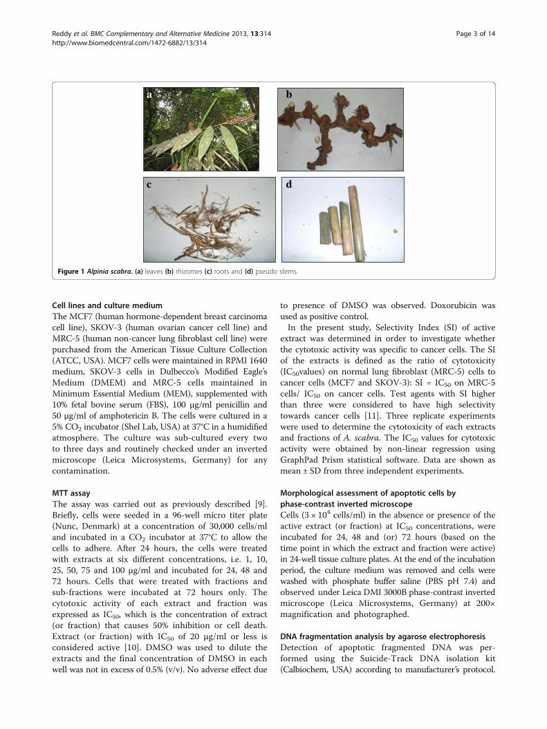
Plant sample collection and identificationThe fresh leaves, rhizomes, roots and pseudo stems ofA. scabra were collected from Genting Highland, Pahang,Malaysia. The plant samples were identified by ProfessorDr Halijah Ibrahim of Institute of Biological Sciences,Faculty of Science, University of Malaya, Malaysia and avoucher specimen (herbarium no. HI 1419) was depositedat the herbarium of Institute of Biological Sciences,Faculty of Science, University of Malaya, Malaysia. Theappearance of A. scabra is shown in Figure 1.
Preparation of extractsThe extracts were prepared as previously described [9]with minor modifications. Briefly, the different parts ofA. scabra (leaves, rhizomes, roots and pseudo stems)were washed and ground to fine powder. The dried andground samples were then extracted with 80% methanolfor three days at room temperature to obtain the crudemethanol extracts. Part of the crude methanol extract wasreserved for the cytotoxicity assay while the remaining por-tions were fractionated with hexane to give the hexane-soluble extracts and hexane-insoluble residues. The hexane-insoluble residues were further partitioned with chloroformand water (1:1, 100 ml: 100 ml) to give the chloroform andwater extracts. The weights of the crude methanol andfractionated extracts (hexane, chloroform and water) weremeasured after solvent evaporation. All the extracts weredissolved in DMSO, except for the water extracts whichwere dissolved in distilled water to form stock solutions of20 mg/ml and stored at−20°C before cytotoxicity testing.
Reddy et al. BMC Complementary and Alternative Medicine 2013, 13:314 Page 2 of 14http://www.biomedcentral.com/1472-6882/13/314
Cell lines and culture mediumThe MCF7 (human hormone-dependent breast carcinomacell line), SKOV-3 (human ovarian cancer cell line) andMRC-5 (human non-cancer lung fibroblast cell line) werepurchased from the American Tissue Culture Collection(ATCC, USA). MCF7 cells were maintained in RPMI 1640medium, SKOV-3 cells in Dulbecco’s Modified Eagle’sMedium (DMEM) and MRC-5 cells maintained inMinimum Essential Medium (MEM), supplemented with10% fetal bovine serum (FBS), 100 μg/ml penicillin and50 μg/ml of amphotericin B. The cells were cultured in a5% CO2 incubator (Shel Lab, USA) at 37°C in a humidifiedatmosphere. The culture was sub-cultured every twoto three days and routinely checked under an invertedmicroscope (Leica Microsystems, Germany) for anycontamination.
MTT assayThe assay was carried out as previously described [9].Briefly, cells were seeded in a 96-well micro titer plate(Nunc, Denmark) at a concentration of 30,000 cells/mland incubated in a CO2 incubator at 37°C to allow thecells to adhere. After 24 hours, the cells were treatedwith extracts at six different concentrations, i.e. 1, 10,25, 50, 75 and 100 μg/ml and incubated for 24, 48 and72 hours. Cells that were treated with fractions andsub-fractions were incubated at 72 hours only. Thecytotoxic activity of each extract and fraction wasexpressed as IC50, which is the concentration of extract(or fraction) that causes 50% inhibition or cell death.Extract (or fraction) with IC50 of 20 μg/ml or less isconsidered active [10]. DMSO was used to dilute theextracts and the final concentration of DMSO in eachwell was not in excess of 0.5% (v/v). No adverse effect due
to presence of DMSO was observed. Doxorubicin wasused as positive control.
In the present study, Selectivity Index (SI) of activeextract was determined in order to investigate whetherthe cytotoxic activity was specific to cancer cells. The SIof the extracts is defined as the ratio of cytotoxicity(IC50values) on normal lung fibroblast (MRC-5) cells tocancer cells (MCF7 and SKOV-3): SI = IC50 on MRC-5cells/ IC50 on cancer cells. Test agents with SI higherthan three were considered to have high selectivitytowards cancer cells [11]. Three replicate experimentswere used to determine the cytotoxicity of each extractsand fractions of A. scabra. The IC50 values for cytotoxicactivity were obtained by non-linear regression usingGraphPad Prism statistical software. Data are shown asmean ± SD from three independent experiments.
Morphological assessment of apoptotic cells byphase-contrast inverted microscopeCells (3 × 10⁴ cells/ml) in the absence or presence of theactive extract (or fraction) at IC50 concentrations, wereincubated for 24, 48 and (or) 72 hours (based on thetime point in which the extract and fraction were active)in 24-well tissue culture plates. At the end of the incubationperiod, the culture medium was removed and cells werewashed with phosphate buffer saline (PBS pH 7.4) andobserved under Leica DMI 3000B phase-contrast invertedmicroscope (Leica Microsystems, Germany) at 200×magnification and photographed.
DNA fragmentation analysis by agarose electrophoresisDetection of apoptotic fragmented DNA was per-formed using the Suicide-Track DNA isolation kit(Calbiochem, USA) according to manufacturer’s protocol.
b
dc
a
Figure 1 Alpinia scabra. (a) leaves (b) rhizomes (c) roots and (d) pseudo stems.
Reddy et al. BMC Complementary and Alternative Medicine 2013, 13:314 Page 3 of 14http://www.biomedcentral.com/1472-6882/13/314
Briefly, MCF7 and SKOV-3 cells were cultured and treatedwith cytotoxic extracts, fractions and doxorubicin(positive control) for 24 hours according to the IC50
values. After 24 hours of incubation, the floating andtrypsinized-adherant of treated and untreated cells werecollected and total DNA was extracted from the cells.Samples were loaded into a 1.5% agarose gel and separatedby electrophoresis. DNA fragments were stained with0.5 μg/ml ethidium bromide and were visualized underUV illumination using a Gene Flash gel documentationsystem (Syngene Bio imaging, UK).
Morphological detection of apoptosis using DAPInuclear stainThe occurrence of apoptosis in MCF7 and SKOV-3cells was evaluated using 4’6-diamidino-2-phenylindole(DAPI, Sigma) staining. Cells (1 × 10⁶) were platedonto 6-well tissue culture plate and incubated in aCO2 incubator at 37°C for 24 hours. After 24 hours,the cells were treated with cytotoxic extracts, fractions,sub-fraction and doxorubicin (positive control) for 24 hoursat concentration corresponding to the IC50 values. Negativecontrol comprised of cells not treated with any extract.After the incubated period, the cells were then harvestedand washed with PBS. The resulting cell pellet was fixedwith acetone at −20°C for 30 min. The cells were thenstained with DAPI solution (1 μg/ml) at 4°C for 30 minutes.Stained cells were spotted onto a slide and cover slips werethen mounted onto glass microscope slides and observedunder fluorescence microscopy (Olympus BX51) using a358 nm excitation and 460 nm emission fluorescent filter.
Bioassay-guided fractionation of leaf chloroform extractThe cytotoxic chloroform extract of leaves (9.0 g) wassubjected to vacuum liquid chromatography (VLC)which is a rapid crude fractionation system. The column(7 cm diameter, 30 cm length) was packed with 540.0 gof silica gel 60 (Merck, 0.063-0.200 mm) as stationaryphase. The elution of components present in the extractwere started with chloroform and then the polarity ofthe eluent was gradually increased with addition ofmethanol and finally with only methanol. Elution wasmonitored by thin layer chromatography (TLC) and theeluent vials were pooled together based on similar patternof TLC spots into a total of 10 fractions labelled as LC1 toLC10. All the 10 fractions were then tested for cytotoxicityagainst MCF7, SKOV-3 and MRC-5 cell lines using MTTassay at 72 hours. Among the 10 fractions, only fractionLC4 was found to be active in the cytotoxicity screeningagainst MCF7 and SKOV-3 cell lines. Thus, LC4 wasselected for further isolation and purification work.
The cytotoxic fraction LC4 which was a dark brown pastewas then purified using VLC. The column was packed with60.0 g of silica gel 60 (Merck, 0.063-0.200 mm) as the
stationary phase and the ratio of the fraction to silica gelwas 1:60. In brief, elution of components in the fractionstarted with chloroform and then its polarity was graduallyincreased with addition of methanol and finally withmethanol. Elution of components from the columnwas monitored by TLC and eluent vials with similarpattern of TLC profile were combined to give 17 sub-fractions (VLC1 to VLC17). Some of the sub-fractions(weight more than 20 mg) were then tested for cytotoxicityagainst MCF7, SKOV–3 and MRC-5 cell lines using MTTassay at 72 hours. A flow chart of the bioassay-guidedfractionation of the cytotoxic leaf chloroform extractisshown in Figure 2.
Bioassay-guided fractionation of rhizome chloroformextractThe chloroform extract of rhizomes (2.0 g) was subjectedto a rapid crude fractionation using VLC. The column(7 cm diameter, 30 cm length) was packed with 120.0 g ofsilica gel (Merck, 0.063-0.200 mm) as the stationary phaseand the ratio of the extract to silica gel was 1:60. In brief,the components in the fraction were initially eluted withchloroform and then its polarity was gradually increasedwith addition of acetone and finally methanol. Elution ofcomponents from the column was monitored by TLC.Eluent vials with similar pattern of TLC profile werecombined to give 18 fractions (RC1 to RC18). All the18 fractions were then tested for cytotoxicity againstSKOV-3 and MRC-5 cells. Figure 3 shows the flowchart of the bioassay-guided fractionation of the rhizomechloroform extract.
GC-MS analysisGC-MS analysis was performed using an AgilentTechnologies 6980 N gas chromatograph equippedwith a 5975 Mass Selective Detector (70 eV direct inlet) asdescribed by Malek et al. [12] with some modifications;the HP-5 ms capillary column (5% phenylmethylsiloxane)with column dimensions 30.0 m × 25 mm × 25 um wasinitially set at 100°C, then the temperature of the ovenwas increased at 5°C per minute to 300°C and hold for10 minutes using helium as carrier gas at flow rate 1 ml/min.The total ion chromatogram obtained was auto integrated byChemStation and the components were identified bycomparison with an accompanying mass spectral database[13]. Only mass spectral fragmentation pattern that gavegreater than 90% match were accepted.
Results and discussionExtraction yield of A. scabra extractsSolvent extraction is the most popular method used inplant sample preparation. The crude methanol extractswere firstly prepared and further fractionated intohexane, chloroform and water extracts as a single solvent
Reddy et al. BMC Complementary and Alternative Medicine 2013, 13:314 Page 4 of 14http://www.biomedcentral.com/1472-6882/13/314

may not be enough to identify certain extracts responsiblefor the activity. The yield of methanol extracts fromdifferent parts of A. scabra is shown in Table 1,whereas the yield of extracts fractionated from crudemethanol extracts is shown in Table 2. The percentage ofcrude methanol extract yield was based on the weight ofdried and ground plant materials and the percentage yieldof fractionated extracts was based on the weight of crudemethanol extract used.
Cytotoxic activities of A. scabra extractsIn the present study, the cytotoxic effect (IC50) of the crudemethanol and fractionated extracts (hexane, chloroform
and water) from the different parts of A. scabra wereinvestigated on two human cancer cells (MCF7 andSKOV-3) and one normal non-cancer cells (MRC-5)using MTT assay. MTT assay is commonly used incell biology for the study of growth factor, cytokinesand cytotoxicity of chemotherapeutic agents as it offers aquantitative and simple method for evaluating cell popula-tion’s response to external factors. Doxorubicin was used asthe positive control in the present study because it is acommonly used chemotherapeutic drug for the treatmentof acute leukemia, lymphomas and different types of solidtumours such as breast, liver and lung cancers [14].Cytotoxic activity (IC50) and Selectivity Index (SI) of the
Leaf chloroform extract (9.0 g)
VLCChloroform: methanol
LC1 (0.067 g)
LC2 (0.067 g)
LC3 (0.052 g)
LC4 (4.063 g)
LC5 (0.977 g)
LC6(0.661 g)
LC7 (0.356 g)
LC8 (0.217 g)
LC9 (0.309 g)
LC10 (0.143 g)
Identified cytotoxic fraction LC4 (4.063 g)
Cytotoxicity screening
VLCChloroform: methanol
Sub-fraction VLC1–VLC17
Cytotoxicity screening
Identified cytotoxic sub-fraction VLC9
Figure 2 Flow chart of bioassay-guided fractionation of cytotoxic leaf chloroform extract.
Rhizome chloroform extract (1.22 g)
VLCChloroform: acetone
RC3(14 mg)
RC8(36 mg)
RC9(12 mg)
RC10(2 mg)
RC11 (7 mg)
RC12 (5 mg)
RC13 (60 mg)
RC14 (9 mg)
RC15 (36 mg)
RC16(21 mg)
RC17 (29 mg)
RC18 (461 mg)
RC1(18mg)
RC2 (4mg)
RC4(24 mg)
RC5(30 mg)
RC6(18 mg)
RC7(20 mg)
Identified cytotoxic fraction RC5
Cytotoxicity screening
Figure 3 Flow chart of bioassay-guided fractionation of cytotoxic rhizome chloroform extract.
Reddy et al. BMC Complementary and Alternative Medicine 2013, 13:314 Page 5 of 14http://www.biomedcentral.com/1472-6882/13/314

extracts of leaves, rhizomes, roots and pseudo stems of A.scabra are summarized in Tables 3, 4, 5 and 6, respectively.
Generally, the root and pseudo stem extracts displayedweaker cytotoxicity profile against all the tested humancell lines compared to the leaf and rhizome extracts. Amongthe extracts, hexane and chloroform extracts showed bettercytotoxic activity against all the tested cell lines comparedto the methanol and water extracts. The hexane andchloroform extracts of leaves demonstrated active cyto-toxic effect against MCF7 and SKOV-3 cells (Table 3).The leaf hexane extract showed high inhibition againstMCF7 cells with IC50 value of 15.30 μg/ml at 48 hours, incomparison to IC50 values of 19.30 and 16.33 μg/ml at 24and 72 hours, respectively. It is interesting to note that thechloroform extract of leaves only demonstrated active
cytotoxicity against MCF7 cells at 24 hours (18.8 μg/ml),but not at 48 and 72 hours.
The hexane and chloroform extracts of leaves and thechloroform extract of rhizomes displayed active cytotoxiceffect against the SKOV-3 cells with IC50 values in de-creasing trend from 24 to 72 hours. This indicates that thelonger the incubation time of the extracts in cells, the
Table 1 Yield of the methanol extracts from differentparts of A. scabra
Plant part Sample/Extract Weight (g) (%)
Leaves Fresh samples 1000.00
Dried and ground plant material 600.00 (60.00)
Methanol extract 41.22 (6.87)
Rhizomes Fresh samples 7000.00
Dried and ground plant material 400.00 (5.71)
Methanol extract 17.86 (4.47)
Roots Fresh samples 900.00
Dried and ground plant material 100.00 (10.00)
Methanol extract 7.11 (7.11)
Pseudo stems Fresh samples 8000.00
Dried and ground plant material 650.00 (8.13)
Methanol extract 47.38 (7.29)
Table 2 Yield of extracts fractionated from the crudemethanol extracts
Plant part Extract Yield ofextracts (g)
Percentage (%)
Leaves Hexane 0.50 1.21
(extracted from 41.22 g ofmethanol extract)
Chloroform 9.64 23.39
Water 21.50 52.16
Rhizomes Hexane 0.65 3.62
(extracted from 17.86 g ofmethanol extract)
Chloroform 1.22 6.83
Water 11.99 67.12
Roots Hexane 0.31 4.36
(extracted from 7.11 g ofmethanol extract)
Chloroform 0.63 8.86
Water 5.14 72.29
Pseudo stems Hexane 0.48 1.01
(extracted from 47.38 g ofmethanol extract)
Chloroform 3.23 6.82
Water 33.05 69.76
Table 3 Cytotoxic activity (IC50) of A. scabra leaf extractsExtracts Treatment
duration(hour)
IC50a(μg/ml) (SIb)
MCF7 SKOV-3 MRC-5
Methanol 24 90.67 ± 6.11 47.00 ± 11.53 >100
48 56.27 ± 9.18 37.67 ± 2.52 57.58 ± 1.25
72 53.33 ± 6.43 34.33 ± 0.58 65.35 ± 1.68
Hexane 24 19.30 ± 5.70 (1.6) 18.00 ± 2.65 (1.7) 31.38 ± 2.31
48 15.30 ± 4.04 (1.0) 6.00 ± 1.00 (2.4) 14.63 ± 2.08
72 16.33 ± 0.58 (1.0) 4.93 ± 0.12 (3.2) 15.90 ± 0.94
Chloroform 24 18.80 ± 1.06 (2.4) 20.00 ± 1.00 (2.3) 45.88 ± 3.81
48 23.67 ± 7.64 14.33 ± 1.53 (2.3) 32.26 ± 2.11
72 25.00 ± 0.00 14.67 ± 0.58 (2.2) 32.90 ± 0.76
Water 24 >100 >100 >100
48 >100 >100 >100
72 >100 >100 >100
Doxorubicinc 24 2.50 ± 0.10 2.50 ± 0.10 >100
48 2.37 ± 0.23 (32.4) 1.77 ± 0.15 (43.4) 76.87 ± 12.64
72 0.39 ± 0.02 (4.3) 1.47 ± 0.15 (1.1) 1.68 ± 0.42
aData are presented as mean ± SD from three independent experimentstriplicate for each. Values in bold characters are considered to have cytotoxicactivity (IC5020 μg/ml or less); bSelectivityIndex (SI); cPositive control.
Table 4 Cytotoxic activity (IC50 μg/ml) of A. scabrarhizome extractsExtracts Treatment
duration(hour)
IC50a(μg/ml) (SIb)
MCF7 SKOV-3 MRC-5
Methanol 24 >100 >100 >100
48 >100 >100 >100
72 >100 >100 >100
Hexane 24 79.67 ± 10.60 40.00 ± 2.00 73.69 ± 2.23
48 60.00 ± 5.66 25.67 ± 0.58 41.31 ± 2.26
72 57.33 ± 1.15 24.00 ± 3.46 53.49 ± 2.71
Chloroform 24 70.67 ± 23.12 21.67 ± 4.73 78.08 ± 7.76
48 39.00 ± 1.41 19.33 ± 0.58 (2.3) 43.56 ± 0.54
72 37.67 ± 0.58 17.33 ± 0.58 (2.6) 44.65 ± 2.57
Water 24 >100 >100 >100
48 >100 >100 >100
72 >100 >100 >100
Doxorubicinc 24 2.50 ± 0.10 2.50 ± 0.10 >100
48 2.37 ± 0.23 (32.4) 1.77 ± 0.15 (43.4) 76.87 ± 12.64
72 0.39 ± 0.02 (4.3) 1.47 ± 0.15 (1.1) 1.68 ± 0.42
aData are presented as mean ± SD from three independent experimentstriplicate for each. Values in bold characters are considered to have cytotoxicactivity (IC5020 μg/ml or less); bSelectivity Index (SI); cPositive control.
Reddy et al. BMC Complementary and Alternative Medicine 2013, 13:314 Page 6 of 14http://www.biomedcentral.com/1472-6882/13/314
better the cytotoxicity results. The hexane extract of leavesshowed an excellent inhibition towards SKOV-3 cells withIC50 value of 4.93 μg/ml at 72 hours, in comparison toIC50 values of 18.00 and 6.00 μg/ml at 24 and 48 hours,respectively. The chloroform extract of leaves possessed thestrongest cytotoxicity at 48 hours with IC50 of 14.33 μg/ml
Table 5 Cytotoxic activity (IC50 μg/ml) of A. scabra rootextractsExtracts Treatment
duration(hour)
IC50a(μg/ml) (SIb)
MCF7 SKOV-3 MRC-5
Methanol 24 70.67 ± 5.86 56.67 ± 1.15 44.98 ± 10.59
48 47.67 ± 4.93 34.67 ± 0.58 47.70 ± 8.68
72 64.00 ± 0.00 34.00 ± 4.36 38.94 ± 5.02
Hexane 24 56.00 ± 2.00 33.33 ± 1.15 30.59 ± 0.36
48 33.67 ± 5.03 28.33 ± 3.51 30.71 ± 3.15
72 40.67 ± 1.15 28.00 ± 1.73 29.59 ± 5.98
Chloroform 24 67. 33 ± 5.03 44.00 ± 5.29 30.27 ± 0.33
48 37.33 ± 4.16 37.00 ± 0.58 51.09 ± 10.80
72 42.67 ± 2.31 33.67 ± 3.21 38.39 ± 5.57
Water 24 >100 >100 >100
48 >100 >100 >100
72 >100 >100 >100
Doxorubicinc 24 2.50 ± 0.10 2.50 ± 0.10 >100
48 2.37 ± 0.23 (32.4) 1.77 ± 0.15 (43.4) 76.87 ± 12.64
72 0.39 ± 0.02 (4.3) 1.47 ± 0.15 (1.1) 1.68 ± 0.42
aData are presented as mean ± SD from three independent experimentstriplicate for each. Values in bold characters are considered to have cytotoxicactivity (IC5020 μg/ml or less); bSelectivity Index (SI); cPositive control.
Table 6 Cytotoxic activity (IC50 μg/ml) of A. scabrapseudo stem extractsExtracts Treatment
duration(hour)
IC50a (μg/ml) (SIb)
MCF7 SKOV-3 MRC-5
Methanol 24 >100 >100 >100
48 >100 >100 >100
72 >100 >100 >100
Hexane 24 >100 40.67 ± 3.51 >100
48 84.00 ± 1.00 34.00 ± 1.73 55.86 ± 16.71
72 67.30 ± 1.15 34.67 ± 0.58 49.74 ± 1.33
Chloroform 24 80.00 ± 15.10 56.00 ± 5.29 >100
48 66.67 ± 1.16 30.67 ± 5.13 52.86 ± 1.75
72 59.30 ± 1.15 33.00 ± 1.73 48.18 ± 2.34
Water 24 >100 >100 >100
48 >100 >100 >100
72 >100 >100 >100
Doxorubicinc 24 2.50 ± 0.10 2.50 ± 0.10 >100
48 2.37 ± 0.23 (32.4) 1.77 ± 0.15 (43.4) 76.87 ± 12.64
72 0.39 ± 0.02 (4.3) 1.47 ± 0.15 (1.1) 1.68 ± 0.42
aData are presented as mean ± SD from three independent experimentstriplicate for each. Values in bold characters are considered to have cytotoxicactivity (IC5020 μg/ml or less); bSelectivity Index (SI); cPositive control.
Table 7 Cytotoxic activity (IC50 μg/ml) of fractions andsub-fraction obtained from leaf chloroform extract
Fraction (LC)/Sub-fraction(VLC)
IC50a(μg/ml) MRC-5
MCF7 SKOV-3
LC1 50.00 ± 5.00 92.30 ± 4.78 >100
LC2 42.20 ± 4.48 56.00 ± 6.60 >100
LC3 33.40 ± 4.05 40.50 ± 2.56 >100
LC4 18.53 ± 1.02 11.12 ± 0.24 >100
LC5 51.18 ± 4.56 39.18 ± 1.34 >100
LC6 >100 >100 >100
LC7 >100 >100 >100
LC8 >100 >100 >100
LC9 >100 >100 >100
LC10 >100 >100 >100
VLC9 15.53 ± 0.50 10.89 ± 0.64 >100
Chloroform 25.00 ± 0.00 14.67 ± 0.58 32.90 ± 0.76aData are presented as mean ± SD from three independent experimentstriplicate for each. Values in bold characters are considered to have cytotoxicactivity (IC5020 μg/ml or less).
Table 8 Cytotoxic activity (IC50 μg/ml) of fractionsobtained from the rhizome chloroform extract
Fraction IC50a(μg/ml)(SIb)
SKOV-3 MRC-5
RC1 94.56 ± 4.32 95.70 ± 3.00
RC2 88.90 ± 4.78 66.00 ± 5. 66
RC3 25.49 ± 2.56 50.80 ± 1.24
RC4 23.35 ± 2.24 71.00 ± 3.40
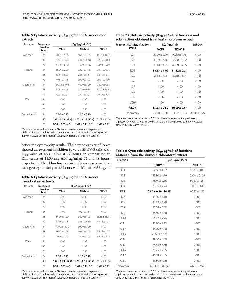
RC5 2.84 ± 0.60 (14.15) 40.20 ± 1.50
RC6 39.90 ± 1.18 >100
RC7 32.63 ± 6.78 >100
RC8 50.24 ± 7.78 >100
RC9 64.50 ± 1.40 >100
RC10 66.82 ± 2.26 >100
RC11 51.30 ± 3.12 >100
RC12 45.70 ± 4.00 >100
RC13 21.60 ± 10.80 >100
RC14 29.70 ± 2.50 >100
RC15 25.33 ± 3.56 >100
RC16 24.75 ± 2.85 >100
RC17 45.08 ± 3.45 >100
RC18 43.89 ± 4.76 >100
Chloroform 17.33 ± 0.58 (2.6) 44.65 ± 2.57aData are presented as mean ± SD from three independent experimentstriplicate for each. Values in bold characters are considered to have cytotoxicactivity (IC5020 μg/ml or less); bSelectivity Index (SI).
Reddy et al. BMC Complementary and Alternative Medicine 2013, 13:314 Page 7 of 14http://www.biomedcentral.com/1472-6882/13/314

compared to IC50 values at 24 and 72 hours. Meanwhile,the chloroform extract from the rhizomes showedhigh inhibition towards SKOV-3 cells with IC50 value of17.30 μg/ml at 72 hours, in comparison to IC50 values of21.67 and 19.33 μg/ml at 24 and 48 hours, respectively.
MRC-5 cell line has been used as a normal cell model inmany similar studies [9,15-17]. In the current study, all thefour samples (leaves, rhizomes, roots and pseudostems)were tested against MRC-5 normal cells and only the leafhexane extract showed cytotoxicity at 48 and 72 hours withthe IC50 values of 14.64 and 15.90 μg/ml, respectively.Selectivity of the active extracts were determined but noneof the active extracts showed selectivity to the cancer cellsas all the selectivity indexes were lower than three, exceptfor the leaf hexane extract which showed selectivity towardsSKOV-3 cells at 72 hours with the SI value of 3.2 (Table 3).
The leaf (hexane and chloroform) and rhizome(chloroform) extracts were selected for the bioassay-guided fractionation as these extracts showed strong
cytotoxic effect against the selected cancer cells (IC50
values of 20 μg/ml or less).
Bioassay-guided fractionation of the leaf chloroform extractThe ten fractions (LC1-LC10) obtained from VLC weretested for cytotoxicity against MCF7, SKOV-3 and MRC-5cell lines using MTT assay. The fraction LC4 was the onlyfraction found to be active in the cytotoxicity screeningagainst MCF7 and SKOV-3 cell lines with IC50values of18.53 and 11.12 μg/ml, respectively (Table 7). Hence,fraction LC4 was warranted for further purification byVLC and yielded 17 sub-fractions (VLC1-VLC17; Figure 2).As shown in Table 7, sub-fraction VLC 9 showedgood cytotoxicity against MCF7 and SKOV-3 cell lines(IC50 values of 15.53 and 10.89 μg/ml, respectively) butweak cytotoxicity profile against the MRC-5 cell line(IC50value >100 μg/ml). As shown in Table 7, the cytotox-icity in ascending order was leaf chloroform extract < LC4 < VLC9. This may be due to the cytotoxic compounds
24 hours 48 hours 72 hours
Con
trol
Lea
f he
xane
Lea
f ch
loro
form
A
A
a
A
B
b Ac
B
dFigure 4 Morphological observation of MCF7 cells treated with the cytotoxic leaf hexane and chloroform extracts underphase-contrast inverted microscope (magnification 200×). Arrows indicate (A) cell shrinkage and (B) membrane blebbing as evidence ofapoptosis. Note that the cells were treated with the following concentrations of extracts: a = 19.3 μg/ml, b = 15.30 μg/ml, c = 16.33 μg/ml andd = 18.80 μg/ml. Figures shown were obtained from at least three independent experiments with similar parameter.
Reddy et al. BMC Complementary and Alternative Medicine 2013, 13:314 Page 8 of 14http://www.biomedcentral.com/1472-6882/13/314
which present in VLC9 after the purification of the leafchloroform extract and LC4 via VLC. Thus, the activeingredients in VLC9 may lead to valuable compounds thathave the ability to kill cancer cells but not toxic againstnormal MRC-5 cells.
Bioassay-guided fractionation of the rhizome chloroformextractThe cytotoxic effect of the fractions (RC1–RC18)derived from the chloroform extract of rhizome by VLCwas evaluated in order to determine the fraction thatgive the highest activity. Table 8 shows the IC50 values ofthe 18 fractions from the chloroform extract of rhizome.Fraction RC5 was the only fraction which exhibited
remarkable cytotoxicity (IC50 value of 2.84 μg/ml) andshowed high selectivity (SI value of 14.15) against theSKOV-3 cells, compared to the rhizome chloroformextract. This exhibited the improvement of cytotoxicityand selectivity after the purification procedure.
GC-MS analysis of leaf hexane extractThe cytotoxic hexane leaf extracts were analysed usingGC-MS in the present study. The identified compoundsare 61.02% methyl palmitate and 24.91% methyl stearatewhich comprise of 85.93% of the total hexane extract.Methyl palmitate EI-MS m/z (%): 270 [M] + (2), 239 (2),227 (5), 213 (2), 199 (4), 185 (5), 171 (8), 157 (2), 143(18), 129 (8), 115 (4), 107 (1), 97 (8), 87 (70), 74 (100),
24 hours 48 hours 72 hours
Con
trol
Lea
f he
xane
Lea
f ch
loro
form
Rhi
zom
e ch
loro
form
A
BC
a b
B
C
c
B
A
dB
A
eB
f
A
g
C
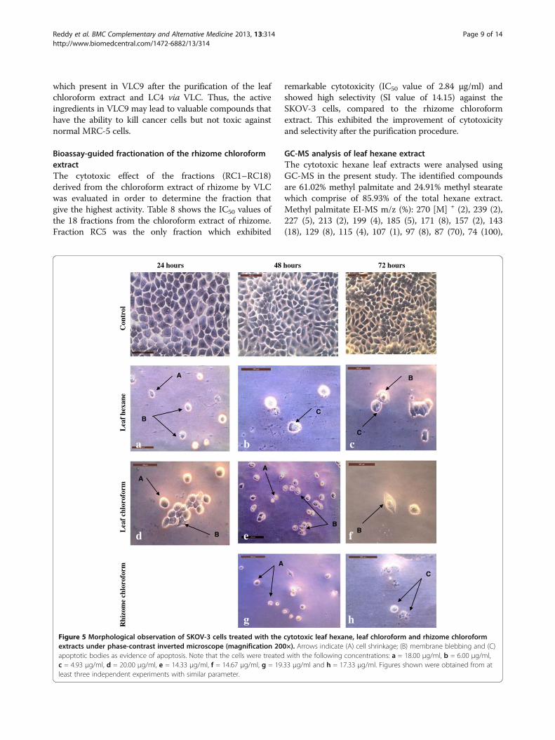
hFigure 5 Morphological observation of SKOV-3 cells treated with the cytotoxic leaf hexane, leaf chloroform and rhizome chloroformextracts under phase-contrast inverted microscope (magnification 200×). Arrows indicate (A) cell shrinkage; (B) membrane blebbing and (C)apoptotic bodies as evidence of apoptosis. Note that the cells were treated with the following concentrations: a = 18.00 μg/ml, b = 6.00 μg/ml,c = 4.93 μg/ml, d = 20.00 μg/ml, e = 14.33 μg/ml, f = 14.67 μg/ml, g = 19.33 μg/ml and h = 17.33 μg/ml. Figures shown were obtained from atleast three independent experiments with similar parameter.
Reddy et al. BMC Complementary and Alternative Medicine 2013, 13:314 Page 9 of 14http://www.biomedcentral.com/1472-6882/13/314

65 (1), 55 (30). Methyl stearate EI-MS m/z (%): 298 [M] +
(6), 255 (8), 241 (2), 213 (4), 199 (8), 185 (6), 143 (26), 129(20), 111 (4), 97 (10), 87 (70), 74 (100), 55(56). Allthe compounds were identified by GC-MS analysis aswell as comparison of its mass spectral data withreported data [18].
Previous report by Sri Nurestri et al. [18] suggestedthat methyl esters might exert cytotoxic effect againstnormal MRC-5 cells but not on KB, MCF7 and HCT116cells. This finding supports the data from the present
study on the cytotoxicity of A. scabra extracts againstMRC-5 cells. As shown in Table 3, the hexane leafextract showed cytotoxic activity on MRC-5 cells at 48and 72 hours with IC50 values of 14.63 and 15.90 μg/ml,respectively. This may be due to the presence of methylpalmitate and methyl stearate in the extract. Furthermore,this finding on cytotoxicity of methyl esters is supportedby Takeara et al. [19] which reported that methylpalmitate showed cytotoxic effect on T-cell leukemiacell line (Molt-4) with IC50 value of 2.28 μg/ml while
MCF7 SKOV-3
Con
trol
Fra
ctio
n L
C4
Sub-
frac
tion
VL
C9
Fra
ctio
n R
C5
b
e
B
A
d
B
A
A
C
c
B
A
a
A
B
Figure 6 Morphological observation of MCF7 and SKOV-3 cells treated with the cytotoxic fractions LC4, VLC9 and RC5 underphase-contrast inverted microscope (magnification 200×). Arrows indicate (A) cell shrinkage; (B) membrane blebbing and (C) apoptoticbodies as evidence of apoptosis. Note that the cells were treated with the following concentrations: a = 18.53 μg/ml, b = 11.12 μg/ml, c = 15.53 μg/ml,d = 10.89 μg/ml and e = 2.84 μg/ml. Figures shown were obtained from at least three independent experiments with similar parameter.
Reddy et al. BMC Complementary and Alternative Medicine 2013, 13:314 Page 10 of 14http://www.biomedcentral.com/1472-6882/13/314

methyl stearate was cytotoxic to acute promyeloblasticleukemia cell line (HL-60) and Molt-4 cell line withIC50 values of 3.08 and 4.65 μg/ml, respectively.
Morphological assessment of apoptotic cells byphase-contrast inverted microscopeThe results from the present study (Figures 4, 5 and 6)showed that there were obvious morphological changesin MCF7 and SKOV-3 cells after treatment with thecytotoxic extracts, fractions and sub-fraction which wereindicative of cell apoptosis. The untreated control MCF7and SKOV-3 cells maintained their original morphologywhich are cuboids and in polygonal shapes, and wereadherent to the plates. The MCF7 cells were treated withhexane and chloroform extracts of leaves (Figure 4)while SKOV-3 cells were treated with leaf hexane, leafchloroform and rhizome chloroform extracts (Figure 5)for 24, 48 and (or) 72 hours according to the IC50 values(Tables 3 and 4). Figure 6 shows MCF7 and SKOV-3cells treated with fraction LC4, sub-fraction VLC9 andfraction RC5 at 72 hours according to the IC50 values inTables 7 and 8. The most recognizable morphologicalfeatures of apoptotic cells observed in this study wereshrinkage of cells due to cytoplasmic condensation,rounding up of cells, bleb formation, chromatin con-densation and apoptotic bodies’ formation. The other
morphological change observed in apoptotic cells was therounded up cells losing contact with neighbouring cells andcaused some sensitive cells to detach from the surface ofthe well plates. This morphological observation of apoptoticcells were in agreement with previous report [15].
DNA fragmentation analysis by agarose electrophoresisDNA fragmentation is a biochemical hallmark of apoptoticcell death. To elucidate whether the active extracts andfractions decrease cell survival by the induction of DNAfragmentation, genomic DNA isolated from MCF7 andSKOV-3 cells were exposed according to the IC50 valueconcentration, electrophoresed and photographed as shownin Figures 7 and 8. Typical DNA ladder formation can beseen clearly in the MCF7 and SKOV-3 cells treatedwith doxorubicin (positive control) whereas DNAfrom untreated MCF7 and SKOV-3 cells did not showany fragmentation or smearing. In MCF7 cells treatedwith cytotoxic active extracts and sub-fractions (Figure 7),the formation of DNA ladder was observed less clearly asthere were interspersing smear in the lanes. This patternwas noticeably observed in MCF7 cells treated withhexane and chloroform extract of leaf, fraction LC4 and
Mar
ker
Unt
reat
ed c
ell
Lea
f he
xane
ext
ract
Lea
f ch
loro
form
ext
ract
Fra
ctio
n L
C4
Sub-
frac
tion
VL
C9
Pos
itiv
e co
ntro
l
Figure 7 DNA fragmentation of MCF7 cells after treated withcytotoxic extracts and sub-fractions for 24 hours.
Mar
ker
Unt
reat
ed c
ell
Pos
itiv
e co
ntro
l
Lea
f he
xane
ext
ract
Lea
f ch
loro
form
ext
ract
Rhi
zom
e ch
loro
form
ext
arct
Fra
ctio
n L
C4
Sub-
frac
tion
VL
C9
Fra
ctio
n R
C5
Figure 8 DNA fragmentation of SKOV-3 cells after treated withcytotoxic active extracts and sub-fractions for 24 hours.
Reddy et al. BMC Complementary and Alternative Medicine 2013, 13:314 Page 11 of 14http://www.biomedcentral.com/1472-6882/13/314

sub-fraction VLC9 at concentrations of 19.30, 18.80, 18.53and 15.53 μg/ml, respectively. The smearing could be dueto some post-apoptotic necrosis cells [15].
For the SKOV-3 cells (Figure 8), the ladder-like appear-ance of DNA observed mildly in the cells treated withhexane and chloroform extract of leaf at concentrations of18.0 and 20.0 μg/ml, respectively. SKOV-3 cells which weretreated with chloroform extract of rhizome, fraction LC4,sub-fraction VLC9 and fraction RC5 at concentrations of21.67, 11.12, 10.89 and 2.84 μg/ml, respectively did notshow any DNA laddering or even smearing effect. Thiscould be due to the concentration of the particular extractand sub-fractions which were used to treat the cells werelow since at lower doses of treatment, only high molecularweight intact DNA was observed [20]. Besides that, in somecases, DNA fragmentation appears to be delayed, partial, orabsent in cells which otherwise meet the morphological cri-teria for apoptosis and maybe show more limited DNAdegradation with the formation of 300-or 50-kb fragments[21]. The large band present at the top of the gel asobserved in SKOV-3 cells treated with fraction LC4and RC5 may represent large semi-fragmented piecesof DNA and indicates incomplete apoptotic fragmentationin the sample material [22].
Morphological detection of apoptosis using DAPI nuclear stainDAPI is a fluorescent stain that allows examination of nu-clei in a fluorescence microscope for morphological assess-ment of changes during apoptosis [23]. Apoptosis isinitially characterized by morphological features, such aschromatin condensation, nuclear fragmentation and mem-brane blebbing [24]. To gain an insight on the effect ofcytotoxic extracts, fractions, sub-fraction and doxorubicinon nuclear alterations, cells were stained with DAPI.Figures 9 and 10 show the apoptotic morphological charac-teristics, as visualized by DAPI staining, of MCF7 andSKOV-3 cells treated (for 24 hours) with cytotoxic extracts,fractions, sub-fraction and doxorubicin (positive control)according to the IC50 values. In the control-untreatedgroup [Figures 9(a) and 10(a)], the cells were rounded inshape and the large nuclei were homogenously stainedwith a less bright blue colour. This is because when healthycells are exposed to DAPI, staining is restricted to chroma-tin. Treated MCF7 and SKOV-3 cells displayed bright bluefluorescence with higher intensity than untreated cellsdue to the highly condensed chromatin.
Besides that, signs of nuclear shrinkage and chromatincondensation which are hallmark of apoptosis were alsoobserved as shown in Figures 9 and 10. Apoptosis is also
a b
c d
e f
Figure 9 Morphological observation with DAPI staining by fluorescence microscope for MCF7 cells at 24 hours (magnification 400×).Arrows indicate signs of nuclear shrinkage and chromatin condensation. DNA samples in the untreated cells were homogenously stained andless intense compared to those in treated cells. Note that the cells were treated with the following extracts/sub-fractions: a = control, b = leafhexane, c = leaf chloroform, d = fraction LC4, e = Sub-fraction VLC9 and f = positive control. Figures shown were obtained from at least threeindependent experiments with similar parameter.
Reddy et al. BMC Complementary and Alternative Medicine 2013, 13:314 Page 12 of 14http://www.biomedcentral.com/1472-6882/13/314
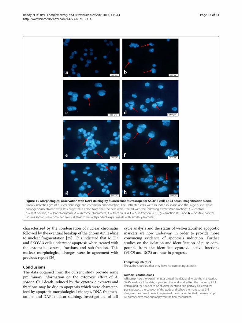
characterized by the condensation of nuclear chromatinfollowed by the eventual breakup of the chromatin leadingto nuclear fragmentation [25]. This indicated that MCF7and SKOV-3 cells underwent apoptosis when treated withthe cytotoxic extracts, fractions and sub-fraction. Thisnuclear morphological changes were in agreement withprevious report [26].
ConclusionsThe data obtained from the current study provide somepreliminary information on the cytotoxic effect of A.scabra. Cell death induced by the cytotoxic extracts andfractions may be due to apoptosis which were character-ized by apoptotic morphological changes, DNA fragmen-tations and DAPI nuclear staining. Investigations of cell
cycle analysis and the status of well-established apoptoticmarkers are now underway, in order to provide moreconvincing evidence of apoptosis induction. Furtherstudies on the isolation and identification of pure com-pounds from the identified cytotoxic active fractions(VLC9 and RC5) are now in progress.
Competing interestsThe authors declare that they have no competing interests.
Authors’ contributionsASR performed the experiments, analyzed the data and wrote the manuscript.SNAM evaluated the data, supervised the work and edited the manuscript. HIdetermined the species to be studied, identified and partially collected theplant, propose the concept of the study and edited the manuscript. SKSdesigned the current project, supervised the work and edited the manuscript.All authors have read and approved the final manuscript.
a b
c d
e f
g h
Figure 10 Morphological observation with DAPI staining by fluorescence microscope for SKOV-3 cells at 24 hours (magnification 400×).Arrows indicate signs of nuclear shrinkage and chromatin condensation. The untreated cells were rounded in shape and the large nuclei werehomogenously stained with less bright blue color. Note that the cells were treated with the following extracts/sub-fractions: a = control,b = leaf hexane, c = leaf chloroform, d = rhizome chloroform, e = fraction LC4, f = Sub-fraction VLC9, g = fraction RC5 and h = positive control.Figures shown were obtained from at least three independent experiments with similar parameter.
Reddy et al. BMC Complementary and Alternative Medicine 2013, 13:314 Page 13 of 14http://www.biomedcentral.com/1472-6882/13/314

AcknowledgementsThe authors wish to acknowledge the Ministry of Higher Education ofMalaysia (MOHE) and the University of Malaya for financial assistancethrough FRGS FP046/2010B and PPP PG065-2012B.
Received: 26 March 2013 Accepted: 29 October 2013Published: 12 November 2013
References1. Ramlan AA: Turning Malaysia into a global herbal producer, a personal
perspective. Malaysia: Universiti Teknologi Malaysia; 2003.2. Lam KS: New aspects of natural products in drug discovery. Trends
Microbiol 2007, 15:279–289.3. Newman DJ, Cragg GM, Snader KM: Natural products as sources of new
drugs over the period 1981-2002. J Nat Prod 2003, 66:1022–1037.4. Holttum RE: The Zingiberaceae of the Malay Peninsula. Gard Bull
Singapore 1950, 13:1–249.5. Surh YJ: Molecular mechanisms of chemopreventive effects of selected
dietary and medicinal phenolic substances. Mutat Res 1999, 428:305–327.6. Lee CC, Houghton P: Cytotoxicity of plants from Malaysia and Thailand
used traditionally to treat cancer. J Ethnopharmacol 2005, 100:237–243.7. Banjerdpongchai R, Punyati P, Nakrob A, Pompimon W, Kongtawelert P:
4’-Hydroxycinnamaldehyde from Alpinia galangal (Linn.) induces humanleukemic cell apoptosis via mitochondrial and endoplasmic reticulumstress pathways. Asian Pac J Cancer Prev 2011, 12:593–598.
8. Ibrahim H, Sim KS, Syamsir DR, MohdNor NR, SriNurestri AM, Awang K:Cytotoxic activity of leaf and rhizome extracts of Alpinia scabra (Blume)Naves, a wild ginger from Peninsular Malaysia. Afr J Pharmacol 2010,4:708–711.
9. Reddy NS, Navanesan S, Sinniah SK, Wahab NA, Sim KS: Phenolic content,antioxidant effect and cytotoxic activity of Leea indica leaves. BMCComplement Altern Med 2012, 12:128–134.
10. Swanson SM, Pezzuto JM: Bioscreening technique for cytotoxicitypotential and ability to inhibit macromolecule biosynthesis. In Drugbioscreening: drug evaluation techniques in pharmacology. Edited byThompson EB. New York: VCH Publishers; 1990:273–297.
11. Mahavorasirikul W, Viyanant V, Chaijoroenkul W, Itharat A, Na-Bangchang K:Cytotoxicity activity of Thai medicinal plants against human cholangio-carcinoma, laryngeal and hepatocarcinoma cells in vitro. BMC Comple-ment Altern Med 2010, 10:1–8.
12. Malek SNA, Phang CW, Ibrahim H, Wahab NA, Sim KS: Phytochemical andcytotoxic investigations of Alpinia mutica rhizomes. Molecules 2011,16:583–589.
13. W9N11: MS Library, Agilent Technologies. CA, USA: Palo; 2011.14. Tokarska-Schlattner M, Wallimann T, Schlattner U: Alterations in myocardial
energy metabolism induced by the anti-cancer drug doxorubicin. C RBiol 2006, 329:657–668.
15. Ramasamy S, Abdul WN, Zainal AN, Manickam S, Zakaria Z: Growthinhibition of human gynecologic and colon cancer cells by Phyllanthuswatsonii through apoptosis induction. PLoS ONE 2012, 7:e34793.
16. Cheng Y, Wang Y, Wang XW: A causal relationship discovery basedapproach to identifying active components of herbal medicine. ComputBiol Chem 2006, 30:146–154.
17. Bézivin C, Tomasi S, Lohezic-Le Devehat F, Boustie J: Cytotoxic activity ofsome lichen extracts on murine and human cancer cell lines.Phytomedicine 2003, 10:499–503.
18. Sri Nurestri AM, Sim KS, Norhanom AW: Phytochemical and cytotoxicinvestigations of Pereskia grandifolia Haw. (Cactaceae) leaves. J Biol Sci2009, 9:488–493.
19. Takeara R, Jimenez PC, Wilke DV, Odorico de Moraes M, Pessoa C, PeporineLopes N, Lopes JL, Monteiro da Cruz Lotufo T, Costa-Lotufo LV: Antileuke-mic effects of Didemnum psammatodes (Tunicata: Ascidiacea) constitu-ents. Comp Biochem Physiol A Mol Integr Physiol 2008, 151:363–369.
20. Yew HC, Fariza JN, Rozie S, Thiam TT, Noor RA, Zakiah I: Combinedxanthorrhizol-curcumin exhibits synergistic growth inhibitory activity viaapoptosis induction in human breast cancer cells MDA-MB-231. CancerCell Int 2009, 9:1.
21. Zakeri ZF, Quaglino D, Latham T, Lockshim RA: Delayed internucleosomalDNA fragmentation in programmed cell death. FASEB J 1993, 7:470–478.
22. Matassov D, Kagan T, Leblanc J, Sikorska M, Zakeri Z: Measurement ofapoptosis by DNA fragmentation. In Methods in molecular biology:
apoptosis methods and protocols. 282nd edition. Edited by Brady HJM.Totowa: Human Press Inc; 2004:17.
23. Kapuscinski J: DAPI: a DNA-specific fluorescent probe. Biotech Histochem1995, 70:220–233.
24. Kerr JF, Wylie AH, Curie AR: Apoptosis: a basic biological phenomenonwith wide-ranging implications in tissue kinetics. Br J Cancer 1972,26:239–257.
25. Willingham MC: Cytochemical methods for the detection of apoptosis.J Histochem Cytochem 1999, 47:1101–1109.
26. Hsiung WY, Kadir HA: Leea indica ethyl acetate fraction induces growth-inhibitory effect in various cancer cell lines and apoptosis in Ca Skihuman cervical epidermoid carcinoma cells. Evid-based Compl Alt 2011,2011:13. Article ID 293060.
doi:10.1186/1472-6882-13-314Cite this article as: Reddy et al.: Cytotoxic effect of Alpinia scabra (Blume)Náves extracts on human breast and ovarian cancer cells. BMCComplementary and Alternative Medicine 2013 13:314.
Submit your next manuscript to BioMed Centraland take full advantage of:
• Convenient online submission
• Thorough peer review
• No space constraints or color figure charges
• Immediate publication on acceptance
• Inclusion in PubMed, CAS, Scopus and Google Scholar
• Research which is freely available for redistribution
Submit your manuscript at www.biomedcentral.com/submit
Reddy et al. BMC Complementary and Alternative Medicine 2013, 13:314 Page 14 of 14http://www.biomedcentral.com/1472-6882/13/314