cytotoxic and mutagenic potencies of various root canal filling materials in eukaryotic and...
TRANSCRIPT
JOURNAL OF ENDODONTICS Printed in U.S.A. Copyright © 1999 by The American Association of Endodontists VOL. 25, NO. 5, MAY 1999
Cytotoxic and Mutagenic Potencies of Various Root Canal Filling Materials in Eukaryotic and Prokaryotic Cells In Vitro
Handan Ersev, DDS, Gottfried Schmalz, DMD, PhD, DDS, G~Jnd6z Bayirli, DMD, PhD, DDS, and Helmut Schweikl, PhD
Cytotoxic and mutagenic effects of root canal filling cements of various chemical composition were de- termined in vitro. Materials set for 24 h and I wk were eluted for 24 h in cell culture medium (cytotoxicity testing) and dimethyl sulfoxide or physiological sa- line (mutagenicity testing). The differences between cytotoxic potencies of eluates of the endodontic ma- terials on L-929 cells were quantified colorimetrically (MTT test). Eluates of Traitment SPAD were about 5- to 30-fold more toxic than silver-free AH26, Tubli- Seal, CRCS, and Endomethsone N. The rank order of the toxic effects depended on the setting time of mixed materials. Dimethyl sulfoxide and saline elu- ates of Traitment SPAD, Tubli-Seal, Endomethasone N, CRCS, and Ketac-Endo were not mutagenic in the Ames test. Both eluates of silver-free AH26 set for 24 h were weakly mutagenic in Salmonella typhi- murium TA100. Weak mutagenicity of saline eluates of the material was also observed in TA97a and TA102. These results point to the possibility that mixed silver-free AH26 might contain small amounts of two mutagenic substances: bisphenol A diglycidyl ether and formaldehyde.
Elimination of microorganisms from root canals by instrumenta- tion and irrigation is essential to minimize the chances of failures in endodontic therapy. Antimicrobial activity of root canal filling cements that contain substances like paraformaldehyde, eugenot, and thymol help to destroy any remaining reservoir of bacteria (1). On the other hand, severe toxicity of a filling cement may be a mason for the damage of periapical soft tissues, thereby abolishing the beneficial effects of the antibacterial properties of the materials.
Tissue reactions in vivo and cytotoxic responses of cells in culture vary immensely, depending on the chemical composition of the root canal filling cements in a given experimental set-up. It has been demonstrated that materials based on zinc oxide-eugenol were moderately to severely toxic in implantation studies. Severe
inflammation was observed especially after short setting times when the cements were injected into subcutaneous connective tissue of rabbits (2). Moderate inflammation was present in apical and periapical tissues of baboons after a 2-yr observation period (3). Formaldehyde-containing materials can induce severe periapical in- flammation even after a few months observation period (4). The resin-based filling cement AH26 caused severe tissue irritation after observation periods of a few days in various biocompatibility studies; however, mild tissue reactions were usually reported from long-term investigations (3, 5, 6). In contrast to these findings, a glass ionomer root canal sealer was tissue-compatible with a mild inflammatory reaction after exposure for 5 days (7). Calcium hydroxide-based filling cements showed mild to moderate tissue irritating activities (2, 5).
The cytotoxicity of the same materials has been intensively studied in vitro. Rank orders of toxic potencies were almost iden- tical to in vivo findings (8-10). The mutagenic and genotoxic effects of endodontic filling materials have been addressed on rare occasions recently. A resin-based material was mutagenic in the Salmonella/microsome assay (Ames test) and in mammalian cells in vitro (11-14). Genotoxicity of a zinc oxide-eugenol filling cement containing paraformaldehyde was observed in vitro, and the resin-based sealer was genotoxic in both in vitro and in vivo assays (15). Bisphenol A diglycidyl ether was identified as a mutagenic component of the resin-based material previously (12, 13).
In the present study, the mutagenicity of clinically used endo- dontic materials was tested 24 h after setting against different Salmonella typhimurium strains to broaden the basis of informa- tion on the mutagenic activities and candidate mutagenic ingredi- ents of the materials. In addition, the cytotoxic effects of eluates of the endodontic sealing cements set for 24 h and 1 wk were quantified colorimetrically (MTT assay) in L929 mouse fibro- blasts. This approach might lead to a reduction of the numbers of implantation studies necessary in the course of biocompatibility testing of endodontic sealing cements.
359
M A T E R I A L S AND M E T H O D S
Test Materials and Sample Preparation
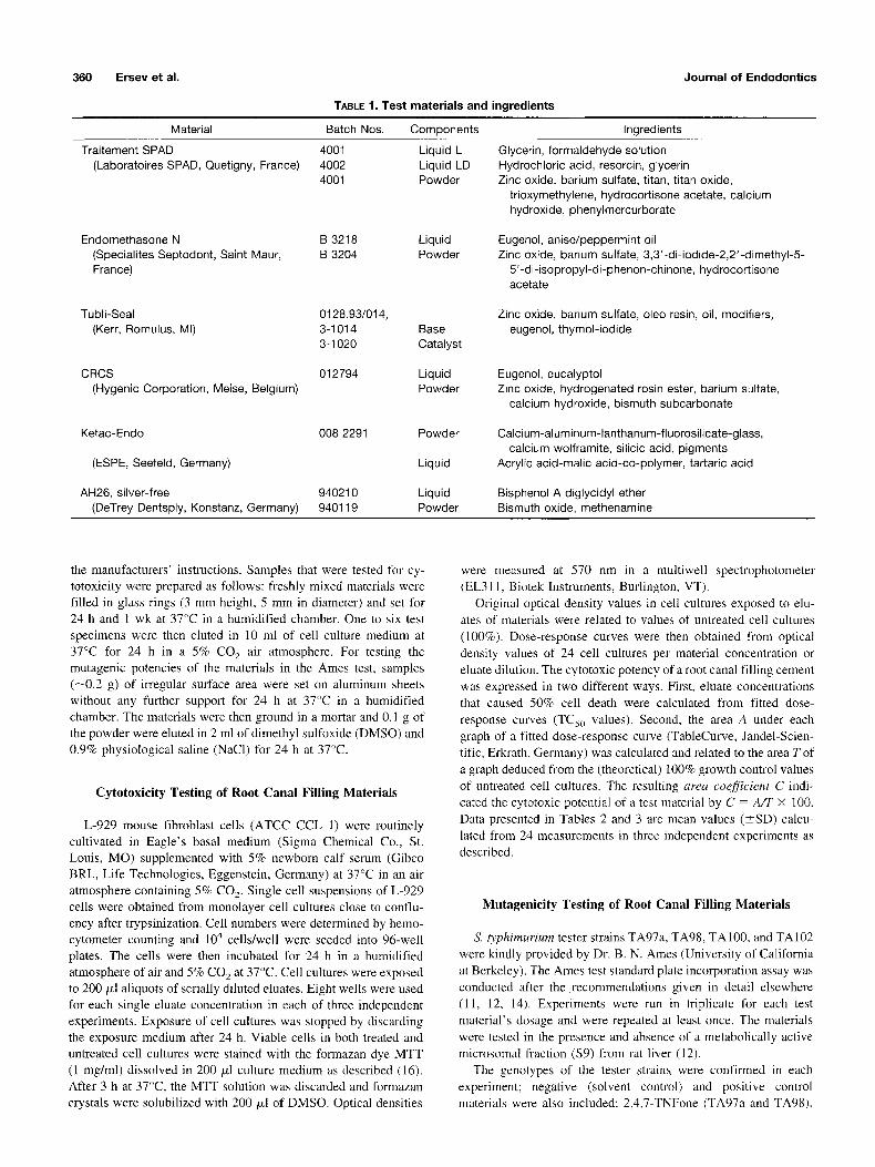
The various root canal sealers that were tested in the present study are listed in Table 1. The materials were mixed according to
360 Ersev et al. Journal of Endodontics
TABLE 1. Test materials and ingredients
Material Batch Nos . Components Ingredients
Traitement SPAD 4001 (Laboratoires SPAD, Quetigny, France) 4002
4001
Liquid L Liquid LD Powder
Endomethasone N B 3218 Liquid (Specialites Septodont, Saint Maur, B 3204 Powder France)
Tubli-Seal 0128.93/014, (Kerr, Romulus, MI) 3-1014 Base
3-1020 Catalyst
CRCS 012794 Liquid (Hygenic Corporation, Meise, Belgium) Powder
Ketac-Endo 008 2291
(ESPE, Seefeld, Germany)
AH26, silver-free 940210 (DeTrey Dentsply, Konstanz, Germany) 940119
Powder
Liquid
Liquid Powder
Glycerin, formaldehyde solution Hydrochloric acid, resorcin, glycerin Zinc oxide, barium sulfate, titan, titan oxide,
trioxymethylene, hydrocortisone acetate, calcium hydroxide, phenylmercurborate
Eugenol, anise/peppermint oil Zinc oxide, barium sulfate, 3,3'-di-iodide-2,2'-dimethyl-5-
5'-di-isopropyl-di-phenon-chinone, hydrocortisone acetate
Zinc oxide, barium sulfate, oleo resin, oil, modifiers, eugenol, thymol-iodide
Eugenol, eucalyptol Zinc oxide, hydrogenated rosin ester, barium sulfate,
calcium hydroxide, bismuth subcarbonate
Calcium-aluminum-lanthanum-fluorosilicate-glass, calcium wolframite, silicic acid, pigments
Acrylic acid-malic acid-co-polymer, tartaric acid
Bisphenol A diglycidyl ether Bismuth oxide, methenamine
the manufacturers ' instructions. Samples that were tested for cy- totoxicity were prepared as follows: freshly mixed materials were filled in glass rings (3 mm height, 5 mm in diameter) and set for 24 h and 1 wk at 37°C in a humidified chamber. One to six test specimens were then eluted in 10 ml of cell culture medium at 37°C for 24 h in a 5% CO 2 air atmosphere. For testing the mutagenic potencies of the materials in the Ames test, samples (--0.2 g) of irregular surface area were set on aluminum sheets without any further support for 24 h at 37°C in a humidified chamber. The materials were then ground in a mortar and 0.1 g of the powder were eluted in 2 ml of dimethyl sulfoxide (DMSO) and 0.9% physiological saline (NaC1) for 24 h at 37°C.
Cytotoxicity Testing of Root Canal Filling Materials
L-929 mouse fibroblast cells (ATCC CCL l) were routinely cultivated in Eagle 's basal medium (Sigma Chemical Co., St. Louis, MO) supplemented with 5% newborn calf serum (Gibco BRL, Life Technologies, Eggenstein, Germany) at 37°C in an air atmosphere containing 5% CO 2. Single cell suspensions of L-929 cells were obtained from monolayer cell cultures close to conflu- ency after trypsinization. Cell numbers were determined by hemo- cytometer counting and 10 4 cells/well were seeded into 96-well plates. The cells were then incubated for 24 h in a humidified atmosphere of air and 5% CO 2 at 37°C. Cell cultures were exposed to 200/M aliquots of serially diluted eluates. Eight wells were used for each single eluate concentration in each of three independent experiments. Exposure of cell eultures was stopped by discarding the exposure medium after 24 h. Viable cells in both treated and untreated cell cultures were stained with the formazan dye MTT (1 mg/ml) dissolved in 200/~1 culture medium as described (16). After 3 h at 37°C, the MTT solution was discarded and formazan crystals were solubilized with 200 ~1 of DMSO. Optical densities
were measured at 570 nm in a multiwell spectrophotometer (EL311, Biotek Instruments, Burlington, VT).
Original optical density values in cell cultures exposed to elu- ates of materials were related to values of untreated cell cultures (100%). Dose-response curves were then obtained from optical density values of 24 cell cultures per material concentration or eluate dilution. The cytotoxic potency of a root canal filling cement was expressed in two different ways. First, eluate concentrations that caused 50% cell death were calculated from fitted dose- response curves (TC~o values). Second, the area A under each graph of a fitted dose-response curve (TableCurve, Jandel-Scien- tific, Erkrath, Germany) was calculated and related to the area T of a graph deduced from the (theoretical) 100% growth control values of untreated cell cultures. The resulting area coefficient C indi- cated the cytotoxic potential of a test material by C = A/T × I00. Data presented in Tables 2 and 3 are mean values (_+SD) calcu- lated from 24 measurements in three independent experiments as described.
Mutagenicity Testing of Root Canal Filling Materials
S. O'phimurium tester strains TA97a, TA98, TA100, and TA102 were kindly provided by Dr. B. N. Ames (University of California at Berkeley). The Ames test standard plate incorporation assay was conducted after the recommendat ions given in detail elsewhere (11, 12, 14). Experiments were run in triplicate for each test material 's dosage and were repeated at least once. The materials were tested in the presence and absence of a metabolically active microsomal fraction ($9) from rat liver (12).
The genotypes of the tester ,strains were confirmed in each experiment; negative (solvent control) and positive control materials were also included: 2,4,7-TNFone (TA97a and TA98),
Vol. 25, No. 5, May 1999 Cytocompatibility of Endodontic Materials 361
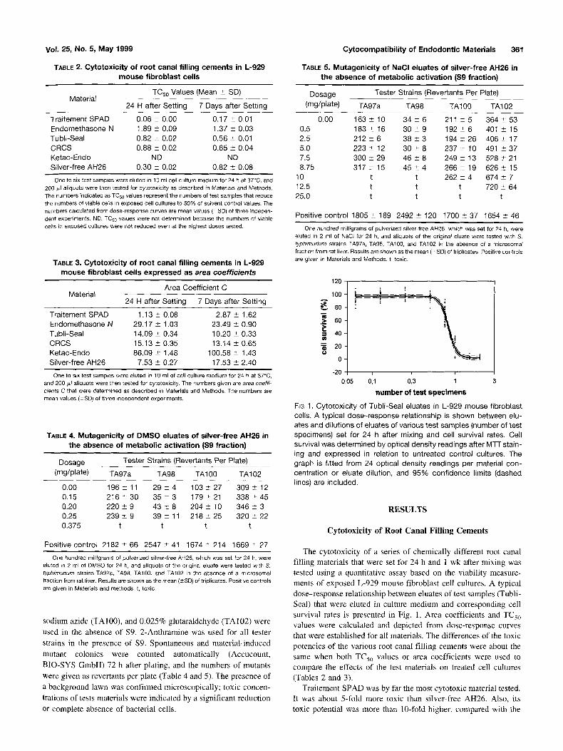
TABLE 2. Cytotoxicity of root canal filling cements in L-929 mouse fibroblast ceils
Material TCso Values (Mean +_ SD)
24 H after Setting 7 Days after Setting
Traitement SPAD 0.06 _+ 0.00 0.17 ± 0.01 Endomethasone N 1.89 ± 0.09 1.37 ± 0.03 Tubli-Seal 0.82 _+ 0.02 0.56 _+ 0.01 CRCS 0.88 ± 0.02 0.65 ± 0.04 Ketac-Endo ND ND Silver-free AH26 0.30 ± 0.02 0.82 ± 0.08
One to six test samples were eluted in 10 mt cell culture medium for 24 h at 37°C, and 200/~1 aliquots were then tested for cytotoxicity as described in Materials and Methods. The numbers indicated as TC50 values represent the numbers of test samples that reduce the numbers of viable cells in exposed cell cultures to 50% of solvent control values. The numbers calculated from dose-response curves are mean values (+SD) of three indepen- dent experiments. ND, TC50 values were not determined because the numbers of viable cells in exposed cultures were not reduced even at the highest doses tested.
TABLE 3. Cytotoxicity of root canal filling cements in L-929 mouse fibroblast cells expressed as area coefficients
Material Area Coefficient C
24 H after Setting 7 Days after Setting
Traitement SPAD 1.13 +_ 0.08 2.87 _+ 1.62 Endomethasone N 29.17 ± 1.03 23.49 _+ 0.90 Tubli-Seal 14.09 _+ 0.34 10.20 _+ 0.33 CRCS 15.13 _+_ 0.35 13.14 +_ 0.65 Ketac-Endo 86.09 _+ 1.48 100.58 _+ 1.43 Silver-free AH26 7.53 _+ 0.27 17.53 _+ 2.40
One to six test samples were eluted in 10 ml of cell culture medium for 24 h at 37°C, and 200/~1 aliquots were then tested for cytotoxicity. The numbers given are area coeffi- cients C that were determined as described in Materials and Methods. The numbers are mean values (-+SD) of three independent experiments.
TABLE 4. Mutagenicity of DMSO eluates of silver-free AH26 in the absence of metabolic activation ($9 fraction)
Dosage Tester Strains (Revertants Per Plate)
(mg/plate) TA97a TA98 TA100 TA102
0.00 196_+ 11 2 9 ± 4 103_+27 3 0 9 ± 12 0.15 216 _+ 30 35 ± 3 179 _+ 21 338 ± 45 0.20 220 _+ 9 43 ± 8 204 _+ 10 346 _+ 3 0.25 239 _+ 9 39 ± 11 218 _+ 25 320 Jr 22 0.375 t t t t
Positive control 2182 ± 66 2547 _+ 41 1674 _+ 214 1669 +_ 27
One hundred milligrams of pulverized silver-free AH26, which was set for 24 h, were eluted in 2 ml of DMSO for 24 h, and aliquots of the original eluate were tested with S. typhimurium strains TA97a, TA98, TA100, and TA102 in the absence of a microsomal fraction from rat liver. Results are shown as the mean (-+SD) of triplicates. Positive controls are given in Materials and methods, t, toxic.
sodium azide (TA100), and 0.025% glutaraldehyde (TA102) were
used in the absence of $9. 2-Anthramine was used for all tester
strains in the presence of $9. Spontaneous and material-induced
mutant colonies were counted automatically (Accucount,
BIO-SYS GmbH) 72 h after plating, and the numbers of mutants
were given as revertants per plate (Table 4 and 5). The presence of
a background lawn was confirmed microscopically; toxic concen-
trations of tests materials were indicated by a significant reduction
or complete absence of bacterial cells.
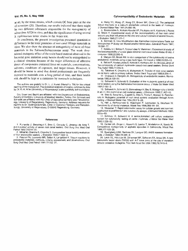
TABLE 5. Mutagenicity of NaCI eluates of silver-free AH26 in the absence of metabolic activation ($9 fraction)
Dosage Tester Strains (Revertants Per Plate)
(mg/plate) TA97a TA98 TA100 TA102
0.00 163 ± 10 34 ± 6 211 +_ 5 364 _+ 53 183 ± 16 30 ± 9 192 _+ 6 401 _+ 15 2 1 2 ± 6 38-+3 1 9 4 ± 2 6 406± 17 223_+ 12 3 0 ± 8 2 3 7 ± 10 491 ± 3 7 300+_29 46+-8 249_+13 5 2 8 ± 2 1 3 1 7 ± 15 4 5 ± 4 2 6 6 ± 19 626± 15
t t 262 ± 4 674 ± 7 t t t 720 _+ 64 t t t t
0.5 2.5 5.0 7.5 8.75
10 12.5 25.0
Positive control 1805 _+ 189 2492 ± 120 1700 ± 37 1654 4- 46
One hundred milligrams of pulverized silver-free AH26, which was set for 24 h, were eluted in 2 ml of NaCI for 24 h, and aliquots of the original eluate were tested with S. typhimurium strains TA97a, "1-A98, TA100, and TA102 in the absence of a microsoma[ fraction from rat liver. Results are shown as the mean (÷SD) of triplicates. Positive controls are given in Materials and Methods. t, toxic.
120
100
~ 8o
.~ 6o
-., 40
= 20 o
0
-20 0,05
F----z-- = h
L i i i
0,1 0,3 1
number of test specimens
F~G 1. Cytotoxicity of Tubli-Seal eluates in L-929 mouse fibroblast cells. A typical dose--response relationship is shown between elu- ates and dilutions of eluates of various test samples (number of test specimens) set for 24 h after mixing and cell survival rates. Cell survival was determined by optical density readings after M-IF stain- ing and expressed in relation to untreated control cultures. The graph is fitted from 24 optical density readings per material con- centration or eluate dilution, and 95% confidence limits (dashed lines) are included.
RESULTS
C y t o t o x i c i t y o f R o o t C a n a l F i l l ing C e m e n t s
The cytotoxicity of a series of chemically different root canal filling materials that were set for 24 h and 1 wk after mixing was tested using a quantitative assay based on the viability measure- ments of exposed L-929 mouse fibroblast cell cultures. A typical dose-response relationship between eluates of test samples (Tubli- Seal) that were eluted in culture medium and corresponding cell survival rates is presented in Fig. 1. Area coefficients and TCso values were calculated and depicted from dose-response curves that were established for all materials. The differences of the toxic potencies of the various root canal filling cements were about the same when both TCso values or area coefficients were used to compare the effects of the test materials on treated cell cultures (Tables 2 and 3).
Traitement SPAD was by far the most cytotoxic material tested. It was about 5-fold more toxic than silver-free AH26. Also, its toxic potential was more than 10-fold higher, compared with the

362 Ersev et al.
reactions elicited by Tubli-Seal and CRCS and about 30-fold higher than Endomethasone N after a setting time of 24 h (Table 2). The toxic potencies of Traitement SPAD and the resin-based silver-free AH26 were equally reduced by a factor of 2 to 3 when the materials were tested after a setting time of 1 wk (Tables 2 and 3). In contrast, the cytotoxicity of eluates of Endomethasone N, Tubli-Seal, and CRCS increased slightly after a setting time of 1 wk after mixing. Consequently, the rank order of cytotoxic effects of the materials in L-929 cells was changed. Silver-free AH26 was less toxic than Tubli-Seal and CRCS after a setting time of 1 wk (Tables 2 and 3). Ketac-Endo had little cytotoxic effect on L-929 cells after a setting time of 24 h and 1 wk. When the cytotoxicity of Ketac-Endo was quantitatively related to those of other mate- rials by its area coefficient, Ketac-Endo was more than 70-fold less toxic than Traitement SPAD after a setting time of 24 h (Table 3).
Mutagenicity of Root Canal Filling Cements
Traitement SPAD, Endomethasone N, Tubli-Seal, CRCS, and Ketac-Endo were not mutagenic in any of the Salmonella tester strains in broad concentration ranges from nontoxic to toxic (data not shown). In contrast, silver-free resin-based AH26 tested pos- itive in the Ames mutation assay. DMSO eluates of silver-free AH26 set for 24 h induced a slight dose-dependent increase of mutant numbers of the tester strain S. typhimurium TA100 in the absence of a metabolically active microsomal fraction from rat liver ($9). Mutant numbers were about 2-fold higher in bacterial cultures exposed to material eluates than in cultures treated with the solvent only (Table 4). The mutagenic effect was reduced when a $9 fraction was present in the reaction mixture (data not shown). Interestingly, DMSO eluates of silver-free AH26 were not active in any other tester strain (Tables 4). However, mutagenicity of silver- free AH26 after elution in NaC1 was also detected in tester strains S. typhimurium TA97a and TA102 when the aliquots tested were about 40- to 50-fold higher than in DMSO eluates (Table 5).
DISCUSSION
Results from biological testing of root canal filling materials varied with the chemical composition of the materials. However, the ranking of toxic cell and tissue reactions caused by these materials is consistent in cytotoxicity tests in vitro, implantation tests, and usage tests in vivo (2-4, 8, 10). The high sensitivity of quantitative cell culture systems can be used as a major advantage in toxicity testing of dental materials to determine the differences between the materials' toxic potencies in addition to listing a rank order of the cytotoxic potentials (17). Herein, the cytotoxicity of eluates of six root canal filling cements was first measured and expressed in dose-response relationships. The amounts of cell culture medium eluates, which led to 50% cell survival after a 24-h exposure, were determined by a quantitative assay, and the mate- rials toxicities were compared accordingly. The use of area coef- ficients calculated from dose-response curves to compare toxic potentials may even underestimate the differences of toxic poten- tials when a material, like Ketac-Endo, was nontoxic. Otherwise, both parameters provide very similar information on the cytotox- icity of mixed materials. It can be easily determined that the cytotoxic potency of, for example, the most toxic endodontic material Traitment SPAD was more than 30-fold higher than the potency of Endomethasone N after a setting time of 24 h. There
Journal of Endodontics
was also a difference of almost 1 order of magnitude between the cytotoxic potencies of Traitment SPAD and Ketac-Endo that was by far the least toxic material tested herein.
The quantitative cytotoxicity assay based on the staining of viable cells by the tetrazolium dye MTT is, of course, also a precise approach to rank the endodontic materials in order of decreasing cytotoxicities depending on the setting time. Rank orders similar to those observed herein were reported from different laboratories (8, 10).
The resin-based AH26 can cause severe toxic reactions imme- diately after mixing in implantation studies and in culture cells in vitro (5, 10). Also, CRCS, a calcium hydroxide-based material containing zinc oxide-eugenol, exhibited mild to moderate toxicity in implantation studies and in cell culture tests (5, 9). Based on these observations and the quantitative approach to determine the cytotoxicity of root canal filling materials presented herein, the so far unknown reaction of a root canal filling material in implanta- tion studies can be predicted from two relationships. First, the relationship of the known toxic reactions of two endodontic ma- terials in quantitative cell culture experiments as described herein (in vitro) and the graded tissue reaction in implantation studies (in vivo) can be used. Second, the relationship between the toxic reactions of these materials and the material of so far unknown properties determined in a quantitative cell culture assay is also useful. This approach might provide information on the toxic potential of a root canal sealing material and, therefore, be helpful in reducing the number of animals to be used in implantation studies.
The mutagenic potential of root canal filling materials in the Ames test is low. None of the materials which contain zinc oxide- eugenol and formaldehyde was mutagenic in any of the four different S. typhimurium tester strains. Similar observations have been reported and discussed recently after testing various materi- als. Likewise, mutagenicity and genotoxicity of mixed AH26 has been shown in previous investigations (11-15). Most important, the mutagenic effects decreased with increasing setting times in both the bacterial and mammalian cell gene mutation assay (12, 13).
There is evidence that the mutagenic effect of AH26 may arise from the liquid component bisphenol A diglycidyl ether, because this showed mutagenic activity with the same characteristics (11- 13). In our investigation, the mutagenic response of silver-free AH26 in TA 100 was also characterized by a steep rise in revertants within a narrow concentration range of DMSO eluates, which was reduced in the presence of metabolic activation ($9 fraction). Therefore, it is concluded that the mutagenic reaction toward TA100 might be caused by the liquid component bisphenol A diglycidyl ether. The mutagenic activity of bisphenol A diglycidyl ether in the Ames test has been shown previously (18).
In contrast to previous findings with formaldehyde (11) and our results in strain TA102 with silver-free AH26 as discussed herein, we were unable to detect any mutagenic response with Traitment SPAD, a root canal filling material containing formaldehyde and hydrochloric acid. This might be due to the strong toxic effect of the material toward the test bacteria, preventing the detection of a mutagenic response. On the other hand, formaldehyde may be the active compound in tester strain TA102, because AH26 releases formaldehyde and the amount of formaldehyde increases up to 2 days of setting to nearly 200 times aver the concentration of freshly mixed AH26 (19). It has been shown that formaldehyde is more active in strain TA102 than TAI00, because it acts preferentially at AT base pairs and oxidative mutagens are detected poorly, or not
Vol. 25, No. 5, May 1999
at all, by the tester strains, which contain GC base pairs at the site of reversion (20). Therefore, our results indicated that there might be two different substances responsible for the mutagenicity of silver-free AH26 in vitro, and thus the significance of using several S. ~phimurium tester strains in the Ames test.
In conclusion, the present investigation presented quantitative differences in the toxic potencies of root canal fillings materials in vitro. We also show the absence of mutagenicity of most of these materials in the Salmonella/microsome assay. The weak dose- related mutagenic effect of the resin-based material observed in the bacterial gene mutation assay herein should not be extrapolated to a clinical situation because of the major differences of effective doses of components extracted from set materials, concentrations, solvents, conditions of exposure, and target tissues. However, it should be borne in mind that dental professionals are frequently exposed to materials over a long period of time, and their health risk should be kept at a minimum by nontouch techniques.
The authors are grateful to Dr. L. d. Nunez (Memphis, TN) for his critical reading of the manuscript. The statistical analyses of original cytotoxicity data by Dr. K.-A. Hiller (University of Regensburg) is also gratefully acknowledged.
Drs. Ersev and Bayirli are affiliated with the Department of Endodontics, Faculty of Dentistry, University of Istanbul, Istanbul, Turkey. Drs. Schmalz and Schweikl are affiliated with the Clinic of Operative Dentistry and Periodontol- ogy, University of Regensburg, Regensburg, Germany. Address requests for reprints to Dr. Gottfried Schmalz, Clinic of Operative Dentistry and Periodon- tology, University of Regensburg, D-93042 Regensburg, Germany.
References
1. Pumarola J, Berastegui E, Brau E, Canalda C, Jimenez de Anta T. Antimicrobial activity of seven root canal sealers. Oral Surg Oral Med Oral Pathol 1992;74:216-20.
2. Mittal M, Chandra S, Chandra S. Comparative tissue toxicity evaluation of four endodontic sealers. J Endodon 1995;21:622-4.
3. Pascon EA, Leonardo MR, Safavi K, Langeland K. Tissue reactions to endodontic materials: methods, criteria, assessment, and observations. Oral Surg Oral Med Oral Pathol 1991 ;72:222-37.
Cy tocompa t i b i l i t y o f Endodon t i c Mate r ia ls 363
4. Hong YC, Wang JT, Hong CY, Brown WE, Chow LC. The periapical tissue reactions to a calcium phosphate cement in the teeth of monkeys. J Biomed Mater Res 1991 ;25:485-98.
5. Economides N, Kotsaki-Kovatsi VP, Poulopoulos A, Kolokuris I, Rozos G, Shore R. Experimental study of the biocompatibility of four root canal sealers and their influence on the zinc and calcium content of several tissues. J Endodon 1995;21:122-7.
6. Schmalz G. Eine Modifikation des Kaninchen-lmplantations-Tests zur biologischen PKJfung von WurzelkanalfOlI-Materialien. Zahn&rztl Praxis 1987; 10:366 -77.
7. Kolokuris I, Beltes P, Economides N, Vlemmas I. Experimental study of the biocompatibility of a new glass-ionomer root canal sealer (Ketac-Endo). J Endodon 1996;22:395-8.
8. Meryon SD, Brook AM. In vitro comparison of the cytotoxicity of twelve endodontic materials using a new technique. Int Ended J 1990;23:203-10.
9. Beltes P, Koulaouzidou E, Kotoula V, Kortsaris AH. In vitro evaluation of the cytotoxicity of calcium hydroxide-based root canal sealers. Ended Dent Traumatol 1995;11:245-9.
10. Takahara K, Onodera A, Matsumoto K. Toxicity of root canal sealers on rat bone cells in primary culture. Ended Dent Traumatol 1990;6:200-7.
11. Orstravik D, Hongslo JK. Mutagenicity of endedontic sealers. Bioma- terials 1985;6:129-32.
12. Schweikl H, Schmalz G. Evaluation of the mutagenic potential of root canal sealers using the SalmonellaJmicrosome assay. J Mater Sci Mater Med 1991;2:181-5.
13. Schweikl H, Schmalz G, Stimmelmayr H, Bey B. Mutagenicity of AH26 in an in vitro mammalian cell mutation assay. J Endodon 1995;21:407-10.
14. Stea S, Savarino L, Ciapetti G, Stea S, Trotta F, Morozzi G, Pizzofer- rate A. Mutagenic potential of root canal sealers: evaluation through Ames testing. J Biomed Mater Res 1994;28:319-28.
15. Hell J, Reifferscheid G, Waldmann P, Leyhausen G, Geurtsen W. Genotoxicity of dental materials. Mutat Res 1996;368:181-94.
16. Mossman T. Rapid colorimetric assay for cellular growth and survival: application to proliferation and cytotoxicity assays. J Immunol Methods 1983; 65:55-63.
17. Schmalz G, Schweikl H. A semiautomated cell culture evaluation system for cytotoxicity testing of dental materials. J Mater Sci Mater Med 1990;1:228-32.
18. Canter DA, Zeiger E, Haworth S, Lawlor T, Mortelsman K, Speck W. Comparative mutagenicity of aliphatic epoxides in Salmonella. Mutat Res 1986;172:105-38.
19. Spangberg LSW, Barbosa SV, Lavigne GD. AH26 releases formalde- hyde. J Endodon 1993;19:596-8.
20. Levin DE, Hollstein M, Christman MF, Schwiers EA, Ames BN. A new Salmonella tester strain (TA102) with A-T base pairs at the site of mutation detects oxidative mutagens. Proc Natl Acad Sci USA 1982;79:7445-9.