cytosolic ascorbate peroxidase 1 is a central component of the
TRANSCRIPT

Cytosolic Ascorbate Peroxidase 1 Is a Central Component ofthe Reactive Oxygen Gene Network of Arabidopsis W
Sholpan Davletova,a Ludmila Rizhsky,b Hongjian Liang,b Zhong Shengqiang,b David J. Oliver,b Jesse Coutu,a
Vladimir Shulaev,c Karen Schlauch,d and Ron Mittlera,1
a Department of Biochemistry and Molecular Biology, University of Nevada, Reno, Nevada 89557b Department of Genetics and Developmental and Cell Biology, Iowa State University, Ames, Iowa 50011c Virginia Bioinformatics Institute, Blacksburg, Virginia 24061d Center for Biomedical Genomics and Informatics, George Mason University, Manassas, Virginia 20110
Reactive oxygen species (ROS), such as O2� and H2O2, play a key role in plant metabolism, cellular signaling, and defense. In
leaf cells, the chloroplast is considered to be a focal point of ROS metabolism. It is a major producer of O2� and H2O2 during
photosynthesis, and it contains a large array of ROS-scavenging mechanisms that have been extensively studied. By
contrast, the function of the cytosolic ROS-scavenging mechanisms of leaf cells is largely unknown. In this study, we
demonstrate that in the absence of the cytosolic H2O2-scavenging enzyme ascorbate peroxidase 1 (APX1), the entire
chloroplastic H2O2-scavenging system of Arabidopsis thaliana collapses, H2O2 levels increase, and protein oxidation
occurs. We further identify specific proteins oxidized in APX1-deficient plants and characterize the signaling events that
ensue in knockout-Apx1 plants in response to a moderate level of light stress. Using a dominant-negative approach, we
demonstrate that heat shock transcription factors play a central role in the early sensing of H2O2 stress in plants. Using
knockout plants for the NADPH oxidase D protein (knockout-RbohD), we demonstrate that RbohD might be required for
ROS signal amplification during light stress. Our study points to a key role for the cytosol in protecting the chloroplast
during light stress and provides evidence for cross-compartment protection of thylakoid and stromal/mitochondrial APXs
by cytosolic APX1.
INTRODUCTION
Reactive oxygen species (ROS) are partially reduced or excited
forms of atmospheric oxygen (O2) continuously produced in cells
during aerobicmetabolism (Halliwell andGutteridge, 1989). They
can cause extensive cell injury or death, but they play a central
role in many signaling pathways in plants involved in stress
perception, photosynthesis regulation, pathogen response, pro-
grammed cell death, hormonal action, and plant growth and
development (Dat et al., 2000; Mittler, 2002; Mullineaux and
Karpinski, 2002; Neill et al., 2002; Apel and Hirt, 2004).
In Arabidopsis thaliana, a network of at least 152 genes
controls ROS metabolism (Mittler et al., 2004). The network is
thought to regulate the rates of ROS production and ROS
scavenging in the different cellular compartments and to mod-
ulate the steady state level of ROS for signaling as well as
defense purposes. In leaf cells, an intricate balance exists
between H2O2 and O2� production in the chloroplast and
peroxisome during photosynthesis and the activities of the
ROS-scavenging enzymes superoxide dismutase (SOD), ascor-
bate peroxidase (APX), and catalase (Asada, 1999; Mittler, 2002;
Apel and Hirt, 2004). The chloroplast contains at least three
different isozymes of APX: a thylakoid-bound APX (At1g77490),
a lumen APX (At4g09010), and a stromal APX (At4g08390;
stromal APX was recently shown to be dually targeted to the
stroma and mitochondria in Arabidopsis and would be refer-
red to as stromal/mitochondrial APX; Chew et al., 2003). The
chloroplast also contains four different isozymes of SOD: a
CuZnSOD (CSD2), threeFeSODs, andenzymesof theascorbate-
glutathione cycle capable of reducing oxidized ascorbic acid
and glutathione (Asada and Takahashi, 1987). By contrast, the
cytosol contains one cytosolic APX (APX1, At1g07890), with an
additional APX (APX2, At3g09640) that is inducible mainly under
extreme light or heat stress conditions (Karpinski et al., 1999;
Panchuk et al., 2002), and different enzymes of the ascorbate-
glutathione cycle (Mittler et al., 2004). Leaf peroxisomes contain
three isozymes of catalase, one isozyme of APX (At4g35000) that
is bound to the outer layer of the peroxisome, and enzymes of the
ascorbate-glutathione cycle (Corpas et al., 2001; Shigeoka et al.,
2002). Comparedwith other cellular compartments, chloroplasts
contain high levels of the antioxidants ascorbic acid and gluta-
thione (up to 25 and 5mM, respectively; Noctor and Foyer, 1998).
Mathematical calculations, as well as computer model simu-
lations, based on enzyme concentration and reaction rate, the
rates of O2� and H2O2 production, and the concentrations of the
antioxidants ascorbic acid and glutathione in chloroplasts,
conclude that the chloroplast is well equipped to scavenge the
1 To whom correspondence should be addressed. E-mail [email protected]; fax 775-784-6911.The author responsible for distribution of materials integral to thefindings presented in this article in accordance with the policy describedin the Instruction for Authors (www.plantcell.org) is: Ron Mittler([email protected]).WOnline version contains Web-only data.Article, publication date, and citation information can be found atwww.plantcell.org/cgi/doi/10.1105/tpc.104.026971.
The Plant Cell, Vol. 17, 268–281, January 2005, www.plantcell.orgª 2004 American Society of Plant Biologists
Dow
nloaded from https://academ
ic.oup.com/plcell/article/17/1/268/6113109 by guest on 16 N
ovember 2021

O2� and H2O2 produced during photosynthesis in plants grown
under controlled conditions or subjected to light stress (Asada
and Takahashi, 1987; Asada, 1999; Polle, 2001). In addition, the
expression of transcripts encoding the stromal/mitochondrial
and thylakoid-bound APXs was not enhanced during light stress,
whereas the expression of the cytosolic APXs was enhanced
(Karpinski et al., 1997; Shigeoka et al., 2002). This finding was
mainly interpreted as supportive of the hypothesis that within
leaf cells the chloroplastic scavenging systems are sufficient
to handle ROS production even under stressful conditions
(Shigeoka et al., 2002).
The signal transduction pathway leading to the enhanced
expression of cytosolic APXs during light stress was proposed
to involve a chloroplast-to-nuclei signal that depends on the pool
of reduced plastoquinone in the chloroplast and may involve
H2O2 (Karpinski et al., 1997; Rodermel, 2001; Mullineaux and
Karpinski, 2002). Moreover, the signal that enhanced APX2
expression in response to high light stress was shown to be
a systemic signal (Karpinski et al., 1999). However, in view of the
supposition that the chloroplast is well equipped to scavenge
ROS produced during photosynthesis, the significance of the
enhanced cytosolic APX expression during light stress is un-
known. Recent analysis of knockout plants deficient in cytosolic
APX1 (KO-Apx1) revealed that in the absence of APX1 photo-
synthetic activity was suppressed, suggesting that cytosolic
APXs might be essential for chloroplast protection during light
stress (Pnueli et al., 2003; Mittler et al., 2004).
In this study, we demonstrate that in the absence of cytosolic
APX1, the entire chloroplastic H2O2-scavenging system of Ara-
bidopsis collapses, H2O2 levels increase, and protein oxidation
occurs. Using Affymetrix GeneChip and matrix-assisted laser-
desorption ionization time of flight (MALDI-TOF) technology, we
identify specific proteins oxidized in KO-Apx1 plants and char-
acterize the signaling events that ensue in KO-Apx1 plants in
response to a moderate level of light stress. Using a dominant-
negative approach, we demonstrate that heat shock transcrip-
tion factors (HSFs) play a key role in the early sensing of H2O2
stress in KO-Apx1 plants, and using knockout plants for the
NADPH oxidase D protein (KO-RbohD), we demonstrate that
RbohD might be required for ROS signal amplification during
light stress in Arabidopsis. Our study points to a key role for the
cytosol in protecting the chloroplast during light stress and
provides evidence for cross-compartment protection of thyla-
koid and stromal/mitochondrial APXs by cytosolic APX1.
RESULTS
H2O2 Accumulation and Protein Oxidation in KO-Apx1Plants
As shown in Figure 1A, KO-Apx1 plants grown under low light
conditions (25 mmol m�2 s�1) and transferred to a moderate light
level of 250 mmol m�2 s�1 accumulated H2O2 in leaves. By
contrast, wild-type plants subjected to the same treatment did
not accumulate H2O2 to the same level. The accumulation of
H2O2 appeared to occur in a homogeneous manner in most leaf
cells and was not confined to a particular tissue, such as the
vascular tissue.
Figure 1. Deficiency in APX1 Results in H2O2 Accumulation and Protein
Oxidation in Arabidopsis Leaves Subjected to a Moderate Level of Light
Stress.
(A) Accumulation of H2O2 in wild-type and knockout-Apx1 (KO-Apx1)
plants in response to a moderate level of light stress. Compared with
wild-type plants, KO-Apx1 plants are shown to accumulate higher levels
of H2O2 (evident by darker staining of leaves at 90 and 180 min after the
application of light stress).
(B) Detection of proteins containing carbonyl groups (indicative of
protein oxidation) in leaf extracts obtained from wild-type and KO-
Apx1 plants by a protein blot assay (top). A protein gel stained with
Coomassie blue is used to demonstrate equal loading of proteins
(bottom). Compared with wild-type plants, KO-Apx1 plants are shown
to accumulate a high level of oxidized proteins. Detection of H2O2 and
protein oxidation was performed as described in Methods. All experi-
ments were repeated at least three times. Representative results are
shown.
Cross-Compartment Protection by APX1 269
Dow
nloaded from https://academ
ic.oup.com/plcell/article/17/1/268/6113109 by guest on 16 N
ovember 2021

To examine whether H2O2 accumulation in KO-Apx1 resulted
in oxidative damage to cells, we tested protein extracts from leaf
cells using a protein gel blot assay that detects protein oxidation
(detects the presence of carbonyl groups on proteins by first
reacting proteins with 2,4-dintrophenylhydrazine (DNP) and then
detecting the DNP-bound proteins on protein blots using a DNP-
specific antibody; Johansson et al., 2004; Rizhsky et al., 2004).
Leaf cells grown under controlled conditions contain a low
baseline level of oxidized proteins (data not shown; Johansson
et al., 2004). As shown in Figure 1B, KO-Apx1 plants subjected to
the light treatment described above (25 to 250 mmol m�2 s�1)
accumulated high levels of oxidized proteins. By contrast, wild-
type plants subjected to the same treatment did not accumulate
a similar level of oxidized proteins. This result suggests that the
absence of APX1 results not only in the accumulation of H2O2
(Figure 1A) but also in damage to specific proteins in leaf cells
(Figure 1B).
To identify specific proteins oxidized in KO-Apx1 plants in
response to the light stress treatment described above, we
performed proteomic analysis of protein extracts and deter-
mined the identity of specific proteins detected by the DNP-
antibody using MALDI-TOF (see Methods for details). As shown
in Table 1, the lack of APX1 resulted in the oxidation of different
proteins belonging to different metabolic pathways. Interest-
ingly, three of the proteins identified by our study as oxidized in
knockout cytosolic Apx1 plants were chloroplastic proteins (Cys
synthase, Asp kinase, and ribulose-1,5-bisphosphate carboxyl-
ase/oxygenase (Rubisco) large subunit; Table 1). Other proteins
oxidized in KO-Apx1 plants are possibly localized to the nuclei,
cytosol, or the secretory pathway. At least one protein was
predicted to be a membrane protein (Table 1). Low levels of
oxidized Rubisco large subunit were also detected in wild-type
plants subjected to the light stress treatment (data not shown;
see also Rizhsky et al., 2004). The accumulation of H2O2 and
the oxidation of chloroplastic proteins in KO-Apx1 plants during
the light shift treatment (Figure 1, Table 1) suggest that, in the
absence of cytosolic APX1, the chloroplastic or peroxisomal
H2O2-scavenging system(s) of Arabidopsis are unable to scav-
enge the H2O2 produced during photosynthesis or photorespi-
ration. The accumulation of H2O2 and protein oxidation (Figure 1,
Table 1) did not result in the activation of cell death in leaves of
wild-type or KO-Apx1 plants, and KO-Apx1 plants were able to
make a complete recovery from the light stress treatment (data
not shown; see also Pnueli et al., 2003).
In the Absence of APX1, the Chloroplastic H2O2-Scavenging
SystemCollapses
The lack of cytosolic APX1 resulted in the oxidation of chloro-
plastic proteins (Table 1), suggesting that APX1 activity might be
important for chloroplast protection. Previous studies have
shown that chloroplasts are extremely sensitive to external
application of H2O2 (Asada, 2000) and that chloroplastic APXs
are inactivated by H2O2 (Mano et al., 2001). The lack of cytosolic
APX1 may therefore affect H2O2 scavenging systems in the
chloroplast. We therefore studied the relationship between
cytosolic APX1 and the chloroplastic APXs, thylakoid and
stromal/mitochondrial APX.
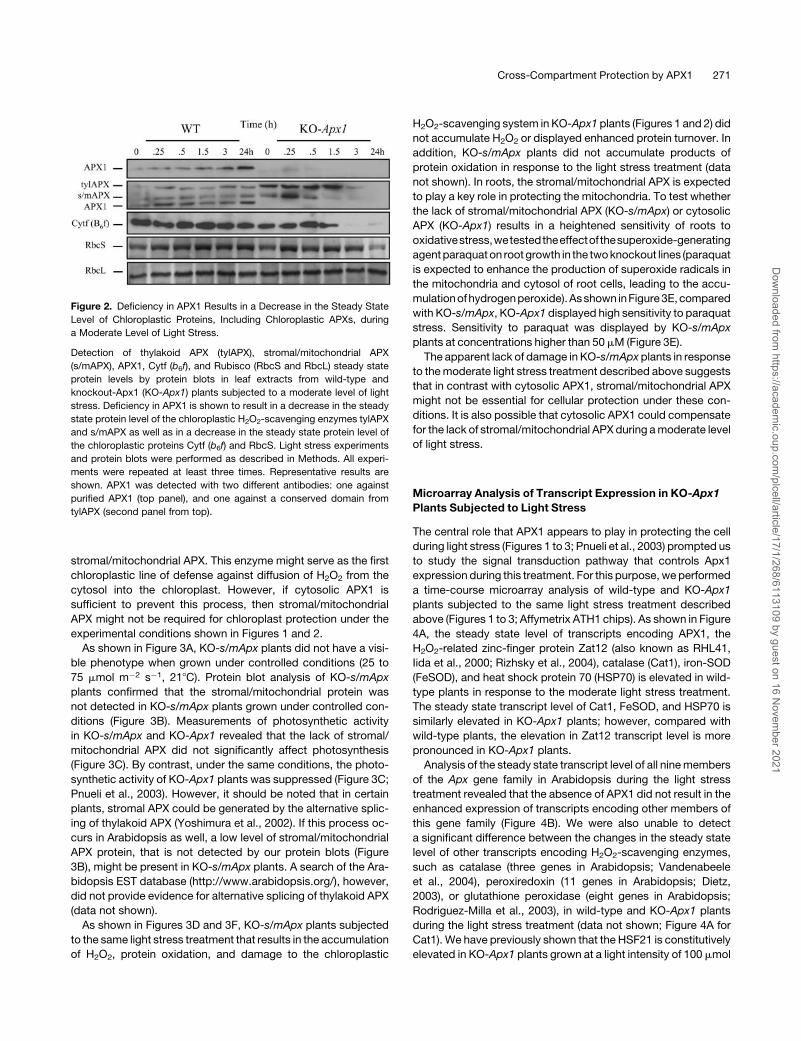
As shown in Figure 2, compared with KO-Apx1 plants, the
steady state protein level of thylakoid and stromal/mitochondrial
APX did not appear to change during this treatment in wild-type
plants. The level of cytosolic APX1 did however increase in wild-
type plants during the light stress treatment (Figure 2). By
contrast, in the absence of APX1 (KO-Apx1), the steady state
level of thylakoid and stromal/mitochondrial APX was elevated,
at least during early time points of light stress. However, the lack
of APX1 resulted in the apparent degradation of thylakoid and
stromal/mitochondrial APXs, a b6f complex subunit protein (Cytf)
and the small subunit protein of Rubisco (Figure 2). These
findings suggest that although APX1 is localized to the cytosol,
it is critical for protecting the H2O2 scavenging system in the
chloroplast during light stress. The inactivation of chloroplastic
APXs byH2O2 (Mano et al., 2001) may serve as the initiation point
for their degradation by chloroplastic proteases.
Cytosolic APX1 Might Be Sufficient to Protect the
Chloroplast in the Absence of Stromal/Mitochondrial APX
To further characterize the relationship between cytosolic APX1
and chloroplastic APXs, we studied knockout plants deficient
in stromal/mitochondrial APX (KO-s/mApx). Because chloro-
plasts are sensitive to the external application of H2O2 (Asada,
2000) and the lack of APX1 might allow H2O2 to diffuse into
the chloroplast from the cytosol, we focused our study on
Table 1. Proteins Oxidized in KO-Apx1 Plants in Response to a Moderate Level of Light Stress
Protein Locus Number No. of Fragments Matched Coverage Predicted Location
Cys synthase [O-acetylserine (thiol)lyase] At3g22460 5 28% Plastid
Asp kinase/homoserine dehydrogenase At1g31230 18 27% Plastid
Putative transposase At2g14140 20 28% Cytosol/nuclear
Rubisco large subunit ArthCp030 14 26% Plastid
Zinc-finger, C3HC4 (RING finger) At5g60710 10 22% Nuclear
Cytochrome P450 At4g15396 22 39% Secretory
Putative cation/Hþ exchanger At5g58460 15 23% Membrane
Transcription activator At3g52910 4 22% Cytosol/nuclear
Proteomic analysis of protein oxidation was determined as described in Methods. Predicted subcellular localization was performed as described in
Mittler et al. (2004).
270 The Plant Cell
Dow
nloaded from https://academ
ic.oup.com/plcell/article/17/1/268/6113109 by guest on 16 N
ovember 2021
stromal/mitochondrial APX. This enzyme might serve as the first
chloroplastic line of defense against diffusion of H2O2 from the
cytosol into the chloroplast. However, if cytosolic APX1 is
sufficient to prevent this process, then stromal/mitochondrial
APX might not be required for chloroplast protection under the
experimental conditions shown in Figures 1 and 2.
As shown in Figure 3A, KO-s/mApx plants did not have a visi-
ble phenotype when grown under controlled conditions (25 to
75 mmol m�2 s�1, 218C). Protein blot analysis of KO-s/mApx
plants confirmed that the stromal/mitochondrial protein was
not detected in KO-s/mApx plants grown under controlled con-
ditions (Figure 3B). Measurements of photosynthetic activity
in KO-s/mApx and KO-Apx1 revealed that the lack of stromal/
mitochondrial APX did not significantly affect photosynthesis
(Figure 3C). By contrast, under the same conditions, the photo-
synthetic activity of KO-Apx1 plants was suppressed (Figure 3C;
Pnueli et al., 2003). However, it should be noted that in certain
plants, stromal APX could be generated by the alternative splic-
ing of thylakoid APX (Yoshimura et al., 2002). If this process oc-
curs in Arabidopsis as well, a low level of stromal/mitochondrial
APX protein, that is not detected by our protein blots (Figure
3B), might be present in KO-s/mApx plants. A search of the Ara-
bidopsis EST database (http://www.arabidopsis.org/), however,
did not provide evidence for alternative splicing of thylakoid APX
(data not shown).
As shown in Figures 3D and 3F, KO-s/mApx plants subjected
to the same light stress treatment that results in the accumulation
of H2O2, protein oxidation, and damage to the chloroplastic
H2O2-scavenging system in KO-Apx1 plants (Figures 1 and 2) did
not accumulate H2O2 or displayed enhanced protein turnover. In
addition, KO-s/mApx plants did not accumulate products of
protein oxidation in response to the light stress treatment (data
not shown). In roots, the stromal/mitochondrial APX is expected
to play a key role in protecting the mitochondria. To test whether
the lack of stromal/mitochondrial APX (KO-s/mApx) or cytosolic
APX (KO-Apx1) results in a heightened sensitivity of roots to
oxidativestress,wetestedtheeffectof thesuperoxide-generating
agentparaquaton rootgrowth in the twoknockout lines (paraquat
is expected to enhance the production of superoxide radicals in
the mitochondria and cytosol of root cells, leading to the accu-
mulationofhydrogenperoxide).Asshown inFigure3E,compared
with KO-s/mApx, KO-Apx1 displayed high sensitivity to paraquat
stress. Sensitivity to paraquat was displayed by KO-s/mApx
plants at concentrations higher than 50mM (Figure 3E).
The apparent lack of damage in KO-s/mApxplants in response
to themoderate light stress treatment described above suggests
that in contrast with cytosolic APX1, stromal/mitochondrial APX
might not be essential for cellular protection under these con-
ditions. It is also possible that cytosolic APX1 could compensate
for the lack of stromal/mitochondrial APX during amoderate level
of light stress.
Microarray Analysis of Transcript Expression in KO-Apx1
Plants Subjected to Light Stress
The central role that APX1 appears to play in protecting the cell
during light stress (Figures 1 to 3; Pnueli et al., 2003) prompted us
to study the signal transduction pathway that controls Apx1
expression during this treatment. For this purpose, weperformed
a time-course microarray analysis of wild-type and KO-Apx1
plants subjected to the same light stress treatment described
above (Figures 1 to 3; Affymetrix ATH1 chips). As shown in Figure
4A, the steady state level of transcripts encoding APX1, the
H2O2-related zinc-finger protein Zat12 (also known as RHL41,
Iida et al., 2000; Rizhsky et al., 2004), catalase (Cat1), iron-SOD
(FeSOD), and heat shock protein 70 (HSP70) is elevated in wild-
type plants in response to the moderate light stress treatment.
The steady state transcript level of Cat1, FeSOD, and HSP70 is
similarly elevated in KO-Apx1 plants; however, compared with
wild-type plants, the elevation in Zat12 transcript level is more
pronounced in KO-Apx1 plants.
Analysis of the steady state transcript level of all ninemembers
of the Apx gene family in Arabidopsis during the light stress
treatment revealed that the absence of APX1 did not result in the
enhanced expression of transcripts encoding other members of
this gene family (Figure 4B). We were also unable to detect
a significant difference between the changes in the steady state
level of other transcripts encoding H2O2-scavenging enzymes,
such as catalase (three genes in Arabidopsis; Vandenabeele
et al., 2004), peroxiredoxin (11 genes in Arabidopsis; Dietz,
2003), or glutathione peroxidase (eight genes in Arabidopsis;
Rodriguez-Milla et al., 2003), in wild-type and KO-Apx1 plants
during the light stress treatment (data not shown; Figure 4A for
Cat1). We have previously shown that the HSF21 is constitutively
elevated in KO-Apx1 plants grown at a light intensity of 100 mmol
Figure 2. Deficiency in APX1 Results in a Decrease in the Steady State
Level of Chloroplastic Proteins, Including Chloroplastic APXs, during
a Moderate Level of Light Stress.
Detection of thylakoid APX (tylAPX), stromal/mitochondrial APX
(s/mAPX), APX1, Cytf (b6f), and Rubisco (RbcS and RbcL) steady state
protein levels by protein blots in leaf extracts from wild-type and
knockout-Apx1 (KO-Apx1) plants subjected to a moderate level of light
stress. Deficiency in APX1 is shown to result in a decrease in the steady
state protein level of the chloroplastic H2O2-scavenging enzymes tylAPX
and s/mAPX as well as in a decrease in the steady state protein level of
the chloroplastic proteins Cytf (b6f) and RbcS. Light stress experiments
and protein blots were performed as described in Methods. All experi-
ments were repeated at least three times. Representative results are
shown. APX1 was detected with two different antibodies: one against
purified APX1 (top panel), and one against a conserved domain from
tylAPX (second panel from top).
Cross-Compartment Protection by APX1 271
Dow
nloaded from https://academ
ic.oup.com/plcell/article/17/1/268/6113109 by guest on 16 N
ovember 2021

m�2 s�1 (Pnueli et al., 2003; see also Figure 4 for plants grown at
25 mmol m�2 s�1 and shifted to 250 mmol m�2 s�1). As shown in
Figure 4C, a gene-specific analysis of the steady state transcript
level of HSFs performed with the DNA chips (ATH1-Affymetrix)
revealed that the steady state level of HSF21 and HSF5 is
elevated in KO-Apx1 plants during early stages of response to
light stress (only HSFs with a change of more than twofold in
expression are shown in Figure 4C).
To study signal transduction events associated with H2O2
accumulation in KO-Apx1 plants, we identified all transcripts that
were significantly elevated in KO-Apx1 plants compared with
wild-type plants during the light stress treatment (see
Figure 3. Characterization of Knockout Plants Deficient in Stromal/Mitochondrial APX.
(A) Photograph of wild-type (WT1 and WT2 for Columbia and Wassilewskija [Ws], respectively), knockout-Apx1 (KO-Apx1, Ws), and knockout stromal/
mitochondrial APX (KO-s/mApx, Columbia) plants grown under controlled conditions. No visible phenotype is shown to be associated with the lack of
s/mAPX under these conditions.
(B) Protein blot analysis showing the lack of the s/mAPX proteins in KO-s/mApx plants.
(C) Measurements of photosynthetic activity (CO2 gas exchange) in WT1, WT2, KO-Apx1, and KO-s/mApx plants. In contrast with KO-Apx1 (APX1)
plants, the photosynthetic activity of KO-s/mApx (s/mAPX) plants is shown not to be suppressed. Conditions for photosynthetic measurements were as
follows: CO2, 400ppm; light intensity, 1000 mmol m�2 s�1; temperature, 218C.
(D) Accumulation of H2O2 in wild-type and knockout-s/mApx (KO-s/mApx) plants in response to a moderate level of light stress. Compared with wild-
type or KO-Apx1 plants (Figure 1A), KO-s/mApx are shown not to accumulate high levels of H2O2 in response to a moderate level of light stress.
(E) Inhibition of root growth in 5-d-old seedlings grown on agar plates in the presence of different concentrations of the superoxide-generating
compound paraquat. In contrast with seedlings of KO-Apx1 plants that show high sensitivity to paraquat (top panel), the root growth of KO-s/mApx
seedlings is shown to be less sensitive to the paraquat treatment.
(F) Detection of thylakoid APX (tylAPX), stromal/mitochondrial APX (s/mAPX), APX1, Cytf (b6f ), and Rubisco (RbcL) steady state protein levels by protein
blots in leaf extracts from wild-type and KO-s/mApx plants subjected to a moderate level of light stress. Compared with wild-type or KO-Apx1 plants
(Figure 2), the steady state protein levels of tylAPX and Cytf (b6f ) are shown not to be suppressed in response to a moderate level of light stress.
Detection of H2O2 in leaves, protein blots, and measurements of photosynthetic activity were performed as described in Methods. No differences were
found between the sensitivity of Ws and Columbia cultivars to the treatments shown in Figures 1 to 3 (data not shown). All experiments were repeated at
least three times. Representative results are shown.
272 The Plant Cell
Dow
nloaded from https://academ
ic.oup.com/plcell/article/17/1/268/6113109 by guest on 16 N
ovember 2021

Methods). Table 2 presents 158 signal transduction, ROS-
related, defense, transcription factor, and zinc-finger protein
transcripts significantly elevated in KO-Apx1 plants compared
with wild-type plants during the light stress treatment (more than
twofold). Supplemental Table 1 (general metabolism and cell
function), Supplemental Table 2 (putative disease resistance
genes), Supplemental Table 3 (P450s), and Supplemental Table
4 (unknowns) online present all other transcripts significantly
elevated in KO-Apx1 (more than twofold) compared with wild-
type plants during the light stress treatment.
Figure 4. Microarray Analysis of Knockout-Apx1 Plants Subjected to a Moderate Level of Light Stress.
(A) RNA gel blots showing an increase in the steady state level of transcripts encoding APX1 (Apx1), the zinc-finger protein Zat12 (Zat12), HSF21,
NADPH oxidase D (RbohD), catalase (Cat1), iron superoxide dismutase (FeSOD), and heat shock protein 70 (Hsp70) in plants subjected to a moderate
level of light stress. RNA gel blots showing the transcript level of transcripts encoding the large subunit of Rubisco (RbcL) as well as a photograph of
total RNA are shown to demonstrate equal loading of RNA. Experiments were repeated at least six times. Representative results are shown.
(B) Changes in steady state transcript level of transcripts encoding all members of the APX gene family (APX1-7, tylAPX, and s/mAPX) in wild-type
(control) and knockout-Apx1 (knockout) plants in response to a moderate level of light stress.
(C) Changes in steady state transcript level of transcripts encoding four HSFs (HSF21, HSF5, HSF4, and a putative HSF [pHSF]) all with an increase in
expression of more than twofold during the light stress treatment.
(D) Changes in steady state level of transcripts encoding all members of the NADPH oxidase gene family (RbohA to J) in control and knockout-Apx1
(knockout) plants in response to a moderate level of light stress.
Time-course microarray analsysis ([B] to [D]) was repeated twice with similar results, and representative results are shown. RNA gel blots and
microarray analysis were performed as described in Methods. Visualization of transcript levels in (B), (C), and (D) was performed with ArrayAssist.
Statistical analysis of microarray results was performed as described in Methods. Transcripts that were significantly elevated in KO-Apx1 compared
with wild-type plants are shown in Table 2 and in the supplemental data online.
Cross-Compartment Protection by APX1 273
Dow
nloaded from https://academ
ic.oup.com/plcell/article/17/1/268/6113109 by guest on 16 N
ovember 2021
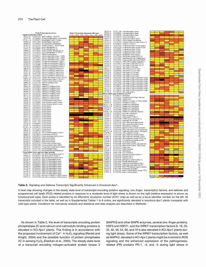
As shown in Table 2, the level of transcripts encoding protein
phosphatase 2C and calcium and calmodulin binding proteins is
elevated in KO-Apx1 plants. This finding is in accordance with
the proposed involvement of Ca2þ in H2O2 signaling (Rentel and
Knight, 2004) and the possible function of protein phosphates
2C in sensing H2O2 (Desikan et al., 2004). The steady state level
of a transcript encoding mitogen-activated protein kinase 3
(MAPK3) and other MAPK enzymes, several zinc-finger proteins,
HSF5 and HSF21, and the WRKY transcription factors 6, 18, 25,
33, 40, 46, 54, 60, and 70 is also elevated in KO-Apx1 plants dur-
ing light stress. Some of the WRKY transcription factors, as well
asMAPK3, elevated in KO-Apx1 plantsmight be involved in ROS
signaling and the enhanced expression of the pathogenesis-
related (PR) proteins PR-1, -3, and -5 during light stress in
Table 2. Signaling and Defense Transcripts Significantly Enhanced in Knockout-Apx1.
A heat map showing changes in the steady state level of transcripts encoding putative signaling, zinc-finger, transcription factors, and defense and
programmed cell death (PCD) related proteins in response to a moderate level of light stress is shown on the right (relative expression is shown as
knockout/wild type). Each probe is identified by its Affymetrix accession number (ATH1 chip) as well as by a locus identifier number on the left. All
transcripts included in the table, as well as in Supplemental Tables 1 to 6 online, are significantly elevated in knockout-Apx1 plants compared with
wild-type plants. Conditions for microarray analysis and statistical and data analysis are described in Methods.
274 The Plant Cell
Dow
nloaded from https://academ
ic.oup.com/plcell/article/17/1/268/6113109 by guest on 16 N
ovember 2021

KO-Apx1 plants (Table 2; Zhang andKlessig, 2001; Apel andHirt,
2004; Ulker and Somssich, 2004). The enhanced expression of
these pathogen defense mechanisms in KO-Apx1 plants sug-
gest a high degree of cross talk between biotic and abiotic
stresses mediated by H2O2 (Bowler and Fluhr, 2000; Mittler,
2002; Dat et al., 2003; see also Supplemental Table 2 online for
disease resistance genes).
ROS-related transcripts enhanced in KO-Apx1 plants com-
pared with wild-type plants included putative metal binding
proteins, mitochondrial alternative oxidase (Aox1A), a class III
peroxidase, thioredoxin, cytosolic glutaredoxin, and cytosolic
monodehydroascorbate reductase (MDAR). Cytosolic glutare-
doxin and MDAR are components of the ascorbate-glutathione
cycle and might be elevated to compensate for the lack of
cytosolic APX1 (Rizhsky et al., 2002; Pnueli et al., 2003; Mittler
et al., 2004). The enhanced level of transcripts encoding mito-
chondrial Aox1A in KO-Apx1 plants support our findings that in
the absence of cytosolic APX1, proteins from other compart-
ments are subjected to oxidative stress (Table 1). The expression
of at least two different transcripts associated with protein
oxidation was also elevated in KO-Apx1 plants compared with
wild-type plants. These encode for Met sulfoxide reductase that
reduces oxidized Met residues in proteins (Bechtold et al., 2004)
and aconitase that is highly sensitive to ROS damage (Yoo and
Regnier, 2004). This finding supports our directmeasurements of
protein oxidation in KO-Apx1 plants (Table 1).
Additional transcripts with a possible involvement in cellular
detoxification elevated in KO-Apx1 plants compared with wild-
type plants included glutathione S-transferase (Table 2) and
cytochrome P-450 (see Supplemental Table 3 online). The
steady state level of transcripts encoding accelerated cell death
2 (Acd2), a lesion mimic gene, is also elevated in KO-Apx1 plants
compared with wild-type plants. ACD2 might be linked to
pathogen responses activated in KO-Apx1, or it might be re-
quired to suppress programmed cell death in KO-Apx1. It is also
possible that the function of ACD2 (chlorophyll catabolite re-
ductase; Mach et al., 2001) is related to oxidative stress induced
in the chloroplast in KO-Apx1 plants.
Interestingly, the steady state level of most signaling and
defense transcripts shown in Table 2 decreased at the 6- and
24-h time points, suggesting that the stress that was imposed
on KO-Apx1 plants by the light shift treatment might have been
transient. It is possible that KO-Apx1 plants were able to
acclimate and decrease the level of stress by activating alterna-
tive H2O2 scavenging mechanisms, by activating photoprotec-
tive mechanisms, or by decreasing the rate of H2O2 production
(Mittler, 2002; Muller-Moule et al., 2004). This finding might
explain why KO-Apx1 plants were able to recover from the light
stress treatment (data not shown).
Because NADPH oxidases were recently proposed to function
asmediators of positive amplification loops duringROS signaling
(Mittler et al., 2004), we tested the steady state transcript level of
all members of the NADPH oxidase gene family during light
stress. As shown in Figure 4D, the steady state level of tran-
scripts encoding the NADPH oxidase D protein (RbohD) was
transiently elevated in KO-Apx1 plants compared with wild-type
plants (see also Figure 4A). The steady state level of transcripts
encoding all other members of this gene family was, however,
unchanged during the moderate light stress treatment in wild-
type and KO-Apx1 plants (Figure 4D).
Functional Analysis of the Role of HSF21 and RbohD in
Apx1 Transcript Expression during Light Stress in
Wild-Type Plants
The steady state level of transcripts encoding the transcription
factors HSF21 and HSF5 is rapidly elevated in KO-Apx1 plants in
response to the light stress treatment (Figure 4C). At least in
Drosophila melanogaster and mammalian cells the oligomeriza-
tion and DNA binding of HSFs were shown to be induced in vivo
and in vitro by H2O2, suggesting that HSFs act as direct sensors
of H2O2 in cells (Zhong et al., 1998; Ahn and Thiele, 2003). The
promoter of Apx1, as well as the promoters of many defense
genes and transcription factors involved in H2O2 signaling and
defense, contains an HSF binding motif (Mittler and Zilinskas,
1992; Rizhsky et al., 2004). Promoter analyses, as well as over-
expression studies of HSF3 in Arabidopsis, suggest that the HSF
binding site at theApx1promoter is functional (Storozhenkoet al.,
1998; Panchuk et al., 2002). However, no genetic evidence was
presented for the role of HSFs in H2O2 signaling in plants.
To study the importance of HSF21 (AtHSFA4a, an A-class
HSF; Nover et al., 2001) to Apx1 expression during the light
stress treatment described above, we generated transgenic
plants that constitutively express a dominant-negative construct
for HSF21. The expressed construct contained the entire HSF21
open reading frame, but without the HSF activation domain
(Figure 5A). As shown in Figure 5B, constitutive expression of
the dominant-negative construct for HSF21 prevented the
accumulation of transcripts encoding APX1 and the zinc-finger
protein Zat12 in response to light stress. By contrast, the
accumulation of transcripts encoding the heat shock protein
HSP70 (Figure 5B) or catalase (data not shown) was not inhibited
in the dominant-negative expressing plants. Interestingly, the
expression of transcripts encoding HSF21 was strongly elevated
in the dominant-negative plants in response to light stress (tested
with a probe for the activation domain of HSF21 that is missing
from the dominant-negative construct; data not shown). Our
findings that Apx1 and Zat12 transcript accumulation is inhibited
in plants expressing the dominant-negative HSF21 construct
suggest that HSF21 function is required for Zat12 and Apx1
expression during light stress (Figure 5B).
To study the relative importance of RbohD to Apx1 expression
during the light stress treatment described above, we used
knockout plants deficient in RbohD (KO-RbohD; Torres et al.,
2002). As shown in Figure 5C, the steady state level of transcripts
encoding APX1 was enhanced in response to light stress in both
wild-type and KO-RbohD plants at 3 h. However, in contrast with
wild-type plants, the steady state level of transcripts encoding
APX1 declined at the 6-h time point in KO-RbohD plants. Similar
results were found with transcripts encoding Cat1 (Figure 5C).
Because the steady state level of transcripts encoding RbohD
was elevated in KO-Apx1 plants at an early time point (0.5 h;
Figure 5D), it is possible that RbohD is required for Apx1 andCat1
expression during late stages of response to light stress, acting
to amplify the signal that controls Apx1 and Cat1 expression (see
Discussion; Figure 5D).
Cross-Compartment Protection by APX1 275
Dow
nloaded from https://academ
ic.oup.com/plcell/article/17/1/268/6113109 by guest on 16 N
ovember 2021

DISCUSSION
Cross-Compartment Protection by APX1
Several recent studies have demonstrated that ROS function as
important signaling molecules involved in the control of pro-
cesses such as pathogen defense, hormonal signaling, stress
response, and plant growth and development (Torres et al.,
2002; Foreman et al., 2003; Kwak et al., 2003). However, ROSare
also toxic molecules capable of injuring or even killing plant cells,
and their level in cells needs to be tightly regulated (Neill et al.,
2002; Mittler et al., 2004). To prevent ROS, produced in the
chloroplast, from damaging cells and possibly interfering with
ROS signaling in other compartments, the chloroplast contains
multiple ROS scavenging systems, including at least two com-
plete pathways for H2O2 removal at the thylakoid (water–water
cycle; Asada, 1999) and the stroma (ascorbate-glutathione cycle;
Asada and Takahashi, 1987). Although these pathways are
thought to be sufficient for proper H2O2 scavenging during
photosynthesis (Asada and Takahashi, 1987; Asada, 1999; Polle,
2001), our findings suggest that the cytosolic ascorbate-
glutathione cycle is required for H2O2 removal during photosyn-
thesis (Figures 1 and 2, Table 1). Moreover, our findings suggest
that the cytosolic pathway could even compensate or protect the
cell when the stromal pathway is absent (Figure 3). This type of
cross-compartment protectionmight also be foundwith other or-
ganelles, such as peroxisomes ormitochondria, because H2O2 is
transported across biological membranes (Willekens et al., 1997;
Figure 5. Functional Analysis of HSF21 and RbohD in Transgenic and Knockout Arabidopsis Plants.
(A) A scheme describing the structure of the HSF21 dominant-negative construct expressed in transgenic plants.
(B) RNA gel blots showing the steady state transcript level of Apx1, Zat12, HSF21, and HSP70 in wild-type plants and transgenic plants expressing the
dominant-negative construct for HSF21 (DN-HSF21) plants in response to a moderate level of light stress.
(C) RNA gel blots showing the steady state transcript level of Apx1 and Cat1 in wild-type plants and knockout plants lacking RbohD in response to
a moderate level of light stress.
(D) Amodel showing the putative signal transduction pathway that controls the expression of Apx1 during amoderate level of light stress in Arabidopsis.
Construction of transgenic plants, light stress experiments, and RNA gel blots were performed as described in Methods.
276 The Plant Cell
Dow
nloaded from https://academ
ic.oup.com/plcell/article/17/1/268/6113109 by guest on 16 N
ovember 2021
Henzler and Steudle, 2000). The extent of cross-compartment
protection, however, might be limited because at least in
tobacco (Nicotiana tabacum) and wheat (Triticum aestivum)
a deficiency in thylakoid-bound APX was shown to have adverse
effects on plant growth and photosynthetic performance (Yabuta
et al., 2002; Danna et al., 2003). These findings suggest that
cytosolic APX1 might not be able to protect the chloroplast
against H2O2 produced at the surface of the thylakoidmembrane
(see also Rizhsky et al., 2003 for the effect of disrupting the
water–water cycle in Arabidopsis).
Interestingly, at least at the steady state transcript level, none
of the other Apx genes, including the cytosolic Apx2 gene,
responded to the deficiency in APX1 (Figure 4B;Apx2 expression
was however elevated in KO-Apx1 plants in response to heat
shock; data not shown; Pnueli et al., 2003). KO-Apx1 plants
grown under controlled growth conditions are stunted and
have a late flowering phenotype; however, they are viable and
productive (Figure 3A; see also Pnueli et al., 2003). Although
the steady state level of transcripts encoding several ROS-
scavenging enzymes, including MDAR and glutaredoxin, is
elevated in KO-Apx1 plants, it is not entirely clear how they
compensate for the lack of APX1 (Table 2; Mittler et al., 2004).
The exposure of KO-Apx1 plants to a moderate level of light
stress does, however, reveal that APX1 is important during early
stages of plant acclimation to this treatment (Figures 1 and 2,
Tables 1 and 2). There may be two different explanations for
the function of APX1 under these conditions: (1) APX1 might be
directly involved in scavenging of H2O2 that leaks from the
chloroplast or peroxisomes, and/or (2) APX1 might be required
for the signal transduction pathway that is activated in plants
during the light stress treatment, and in its absence the activation
of certain defense mechanisms is prevented. We were unable to
find significant differences in transcript expression between the
wild type and KO-Apx1 that could account for the later explana-
tion. Asada (2000) demonstrated that intact chloroplasts are
sensitive to the external application of H2O2. In view of this report,
the enhanced production of H2O2 in KO-Apx1 plants and the
accumulation of oxidized proteins from different cellular com-
partments in KO-Apx1, we favor the first explanation suggesting
that APX1 is directly involved in H2O2 scavenging during light
stress. As a key cytosolic enzyme responsible for H2O2 scav-
enging in Arabidopsis, it is possible that APX1 functions as
a defense barrier between the three major ROS-producing
organelles of plant cells (i.e., the chloroplast, mitochondria, and
peroxisomes) and/or the redox- and oxidative stress–sensitive
regulatory mechanisms of the nuclei. The damage to chloroplas-
tic APXs in KO-Apx1 plants (Figure 2) suggests that a key role of
cytosolic APX1 is to protect the chloroplast from H2O2 that may
diffuse from the mitochondria, cytosol, or peroxisome during
photosynthesis.
Because our experimental design involved acclimating plants
to a low level of light intensity before the application of light stress,
it is possible that some photoprotective mechanisms were not
activated in our pretreated plants, exacerbating the effect of the
light stress treatment and making the dependence on H2O2-
scavenging systems central to the acclimation of plants to this
treatment. At later time points, these mechanisms might be
activated to protect the cell, and the dependence on APXs
might be reduced (see above for Table 2; Muller-Moule et al.,
2004).
Apx1 Signal Transduction: A Key Role for HSFs and Cross
Talk between Biotic and Abiotic Stresses
At least three different types of sensor molecules might be
involved in H2O2 perception in plants. Hydrogen peroxide might
be sensed by a two-component kinase systemor other receptor-
typemolecules, by direct inhibition of phosphatase activity, or by
redox-sensitive transcription factors such as HSF (Desikan et al.,
2004; Mittler et al., 2004). Transcripts encoding some of these
putative sensing mechanisms might have been identified by our
microarray analysis (Table 2; see supplemental data online). The
finding that expression of a dominant-negative construct for
HSF21 in transgenic Arabidopsis plants prevented the accumu-
lation of transcripts encoding APX1 during light stress (Figure 5)
strongly suggests that HSFs are involved in the signal trans-
duction pathway controlling Apx1 expression under the condi-
tions tested. Expression of the dominant-negative construct also
prevented the accumulation of transcripts encoding Zat12, an
H2O2-responsive zinc-finger protein required for Apx1 expres-
sion during oxidative stress (Figure 5; Rizhsky et al., 2004). This
finding suggests that HSF21 is required at a relatively early stage
of the oxidative stress acclimation response. It should be noted
that the promoter ofZat12 contains at least two copies of theHSF
binding element (Rizhsky et al., 2004), suggesting that HSF21
might function by directly binding to the promoter of Zat12.
Although the dominant-negative construct was designed
using the HSF21 open reading frame (Figure 5A), there are 21
different HSF genes in Arabidopsis, and oligomers (three sub-
units per DNA binding domain) of HSFs could contain multiple
copies of a particular factor or monomers encoded by different
HSF genes (Nover et al., 2001). Thus, it is not possible to
determine without further experimental work the specificity of
the effect of the dominant-negative construct to the HSF21
protein. Nonetheless, our findings with the dominant-negative
construct for HSF21 could be viewed as the first genetic
evidence that HSFs are important sensors for H2O2. HSF21
might be a good candidate for a sensor because it is constitu-
tively expressed in cells in the absence of stress and its
expression is elevated in KO-Apx1 plants (Figure 4C).
The putative order of events that follows the accumulation of
H2O2 in Arabidopsis leaf cells in response to a moderate level of
light stress is shown in Figure 5D. NADPHoxidaseswere recently
proposed to function as generators of ROS for different signal
transduction purposes (Torres et al., 2002; Foreman et al., 2003;
Kwak et al., 2003). Our microarray analysis (Figure 4D), as well as
our analysis of KO-RbohD plants (Figure 5C), suggest that the
NADPH oxidase protein RbohD might be involved in the signal
transduction pathway of Apx1 as a positive amplification factor
that maintains the expression of the Apx1 transcript at a high
steady state level (Figure 5D). Thus, in the absence of RbohD, the
steady state level of transcripts encoding APX1 is elevated at 3 h
but declines at 6 h. It is not clear, however, whether RbohD
functions as part of the oxidative stress-response pathway of
Arabidopsis orwhether it is activated inKO-Apx1plants as part of
the pathogen response pathway, similar to transcripts encoding
Cross-Compartment Protection by APX1 277
Dow
nloaded from https://academ
ic.oup.com/plcell/article/17/1/268/6113109 by guest on 16 N
ovember 2021
PR proteins elevated in KO-Apx1 plants (Table 2). Perhaps
these two responses (i.e., the oxidative stress response and the
pathogen response) could not be separated in KO-Apx1 plants
because the APX1 protein is involved in both (Mittler et al., 1998,
1999). The large number of pathogen response transcripts,
including disease resistance genes (see Supplemental Table 2
online), elevated in KO-Apx1 plants in response to the light stress
treatment, demonstrate the high degree of overlap that exists
between biotic and abiotic stresses. It is likely that H2O2 is the
mediator of this cross talk (Bowler and Fluhr, 2000; Mittler, 2002;
Dat et al., 2003). Because the steady state level of Apx1 is
enhanced in response to oxidative stress, as well as pathogen
attack (Mittler and Zilinskas, 1992; Mittler et al., 1998), it is
possible that the elevation in Apx1 steady state transcript level at
the 3-h time point is a result of activating the oxidative stress
response pathway, whereas the elevation of Apx1 transcripts at
the 6-h time point is a result of activating the pathogen response
pathway (Figure 5C). In KO-RbohD plants, the later pathway
might be suppressed, whereas in the dominant-negative HSF21
plants, both are suppressed (Figure 5). Our findings might
suggest that the expression of transcripts encoding other ROS
response and general defense proteins, in response to abiotic
stress, should be reevaluated in view of the cross talk between
biotic and abiotic stresses that might result from the accumula-
tion of H2O2 in cells. Thus, a biphasic mechanism might control
the expression of these transcripts. In the first phase, their
expression might be elevated because of direct sensing of
abiotic stress–generated H2O2 in cells, and in the second phase
their expression might be elevated as an integral part of the
pathogen response pathway activated during abiotic stress (this
pathwaymight be suppressed in KO-RbohDplants; Figure 5C). A
similar biphasic process might be activated in response to
pathogen attack; however, during this process, the pathogen
response pathway is activated first (this pathway will generate
H2O2 in cells through the action of NADPH oxidases), and the
abiotic stress, H2O2 response pathway is second.
Proteomics of Protein Oxidation
Protein carbonylation is an irreversible oxidative process leading
to a loss of function and often proteolysis of the modified protein
(Stadtman, 1992). Proteomic analysis of protein oxidation is
emerging as a new tool to study oxidative stress and damage
caused to cells during disease or different environmental
stresses (Akenov et al., 2001; Rizhsky et al., 2004). Johansson
et al. (2004) have identified different proteins oxidized in Arabi-
dopsis plants grown under controlled conditions. They have
identified seven chloroplastic and five mitochondrial proteins
oxidized in cells at the end of the vegetative phase of develop-
ment (i.e., before bolting). Interestingly, only one of the proteins
Johansson et al. (2004) have identified in their study (Rubisco
large subunit; RbcL) was identified by our proteomic analysis of
proteins oxidized in KO-Apx1 plants during the moderate light
stress treatment (Figure 1B, Table 1). The RbcL protein was
initially identified by Rizhsky et al. (2004) as the major protein
carbonylated in knockout-Zat12 plants in response to oxidative
stress. Prior analysis of Rubisco has demonstrated that this
protein is prone to different types of oxidative damage (Mehta
et al., 1992). However, because of the high abundance of the
RbcL protein in chloroplasts, it is not entirely clear whether it is
oxidized as a general byproduct of oxidative stress in plants or
whether its oxidation is a specific process associated with
oxidative damage to cells and inhibition of photosynthesis.
Proteomic analysis of protein oxidation during stress, espe-
cially in different genetic backgrounds lacking specific ROS-
defense enzymes, is a particularly powerful approach because it
can pinpoint the specific proteins damaged in specific mutants
and link between enzymatic activities in particular compartments
and oxidative damage at the cellular level. Thus, our findings that
chloroplastic, mitochondrial, and membrane-bound proteins are
oxidized in mutant plants lacking a cytosolic H2O2-scavenging
enzyme (KO-Apx1; Table 1) provides direct evidence that this
enzyme is key to the protection of adjacent compartments (see
also Figures 1 to 3). The notion that each individual compartment
is protected by its own set of ROS-scavenging enzymes should
therefore be reevaluated, and a new view of the ROS network
should be adopted as a highly interlinked network that requires
the coordinated function of ROS-scavenging pathways from
different cellular compartments to modulate the level of ROS in
cells, prevent cellular damage, and control ROS signaling.
METHODS
Plant Material and Growth Conditions
Arabidopsis thaliana (cv Columbia and Ws) plants were grown in growth
chambers (Percival E-30, AR-66; Percival Scientific, Perry, IA) under
controlled conditions: 21 to 228C, constant light, 25 mmol m�2 s�1, and
a relativehumidityof70%.KnockoutArabidopsis linescontainingaT-DNA
insert in the stromal/mitochondrial APX gene (s/mApx, At4g08390; ob-
tained through the SIGnAL project, http://signal.salk.edu/tabout.html)
were out-crossed and selfed to check for segregation as previously
described (Pnueli et al., 2003; Rizhsky et al., 2004). Screening for
expression of s/mAPX was performed by protein blots using an antibody
raised against a conserved domain of cytosolic and chloroplastic APXs.
Knockout plants deficient in cytosolic Apx1 (KO-Apx1) were obtained as
described by Pnueli et al. (2003). Light stress treatments were performed
by changing the light intensity from 25 to 250 mmol m�2 s�1, and all other
parameters were maintained constant. At different times, leaves were
excised, flash frozen in liquid nitrogen, and stored for further analysis at
�808C. All experiments were performed in triplicate and repeated at least
three times (with the exception of DNA arrays that were performed as
describedbelow).Representative resultsareshown inthedifferentfigures.
Analysis of seedling tolerance to paraquat was performed on agar plates
as described by Rizhsky et al. (2004). Vector construction, PCR cloning
and sequencing, and transformation of Arabidopsis plantswasperformed
as described byRizhsky et al. (2004). Transgenic plants were screened by
RNA and protein blots (Pnueli et al., 2003; Rizhsky et al., 2004).
Molecular, Physiological, and Biochemical Analysis
RNA and protein were isolated and analyzed by RNA and protein blots as
previously described (Pnueli et al., 2003; Rizhsky et al., 2004). RNA
staining or a ribosomal 18S rRNA probe were used to control for RNA
loading. Coomassie Brilliant Blue staining of protein gels was used to
control for protein loading. Photosynthetic activity was measured with
a LI-6400 apparatus (LI-COR Biosciences, Lincoln, NE) as described by
278 The Plant Cell
Dow
nloaded from https://academ
ic.oup.com/plcell/article/17/1/268/6113109 by guest on 16 N
ovember 2021

Rizhsky et al. (2003). Detection of H2O2 was performed by infiltrating
excised leaves with a solution of 1 mg/mL diaminobenzidine prepared in
water. Leaveswere placed on two layers of 3MMWhatman paper (Clifton,
NJ) soaked with water and exposed to light for different times. Leaves
were then fixed with a solution of 3:1:1 (v:v:v) ethanol:lactic acid:glycerol,
washed with 75, 50, and 25% ethanol, equilibrated with water, and
photographed. Detection of protein oxidation by protein blots was
performed with the OxyBlot protein oxidation kit (Chemicon International,
Temecula, CA) as recommended by the manufacturer (see also Rizhsky
et al., 2004). Antibodies against APX1 were obtained as described by
Mittler and Zilinskas (1991). Polyclonal antibodies that react with tylAPX,
s/mAPX, and cAPX (APX1) were prepared in rabbits against a fragment of
thyAPX (from Lys100 to Ile341) cloned into pCAL-n, expressed in bacteria
and purified by affinity chromatography. Antibodies against RbcL, RbcS,
and Cytf (a component of the b6f complex) were a gift of R. Nechushtai
(Hebrew University, Jerusalem, Israel).
Proteomic Analysis of Oxidized Proteins
Oxidized proteins were extracted by grinding 200 mg of tissue in liquid
nitrogen. The powder was dissolved in 1 mL of extraction buffer (25 mM
Tris-Cl, pH 8, 0.1% Triton X-100, 50 mMDTT, and the protease inhibitors
leupeptin [0.5 mg/mL], trypsin inhibitor [0.5 mg/mL], and PMSF [40 mg/
mL]). After centrifugation, the supernatant was stored at �808C until
analysis. The protein (250 mL containing ;500 mg protein) was derivat-
ized by adding 1 mL of 10 mM 2,4-dinitrophenylhydrazine in 2 M HCl and
incubating for 30 min. The protein was precipitated with 25% trichloro-
acetic acid, and the pellet, collected by centrifugation, was washed with
cold ethanol/ethyl acetate (1:1; v:v), dried in a vacuum, and redissolved in
100 mL of rehydration buffer (7 M urea, 2 M thiourea, 2 mM tributyl
phosphine, 4% CHAPS, and 40 mM Tris). After centrifugation, the
resulting solution was diluted with 2mL of coupling buffer (0.1MNaHCO3
and 0.5 M NaCl, pH 8.3 to 8.5) and mixed with 15 mg of anti-DNP affinity
column matrix. The column matrix was produced using cyanogen
bromide–activated beads (Sigma-Aldrich, St. Louis, MO) and anti-
dinitrophenylhydrazone (DNP) antibody (Bethyl Lab, Montgomery, TX)
using the methods included with the beads. After incubating for 1 h, the
matrix was loaded into a columnmade from a 200-mL pipette tip, washed
with 4 mL of coupling buffer, and eluted with 200 mL of 5%SDS. Samples
were resolved on one- or two-dimensional PAGE. The oxidized proteins
were then detected by protein gel blotting.
For MALDI-TOF analysis, proteins identified on the protein gel blots
were punched out of a Coomassie Brilliant Blue–stained gel and pro-
cessed as described by Porubleva et al. (2001). Peptide masses were
analyzedwithMS-Fit (http://prospector.ucsf.edu/ucsfhtml4.0/msfit.htm).
Matches were deemed significant when at least four fragments were
matched, covering more than 20% of the protein.
GeneChip Microarray Experiments
In six independent experiments, RNA was isolated from wild-type and
KO-Apx1 plants grown under controlled conditions and subjected to
a moderate level of light stress as described above (samples were
obtained at 0, 0.25, 0.5, 1.5, 3, 6, and 24 h after light stress application, 15
plants per time point, and RNA was isolated using Trizol; Pnueli et al.,
2003; Rizhsky et al., 2004). For each time point, RNA from three
independent experiments was pooled to generate 28 RNA pools (two
RNA pools for each time point for a total of 14 wild-type and 14 KO-Apx1
pools). These RNA samples were used to perform chip hybridization
analyses (Arabidopsis ATH1 chips; Affymetrix, Santa Clara, CA) at the
Virginia Bioinformatics Institute Core LaboratoryGene Expression Facility
(https://www.vbi.vt.edu/). Conditions for RNA isolation, labeling, hybrid-
ization, and data analysis are described byPnueli et al. (2003) andRizhsky
et al. (2004). Data visualization and analysis were performed with the
GeneChip mining tool version 5.0 (Silicon Genetics, Redwood City, CA),
GeneSpring version 5.1, and ArraAassit (IobionLab, La Jolla, CA). Some
of the results were confirmed by RNA gel blots. See details for data
analysis below.
GeneChip Data Processing and Analysis
All GeneChip arrays were processed first by robust multiarray average
(RMA) (Irizarry et al., 2003) using the R package affy (Gautier et al., 2004).
Specifically, expression values were computed from rawCEL files by first
applying the RMA model of probe-specific correction of perfect match
probes. These corrected probe values were then normalized via quantile
normalization,andamedianpolishwasappliedtocomputeoneexpression
measure from all probe values. Resulting RMA expression values were
log2-transformed. (Please see the affy manual at www.bioconductor.
org /repository/devel/vignette/affy.pdf for details.) Density plots and box
plots of RMA expression value distributions of all arrays were very similar
with no apparent outlying arrays (see Supplemental Figure 1 onlinw).
Digestioncurvesdescribing trends inRNAdegradationbetween the59end
and the 39 end in each probe set were generated, and all 28 proved very
similar, with a downward trend at the 59 end (see Supplemental Figure 2
online).
Pearson correlation coefficients and Spearman rank coefficients were
computed on the RMA expression values (log base 2) for each set of
biological replicates. Spearman coefficients ranged from 0.977 to 0.994;
Pearson coefficients ranged between 0.982 and 0.995 (see Supplemental
Table 5 online). A scatter plot of RMA expression values between the two
wild-type biological replicates at 6 h is presented in Supplemental Figure
3 online.
To determine whether genes were differentially expressed between
genotypes across the temporal states, analysis of variance (ANOVA) was
performed on the RMA expression values. (For an overview on the
application of ANOVA to microarray data, please see Kerr et al., 2000.)
The following model was used for this analysis: yijk ¼ Vi þ Tj þ (VT)ij þ eijk,where yijk denotes the log2 signal measured for variety i, time j, and
biological replicate k, with 1# i# 2, 1# j# 7, and 1# k# 2. The terms Vi
and Tj measure the effect of the variety and time point, respectively, and
the interaction term (VT)ij accounts for the interaction between variety and
time. An ANOVA was performed on each gene using the linear model
above, and six contrasts based on differences of genotypes with respect
to the last six time points. (Differences between genotypes at time 0 were
not of interest in this study.) The R package limma was used for ANOVA
methods (www.bioconductor.org/repository/devel/vignette/affy.pdf). A
multiple testing correction (Benjamini and Hochberg, 1995) was applied
to the P values of the F-statistics to adjust the false discovery rate. Genes
with adjusted F-statistic P values < 0.05 were extracted for further
analysis. This resulted in 3915 geneswith significant F-statistics based on
the ANOVA above (see Supplementary Table 6 online). Expression values
of this selection were then inverse log transformed, and genes with
differential expression between wild-type and knockout expression
values of more than twofold during at least one time point were selected.
This selection included 843 genes. Similarly, genes with differential ex-
pression between wild-type and knockout expression values of <0.5-fold
during at least one time point were selected as suppressed (see
Supplemental Table 7 online). Interestingly, no gene was both upregu-
lated and downregulated by more than twofold in knockout across the
time series, enabling a clear separation between upregulated and down-
regulated genes. Storey multiple testing adjustments (Storey and
Tibshirani, 2003) were also performed on the P values of the F-statistics.
All P values found significant by the Benjamini-Hochberg correction were
also found to be significant in the Storey correction. The Storey correction
contained 691 additional genes that were not included in the analysis.
Cross-Compartment Protection by APX1 279
Dow
nloaded from https://academ
ic.oup.com/plcell/article/17/1/268/6113109 by guest on 16 N
ovember 2021

ACKNOWLEDGMENTS
We thank Jeffery Dangl and Miguel Angel Torres for the gift of the
RbohD probe and KO-RbohD seeds and Rachel Nechushtai for the gift
of antisera to RbcL, RbcS, and Cytf. This work was supported by
funding from the National Science Foundation (NSF-0431327), the Plant
Sciences Institute at Iowa State University, and the Biotechnology
Council of Iowa State University.
Received August 16, 2004; accepted November 10, 2004.
REFERENCES
Ahn, S.G., and Thiele, D.J. (2003). Redox regulation of mammalian heat
shock factor 1 is essential for Hsp gene activation and protection from
stress. Genes Dev. 17, 516–528.
Akenov, M.Y., Aksenova, M.V., Butterfield, D.A., Geddes, J.W., and
Markesbery, W.R. (2001). Protein oxidation in the brain in Alzheimer’s
disease. Neuroscience 103, 373–383.
Apel, K., and Hirt, H. (2004). Reactive oxygen species: Metabolism,
oxidative stress, and signal transduction. Annu. Rev. Plant Biol. 55,
373–399.
Asada, K. (1999). The water-water cycle in chloroplasts: Scavenging of
active oxygen and dissipation of excess photons. Annu. Rev. Plant
Physiol. Plant Mol. Biol. 50, 601–639.
Asada, K. (2000). The water-water cycle as alternative photon and
electron sinks. Philos. Trans. R. Soc. Lond. B Biol. Sci. 355, 1419–
1431.
Asada, K., and Takahashi, M. (1987). Production and scavenging
of active oxygen in photosynthesis. In Photoinhibition, D.J. Kyle,
C.B. Osmond, and C.J. Arntzen, eds (Amsterdam: Elsevier), pp.
227–287.
Bechtold, U., Murphy, D.J., and Mullineaux, P.M. (2004). Arabidopsis
peptide methionine sulfoxide reductase2 prevents cellular oxidative
damage in long nights. Plant Cell 16, 908–919.
Benjamini, Y., and Hochberg, Y. (1995). Controlling the false discovery
rate: A practical and powerful approach to multiple testing. J. R. Stat.
Soc. B 57, 289–300.
Bowler, C., and Fluhr, R. (2000). The role of calcium and activated
oxygens as signals for controlling cross-tolerance. Trends Plant Sci.
5, 241–246.
Chew, O., Whelan, J., and Millar, A.H. (2003). Molecular definition of
the ascorbate-glutathione cycle in Arabidopsis mitochondria reveals
dual targeting of antioxidant defenses in plants. J. Biol. Chem. 278,
46869–46877.
Corpas, F.J., Barroso, J.B., and del Rio, L.A. (2001). Peroxisomes as
a source of reactive oxygen species and nitric oxide signal molecules
in plant cells. Trends Plant Sci. 6, 145–150.
Danna, C.H., Bartoli, C.G., Sacco, F., Ingala, L.R., Santa-Maria, G.E.,
Guiamet, J.J., and Ugalde, R.A. (2003). Thylakoid-bound ascorbate
peroxidase mutant exhibits impaired electron transport and photo-
synthetic activity. Plant Physiol. 132, 2116–2125.
Dat, J., Vandenabeele, S., Vranova, E., Van Montagu, M., Inze, D.,
and Van Breusegem, F. (2000). Dual action of the active oxygen
species during plant stress responses. Cell. Mol. Life Sci. 57,
779–795.
Dat, J.F., Pellinen, R., Beeckman, T., Van De Cotte, B., Langebartels,
C., Kangasjarvi, J., Inze, D., and Van Breusegem, F. (2003).
Changes in hydrogen peroxide homeostasis trigger an active cell
death process in tobacco. Plant J. 33, 621–632.
Desikan, R., Cheung, M.K., Bright, J., Henson, D., Hancock, J.T.,
and Neill, S.J. (2004). ABA, hydrogen peroxide and nitric oxide
signalling in stomatal guard cells. J. Exp. Bot. 55, 205–212.
Dietz, K.J. (2003). Plant peroxiredoxins. Annu. Rev. Plant Biol. 54,
93–107.
Foreman, J., Demidchik, V., Bothwell, J.H., Mylona, P., Miedema, H.,
Torres, M.A., Linstead, P., Costa, S., Brownlee, C., Jones, J.D.,
Davies, J.M., and Dolan, L. (2003). Reactive oxygen species pro-
duced by NADPH oxidase regulate plant cell growth. Nature 422,
442–446.
Gautier, L., Cope, L., Bolstad, B., and Irizarry, R.A. (2004). Affy—
Analysis of Affymetrix GeneChip data at the probe level. Bioinfor-
matics 20, 307–315.
Halliwell, B., and Gutteridge, J.M.C. (1989). Free Radicals in Biology
and Medicine. (Oxford: Clarendon Press).
Henzler, T., and Steudle, E. (2000). Transport and metabolic degrada-
tion of hydrogen peroxide in Chara corallina: Model calculations and
measurements with the pressure probe suggest transport of H(2)O(2)
across water channels. J. Exp. Bot. 51, 2053–2066.
Iida, A., Kazuoka, T., Torikai, S., Kikuchi, H., and Oeda, K. (2000). A
zinc finger protein RHL41 mediates the light acclimatization response
in Arabidopsis. Plant J. 24, 191–203.
Irizarry, R.A., Hobbs, R., Collin, R., Beazer-Barclay, Y.D., Antonellis,
K.J., Scherf, U., and Speed, T.P. (2003). Exploration, normalization,
and summaries of high density oligonucleotide array probe level data.
Biostatistics 4, 249–264.
Johansson, E., Olsson, O., and Nystrom, T. (2004). Progression and
specificity of protein oxidation in the life cycle of Arabidopsis thaliana.
J. Biol. Chem. 279, 22204–22208.
Karpinski, S., Escobar, C., Karpinska, B., Creissen, G., and
Mullineaux, P.M. (1997). Photosynthetic electron transport regulates
the expression of cytosolic ascorbate peroxidase genes in Arabidop-
sis during excess light stress. Plant Cell 9, 627–640.
Karpinski, S., Reynolds, H., Karpinska, B., Wingsle, G., Creissen, G.,
and Mullineaux, P. (1999). Systemic signaling and acclimation in
response to excess excitation energy in Arabidopsis. Science 284,
654–657.
Kerr, M.K., Martin, M., and Churchill, G.A. (2000). Analysis of variance
for gene expression microarray data. J. Comput. Biol. 7, 819–837.
Kwak, J.M., Mori, I.C., Pei, Z.M., Leonhardt, N., Torres, M.A., Dangl,
J.L., Bloom, R.E., Bodde, S., Jones, J.D., and Schroeder, J.I.
(2003). NADPH oxidase AtrbohD and AtrbohF genes function in
ROS-dependent ABA signaling in Arabidopsis. EMBO J. 22, 2623–
2633.
Mach, J.M., Castillo, A.R., Hoogstraten, R., and Greenberg, J.T.
(2001). The Arabidopsis-accelerated cell death gene ACD2 encodes
red chlorophyll catabolite reductase and suppresses the spread of
disease symptoms. Proc. Natl. Acad. Sci. USA 98, 771–776.
Mano, J., Ohno, C., Domae, Y., and Asada, K. (2001). Chloroplastic
ascorbate peroxidase is the primary target of methylviologen-induced
photooxidative stress in spinach leaves: Its relevance to monodehy-
droascorbate radical detected with in vivo ESR. Biochim. Biophys.
Acta 1504, 275–287.
Mehta, R.A., Fawcett, T.W., Porath, D., and Mattoo, A.K. (1992).
Oxidative stress causes rapid membrane translocation and in vivo
degradation of ribulose-1,5-bisphosphate carboxylase/oxygenase.
J. Biol. Chem. 267, 2810–2816.
Mittler, R. (2002). Oxidative stress, antioxidants and stress tolerance.
Trends Plant Sci. 7, 405–410.
Mittler, R., Feng, X., and Cohen, M. (1998). Post-transcriptional
suppression of cytosolic ascorbate peroxidase expression during
pathogen-induced programmed cell death in tobacco. Plant Cell 10,
461–474.
280 The Plant Cell
Dow
nloaded from https://academ
ic.oup.com/plcell/article/17/1/268/6113109 by guest on 16 N
ovember 2021

Mittler, R., Hallak-Herr, E., Orvar, B.L., Van Camp, W., Willekens, H.,
Inze, D., and Ellis, B. (1999). Transgenic tobacco plants with reduced
capability to detoxify reactive oxygen intermediates are hyper-
responsive to pathogen infection. Proc. Natl. Acad. Sci. USA 96,
14165–14170.
Mittler, R., Vanderauwera, S., Gollery, M., and Van Breusegem, F.
(2004). The reactive oxygen gene network of plants. Trends Plant Sci.
9, 490–498.
Mittler, R., and Zilinskas, B. (1991). Purification and characterization of
pea cytosolic ascorbate peroxidase. Plant Physiol. 97, 962–968.
Mittler, R., and Zilinskas, B. (1992). Molecular cloning and character-
ization of a gene encoding pea cytosolic ascorbate peroxidase. J. Biol.
Chem. 267, 21802–21807.
Muller-Moule, P., Golan, T., and Niyogi, K.K. (2004). Ascorbate-
deficient mutants of Arabidopsis grow in high light despite chronic
photooxidative stress. Plant Physiol. 134, 1163–1172.
Mullineaux, P., and Karpinski, S. (2002). Signal transduction in re-
sponse to excess light: getting out of the chloroplast. Curr. Opin. Plant
Biol. 5, 43–48.
Neill, S., Desikan, R., and Hancock, J. (2002). Hydrogen peroxide
signalling. Curr. Opin. Plant Biol. 5, 388–395.
Noctor, G., and Foyer, C. (1998). Ascorbate and glutathione: Keeping
active oxygen under control. Annu. Rev. Plant Physiol. Plant Mol. Biol.
49, 249–279.
Nover, L., Bharti, K., Doring, P., Mishra, S.K., Ganguli, A., and
Scharf, K.D. (2001). Arabidopsis and the heat stress transcription
factor world: How many heat stress transcription factors do we need?
Cell Stress Chaperones 6, 177–189.
Panchuk, I.I., Volkov, R.A., and Schoffl, F. (2002). Heat stress- and
heat shock transcription factor-dependent expression and activity of
ascorbate peroxidase in Arabidopsis. Plant Physiol. 129, 838–853.
Pnueli, L., Hongjian, L., and Mittler, R. (2003). Growth suppression,
abnormal guard cell response, and augmented induction of heat
shock proteins in cytosolic ascorbate peroxidase (Apx1)-deficient
Arabidopsis plants. Plant J. 34, 187–203.
Polle, A. (2001). Dissecting the superoxide dismutase-ascorbate
peroxidase-glutathione pathway in chloroplasts by metabolic model-
ing. Computer simulations as a step towards flux analysis. Plant
Physiol. 126, 445–462.
Porubleva, L., Vander Velden, K., Kothari, S., Oliver, D.J., and
Chitnis, P.R. (2001). The proteome of maize leaves: Use of gene
sequences and EST data for identification of proteins with peptide
mass fingerprints. Electrophoresis 22, 1724–1783.
Rentel, M.C., and Knight, M.R. (2004). Oxidative stress-induced
calcium signaling in Arabidopsis. Plant Physiol. 135, 1471–1479.
Rizhsky, L., Davletova, S., Liang, H., and Mittler, R. (2004). The zinc-
finger protein Zat12 is required for cytosolic ascorbate peroxidase 1
expression during oxidative stress in Arabidopsis. J. Biol. Chem. 279,
11736–11743.
Rizhsky, L., Hallak-Herr, E., Van Breusegem, F., Rachmilevitch, S.,
Rodermel, S., Inze, D., and Mittler, R. (2002). Double antisense
plants with suppressed expression of ascorbate peroxidase and
catalase are less sensitive to oxidative stress than single antisense
plants with suppressed expression of ascorbate peroxidase or
catalase. Plant J. 32, 329–342.
Rizhsky, L., Liang, H., and Mittler, R. (2003). The water-water cycle is
essential for chloroplast protection in the absence of stress. J. Biol.
Chem. 278, 38921–38925.
Rodermel, S. (2001). Pathways of plastid-to-nucleus signaling. Trends
Plant Sci. 6, 471–478.
Rodriguez-Milla, M.A., Maurer, A., Rodriguez-Huete, A., and
Gustafson, J.P. (2003). Glutathione peroxidase genes in Arabidopsis
are ubiquitous and regulated by abiotic stresses through diverse
signaling pathways. Plant J. 36, 602–615.
Shigeoka, S., Ishikawa, T., Tamoi, M., Miyagawa, Y., Takeda, T.,
Yabuta, Y., and Yoshimura, K. (2002). Regulation and function of
ascorbate peroxidase isoenzymes. J. Exp. Bot. 53, 1305–1319.
Stadtman, E.R. (1992). Protein oxidation and aging. Science 257, 1220–
1224.
Storey, J.D., and Tibshirani, R. (2003). Statistical significance for
genome-wide experiments. Proc. Natl. Acad. Sci. USA 100, 9440–
9445.
Storozhenko, S., De Pauw, P., Van Montagu, M., Inze, D., and
Kushnir, S. (1998). The heat-shock element is a functional component
of the Arabidopsis APX1 gene promoter. Plant Physiol. 118, 1005–
1014.
Torres, M.A., Dangl, J.L., and Jones, J.D. (2002). Arabidopsis
gp91phox homologues AtrbohD and AtrbohF are required for accu-
mulation of reactive oxygen intermediates in the plant defense re-
sponse. Proc. Natl. Acad. Sci. USA 99, 517–522.
Ulker, B., and Somssich, I.E. (2004). WRKY transcription factors: From
DNA binding towards biological function. Curr. Opin. Plant Biol. 7,
491–498.
Vandenabeele, S., Vanderauwera, S., Vuylsteke, M., Rombauts, S.,
Langebartels, C., Seidlitz, H.K., Zabeau, M., Van Montagu, M.,
Inze, D., and Van Breusegem, F. (2004). Catalase deficiency
drastically affects gene expression induced by high light in Arabidop-
sis thaliana. Plant J. 39, 45–58.
Willekens, H., Chamnongpol, S., Davey, M., Schraudner, M.,
Langebartels, C., Van Montagu, M., Inze, D., and Van Camp, W.
(1997). Catalase is a sink for H2O2 and is indispensable for stress
defence in C-3 plants. EMBO J. 16, 4806–4816.
Yabuta, Y., Motoki, T., Yoshimura, K., Takeda, T., Ishikawa, T., and
Shigeoka, S. (2002). Thylakoid membrane-bound ascorbate peroxi-
dase is a limiting factor of antioxidative systems under photo-
oxidative stress. Plant J. 32, 915–925.
Yoo, B.S., and Regnier, F.E. (2004). Proteomic analysis of carbonyl-
ated proteins in two-dimensional gel electrophoresis using avidin-
fluorescein affinity staining. Electrophoresis 25, 1334–1341.
Yoshimura, K., Yabuta, Y., Ishikawa, T., and Shigeoka, S. (2002).
Identification of a cis element for tissue-specific alternative splicing of
chloroplast ascorbate peroxidase pre-mRNA in higher plants. J. Biol.
Chem. 277, 40623–40632.
Zhang, S., and Klessig, D.F. (2001). MAPK cascades in plant defense
signaling. Trends Plant Sci. 6, 520–527.
Zhong, M., Orosz, A., and Wu, C. (1998). Direct sensing of heat shock
and oxidation by Drosophila heat shock transcription factor. Mol. Cell
2, 101–108.
Cross-Compartment Protection by APX1 281
Dow
nloaded from https://academ
ic.oup.com/plcell/article/17/1/268/6113109 by guest on 16 N
ovember 2021