cytology in veterinary practice: sample collection, slide ... · if the slide preparation is over...
TRANSCRIPT

Cytology in Veterinary Practice: Sample Collection, Slide Preparation and Interpretation Guidelines
A. Rick Alleman, DVM, PhD, DABVP, DACVP Lighthouse Veterinary Consultants, LLC
Gainesville, FL 32606 ASPIRATION OF A MASS 1. Hold the mass firmly with one hand. 2. Insert the needle, with attached syringe, into the mass.
(** Note: For small masses a more precise collection can be accomplished by using a needle without an attached syringe**)
3. Pull back the plunger and hold to apply and maintain slight negative pressure.
4. The needle is then redirected in the mass several times while maintaining a negative pressure in the syringe.
5. Once material appears in the hub of the needle, the plunger is released and the needle can then be removed from the mass.
6. Disconnect the needle from the syringe and pull the plunger back. 7. Re-attach the needle to the syringe and depress the plunger to expel the aspirated material
onto a clean glass slide. 8. A second glass slide is gently placed over the aspirated material and the material is allowed to
diffuse out to form a thin layer between the two slides. 9. The two slides are gently slid apart forming a monolayer of aspirated cells. 10. The material is then allowed to air-dry prior to staining. SLIDE PREPARATION In cytology, cells that are properly smeared and stained can be described as “fried eggs” because of the similarity in the appearance of the nucleus and cytoplasm to the egg yolk and white. If the preparation is too thick, or is improperly stained, the cell outline may be seen, but intracellular detail will not be visible. These are the undesirable “hard-boiled eggs”. Slide preparations that are smeared too thin will cause
cell lysis and disruption of cellular architecture. These “scrambled eggs” are also undesirable. A properly prepared smear will have an area of “fried eggs” for viewing. There are many stains available for routine cytologic evaluation in private practice. Most of these are Romanowsky-type stains such as Diff QuikTM. Alternatively, a true “Wright-Giemsa Stain Kit” from Volu-Sol, Inc. item number VWG-300 (www.volusol.com) is available for use in a clinical practice laboratory. This stain kit has the advantage of

having a more consistent and better color distinction and better ability to stain mast cell granules. The exact staining procedure may vary depending on the stain used and manufacture’s recommendations should be used as general guidelines. However, for most Diff-Quik-type stains it is advisable to allow the slides to sit in the fixative for a minimum of 2 to 3 minutes before proceeding to the eosinophilic and basophilic stains. The number of dips in each stain will vary depending on the age of the stain and the thickness of the preparation, but usually 6 to 8 one-second dips in the eosinophilic stain and 5 to 6 one-second dips in the basophilic stain is sufficient. If the slide is under-stained, it should be adjusted by returning the slide to the appropriate stain color. If the slide preparation is over-stained, the slide may be placed in methanol for several minutes to de-stain and then re-stained as before. The assessment of staining intensity is made by visualizing good color contrast between the cell nucleus and cytoplasm. The most important aspect of staining and slide preparation is to be able to visualize good nuclear and cytoplasmic detail. Without good slide preparation and staining, a reliable cytologic interpretation cannot be made. INTERPRETATION The first step in making a cytological diagnosis is to classify the lesion into one of five general categories of disease processes: 1) inflammation, 2) cyst formation, 3) hemorrhagic lesion, 4) neoplasia, or 5) mixed cell population. In some cases, more than one pathologic process may be occurring simultaneously in a single lesion. For instance, there may be hemorrhage within a neoplasm or inflammation within a cyst. However, when the population is not mixed and they are in their purist form, the categories are easily distinguished by the cell population present. The general cellular characteristics of the 5 categories of lesions are listed below. 1. Inflammation - characterized by the presence of neutrophils above what would be expected
from any blood contamination 2. Cyst formation - large numbers of mature, keratinized, squamous epithelial cells or amorphous
material of low cellularity. 3. Hemorrhagic lesion - blood in the presence of macrophages, some of which contain engulfed
erythrocytes (erythrophagia) or hemosiderin. 4. Neoplasia - a homogeneous population of cells all from the same tissue of origin. 5. Mixed cell population - preparation contains both inflammatory (neutrophils, macrophages, etc.)
and noninflammatory cells (epithelial or mesenchymal) INFLAMMATION There are 3 types of inflammation that commonly occur, 1) Purulent inflammation, 2) Pyogranulomatous inflammation, and 3) Eosinophilic inflammation. Each type will contain neutrophils as a major or minor component, however, they are distinguished from each other by the presence or absence of other cell types. The type of inflammatory response present may give some indication of the disease process that caused it. 1. Purulent inflammation : Purulent inflammation is characterized by the presence of a predominant (usually > 85%) population of neutrophils. The neutrophils in any inflammatory response, but particularly in purulent inflammation, need to be evaluated for the presence or absence of degenerative changes.

2. Pyogranulomatous inflammation : characterized by the presence of neutrophils with a 15% or greater component of macrophages (Figure below right). When pyogranulomatous inflammation is present a different set of disease processes must be considered. The disease processes that may induce a pyogranulomatous response include:
Fungal infections Foreign bodies Atypical mycobacteria Actinomycosis/nocardiosis Panniculitis Tissue reaction to injections 3. Eosinophilic inflammation : characterized by an inflammatory process in which greater than 10%
of the inflammatory cells are eosinophils. The types of diseases processes that elicit an eosinophilic response in tissues are listed.
Eosinophilic granuloma complex/rodent ulcer Allergic/hypersensitivity reactions
Parasitic migration (Paragonimus, heartworms, lung worms) Mast cell tumors Pythiosis and some fungal infections Foreign body, if a hypersensitivity reaction develops Rare: Lymphoid neoplasia (lymphoma, lymphomatoid granulomatosis) CYST FORMATION There are 3 types of cutaneous cysts commonly seen in veterinary medicine, the epidermal inclusion cyst (follicular cyst), the apocrine cyst, and the sebaceous cyst. The cytologic and gross appearance of material aspirated from these lesions is described below. Other less common cysts may occur associated with various glandular structures of the body such as the mammary gland, prostate gland, or ovary. Cystic fluid drawn from glandular organs is typically of low cellularity, containing only low numbers of glandular epithelium in a proteinaceous background. The epidermal inclusion cyst (follicular cyst) is a common cutaneous lesion. Cytologically, large numbers of mature, keratinized squamous epithelial cells are seen. Eventually, the epithelial cells degenerate leaving an amorphous, basophilic cellular debris with cholesterol crystals. If the cystic structure ruptures, a neutrophilic inflammatory response occurs. Apocrine cysts are secretory accumulations from apocrine sweat glands. When these lesions are aspirated, a clear fluid is seen. This may cause the swelling to disappear, sometimes completely. Cytologically this fluid is of very low cellularity. Although the lesion can be temporarily resolved by aspiration, the fluid often reaccumulates. Repeated aspiration may eventually resolve the cyst permanently. Sebaceous cysts are secretory accumulations from the sebaceous glands. These cutaneous lesions may occur anywhere, but are often found on the head or in the ears, particularly in animals with chronic otitis externa. The aspirated fluid is brown and oily. It is of low cellularity, containing only an

amorphous, basophilic, proteinaceous material. HEMORRHAGIC LESION
Blood can be found in aspirates taken from almost any type of lesion. However, cytologically, a diagnosis of hemorrhage is made by identifying hemosiderin pigment within macrophages or if whole erythrocytes are identified as being phagocytized by macrophages. Hemorrhage may be seen as a primary lesion, for example, in a hematoma induced by trauma or a bleeding disorder. Post-operative seroma formation will also appear as a proteinaceous fluid with evidence of hemorrhage. However, hemorrhage may also be a secondary component of a neoplastic
process. Hemorrhage may be seen in a number of neoplasms; however, aspirates from hemangiomas and hemangiosarcomas, due to the low cellularity of the sample, may only yield evidence of blood and hemorrhage. The lesion is classified as hemorrhagic if hemorrhage is the only component of the lesion observed cytologically. NEOPLASIA Cytologically, neoplasia is characterized by the presence of a homogeneous population of cells that appear to have come from the same tissue of origin. This is best appreciated by the presence of cells with the same cytoplasmic characteristics. If a neoplasm is diagnosed, two important questions should be addressed; 1) is the lesion benign or malignant and 2) what is the tissue of origin of the neoplastic cell population. BENIGN VS. MALIGNANT Primarily nuclear characteristics are used to make a determination of whether or not a neoplastic cell population is benign or malignant. Benign neoplasia or hyperplasia is characterized by the presence of a uniform population of cells. There should be uniformity in the cytoplasmic and
nuclear size, shape, and nuclear to cytoplasmic ratio (N:C). Nucleoli may be seen in benign cells, however, if they are present they should be of consistent size, shape, and number between individual cells. Malignant populations of cells display abnormal nuclear features that represent the cytologic criteria for malignancy. Commonly observed nuclear features of malignancy include: 1) anisokaryosis (a variation in nuclear size of 1.5 times or greater), 2) pleomorphism (variability in shape between nuclei in a cell population), 3) high or variable N:C ratio (normal, non-lymphoid cells have a N:C ratio of 1:3 to 1:8; ratios of 1:2 or higher suggest malignancy), 4) increased mitotic activity
(mitotic figures are uncommon in normal tissues), 5) nucleoli that vary in size, shape, and number
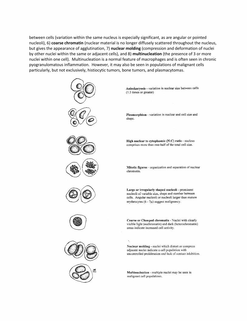
between cells (variation within the same nucleus is especially significant, as are angular or pointed nucleoli), 6) coarse chromatin (nuclear material is no longer diffusely scattered throughout the nucleus, but gives the appearance of agglutination, 7) nuclear molding (compression and deformation of nuclei by other nuclei within the same or adjacent cells), and 8) multinucleation (the presence of 3 or more nuclei within one cell). Multinucleation is a normal feature of macrophages and is often seen in chronic pyogranulomatous inflammation. However, it may also be seen in populations of malignant cells particularly, but not exclusively, histiocytic tumors, bone tumors, and plasmacytomas.
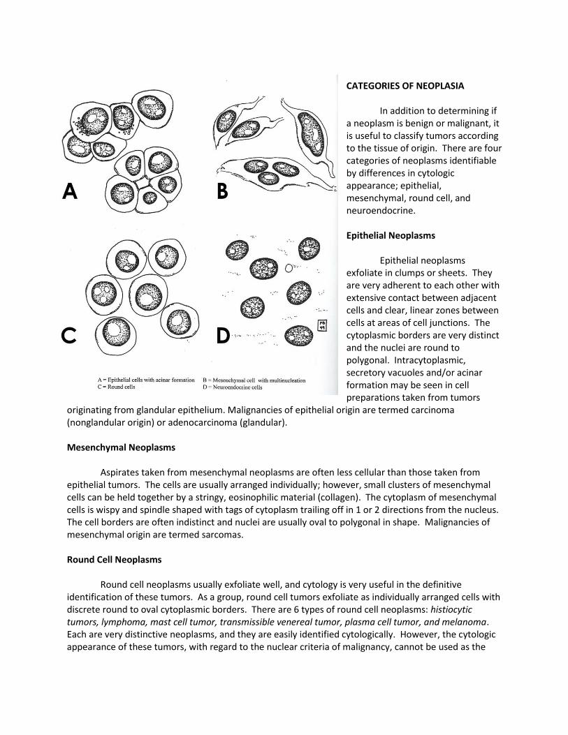
CATEGORIES OF NEOPLASIA In addition to determining if a neoplasm is benign or malignant, it is useful to classify tumors according to the tissue of origin. There are four categories of neoplasms identifiable by differences in cytologic appearance; epithelial, mesenchymal, round cell, and neuroendocrine. Epithelial Neoplasms Epithelial neoplasms exfoliate in clumps or sheets. They are very adherent to each other with extensive contact between adjacent cells and clear, linear zones between cells at areas of cell junctions. The cytoplasmic borders are very distinct and the nuclei are round to polygonal. Intracytoplasmic, secretory vacuoles and/or acinar formation may be seen in cell preparations taken from tumors
originating from glandular epithelium. Malignancies of epithelial origin are termed carcinoma (nonglandular origin) or adenocarcinoma (glandular). Mesenchymal Neoplasms Aspirates taken from mesenchymal neoplasms are often less cellular than those taken from epithelial tumors. The cells are usually arranged individually; however, small clusters of mesenchymal cells can be held together by a stringy, eosinophilic material (collagen). The cytoplasm of mesenchymal cells is wispy and spindle shaped with tags of cytoplasm trailing off in 1 or 2 directions from the nucleus. The cell borders are often indistinct and nuclei are usually oval to polygonal in shape. Malignancies of mesenchymal origin are termed sarcomas. Round Cell Neoplasms Round cell neoplasms usually exfoliate well, and cytology is very useful in the definitive identification of these tumors. As a group, round cell tumors exfoliate as individually arranged cells with discrete round to oval cytoplasmic borders. There are 6 types of round cell neoplasms: histiocytic tumors, lymphoma, mast cell tumor, transmissible venereal tumor, plasma cell tumor, and melanoma. Each are very distinctive neoplasms, and they are easily identified cytologically. However, the cytologic appearance of these tumors, with regard to the nuclear criteria of malignancy, cannot be used as the

sole means of predicting the biological behavior. Histiocytic tumors - These include benign histiocytomas and malignant histiocytosis. Histiocytomas contain cells with moderate amounts of pale, lightly basophilic cytoplasm. Nuclei are round to pleomorphic with fine chromatin and indistinct nucleoli. These are benign, cutaneous tumors, typically seen in young dogs less than 3 years of age. They often become infiltrated with lymphocytes (most of which are small) and then spontaneously regress. Malignant histiocytosis is a malignant neoplasm of histiocytic origin. There is a breed predilection in Rottweilers and Golden retrievers. These tumors may occur
anywhere on the body and are often systemic, involving the bone marrow and/or spleen. Cells contain variable amounts of basophilic, vacuolated cytoplasm with pleomorphic nuclei that contain several criteria of malignancy. Multinucleation may be present. Leukophagia and erythrophagia is a common occurrence with these tumors. Lymphoma - cells contain scant to moderate amounts of deeply basophilic cytoplasm. Nuclei are round to polygonal with high N:C ratio. The background often contains small tags of cytoplasmic fragments called “Lymphoglandular bodies”. All lymphomas are considered malignant. Mast cell tumor - cytoplasm frequently contains numerous purple (metachromatic) granules which often obstruct visualization of the nuclei. Nuclei are round, if visible. Anaplastic cells from undifferentiated mast cell tumors may contain few to no granules. In the dog, inflammation with eosinophils is often present. These tumors are considered potentially malignant and likelihood of metastasis is evaluated using 2 criteria, the cytologic differentiation of the tumor cells, and the location of the tumor in the body (Alleman and Bain, Vet. Med., 95:204, 2000). Transmissible venereal tumor - large numbers of round cells with variable amounts of moderately basophilic cytoplasm. The cytoplasm often contains small punctate vacuoles. Nuclei are round to polygonal with coarse chromatin and 1 or 2 large prominent nucleoli. These tumors may be located in or around the genitalia or nasal cavity. Plasmacytoma - cells have variable amounts of deeply basophilic cytoplasm. Nuclei are round and often eccentrically located in the cells. Perinuclear

Golgi zones may be seen in some cells. There is marked anisokaryosis and binucleation and multinucleation is frequently seen. Most in the dog are benign, but most in the cat have been aggressive in behavior. Melanoma - Melanomas are cutaneous tumors arising from neoplastic melanocytes. Melanomas may occur anywhere on the skin and in the oral cavity. Melanomas are actually tumors of neuroectodermal origin. Some cytologist, prefer to group these tumors in the round cell category because they cytologically and biologically share similarities with mast cell tumors, and they do not fit well into other categories. In general, the well-differentiated tumors have more individualized cells (spindle-shaped), where as the poorly differentiated tumors may attempt to form clumps (epithelioid). The cytoplasm often contains variable amounts of fine pigment granules. However, the granules of melanocytes are brown-black to green-black, distinctively different from the purple granules of the mast cells. Some melanomas are classified as amelanotic because they appear to lack detectable pigment. However, cytologically small amounts of pigment can usually be found, even in poorly differentiated tumors. If cells aspirated from a mass appear very anaplastic, and contain both mesenchymal and epithelial features, an amelanotic melanoma should be suspected. Neuroendocrine/Endocrine Neoplasms Neuroendocrine/endocrine tumors are tumors of chemoreceptors (carotid and aortic bodies) or endocrine glands such as the thyroid, parathyroid, pancreas, and adrenal gland. These tumors share a characteristic cytologic feature. Slide preparations appear as free or “naked” nuclei embedded in a background of cytoplasm. Few distinct cytoplasmic borders are visualized. In general, neoplasms from this category may not have significant criteria of malignancy, and caution must be used when interpreting malignant potential of these lesions based on appearance alone. The thyroid tumors are the neuroendocrine tumors most frequently evaluated. Canine thyroid tumors are usually located on the neck or near the thoracic inlet. Aspirates from thyroid tumors contain clumps of epithelial cells that appear as free nuclei embedded in a background of pale blue cytoplasm with infrequent visualization of cytoplasmic membranes or borders. Amorphous pink material (colloid) may be associated with some cell clusters. Dark, blue-black pigment (tyrosine granules) is sometimes seen in the cytoplasm of epithelial cells. Most thyroid tumors, even adenocarcinomas, will be composed of a fairly uniform population of cells, having few if any criteria of malignancy. However, in the dog over 90% of the

clinically apparent thyroid tumors are adenocarcinomas. In contrast, most thyroid tumors in the cat are benign adenomas or adenomatous hyperplasia. Adenocarcinomas may occur in the cat, but are uncommon. INFLAMMATION AND NEOPLASIA (THE MIXED CELL POPULATION) The malignant criteria must be interpreted with caution when inflammation is present. Inflammation causes reactive changes in epithelial and mesenchymal cells that mimic malignancy. Unless ulcerated or necrotic, most mesenchymal tumors are not infiltrated with inflammatory cells. Therefore, in many cases, when a mixed population of anaplastic mesenchymal cells is seen along with neutrophils and/or macrophages, a reactive fibroplasia should be suspected until confirmed otherwise by histologic evaluation. Tumors associated with inflammation may include squamous cell carcinomas, nasal tumors, and bladder tumors. Most lesions with mixed cell populations will require histopathology to confirm a diagnosis. THE CYTOLOGIC APPEARANCE OF SELECTED NEOPLASMS
Squamous Cell Carcinoma In the cat, squamous cell carcinomas (SCC) have a site predilection for the head. They may occur on the nose, ears, eyelids or anywhere in the oral cavity. Cytologic Appearance: Squamous cell carcinomas have some distinguishing characteristics that help aid in the identification of this tumor. Unlike other epithelial tumors, cells from these tumors may be more individually arranged, with angular cytoplasmic borders. Most tumors are composed of anaplastic appearing squamous cells. The cells are usually very pleomorphic, with variable N:C ratio. They contain moderate amounts of lightly basophilic to aqua-blue cytoplasm (keratinized). Some cells contain small, clear, punctate perinuclear vacuoles called keratohyalin granules. One prominent feature of squamous cell carcinomas is the presence of dysplasia and dyskeratosis, both of which indicate an abnormal cell development. Dysplasia is recognized when mature, angular squamous cells, some with keratin or keratohyalin granules, contain immature nuclei. When keratin is being produced by cells with immature nuclei to a

point where it is seen cytologically, sometimes compartmentalized in the cells, there is dyskeratosis (abnormal production of keratin). "Tadpole- like” cells with eccentrically located nuclei and single blunted cytoplasmic tails are occasionally seen. The nuclei are pleomorphic with coarse chromatin and prominent, multiple nucleoli. Well-differentiated SCCs may contain large numbers of fairly mature squamous epithelium. SCCs often become inflamed, making the cytologic identification of the neoplasm difficult. Unless metastasis can be identified in a regional lymph node, histological confirmation must be used to distinguish reactive versus neoplastic squamous cells. Biological Behavior: The biological behavior of oral SCC is site dependent and aggressive. SCCs with cutaneous locations or on the nasal cavity tend to metastasize late in the course of the disease, but are locally very aggressive. SCCs of the tonsils or caudal tongue are more likely to metastasize to lymph nodes and lungs, often by the time of diagnosis. Basal Cell Tumors
Basal cell tumors are common on the head and neck of the cat. They commonly occur in the dog as well, but rare in other domestic species. They are one of the most commonly reported cutaneous tumors in the feline. They have also been called basal cell carcinomas, but since they are usually benign, this term does not accurately describe their biological behavior in domestic animals. Cytologic appearance: Cytologically, these tumors contain tight clumps of epithelium with deep blue cytoplasm and high N:C ratio. Mitotic figures may occasionally be observed. There may be low numbers of mature sebaceous gland epithelium mixed within the clumps of basal cells. Sebaceous differentiation is associated with some basal cell tumors. Some basal cell tumors, especially in the cat, contain variable amounts of melanin pigment and must be distinguished from true melanocytic tumors. The cohesiveness of basal cells and the very high N:C ratio are distinguishing features. However, this distinction may at times be cytologically difficult since melanin pigment may be within the basal cells themselves. Biological behavior: These tumors are slow growing and rarely metastasize. Surgical excision is usually curative, but recurrence may occur after incomplete excision. Although some basal cells may cytologically appear anaplastic, this usually has no prognostic value in predicting the biological behavior. Mast Cell Tumors Two distinct forms of mast cell tumors (MCT) occur in the cat; the cutaneous/subcutaneous form and the visceral form. The two forms rarely co-exist. Approximately ½ of the cutaneous mast cell tumors in the cat are located on the head and neck. The vast majority of cutaneous mast cell tumors in the cat are located on the head, neck and trunk. Very few cutaneous tumors are located on the limbs. There are two clinical presentations of cutaneous MCTs in cats. The solitary form occurs as a single, well-circumscribed mass. These often occur on cats > 4 years of age and there is no breed predisposition.

The multiple form, or histiocytic form, occurs as multiple, raised nodules. These have reportedly been seen more commonly in younger cats, <4 years of age, with a breed predisposition in the Siamese. The visceral forms are either primary splenic form or the alimentary (intestinal) form. The splenic form is more often associated with mastocythemia and more frequently causes vomiting and gastric/intestinal ulceration. These cats have severe splenomegaly with marked infiltration of mast cells. Cytologic Appearance: Mast cell tumors are in the cytologic category of round cell tumors. The tumors in this category are composed of individually arranged, round cells that are fairly abundant on cytologic preparations. The background of such preparations often contains numerous small, purple granules from ruptured cells. The cytoplasm of intact cells frequently contains numerous purple (metachromatic) granules which can obstruct visualization of the nuclei. Nuclei are round if visible. Anaplastic cells from undifferentiated mast cell tumors may contain few to no granules. Inflammation with eosinophils is often present in canine MCTs but is rare in feline MCTs. Biological Behavior: Solitary, cutaneous MCTs are usually benign, and unlike in the dog, the location of the tumor does not seem to have an influence on the biological behavior. Cats with multiple MCTs or cats with solitary tumors that recur after surgical excision may have a more guarded prognosis. With regard to visceral tumors, the splenic form seems to have a better long term-prognosis than the alimentary form. Plasmacytoma (extramedullary) Extramedullary plasmacytomas are uncommon neoplasms in the cat with site predilections for the skin, oral cavity and spleen. This tumor more commonly affects dogs; however, there are reported cases in the cat. Cytologic Appearance: Plasmacytomas have the cytologic appearance of other round cell tumors with aspirates yielding moderate to large numbers of individually arranged, round to oval cells with discrete cytoplasmic borders. These cells contain variable amounts of deeply basophilic cytoplasm. Many of the cells will have a characteristic "plasmacytoid" appearance with a round nucleus which is often eccentrically located in the cell. A perinuclear clear area "Golgi zone" is sometimes seen in the cytoplasm. There is moderate to marked anisokaryosis, and binucleation and multinucleation is occasionally observed.

Biological Behavior: In the dog, these tumors are usually considered benign and surgical removal is curative in most cases. However, local tumor recurrence following incomplete surgical removal has been observed. Even though the cytological appearance may be identical in either species, in the cat, the tumor may be more aggressive and become systemic with associated metastasis and monoclonal gammopathies. Frequent metastasis to regional lymph nodes, liver, and spleen has been reported. Cats with extramedullary plasmacytomas often have an associated monoclonal gammopathy. In either species, plasmacytomas can, on occasion, be associated with local amyloid production, and rarely with amyloidosis or hypercalcemia. Feline Thyroid Tumors These tumors are commonly seen in older cats and are biologically active. They most often are located in the cervical area lateral to the trachea, but may be seen anywhere along the neck, at the thoracic inlet, or in the thoracic cavity. Cytologic Appearance: Cytologically, thyroid adenomas, or adenomatous hyperplasia, appears as clumps of epithelial cells scattered throughout the preparation. These clumps will contain free nuclei embedded in a background of pale blue cytoplasm with infrequent visualization of cytoplasmic membranes or borders. Amorphous pink material (colloid) may be associated with some clumps. Dark, blue-black pigment is sometimes seen in the cytoplasm (tyrosine granules). This pigment, along with the neuroendocrine appearance of the cells, may be used to definitively identify the tissue as thyroid in origin. Nuclei are round to oval, and fairly uniform in size and shape. Adenocarcinomas are uncommon, but distinction between adenomas and adenocarcinomas is difficult, if not impossible, to do cytologically. In the cat, most thyroid tumors should be considered benign until proven otherwise. Histological evaluation of capsular or lymphatic invasion is often required to identify adenocarcinomas of thyroid origin. Biological Behavior: Unlike the canine, most thyroid tumors (adenomas and adenocarcinomas) in the cat actively secrete thyroid hormones. Adenomas are usually well encapsulated and prognosis is excellent with surgical removal. If bilateral thyroidectomy is performed the patient must be monitored for signs of hypothyroidism and/or hypocalcemia resulting from removal of the parathyroid glands. Adenocarcinomas are identified in only 3% to 5% of cats with hyperparathyroidism. These lesions are locally invasive and metastasize to regional lymph nodes. Metastatic disease was reported in 40% to 71% of cats with adenocarcinomas. Fibrosarcoma
Fibrosarcomas are malignant neoplasms of fibroblasts. They most commonly occur in the skin and subcutis of the extremities and trunk. Soft tissue fibrosarcomas are commonly seen in cats. The high

incidence in this species has been associated with tumor growth at sites of previous inoculations, and as the results of infections with Feline Sarcoma Virus. Cytologic Appearance: Preparations contain low to moderate numbers of individually arranged, spindle-shaped cells. Nuclei are oval to pleomorphic and anisokaryosis is usually present. Nucleoli are frequently prominent and multiple. Nuclear chromatin may be coarse. Mitotic figures are often recognized and multinucleated cells are frequently observed. Biological Behavior: These tumors are usually locally invasive but are typically slow to metastasize. Because they are slow to metastasize, surgical resection or limb amputation is the treatment of choice. Increased incidence of vaccine-associated fibrosarcomas coincided with the introduction of mandatory rabies vaccinations and the FeLV vaccine. Aluminum hydroxide or aluminum phosphate adjuvants are implicated in the pathogenesis. The estimated incidence is about 1 in 5,000. The tumors are best managed with aggressive surgical resection. Recommendations are to vaccinate with RV and FeLV on the rear limbs in order to allow amputation should tumors occur. Post-operative radiation may be considered for microscopic residual tumor or unclean margins, but reports thus far indicate only moderate response. Unless limb amputation is an option, most cats develop local tumor recurrence and are euthanized. Metastasis occurs in 11% of reported cases and survival rates post-surgery vary, but 2 year survival is generally considered poor (<20%). Survival rates may be higher in tumors located on the limbs where amputation is possible (JAVMA 216:58-62, 2001). Osteosarcoma Osteosarcoma is the most common malignant bone tumor of the limbs. They frequently occur in giant and large breed dogs, but may also be seen in small dogs and, occasionally, cats. In the feline, most reported cases have occurred on the rear leg, particularly the femur. Although they are bony neoplasms, tissue aspirates from osteosarcomas are usually easily obtained using a standard 20 gauge or 22 gauge needle and syringe. If dense cortical bone is still present around the lesion a bone marrow aspirate needle may be used to penetrate the cortical bone, and the stylet can then be removed once the needle is embedded in the mass. Cytologic Appearance: Aspirates contain moderate to large numbers of individualized, oval to spindle shaped cells (malignant osteoblasts). These cells contain moderate amounts of deeply basophilic cytoplasm with discrete cytoplasmic borders, giving some osteosarcomas more of a round cell appearance. Nuclei are round to oval and often eccentrically located in the cytoplasm (Flag cells). There is marked anisokaryosis (variation in nuclear size) and nuclei contain clumped chromatin with prominent, multiple, pleomorphic nucleoli. Multinucleation is a distinguishing feature, along with the presence of amorphous pink material (osteoid) that can be seen both extracellularly and as granules within the cytoplasm of neoplastic cells.

Biological Behavior: In the feline species, osteosarcomas are less aggressive and typically metastasize late in the course of the disease. Reported median survival time following surgical removal is 4 to 5 years. Hemagiopericytoma (Spindle cell tumor, nerve sheath tumor or neurofibrosarcoma) Hemangiopericytomas are mesenchymal neoplasms of undetermined origin. Some investigators believe the tumor arises from pericytes that line the blood vessels, hence the name. Recent immunohistochemical characterization (vimentin and actin positive) supports this hypothesis. Thus far, the dog is the only domestic animal in which this neoplasm has been reported, however, these tumors are fairly common in the canine species. Site predilection: The most common site for the hemangiopericytoma is the extremities, particularly the lateral surface of the hind limbs. Tumors may present as a firm nodule or as a soft gelatinous swelling, mimicking a lipoma. Cytological appearance: Cytologically, preparations of needle aspirates taken from hemangiopericytomas are usually very cellular. In fact, an extremely cellular aspirate from a mesenchymal tumor is a diagnostic feature of the hemangiopericytoma. Aspirates contain large numbers of individually arranged mesenchymal cells with very thin, wispy cytoplasmic tails. Low numbers of small, punctate, clear, cytoplasmic vacuoles are frequently observed. Nuclei are round to oval, plump, and often contain one or two prominent nucleoli. Anisokaryosis is moderate to marked. Binucleation and sometimes small multinucleated cells (3 or 4 nuclei) are occasionally seen. Biological behavior: These tumors are locally invasive and recurrence following local resection has been reported in 26% to 60% of the cases. Metastasis is rare (<5% of cases). Aggressive surgical excision is the treatment of choice. These tumors may become more infiltrative and aggressive with each surgical excision, therefore, the initial attempt at surgical removal provides the best opportunity for a cure.