cytokinins in arabidopsis, tools, pathways and interaction
TRANSCRIPT
Cytokinins in Arabidopsis, Tools, Pathways and Interaction with Auxin
Anders NordströmDepartment of Forest Genetics and Plant Physiology
Umeå
Doctoral thesisSwedish University of Agricultural Sciences
Umeå 2004

Acta Universitatis Agriculturae SueciaeSilvestria 317
ISSN 1401-6230ISBN 91 576 6701 2© 2004 Anders Nordström. UmeåPrinted by: SLU, Grafiska Enheten, Umeå, Sweden, 2004

Abstract
Nordström, A. 2004. Cytokinins in Arabidopsis, Tools, Pathways and Interaction with Auxin. Doctoral thesis.Silvestria 317. ISSN 1401-6230, ISBN 91-576-6701-2
Cytokinins are plant hormones, and their relative abundance in proportion to auxin determines whether shoots or roots are formed in callus tissue. The cytokinin to auxin ratio is also believed to regulate apical dominance in plants. The suggested biosynthetic route for cytokinin involves formation of the nucleotide iPMP as the first metabolite synthesized, which is then subsequently hydroxylated to form the nucleotide ZMP. In the work underlying this thesis the potential role of an iPMP-independent biosynthetic route, with direct formation of the nucleotide ZMP was explored. Using an inducible IPT line of Arabidopsis, and in vivo deuterium labelling coupled to analysis with LC-MS/MS, we concluded that ZMP could indeed be formed independently of iPMP. In a similar experiment with wild type Arabidopsis, ZMP was also formed independently of iPMP. Furthermore, the two plant hormones cytokinin and auxin have been shown to regulate each other’s metabolism. In experiments using the IAA analogue NAA, a very rapid decrease in the biosynthesis and pool size of cytokinins was observed. The effect of NAA was mediated through the iPMP-independent pathway. A negative effect of cytokinin on auxin biosynthesis and pool size was also detected. This regulation was found to be slower, and probably related to alterations in development. Arabidopsis and tobacco leaves and roots were separately incubated in media containing 2H2O. Cytokinin biosynthesis capacity was detected in all tissues, and small leaves seemed to possess the highest de novo biosynthesis capability. An Arabidopsis mutant defective in production of the ADK enzyme responsible for conversion of adenosine to adenosine 5´monophosphate and the corresponding cytokinins was found to contain elevated levels of cytokinins, while its IAA levels were lower than in wild type. The increased cytokinin content was due to increased cytokinin biosynthesis. Finally, a LC-MS/MS method capable of quantifying central cytokinin metabolites in sample sizes <50mg FW including bases, ribosides and intact nucleotides at a rate of 70 samples/day was developed.
Keywords: cytokinin, auxin, biosynthesis, Arabidopsis thaliana, liquid chromatography, ESI, MS/MS, interaction, development, plant hormones
Authors address: Umeå Plant Science Centre, Department of Forest Genetics and Plant Physiology, SE-901 83 Umeå Sweden

I stunder av,
Högmod eller SvårmodHybris eller Ödmjukhet
har jag funnit ro i följande uppmuntrande ord signerade G.S.
”Du skall veta Anders, jag kan få en apa genom forskarutbildningen”

ContentsBackground…………………………..........……………….....7 Introduction…………………………...........……………….... 7Objectives…………………………………………….............. 9Cytokinin Biosynthesis………………………………............. 9 Metabolism and Catabolism………………………….............. 14Cytokinin Sites of Synthesis and Translocation………............ 15Cytokinin perception and signal transduction…………........... 17Cytokinin Action………………………………………........... 19Auxin …………………………………………………........ 20Cytokinin – Auxin Interactions………………………............. 22
Experimental…………………………………………............ 24Plant material and growth conditions…………………............ 24Sample purification……………………………………........... 24Electrospray Mass Spectrometry (ESI-MS)………….............. 25Plant hormone quantification…………………………............ 27In Vivo deuterium labelling experiments……………….......... 27
Results and Discussion………………………………............ 29De novo ZMP synthesis (I)……………………………........... 29Reciprocal Regulation of Auxin – Cytokinin Pool Sizes and de novo Biosynthesis (II)……………............. 31Mechanism of IAA regulation of Cytokinin Biosynthesis (II)…………………………............... 32Sites of Cytokinin Biosynthesis (II)………………….............. 33Derivatization of Cytokinins for ESI-MS/MS (III)….............. 34Cytokinin – Auxin Interaction in Arabidopsis mutant,disrupted in the ADK gene (IV)………………………............ 36
Conclusions…………………………………………….......... 38
Further Perspectives…………………………………........... 39
Acknowledgements…………………………………….......... 40
References……………………………………………............ 42

7
AppendixList of Papers
The present thesis is based on the following papers, which will be referred to by their Roman numerals:
I. Åstot, C., Dolezal, K., Nordström, A., Wang, Q., Kunkel, T., Moritz, T., Chua, N-H. and Sandberg, G. (2000) An alternative cytokinin biosynthesis pathway. Proceedings of the National Academy of Sciences of the United States of America. 97, 14778-14783.
II. Nordström, A., Tarkowski, P., Tarkowska, D., Norbaek, R., Åstot, C., Dolezal, K. and Sandberg, G. (2004) Auxin regulation of cytokinin biosynthesis in Arabidopsis thaliana: A factor of potential importance for auxin-cytokinin-regulated development. Proceedings of the National Academy of Sciences of the United States of America. 101, 8039-8044.
III. Nordström, A., Tarkowski, P., Tarkowska, D., Dolezal, K., Åstot, C., Sandberg, G. and Moritz, T. (2004) Derivatization for LC-Electrospray Ionization-MS: A tool for improving reversed-phase separation and ESI responses of bases, ribosides, and intact nucleotides. Analytical Chemistry. 76, 2869-2877.
IV. Para, A., Nordström, A., Wilson, K., Moffat, B.A., Sandberg, G. and Sundås-Larsson, A. (2004) Disruption of the ADK1 gene causes meristem distortion and a cytokinin overproduction phenotype in Arabidopsis thaliana. Manuscript.
Papers I and II are reproduced with kind permission from National Academy of Sciences, USA and paper III is reproduced with kind permission from the American Chemical Society.
Also published by the author but not included in the thesis:
Gullberg, J., Jonsson, P., Nordström, A., Sjöström, M. and Moritz, T. (2004) Design of experiments: an efficient strategy to identify factors influencing extraction and derivatization of Arabidopsis thaliana samples in metabolomic studies with gas chromatography/mass spectrometry. Analytical Biochemistry. 331, 283-295.
Jonsson, P., Gullberg, J., Nordström, A., Kusano, M., Kowalczyk, M., Sjöström, M. and Moritz, T. (2004) A strategy for identifying differences in large series of metabolomic samples analyzed by GC/MS. Analytical Chemistry. 76, 1738-1745.
Viklund, C., Nordström, A., Irgum, K., Svec, F. and Frechet, J.M.J. (2001) Preparation of porous poly(styrene-co-divinylbenzene) monoliths with controlled pore size distributions initiated by stable free radicals and their pore surface functionalization by grafting. Macromolecules. 34, 4361-4369.

7
BackgroundIntroduction
Multi-cellular organisms need signalling pathways to co-ordinate responses within the system. This enables each organism to proceed through the series of events that takes place during its life cycle. The signalling system reacts to both intrinsic and external factors, integrates the cues and initiates functional responses. In plants, several compounds are believed to be parts of either paracrine or long-distance signalling pathways. Some of these are referred to as “plant hormones” or “growth regulators”. Cytokinins and auxins are two classes of compounds that act as plant hormones.
This thesis deals with the biosynthesis of cytokinins and the interaction between cytokinins and auxin at the biosynthetic level. It seems logical to study these two compounds in conjunction with each other, since they have proven to regulate developmental processes in both antagonistic and synergistic manners. Moreover, their discovery can also be traced to the same group of researchers.
Folke Skoog (1908-2001) was working in the early 1930´s as an undergraduate/graduate student in the laboratories of Frits Went and Kenneth Thimann at the California Institute of Technology. He participated in the work leading to the discovery of IAA as a growth substance (Thimann & Koepfli, 1935). He and his collaborators found that material promoting growth in oat coleoptiles could be obtained from terminal buds of beans by allowing it to diffuse into agar blocks. Furthermore, when excised terminal buds of beans were placed directly on cut surfaces of decapitated oat plants, inhibition of lateral buds could be maintained. Auxin was the factor that inhibited growth of the lateral buds. They suspected that a second factor was involved in the process. In the mid 1950´s, Miller and Skoog described the first cytokinin, kinetin (Fig. 1), which they obtained from heated DNA (Miller & Skoog, 1955; Miller et al., 1956). Although this was not an endogenous cytokinin, it displayed the ability to induce cell division and shoot formation from callus growing in a cell culture in concert with IAA (Skoog & Miller, 1957). The cited authors demonstrated that the ratio of kinetin to auxin determined shoot or root growth from a tobacco callus. If the ratio was high, the callus formed shoots and if the ratio was low, the callus formed root tissue. The conclusion was that the cytokinin to auxin balance is involved in organogenesis. However, it was not until 1958 that Thimann´s group showed that the “other factor” was a cytokinin and demonstrated its antagonistic effect to auxin (Wickson & Thimann, 1958). They excised stem parts and placed them in a growth medium. In a control (just medium) the lateral buds were released. If auxin was added this was inhibited. If both auxin and kinetin were added, the buds were released, demonstrating the antagonistic effect of these two compounds (Wickson & Thimann, 1958).
The name cytokinin derives from cytokinesis (cell division) and a cytokinin is now defined as “A compound that in the presence of auxin induces cell division in a suitable assay material grown on a defined medium” (Shaw, 1994).

8 9
The first naturally occurring cytokinin that was identified was trans-6-(4-hydroxy-3-methylbut-2-enylamino)-purine (Letham, 1963; Letham, et al., 1964). The compound was named Zeatin (Fig. 1) after the plant Zea mays in which it was found. Zeatin was present at a very low concentration. Letham only obtained 1 mg of Zeatin from 70 kg of tissue! (Letham et al., 1964). Several naturally occurring cytokinins (as well as synthetic analogues) have been identified since then. The naturally occurring cytokinins are all adenines, substituted at N6 with an isoprenoid or aromatic side chain some of which are displayed in Fig. 1. Cytokinins have
N
N NH
N
HN
N
N NH
N
HN
OH
N
N NH
N
HN OH
N
N NH
N
HN
OH
N
N N
N
HN
O
OHHO
HO
N
N N
N
HN
OH
O
OHHO
HO
N
N NH
N
NH
HO
O
OH
OH
OHHO
N
N N
N
HN
OH
O
HO
HO OH
HO
N
N NH
N
HN
O
O
OH
OH
OHHO
N
N N
N
HN
OH
O
OHHO
OP
O
OH
O
N
N N
N
HN
OH
O
OHHO
OP
O
OH
OP
O
OH
O
N
N N
N
HN
OH
O
OHHO
OP
O
OH
OP
O
OH
OP
O
OH
O
N
N N
N
HN
O
OHHO
OP
O
OH
O
N
N N
N
HN
O
OHHO
OP
O
OH
OP
O
OH
O
N
N N
N
HN
O
OHHO
OP
O
OH
OP
O
OH
OP
O
OH
O
NH
OH
O
O
N
N NH
N
HN
Auxin (IAA) Kinetin isopentenyl adenine (iP) trans-Zeatin (t-Z) cis-Zeatin (c-Z)
dihydro-zeatin (DHZ) Zeatin riboside (ZR) isopentenyl adenosine (iPA) Zeatin-9-glucoside (Z9G)
Zeatin-7-glucoside (Z7G) Zeatin-O-glucoside (ZOG)
isopentenyl adenosine 5´monophosphate (iPMP)
Zeatin riboside 5´monophosphate (ZMP)
isopentenyl adenosine 5´diphosphate (iPDP)
isopentenyl adenosine 5´triphosphate (iPTP)
Zeatin riboside 5´triphosphate (ZTP)
Zeatin riboside 5´diphosphate (ZDP)
Figure 1. Selected cytokinin and Auxin metabolites

8 9
been found to play a part in many aspects of plant growth and development either antagonistically/synergistically with other plant growth regulators or by themselves. In the last few years, molecular evidence for endogenous cytokinin biosynthesis in plants (Kakimoto, 2001; Takei et al., 2001a), and their ability to perceive cytokinins, via the identification of cytokinin receptors, has been obtained (Inoue et al., 2001). Cytokinins have thus been definitively established as endogenously produced signalling compounds. For reasons of convenience, throughout this thesis I will refer to cytokinins as the naturally occurring, N6 isoprenoid-substituted adenines.
Objectives
The main objectives of the work underlying this thesis were to investigate how cytokinins are synthesised de novo (Paper I) and how cytokinin and auxin regulate each other’s metabolism (Paper II). Furthermore, we wanted to investigate potential sites of cytokinin biosynthesis (Paper II). The need for high-throughput quantification methods, capable of analysing cytokinins in sample sizes (< 100 mg FW) which are potentially more relevant for physiological studies than the large samples used in previous investigations was addressed in Paper III. How cytokinin-auxin homeostasis is altered in an Arabidopsis mutant defective in central cytokinin metabolism was examined in Paper IV.
Cytokinin Biosynthesis
There have been three main views on cytokinin synthesis, holding that:
A: They are not endogenously produced by plants, but rather produced by bacteria, and assimilated by plants in a symbiotic fashion (Holland, 1997).B: They can be synthesised indirectly via plant tRNA.C: They can be synthesised directly, de novo, by plants.
A: The first view has been proved wrong, at least in the sense that cytokinins cannot be synthesised by plants. Genes encoding enzymes for de novo cytokinin synthesis have been found in Arabidopsis (Kakimoto, 2001; Takei et al., 2001a). Moreover, aseptically grown plants have also demonstrated de novo cytokinin biosynthesis.
B: Regarding the second view, cytokinins were found to be located adjacent to the 3´-end of the anticodon in tRNA obtained from yeast (Zachau et al., 1966; Bieman et al., 1966). Since then, cytokinin-active ribonucleosides have been reported in tRNAs from virtually all organisms tested. It has been suggested that the action of cytokinins might be mediated through the function of cytokinins in tRNA on protein synthesis. Six different cytokinins have been discovered in plant tRNA, including cis/trans-ZR and cis/trans-iPA, of which cis-ZR seems to be the most abundant (Taller, 1994). Recently, a gene coding for a tRNA isopentenyltransferase enzyme was cloned in Arabidopsis (Golovko et al., 2002). In incorporation studies of radioactive adenine in intact bean root, most of the cytokinin subsequently detected appeared to originate from RNA hydrolysis (Maaβ & Klämbt, 1981). The fact that cis-type cytokinins are the most abundant cytokinins in tRNA has raised the question

10 11
if tRNA actually can act as a source of cytokinins, since cis-type cytokinins are biologically inactive (Murai, 1994). The occurrence of cis/trans isomerases (Bassil et al., 1993), might partially explain how biologically inactive cis-cytokinins, derived from tRNA are modified to become active cytokinins. However, tRNAs are estimated to account for at most 40% of the cytokinin biosynthesis when calculated from the tRNA turnover rate based on radioactive feeding experiments (Barnes et al., 1980), and it is generally assumed that tRNAs play only a minor role, if any, as sources of cytokinin precursors (Barnes et al., 1980; Klämbt, 1992).
C: The generally accepted view has been that plants do synthesise cytokinins endogenously. Dictyostelium discoideum (slime mold) gave the first evidence of direct de novo cytokinin biosynthesis from AMP and an isoprenoid side chain. A partially purified enzyme from this organism catalysed the transfer of the isopentenyl moiety from dimethylallyldiphosphate (DMAPP) to AMP as illustrated on the cytosolic side
Figure 2. Proposed compartmentalization of cytokinin metabolism in plant cells. In the cytosolic Mevalonate (MVA) pathway, which can be inhibited by mevastatin, isopentenyl transferase, IPT, catalyses the transfer of DMAPP to AMP/ADP/ATP leading to the formation of iPMP. In the plastid localized 1-Deoxy-D-xylulose 5-phosphate- (DXP) pathway, also referred to as the Methyl-erythritol-phosphate (MEP) pathway, the cytokinin ZMP is formed by the addition of HMBPP to AMP/ADP/ATP.
N
N N
N
NH2
O
OHOH
OP
O
OH
O
OPOPO
OO
OH OH
AMP
3 Acetyl-CoA
Mevalonate
N
N N
N
HN
O
OHHO
OP
O
OH
O
iPMP
HMG-CoA
IPT
Mevastatin
CYTOSOLMevalonate-Pathway
DMAPP IPP
MEP
OPOPO
OO
OH OH
OH
N
N N
N
NH2
O
OHOH
OP
O
OH
O
DXP
DMAPPIPP
AMP
N
N N
N
HN
OH
O
OHHO
OP
O
OH
O
ZMP
PLASTID (CHLOROPLAST)DXP-Pathway
HMBPP
??
?
iPMP
ZMP
Hydroxylation
Transport?
Transport?
Hydroxylation
IPT

10 11
in figure 2. This was referred to as DMAPP:AMP isopentenyltransferase (iptase) activity and the enzyme was referred to as an isopentenyl transferase IPT (Taya et al., 1978). The same activity was detected in cell-free extracts of cytokinin-autotrophic tobacco tissue culture (Chen & Melitz, 1979). The gene that encoded a similar IPT was cloned in Agrobacterium tumefaciens (bacteria) from the Ti-plasmid (Barry et al., 1984; Akiyoshi et al., 1984). Activity of IPT from partially purified corn kernels was demonstrated by Reinecke et al. (1991), who further demonstrated that the enzymatic activity decreased during Zea mays kernel development (Reinecke et al., 1992). Similarly, IPT activity was described by Blackwell & Horgan (1994) from crude extracts of Zea mays kernels. The IPT enzyme is very unstable and it has proven to be difficult to purify to homogeneity. Another cytokinin biosynthetic gene, TZS (trans-Zeatin synthase), is present in the virulence region of the Ti-plasmid (that is, it is not located in the T-DNA which is actually transferred to the plants at infection) in some strains of Agrobacterium tumefaciens (Kaiss-Chapman & Morris, 1977). This gene, when cloned and expressed in Escherichia coli resulted in the secretion of Zeatin (Beaty et al., 1986). The conclusion from all these findings is that the initial step in cytokinin biosynthesis is the catalytic transfer by an IPT-enzyme of DMAPP to AMP (Fig. 2), producing iPMP as the first cytokinin metabolite.
Kakimoto explored the Arabidopsis genome database and searched for genes with similarity to the Agrobacterium IPT sequence, finding nine putative Arabidopsis IPT genes which were named AtIPT1 to AtIPT9 (Kakimoto, 2001). Independently, another Japanese research group made the same discovery (Takei et al., 2001a). In an attempt to further characterise these putative cytokinin biosynthesis genes, Kakimoto over-expressed the AtIPT4 gene in Arabidopsis calli. The calli were able to regenerate shoots even in the absence of cytokinins in analogy with the experiments performed by Skoog and Miller (1957). This demonstrated the ability of AtIPT4 to synthesise cytokinins (Kakimoto, 2001). In a second experiment the ATIPT4 gene was expressed in E. coli. In kinetic studies subsequently performed on the gene product, Kakimoto found that the AtIPT4 was a DMAPP:ATP/ADP isopentenyl transferase rather than a DMAPP:AMP isopentenyl transferase. This implies that the first cytokinin metabolites formed could be iPTP or iPDP rather than iPMP (Fig. 3) (Kakimoto, 2001). Takei and co-workers expressed AtIPT1 to ATIPT8 in E. coli and detected Z and iP in the culture medium (Takei et al., 2001a). The relative abundance of Z and iP varied somewhat, and for AtIPT1, 4, 7, 8, the Z/iP ratio was higher than for AtIPT3, 5, 6 (Takei et al., 2001a). Further, in vitro characterisation of the purified AtIPT1 gene product, revealed DMAPP:AMP isopentenyl transferase activity (Takei et al., 2001a).
The finding by Kakimoto that AtIPT4 had much higher substrate specificity for ATP and ADP than AMP (Kakimoto, 2001) is rather intriguing. As already mentioned, this implies that early cytokinin metabolites might be di- or tri-phosphates rather than mono-phosphates (Fig. 3). The existence of such cytokinin metabolites have been suspected for quite some time (Miller, 1967). For instance, Laloue et al. (1974) demonstrated that these metabolites could be formed in plants. They fed cytokinin-dependent tobacco cell culture with radiolabelled iPA and demonstrated that iPDP and iPTP were formed (Laloue et al., 1974). These two metabolites accounted for 77% of the extracted radioactivity. In order to minimise potential interference from
12 13
other labelled purines present in the extraction mix, they also demonstrated that di- and tri-phosphate metabolites were formed from labelled BAP (benzyl amino purine) (Laloue et al., 1974). The formation of di- and tri-phosphate cytokinin metabolites in feeding experiments with radiolabelled compounds has since been shown in tobacco cell culture (Laloue et al., 1977a; Laloue & Pethe, 1982) and cell cultures of the moss Physcomitrella patens (Schwartzenberg et al., 2003).
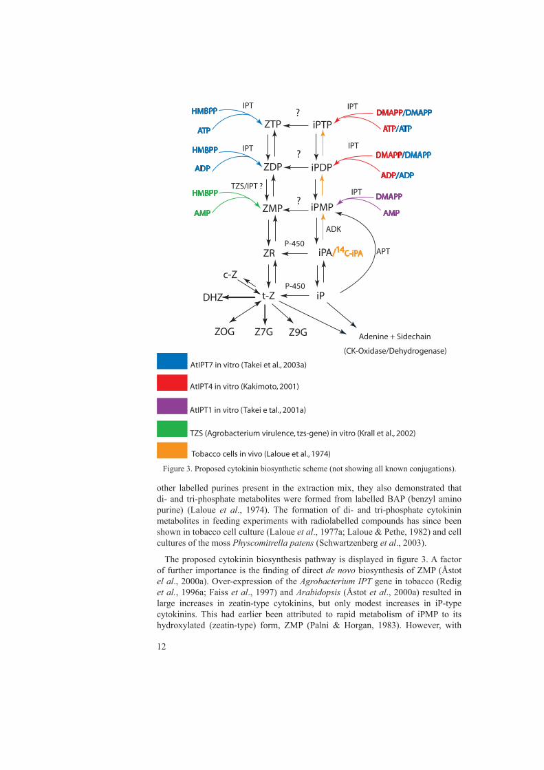
The proposed cytokinin biosynthesis pathway is displayed in figure 3. A factor of further importance is the finding of direct de novo biosynthesis of ZMP (Åstot el al., 2000a). Over-expression of the Agrobacterium IPT gene in tobacco (Redig et al., 1996a; Faiss et al., 1997) and Arabidopsis (Åstot et al., 2000a) resulted in large increases in zeatin-type cytokinins, but only modest increases in iP-type cytokinins. This had earlier been attributed to rapid metabolism of iPMP to its hydroxylated (zeatin-type) form, ZMP (Palni & Horgan, 1983). However, with
Figure 3. Proposed cytokinin biosynthetic scheme (not showing all known conjugations).
ZTP
ZDP
ZMP
iPTP
iPDP
iPMP
ZR
t-Z
ZOG Z7G Z9G
iPA/14C-iPA
iP
DMAPP/DMAPPHMBPP
ATP
ADP
AMP
ATP/ATP
ADP/ADP
DMAPP/DMAPP
DMAPP
AMP
HMBPP
HMBPP
Adenine + Sidechain
IPT
IPT
IPT
IPT
IPT?
?
c-Z
DHZ
TZS/IPT ?
APT
ADK
(CK-Oxidase/Dehydrogenase)
AtIPT7 in vitro (Takei et al., 2003a)
P-450
P-450
AtIPT4 in vitro (Kakimoto, 2001)
AtIPT1 in vitro (Takei e tal., 2001a)
TZS (Agrobacterium virulence, tzs-gene) in vitro (Krall et al., 2002)
?
Tobacco cells in vivo (Laloue et al., 1974)

12 13
in vivo deuterium labelling and double tracer feeding it became evident that there is an iPMP-independent pathway for the synthesis of ZMP (Åstot et al., 2000a). The proposed iPMP-independent pathway would use 4-hydroxy-3-methyl-2-(E)-butenyldiphosphate (HMBPP), rather than DMAPP as the side chain precursor (Fig. 2+3). Plants have two possible biosynthesis pathways for the isoprenoid side chain, the DXP (1-Deoxy-D-xylulose 5-phosphate) pathway in plastids (chloroplasts) and the MVA (Mevalonate) pathway in the cytosol (Lichtenthaler, 1999; Rohmer, 2003) (Fig.2). It should be mentioned that the DXP-pathway is also referred to as the MEP (methylerythritolphosphate) pathway. Both pathways can supply DMAPP and IPP, but HMBPP has only been found to be an intermediate in the plastid localised DXP-pathway (Hecht et al., 2001) (Fig. 2).
In an attempt to investigate the possible origins of the isoprenoid side chains of cytokinins, Kasahara and co-workers utilised 13C-substrate feeding and a two-way approach involving both Arabidopsis mutants and an enzyme inhibitor (Kasahara et al., 2004). An Arabidopsis mutant, cla1, which is defective in the 1-deoxy-D-xylulose-5-phsophate synthase (DXP synthase) of the DXP pathway (Estevez et al., 2000) was used for studying 13C-labelling patterns generated when the DXP pathway is defective. Mevastatin, which selectively blocks the HMG coA reductase of the MVA (Mevalonic acid) pathway (Fig 2) was used to investigate the labelling pattern when the MVA-pathway was blocked (Kasahara et al., 2004). The cited authors demonstrated that trans-Z and iP had mainly plastid origins, whereas the cis-Z side chain had a cytosolic origin (Kasahara et al., 2004). A finding that further strengthens the hypothesis that cytokinin isoprenoid side chains are of plastid (DXP-pathway origin) was the discovery that cytokinin levels were not affected in a mutant with a dysfunctional hmg1 gene, which encodes the HMG reductase of the cytosolic pathway (Suzuki et al., 2004). Moreover, it was previously demonstrated that expression of the Agobacterium TZS gene in E. coli results in secretion of Z (Beaty et al., 1986). The gene encoding this enzyme was cloned, expressed and the enzyme was purified and studied in vitro by Krall and co-workers. With the radioactively labelled substrate HMBPP and AMP they demonstrated conversion to ZMP (Krall et al., 2002). Takei and co-workers prepared Z- and iP-type cytokinin di- and tri-phosphates standards by expressing AtIPT7 in E. coli and subsequently utilised in vitro reactions for the actual synthesis. They found that the reaction efficiency was lower when using HMBPP as a substrate than DMAPP, indicating that the gene product of AtIPT7 has higher specificity for DMAPP (Takei et al., 2003a). This group also developed a method for separating these di- and tri-phosphates from plant material (Takei et al., 2003b). However, they did not demonstrate unambiguously the presence of the compounds in planta. In contrast, the above findings, which support de novo ZMP synthesis independent of iPMP biosynthesis, over-expression of AtIPT8 in Arabidopsis led to an accumulation of iP-type cytokinins in a study by Sun et al. (2003).
Taken together, these findings show that plants have the ability to synthesise cytokinins endogenously. There seems to be two pathways for de novo cytokinin biosynthesis, but their relative importance is not fully understood.

14 15
Metabolism and Catabolism
A summarised version of some of the known cytokinin metabolic conversions is displayed in figure 3. Inter-conversions between cytokinin bases, ribosides and nucleotides are mediated by the same enzymes that convert their adenyl analogues (Mok & Martin, 1994). Generally these enzymes show higher affinity for the adenine compounds than for their cytokinin relatives, as exemplified in kinetic studies in Arabidopsis with adenosine phosphoribosyl transferase APT (Allen et al., 2002) and adenosine kinase (ADK) (Moffat et al., 2000). Other changes to the adenine structure are more specific for the adenine type cytokinins. These include glucolysation at the 3, 7 or 9 position at the purine ring by glucosyl transferase (Entsch et al., 1979). These metabolites are not subject to hydrolysis of the glucoside unit by β-glucosidase and are biologically inactive (Laloue, 1977b). They seem to be very stable conjugates and are considered to be permanently inactivated forms. There is conflicting evidence regarding their ability to serve as substrates for cytokinin oxidase (Schmülling et al., 2003). Conjugates of cytokinins may also occur with alanine at the 9 position, lupinic acid (Parker et al., 1975).
Modification to the side chain is more specific to cytokinins. A reductase that is highly specific for trans-Z has been isolated in Phaseolus (Martin et al., 1989) that is responsible for reduction of the isoprenoid side-chain double bond. The enzyme did not modify cis-Z, ZR or iP. Cytokinin oxidase does not seem to use DHZ as a substrate (Bilyeu et al., 2001), and hence the formation of dihydro-metabolites might be important in maintaining cytokinin homeostasis. Glycosylation of the isoprenoid side-chain hydroxyl group of Z seems to be a key process in the machinery that regulates the level of active cytokinins. Zeatin-O-glucoside, ZOG, and the corresponding Zeatinriboside-O-glucoside, ZROG, display low biological activity and are converted back to their active Z or ZR forms by β-glucosidases (Brzobohaty et al., 1993; Mok & Mok, 2001). The glycosylation is mediated by Zeatin-O-glycosyltransferase. The gene that encodes this enzyme, ZOG1, has been cloned in phaseolus lunatus (Martin et al., 1999). The enzyme responsible for O-glucosylation of cis-Z has also been identified in maize (Martin et al., 2001; Veach et al., 2003). Conversion of iP-type cytokinins (iP and iPA) to their hydroxylated Z-type counterparts is believed to be mediated by microsomal, mixed function oxidases, particularly cytochrome P-450 enzymes (Chen & Leisner, 1984). Activity of cytokinin cis-trans isomerases has been detected in extracts from Phaseolus (Bassil et al., 1993). This enzyme might participate in regulation of the pool of active cytokinins by converting less active cis-Z to active trans-Z. Incidentally, in maize, cis-Z seems to be an active cytokinin (Yonekura-Sakakibara, 2004). Therefore one might draw erroneous conclusions about cytokinin metabolism and its regulation, when comparing data from many different species.
Cytokinin oxidase catalyses the irreversible degradation of iP, Z and their corresponding ribosides in a single step by oxidative side-chain cleavage (Schmülling et al., 2003). The enzyme seems to be expressed from a family of seven genes in Arabidopsis (Schmülling et al., 2003). However, it has been demonstrated that the enzymes do not need molecular oxygen as an electron acceptor but rather use a flavin co-factor (Galuszka et al., 2001). This implies that the enzyme should be

14 15
named cytokinin dehydrogenase rather than cytokinin oxidase. Cytokinin oxidase/dehydrogenase activity is up-regulated by cytokinins (Dietrich et al., 1995; Motyka et al., 1996; Motyka et al., 2003). Predicted sub-cellular localizations derived from signal peptide analysis of the gene sequences include extra-cellular, cytosolic and plastid locations for the different gene products (Emanuelsson et al., 2000). One should be careful when interpreting such results, but experimental data have so far supported an extra-cellular localization since cytokinin oxidase/dehydrogenase activity has been found in the media of moss protoplast (Houba-Herin et al., 1999), yeast (Werner et al., 2001) and tobacco (Motyka et al., 2003) cultures. This is consistent with the finding that the CRE1-cytokinin receptor (Inoue et al., 2001) seems to be localised in the cell membrane, suggesting that some cytokinin signalling (at least) originates from the extra-cellular space, and thus regulation of the cytokinin content should be controlled, at least partially, by processes that occur outside of the cell wall. Recently a transgenic Arabidopsis was engineered to over-express cytokinin oxidase/dehydrogenase (Werner et al., 2003). Werner and co-workers found in fluorescently labelled fusion protein experiments that some cytokinin oxidases were targeted for the vacuole and the endoplasmatic reticulum (ER), supporting the hypothesis that some of the products of this gene family might be secreted from the cell (Werner et al., 2003). Expression studies using GUS-constructs revealed tissue-specific expression for the different genes in the cytokinin oxidase/dehydrogenase family (Werner et al., 2003). The transgenic plant displayed lowered levels of endogenous cytokinins, and would be highly suitable for investigating the significance of cytokinins in planta.
Taken together, these data suggest that cytokinin levels are regulated in plants at the biosynthetic level, by base to nucleotide/riboside conversions, irreversible inactivation to N-glucosides, reversible inactivation to O-glucosides and via permanent inactivation by oxidative cleavage of the side-chain by cytokinin oxidase/dehydrogenase.
Cytokinin Sites of Synthesis and Translocation
Cytokinins have been found in roots, stems, leaves, flowers, fruits and seeds, and they are probably present in every tissue in a living plant. This, however, does not necessarily imply that they are synthesised by every cell. A large set of data indicates that roots, especially root tips, are major sites of cytokinin biosynthesis (reviewed by Letham, 1994). During aseptic culture of maize roots by Van Staden & Smith (1978) cytokinins were released into the medium. In a similar experiment by Koda & Okazawa (1978) it was demonstrated that after growth of excised tomato root tips for seven days the cytokinin level in the media was eight times higher than in the root tissue. Studies on extracts of sunflower root apices 0-1 and 1-3 mm from the tip have found a 100-fold higher activity of cytokinins in the former location (Weiss & Vaadia, 1965). Chen and co-workers demonstrated incorporation of 14C-adenine into cytokinins in pea and carrot roots when grown on media (Chen et al., 1985). Their results indicated that roots are the major source of cytokinin biosynthesis, but not the only one. Indirect evidence of root tip localised cytokinin biosynthesis has also been obtained, in studies involving, for instance, immunochemical staining (Sossountzov et al., 1988) and decapitation of root tips followed by observed

16 17
reductions in cytokinin contents (Feldman, 1979). Although it must be stated that some of these measurements lack the precision required by today’s standards and consequently some of the evidence for root-localised cytokinin biosynthesis should be questioned. Some investigations have also pointed towards a possible role of apical plant parts in cytokinin biosynthesis. Incorporation of 14C-adenine into cytokinin in apparently rootless tobacco plants suggests possible apical cytokinin synthesis (Chen & Petschow. 1978).
After identification of the cytokinin biosynthesis genes AtIPT1 to AtIPT9 (Kakimoto, 2001; Takei et al., 2001a), expression studies of these genes have been performed (Miyawaki et al., 2004). These investigators used beta-glucoronidase (GUS) fusion constructs with all of the AtIPT genes in Arabidopsis. They concluded that the genes were predominantly expressed in the following tissues: AtIPT1::GUS in xylem precursor cell files in the root tip, leaf axils, ovules and immature seeds, AtIPT3::GUS in phloem tissues, AtIPT4::GUS and AtIPT8::GUS in immature seeds with highest expression in the chalazal endosperm (CZE), a structure in the nutritious tissue that surrounds the embryo, AtIPT5::GUS in root primordia, root caps, upper parts of the young inflorescence and fruit abscission zones, and AtIPT7::GUS in endodermis of the root elongation zone and trichomes (outgrowths) on young leaves. AtIPT2 and AtIPT9, which are responsible for tRNA isopentenyl transferases, were expressed ubiquitously (Miyawaki et al., 2004). These findings suggest that cytokinin biosynthetic capacities occur throughout the plant, and that many of the genes are expressed in different parts of the root structure.
Different sites of synthesis and action imply translocation. There are numerous demonstrations of cytokinin activity in xylem sap (reviewed by Letham, 1994). A gene family of high affinity transporters for adenine and purine derivatives in Arabidopsis has been identified (Gillissen et al., 2000; Bürkle et al., 2003). Expression studies of these genes have indicated that they have a role in retrieving root-synthesised cytokinins from the xylem and thus facilitating long-distance transport of cytokinins (Bürkle et al., 2003). Further evidence of xylem translocation of cytokinins was obtained by Takei and co-workers when studying the cytokinin response to nitrogen availability in maize. Upon addition of nitrogen to nitrogen-depleted plants, iPMP started to accumulate in the roots within an hour. This preceded the accumulation of ZMP and Z in the xylem sap. In the leaf tissue, Z started to accumulate 4 h after addition of nitrogen, and the elevated Z levels were maintained for at least 24 h (Takei et al., 2001b). Furthermore, in a subsequent investigation they also observed that the ZOG levels started to decrease in the root tissue upon nitrogen availability, suggesting increased conversion of inactive ZOG to active Z (Takei et al., 2002). The potential role of cytokinins in the phloem is not well understood. There are few reports, and only a few cytokinins have been identified with certainty: these include nucleotides (Vonk, 1978) and more recently Z and ZR were identified in phloem sap from the bean plant Ricinus (Kamboj et al., 1998). Translocation of cytokinins within the germinating seed is fairly well established. The incorporation of radioactively-labelled adenine into DHZR (Dihydrozeatin riboside) and DHZRMP (Dihydrozeatin riboside 5´monophosphate) has been demonstrated, and the subsequent polar movement of these compounds from embryo to emerging cotyledons in lupin seeds (Nandi et al., 1988; Nandi & Palni, 1989). Furthermore, excision of the embryonic axis by Munoz et al. (1990)

16 17
led to reduced cytokinin levels in the cotyledons. However, in the light of very recent discoveries, one must question these findings, at least as general conclusions. It appears that in Arabidopsis, no cytokinin receptors are expressed, either in the embryo or the developing seedling (Higuchi et al., 2004). Without any receptors, what would the purpose of cytokinin be as a signal? Indeed, there are studies that question whether cytokinins are long-distance signalling compounds, and rather propose a paracrine mode of action. Grafting experiments using tobacco rootstock with inducible cytokinin over-production and wild type shoots resulted in a 50-fold elevation of ZR in the roots 24 h after induction. However, no elevation of cytokinin in the shoots was detected (Faiss et al., 1997). To circumvent potential translocation problems due to grafting, Böhner used a dexamethasone-inducible/tetracycline-repressible expression system of the bacterial IPT gene in tobacco (Böhner & Gatz, 2001). When the IPT gene was induced in the tobacco plants, release of axillary buds was observed. Following this, tetracycline was applied locally to leaf-axils, causing repression of IPT expression. This immediately arrested the axillary bud outgrowth, and demonstrated that cytokinin produced elsewhere in the plant was not sufficient to release the bud outgrowth (Böhner & Gatz, 2001). These experiments raise questions about cytokinins as long-distance signalling compounds.
Cytokinin perception and signal transduction
Regardless of the possible role of cytokinins as long-distance or paracrine signalling substances, they are perceived and the signals are somehow relayed to gene expression and an effect is mediated. The first indication that histidine kinases might be involved in signal transduction of cytokinins came with the identification of the cki1 mutant (Kakimoto, 1996). Over-expression of CKI1 (AHK1) induced typical cytokinin responses independently of cytokinins. However, it later became evident that CKI1 does not encode a receptor since it was constitutively active as a histidine kinase when expressed in E. coli, and not only in response to applied cytokinins. The discovery of the first true cytokinin receptor was made by three independent groups and designated CRE1 by Inoue et al. (2001) or AHK4 (Suzuki et al., 2001a; Ueguchi et al., 2001a). The CRE1 (Inoue et al., 2001) gene was discovered by screening Arabidopsis calli for calli that were resistant to cytokinins. They found the cytokinin response1-1 gene (cre1), which was identical to wol (Scheres et al., 1995; Mähönen et al., 2000) and AHK4 (Ueguchi et al., 2001a). The CRE1 gene codes for a histidine kinase. To determine the molecular role of the gene, Inoue and co-workers used a mutant of Saccaromyses cerevisiae in which the only histidine kinase was disrupted (sln1). This mutant is lethal to yeast owing to the lack of phosphate-transfer. When CRE1 was introduced in sln1, rescue from the lethality only took place in the presence of cytokinins (Inoue et al., 2001). This experiment, coupled with the cre1 mutant’s insensitivity to cytokinins, clearly demonstrated the role of CRE1 as a cytokinin receptor. It has now become apparent that cytokinin perception and signal transduction in Arabidopsis is mediated via a two-component system (Fig. 4), consisting of the histidine kinase and a response regulator. Most histidine kinases are trans-membrane receptors with a signal sensing domain in the extra-cellular space and a signal transducing domain in the cytoplasm. Response regulators have a receiver domain and often an output domain which, for example,

18 19
can bind to DNA and affect transcription. As illustrated in figure 4, a histidine kinase can sense an extra-cellular signal such as Zeatin, causing a histidine residue on the cytoplasmic side to become phosphorylated. This phosphate will then be relayed to an asparagine (asp) residue and then further to an Arabidopsis histidine-containing phosphotransfer factor (AHP). The AHP in turn will diffuse through the nuclear membrane and transfer the phosphate group to an Arabidopsis response regulator (ARR) which, in turn, will affect transcription of DNA by a cytokinin response gene (Fig. 4).
Yamada and co-workers demonstrated in vitro that cytokinins physically interact with the CRE1 gene product and mediate a signal through the membrane (Yamada et al., 2001). Moreover, it has been demonstrated that AHP physically interacts with ARR1 (Suzuki et al., 2001b), and that the AHP interacts functionally with CRE1 (Suzuki et al., 2001a). Sakai demonstrated that ARR1 is, indeed, a signal transducer that directly activates a primary cytokinin response gene (Sakai et al., 2001). In situ-hybridization revealed that vascular cylinder and pericycle cells specifically express CRE1 in primary roots in Arabidopsis (Mähönen et al., 2000), therefore, it is likely that cytokinin signalling is involved in proliferation of root vascular precursor cells. Reverse transcriptase PCR analysis showed that expression of CRE1 as well as AHK2 and AHK3 (two other cytokinin receptors that have been identified) overlaps
Cytokinin
CRE1
ATP ADP
P
P
AHP
AHP
ARR inactiveARR active
Cytokinin responsive gene
P
His
Asp
Figure 4. Cytokinin signal transduction (adapted from Aoyama & Oka, 2003). The CRE1 (AHK 1) histidine kinases (also AHK2 and AHK3) perceive cytokinins and their conserved histidine residues are phosphorylated. The phosphate is transferred to the Arabidopsis histidine-containing phosphotransfer factor, AHP, via the C-terminal receiver domain of the histidine kinase. The AHP carries the phosphate group into the nucleus and transfers it to an ARR (Arabidopsis response regulator), which transactivates cytokinin-responsive genes and an effect is mediated.

18 19
in roots, leaves, stems and flowers (Ueguchi et al., 2001b; Nishimura et al., 2004). Nishimura also demonstrated genetic redundancy for these genes when studying single, double and triple mutants of the CRE1, AHK2 and AHK3 (Nishimura et al., 2004). The Arabidopsis response regulators (ARRs) have been classified as type-A and type-B ARRs. Type-A ARRs are rapidly induced in response to exogenous cytokinins and this induction occurs in the absence of de novo protein synthesis (D`agostino et al., 2000). In contrast, the type-B ARRs have been shown to be the transcription factors that positively mediate cytokinin responses (Hwang & Sheen, 2001; Sakai et al., 2001). To and co-workers demonstrated with single to hextuple mutants in type-A ARR:s a progressively increasing sensitivity towards cytokinins, indicating that the type-A ARRs are negative regulators of type-B ARRs, possibly mediated by competition for phosphate groups from the AHPs (To et al., 2004). Cytokinin perception and signal transduction has been thoroughly reviewed by Kakimoto (2003) and Aoyama & Oka (2003).
Cytokinin Action
Cytokinins are believed to be involved in many processes throughout the plant life cycle. These include seed dormancy and germination, de novo bud formation, release of buds from apical dominance, leaf expansion, reproductive development and delay of senescence (reviewed by Mok, 1994). Furthermore, cytokinins are thought to be involved in blue light responses (Karnachuk, et al., 2001), red light perception (Fankhauser, 2002), regulation of source-sink relations (Roitsch & Ehness, 2000), water stress tolerance (Zhang et al., 2000), apoptotic induction (Carimi et al., 2003) and floral transition (Corbesier et al., 2003).
A central element of the function of cytokinins seems to be the ability to induce cell division via involvement in the cell cycle stages. Cell division proceeds via a continuous cycle of phases. Cells in the G1-phase expand and prepare for DNA replication which takes place during S-phase. After S-phase cells enter the G2-phase where they continue to expand and prepare for mitosis (cell division) which occurs during M-phase. In plant cells this process can be arrested at the G1 to S phase transition or at the G2 to M transition (den Boer & Murray, 2000). When cells are kept in the cycle, they keep on dividing and thus are prevented from undergoing further differentiation and assignation to a certain cell fate. This feature is utilized when growing callus (undifferentiated cells culture). Callus formation and growth require both cytokinins as well as auxins to be present. Reports of cytokinin increasing the amount of G1 Cyclin D3 (CycD3) required for the G1-S transition (Soni et al., 1995) and the demonstration that Arabidopsis callus constitutively expressing CycD3 can grow independently from cytokinin (Riou-Khamlichi et al., 1999) suggests that cytokinin is involved in the G1-S transition and thus keep cells dividing. Using synchronized tobacco cells, Redig demonstrated peaks of endogenous Z content around the S and M phases (Redig et al., 1996b). The potential role of cytokinin as a requirement in the G2-M phase transition was demonstrated by the finding that applications of lovastatin, an inhibitor of the mevalonate pathway (and therefore potential inhibitor of cytokinin biosynthesis to some extent) inhibited mitosis. Application of Z to these cells released them and they proceeded with mitosis (Laureys et al., 1998). The regulation of the cell cycle

20 21
is very complex and not completely understood. In plants more compounds, such as auxin, ABA, jasmonic acid and gibberelins appear to be involved (reviewed by Horvath et al., 2003).
By constitutively over-expressing cytokinin oxidase/dehydrogenase genes, Werner and co-workers managed to reduce endogenous cytokinin content by 30% to 60% (Werner et al., 2003). The phenotype included a severe reduction in the growth of aerial parts and reductions in internode length, leaf size and size of the shoot apical meristem (SAM). The cited authors showed that these phenotypical traits were due to a reduced rate of cell division, i.e. cell size increased while cell number decreased (Werner et al., 2003). They also demonstrated an increase in total root mass. It was therefore proposed that cytokinins are positive regulators of cell division in the shoot apical meristem and negative regulators of cell division in the root apical meristem. Furthermore, the transgenic tobacco displayed less apical dominance and delayed senescence. These findings conflict with the general view that cytokinins are positive regulators of lateral buds (resulting in less apical dominance) and that they delay senescence. Their endogenous cytokinin quantifications were, however, based on whole plant measurements and might not therefore reflect local conditions in planta, which might be of the greatest importance in cytokinin-regulated development (Faiss et al., 1997; Böhner & Gatz, 2001).
Another approach to investigate the roles of cytokinin in planta was taken by Rashotte et al. (2003). They applied various types of cytokinins in different concentrations to Arabidopsis plants and monitored subsequent gene expression over a time course by global mRNA profiling. Genes encoding the type-A Arabidopsis response regulators (type-A ARRs) and cytokinin oxidase were amongst the earliest and most significant to show positive responses in transcription. However, they also noticed increased expression of a specific cytochrome P-450 and of genes involved in auxin responses. Therefore, it seems that many of the early responses of cytokinins mediate changes in genes modulating either the cytokinin response (ARRs) or genes that can potentially regulate the active cytokinin pool (cytokinin oxidase and perhaps an iP-type hydroxylating cytochrome P-450). A similar experiment was performed by Hoth and co-workers. They used a dexamethasone-inducible system to elevate endogenous cytokinin levels (Hoth et al., 2003). They also found an induction of type-A ARRs as well as a transient induction of AHK4. Interestingly, they also noted a transient induction of AtIPT3, indicating that cytokinins might mediate positive feedback of their own biosynthesis (Hoth et al., 2003)
Taken together, these findings indicate that cytokinins are involved in many developmental processes and can respond to external signals during the course of plant life. The involvement of cytokinins in the cell cycle appears to be of central importance to cytokinin responses.
Auxin
Auxins are a class of plant hormones that naturally occur in all plants. The structure of auxin was determined by Thimann & Koepli (1935), who revealed it to be an indole derivative, indole-3-acetic acid (IAA). As well as their involvement in gravitropic and phototropic responses, auxins have been found to be involved in

20 21
many developmental processes, such as embryo development, differentiation of leafs and vascular tissue, primary and lateral root development, apical dominance and fruit development. The physiologically active form is the free acid (Fig. 1), but IAA can also be found in different conjugated forms, including ester-types with the carboxyl group linked via oxygen to a sugar (for example glucose) and amide-types with the carboxyl group forming an amide (peptide bond) to amino acids or polypeptides (thoroughly examined in Kowalczyk, 2002).
There appears to be a high level of redundancy in biosynthetic pathways leading to IAA. Two main routes are believed to exist (reviewed in Ljung et al., 2002). In one of the pathways tryptophan is the precursor. Ti plasmids of Agrobacterium tumefaciens T-DNA contain two IAA biosynthetic genes, iaaM and iaaH. Tryptophan is converted to indole-3-acetamide by tryptophan 2-monooxygenase (the gene product of iaaM) and then further hydrolysed to IAA by indole-3-acetamide hydrolase (the iaaH gene product). However, this is not believed to be the native biosynthetic route in plants. In endogenous biosynthesis, three intermediates in three separate pathways are candidates for the conversion of tryptophan to IAA. The intermediates are indole-3-pyruvic acid, tryptamine and indole-3-acetonitrile. In the tryptophan-independent pathway, indole-3-glycerol phosphate (IGP) is the putative precursor. In Arabidopsis, the greatest capacity to synthesise IAA de novo has been found in very young leafs, less than 0.5 mm in length, but all parts of the young Arabidopsis plant possessed the capacity to synthesise IAA de novo as demonstrated elegantly by Ljung and colleagues (Ljung et al., 2001).
The only good candidate identified so far for an auxin receptor is the auxin binding protein 1 (ABP1) (Timpte, 2001). One very important aspect of auxin responses is the modulation of gene expression. The most extensively studied subgroup of responsive genes consists of the early response genes, the induction of which occurs within minutes of exposure to elevated IAA levels and does not require protein synthesis. The Aux/IAA genes are included in this family of genes. In the current model for auxin-regulated gene expression Aux/IAA repressor proteins are associated with an Auxin response factor (ARF). This complex inhibits the expression of early auxin response genes. Auxin promotes the ubiquitination of Aux/IAA proteins, leading to their rapid degradation by the proteasome (Reed, 2001). In the absence of Aux/IAA repressor proteins, early response genes will become activated by ARF transcription factors and the auxin response is mediated by expression of early auxin response genes. With the use of Luciferase (LUC) fusion proteins Zenser and co-workers recently tested several plant hormones for their effect on IAA signal transduction. They demonstrated that only auxin can accelerate Aux/IAA proteolysis (Zenser et al., 2003). Two auxin resistant (auxin insensitive) mutants have been identified: axr1 (Estelle & Sommerville, 1987) and axr4 (Hobbie & Estelle, 1995). The AXR1gene product is believed to positively modulate ubiquitination, so the axr1 mutant is insensitive to auxin when the degradation of Aux/IAA does not take place (Gray & Estelle, 2000). Axr4 is probably not auxin insensitive in the sense that it causes a defect in the plant’s auxin response, but is rather involved in the influx process of auxin into the cell (Yamamoto & Yamamoto, 1999). This was illustrated by the addition of NAA, which recovered the wild type phenotype, NAA as opposed to IAA possesses the ability to penetrate cells by means of diffusion rather than active uptake (Yamamoto & Yamamoto, 1999).

22 23
Cytokinin – Auxin Interactions
Cytokinins and auxins have been shown to interact in many different fashions, antagonistically, synergistically and additively (reviewed by Coenen & Lomax, 1997). Skoog and Miller demonstrated the importance of the kinetin/IAA ratio in organ formation from callus (Skoog & Miller 1957). The discovery by Wickson and Thimann (1958) that kinetin released outgrowth of buds in pea has been followed by more detailed studies of these interactions. One possible mechanism for the observed antagonistic behaviour would be reciprocal regulation of cytokinin and IAA, i.e. that the two groups of hormones control each other’s biosynthesis and/or metabolism. Insertion of T-DNA from Agrobacterium tumefaciens has yielded crown gall tumours with increased cytokinin/IAA ratios (Akiyoshi et al., 1983; Smigocki & Owens, 1989). Bangerth observed an increase in cytokinin contents of xylem sap following decapitation in Phaseolus (Bangerth, 1994). A similar increase was observed in the node and internode below a point of decapitation in pea (Li et al., 1995) that preceded the outgrowth of the lateral buds. Expression of the bacterial IPT gene in tobacco has resulted in increased cytokinin levels and subsequent application of IAA has reduced the expression level of this gene as well as cytokinin levels (Zhang X. D. et al., 1996). Transgenic tobacco harbouring the bacterial IPT gene under the control of an auxin-inducible promoter, SAUR, showed increased tolerance to exogenously applied auxin in studies by Li et al. (1994). Unfortunately, no IAA quantifications were performed, but this could indicate lowered IAA levels. Eklöf and colleagues demonstrated that auxin over-producing plants displayed lower levels of cytokinins and lower cytokinin oxidase activity (Eklöf et al., 1997). Kotov and Kotova investigated hormonal changes in decapitated pea seedlings. Decapitation of 7-day old pea seedlings resulted in a 2-fold decrease in IAA levels in the 1st and 2nd internode, while the Z/ZR contents were increased 5-fold (Kotov & Kotova, 2000) Recently, an experiment with apically decapitated pea demonstrated a decrease in IAA in the stem followed by 5-6 fold increases in Z-type cytokinins and 1.5-2 fold increases in iP-type cytokinins in the stem (Kotova et al., 2004). In the roots, levels of iP-type cytokinins remained unchanged whereas contents of Z-type cytokinins increased 1.5-2 fold. In the root IAA levels remained constant compared to the control (Kotova et al., 2004).
These experiments give the impression that more IAA results in less cytokinins and less IAA results in more cytokinins. How this effect of IAA on cytokinin metabolism is mediated has not been completely clarified. However, some possible mechanisms have been proposed. The IAA conjugate IAA-glucose was found to inhibit the β-glucosidases that convert ZOG to the free base form, Z, thus potentially lowering levels of active cytokinins (Brzobohaty et al., 1994). Furthermore, auxin has been proposed to induce activity of cytokinin oxidase. This was demonstrated in tobacco pith explants in which oxidative breakdown of radio-labelled ZR was increased after the addition of NAA (Palni et al., 1988). Additional evidence was provided by the in vitro observation that the Z to adenine conversion rate increased after NAA treatment (Zhang et al., 1995). However, Motyka et al. (1992) did not detect any increases in cytokinin oxidase activity after applying several synthetic auxins to the surface of tobacco calli. Eklöf and co-workers detected a reduction

22 23
in cytokinin oxidase/dehydrogenase acitivity in IAA overproducing tobacco plants (Eklöf et al., 1997).
Cytokinin effects on auxin levels have also been investigated. Expression of the bacterial IPT gene from Agrobacterium can cause auxin autonomy and increase the IAA content in Nicotiana glutinosa cells (Binns et al., 1987), while Zea mays roots immersed in media containing Z have displayed elevated IAA levels (Bourquin & Pilet, 1990). Increases in IAA levels have been observed following applications of BAP (benzyl amino purine) to pea (Pisum sativum) root tips (Bertell & Eliasson, 1992) and application of exogenous synthetic cytokinins to pea apices resulted in increased levels of free and conjugated IAA in the second node and internode in a study by Li & Bangerth (2003).
In contrast to these findings, using a bacterial IPT over-expressing tobacco system Eklöf and co-workers demonstrated a reduction in IAA pool sizes and biosynthesis at the whole plant level (Eklöf et al., 1997). Furthermore, perceptions about the importance of the cytokinin/auxin ratio became somewhat more complex when plants over-expressing both IAA and cytokinin biosynthetic genes were examined. Whole plant extracts of these plants had wild type levels of both hormones. However, they simultaneously displayed both auxin and cytokinin over-producing phenotypes (Eklöf et al., 2000). Furthermore, in an Arabidopsis mutant, SUPERSHOOT, which is defective in the sps gene encoding a cytochrome P-450, zeatin levels were elevated 3-9 fold compared to wild type (Tantikanjana et al., 2001). Simultaneously, the IAA level was eight times higher than wild type (Tantikanjana et al., 2001).
How cytokinins regulate auxin metabolism has also not been clarified. However, cytokinins inhibited the conjugation of IAA to IAA-aspartate in mungbean hypocotyls in experiments by Yip & Yang (1986), thereby possibly increasing the levels of free IAA.
The synergistic effect of cytokinins and auxins in the cell cycle has also been demonstrated. It was early shown that kinetin alone does not induce mitosis (Das et al., 1956). However, addition of IAA and kinetin to cultured tobacco pith tissue induced mitosis (Das et al., 1956). Regulation of the G2-M transition is probably mediated by activation of Cyclin Dependent Kinases (CDKs). In tobacco pith explants, application of auxin increased the immuno-detectable amount of CDK protein and additional application of cytokinin resulted in activation of CDK, possibly through de-phosphorylation of CDK (Zhang K. et al., 1996). Auxin induces expression of the cyc2 class of CDKs, and cytokinin increased the catalytic activity of its gene products when examined in shoot-derived cell suspensions of tobacco by John et al. (1993). Another CDK transcript, CycD3, was found to be induced by cytokinins in a cytokinin over-producing mutant (Nogue et al., 2000). Furthermore, cell division can be induced and maintained in the absence of exogenous cytokinins in transgenic plants over-expressing CycD3 (Riou-Khamlichi et al., 1999). Therefore, cytokinins and auxin appear to co-regulate shoot cell proliferation by controlling the activation and expression of cell cycle components. A gene (PROPORZ1) possibly involved in the cytokinin and auxin mediated signalling during the shift from cell proliferation to differentiation was recently identified (Sieberer et al., 2003). When the mutant was grown in callus tissue, it lacked the ability to form organs in response to altered cytokinin/auxin ratios.

24 25
ExperimentalPlant material and growth conditions
Paper I describes studies with Arabidopsis thaliana ecotype Landsbergis erecta and a line (3-2) transformed with the Agrobacterium tumefaciens IPT gene. In the 3-2 line the IPT gene was under the control of a glucocorticoid-inducible expression system which was induced by the addition of dexamethasone to the growth media (Aoyama & Chua, 1997; Kunkel et al., 1999). The use of an inducible system circumvents problems associated with constitutive over-production of a hormone and the potential alterations in growth compared with wild type control. For the study in paper II, Arabidopsis thaliana ecotype Columbia was used, as well as the inducible (3-2) line expressing the Agrobacterium tumefaciens IPT gene. In paper II Nicotiana tabacum L. cv. Petit Havana SR1 was also examined, as well as Arabidopsis mutants axr1-3, (Estelle & Sommerville, 1987) axr4-2 (Hobbie & Estelle, 1995) and the Arabidopsis double mutant axr4-2 x aux1-7 (Hobbie & Estelle, 1995). To mimic the effect of IAA we used NAA (1-naphthaleneacetic acid), which (unlike IAA) can enter cells by diffusion, thus ensuring a direct effect mediated simultaneously throughout the plant. For paper III Arabidopsis thaliana ecotype Columbia was used. Transgenic line gt6-2 of Arabidospsis thalina ecotype Landsbergis erecta, and a double mutant gt6-2 x stm1 were used in paper IV.
In the studies described in Papers I and II, Arabidopis were grown sterile in liquid culture in 1/1-Murashige and Skoog (MS) media (I) and 1/2 MS media (II). The media contained 1.5% sucrose (1/2 MS) and the pH was adjusted to 5.6 using KOH. Approximately 25 seeds were added to each 250 ml Erlenmeyer flask which contained 50 ml media. Seedlings were grown for three weeks at 22°C under long day conditions (18h light/6h dark). In II tobacco was grown for 6 weeks on soil before incubation. In III Arabidopsis was grown on soil under short day conditions (9h light) for three months.
Sample purification
In these studies we employed two different extraction and purification strategies. In I and II, Bielesky buffer extraction (60% methanol, 25% chloroform, 10% formic acid and 5% water) was used followed by purification through SCX (a cation exchanger), DEAE (an anion exchanger), C18 (reversed phase) solid phase extraction (SPE) cartridges, and finally immunoaffinity chromatography (IAC) as described in Åstot et al. (1998). In III and IV, a new strategy was developed which was based on cytokinin purification as described by Dobrev & Kaminek (2002). This approach used an acidified water-methanol mixture for extraction followed by C18 and MCX (Mixed Mode; Cation-Reversed Phase, purification cartridge). A schematic presentation of the setup is displayed in figure 5. No immunoaffinity step was utilised, and a certain compromise on purity was observed. However, this strategy proved to be sufficiently robust, moreover it proved to be easy to automate using a positive-pressure SPE-robot.

24 25
Electrospray Mass Spectrometry (ESI-MS)
Cytokinin quantifications described in Paper I were performed with capillary liquid chromatographic/frit-fast atom bombardment. The use of liquid chromatography coupled with fast atom bombardment (FAB) for cytokinin analysis is described in detail in Åstot et al. (1998). In II, III and IV liquid chromatography hyphenated via electrospray interface (ESI) to a triple quadrupole mass spectrometer was used for cytokinin separation and detection. In Liquid Chromatography-Electro Spray Interfacing/Ionisation - Mass Spectrometry (LC-ESI-MS), the LC effluent is transported through a capillary to which a high voltage (2-5 kV) is applied. This voltage can be either positive or negative, depending on the analytes. The applied voltage provides the electric field gradient that gives rise to charge separation on the surface of the liquid. A “Taylor cone” is generated, which protrudes from the capillary tip (Fig. 6). When the solution creating the Taylor cone reaches a point at which the Columbic repulsion of the surface charge is equal to the surface tension of the solution, the Rayleigh limit (Taflin et al., 1989), droplets that contain excess positive or negative charge detach from the tip. These droplets move through the
Plant materialInternal standard
Tungsten grinding ball
Shake, 3min @ 30Hz
Centrifuge 14000 RPM, 4minTransfer supernatant to glass vialTransfer to Robot
Glass Vialswith extractionsupernatant
C18 1ml SPEColumns
4-Position Needle 1. Collect flowthrough
(Extractionbuffer)
Extraction buffer, 750:200:50MeOH:H2O:Formic acid
2. Evaporate, dissolvein 1M Formic acid
Sample in 1MFormic acid
SCX 1ml SPEColumns
HPLC-vials
Evaporate, derivatization, Analysis
4. Elute 0.35M NH4OH in 60% MeOH directly to HPLC-vials
Gilson SPE positive pressure robot
3. Application of sample to SCX wash with MeOH.
Figure 5. Extraction and purification work flow (III)

26 27
atmosphere towards the entrance of the mass spectrometer and generate ions by a mechanism that is still to be fully explained (Kebarle & Peschke, 2000). Charging of the new surface as the droplets form and leave the tip requires a continuous flow of charge from the power supply and the electrospray process can be described as one in which oxidation takes place at the spray tip and reduction on the counter metal plate (Fig. 6) in positive ESI (in negative ESI oxidation/reduction is reversed).
Ionization takes place under atmospheric pressure in ESI, in contrast to FAB, where the molecules are continuously introduced to the ion source which is held at low pressure. The utilization of atmospheric ionisation puts less constraints on the flow rates which can be used for the separation system i.e. higher flow rates can be
Injector, Capillary Pumps Magnified
Capillary tip
Oxidation
Reduction
Q1
Q2
Q3
Detector
LC-Column
+
-++
++
++
++++
+
+++
+
++
+
+
++
+
++
+
+
++
+
+
+
+
+
+ +
+
+
+
+
+ +
+
+
+
+
+
++
-- -
m1+m2
+m3
+
m4+
m1+
f1 f2 f3
f1
f3
f2
+ + +
+
+
+
Quadrupole 1 (Q1)
Quadrupole 2 (Q2) Collision Cell
Quadrupole 3 (Q3)
ArAr
Ar
Ar
Figure 6. Schematic view of Electro Spray Ionization (ESI). Liquid is transferred from the separation column through a stainless steel capillary to which a voltage is applied. At the tip of the capillary, a “Taylor cone” is formed. Droplets are released from the cone when the Rayleigh limit is reached. Eventually this leads to formation of free ions. Uncharged molecules, such as solvent, are not extracted with a setup like this (orthogonal ion extraction). In Multiple Reaction Monitoring, parent ions are selected in the first quadrupole. Only m1 passes Q1 with a stable trajectory. In Q2 (the collision cell), fragmentation is induced with argon gas and an energy field. Only a certain daughter fragment (f2) passes Q3 with a stable trajectory and is detected at the detector. Only an electronic signal is generated in this mode, and no spectral information is obtained in MRM analysis.

26 27
used. After the charged molecules have been produced in the ESI process, they enter the first quadrupole. This is a low pressure region, evacuated by rotary pumps and turbo pumps to the region of 10-6 torr. The quadrupole consist of two pairs of rods arranged orthogonally to each other. Each pair of rods employs a combination of direct-current (DC) and radiofrequency (RF) fields as a mass “filter”. The “filter” is scanned by ramping the magnitude of the RF amplitude and DC voltages at a fixed ratio. The RF frequency is held constant. For any given set of RF and DC voltages only ions of a specific m/z will obtain a stable trajectory and reach the detector. The mass spectrum is scanned as the voltages are swept from a pre-established minimum to a maximum value at a constant DC:RF ratio. In II, III and IV, we utilized a triple quadrupole which has three quadrupoles aligned in series. The middle one is operated solely with radio frequency and will not select ions of a particular m/z, but is rather used as a collision cell. The triple quadrupole can perform many different ms/ms operations. We have utilised it for Multiple Reaction Monitoring (MRM), which is the optimal mode for targeted analysis. The first quadrupole is set to pass only a specific mother fragment, in our case the Molecular ion + Proton (M+H+). The chosen ion is then induced to fragment in the collision cell with the aid of energy and a gas (Argon), and a specific fragment is selected in the third quadrupole (Fig. 6). The result is highly specific monitoring of a certain parent ion to daughter ion m/z transition. The quadrupoles are swept constantly between certain transitions and the output is a measured current in the detector, which can be integrated and a signal proportional to the amount of analyte is obtained.
Plant hormone quantification
In order to compensate for possible losses during sample clean up or derivatization and variations in the amounts injected onto the columns, we used internal standards. These were cytokinins labelled with five to six stable-isotope deuterium (2H) atoms. The nucleotides were also labelled, with one 15N-atom. Quantifications were based on calculated response ratios in the samples between the endogenous, unlabelled, cytokinins and their labelled counterparts. These response factors were then related to a pre-constructed calibration curve with different ratios between the endogens and the internal standards. Using this procedure, an absolute value for the endogenous concentration present in the sample can be obtained. A summary of standards used is displayed in table 1.
In Vivo deuterium labelling experiments
In I, II and IV in vivo deuterium labelling experiments were performed to trace de novo cytokinin biosynthesis. Plants were grown to a certain stage on normal MS media than transferred to media that contained 30% 2H2O. Heavy water is a tracer that avoids certain problems associated with stable isotope labelled precursors, such as compartmentalization in a non-native fashion, as illustrated with 2H5-Trypthophan when tracing IAA biosynthesis (Rapparini et al., 1999). 2H2O will have access to all compartments of the cell and the introduction of this tracer will start a general labelling process in all metabolic pathways of the plant. An LC-(frit)-FAB method for measuring in vivo deuterium labelling in planta was developed by Åstot

28 29
and co-workers (Åstot et al., 2000b). The plants rapidly incorporated deuterium into the side chain and ribose moiety. In the adenine moiety of cytokinins not much label was found, demonstrating recycling of this structure by the plants. In order to measure the biosynthetic rate of cytokinins specifically, the MRM transition from a fragment that did not contain the ribose moiety was required. For Z type cytokinins the m/z transitions used in I, II and IV were: 276-202, 277-203, 278-204 and 279-205. These transitions reflected the loss of the propionyl group from the “base and side chain-propionyl group” fragment. In I single ion monitoring (m/z 204, 205, 206 and 207) in the high resolution mode was utilised for studying the incorporation into iP-type cytokinins. In II and IV MRM was also used for the iP-type using an ESI-triple quadrupole configuration. This was possible since the loss of the side chain fragments at m/z 69, 70 and 71 was observed from the corresponding “base-side chain” fragments at m/z 204, 205 and 206. The incorporation was then calculated and presented as a tracer/tracee ratio (t/t-ratio). The t/t-ratio is obtained by calculating the natural isotope distribution in a set of standards, then subtracting the natural isotope contribution for each isotopomer in the sample by multiplying the natural isotope fraction (%) found in the standards by the I0 isotope (M+H+) and subtracting the resulting value from the respective isotopomers (I1, I2 etc.). This is followed by dividing the summed natural isotope corrected-intensities in I1, I2 etc by the sum of the natural isotopes (Fig. 7).
Endogen
Internal Standard
Endogen m/z-transition
Internal Standard
m/z-transition
iP
2H6-iP
204-136
210-137
Z
2H5-Z
276-202
281-207
DHZ
2H5-Z
278-204
281-207
iPA
2H6-iPA
504-204
510-210
ZR
2H5-ZR
576-276
581-281
DHZR
2H5-ZR
578-278
581-281
iPMP
2H6,15N1-iPMP
528-204
535-211
ZMP
2H5,15N1-ZMP
600-276
606-282
ZOG
2H5-ZOG
606-202
611-207
ZROG
2H5-ZROG
906-606
911-611
Z7G
2H5-Z7G
662-276
667-281
Z9G
2H5-Z9G
662-276
667-281
Table 1. Standards and m/z transitions used in the work.

28 29
Results and DiscussionDe novo ZMP synthesis (I)
The primary event of cytokinin biosynthesis has generally been believed to be the formation of iPMP by the attachment of DMAPP to AMP via the action of an isopentenyltransferase enzyme (Fig. 2) (Chen, 1997), which was identified in Agrobacterium tumefaciens as IPT (Barry et al., 1984; Akiyoshi et al., 1984). In I we used an inducible transgenic Arabidopsis line that expresses the bacterial IPT after induction with the glucocorticoid dexamethasone (Kunkel et al., 1999). It was found that ZMP levels increased 100-fold within 12 h of induction whereas over-
I0 I1 I2 I3 S-I0 S-I1 S-I2 S-I3Natural Isotope Incorporated label
(2H)
Standard Sample
x0
x1x2 x3
y3
y1
y2
xbxc xd
y3y0
S-I0
S-I1
S-I2
S-I3
I0
I1
I2
I3
Standard
Sample
MRM-Traces (m/z - m/z)
Time
Inte
nsi
ty
276-202
277-203
278-204
279-205
276-202
277-203
278-204
279-205
1. Calculate % natural isotope by dividingI1/I0, I2/I0, I3/I0 in standard(x1/x0, x2/x0, x3/x0)
2. Find % natural isotope (white areas in the graph, the Tracee) in sample by multiplyingfractions obtained in 1. with S-I0xb=(x1/x0) * S-I0xc=(x2/x0) * S-I0xd=(x3/x0) * S-I0
4. Calculate t/t-ratio by division of summedvalues obtained in 3. withsummed valuesobtained in 2.t/t-ratio = (y1+y2+y3) / (y0+xb+xc+xd)
3. Calculate black areas (Tracer)y1=(S-I1)-xby2=(S-I2)-xcy3=(S-I3)-xd
Bars represents areas as integrated from the MRMtraces (I0, I1, I2, I3 and S-I0, S-I1, I-S2, S-I3). White parts is the natural isotope contribution to respective area. Black part is the contribution of the label (2H) to I1, I2 and I3.
Tracer/Tracee ratio is the ratio between theblack area (Tracer) and the white area (Tracee)
Figure 7. Schematic presentation of calculation of t/t-ratios (de novo synthesis)

30 31
expression of the bacterial IPT caused not even a transient increase in the level of iPMP (I). This suggested either that ZMP was synthesised de novo or that iPMP to ZMP conversion remained very rapid even with a dramatic increase in iPMP biosynthesis. Further experiments in which we measured de novo biosynthesis of ZMP and iPMP in the inducible Arabidopsis IPT line with in vivo deuterium labelling revealed that the iPMP biosynthetic rate was close to linearly constant during a 24h experiment (I). For ZMP, on the other hand, there was a distinct increase in the biosynthetic rate shortly after induction with dexamethasone. After 24 hours, the t/t-ratios were 2.5 and 3.5 for iPMP and ZMP, respectively (I). This is not consistent with an iPMP biosynthetic pathway. In a linear biosynthetic pathway a precursor cannot have a lower level of enrichment than its product. In a further attempt to explain the data a double tracer experiment was performed, as illustrated in figure 8. Feeding plants with simultaneously with 2H6-iPA and 2H2O would result, if most ZMP is formed via iPMP, in a distinct increase in the +5 isotopomer of ZMP due to the five deuterium atoms that follow the iPA side-chain via hydroxylation of iPMP in the conversion to ZMP (Fig. 8). Simultaneous feeding of 30% 2H2O and 2H6-iPA to transgenic Arabidopsis induced to over-express the bacterial IPT gene resulted in an isotopomer pattern typical of de novo synthesis (I) and no distinct signal in the +5 channel of ZMP. From this we can conclude that in the inducible IPT system ZMP is synthesised de novo independently of iPMP. Also, when we blocked hydroxylation of iPMP to ZMP with metyrapone (Fig. 8) we observed de novo ZMP synthesis (I). But what is the case for wild type (wt) Arabidopsis? In a similar double tracer feeding experiment using wild type plants, it was demonstrated that some ZMP formed originated from hydroxylated iPMP (giving a distinct signal for the +5 isotopomer for ZMP) (I). But there was also de novo synthesis when hydroxylation was blocked (Fig 8). This demonstrated that wild type Arabidopsis possesses the capacity to synthesise ZMP de novo (I). An experiment with mevastatin, an inhibitor of the cytosolic mevalonate pathway and metyrapone, a P-450 inhibitor, inhibiting hydroxylation, resulted in significantly reduced ZMP de novo biosynthesis (I), indicating that the side chain precursor is synthesised in the cytosol. However, it should be noted that this is not entirely consistent with recent findings (see below).
iPMP / 2Hn-iPMP / 2H6-iPMP iPA/2H6-iPA
2H6-iPA
ZMP / 2Hn-ZMP / 2H5-ZMP ZR/2Hn-ZR2H2O
HMBPP?
AMP/ADP/ATP?IPT
IPT
DMAPP
2H2O
AMP/ADP/ATP?
Metyrapone
Figure 8. Double tracer feeding experiment. Plants are incubated with both 2H6-iPA and 2H2O. The labelling pattern reveals de novo biosynthesis of ZMP if there is no distinct +5 isotopomer for ZMP when hydroxylation is blocked with metyrapone, and labelling is distributed instead amongst the I1 to I5 isotopomers.

30 31
The putative side chain precursor would be HMBPP, this molecule has been identified as an intermediate in the non-mevalonic, and plastid-localized, DXP-pathway of E. coli (Hecht et al., 2001) (Fig. 2). In our experiment we utilized mevastatin, which is known to selectively inhibit HMGA coA reducatase in the mevalonic pathway, which is localised in the cytosol (Endo et al., 1976). It has recently been demonstrated that trans-cytokinins in Arabidopsis are of plastid origin, whereas cis-cytokinins seem to be of cytosolic origin (Kasahara et al., 2004). Furthermore, a loss of function mutant in Arabidospsis hmg1, which has a malfunction in expression of the HMG coA reductase of the mevalonic pathway, were not affected in cytokinin levels (Suzuki et al., 2004), indicating that cytokinins are synthesised from side chains that are of non-cytosolic origin. There has been speculation concerning the possible transfer of IPP between the plastid and cytosol (Lichtenthaler, 1999). If IPP could diffuse between these two compartments, IPP could be transformed to DMAPP in the cytosol and thus act as a side chain precursor to AMP, subsequently yielding cytokinins even in the presence of mevastatin. However, we also added metyrapone, thereby blocking the possibility of ZMP arising from iPMP. Furthermore, recent studies using virus-induced gene silencing in tobacco have ruled out the possibility of significant transfer of IPP between the plastid and the cytosol (Page et al., 2004). The reduction in de novo ZMP biosynthesis we observed when adding mevastatin is difficult to explain in the light of these findings. However, the finding of de novo iPMP-independent ZMP biosynthesis still appears to be valid, and is further supported by observations of Krall et al. (2002) who recorded ZMP formation in vitro following applications of radiolabelled HMBPP and AMP using the Agrobacterium TZS enzyme. Also, Takei observed production of Z-type di- and tri-phosphates using AtIPT7 enzyme obtained from heterologous expression in E. coli, with HMBPP as substrate (Takei et al., 2003).
Reciprocal Regulation of Auxin – Cytokinin Pool Sizes and de novo Biosynthesis (II)
In Paper II we explored the metabolic basis of observed reciprocal cytokinin-auxin effects on each other’s metabolism. Wild type Arabidopsis plants were grown in liquid culture as described in the Experimental section. The IAA analogue 1-napthaleneacetic acid (NAA) was applied and 24 h after incubation with NAA a reduction in the level of several cytokinins was observed (II). The use of NAA instead of IAA is preferred because NAA can rapidly access cell compartments and thus ensure fast effects, whereas IAA does not have free access to the cell through diffusion. The next step was to design a dose-time experiment in which NAA was applied at different concentrations and samples were collected in a time course. De novo synthesis was measured for ZMP, ZR and Z9G and the results are shown in (II). The negative effect of NAA on cytokinin biosynthesis was observed after just 6 h for ZMP and ZR. After 24 h a significant decrease in Z9G biosynthesis was observed. This is in agreement with the view that cytokinins are synthesised de novo at the nucleotide level (Fig. 3).

32 33
We then proceeded with a similar experiment where we used the inducible IPT system in order to monitor the effect of elevated cytokinin levels on IAA levels and biosynthesis (II). Using an inducible system has the advantage that there are no growth alterations as compared with wild type up until induction. Constitutive over-production of a hormone might induce changes prior to measurements and make it difficult to distinguish between primary and secondary effects. The very steep increases in ZMP and Z levels did not have an immediate effect on the IAA pool size or de novo synthesis. A reduction in IAA biosynthesis was observed between 24 and 36 h, and after 48 h a significant reduction in IAA compared with the control was observed. The conclusion was that IAA exercises very tight control of de novo cytokinin biosynthesis, whereas cytokinins seem to affect IAA biosynthesis indirectly through developmental changes rather than via a direct control mechanism (II). This can be contrasted to other findings suggesting a positive effect of cytokinins on IAA levels (Li & Bangerth, 2003). The cited authors measured an increase in the amount of IAA released below a point of treatment with synthetic cytokinins in pea. One must however remember that our conclusions are based on data obtained from whole plant measurements. What we can conclude with certainty is that the net effect of NAA on cytokinin metabolism is a reduction in biosynthesis and that cytokinins have similar effects on net IAA biosynthesis and pool size. Locally, auxin/cytokinin ratios might be very different, as observed by Eklöf et al. (2000). In tobacco plants over-expressing both the cytokinin IPT gene and IAA biosynthetic genes, wild type levels of both hormones were observed. At the same time the plant displayed phenotypical traits of both cytokinin and IAA over-production (Eklöf et al., 2000).
Mechanism of IAA regulation of Cytokinin Biosynthesis (II)
In order to investigate how NAA exercises its effects on cytokinin biosynthesis in wild type plants we designed an experiment where we decoupled the two de novo biosynthetic routs of cytokinins from each other by adding metyrapone to block the hydroxylation of iPMP to ZMP (Fig. 8), and 0/25 µM NAA. The results were striking (II). There was no difference in de novo iPMP biosynthesis following the addition of 0 or 25µM NAA, but there was a significant decrease in the biosynthesis of ZMP when NAA was added. This implies that NAA acts primarily on the iPMP-independent, de novo ZMP biosynthetic pathway. Furthermore, we examined the effects of adding NAA on de novo ZMP biosynthesis in an auxin-insensitive mutant (axr1) and an auxin mutant (axr4) which has altered IAA influx capability (Yamamoto & Yamamoto, 1999) (II). Since axr4 is basically restored with the addition of freely diffusible NAA whereas axr1 is defective in auxin signal transduction, it was judged that this experiment would provide strong indications about the potential role of auxin signal transduction in IAA-regulated cytokinin biosynthesis. The auxin mutant axr4 displayed a similar reduction in biosynthesis of ZMP as the wild type control, whereas NAA had less effect on ZMP biosynthesis in the auxin-insensitive mutant axr1. This indicates that normal auxin perception and signal transduction is needed for the auxin effect on cytokinin biosynthesis to be mediated (II).

32 33
Sites of Cytokinin Biosynthesis (II)
The current model of cytokinin-auxin regulated development is based on concentration ratios of these two hormones. Our results, showing a very rapid control of cytokinin biosynthesis by IAA impacts this model, especially since cytokinins have been found to exert their effects to some extent in a paracrine fashion (Faiss et al., 1997; Böhner & Gatz, 2001) rather than being a long distance signalling molecule. Furthermore, the plants’ potential capacity for IAA synthesis in different sites was recently investigated by Ljung and co-workers. They found that young leaves, which contain tissues undergoing intense cell division, had the highest IAA biosynthetic capacity (Ljung et al., 2001). Moreover, they showed that all parts of the Arabidopsis plants had capacity for IAA synthesis. Therefore, we wished to investigate potential sites of cytokinin biosynthesis. Firstly we dissected Arabidopsis root and shoot tissues and incubated them separately in 30% 2H2O. The resulting isotopomer patterns are displayed in (II). The istopomer profiles revealed de novo ZMP synthesis in both root and shoot tissues. Even though most evidence has indicated that cytokinins are synthesised in roots (reviewed by Letham, 1994), some experiments have indicated that cytokinin synthesis may occur in the shoot apex (Chen & Petschow, 1978). Expression studies of endogenous AtIPT cytokinin biosynthesis genes by Miyawaki and co-workers demonstrated that AtIPT1 and AtIPT7 were expressed in apical parts (Miyawaki et al., 2004) further strengthening the possibility of apical de novo synthesis of cytokinins.
In order to obtain a higher resolution understanding of the localisation of cytokinin biosynthesis in the apex we switched to Nicotiana tabacum (II). Leaves of six-week-old tobacco plants were dissected out according to their size. Large leaves (>20 cm length), medium-sized leaves (between 20 and 5 cm in length) and small leaves (<5 cm in length) were all incubated separately in 2H2O. The highest biosynthetic capacities of ZMP and ZR were found in the smallest leafs, which also contained the largest pools of these metabolites (II). This should be related to the findings of Ljung, who found that small leaves possess the highest IAA biosynthesis capacity (Ljung et al., 2001). How does this relate to our finding that IAA rapidly regulates cytokinin biosynthesis? Ljung also found a concentration gradient of IAA in the leaves, with IAA concentrations increasing towards the petiole (Ljung et al., 2001). One possible explanation for these findings is that the auxin-cytokinin interaction and regulation may occur in highly localised “micro environments” in the developing plant. If so, whole plant experiments might give misleading results, since tobacco plants over-expressing both cytokinin and auxin biosynthetic genes display phenotypical traits of both cytokinin and auxin over-production, but have wild type levels of both hormones (Eklöf et al., 2000). In Paper II we further addressed questions concerning root and shoot localised biosynthesis by using the Arabidopsis double mutant axr4-1 x aux1-7. This mutant has impaired lateral root formation, but neither de novo ZMP biosynthesis nor the ZMP pool was altered as compared to wild type. However, the iPMP-pool was strongly reduced, suggesting that different cytokinin metabolites may have different sites of synthesis (II).

34 35
Derivatization of Cytokinins for ESI-MS/MS (III)
Cytokinin analysis poses a great challenge for the investigator. Cytokinins are generally present in very low amounts in plants: 0.05 – 50 pg / mg fresh weight. Plants also contain thousands of other compounds of widely differing sizes (proteins to small molecules), each with different physical characteristics. Therefore the cytokinins first have to be enriched by suitable means of sample purification, then separated from each other and detected. Cytokinins as a group are quite diverse, ranging from free bases (Z and iP) through ribosides and glucosides (ZR, iPA, ZOG) to polar nucleotides (ZMP and iPMP). However, they share some common physical features that can be used for analyte enrichment. All cytokinins are ionisable depending on pH. The exocyclic amino group at position 6 of the purine has a pKa ≈ 4 giving cytokinins a positive charge at pH<2. The imidazole hydrogen (NH) at position 9 has a pKa ≈ 10, giving rise at pH>12 to a negative charge (Horgan & Scott, 1987). The cytokinin nucleotides, in addition to their basic pKa at ≈ 4, have acidic pKa values at ≈ 1 and 6 associated with the phosphate group. Thus at pH ≈ 2 the nucleotides will behave as zwitterions with no net charge and as pH is increased above 6 they will become doubly negatively charged (Horgan & Scott, 1987). Furthermore, the hydrophobicity of cytokinins differs from that of analagous adenine structures because of the side chain structure attached at the N6 position. Different strategies to enrich cytokinins have been employed (Prinsen et al., 1995; Åstot et al., 1998; Witters et al., 1999; Dobrev & Kaminek, 2002; Novak et al., 2003). All of these authors but Dobrev & Kaminek used immunoaffinity purification as the final step. This generates highly enriched samples since immunoaffinity-chromatography (IAC) offers retention factors (k) approaching infinity for their specific analytes, whereas the retention factors for all other compounds are theoretically zero, that is, only the analyte for which the columns have specific affinity are retained, and all other metabolites pass through them. However, this purification strategy is not easy to automate and the IAC-columns available today have no affinity for N7-glucosides and O-glucosides or intact nucleotides. Therefore, we developed a purification scheme (III) based on information in Dobrev & Kaminek (2002) (Fig. 5). We automated this purification procedure using a positive pressure SPE robot and obtained a throughput of 70 samples/day (routinely). This can be compared with 6-12 samples/day using previous methods (III).
In the studies described in Paper I we used a Liquid Chromatography – Fast Atom Bombardment setup (LC-FAB) for separating and detecting cytokinins. A derivatization scheme based on the propionylation of hydroxyl groups was developed by Åstot and co-workers for improved sensitivity (Åstot et al., 1998). In III we further developed the derivatization strategy and employed it for Electrospray Ionisation (ESI) mass spectrometry. The principles of ESI are outlined in figure 6. By coupling a propionyl or benzoyl group to available hydroxyl groups, a more hydrophobic cytokinin derivative is obtained (Fig. 9). This gives the molecules greater “surface activity”, which is a desirable feature during the ionization process in ESI. The more hydrophobic molecules tend to concentrate at the interface between the droplet surface and atmosphere. Thus, they are easier to ionize than more polar molecules, which more readily dissolve in the solvent droplet and, hence, are discriminated against when repulsion causes the droplets to shrink (Tang

34 35
& Kebarle, 1993; Cech & Enke, 2001a). The possibility of using derivatization for ESI was illustrated in work by Okamoto who achieved a 5000-fold increase in the ESI signal for maltopentaose by reacting it with trimethyl (p-aminophenyl)-ammonium (Okamoto et al., 1995). The effect of derivatization on ESI signal intensity is illustrated for some cytokinin derivatives and ADP in (III).
Controlling retention of the analytes in the chromatographic system is also an important step when seeking to improve sensitivity (III). Signal suppression is often caused by the simultaneous ionization of analytes of interest and interfering substances. Therefore, optimal sensitivity in ESI is obtained if analytes can be eluted completely separately. The signal intensity in ESI is also correlated to the nature of the solvents used. Optimal ionization in ESI is often observed with organic modifier concentrations of around 50% (Cech & Enke, 2001b). Derivatization through propionylation also proved to be excellent for controlling retention and for enabling compounds to be eluted in higher fractions of organic modifier (III). The striking effect of derivatization on chromatographic retention is shown in (III, fig. 4). Without derivatization, virtually no retention at all was observed, whereas with the derivatization representative bases, ribosides and nucleotides separated very well. A further gain was the demonstrated ability to analyse all major cytokinin metabolites in the same injection. Previous methods have analyzed nucleotides as a separate riboside fraction after de-phosphorylation. Analysis of intact nucleotides simultaneously with other metabolites further increases the speed and potential accuracy of the analysis. The analysis protocol presented in Paper III was tested with a real sample and the results for some of the cytokinin metabolites are presented
N
N N
N
HN
O
O
OO
O
O
OO
OO
OO
N
N N
N
HN
O
O
OO
O
O
OO
O
O
O
Benzoic-anhydride
Propionic-anhydride
N
N N
N
HN
OH
O
OHHO
HO
N
N N
N
HN
OH
O
OHHO
HO N-MetylimidazoleAcetonitrile37 degrees C, 30min
N-MetylimidazoleAcetonitrile37 degrees C, 30min
O
Figure 9. Derivatization with propionic anhydride/benzoic anhydride creates a more hydrophopic, surface active derivative.
36 37
in (III, fig. 6). It is important to be able to perform analysis in as small a sample as possible in order to achieve the highest possible biological resolution. In Paper III, we demonstrated the strength of derivatization through either propionylation or benzoylation by analyzing 20 mg samples of Arabidopsis. The signal to noise ratios that can be estimated from (III, fig. 6) implies that it is probably possible to use this protocol for even smaller samples.
Cytokinin – Auxin Interaction in Arabidopsis mutant, disrupted in the ADK gene (IV)
Paper IV details investigations of the cytokinin and auxin contents of a mutant with phenotypic traits of a cytokinin over-producer and an over-proliferating shoot apical meristem (SAM) using methodology developed in III. The mutant was found to have a disruption at the ADENOSINE KINASE1 (ADK1) locus. The Arabidopsis genome contains two genes encoding the enzyme adenosine kinase, ADK1 and ADK2 (Moffat et al., 2000). The ADK enzyme is responsible for converting iPA to iPMP as indicated in figure 3. Assays of ADK activity in crude extracts of 3-week-old plants indicated that adk1 is responsible for 40% of the activity detected in wild type plants (IV). The residual ADK activity in the adk1 mutant is probably due to the presence of ADK2 enzyme that is not affected by the mutation (IV). In a root growth experiment where seedlings were grown on hormone-free media for five days before transfer to media containing hormone, adk1 showed decreased sensitivity towards benzyl amino purine (BAP) (IV). The decreased sensitivity was manifested in the form of weaker inhibition of root growth, indicating that the mutant had altered cytokinin levels and/or responses. Cytokinin quantifications revealed a tendency towards increased contents of all measured cytokinins (IV). The most pronounced increases were in the ZMP, iPMP, Z7G and Z9G pools and, to a lesser extent, in the ribosides and bases. The ADK enzyme is involved in the conversion of adenosine (Ado) to adenosine monophosphate (AMP), but it has also been proven to convert iPA to iPMP (Moffat et al., 2000). To address the question whether the elevated cytokinin levels were due to increased release from conjugated forms or to increased de novo cytokinin biosynthesis, we probed the adk1 and wild type with an in vivo deuterium labelling experiment. For iPMP and ZR we detected significantly higher incorporation of 2H and, hence, concluded that the increase in the cytokinin pool was due to an elevation of de novo synthesis (IV). This is not expected as a direct consequence of the disruption of ADK enzyme activity. There is some enzymatic redundancy in the conversion of bases and ribosides to nucleotides. As well as the ADK converting iPA to iPMP, APT adeninephosphoribosyl transferase has been detected in Arabidopsis (Allen et al., 2002). This means that iPMP could be produced via either ADK or APT enzyme activity. If the iPA is first converted to the free base iP it can be subsequently converted directly to iPMP by the action of APT.
It is possible that lower than wild type levels of iPMP occur in the plant due to the disruption of ADK. As a consequence of this iPMP depletion, the de novo biosynthesis machinery may be started, causing increased levels of iPMP biosynthesis and pool size. In a further experiment we measured the levels of free IAA and found them to be significantly lower in adk1 compared to wild type

36 37
(IV). Could this be related to the findings in paper II? We can only speculate. The increased levels of cytokinins were detected at a late developmental stage (we only measured them at one time point). They might arise as a direct consequence of the ADK1 enzyme malfunction or via changes in the growth pattern caused by changes in central metabolites, and then only as a secondary response. Once the cytokinin levels are elevated they might affect IAA biosynthesis in analogy with findings in (II). The lowered levels of IAA would then in turn stimulate cytokinin biosynthesis further and so forth in a positive feedback loop. The changed cytokinin/auxin homeostasis could then alter the development of the plant and yield phenotypic traits such as those observed in (IV).
Further time-course experiments, or analyses of plants with inducible repression of ADK1, could reveal the potential role of the altered cytokinin/auxin homeostasis observed in IV. It is difficult to resolve causes and effects of the changed hormone homeostasis and its interrelationships, but using techniques developed in III we have observed a changed cytokinin/auxin content ratio and an increase in cytokinin biosynthesis in the described mutant.

38 39
Conclusions From the work performed in this investigation, we would like to propose the following model of cytokinin biosynthesis and cytokinin interaction with auxin:
Cytokinins can be synthesised de novo both via iPMP and ZMP. Auxin rapidly regulates cytokinin pools by reducing their biosynthesis. The reduction in biosynthesis is caused by an effect on the ZMP biosynthetic pathway and primarily causes a reduction in the pool of Z-type cytokinins. Normal IAA perception and signal transduction is required for the negative regulatory mechanism to act on cytokinin biosynthesis. Cytokinins regulate IAA biosynthesis and the IAA pool more slowly, probably via developmental changes. Cytokinins can be potentially synthesised throughout the plant with the highest capacity in small leaves and roots. Cytokinins of the iP-type are primarily synthesised in the root whereas Z-types are synthesised in the apical part, as summarized in figure 10.
A mutant affected in the ADK enzyme, displaying phenotypic traits of cytokinin over-production was investigated for auxin and cytokinin content and biosynthesis. Changes in its homeostasis were detected and preliminary results indicate that this might be due to increased de novo cytokinin biosynthesis.
Furthermore, a very potent method for cytokinin analysis has been developed. The method can separate and measure, with high sensitivity and accuracy, all major cytokinin metabolites, ranging from free bases to intact nucleotides in minute amounts of plant material (<50 mg). This can be done at high sample throughputs (≈70 samples/day).
Figure 10. Model: both ZMP and iPMP are potentially synthesized de novo. IAA negatively regulates cytokinin biosynthesis through IAA signal transduction in the de novo ZMP biosynthesis pathway. ZMP is predominantly synthesized in the apical part, whereas iPMP is predominantly synthesized in the root region. The capacity to synthesise cytokinins de novo is present in both the apical and the root region.
Apical Region
Root Region
iPMP iPA iP
ZMP ZR Z
IAA
IAABiosynthesis
IAA Perception/Signaltransduction Changed growth
ZMP ZR Z
IAA
IAABiosynthesis
IAA Perception/Signaltransduction Changed growth
iPMP iPA iP

38 39
Further Perspectives Many details of the molecular basis of cytokinin biosynthesis and perception have been discovered during the last few years. Endogenous Arabidopsis cytokinin biosynthesis genes have been cloned and cytokinin receptors have been identified. On the metabolite side, however, many questions remain to be answered. Answering some of these questions will require:
• Identification of the HMBPP metabolite in planta• Unambiguous identification of the first cytokinin biosynthetic products:
ZTP, ZDP, iPTP and iPDP in planta• Comparison of the relative importance of the biosynthetic genes AtIPT1
through 9 for the synthesis of Z and iP type cytokinins, respectively, through in vivo deuterium feeding experiments.

40 41
AcknowledgementsEftersom detta är det enda Ni läser kan jag lika gärna skriva mycket!
Först av alla vill jag tacka min handledare Göran Sandberg. Du är inte bara en extraordinär forskare och en förträfflig läromästare utan även en god människokännare som verkligen kan ge stödet när det behövs som bäst – Tack! Thomas Moritz, den mannen... vad vore hormonforskningen i detta labbet utan dig! Du har lärt mig allt jag kan om mass spektrometri plus en oändlig mängd goda garv! (Cigarren i North Carolina... eine kleine i Bremen...........) Ett särskilt tack till Thomas även för en utmärkt insats som vice prefekt!
Gertrud o Maria, tack för alla uppmuntrande o lugnande ord samt praktisk hjälp! Jag fattar inte hur ni orkar...??! Ingabritt – du är en verklig pärla! Tack för allt! Nu tar jag min stökiga labb-bänk och drar vidare! Roger – Lugnet personifierat, tack för all hjälp med odlingar av växter, jag fattade aldrig riktigt hur man gjorde! Stefan L – Nu får du klara dig utan pitebon! Tack för all data hjälp! Anders E. En kemist mindre på labbet! Jag lämnar hyllorna på kontoret, det blir nog lugnast så!! Tack skall du ha!!
For many good laughs and Bombay Sapphire drinks, I thank my eminent friend Mariusz although I never learnt to spell your last name! Janne Eklöf, Världens snällaste öl-bryggare! Mången goda stunder i tillvaron har du bidragit med!! Tack! Karin Ljung – De Utomordentligt Dåliga Ordvistarnas Okrönta Drottning! Tack! Crister Åstot – Tack för att du guidade in mig i branshen! Camilla Viklund det var du som såg till att jag blev doktorand här – Tack! Thanks to John Blackwell for giving my introduction + paper III the English edge.
The Czech Mafia: Karel, Petr and Dana – You are indeed the last true defenders of the “Cytokinin Business” Thanks for many good laughs and friendship!!
To my room-mates/mates/friends/colleagues/fellow pumpkin/tomato-growers;Jonas - Västkustens Pärla - Gullberg vi har haft himmmmmla sköööj! Tack för det och för god vänskap! Jonathan Love! Thanks for all good laughs! I am looking forward to invite you for an afternoon drink at my future severely oversized veranda! By the way, does Lear Jet have business class? Annika – Du kommer att göra stora vetenskapliga upptäckter – se oxå till att hålla lite ordning på grabbarna på kontoret! To former room mates Kazou and Avano thanks!
Till alla doktorander/doktorer som verkligen vet hur en efterfest skall festas! Hur en barvagn skall dras! Eller hur lång en kafferast bör vara! Utan Er hade vardagen blivit tråkigare, uppgifterna omöjliga, festerna torrare och efterfesterna kortare! Vad roligt vi har haft! Speciellt; Sara, Jenny H, Jarmo, Henrik B + Karin, Sven, Magnus H, Mattias H, Jeanette

40 41
Till mina vänner i det ”civila livet” Vad trist det hade varit utan Er!Jojje + Anna + EllaSteffo + Malin + Mitt fadderbarn Bella :-) + Kerstin
A special tribute to Sekten, Mats (Määds), Crille (Du skall vara stolt att...), Matte (Kabin-mannen).
Samt de gamla trotjänarna :-) Sandberg (Hormone Hank), Isak (Isaksson), Erik (Startråttan), Olson (Fänriken).
Min Familj, Mamma och Pappa jag har haft en bra uppväxt!! :-) Tack, Tack Tack! Kristin, Lars o Lilla Malin (Jag ser fram emot nästa lördagsfika!), Johan (Jag har alltid undrat över dessa saker...) och Franzi, Gurre (är det min tur att diska?) och världens BÄSTA Britta! (Kjam!). En stor kram till Farmor Stina och Mormor Karina och alla andra släktingar!! Ett särsklilt tack till den norrländska motsträviga taxen Wilma (Rauuu), de amerikanska cockerspanilarna Ronja o Murklan, sköldpaddan August samt hästen Kaka! Puh!
Det enda tacket med tillhörande kyss!!! Maria min Kärlek!! Resan har bara börjat!
This work was carried out with funding from The Swedish Research Council (VR) and The Foundation for Strategic Research (SSF)

42 43
ReferencesAkiyoshi D.E., Morris R.O., Hinz R., Mischke B.S., Kosuge T., Garfinkel D.J.,
Gordon M.P., Nester E.W. 1983. Cytokinin/auxin balance in crown gall tumors is regulated by specific loci in the T-DNA. Proceedings of the National Academy of Sciences of the United States of America. 80, 407-411.
Akiyoshi D.E., Klee H., Amasino R.M., Nester E.W., Gordon M.P. 1984. T-DNA of Agrobacterium tumefaciens encodes an enzyme of cytokinin biosynthesis. Proceedings of the National Academy of Sciences of the United States of America. 81, 5994-5998.
Allen M., Qin W.S., Moreau F., Moffatt B. 2002. Adenine phosphoribosyltransferase isoforms of Arabidopsis and their potential contributions to adenine and cytokinin metabolism. Physiologia Plantarum. 115, 56-68.
Aoyama T., Chua N.H. 1997. A glucocorticoid-mediated transcriptional induction system in transgenic plants. Plant Journal. 11, 605-612.
Aoyama T., Oka A. 2003. Cytokinin signal transduction in plant cells. Journal of Plant Research. 116, 221-231.
Bangerth F. 1994. Response of cytokinin concentration in the xylem exudate of bean (Phaseolus vulgaris L) plants to decapitation and auxin treatment, and relationship to apical dominance. Planta. 194, 439-442.
Barnes M.F., Tien C.L., Gray J.S. 1980. Biosynthesis of cytokinins by potato cell cultures. Phytochemistry. 19, 409-412.
Barry G.F., Rogers S.G., Fraley R.T., Brand L. 1984. Identification of a cloned cytokinin biosynthetic gene. Proceedings of the National Academy of Sciences of the United States of America. 81, 4776-4780.
Bassil N.V., Mok D.W.S., Mok M.C. 1993. Partial-purification of a cis-trans-isomerase of zeatin from immature seed of Phaseolus vulgaris L. Plant Physiology. 102, 867-872.
Beaty J.S., Powell G.K., Lica L., Regier D.A., MacDonald E.M.S., Hommes N.G., Morris R.O. 1986. Tzs, a nopaline TI plasmid gene from Agrobacterium tumefaciens associated with trans-zeatin biosynthesis. Molecular and General Genetics. 203, 274-280.
Bertell G., Eliasson L. 1992. Cytokinin effects on root-growth and possible interactions with ethylene and indole-3-acetic-acid. Physiologia Plantarum. 84, 255-261.
Bieman K., Tsunakawa S., Sonnenbickler J., Feldmann H., Dutting D., Zachau H.G. 1966. Structure of an odd nucleoside from serine-specific transfer ribonucleic acid. Angewandte Chemie. 78, 600-601.
Bilyeu K.D., Cole J.L., Laskey J.G., Riekhof W.R., Esparza T.J., Kramer M.D., Morris R.O. 2001. Molecular and biochemical characterization of a cytokinin oxidase from maize. Plant Physiology. 125, 378-386.
Binns A.N., Labriola J., Black R.C. 1987. Initiation of auxin autonomy in Nicotiana glutinosa cells by the cytokinin-biosynthesis gene from Agrobacterium tumefaciens. Planta. 171, 539-548.
Blackwell J.R., Horgan R. 1994. Cytokinin biosynthesis by extracts of Zea mays. Phytochemistry. 35, 339-342.

42 43
Bourquin M., Pilet P.E. 1990. Effect of zeatin on the growth and indolyl-3-acetic acid and abscisic acid levels in maize roots. Physiologia Plantarum. 80, 342-349.
Brzobohaty B., Moore I., Kristoffersen P., Bako L., Campos N., Schell J., Palme K. 1993. Release of active cytokinin by a beta-glucosidase localized to the maize root meristem. Science. 262, 1051-1054.
Brzobohaty B., Moore I., Palme K. 1994. Cytokinin metabolism - implications for regulation of plant growth and development. Plant Molecular Biology. 26, 1483-1497.
Bürkle L., Cedzich A., Dopke C., Stransky H., Okumoto S., Gillissen B., Kuhn C., Frommer W.B. 2003. Transport of cytokinins mediated by purine transporters of the PUP family expressed in phloem, hydathodes, and pollen of Arabidopsis. Plant Journal. 34, 13-26.
Böhner S., Gatz C. 2001. Characterisation of novel target promoters for the dexamethasone-inducible/tetracycline-repressible regulator TGV using luciferase and isopentenyl transferase as sensitive reporter genes. Molecular and General Genetics. 264, 860-870.
Carimi F., Zottini M., Formentin E., Terzi M., Lo Schiavo F. 2003. Cytokinins: new apoptotic inducers in plants. Planta. 216, 413-421.
Cech N.B., Enke C.G. 2001a. Effect of affinity for droplet surfaces on the fraction of analyte molecules charged during electrospray droplet fission. Analytical Chemistry. 73, 4632-4639.
Cech N.B., Enke C.G. 2001b. Practical implications of some recent studies in electrospray ionization fundamentals. Mass Spectrometry Reviews. 20, 362-387.
Chen C.-M., Petschow B. 1978. Cytokinin biosynthesis in cultured rootless tobacco plants. Plant Physiology. 62, 861-865.
Chen C.-M., K. M.D. 1979. Cytokinin biosynthesis in a cell free system from cytokinin-autotrophic tobacco tissue cultures. Febs Letters. 107, 15-20.
Chen C.-M., Leisner M.S. 1984. Modification of cytokinins by cauliflower microsomal enzymes. Plant Physiology. 75, 442-446.
Chen C.-M., Ertl J.R., Leisner M.S., Chang C.-C. 1985. Localization of cytokinin biosynthetic sites in pea plants and carrot roots. Plant Physiology. 78, 510-513.
Chen C.-M. 1997. Cytokinin biosynthesis and interconversion. Physiologia Plantarum. 101, 665-673.
Coenen C., Lomax T.L. 1997. Auxin-cytokinin interactions in higher plants: old problems and new tools. Trends in Plant Science. 2, 351-356.
Corbesier L., Prinsen E., Jacqmard A., Lejeune P., Van Onckelen H., Perilleux C., Bernier G. 2003. Cytokinin levels in leaves, leaf exudate and shoot apical meristem of Arabidopsis thaliana during floral transition. Journal of Experimental Botany. 54, 2511-2517.
D’Agostino I.B., Deruere J., Kieber J.J. 2000. Characterization of the response of the Arabidopsis response regulator gene family to cytokinin. Plant Physiology. 124, 1706-1717.
Das N.K., Patau K., Skoog F. 1956. Initiation of mitosis and cell division by kinetin and indoleacetic acid in excised tobacco pith tissue. Physiologia Plantarum. 9, 640-651.

44 45
den Boer B.G.W., Murray J.A.H. 2000. Control of plant growth and development through manipulation of cell-cycle genes. Current Opinion in Biotechnology. 11, 138-145.
Dietrich J.T, Kaminek M., Blevins D.G., Reinbott T.M., Morris R.O. 1995. Changes in cytokinins and cytokinin oxidase activity in developing maize kernels and the effects of exogenous cytokinin on kernel development. Plant Physiology and Biochemistry. 33, 327-336.
Dobrev P.I., Kaminek M. 2002. Fast and efficient separation of cytokinins from auxin and abscisic acid and their purification using mixed-mode solid-phase extraction. Journal of Chromatography A. 950, 21-29.
Eklöf S., Åstot C., Blackwell J., Moritz T., Olsson O., Sandberg G. 1997. Auxin-cytokinin interactions in wild-type and transgenic tobacco. Plant and Cell Physiology. 38, 225-235.
Eklöf S., Åstot C., Sitbon F., Moritz T., Olsson O., Sandberg G. 2000. Transgenic tobacco plants co-expressing Agrobacterium iaa and ipt genes have wild-type hormone levels but display both auxin- and cytokinin-overproducing phenotypes. Plant Journal. 23, 279-284.
Emanuelsson O., Nielsen H., Brunak S., von Heijne G. 2000. Predicting subcellular localization of proteins based on their N-terminal amino acid sequence. Journal of Molecular Biology. 300, 1005-1016.
Endo A., Kuroda M., Tanzawa K. 1976. Competitive inhibition of 3-hydroxy-3-methylglutaryl coenzyme A reductase by ML-236-A and ML-236-B fungal metabolites having hypocholesterolemic activity. Febs Letters. 72, 323-326.
Entsch B., Parker C.W., Letham D.S., Summons R.E. 1979. Preparation and characterization, using high performance liquid chromatography, of an enzyme forming glucosides of cytokinins. Biochimica et Biophysica Acta. 570, 124-139.
Estelle M.A., Somerville C. 1987. Auxin-resistant mutants of Arabidopsisthaliana with an altered morphology. Molecular and General Genetics. 206, 200-206.
Estevez J.M., Cantero A., Romero C., Kawaide H., Jimenez L.F., Kuzuyama T., Seto H., Kamiya Y., Leon P. 2000. Analysis of the expression of CLA1, a gene that encodes the 1-deoxyxylulose 5-phosphate synthase of the 2-C-methyl-D-erythritol-4-phosphate pathway in Arabidopsis. Plant Physiology. 124, 95-103.
Faiss M., Zalubilova J., Strnad M., Schmulling T. 1997. Conditional transgenic expression of the ipt gene indicates a function for cytokinins in paracrine signaling in whole tobacco plants. Plant Journal. 12, 401-415.
Fankhauser C. 2002. Light perception in plants: cytokinins and red light join forces to keep phytochrome B active. Trends in Plant Science. 7, 143-145.
Feldman L.J. 1979. Cytokinin biosynthesis in roots of corn. Planta. 145, 315-321.
Galuszka P., Frebort I., Sebela M., Sauer P., Jacobsen S., Pec P. 2001. Cytokinin oxidase or dehydrogenase? Mechanism of cytokinin degradation in cereals. European Journal of Biochemistry. 268, 450-461.
Gillissen B., Burkle L., Andre B., Kuhn C., Rentsch D., Brandl B., Frommer W.B. 2000. A new family of high-affinity transporters for adenine, cytosine, and purine derivatives in Arabidopsis. Plant Cell. 12, 291-300.
44 45
Golovko A., Sitbon F., Tillberg E., Nicander B. 2002. Identification of a tRNA isopentenyltransferase gene from Arabidopsis thaliana. Plant Molecular Biology. 49, 161-169.
Gray W.M., Estelle M. 2000. Function of the ubiquitin-proteasome pathway in auxin response. Trends in Biochemical Sciences. 25, 133-138.
Hecht S., Eisenreich W., Adam P., Amslinger S., Kis K., Bacher A., Arigoni D., Rohdich F. 2001. Studies on the nonmevalonate pathway to terpenes: The role of the GcpE (IspG) protein. Proceedings of the National Academy of Sciences of the United States of America. 98, 14837-14842.
Higuchi M., Pischke M.S., Mähönen A.P., Miyawaki K., Hashimoto Y., Seki M., Kobayashi M., Shinozaki K., Kato T., Tabata S., Helariutta Y., Sussman M.R., Kakimoto T. 2004. In Planta functions of the Arabidopsis cytokinin receptor family. Proc. Natl. Acad. Sci. USA. 101, 8821-8826.
Hobbie L., Estelle M. 1995. The Axr4 auxin-resistant mutants of Arabidopsisthaliana define a gene important for root gravitropism and lateral root initiation. Plant Journal. 7, 211-220.
Holland M.A. 1997. Occam’s razor applied to hormonology - Are cytokinins produced by plants? Plant Physiology. 115, 865-868.
Horgan R., Scott I.M. 1987. Cytokinins. In; Principles and practice of plant hormone analysis; Volume 2. River L, Crozier A. W. and G. Baird Ltd., The Greystone Press. Antrim, Northern Ireland. 0-12-198376-5
Horvath D.P., Anderson J.V., Chao W.S., Foley M.E. 2003. Knowing when to grow: signals regulating bud dormancy. Trends in Plant Science. 8, 534-540.
Hoth S., Ikeda Y., Morgante M., Wang X.J., Zuo J.R., Hanafey M.K., Gaasterland T., Tingey S.V., Chua N.H. 2003. Monitoring genome-wide changes in gene expression in response to endogenous cytokinin reveals targets in Arabidopsis thaliana. Febs Letters. 554, 373-380.
Houba-Herin N., Pethe C., d’Alayer J., Laloue M. 1999. Cytokinin oxidase from Zea mays: purification, cDNA cloning and expression in moss protoplasts. Plant Journal. 17, 615-626.
Hwang I., Sheen J. 2001. Two-component circuitry in Arabidopsis cytokinin signal transduction. Nature. 413, 383-389.
Inoue T., Higuchi M., Hashimoto Y., Seki M., Kobayashi M., Kato T., Tabata S., Shinozaki K., Kakimoto T. 2001. Identification of CRE1 as a cytokinin receptor from Arabidopsis. Nature. 409, 1060-1063.
John P.C.L., Zhang K., Dong C., Diederich L., Wightman F. 1993. P34(Cdc2) Related proteins in control of cell-cycle progression, the switch between division and differentiation in tissue-development, and stimulation of division by auxin and cytokinin. Australian Journal of Plant Physiology. 20, 503-526.
Kaiss-Chapman R.W., Morris R.O. 1977. Trans-Zeatin in culture filtrates of Agrobacterium tumefaciens. Biochemical and biophysical research communications. 76, 453-459.
Kakimoto T. 1996. CKI1, a histidine kinase homolog implicated in cytokinin signal transduction. Science. 274, 982-985.
Kakimoto T. 2001. Identification of plant cytokinin biosynthetic enzymes as dimethylallyl diphosphate : ATP/ADP isopentenyltransferases. Plant and Cell Physiology. 42, 677-685.

46 47
Kakimoto T. 2003. Perception and signal transduction of cytokinins. Annual Review of Plant Biology. 54, 605-627.
Kamboj J.S., Blake P.S., Baker D.A. 1998. Cytokinins in the vascular saps of Ricinus communis. Plant Growth Regulation. 25, 123-126.
Karnachuk R.A., Tishchenko S.Y., Golovatskaya I.F. 2001. Endogenous phytohormones and regulation of morphogenesis of Arabidopsis thaliana by blue light. Russian Journal of Plant Physiology. 48, 226-230.
Kasahara H., Takei K., Ueda N., Hishiyama S., Yamaya T., Kamiya Y., Yamaguchi S., Sakakibara H. 2004. Distinct isoprenoid origins of cis- and trans-zeatin biosyntheses in Arabidopsis. Journal of Biological Chemistry. 279, 14049-14054.
Kebarle P., Peschke M. 2000. On the mechanisms by which the charged droplets produced by electrospray lead to gas phase ions. Analytica Chimica Acta. 406, 11-35.
Klämbt D. 1992. The biogenesis of cytokinins in higher plants: our present knowledge. In; Physiology and biochemistry of cytokinins in plants. Kaminek M, Mok, D. W. S., Zazimalova, E. SPB Academic Publishing. The Hague. 90-5103-066-5 p.25-27
Koda Y., Okazawa Y. 1978. Cytokinin production by tomato root: occurrence of cytokinins in staled medium of root culture. Physiologia Plantarum. 44, 412-416.
Kotov A.A., Kotova L.M. 2000. The contents of auxins and cytokinins in pea internodes as related to the growth of lateral buds. Journal of Plant Physiology. 156, 438-448.
Kotova L.M., Kotov A.A., Kara A.N. 2004. Changes in phytohormone status in stems and roots after decapitation of pea seedlings. Russian Journal of Plant Physiology. 51, 107-111.
Kowalczyk M. 2002. Metabolism and homeostasis of indole-3-acetic acid in Arabidospsis thaliana. Department of forest genetics and plant physiology. Swedish University of Agricultural science. ISBN 91-576-6340-8.
Krall L., Raschke M., Zenk M.H., Baron C. 2002. The Tzs protein from Agrobacterium tumefaciens C58 produces zeatin riboside 5 ‘-phosphate from 4-hydroxy-3-methyl-2-(E)-butenyl diphosphate and AMP. Febs Letters. 527, 315-318.
Kunkel T., Niu Q.W., Chan Y.S., Chua N.H. 1999. Inducible isopentenyl transferase as a high-efficiency marker for plant transformation. Nature Biotechnology. 17, 916-919.
Laloue M., Terrine C., Gawer M. 1974. Cytokinins: Formation of the nucleoside-5´-triphosphate in tobacco and acer cells. Febs Letters. 46, 45-50.
Laloue M., Terrine C., Guern J. 1977a. Cytokinins: metabolism and biological activity of N6-(isopentenyl)adenosine and N6-(isopentenyl)adenine in tobacco cells and callus. Plant Physiology. 59, 478-483.
Laloue M. 1977b. Cytokinins: 7-glucolysation is not a prerequisite of the expression of their biological activity. Planta. 134, 273-275.
Laloue M., Pethe C. 1982. Dynamics of cytokinin metabolism in tobacco cells. In: Plant growth substances. Wareing PF. Academic Press inc. Kent. p.185-195.

46 47
Laureys F., Dewitte W., Witters E., Van Montagu M., Inze D., Van Onckelen H. 1998. Zeatin is indispensable for the G(2)-M transition in tobacco BY-2 cells. Febs Letters. 426, 29-32.
Letham D.S. 1963. Zeatin, a factor inducing cell division isolated from Zea mays. Life sciences. 8, 569-573.
Letham D.S., Shannon J.S., McDonald I.R. 1964. The structure of Zeatin, a factor inducing cell division. Proceedings of the chemical society. 230-231.
Letham D.S. 1994. Cytokinins as phytohormones - sites of biosynthesis, translocation, and function of translocated cytokinin. In; Cytokinins chemistry, activity and function. Mok DWS, Mok MC. CRC Press. Boca raton. 0-8493-6252-0 p.57-80
Li Y., Shi X.Y., Strabala T.J., Hagen G., Guilfoyle T.J. 1994. Transgenic tobacco plants that overproduce cytokinins show increased tolerance to exogenous auxin and auxin transport inhibitors. Plant Science. 100, 9-14.
Li C.J., Guevara E., Herrera J., Bangerth F. 1995. Effect of apex excision and replacement by 1-Naphthylacetic Acid on cytokinin concentration and apical dominance in pea plants. Physiologia Plantarum. 94, 465-469.
Li C.J., Bangerth F. 2003. Stimulatory effect of cytokinins and interaction with IAA on the release of lateral buds of pea plants from apical dominance. Journal of Plant Physiology. 160, 1059-1063.
Lichtenthaler H.K. 1999. The 1-deoxy-D-xylulose-5-phosphate pathway of isoprenoid biosynthesis in plants. Annual Review of Plant Physiology and Plant Molecular Biology. 50, 47-65.
Ljung K., Bhalerao R.P., Sandberg G. 2001. Sites and homeostatic control of auxin biosynthesis in Arabidopsis during vegetative growth. Plant Journal. 28, 465-474.
Ljung K., Hull A.K., Kowalczyk M., Marchant A., Celenza J., Cohen J.D., Sandberg G. 2002. Biosynthesis, conjugation, catabolism and homeostasis of indole-3-acetic acid in Arabidopsis thaliana. Plant Molecular Biology. 49, 249-272.
Maaβ H., Klämbt D. 1981. On the biogenesis of cytokinins in roots of Phaseolus vulgaris. Planta. 151, 353-358.
Martin R.C., Mok M.C., Shaw G., Mok D.W.S. 1989. An enzyme mediating the conversion of zeatin to dihydrozeatin in Phaseolus embryos. Plant Physiology. 90, 1630-1635.
Martin R.C., Mok M.C., Mok D.W.S. 1999. Isolation of a cytokinin gene, ZOG1, encoding zeatin O-glucosyltransferase from Phaseolus lunatus. Proceedings of the National Academy of Sciences of the United States of America. 96, 284-289.
Martin R.C., Mok M.C., Habben J.E., Mok D.W.S. 2001. A maize cytokinin gene encoding an O-glucosyltransferase specific to cis-zeatin. Proceedings of the National Academy of Sciences of the United States of America. 98, 5922-5926.
Miller C.O., Skoog F. 1955. Kinetin, a cell division factor from deoxyribonucleic acid. Journal of the American Chemical Soceity. 77, 1392.
Miller C.O., Skoog F., Okumura F.S., Von Saltza M.H., Strong F.M. 1956. Isolation, structure and synthesis of kinetin, a substance promoting cell division. Journal of the American Chemical Soceity. 78, 1375-1380.

48 49
Miller C.O.1967. Cytokinins in Zea mays. Annals of the New York Academy of Sciences. 144, 251-257.
Miyawaki K., Matsumoto-Kitano M., Kakimoto T. 2004. Expression of cytokinin biosynthetic isopentenyltransferase genes in Arabidopsis: tissue specificity and regulation by auxin, cytokinin, and nitrate. Plant Journal. 37, 128-138.
Moffatt B.A., Wang L., Allen M.S., Stevens Y.Y., Qin W.S., Snider J., von Schwartzenberg K. 2000. Adenosine kinase of arabidopsis. Kinetic properties and gene expression. Plant Physiology. 124, 1775-1785.
Mok D.W.S., Martin R.C. 1994. Cytokinin metabolic enzymes. In; Cytokinins Chemistry, activity and function. Mok DWS, Mok MC. CRC Press. Boca Raton. 0-8493-6252-0 p.129-137
Mok M.C. 1994. Cytokinins and plant development. In; Cytokinins Chemistry, activity and function. Mok DWS, Mok MC. CRC Press. Boca Raton. 0-8493-6252-0 p.155-166
Mok D.W.S., Mok M.C. 2001. Cytokinin metabolism and action. Annual Review of Plant Physiology and Plant Molecular Biology. 52, 89-118.
Motyka V., Kaminek M. 1992. Characterization of cytokinin oxidase from tobacco and poplar callus cultures. In: Physiology and biochemistry of cytokinins in plants. Kaminek M, Mok DWS, Zazimalova E. SPB Academic Publishing III. Hague. 90-5103-066-5 p.33-39
Motyka V., Faiss M., Strnad M., Kaminek M., Schmulling T. 1996. Changes in cytokinin content and cytokinin oxidase activity in response to derepression of ipt gene transcription in transgenic tobacco calli and plants. Plant Physiology. 112, 1035-1043
Motyka V., Vankova R., Capkova V., Petrasek J., Kaminek M., Schmulling T. 2003. Cytokinin-induced upregulation of cytokinin oxidase activity in tobacco includes changes in enzyme glycosylation and secretion. Physiologia Plantarum. 117, 11-21.
Munoz J.L., Martin L., Nicolas G., Villalobos N. 1990. Influence of endogenous cytokinins on reverse mobilization in cotyledons of Cicer arietinum L - reproduction of endogenous levels of total cytokinins, zeatin, zeatin riboside, and their corresponding glucosides. Plant Physiology. 93, 1011-1016.
Murai N. 1994. Cytokinin biosynthesis in tRNA and cytokinin incorporation into plant RNA. In: Cytokinins Chemistry, activity, and function. Mok DWS, Mok MC. CRS Press. Boca Raton. 0-8493-6252-0 p.87-99
Mähönen A.P., Bonke M., Kauppinen L., Riikonen M., Benfey P.N., Helariutta Y. 2000. A novel two-component hybrid molecule regulates vascular morphogenesis of the Arabidopsis root. Genes & Development. 14, 2938-2943.
Nandi S.K., Palni L.M.S., Letham D.S., Knypl J.S. 1988. The biosynthesis of cytokinins in germinating lupin seeds. Journal of Experimental Botany. 39, 1649-1665.
Nandi S.K., Palni L.M.S. 1989. Transport and metabolism of dihydrozeatin riboside in germinating lupin seeds. Journal of Experimental Botany. 40, 615-621.

48 49
Nishimura C., Ohashi Y., Sato S., Kato T., Tabata S., Ueguchi C. 2004. Histidine kinase homologs that act as cytokinin receptors possess overlapping functions in the regulation of shoot and root growth in Arabidopsis. Plant Cell. 16, 1365-1377.
Nogue F., Grandjean O., Craig S., Dennis S., Chaudhury M. 2000. Higher levels of cell proliferation rate and cyclin CycD3 expression in the Arabidopsis amp1 mutant. Plant Growth Regulation. 32, 275-283.
Novak O., Tarkowski P., Tarkowska D., Dolezal K., Lenobel R., Strnad M. 2003. Quantitative analysis of cytokinins in plants by liquid chromatography-single-quadrupole mass spectrometry. Analytica Chimica Acta. 480, 207-218.
Okamoto M., Takahashi K.I., Doi T. 1995. Sensitive detection and structural characterization of trimethyl(p-aminophenyl)ammonium-derivatized oligosaccharides by electrospray-ionization mass-spectrometry and tandem mass-spectrometry. Rapid Communications in Mass Spectrometry. 9, 641-643.
Page J.E., Hause G., Raschke M., Gao W.Y., Schmidt J., Zenk M.H., Kutchan T.M. 2004. Functional analysis of the final steps of the 1-Deoxy-D-xylulose 5-phosphate (DXP) pathway to isoprenoids in plants using virus-induced gene silencing. Plant Physiology. 134, 1401-1413.
Palni L.M.S., Horgan R. 1983. Cytokinin biosynthesis in crown gall tissue of Vinca rosea: metabolism of isopentenyl adenine. Phytochemistry. 22, 1597-1601.
Palni L.M.S., Burch L., Horgan R. 1988. The effect of auxin concentration on cytokinin stability and metabolism. Planta. 174, 231-234.
Parker C.W., Letham D.S., Wilson M.M., Jenkins J.D., MacLeod J.K., Summons R.E. 1975. The identity of two new cytokinin metabolites. Annals of Botany. 39, 375-376.
Prinsen E., Redig P., Vandongen W., Esmans E.L., Van Onckelen H.A. 1995. Quantitative-analysis of cytokinins by electrospray tandem mass-spectrometry. Rapid Communications in Mass Spectrometry. 9, 948-953.
Rapparini F., Cohen J.D., Slovin J.P. 1999. Indole-3-acetic acid biosynthesis in Lemna gibba studied using stable isotope labeled anthranilate and tryptophan. Plant Growth Regulation. 27, 139-144.
Rashotte A.M., Carson S.D.B., To J.P.C., Kieber J.J. 2003. Expression profiling of cytokinin action in Arabidopsis. Plant Physiology. 132, 1998-2011.
Redig P., Schmulling T., VanOnckelen H. 1996a. Analysis of cytokinin metabolism in ipt transgenic tobacco by liquid chromatography tandem mass spectrometry. Plant Physiology. 112, 141-148.
Redig P., Shaul O., Inze D., Van Montagu M., Van Onckelen H. 1996b. Levels of endogenous cytokinins, indole-3-acetic acid and abscisic acid during the cell cycle of synchronized tobacco BY-2 cells. Febs Letters. 391, 175-180.
Reed J.W. 2001. Roles and activities of Aux/IAA proteins in Arabidopsis. Trends in Plant Science. 6, 420-425.
Reinecke D.M., Brenner, M. L. & Rubenstein, I. 1991. Cytokinin biosynthesis in developing corn kernels. Plant. Physiol. 96 (suppl), 77.
Reinecke D.M., Brenner, M. L. & Rubenstein, I. 1992. Cytokinin biosynthesis in developing Zea mays kernels. Plant. Physiol. 99 (suppl), 66.

50 51
Riou-Khamlichi C., Huntley R., Jacqmard A., Murray J.A.H. 1999. Cytokinin activation of Arabidopsis cell division through a D-type cyclin. Science. 283, 1541-1544.
Rohmer M. 2003. Mevalonate-independent methylerythritol phosphate pathway for isoprenoid biosynthesis. Elucidation and distribution. Pure and Applied Chemistry. 75, 375-387.
Roitsch T., Ehness R. 2000. Regulation of source/sink relations by cytokinins. Plant Growth Regulation. 32, 359-367.
Sakai H., Honma T., Aoyama T., Sato S., Kato T., Tabata S., Oka A. 2001. ARR1, a transcription factor for genes immediately responsive to cytokinins. Science. 294, 1519-1521.
Scheres B., Dilaurenzio L., Willemsen V., Hauser M.T., Janmaat K., Weisbeek P., Benfey P.N. 1995. Mutations affecting the radial organization of the Arabidopsis root display specific defects throughout the embryonic axis. Development. 121, 53-62.
Schmülling T., Werner T., Riefler M., Krupkova E., Manns I.B.Y. 2003. Structure and function of cytokinin oxidase/dehydrogenase genes of maize, rice, Arabidopsis and other species. Journal of Plant Research. 116, 241-252.
Schwartzenberg v.K., Pethe, C. & Laloue, M. 2003. Cytokinin metabolism in Physcomitrella patens - differences and similarities to higher plants. Plant Growth Regulation. 39, 99-106.
Shaw G. 1994. Chemistry of adenine cytokinins. In: Cytokinins Chemistry, activity and function. Mok DWS, Mok MC. CRC Press. Boca Raton. 0-8493-6252-0 p.15-34
Sieberer T., Hauser M.T., Seifert G.J., Luschnig C. 2003. PROPORZ1, a putative Arabidopsis transcriptional adaptor protein, mediates auxin and cytokinin signals in the control of cell proliferation. Current Biology. 13, 837-842.
Skoog F., Miller C.O. 1957. Chemical regulation of growth and organ formation in plant tissues cultured in vitro. Symposia of the Society for Experimental Biology. 11, 118-131.
Smigocki A.C., Owens L.D. 1989. Cytokinin-to-auxin ratios and morphology of shoots and tissues transformed by a chimeric isopentenyl transferase gene. Plant Physiology. 91, 808-811.
Soni R., Carmichael J.P., Shah Z.H., Murray J.A.H. 1995. A family of cyclin D-homologs from plants differentially controlled by growth-regulators and containing the conserved retinoblastoma protein-interaction motif. Plant Cell. 7, 85-103.
Sossountzov L., Maldiney R., Sotta B., Sabbagh I., Habricot Y., Bonnet M., Miginiac E. 1988. Immunocytochemical localization of cytokinins in Craigella tomato and a sideshootless mutant. Planta. 175, 291-304.
Sun J.Q., Niu Q.W., Tarkowski P., Zheng B.L., Tarkowska D., Sandberg G., Chua N.H., Zuo J.R. 2003. The Arabidopsis AtIPT8/PGA22 gene encodes an isopentenyl transferase that is involved in de novo cytokinin biosynthesis. Plant Physiology. 131, 167-176.
Suzuki T., Miwa K., Ishikawa K., Yamada H., Aiba H., Mizuno T. 2001a. The Arabidopsis sensor His-kinase, AHK4, can respond to cytokinins. Plant and Cell Physiology. 42, 107-113.

50 51
Suzuki T., Sakurai K., Ueguchi C., Mizuno T. 2001b. Two types of putative nuclear factors that physically interact with histidine-containing phosphotransfer (Hpt) domains, signaling mediators in His-to-Asp phosphorelay, in Arabidopsis thaliana. Plant and Cell Physiology. 42, 37-45.
Suzuki M., Kamide Y., Nagata N., Seki H., Ohyama K., Kato H., Masuda K., Sato S., Kato T., Tabata S., Yoshida S., Muranaka T. 2004. Loss of function of 3-hydroxy-3-methylglutaryl coenzyme A reductase 1 (HMG1) in Arabidopsis leads to dwarfing, early senescence and male sterility, and reduced sterol levels. Plant Journal. 37, 750-761.
Taflin D.C., Ward T.L., Davis E.J. 1989. Electrified droplet fission and the Rayleigh limit. Langmuir. 5, 376-384.
Takei K., Sakakibara H., Sugiyama T. 2001a. Identification of genes encoding adenylate isopentenyltransferase, a cytokinin biosynthesis enzyme, in Arabidopsis thaliana. Journal of Biological Chemistry. 276, 26405-26410.
Takei K., Sakakibara H., Taniguchi M., Sugiyama T. 2001b. Nitrogen-dependent accumulation of cytokinins in root and the translocation to leaf: Implication of cytokinin species that induces gene expression of maize response regulator. Plant and Cell Physiology. 42, 85-93.
Takei K., Takahashi T., Sugiyama T., Yamaya T., Sakakibara H. 2002. Multiple routes communicating nitrogen availability from roots to shoots: a signal transduction pathway mediated by cytokinin. Journal of Experimental Botany. 53, 971-977.
Takei K., Dekishima Y., Eguchi T., Yamaya T., Sakakibara H. 2003a. A new method for enzymatic preparation of isopentenyladenine-type and trans-zeatin-type cytokinins with radioisotope-labeling. Journal of Plant Research. 116, 259-263.
Takei K., Yamaya T., Sakakibara H. 2003b. A method for separation and determination of cytokinin nucleotides from plant tissues. Journal of Plant Research. 116, 265-269.
Taller B.J. 1994. Distribution, Biosynthesis, and Function of Cytokinins in tRNA. In: Cytokinins Chemistry, activity, and function. Mok DWS, Mok MC. CRC Press. Boca Raton. 0-8493-6252-0 p.101-112
Tang L., Kebarle P. 1993. Dependence of ion intensity in electrospray mass-spectrometry on the concentration of the analytes in the electrosprayed solution. Analytical Chemistry. 65, 3654-3668.
Tantikanjana T., Yong J.W.H., Letham D.S., Griffith M., Hussain M., Ljung K., Sandberg G., Sundaresan V. 2001. Control of axillary bud initiation and shoot architecture in Arabidopsis through the SUPERSHOOT gene. Genes & Development. 15, 1577-1588.
Taya Y., Tanaka Y., Nishimura S. 1978. 5´-AMP is a direct precursor of cytokinin in Dictyostelium discoideum. Nature. 271, 545-547.
Thimann K.V., Koepfli J.B. 1935. Identity of the growth-promoting and root-forming substances of plants. Nature. 135, 101-102.
Timpte C. 2001. Auxin binding protein: curiouser and curiouser. Trends in Plant Science. 6, 586-590.

52 53
To J.P.C., Haberer G., Ferreira F.J., Deruere J., Mason M.G., Schaller G.E., Alonso J.M., Ecker J.R., Kieber J.J. 2004. Type-A Arabidopsis response regulators are partially redundant negative regulators of cytokinin signaling. Plant Cell. 16, 658-671.
Ueguchi C., Sato S., Kato T., Tabata S. 2001a. The AHK4 gene involved in the cytokinin-signaling pathway as a direct receptor molecule in Arabidopsis thaliana. Plant and Cell Physiology. 42, 751-755.
Ueguchi C., Koizumi H., Suzuki T., Mizuno T. 2001b. Novel family of sensor histidine kinase genes in Arabidopsis thaliana. Plant and Cell Physiology. 42, 231-235.
Van Staden J., Smith A.R. 1978. The synthesis of cytokinins in excised roots of maize and tomato under aseptic conditions. Annals of Botany. 42, 751-753.
Veach Y.K., Martin R.C., Mok D.W.S., Malbeck J., Vankova R., Mok M.C. 2003. O-glucosylation of cis-zeatin in maize. Characterization of genes, enzymes, and endogenous cytokinins. Plant Physiology. 131, 1374-1380.
Vonk C.R. 1978. Formation of cytokinin nucleotides in a detached inflorescence stalk and the occurrence of nucleotides in phloem exudate from attached Yucca plants. Physiologia Plantarum. 44, 161-166.
Weiss C., Vaadia Y. 1965. Kinetin-like activity in root apices of sunflower plants. Life Sciences. 4, 1323-1326.
Werner T., Motyka V., Strnad M., Schmulling T. 2001. Regulation of plant growth by cytokinin. Proceedings of the National Academy of Sciences of the United States of America. 98, 10487-10492.
Werner T., Motyka V., Laucou V., Smets R., Van Onckelen H., Schmulling T. 2003. Cytokinin-deficient transgenic Arabidopsis plants show multiple developmental alterations indicating opposite functions of cytokinins in the regulation of shoot and root meristem activity. Plant Cell. 15, 2532-2550.
Wickson M., Thimann K.V. 1958. The antagonism of auxin and kinetin in apical dominance. Physiologia Plantarum. 11, 62-74.
Witters E., Vanhoutte K., Dewitte W., Machackova I., Benkova E., Van Dongen W., Esmans E.L., Van Onckelen H.A. 1999. Analysis of cyclic nucleotides and cytokinins in minute plant samples using phase-system switching capillary electrospray-liquid chromatography-tandem mass spectrometry. Phytochemical Analysis. 10, 143-151.
Yamada H., Suzuki T., Terada K., Takei K., Ishikawa K., Miwa K., Yamashino T., Mizuno T. 2001. The Arabidopsis AHK4 histidine kinase is a cytokinin-binding receptor that transduces cytokinin signals across the membrane. Plant and Cell Physiology. 42, 1017-1023.
Yamamoto M., Yamamoto K.T. 1999. Effects of natural and synthetic auxins on the gravitropic growth habit of roots in two auxin-resistant mutants of Arabidopsis, axr1 and axr4: Evidence for defects in the auxin influx mechanism of axr4. Journal of Plant Research. 112, 391-396.
Yip W.K., Yang S.F. 1986. Effect of thidiazuron, a cytokinin-active urea derivative, in cytokinin-dependent ethylene production systems. Plant Physiology. 80, 515-519.
Yonekura-Sakakibara K., Kojima M., Yamaya T., Sakakibara H. 2004. Molecular characterization of cytokinin-responsive histidine kinases in maize. Differential ligand preferences and response to cis-zeatin. Plant Physiology. 134, 1654-1661.

52 53
Zachau H.G., Dutting D., Feldman H. 1966. Nucleotide sequence of two serine-specific transfer ribonucleic acid. Angewandte Chemie. 78, 392-393.
Zenser N., Dreher K.A., Edwards S.R., Callis J. 2003. Acceleration of Aux/IAA proteolysis is specific for auxin and independent of AXR1. Plant Journal. 35, 285-294.
Zhang R., Zhang X., Wang J., Letham D.S., McKinney S.A., Higgins T.J.V. 1995. The effect of auxin on cytokinin levels and metabolism in transgenic tobacco tissue expressing an ipt gene. Planta. 196, 84-94.
Zhang K., Letham D.S., John P.C.L. 1996. Cytokinin controls the cell cycle at mitosis by stimulating the tyrosine dephosphorylation and activation of p34(cdc2)-like H1 histone kinase. Planta. 200, 2-12.
Zhang X.D., Letham D.S., Zhang R., Higgins T.J.V. 1996. Expression of the isopentenyl transferase gene is regulated by auxin in transgenic tobacco tissues. Transgenic Research. 5, 57-65.
Zhang J., Van Toai T., Huynh L., Preiszner J. 2000. Development of flooding-tolerant Arabidopsis thaliana by autoregulated cytokinin production. Molecular Breeding. 6, 135-144.
Åstot C., Dolezal K., Moritz T., Sandberg G. 1998. Precolumn derivatization and capillary liquid chromatographic/frit-fast atom bombardment mass spectrometric analysis of cytokinins in Arabidopsis thaliana. Journal of Mass Spectrometry. 33, 892-902.
Åstot C., Dolezal K., Nordstrom A., Wang Q., Kunkel T., Moritz T., Chua N.H., Sandberg G. 2000a. An alternative cytokinin biosynthesis pathway. Proceedings of the National Academy of Sciences of the United States of America. 97, 14778-14783.
Åstot C., Dolezal K., Moritz T., Sandberg G. 2000b. Deuterium in vivo labelling of cytokinins in Arabidopsis thaliana analysed by capillary liquid chromatography/frit-fast atom bombardment mass spectrometry. Journal of Mass Spectrometry. 35, 13-22.