cytokinin gene andzeatinlevels in - pnas. nati. acad. sci. usa vol. 85, pp. 5131-5135, july 1988...
TRANSCRIPT

Proc. Nati. Acad. Sci. USAVol. 85, pp. 5131-5135, July 1988Developmental Biology
Cytokinin gene fused with a strong promoter enhances shootorganogenesis and zeatin levels in transformed plant cells
(isopentenyl transferase gene/Agrobacteinum/phytohormones/Nicotiana/Cucumis)
A. C. SMIGOCKI* AND L. D. OWENSTissue Culture and Molecular Biology Laboratory, Agricultural Research Service, U.S. Department of Agriculture, Beltsville, MD 20705
Communicated by Folke Skoog, March 3, 1988 (received for review August 26, 1987)
ABSTRACT The isopentenyltransferase (ipt) gene associ-ated with cytokinin biosynthesis in plants was cloned from atumor-inducing plasmid carried by Agrobacterium tumefaciensand placed under the control of promoters of differing activ-ities, the cauliflower mosaic virus 35S promoter and thenopaline synthase promoter. These promoter-gene constructswere introduced into wounded Nicotana stems, leaf pieces, andcucumber seedlings by A. tumefaciens infection. Shoots wereobserved in the infection site on all responding genotypes ofNicotana plants infected with the 35S promoter construct(35S-ipt), whereas only 41% responded similarly to infectionwith the unmodified gene. Furthermore, shoots were observed19 days after infection with the 35S-ipt gene but not until 28 to45 days with the unaltered ipt gene. Shoots were more numer-ous (>40) on galls incited by 35S-ipt and were up to 6 timestaller than shoots induced by the native gene. On Cucumis(cucumber), shoots were observed only on galls incited by the35S-ipt construct. These galls were on the average 7.5 timeslarger than those incited by the nopaline synthase promoterconstruct (NOS-4pt) or the unmodified ipt gene. Zeatin andzeatinriboside concentrations averaged 23 times greater in the35Spt transformed shoots than in ones transformed with thenative ipt gene. These results suggest that a more activepromoter on the ipt gene can enhance or change the morpho-genic potential of transformed plant cells by increasing theirendogenous cytokinin levels.
Patternsiof differentiation in plants can be affected by severalphytohormones including two major classes, the cytokininsand auxins. The relative levels of cytokinins and auxins inplants appear to be critically important. When cultured invitro, cells of most plants require exogenous cytokinin andauxin in the medium for cell division to occur, presumablybecause of low endogenous pools of these phytohormones.Moreover, regeneration of shoots from cultured cells can beachieved with many plant species by increasing the cytoki-nin-to-auxin ratio in the medium (1, 2). However, failure ofsome species, among them important crop species, to re-spond in a morphogenic fashion to such treatment may reflectproblems in hormone uptake, compartmentalization, or me-tabolism. The availability of phytohormone-specifying geneswith plant regulatory sequences from Agrobacterium tume-faciens provides an alternate genetic approach to the study ofmorphogenesis through the in vivo manipulation of hormoneratios (3).A widely studied method of gene transfer to dicotyledon-
ous plants is based on the tumor-inducing (Ti) plasmid of A.tumefaciens (4, 5). When a plant cell is wounded and infectedwith A. tumefaciens, a particular segment of the Ti plasmid,the T-DNA, is transferred and integrated into the plantnuclear genome (6-8). Two ofthe T-DNA genes expressed in
plant cells code for enzymes involved in auxin biosynthesis(9, 10), and a third gene codes for isopentenyl transferase, akey enzyme in cytokinin biosynthesis (11, 12). Inactivation ofany phytohormone gene in the T-DNA by transposon inser-tion (13) leads to altered cytokinin-to-auxin balances intobacco crown galls incited by such mutagenized T-DNA andto altered tumor morphology (14). Thus, inactivation of thecytokinin precursor gene produces a "rooty" tumor mor-phology, whereas inactivation of either of the auxin genesproduces a "shooty" tumor morphology. The shooty tumorresponse was attributed to the isopentenyltransferase (ipt)gene in T-DNA (11). Isopentenyltransferase is the firstspecific enzyme in the pathway for cytokinin biosynthesis(15).We prepared constructs ofthe ipt gene in which expression
was placed under the control of promoters with differingactivities. The in vivo effects on shoot organogenesis of suchgene constructs were studied by infecting Nicotiana stemsand leaf pieces and cucumber seedlings with agrobacteriacarrying these constructs in binary vectors.
METHODSBacterial Media and Strains. All Escherichia coli and A.
tumefaciens strains were grown in LB (16) or YEB (0.1%yeast extract/0.5% beef extract/0.5% peptone/0.05% Mg-SO4-7H2O/0.5% sucrose, pH 7) medium with aeration at 37°Cor 30°C, respectively. Media were supplemented with anti-biotics at the following concentrations: ampicillin, 100,ug/ml; kanamycin, 50 ,ug/ml; and rifampicin, 50 ug/ml. A.tumefaciens strain A136, obtained from Andrew Binns (Uni-versity of Pennsylvania, Philadelphia, PA), is a derivative ofstrain C58 cured of its nopaline Ti plasmid (17). A348 is strainA136 carrying the octopine Ti plasmid pTiA6, and straintms328::Tn5 is a shooty mutant of A348 resulting from a TnSinsertion in auxin gene 1 (13). These two strains wereobtained from Milton Gordon (13). Strain LBA4404(pAL-4404), from Michael Bevan (Plant Breeding Institute, Cam-bridge, England), is strain Ach5 carrying a mutated octopineTi plasmid with the entire T-DNA region deleted (18, 19). E.coli strains JM83 and JM105 (20, 21) were used in cloning andsequencing, and strain MM294(pRK2013) was used as thehelper strain in triparental matings (22).
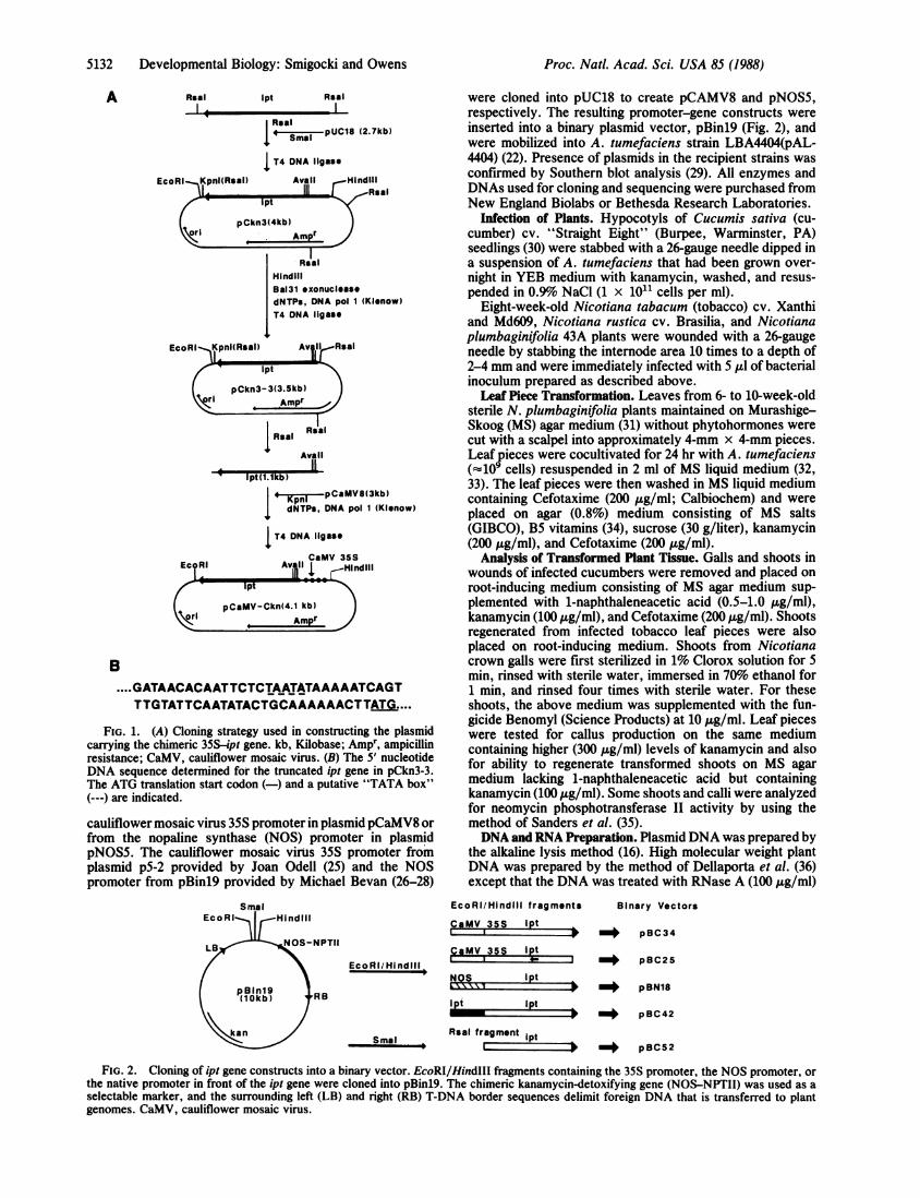
Construction of Binary Vectors. The source of the ipt genewas a 7.3-kilobase EcoRI fragment from the T-DNA region ofpTiB6S3 (23) cloned into pBR325 by Dean Cress (Rohm andHaas). From that fragment, a 1.3-kilobase ipt-containingfragment was cloned into pUC18 (Fig. LA). The promoterregion was removed with BAL-31 exonuclease, and the 5'region of the truncated gene was sequenced (Fig. 1B; ref. 24).A 1.1-kilobase Rsa I fragment from pCkn3-3 (Fig. 1A) wasthen inserted in both orientations downstream from either the
Abbreviations: Ti, tumor inducing; T-DNA, transferred DNA; NOS,nopaline synthase; Kanr, kanamycin resistant.*To whom reprint requests should be addressed.
5131
The publication costs of this article were defrayed in part by page chargepayment. This article must therefore be hereby marked "advertisement"in accordance with 18 U.S.C. §1734 solely to indicate this fact.
5132 Developmental Biology: Smigocki and Owens
A Raal Ipt Rsal
Real
I SR I pUC18 (2.7kb)
I T4 DNA Ilgase
(Klenow)
EcoRl Kpnl(Rsal) Av lI Real
Ipt
pCkn3- 3(3.5kb)
I , Ampi Ri
AvT I
' pt(1.lkb)
| Kpnl -pCaMV8(3kb)I dNTPs, DNA pol 1 (Klenow)
IT4 DNA ligase
CaMV 35SEc RI 11A Hindlil
Ipt
pCaMV-Ckn(4.1 kb)Ampr
B....GATAACACAATTCTCTAATATAAAAATCAGT
TTGTATTCAATATACTGCAAAAAACTTATG....
FiG. 1. (A) Cloning strategy used in constructing the plasmidcarrying the chimeric 35S-ipt gene. kb, Kilobase; Ampr, ampicillinresistance; CaMV, cauliflower mosaic virus. (B) The 5' nucleotideDNA sequence determined for the truncated ipt gene in pCkn3-3.The ATG translation start codon (-) and a putative "TATA box"(---) are indicated.
cauliflower mosaic virus 35S promoter in plasmid pCaMV8 orfrom the nopaline synthase (NOS) promoter in plasmidpNOS5. The cauliflower mosaic virus 35S promoter fromplasmid p5-2 provided by Joan Odell (25) and the NOSpromoter from pBinl9 provided by Michael Bevan (26-28)
were cloned into pUC18 to create pCAMV8 and pNOS5,respectively. The resulting promoter-gene constructs wereinserted into a binary plasmid vector, pBinl9 (Fig. 2), andwere mobilized into A. tumefaciens strain LBA4404(pAL-4404) (22). Presence of plasmids in the recipient strains wasconfirmed by Southern blot analysis (29). All enzymes andDNAs used for cloning and sequencing were purchased fromNew England Biolabs or Bethesda Research Laboratories.
Infection of Plants. Hypocotyls of Cucumis sativa (cu-cumber) cv. "Straight Eight" (Burpee, Warminster, PA)seedlings (30) were stabbed with a 26-gauge needle dipped ina suspension of A. tumefaciens that had been grown over-night in YEB medium with kanamycin, washed, and resus-pended in 0.9o NaCl (1 x 10¹¹ cells per ml).
Eight-week-old Nicotiana tabacum (tobacco) cv. Xanthiand Md609, Nicotiana rustica cv. Brasilia, and Nicotianaplumbaginifolia 43A plants were wounded with a 26-gaugeneedle by stabbing the internode area 10 times to a depth of2-4 mm and were immediately infected with 5 1.d of bacterialinoculum prepared as described above.Leaf Piece Transformation. Leaves from 6- to 10-week-old
sterile N. plumbaginifolia plants maintained on Murashige-Skoog (MS) agar medium (31) without phytohormones werecut with a scalpel into approximately 4-mm x 4-mm pieces.Leaf pieces were cocultivated for 24 hr with A. tumefaciens(Q109 cells) resuspended in 2 ml of MS liquid medium (32,33). The leaf pieces were then washed in MS liquid mediumcontaining Cefotaxime (200 Ag/ml; Calbiochem) and wereplaced on agar (0.8%) medium consisting of MS salts(GIBCO), B5 vitamins (34), sucrose (30 g/liter), kanamycin(200 gg/ml), and Cefotaxime (200,ug/ml).
Analysis of Transformed Plant Tissue. Galls and shoots inwounds of infected cucumbers were removed and placed onroot-inducing medium consisting of MS agar medium sup-plemented with 1-naphthaleneacetic acid (0.5-1.0 ,ug/ml),kanamycin (100 tug/ml), and Cefotaxime (200 t&g/ml). Shootsregenerated from infected tobacco leaf pieces were alsoplaced on root-inducing medium. Shoots from Nicotianacrown galls were first sterilized in 1% Clorox solution for 5min, rinsed with sterile water, immersed in 70% ethanol for1 min, and rinsed four times with sterile water. For theseshoots, the above medium was supplemented with the fun-gicide Benomyl (Science Products) at 10 ,ug/ml. Leaf pieceswere tested for callus production on the same mediumcontaining higher (300 ,ug/ml) levels of kanamycin and alsofor ability to regenerate transformed shoots on MS agarmedium lacking 1-naphthaleneacetic acid but containingkanamycin (100 Ag/ml). Some shoots and calli were analyzedfor neomycin phosphotransferase II activity by using themethod of Sanders et al. (35).DNA and RNA Preparation. Plasmid DNA was prepared by
the alkaline lysis method (16). High molecular weight plantDNA was prepared by the method of Dellaporta et al. (36)except that the DNA was treated with RNase A (100 ttg/ml)
SmalEcoRI HIndlIl
LB NOS-NPTII
pBlnl9(10kb) RB
kin
EcoRI/Hindlil fragments
CaMV 35S lpt
CaMV 35S lptEcoR I/Hi nd ill
Ipt
Ipt
Smal
lPt
Rsal fragment Ipt
+_ pBC34
3 -_ pBC25
$ _+ pBN18
> -> pBC42
> =+ pBC52
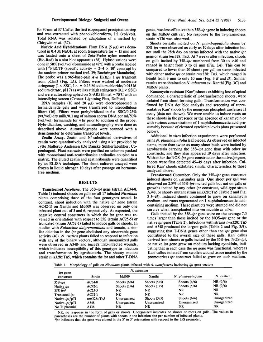
FIG. 2. Cloning of ipt gene constructs into a binary vector. EcoRI/HindIII fragments containing the 35S promoter, the NOS promoter, or
the native promoter in front of the ipt gene were cloned into pBinl9. The chimeric kanamycin-detoxifying gene (NOS-NPTII) was used as a
selectable marker, and the surrounding left (LB) and right (RB) T-DNA border sequences delimit foreign DNA that is transferred to plantgenomes. CaMV, cauliflower mosaic virus.
Binary Vectors
Proc. Natl. Acad. Sci. USA 85 (1988)
Proc. Natl. Acad. Sci. USA 85 (1988) 5133
for 30 min at 37°C after the first isopropanol precipitation stepand was extracted with phenol/chloroform, 1:1 (vol/vol).Total RNA was isolated by adaptation of a method byChirgwin et al. (37).
Nucleic Acid Hybridizations. Plant DNA (5 jig) was dena-tured in 0.4 M NaOH at room temperature for 15 min and
was loaded onto a sheet of Zeta-Probe nylon membrane(Bio-Rad) in a slot blot apparatus (38). Hybridizations were
done in 50% (vol/vol) formamide at 42°C with a probe labeledwith [32P]dCTP (specific activity = 2-4 x 10' cpm/,ug) bythe random primer method (ref. 39; Boehringer Mannheim).The probe was a 965-base-pair Ava II/Kpn I ipt fragmentfrom pCkn3 (Fig. LA). Filters were washed at moderatestringency (1 x SSC, 1 x = 0.15 M sodium chloride/0.015 Msodium citrate, pH 7) as well as at high stringency (0.1 x SSC)and were autoradiographed on X-AR5 film at - 70°C with an
intensifying screen (Cronex Lightning Plus, DuPont).RNA samples (10 and 20 ,g) were electrophoresed in
formaldehyde gels and were transferred to nitrocellulosefilters (16). Filters were prehybridized in 6 x SSC/0.25%(wt/vol) dry milk/0.1 mg of salmon sperm DNA per ml/50%(vol/vol) formamide for 4 hr prior to addition of the probe.Hybridization, washing, and autoradiography were done as
described above. Autoradiographs were scanned with a
densitometer to determine transcript levels.Zeatin Assay. Zeatin and N9-substituted derivatives of
zeatin were quantitatively analyzed using a kit provided byJytte Mollerup Andersen (De Danske Sukkerfabrikker, Co-penhagen). Plant extracts were purified on columns packedwith monoclonal anti-zeatinriboside antibodies coupled to amatrix. The eluted zeatin and zeatinriboside were quantifiedby an ELISA technique. The shoot cultures assayed werefrozen in liquid nitrogen 10 days after passage on hormone-free medium.
RESULTS
Transformed Nicotiana. The 35S-ipt gene (strain AC34-8,Table 1) induced shoots on galls on all 17 infected Nicotianaplants comprising three of the four genotypes tested. Incontrast, shoot induction with the native ipt gene (strainAC42-1) on Xanthi and Md609 was observed on only oneinfected plant out of 5 and 6, respectively. As expected, thenegative control constructs in which the ipt gene was re-versed in orientation with respect to 35S (strain AC25-5) ortruncated (strain AC52-1) failed to induce galls or shoots. Instudies with Kalanchoe daigremontiana and tomato, a sim-ilar deletion in the ipt gene abolished any observable geneactivity (40). N. rustica plants failed to respond to infectionwith any of the binary vectors, although unorganized gallswere observed in A348- and tms328::Tn5-infected wounds,which indicates susceptibility of this genotype to infectionand transformation by agrobacteria. The shooty mutantstrain tms328::Tn5, which contains the ipt and other T-DNA
genes, was less effective than 35S-ipt gene in inducing shootson the Md609 cultivar. No response to the Ti-plasmidlessstrain A136 was observed.
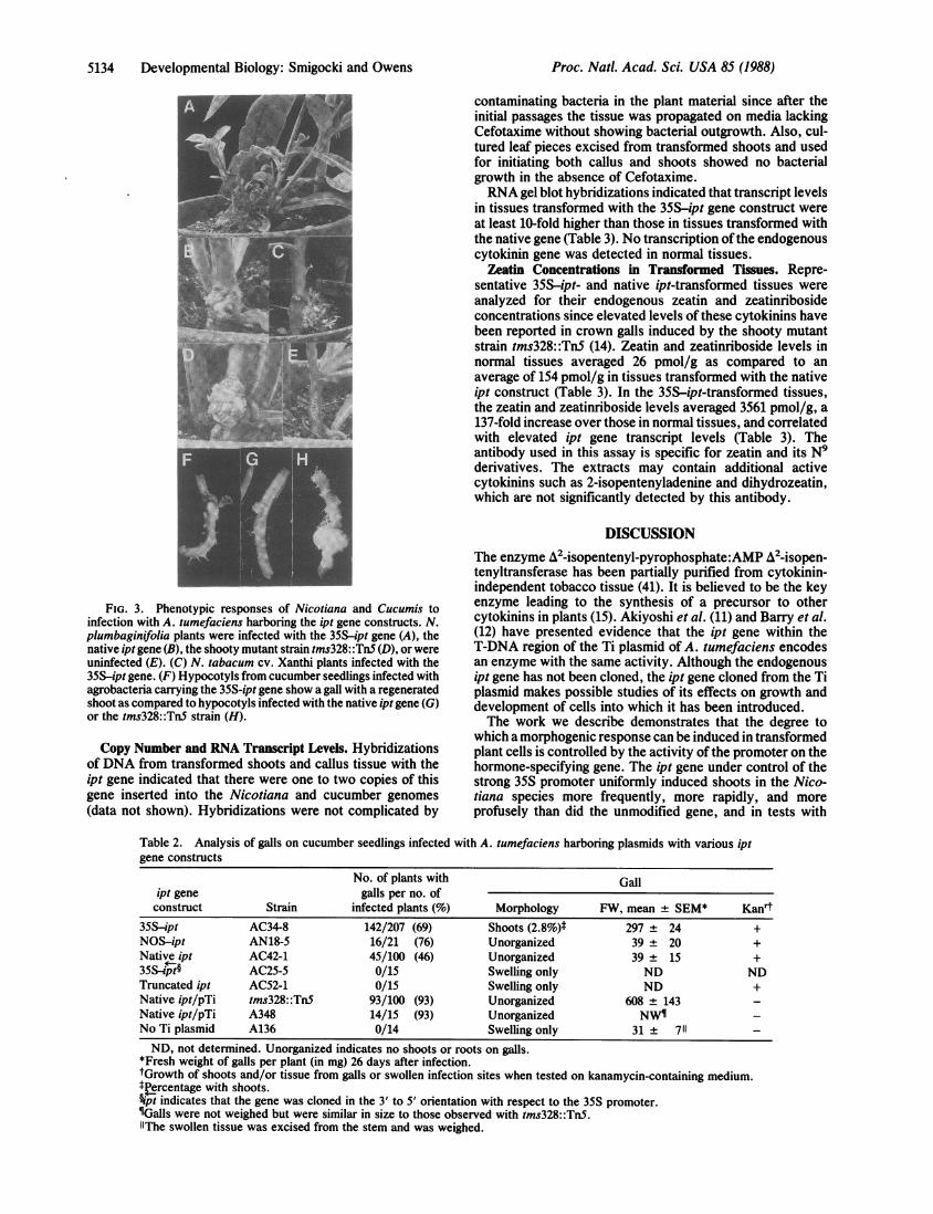
Shoots on galls incited on N. plumbaginifolia stems by35S-ipt were observed as early as 19 days after infection butnot until the 28th day on stems infected with the native iptgene or strain tms328::TnS. At 7 weeks after infection, shootson galls incited by 35S-ipt numbered from 30 to >40 andranged in height from 3 to 62 mm (Fig. 3A). This can becompared to fewer than 20 shoots per gall on stems infectedwith either native ipt or strain tms328::TnS, which ranged inheight from 3 mm to only 10 mm (Fig. 3 B and D). Similarresults were obtained on N. tabacum cv. Xanthi (Fig. 3C) andMd609 plants.
Kanamycin-resistant (Kanr) shoots exhibiting loss ofapicaldominance, a characteristic of ipt-transformed shoots, were
isolated from shoot-forming galls. Transformation was con-
firmed by DNA dot blot analysis and screening of repre-sentative Kanr shoots by the neomycin phosphotransferase IIassay (data not shown). We were unable to induce roots onthese shoots in the presence or the absence of kanamycin orwith various concentrations of 1-naphthaleneacetic acid pre-
sumably because of elevated cytokinin levels (data presentedbelow).
Additional in vitro infection experiments were performedusing N. plumbaginifolia leaf pieces. As observed on infectedstems, more than twice as many shoot buds were incited byagrobacteria carrying the 35S-ipt gene than with other iptconstructs, and they also appeared 19 days after infection.With either the NOS-ipt gene construct or the native ipt gene,shoots were first detected 45-49 days after infection. Cul-tured Kanr shoots exhibited similar characteristics to thoseanalyzed above.Transformed Cucumber. Only the 35S-ipt gene construct
induced shoots on %icumber galls. One shoot per gall wasobserved on 2.8% of 35S-ipt-incited galls but not on galls orgrowths incited by any other ipt construct, wild-type strainA348, or shooty mutant strain tms328::TnS (Table 2 and Fig.3 F-H). Induced shoots continued to grow on kanamycinmedium, and roots regenerated on 1-naphthaleneacetic acid-containing medium. These plantlets were stunted and did notsurvive when transplanted into vermiculite in vitro.
Galls incited by the 35S-ipt gene were on the average 7.5times larger than those incited by the NOS-ipt gene or thenative ipt gene (Table 2). Infections with strains tms328: :TnSand A348 produced the largest galls (Table 2 and Fig. 3H),suggesting that T-DNA genes other than the ipt gene alsocontributed to the overall size of these galls. Kanr callusderived from shoots or galls incited by the 35S-ipt, NOS-ipt,or native ipt gene grew on medium lacking cytokinin, indi-cating that in each case the ipt gene was functional, whereasKanr callus isolated from swollen wound tissue incited by thepromoterless ipt construct failed to grow on such medium.
Table 1. Morphology of galls on Nicotiana plants infected with A. tumefaciens harboring ipt gene vectors
N. tabacumip! geneconstruct Strain Md609 Xanthi N. plumbaginifolia N. rustica
35S-ipt AC34-8 Shoots (6/6) Shoots (5/5) Shoots (6/6) NR (0/6)Native ipt AC42-1 Shoots (1/6) Shoots (1/5) Shoots (5/6) NR (0/6)35S- pt* AC25-5 NR NR NR NRTruncated ipt AC52-1 NR NR NR NRNative ipt/pTi tms328::Tn5 Unorganized Shoots (3/5) Shoots (6/6) UnorganizedNative ipt/pTi A348 Unorganized Unorganized Unorganized UnorganizedNo Ti plasmid A136 NR NR NR NR
NR, no response in the form of galls or shoots. Unorganized indicates no shoots or roots on galls. The values inparentheses are the number of plants with shoots in the infection site per number of infected plants.*Pipt indicates that the gene was cloned in the 3' to 5' orientation with respect to the 35S promoter.
Developmental Biology: Smigocki and Owens
5134 Developmental Biology: Smigocki and Owens
FIG. 3. Phenotypic responses of Nicotiana and Cucumis toinfection with A. tumefaciens harboring the ipt gene constructs. N.plumbaginifolia plants were infected with the 35S-ipt gene (A), thenative ipt gene (B), the shooty mutant strain tms328::Tn5 (D), or wereuninfected (E). (C) N. tabacum cv. Xanthi plants infected with the35S-ipt gene. (F) Hypocotyls from cucumber seedlings infected withagrobacteria carrying the 35S-ipt gene show a gall with a regeneratedshoot as compared to hypocotyls infected with the native ipt gene (G)or the tms328::TnS strain (H).
Copy Number and RNA Transcript Levels. Hybridizationsof DNA from transformed shoots and callus tissue with theipt gene indicated that there were one to two copies of thisgene inserted into the Nicotiana and cucumber genomes(data not shown). Hybridizations were not complicated by
contaminating bacteria in the plant material since after theinitial passages the tissue was propagated on media lackingCefotaxime without showing bacterial outgrowth. Also, cul-tured leaf pieces excised from transformed shoots and usedfor initiating both callus and shoots showed no bacterialgrowth in the absence of Cefotaxime.RNA gel blot hybridizations indicated that transcript levels
in tissues transformed with the 35S-ipt gene construct wereat least 10-fold higher than those in tissues transformed withthe native gene (Table 3). No transcription ofthe endogenouscytokinin gene was detected in normal tissues.
Zeatin Concentrations in Transformed Tissues. Repre-sentative 35S-ipt- and native ipt-transformed tissues wereanalyzed for their endogenous zeatin and zeatinribosideconcentrations since elevated levels of these cytokinins havebeen reported in crown galls induced by the shooty mutantstrain tms328::TnS (14). Zeatin and zeatinriboside levels innormal tissues averaged 26 pmol/g as compared to anaverage of 154 pmol/g in tissues transformed with the nativeipt construct (Table 3). In the 35S-ipt-transformed tissues,the zeatin and zeatinriboside levels averaged 3561 pmol/g, a137-fold increase over those in normal tissues, and correlatedwith elevated ipt gene transcript levels (Table 3). Theantibody used in this assay is specific for zeatin and its N9derivatives. The extracts may contain additional activecytokinins such as 2-isopentenyladenine and dihydrozeatin,which are not significantly detected by this antibody.
DISCUSSIONThe enzyme A2-isopentenyl-pyrophosphate:AMP A2-isopen-tenyltransferase has been partially purified from cytokinin-independent tobacco tissue (41). It is believed to be the keyenzyme leading to the synthesis of a precursor to othercytokinins in plants (15). Akiyoshi et al. (11) and Barry et al.(12) have presented evidence that the ipt gene within theT-DNA region of the Ti plasmid of A. tumefaciens encodesan enzyme with the same activity. Although the endogenousipt gene has not been cloned, the ipt gene cloned from the Tiplasmid makes possible studies of its effects on growth anddevelopment of cells into which it has been introduced.The work we describe demonstrates that the degree to
which a morphogenic response can be induced in transformedplant cells is controlled by the activity of the promoter on thehormone-specifying gene. The ipt gene under control of thestrong 35S promoter uniformly induced shoots in the Nico-tiana species more frequently, more rapidly, and moreprofusely than did the unmodified gene, and in tests with
Table 2. Analysis of galls on cucumber seedlings infected with A. tumefaciens harboring plasmids with various iptgene constructs
No. of plants with Gallipt gene galls per no. ofconstruct Strain infected plants (%) Morphology FW, mean ± SEM* Kanrt
35S-ipt AC34-8 142/207 (69) Shoots (2.8%)* 297 ± 24 +NOS-ipt AN18-5 16/21 (76) Unorganized 39 ± 20 +Native ipt AC42-1 45/100 (46) Unorganized 39 ± 15 +35S4pt§ AC25-5 0/15 Swelling only ND NDTruncated ipt AC52-1 0/15 Swelling only ND +Native ipt/pTi tms328::Tn5 93/100 (93) Unorganized 608 ± 143Native ipt/pTi A348 14/15 (93) Unorganized NW-No Ti plasmid A136 0/14 Swelling only 31 + 711ND, not determined. Unorganized indicates no shoots or roots on galls.
*Fresh weight of galls per plant (in mg) 26 days after infection.tGrowth of shoots and/or tissue from galls or swollen infection sites when tested on kanamycin-containing medium.trercentage with shoots.§ipt indicates that the gene was cloned in the 3' to 5' orientation with respect to the 35S promoter.IGalls were not weighed but were similar in size to those observed with tms328::Tn5.lThe swollen tissue was excised from the stem and was weighed.
Proc. Natl. Acad. Sci. USA 85 (1988)

Proc. Natl. Acad. Sci. USA 85 (1988) 5135
Table 3. Zeatin (Z) and zeatinriboside (ZR) concentrations andipt gene transcript levels in transformed shoots and tissues
Z + ZR, TranscriptTissue ipt gene pmol/g levels
N. plurbacinifolia
nonral leaves erogenus 36 (-*
transformed shoots native Ti 226t 1)
transformed shoots 35S-ipt 2416 (14)
N. tabacum cv xanthi
normal leaves endogenous 15 nat
transformed shoots 35S-jpt 4009§ (35)
N. tabacum cv MD609
normal leaves erdogenous 23 na
transformed shoots 35S-ipt 2478 (10)
Cucumber hypocotyls
normal hypocotyls erdogemous 31 (-)
transformed callus native Ti 83 na
transformed callus 35S-ipt 5340 na
*Relative levels as determined on a densitometer. , Not detected.tThis value represents an average of three samples.4Not analyzed.§The actual determined value of 8018 pmol/g was normalized to asingle gene copy of the ipt gene per genome.
cucumber, a species not of the Solonaceae family, it not onlyinduced larger galls than the other ipt gene constructs, but,more interestingly, it uniquely induced shoots. This enhance-ment in shoot induction was correlated with highly elevatedcytokinin levels in the transformed shoots and tissues. It iswell established that the formation of shoots and roots fromsome plant tissue cultures can be regulated by the ratio ofcytokinin to auxin provided in the medium (1) or provided invitro by tumor genes (2, 14) and that an elevated cytokinin-to-auxin ratio promotes shoot formation. In our experiments,the high endogenous levels of zeatin and zeatinribosidecaused by transformation with the strongly promoted ipt geneconstruct appears to have sufficiently increased the cytoki-nin-to-auxin ratio to induce shoot formation. This in vivomanipulation ofendogenous cytokinin levels eliminates prob-lems associated with uptake and transport of exogenouslyapplied hormones and permits characterization of the effectsof altered cytokinin concentrations on plant development.Our work demonstrates that the overproduction of cytokininwithin plant cells enhances the cells' ability to undergo shootorganogenesis. The mechanism by which this occurs remainsto be determined.
Note. While this paper was under review, further analyses of normalleaves ofN. tabacum cv. Md609 and shoots transformed by the 35S-ipt gene disclosed no significant differences in auxin concentrations.This supports the hypothesis that elevated cytokinin levels intransformed tissues sufficiently increased the cytokinin-to-auxinratio to induce shoot formation.
We thank Debra Eberts and George Pittarelli for excellent tech-nical assistance with the ELISA assays and in growing the tobaccoplants, respectively, and Jytte Mollerup Andersen (De DanskeSukkerfabrikker) for providing the zeatin assay kit.
1. Skoog, F. & Miller, C. 0. (1957) Symp. Soc. Exp. Biol. 11, 118-130.2. Amasino, R. M. & Miller, C. 0. (1982) Plant Physiol. 69, 389-392.
3. Klee, H. J., Horsch, R. B., Hinchee, M. A., Hein, M. B. &Hoffmann, N. L. (1987) Genes Dev. 1, 86-96.
4. Ream, L. W. & Gordon, M. P. (1982) Science 218, 854-859.5. Caplan, A., Herrera-Estrella, L., Inze, D., Van Haute, E., Van
Montagu, M., Schell, J. & Zambryski, P. (1983) Science 222, 815-821.
6. Chilton, M.-D., Drummond, M. H., Merlo, D. J., Sciaky, D.,Montoya, A. L., Gordon, M. P. & Nester, E. W. (1977) Cell 11,263-271.
7. Chilton, M.-D., Saiki, R. K., Yadav, N., Gordon, M. P. & Quetier,R. (1980) Proc. Natl. Acad. Sci. USA 77, 4060-4064.
8. Willmitzer, L., De Beuckeleer, M., Lemmers, M., Van Montagu,M. & Schell, J. (1980) Nature (London) 287, 359-361.
9. Schroder, G., Waffenschmidt, S., Weiler, E. W. & Schroder, J.(1984) Eur. J. Biochem. 138, 387-391.
10. Van Onckelen, H., Prinzen, E., Inze, D., Rudelsheim, P., VanLijsebettens, M., Follin, A., Schell, J., Van Montagu, M. & DeGreef, J. (1986) FEBS Lett. 198, 357-360.
11. Akiyoshi, D. E., Klee, H., Amasino, R. M., Nester, E. W. &Gordon, M. P. (1984) Proc. Natl. Acad. Sci. USA 81, 5994-5998.
12. Barry, G. F., Rogers, S. G., Fraley, R. T. & Brand, L. (1984) Proc.Natl. Acad. Sci. USA 81, 4776-4780.
13. Garfinkel, D. J., Simpson, R. B., Ream, L. W., White, F. F.,Gordon, M. P. & Nester, E. W. (1981) Cell 27, 143-153.
14. Akiyoshi, D. E., Morris, R. O., Hinz, R., Mischke, B. S., Kosuge,T., Garfinkel, D. J., Gordon, M. P. & Nester, E. W. (1983) Proc.Natl. Acad. Sci. USA 80, 407-411.
15. Letham, D. S. & Palni, L. M. S. (1983) Annu. Rev. Plant Physiol.34, 163-197.
16. Maniatis, T., Fritsch, E. F. & Sambrook, J. (1982) MolecularCloning: A Laboratory Manual (Cold Spring Harbor Lab., ColdSpring Harbor, NY), pp. 90-91.
17. Watson, B., Currier, T. C., Gordon, M. P., Chilton, M.-D. &Nester, E. W. (1975) J. Bacteriol. 123, 255-264.
18. Hoekema, A., Hirsch, P. R., Hooykaas, P. J. J. & Schilperoort,R. A. (1983) Nature (London) 303, 179-180.
19. Ooms, G., Hooykaas, P. J. J., Van Veen, R. J. M., Beelen, P. V.,Regensburg-Tuink, T. J. G. & Schilperoort, R. A. (1982) Plasmid 7,15-29.
20. Vieira, J. & Messing, J. (1982) Gene 19, 259-268.21. Messing, J. (1983) Methods Enzymol. 101, 27-78.22. Ditta, G., Stanfield, S., Corbin, D. & Helinski, D. R. (1980) Proc.
Natl. Acad. Sci. USA 77, 7347-7351.23. Van Larebeke, N., Engler, G., Holsters, M., Van den Elsecher, S.,
Zaenen, I., Schilperoort, R. A. & Schell, J. (1974) Nature (London)252, 169-170.
24. Sanger, F., Nicklen, S. & Coulson, A. R. (1977) Proc. Natl. Acad.Sci. USA 74, 5463-5467.
25. Odell, J. T., Nagy, F. & Chua, N.-H. (1985) Nature (London) 313,810-812.
26. Shaw, C. H., Carter, G. H., Watson, M. D. & Shaw, C. H. (1984)Nucleic Acids Res. 12, 7831-7846.
27. Bevan, M., Barnes, W. M. & Chilton, M.-D. (1983) Nucleic AcidsRes. 11, 369-385.
28. Bevan, M. (1984) Nucleic Acids Res. 12, 8711-8721.29. Southern, E. M. (1975) J. Mol. Biol. 98, 503-517.30. Matsumoto, S., Machida, Y. & Takebe, I. (1986) Plant Mol. Biol.
Rep. 4, 42-47.31. Murashige, T. & Skoog, F. (1962) Physiol. Plant. 15, 473-497.32. An, G., Watson, B. D., Stachel, S., Gordon, M. P. & Nester, E. W.
(1985) EMBO J. 4, 277-284.33. Horsch, R. B., Fry, J. E., Hoffman, N. L., Eichholtz, D., Rogers,
S. G. & Fraley, R. T. (1985) Science 227, 1229-1231.34. Gamborg, 0. (1970) Plant Physiol. 45, 372-375.35. Sanders, P. R., Winter, J. A., Barnason, A. R., Rogers, S. G. &
Fraley, R. T. (1987) Nucleic Acids Res. 15, 1543-1558.36. Dellaporta, S. L., Wood, J. & Hicks, J. B. (1984) in Molecular
Biology of Plants: A Laboratory Course Manual, eds. Malmberg,R., Messing, J. & Sussex, I. (Cold Spring Harbor Lab., Cold SpringHarbor, NY), pp. 36-37.
37. Chirgwin, J. M., Przybyla, A. E., MacDonald, R. J. & Rutter,W. J. (1979) Biochemistry 18, 5294-5299.
38. Reed, K. C. & Mann, D. A. (1985) Nucleic Acids Res. 13, 7207-7220.
39. Feinberg, A. P. & Vogelstein, B. (1983) Anal. Biochem. 132, 6-13.40. dePater, B. S., Klinkhamer, M. P., Amesz, P. A., deKam, R. J.,
Memelink, J., Hoge, J. H. C. & Schilperoort, R. A. (1987) NucleicAcids Res. 15, 8267-8282.
41. Chen, C. M. & Melitz, D. K. (1979) FEBS Lett. 107, 15-20.
Developmental Biology: Smigocki and Owens