cytogenetics - university of · pdf fileideogram of human chromosomes. 6 human karyotype. 7...
TRANSCRIPT

1
Cytogenetics

2
Chromosomal Disorders
• 50% of 1st trimester miscarriages
• 5% of stillbirths
• 0.5% of liveborns– Down syndrome—trisomy 21
– Fragile X syndrome
• Somatic cell abnormalities in cancers
History
• Bateson (1916) “It is inconceivable thatparticles of chromatin….can posses thepowers which must be assigned to ourfactors(genes).”
• (~1955) Human cells were thought to have48 chromosomes

3
Cytogenetic Technology
• Peripheral blood lymphocyte culture– Phytohemagglutinin– Hypotonic swelling
• Banding---Giemsa– 350 – 550 bands/N (haploid set)– 850 in prometaphase– G-bands (dark): AT-rich, fewer transcribed genes,
LINES– R-bands (light): GC-rich, more transcribed genes,
SINES (Alu)
Metaphase spread

4
Prometaphase spread
Banding nomenclature
5
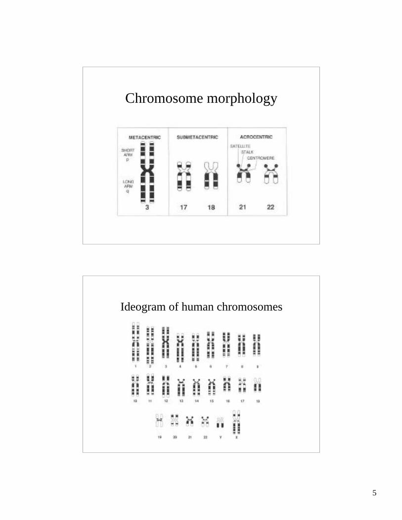
Chromosome morphology
Ideogram of human chromosomes
6
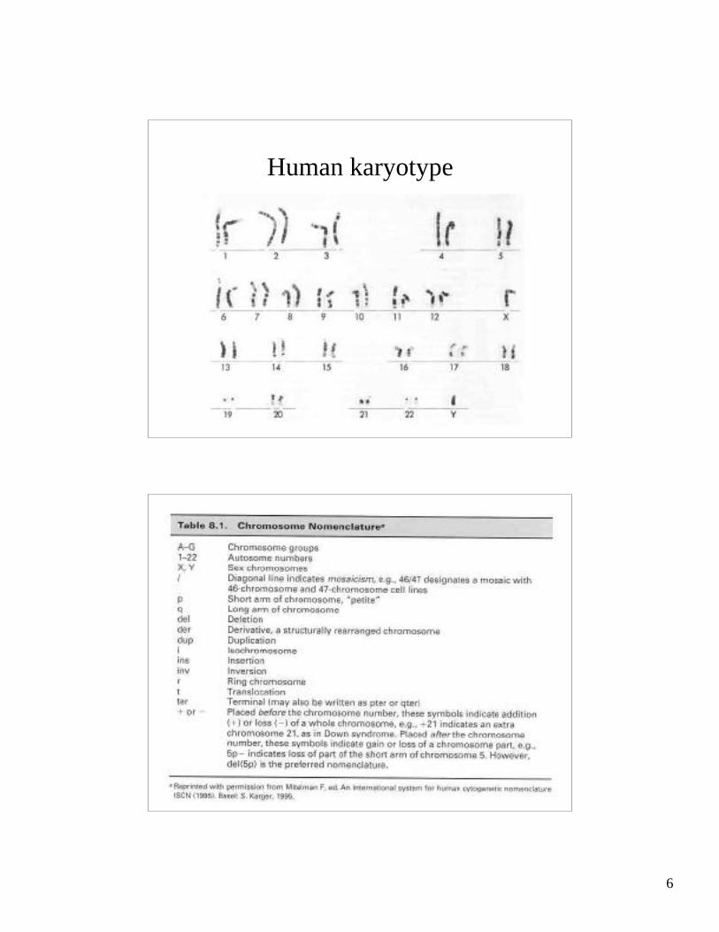
Human karyotype

7
Fluorescence in situ hybridizationFISH
Locus-specific probes
Ch 15 centromere (green)Ch 15 PWS critical region (red)

8
Centromeric probes
Trisomy 9(leukemia)
Centromeric probes(Ch 13 red, Ch18 pink, Ch 21 green, X yellow, Y white)

9
Centromeric probes(Ch 8 red, Y yellow)
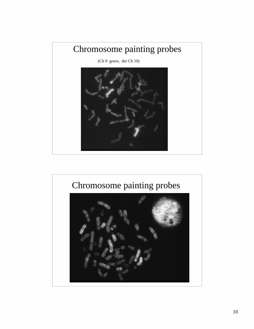
Chromosome painting probes
10
Chromosome painting probes(Ch 9 green, der Ch 10)
Chromosome painting probes
11
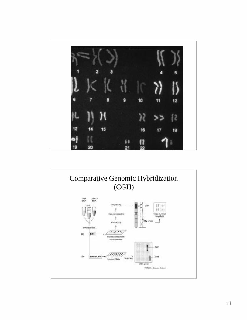
Comparative Genomic Hybridization(CGH)
12
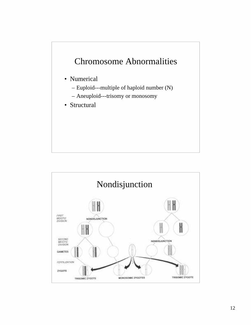
Chromosome Abnormalities
• Numerical– Euploid---multiple of haploid number (N)
– Aneuploid---trisomy or monosomy
• Structural
Nondisjunction

13
Meiotic Nondisjunction
• Usually maternal (maternal age effect)
• Usually MI (meiosis I)– Starts at 20 weeks fetal
– Arrests for 10 to 45 years
– Finishes MI at ovulation
– Meiosis II at fertilization
Meiotic nondisjunction
14
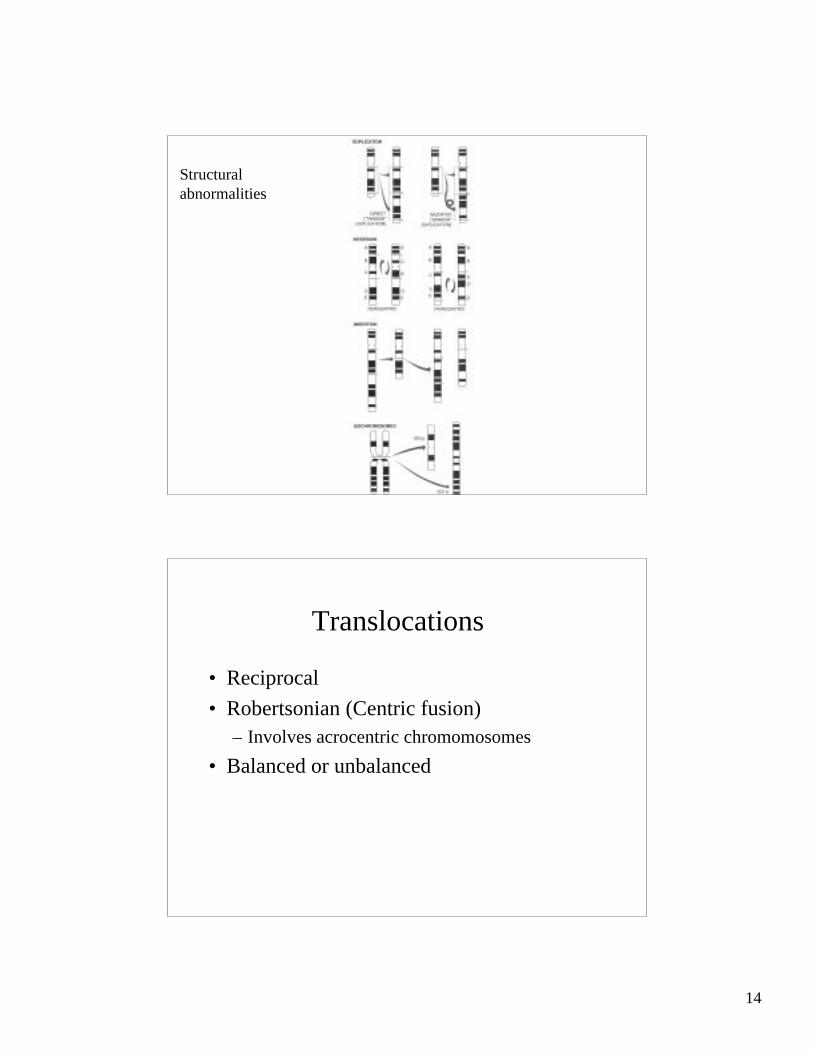
Structuralabnormalities
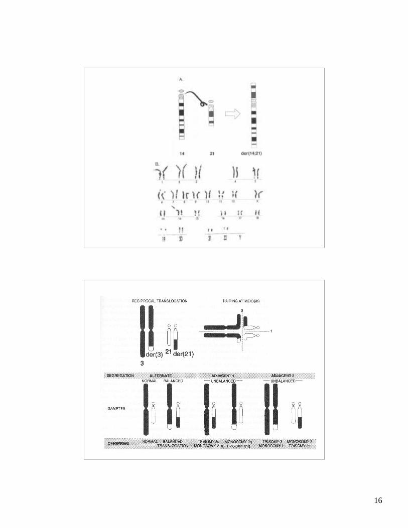
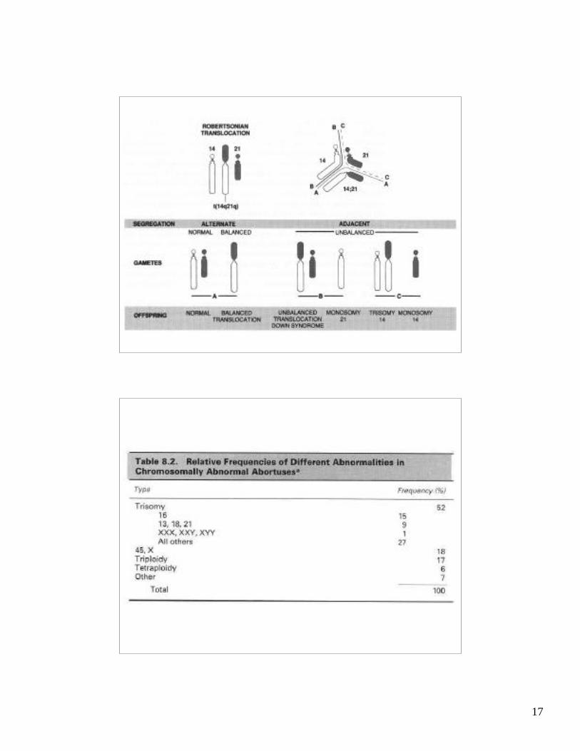
Translocations
• Reciprocal
• Robertsonian (Centric fusion)– Involves acrocentric chromomosomes
• Balanced or unbalanced
15
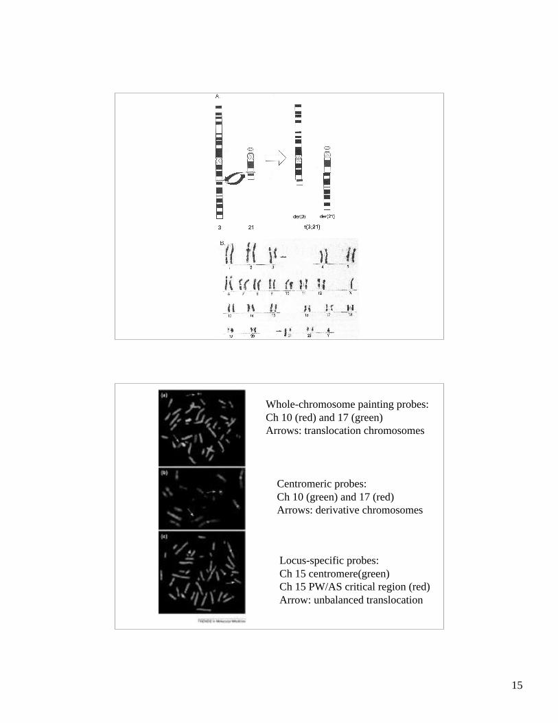
Whole-chromosome painting probes:Ch 10 (red) and 17 (green)Arrows: translocation chromosomes
Centromeric probes: Ch 10 (green) and 17 (red)Arrows: derivative chromosomes
Locus-specific probes: Ch 15 centromere(green)Ch 15 PW/AS critical region (red)Arrow: unbalanced translocation
16
17
18
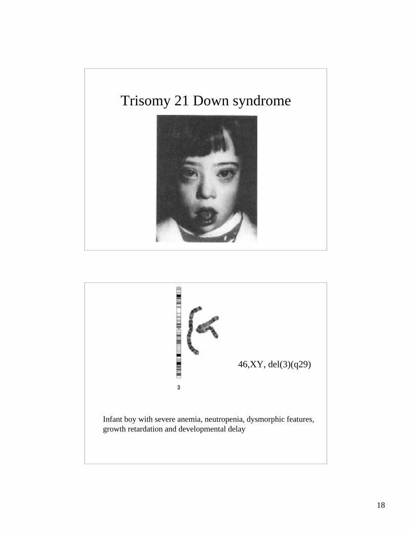
Trisomy 21 Down syndrome
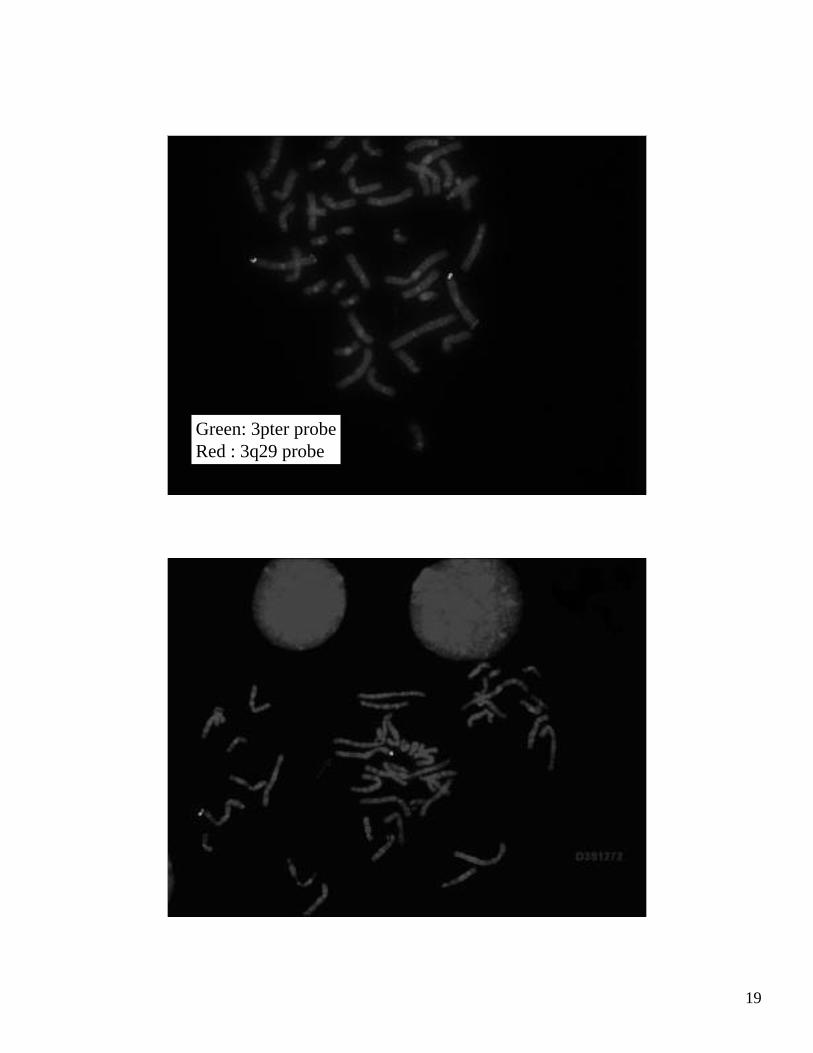
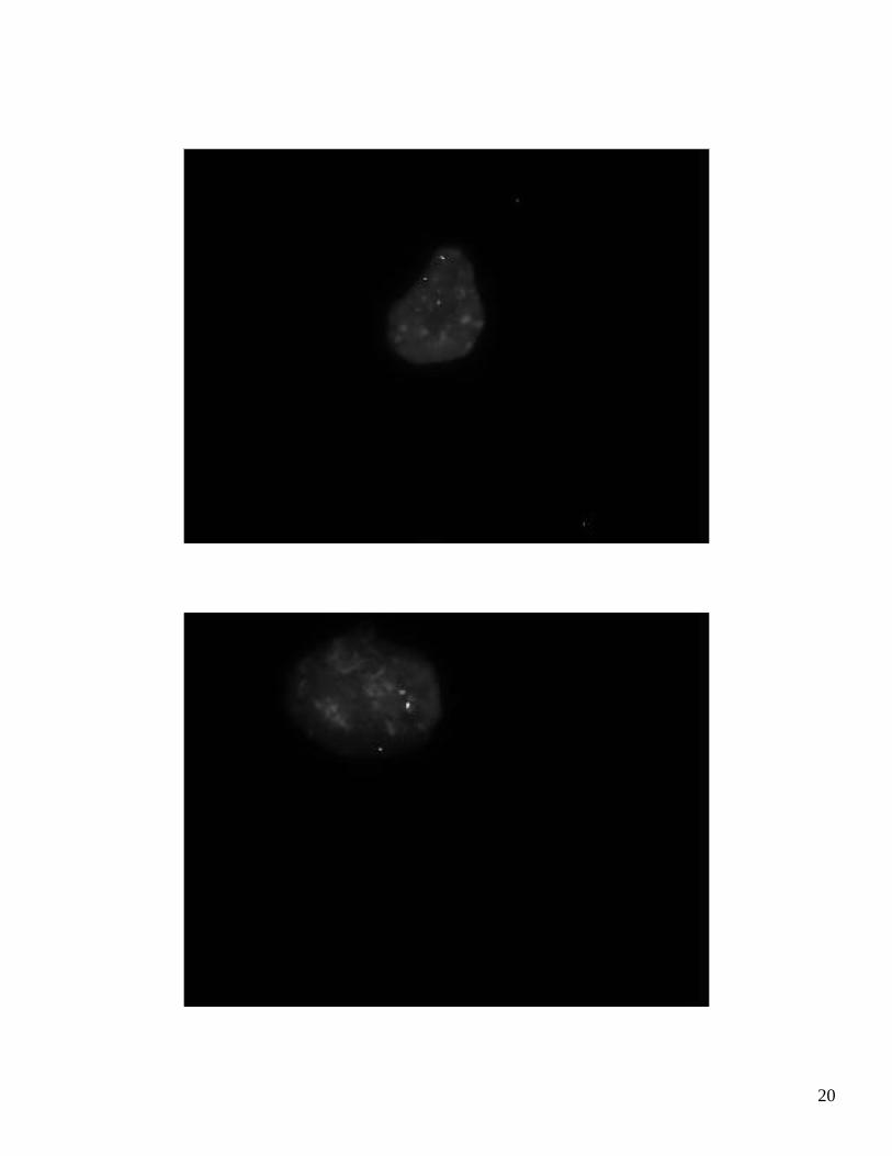
Infant boy with severe anemia, neutropenia, dysmorphic features,growth retardation and developmental delay
46,XY, del(3)(q29)
19
Green: 3pter probeRed : 3q29 probe
20
21
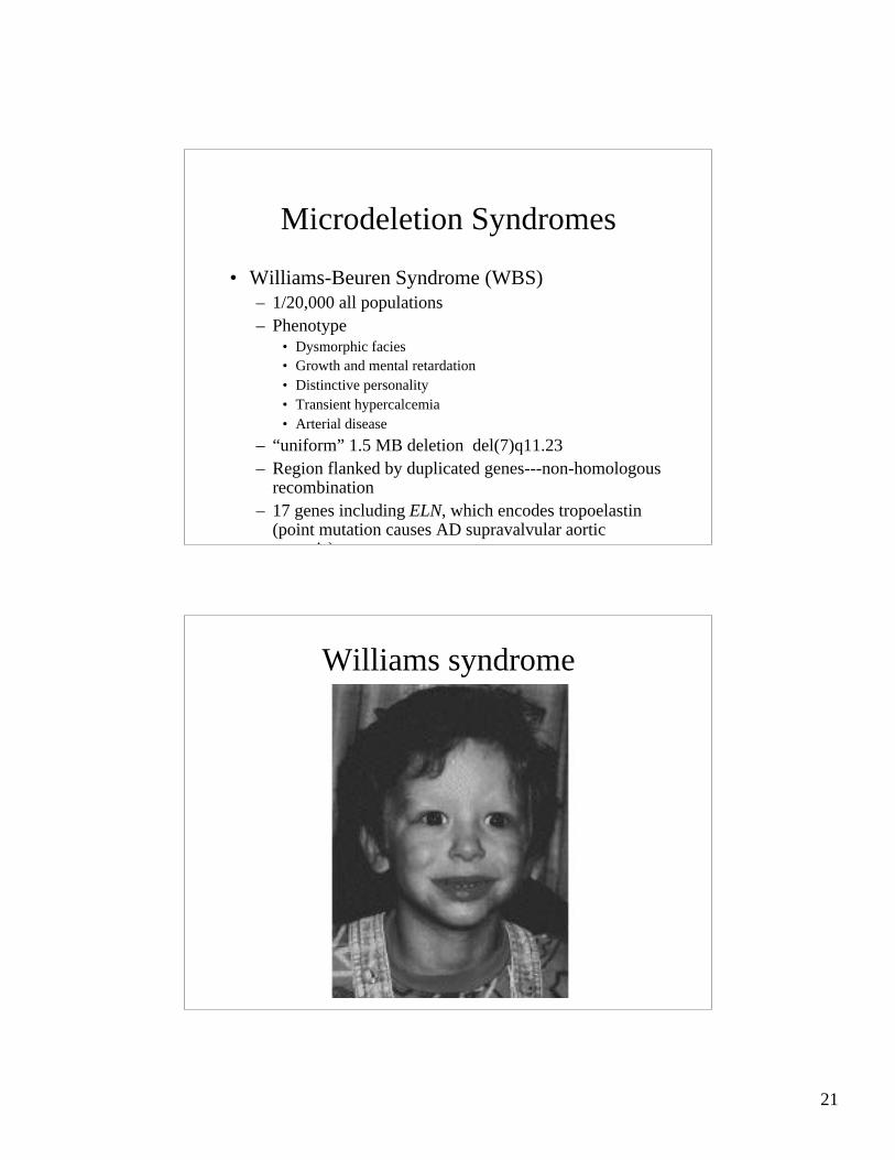
Microdeletion Syndromes
• Williams-Beuren Syndrome (WBS)– 1/20,000 all populations– Phenotype
• Dysmorphic facies• Growth and mental retardation• Distinctive personality• Transient hypercalcemia• Arterial disease
– “uniform” 1.5 MB deletion del(7)q11.23– Region flanked by duplicated genes---non-homologous
recombination– 17 genes including ELN, which encodes tropoelastin
(point mutation causes AD supravalvular aorticstenosis)
Williams syndrome

22
FISH DiagnosisDel(7)q11.23
23
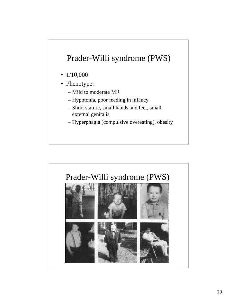
Prader-Willi syndrome (PWS)
• 1/10,000
• Phenotype:– Mild to moderate MR
– Hypotonia, poor feeding in infancy
– Short stature, small hands and feet, smallexternal genitalia
– Hyperphagia (compulsive overeating), obesity
Prader-Willi syndrome (PWS)
24
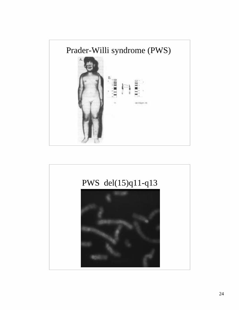
Prader-Willi syndrome (PWS)
PWS del(15)q11-q13

25
Prader-Willi syndrome (PWS)
• 1/10,000• Phenotype:
– Mild to moderate MR– Hypotonia, poor feeding in infancy– Short stature, small hands and feet, small external
genitalia– Hyperphagia (compulsive overeating), obesity
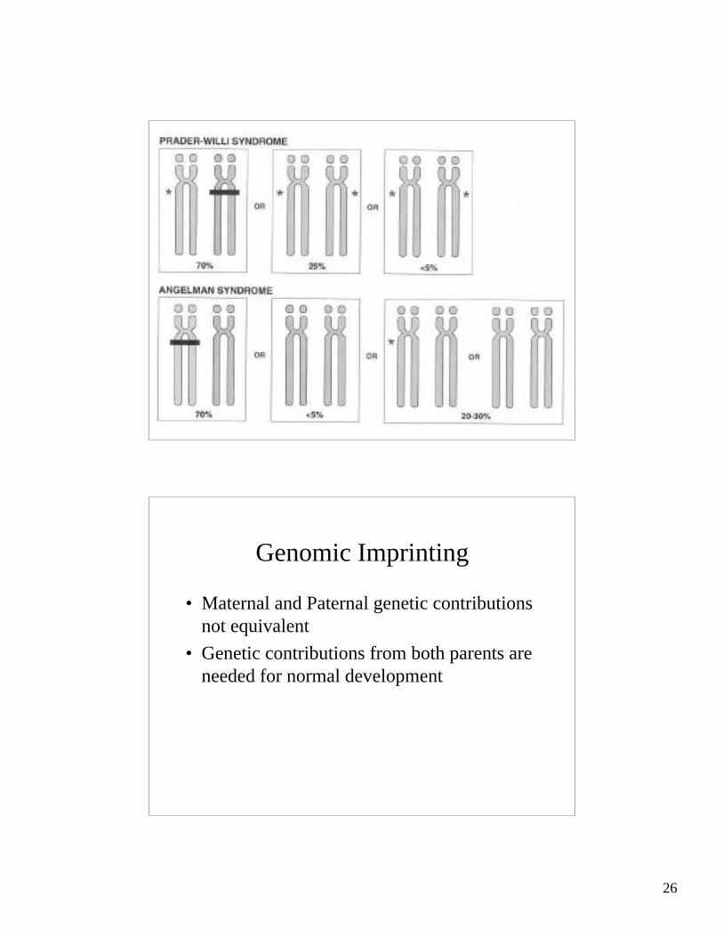
• Del(15)q11-13…..Paternal• Uniparental Disomy
Angelman syndrome
• Severe MR, absence of speech
• Jerky movements
• Inappropriate laughter
• Large jaw
• Del(15)q11-13----but Maternal
26
Genomic Imprinting
• Maternal and Paternal genetic contributionsnot equivalent
• Genetic contributions from both parents areneeded for normal development

27
Evidence for Imprinting
• Mouse Embryos– Gynogenetic---poorly developed extra-
embryonic membranes– Androgenetic—abnormal embryonic structures
• Human tumors– Hydatidiform moles—placental tumors with
two paternal haploid sets of chromosomes– Ovarian teratomas---benign differentiated
tumors with two maternal haploid sets
Mechanism of Imprinting
• Some genes are preferentially inactivated(imprinted) during gametogenesis in male andfemale parents
• Differential DNA methylation/histone acetylation
• Deletion of the active allele----functionalnullisomy
• Uniparental disomy for the inactiveallele—functional nullisomy

28
PWS/AS region