cv update newsletter v12n3 2014 - mc5234-1014
TRANSCRIPT
INSIDE THIS ISSUE
Leadless Pacing Available for Selected Patients
Implantable Loop Re-corders in Patients With Cryptogenic Stroke
Arm Ergometer Provides Alternative to Conven-tional Stress Testing
346
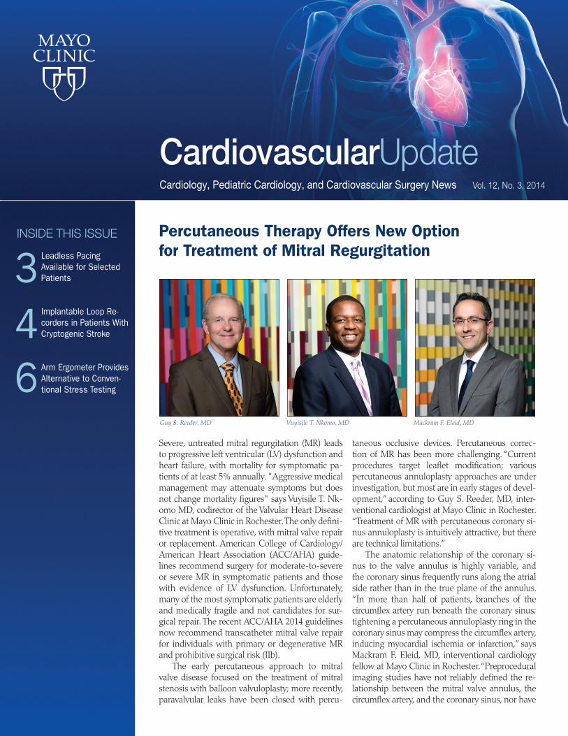
CardiovascularUpdateCardiology, Pediatric Cardiology, and Cardiovascular Surgery News Vol. 12, No. 3, 2014
Severe, untreated mitral regurgitation (MR) leads to progressive left ventricular (LV) dysfunction and heart failure, with mortality for symptomatic pa-tients of at least 5% annually. "Aggressive medical management may attenuate symptoms but does not change mortality figures" says Vuyisile T. Nk-omo MD, codirector of the Valvular Heart Disease Clinic at Mayo Clinic in Rochester. The only defini-tive treatment is operative, with mitral valve repair or replacement. American College of Cardiology/American Heart Association (ACC/AHA) guide-lines recommend surgery for moderate-to-severe or severe MR in symptomatic patients and those with evidence of LV dysfunction. Unfortunately, many of the most symptomatic patients are elderly and medically fragile and not candidates for sur-gical repair. The recent ACC/AHA 2014 guidelines now recommend transcatheter mitral valve repair for individuals with primary or degenerative MR and prohibitive surgical risk (IIb).
The early percutaneous approach to mitral valve disease focused on the treatment of mitral stenosis with balloon valvuloplasty; more recently, paravalvular leaks have been closed with percu-
Guy S. Reeder, MD
Percutaneous Therapy Offers New Option for Treatment of Mitral Regurgitation
taneous occlusive devices. Percutaneous correc-tion of MR has been more challenging. “Current procedures target leaflet modification; various percutaneous annuloplasty approaches are under investigation, but most are in early stages of devel-opment,” according to Guy S. Reeder, MD, inter-ventional cardiologist at Mayo Clinic in Rochester. “Treatment of MR with percutaneous coronary si-nus annuloplasty is intuitively attractive, but there are technical limitations.”
The anatomic relationship of the coronary si-nus to the valve annulus is highly variable, and the coronary sinus frequently runs along the atrial side rather than in the true plane of the annulus. “In more than half of patients, branches of the circumflex artery run beneath the coronary sinus; tightening a percutaneous annuloplasty ring in the coronary sinus may compress the circumflex artery, inducing myocardial ischemia or infarction,” says Mackram F. Eleid, MD, interventional cardiology fellow at Mayo Clinic in Rochester. “Preprocedural imaging studies have not reliably defined the re-lationship between the mitral valve annulus, the circumflex artery, and the coronary sinus, nor have
Vuyisile T. Nkomo, MD Mackram F. Eleid, MD
2 MAYO CLINIC | CardiovascularUpdate
they predicted clinical outcomes. Also, it is not known how coronary sinus annuloplasty devices affect the use of coronary sinus leads for biven-tricular pacing.”
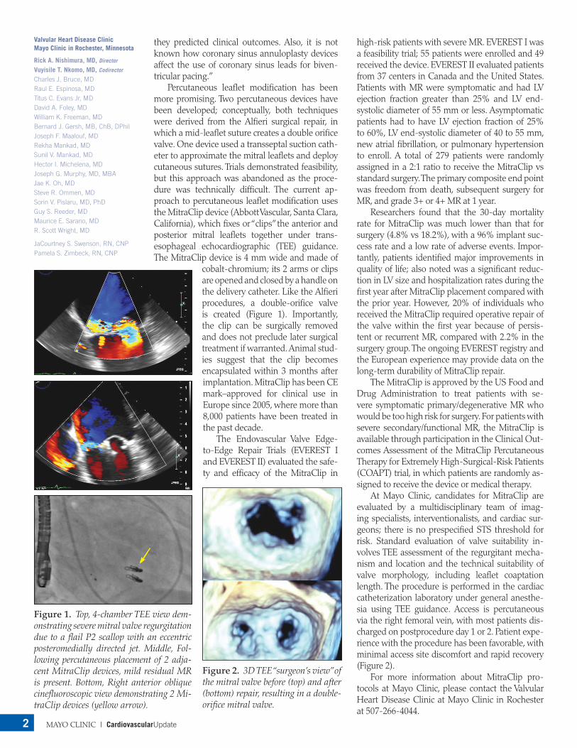
Percutaneous leaflet modification has been more promising. Two percutaneous devices have been developed; conceptually, both techniques were derived from the Alfieri surgical repair, in which a mid-leaflet suture creates a double orifice valve. One device used a transseptal suction cath-eter to approximate the mitral leaflets and deploy cutaneous sutures. Trials demonstrated feasibility, but this approach was abandoned as the proce-dure was technically difficult. The current ap-proach to percutaneous leaflet modification uses the MitraClip device (Abbott Vascular, Santa Clara, California), which fixes or “clips” the anterior and posterior mitral leaflets together under trans-esophageal echocardiographic (TEE) guidance. The MitraClip device is 4 mm wide and made of
cobalt-chromium; its 2 arms or clips are opened and closed by a handle on the delivery catheter. Like the Alfieri procedures, a double-orifice valve is created (Figure 1). Importantly, the clip can be surgically removed and does not preclude later surgical treatment if warranted. Animal stud-ies suggest that the clip becomes encapsulated within 3 months after implantation. MitraClip has been CE mark–approved for clinical use in Europe since 2005, where more than 8,000 patients have been treated in the past decade.
The Endovascular Valve Edge-to-Edge Repair Trials (EVEREST I and EVEREST II) evaluated the safe-ty and efficacy of the MitraClip in
Valvular Heart Disease ClinicMayo Clinic in Rochester, Minnesota
Rick A. Nishimura, MD, DirectorVuyisile T. Nkomo, MD, CodirectorCharles J. Bruce, MDRaul E. Espinosa, MDTitus C. Evans Jr, MDDavid A. Foley, MDWilliam K. Freeman, MDBernard J. Gersh, MB, ChB, DPhilJoseph F. Maalouf, MDRekha Mankad, MDSunil V. Mankad, MDHector I. Michelena, MDJoseph G. Murphy, MD, MBAJae K. Oh, MDSteve R. Ommen, MDSorin V. Pislaru, MD, PhDGuy S. Reeder, MDMaurice E. Sarano, MDR. Scott Wright, MD
JaCourtney S. Swenson, RN, CNPPamela S. Zimbeck, RN, CNP
high-risk patients with severe MR. EVEREST I was a feasibility trial; 55 patients were enrolled and 49 received the device. EVEREST II evaluated patients from 37 centers in Canada and the United States. Patients with MR were symptomatic and had LV ejection fraction greater than 25% and LV end-systolic diameter of 55 mm or less. Asymptomatic patients had to have LV ejection fraction of 25% to 60%, LV end-systolic diameter of 40 to 55 mm, new atrial fibrillation, or pulmonary hypertension to enroll. A total of 279 patients were randomly assigned in a 2:1 ratio to receive the MitraClip vs standard surgery. The primary composite end point was freedom from death, subsequent surgery for MR, and grade 3+ or 4+ MR at 1 year.
Researchers found that the 30-day mortality rate for MitraClip was much lower than that for surgery (4.8% vs 18.2%), with a 96% implant suc-cess rate and a low rate of adverse events. Impor-tantly, patients identified major improvements in quality of life; also noted was a significant reduc-tion in LV size and hospitalization rates during the first year after MitraClip placement compared with the prior year. However, 20% of individuals who received the MitraClip required operative repair of the valve within the first year because of persis-tent or recurrent MR, compared with 2.2% in the surgery group. The ongoing EVEREST registry and the European experience may provide data on the long-term durability of MitraClip repair.
The MitraClip is approved by the US Food and Drug Administration to treat patients with se-vere symptomatic primary/degenerative MR who would be too high risk for surgery. For patients with severe secondary/functional MR, the MitraClip is available through participation in the Clinical Out-comes Assessment of the MitraClip Percutaneous Therapy for Extremely High-Surgical-Risk Patients (COAPT) trial, in which patients are randomly as-signed to receive the device or medical therapy.
At Mayo Clinic, candidates for MitraClip are evaluated by a multidisciplinary team of imag-ing specialists, interventionalists, and cardiac sur-geons; there is no prespecified STS threshold for risk. Standard evaluation of valve suitability in-volves TEE assessment of the regurgitant mecha-nism and location and the technical suitability of valve morphology, including leaflet coaptation length. The procedure is performed in the cardiac catheterization laboratory under general anesthe-sia using TEE guidance. Access is percutaneous via the right femoral vein, with most patients dis-charged on postprocedure day 1 or 2. Patient expe-rience with the procedure has been favorable, with minimal access site discomfort and rapid recovery (Figure 2).
For more information about MitraClip pro-tocols at Mayo Clinic, please contact the Valvular Heart Disease Clinic at Mayo Clinic in Rochester at 507-266-4044.
Figure 2. 3D TEE “surgeon’s view” of the mitral valve before (top) and after (bottom) repair, resulting in a double-orifice mitral valve.
Figure 1. Top, 4-chamber TEE view dem-onstrating severe mitral valve regurgitation due to a flail P2 scallop with an eccentric posteromedially directed jet. Middle, Fol-lowing percutaneous placement of 2 adja-cent MitraClip devices, mild residual MR is present. Bottom, Right anterior oblique cinefluoroscopic view demonstrating 2 Mi-traClip devices (yellow arrow).
MAYO CLINIC | CardiovascularUpdate 3
Leadless Pacing Available for Selected Patients
Early pacing devices offered single-chamber, fixed-rate ventricular pacing for life-threatening conduc-tion system disease. Advances in generator and lead technology and the results of clinical trials over the past 60 years have expanded the indications for device therapy. As a result, more individuals are receiving device therapy; approximately 190,000 pacemakers are implanted every year in the United States. Over time, the patient population receiving pacing therapy has become older and more com-plex. The “weak link” in device therapy has been the leads. While transvenous leads typically have low-er thresholds and better longevity than epicardial leads, they are associated with increased morbid-ity and mortality. Complications at implant include bleeding, vascular damage, cardiac perforation, pneumothorax, and dislodgment. Potential long-term concerns include lead fracture, malfunction, venous obstruction, tricuspid valve regurgitation, and the risks associated with lead extraction. Trans-venous leads are contraindicated in the presence of right-to-left shunt and in some patients with con-genital heart disease. The concept of leadless pacing was first pro-posed in 1970 by Spickler and colleagues, but it is
Yong-Mei Cha, MD, Paul A. Friedman, MD
Heart Rhythm ServicesMayo Clinic in Rochester, Minnesota
Douglas L. Packer, MD, DirectorMichael J. Ackerman, MD, PhD‡Samuel J. Asirvatham, MDBarry A. Boilson, MDDavid J. Bradley, MD, PhDPeter A. Brady, MB, ChB, MDBryan C. Cannon, MD‡Yong-Mei Cha, MDFreddy Del Carpio Munoz, MD*Raul E. Espinosa, MDPaul A. Friedman, MDBernard J. Gersh, MB, ChB, DPhilDavid L. Hayes, MDSuraj Kapa, MDMargaret A. Lloyd, MD, MBAChristopher J. McLeod, MB, ChB, PhDSiva K. Mulpuru, MDThomas M. Munger, MDPeter A. Noseworthy, MDMichael J. Osborn, MDRobert F. Rea, MDAndre Terzic, MD, PhDPhilip L. Wackel, MD‡
Donna M. Kania-Lachance, RN, CNPCharissa L. Koski, RN, CNPJill J. Nagel, PA-CJill M. Olmscheid, RN, CNP
*Mayo Clinic Health System ‡Pediatric Cardiology
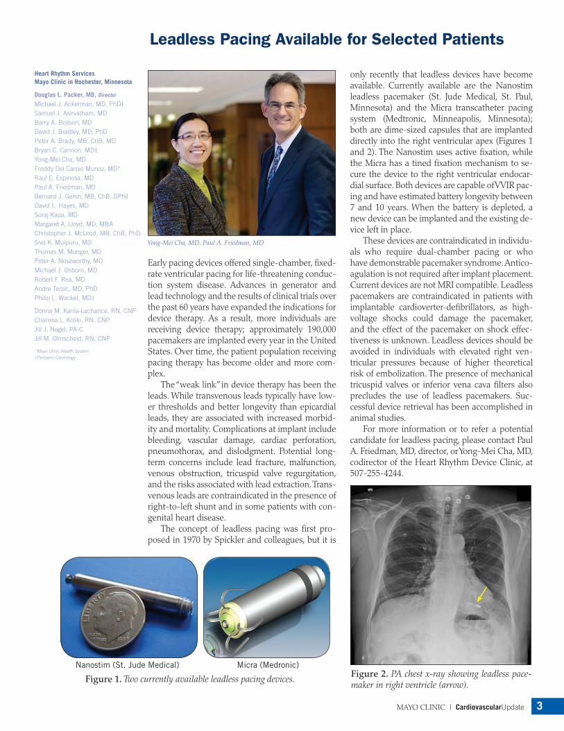
only recently that leadless devices have become available. Currently available are the Nanostim leadless pacemaker (St. Jude Medical, St. Paul, Minnesota) and the Micra transcatheter pacing system (Medtronic, Minneapolis, Minnesota); both are dime-sized capsules that are implanted directly into the right ventricular apex (Figures 1 and 2). The Nanostim uses active fixation, while the Micra has a tined fixation mechanism to se-cure the device to the right ventricular endocar-dial surface. Both devices are capable of VVIR pac-ing and have estimated battery longevity between 7 and 10 years. When the battery is depleted, a new device can be implanted and the existing de-vice left in place. These devices are contraindicated in individu-als who require dual-chamber pacing or who have demonstrable pacemaker syndrome. Antico-agulation is not required after implant placement. Current devices are not MRI compatible. Leadless pacemakers are contraindicated in patients with implantable cardioverter-defibrillators, as high-voltage shocks could damage the pacemaker, and the effect of the pacemaker on shock effec-tiveness is unknown. Leadless devices should be avoided in individuals with elevated right ven-tricular pressures because of higher theoretical risk of embolization. The presence of mechanical tricuspid valves or inferior vena cava filters also precludes the use of leadless pacemakers. Suc-cessful device retrieval has been accomplished in animal studies. For more information or to refer a potential candidate for leadless pacing, please contact Paul A. Friedman, MD, director, or Yong-Mei Cha, MD, codirector of the Heart Rhythm Device Clinic, at 507-255-4244.
Figure 2. PA chest x-ray showing leadless pace-maker in right ventricle (arrow).
Figure 1. Two currently available leadless pacing devices.
Nanostim (St. Jude Medical) Micra (Medronic)

4 MAYO CLINIC | CardiovascularUpdate
Implantable Loop Recorders in Patients With Cryptogenic Stroke
Stroke is a major cause of disability and death. Many strokes have a likely ex-planation, such as carotid disease, poorly controlled hypertension, diabetes, hy-perlipidemia, smoking, in-herited blood clotting condi-tions, and atrial fibrillation (AF). However, a quarter of patients have none of the defined risk factors and yet face the consequences of stroke or transient ischemic attack (TIA; stroke with res-olution of symptoms within 24 hours). These cryptogenic strokes (no specific risk fac-tor or cause found) create
anxiety as it is unclear what can be done to pre-vent additional strokes. Cryptogenic strokes have been shown to have a higher rate of recurrence than other strokes. AF causes approximately one-sixth of all strokes, and anticoagulation substantially lowers the risk of stroke in patients with AF. AF can be intermittent, short lived, and asymptomatic, mak-ing it challenging to identify the arrhythmia with conventional electrocardiography, ambulatory monitoring, and a 30-day event recorder. How-ever, if a patient is detected to have AF and has a CHA2DS2-VASc score higher than 2, then there is compelling evidence that anticoagulation, with either warfarin or novel anticoagulants, reduces the stroke risk by approximately 65% (Table). One study conducted in patients with implant-ed pacemakers documented that stroke risk was significantly increased in those individuals with AF lasting more than 6 minutes (N Engl J Med 2012;366:120-129). Asymptomatic AF as a risk factor for stroke is frequently considered but difficult to prove. “An implanted device is vastly superior to surface monitoring in detecting AF owing to continu-ous analysis over years, generally better signal-to-noise ratio, and lack of compliance issues frequently encountered with the use of external recorders,” says Komandoor Srivathsan, MD, di-rector of Heart Rhythm Services at Mayo Clinic in Arizona. “Even among implantable devices, intra-cardiac electrogram acquisition through a pace-maker or an implantable cardiac defibrillator has more sophisticated rhythm recognition capabili-ties because of the spatial clarity of the obtained
Komandoor Srivathsan, MD
Heart Rhythm ServicesMayo Clinic in Arizona
Komandoor Srivathsan, MD, DirectorJohn F. Beshai, MDDan Sorajja, MDLuis R. Scott, MDWin-Kuang Shen, MD
Susan E. Hendrick, PA-C
electrogram when compared with the subcutane-ous recording of an implanted loop recorder.” Many cryptogenic strokes are thought to be related to unrecognized AF. The use of implant-able cardiac monitoring (ICM) to identify as-ymptomatic AF and associated stroke risk was explored in a recently published study (N Engl J Med 2014;370:2478-2486). This study indicated that an additional 10% of patients with AF can be detected at 12 months of ICM compared with other monitoring techniques, and more wide-spread use of ICM in individuals with cryptogenic stroke might allow for earlier identification of AF and anticoagulation treatment. This hypothesis is tempered by the following issues:
1. Ten patients have to have ICM to detect 1 additional patient with AF. Assuming thera-peutic anticoagulation is of equal benefit to symptomatic AF patients at the time of stroke, approximately 100 patients will have to be implanted with a device to prevent 1 re-current stroke (currently cost-prohibitive).
2. Infection, extraction, and reimplantation add to morbidity and cost.
3. Device detection issues and the occurrence of asymptomatic 30-second episodes of AF re-main a concern.
4. The same issue of the journal reported that a 30-day event recorder was able to detect AF in a significant number of patients and mini-mized the need for ICM.
5. Competing risk factors are frequent in the age group of patients who experience stroke, and the etiology of a specific stroke ascribable to a cause is nebulous in many instances.
Table. CHA2DS2-VASc Scoring System Congestive heart failure 1 point
Hypertension 1 point
Age, y
>75 2 points
65-75 1 point
Diabetes 1 point
Stroke 2 points
Female sex 1 point
Vascular disease 1 point
Potential total 9 points
MAYO CLINIC | CardiovascularUpdate 5
6. The majority of cryptogenic strokes remain cryptogenic even after these investigations.Nevertheless, this study adds important in-
formation that some cryptogenic stroke patients likely have AF, and AF should be high on the list of differential diagnoses in these individuals. The prevention of recurrent strokes in these patients is feasible through enhanced detection provided by ICM, but the subset of patients who would ben-efit from intensive monitoring needs to be better defined. Additional studies will need to confirm that anticoagulation is of equal benefit to those with cryptogenic stroke due to asymptomatic AF prior to its widespread adoption. The ICM size has recently been greatly reduced to the size of a large capsule, and implantation techniques have become simpler. Implantation of these smaller devices may eventually become an in-office, out-patient procedure, which would change the cost-benefit equation.
Summary Points • Implantable cardiac monitoring with a
subcutanous loop recorder can detect
asymptomatic, subclinical atrial fibrillation
in 10% of patients with cryptogenic stroke
at 1 year.
• The rate of detection of asymptomatic
atrial fibrillation in cryptogenic stroke
through implantable cardiac monitoring
is low.
• The number of patients who need to
receive implants to detect atrial fibrillation
in cryptogenic stroke and prevent
recurrence is currently too high to be cost-
effective.
Amir Lerman, MD, received the Outstanding Investigator Award; recipients have pri-mary research appointments, and the award is based on scientific accomplishments, publications, and grant sup-port.
RECOGNITION
The Department of Medicine at Mayo Clinic in Rochester has announced 2014 award recipients. All are members of the Division of Cardiovascular Diseases.
Joerg Herrmann, MD, re-ceived the New Investigator Award, given to young fac-ulty whose work has led to important new insights in the field of biomedical science.
George M. Gura, MD, re-ceived the Laureate Award, presented for professional excellence and personal in-tegrity.
Samuel J. Asirvatham, MD, received the Teaching Ex-cellence Award for teaching noteworthy among peers.
Carole A. Warnes, MD, re-ceived the Lifetime Achieve-ment Award for Outstanding Contributions to Medical Edu-cation. This honor is given for a career-long commitment to medical education that has resulted in substantial and enduring impact at national and international levels.
NEW STAFF
David L. Joyce, MD, has joined the Division of Cardiovas-cular Surgery at Mayo Clinic in Rochester. Dr Joyce gradu-ated from the US Air Force Academy and Harvard Medical School. He did his general surgery fellowship at Johns Hopkins University and his cardiovascular surgery fellow-ship at Stanford University. His areas of interest include heart and lung transplantation, mechanical circulatory sup-port, and minimally invasive coronary surgery.
6 MAYO CLINIC | CardiovascularUpdate
Arm Ergometer Provides Alternative to Conventional Stress Testing
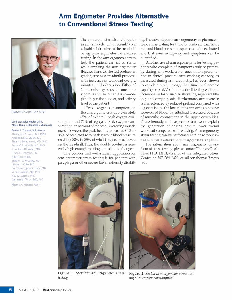
The arm ergometer (also referred to as an “arm cycle” or “arm crank”) is a valuable alternative to the treadmill or leg cycle ergometer for exercise testing. In the arm ergometer stress test, the patient can sit or stand while cranking the arm ergometer (Figures 1 and 2). The test protocol is graded, just as a treadmill protocol, with increases in workload every 2 minutes until exhaustion. Either of 2 protocols may be used—one more vigorous and the other less so—de-pending on the age, sex, and activity level of the patient.
Peak oxygen consumption on the arm ergometer is approximately 65% of treadmill peak oxygen con-
sumption and 70% of leg cycle peak oxygen con-sumption on account of the small exercising muscle mass. However, the peak heart rate reaches 90% to 95% of predicted with peak systolic blood pressure reaching 80% to 85% of what is typically achieved on the treadmill. Thus, the double product is gen-erally high enough to bring out ischemic changes.
One obvious and well-studied application for arm ergometer stress testing is for patients with paraplegia or other severe lower extremity disabil-
Thomas G. Allison, PhD, MPH
ity. The advantages of arm ergometry vs pharmaco-logic stress testing for these patients are that heart rate and blood pressure responses can be evaluated and that exercise capacity and symptoms can be quantified.
Another use of arm ergometry is for testing pa-tients who complain of symptoms only or primar-ily during arm work, a not uncommon presenta-tion in clinical practice. Arm working capacity, as measured during arm ergometry, has been shown to correlate more strongly than functional aerobic capacity or peak Vo2 from treadmill testing with per-formance on tasks such as shoveling, repetitive lift-ing, and carryingloads. Furthermore, arm exercise is characterized by reduced preload compared with leg exercise, as the lower limbs can act as a passive reservoir of blood, but afterload is elevated because of muscular contractions in the upper extremities. These hemodynamic aspects of arm work explain the generation of angina despite lower overall workload compared with walking. Arm ergometry stress testing can be performed with or without si-multaneous measurement of oxygen consumption.
For information about arm ergometry or any form of stress testing, please contact Thomas G. Al-lison, PhD, MPH, director of the Integrated Stress Center at 507-284-6320 or allison.thomas@mayo .edu.
Figure 1. Standing arm ergometer stress testing.
Figure 2. Seated arm ergometer stress test-ing with oxygen consumption.
Cardiovascular Health ClinicMayo Clinic in Rochester, Minnesota
Randal J. Thomas, MD, DirectorThomas G. Allison, PhD, MPHAnanda Basu, MBBS, MDThomas Behrenbeck, MD, PhDFrank V. Brozovich, MD, PhDJ. Richard Hickman, MDBruce D. Johnson, PhDBirgit Kantor, MDStephen L. Kopecky, MDIftikhar J. Kullo, MDFrancisco Lopez-Jimenez, MDVirend Somers, MD, PhDRay W. Squires, PhDCarmen M. Terzic, MD, PhD
Martha A. Mangan, CNP
MAYO CLINIC | CardiovascularUpdate 7
RECOGNITION
Bernard J. Gersh, MB, ChB, DPhil, David R. Holmes Jr, MD, and Veronique L. Roger, MD, cardiologists at Mayo Clinic in Rochester, have been included on the list of “The World’s Most Influential Scientific Minds,” published by Thomson Reuters.
IN THE NEWS
Mayo Clinic is honored to be ranked as U.S. News & World Report’s top hospital. Mayo Clinic in Minnesota is also recognized as one of the top Cardiology & Heart Surgery hospitals in the nation for 2014-2015 by U.S. News & World Report.
CARD IOVASCULAR SELF -STUDY
https://cardiovascular .education-registration.com /selfstudy
RECOGNITION
Bernard J. Gersh, MB, ChB, DPhil, a cardi-ologist at Mayo Clinic in Rochester, has re-ceived honorary membership in the British Cardiovascular Society. Honorary member-ship recognizes esteemed individuals who have made an outstanding contribution to the practice of cardiology worldwide and who are held in high regard by the British cardiology community.
C O N N E C T W I T H U S
Follow us on Twitter!Receive real-time news and thought-leading insights by following @MayoClinicCV
Check out Mayo Clinic on Medscape Cardiology
Mayo joins forces with Medscape Cardiology to bring you the latest perspective on clinical trials, patient care and news: http://www.medscape.com/partners/mayoclinic
Subscribe to our Cardiovascular CME blogConnect with Mayo Clinic’s heart special-ists to keep up-to-date in cardiovascular medicine: http://cvblog.mayo.edu/
Bookmark our Medical Professional Video CenterWatch Grand Rounds lectures and other presenta-tions and videos describing advances in disease and condition treatment, procedures and surgeries: http://www.mayoclinic.org/medical-professionals /video-center

MC5234-1014
Continuing MediCal eduCation, Mayo CliniCFor additional information:Web: www.mayo.edu/cme/cardiovascular-diseasesEmail: [email protected]: 800-283-6296, 507-266-0677, or 507-266-6703
Mayo Clinic Satellite Educational Symposia at AHA 2014*Hot Topics in Arrhythmia Management: What Practitioner Needs to Know*Trendy Topics in Preventive Cardiology*Genetics in CardiologyNov 15-17, 2014, Chicago, IL
Coronary Artery Disease: Case-Based Learning Nov 21-23, 2014, Las Vegas, NV
Echo on Marco Island: Case-Based Approach Dec 4-7, 2014, Marco Island, FL
3rd Annual Mayo Clinic ECG and Heart Rhythm Course: A Case-Based ApproachDec 4-7, 2014, Phoenix, AZ
The Heart Beat of Cardiology: Practical Application of EchocardiographyDec 11-13, 2014, Chicago, IL
Mayo Clinic Cardiology Update at South Beach: A Focus on PreventionJan 7-10, 2015, Miami Beach, FL
Hawaii Heart 2015: Echocardiography and Multimodality Imaging: Case-Based Clinical Decision Making Jan 26-30, 2015, Wailea, Maui, Hawaii
22nd Annual Arrhythmias and the Heart: A Cardiovascular UpdateFeb 2-6, 2015, Kauai, Hawaii
40th Annual Cardiovascular Conference at SnowbirdFeb 13-16, 2015, Snowbird, UT
20th Annual Cardiology at Cancun: Topics in Clinical Cardiology Feb 23-27, 2015, Cancun, Mexico
22nd Annual Echocardiographic Workshop on 2-D and Doppler Echocardiography at VailMar 9-12, 2015, Vail, CO
Heart Failure Management for Nurse Practitioners, Physician Assistants, and Primary Care Providers Mar 19-21, 2015, Orlando, FL
Mayo Clinic Cardiovascular Reviews in BahrainMar 25-28, 2015, Manama, Bahrain
Echo Fiesta: An In-depth Review of Adult Echo-cardiography for Sonographers and PhysiciansMar 26-29, 2015, San Antonio, TX
4th Annual Innovations in Valve and Structural Heart DiseaseApr 2-4, 2015, Nassau, Bahamas
Mayo Clinic Cardiovascular Update
Medical Editor: Margaret A. Lloyd, MD, MBA
Editorial Board: Charanjit S. Rihal, MD, MBA Win-Kuang Shen, MD Joseph A. Dearani, MDFrank Cetta, MD Nicole B. Engler Marjorie G. Durhman
Managing Editor: Jane C. Wiggs, MLA, ELS
Art Director: Marjorie G. Durhman
Photography: Amanda R. Durhman
Mayo Clinic Cardiovascular Update is written for
physicians and should be relied upon for medical
education purposes only. It does not provide a
complete overview of the topics covered and should
not replace the independent judgment of a physician
about the appropriateness or risks of a procedure for a
given patient.
Case Studies From the Heart of Manhattan: A Mayo Clinic Cardiovascular UpdateApr 16-18, 2015, New York, NY
Imaging in Adult Congenital Heart Disease: Pearls for All Cardiac ProvidersApr 24-26, 2015, Ponte Vedra Beach, FL
Echocardiography Review Course for Boards and CertificationApr 25-28, 2015, Rochester, MN
Echocardiography in the Nation’s Capital: Focus for the Physician and SonographerMay 8-10, 2015, Washington, DC
Basic to Advanced Echocardiography: From the Blue Ridge Mountains of AshevilleMay 13-16, 2015, Asheville, NC
Cardiac Rhythm Device Summit Implantation, Management, and Follow-upJun 26-28, 2015, Chicago, IL
29th Annual Echocardiographic Symposium at Vail: New Technologies, Live Scanning, and Clinical UpdateJul 20-23, 2015, Vail, CO
Success With Failure: Strategies for the Evaluation and Treatment of Heart Failure in Clinical PracticeAug 10-12, 2015, Dana Point, CA
Cardiovascular Board ReviewsAug 22-27, 2015, Rochester, MN
Challenges in Clinical Cardiology: A Case-Based UpdateSep 18-20, 2015, Chicago, IL
Mayo Clinic Interventional Cardiology Board ReviewSep 25-27, 2015, Rochester, MN
Advanced Catheter Ablation: New Tips, Techniques, and Technologies for Complex ArrhythmiaOct 2-6, 2015, Washington, DC
31st Annual Echocardiography in Pediatric and Adult Congenital Heart DiseaseOct 8-11, 2015, Phoenix, AZ
Electrophysiology Review for Boards and RecertificationOct 9-11, 2015, Rochester, MN
Cases in Echocardiography, Cardiac CT and MRIOct 21-24, 2015, Napa, CA
Coronary Artery Disease: Prevention, Detection, and TreatmentNov 20-22, 2015, Las Vegas, NV
5th Annual Echo on Marco Island: Case-Based ApproachDec 3-6, 2015, Marco Island, FL
8th Annual The Heart Beat of Cardiology: Practical Application of EchocardiographyDec 10-12, 2015, Chicago, IL
Contact UsMayo Clinic welcomes inquiries and referrals, and a request to a specific physician is not required to refer a patient.
Phoenix/Scottsdale, Arizona 866-629-6362
Jacksonville, Florida 800-634-1417
Rochester, Minnesota 800-533-1564
Resourcesmayoclinic.org/medicalprofs Clinical trials, CME, Grand Rounds, scientific videos, and online referrals