cutting edge technology nasa smart probes
TRANSCRIPT
NASA SMART ProbesCutting Edge Technology
Application to Breast Cancer Diagnosis
Robert W. Mah, Ph.D., NASAStefanie S. Jeffrey, MD., Stanford Medical Center

NASA SMART Probe: Breast Cancer Application
Presenter: Robert W. Mah, Ph.D.
ABSTRACT
There is evidence in breast cancer and other malignancies that the physiologic environment within a tumorcorrelates with clinical outcome. We are developing a unique percutaneous Smart Probe to be used at the time ofneedle biopsy of the breast. The Smart Probe will simultaneously measure multiple physiologic parameterswithin a breast tumor. Direct and indirect measurements of tissue oxygen levels, blood flow, pH, and tissue fluidpressure will be analyzed in real-time. These parameters will be interpreted individually and collectively byinnovative neural network techniques using advanced intelligent software.
The goals are 1) develop a percutaneous Smart Probe with multiple sensor modalities and applying advancedInformation Technologies to provide real-time diagnostic information of the tissue at tip of the probe, 2) test thepercutaneous Smart Probe in women with benign and malignant breast masses who will be undergoing surgicalbiopsy, 3) correlate probe sensor data with benign and malignant status of breast masses, 4) determine whetherthe probe can detect physiologic differences within a breast tumor, at its margins, and in adjacent normal breasttissue, 5) correlate probe sensor data with known prognostic factors for breast cancer, including tumor size,tumor grade, axillary lymph node metastases, estrogen receptor and progesterone receptor status.
Use of the Smart Probe will be integrated into existing Stanford programs focusing on minimally invasivebreast cancer diagnosis and treatment using 3-D breast ultrasound, breast MRI, percutaneous needle biopsy, andpercutaneous radiofrequency ablation of breast cancer.
The long-term goal of this project is to develop a device (the NASA SMART Probe) for minimally invasivereal-time identification and characterization of biological tissues. There are two basic concepts: (1) multiplemicrosensors measure different properties of the tissue under investigation in real-time; (2) "biologically-inspired"soft computing methodologies (hybrid neural networks and fuzzy logic) provide instantaneous synthesis of thehigh-dimensional data gathered by the multiple microsensors. The resulting unique "signature" for the tissue atthe tip of the probe can then be compared with an increasingly rich databank of information gathered from otherpatients and normal individuals.

23
NASA SMART Probes
Biomedical Research Probe
Geological Probe
Application of Advanced Information TechnologiesTo improve the safety, accuracy, and efficiency of critical procedures
To reduce the skill level to perform the procedure
Medical Diagnosis Probe

3
Breast Cancer SMART Probe
• In vivo sensing of physiologic parameters o• Real-time diagnosis of breast tissue at tip• Enable accurate & efficient treatment• Monitoring effects of treatment

4
Breast Surgeons Collaborating in the Development ofa Breast Cancer SMART Probe
Stefanie S. Jeffrey, M.D.Chief of Breast Surgery
Stanford University School of Medicine
Diana O. Cua, M.D.Breast Surgeon — Makati Medical Center
Manila, Philippines

5
SMART Probe Image-guided / Multi-modality Sensors

6
SMART Probe ControlSimple User Interface

7
SMART Probe DiagnosisIn Vivo, Real-time Interpretation
Mal
igna
ntB
enig
nN
orm
al

8
SMART Software InputsInternal / External / Heuristic Information
Information Processing
Internal Measurements:• elastic parameters (stiffness)• optical reflectance• pO2• blood flow• interstitial fluid pressure• electric parameters• thermal parameters• pH• molecular fingerprint (nanotechnology biosensors)• etc.
External Measurements:• mammograms• ultrasound scans• MRI scans• lymph node sampling• manual palpitation• target size/shape• spiculation• etc.
Heuristic information:• prior medical history• family medical history• etc.
SMART Software

9
Breast CancerDiagnostic Parameters
¥ Mean and median pO2
—malignant tumor < normal tissue
¥ Hypoxic fraction in solid tumors may—Influence tumor growth—Increase malignant potential—Reduce sensitivity to non-surgical treatment modalities

10
Breast CancerDiagnostic Parameters

11
Breast CancerDiagnostic Parameters
¥ Mean blood flow—Normal 311 +/- 157 flux
—Benign 482 +/- 209 flux
—Malignant 711 +/- 280 flux
Normal < benign < malignant

12
Breast CancerDiagnostic Parameters
13
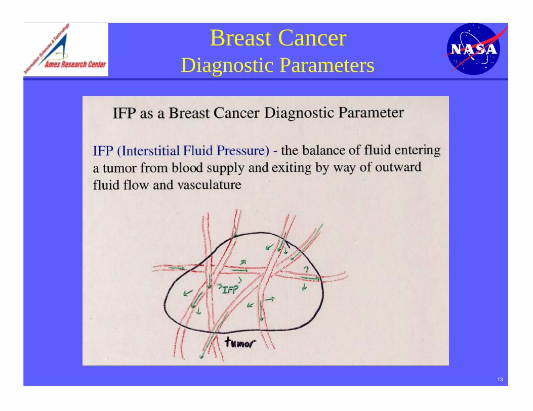
Breast CancerDiagnostic Parameters

14
Breast CancerDiagnostic Parameters
Importance of IFP measurement
¥ Studies confirm that even the smallest invasivebreast cancers exhibit a high IFP wherever theneedle tip is located within the tumor.
¥ Only invasive breast cancer has consistently highIFP.
¥ Due to the elevated IFP in invasive malignant tumorand significant drop-off values at periphery, IFPreadings show great value discrimination betweenmalignant and benign tumors.

15
Breast CancerDiagnostic Parameters

16
Breast CancerDiagnostic Parameters
High microvessel counts represent increased tumorangiogenesis and is correlated with tumor aggressiveness

17
Breast CancerDiagnostic Parameters

18
Elasticity¥ Malignant tumors tend to be 10-100x stiffer than normal breast tissue¥ Some cancers, such as noninvasive ductal, medullary, and mucinous
carcinomas, can be relatively soft
Bioimpedance¥ Electrical and dielectric properties measured on normal and cancerous
breast tissues:— Normal tissues, surrounding tissues, and carcinoma are significantly
different (Chauveau; Jossinet & Schmitt)—Increased capacitance & resistance in malignant tumors
Breast CancerDiagnostic Parameters

19
Computer-aided tools¥ 3D tumor reconstruction (ultrasound sensor attached to robotic arm)¥ Feature extraction¥ Pattern search / Pattern recognition
Breast CancerDiagnostic Parameters
External imaging information:

20
Breast CancerPrognostic Parameters
¥ mean pO2
—metastasizing tumors 7.5 mmHg—nonmetastasing tumors 20 mmHg
—Disease free survival (70-80%) >10 mm Hg—Disease free survival (30-35%) <10 mm Hg
¥ Occurrence of hypoxia and O2 patterns do not correlate withclinical stages
¥ pO2 values in malignant tumors are heterogenousthroughout hypoxic tissue

21
Breast CancerPrognostic Parameters
¥ MVC (microvessel count)—MVC has been shown in many studies to be an independent
prognostic indicator for relapse free survival¥ disease-free < 80 vessels/mm < relapse
¥ Microvessel morphology pattern—none < spotted < linear < mixed patterns < branching
¥ mean malignant tumor size increase in this order¥ all malignant tumors with spotted or linear proved to be invasive
—branching has highest predictive value for malignant tumor—lesions with branching pattern in one study — positive predictive
value for malignancy of 97%—no benign lesions demonstrated a predominant branching pattern

22
SMART Probe SoftwareAdvanced Information Technologies
Neural Networks Fuzzy Logic
Data MiningPattern Recognition
Knowledge Discovery
Information Fusion
INPUTS:Internal / External Measurements
Heuristic Information
OUTPUTS:Diagnosis / Prognosis

23
Neural Network Technology
¥ Generic nonlinear functional element¥ Functionality (defined by weights) set through training process¥ Several applications in industrial process control, credit scoring, fraud
detection, target recognition, optical character recognition, etc.
Neural network — signal processing technology inspired byand loosely modeled after the human brain
OutputsInputs
Neurons(nonlinear
processing units)
Weights (specify connectionstrength between neurons)

24
Fuzzy Logic Technology
¥ Functionality set through correlation process¥ Several applications in industrial process control, credit scoring, fraud
detection, target recognition, optical character recognition, etc.
Fuzzy logic — signal processing technology inspired by andloosely modeled after human processing of qualitative(fuzzy inference) inputs.

25
Detection of Biomolecular Signatures:
• Developing biosensors to study origins of life
• Collaborating with National Cancer Institute (NCI) to develop sensors for cancer diagnosis
NanotechnologyMicro/Nanoscale Biosensors

26
SUMMARYOther Medical Applications
Neurosurgery SMART Probe

27
SMART ProbeBenefits
¥ Real-time tissue diagnosis¥ Avoid injury to arteries, nerves, etc.¥ Determine tumor margins¥ Accurate localization of tumor¥ Accurate treatment & monitoring¥ etc.

28
SMART ProbeBreast Cancer Diagnosis
¥ Information Technologies to assist astronauts inresponding to medical emergencies during space missionswill be employed to improve medical care in the form ofsmart medical tools.
¥ This technology has great potential for the diagnosis &treatment of cancer.
¥ Joint research project between NASA & Stanford MedicalCenter - to develop a smart probe to identify & diagnosewhether a suspicious breast tissue is benign or malignant.

NASA SMART ProbesCutting Edge Technology
Application to Breast Cancer Diagnosis
Robert W. Mah, Ph.D., NASAStefanie S. Jeffrey, MD., Stanford Medical Center

30
• Image enhancement• Pattern search / Pattern recognition• 3D tumor reconstruction• Feature extraction
SMART ProbeComputer-aided Tools

31
SMART ProbeData Sample (optical reflectance)
Normal tissues
Mammary tumor(MCF – 7)

32
SMART ProbeCharacterizing Tissue Types
Parameter Clustering in High Dimensional Space

33
Final RemarkCommercial Version
Silicon Valley start up company is developing a commercialversion of NASA s Breast Cancer SMART Probe

34
Breast CancerDiagnostic Parameters

35
Breast CancerDiagnostic Parameters