cutaneous neuroendocrine carcinoma of the external auditory
TRANSCRIPT
Hindawi Publishing CorporationCase Reports in OtolaryngologyVolume 2012, Article ID 941065, 4 pagesdoi:10.1155/2012/941065
Case Report
Cutaneous Neuroendocrine Carcinoma of the ExternalAuditory Canal: A Case Report and Review of the Literature
Yi-Ke Li,1 Fang-Lu Chi,2 Shu-Yi Wang,3 Wu-Qing Wang,2 Juan-Mei Yang,2 and Yi-Bo Huang2
1 Department of Otolaryngology, School of Medicine, Vanderbilt University, Nashville, TN 37232, USA2 Department of Otology and Skull Base Surgery, Eye Ear Nose and Throat Hospital, Fudan University, Shanghai 200031, China3 Department of Pathology, Eye Ear Nose and Throat Hospital, Fudan University, Shanghai 200031, China
Correspondence should be addressed to Yi-Ke Li, [email protected]
Received 17 April 2012; Accepted 8 July 2012
Academic Editors: A. Harimaya and G. J. Petruzzelli
Copyright © 2012 Yi-Ke Li et al. This is an open access article distributed under the Creative Commons Attribution License, whichpermits unrestricted use, distribution, and reproduction in any medium, provided the original work is properly cited.
Cutaneous neuroendocrine carcinoma (cNEC) is rarely seen in the external ear. In this paper, we newly describe a patient withcNEC in his right external auditory canal, followed by a further discussion on the clinical features, diagnosis, and treatments ofcNEC of the external ear. A review of the literature showed that cNEC of the external auditory canal generally presents as asymp-tomatic and that pathology yields the most confirmative diagnosis. A wide resection with adjuvant radiotherapy and chemotherapyis recommended. The overall prognosis of this condition is poor.
1. Introduction
Neuroendocrine carcinoma (NEC) constitutes a heteroge-neous group of neoplasms that have been postulated to origi-nate from a common precursor cell population. Thoughcommonly associated with the gastrointestinal tract andbronchopulmonary system, a substantial number of thesetumors originate in less common anatomical sites and canrange from indolent, unrecognized entities to highly active,metastatic secretory tumors. In 1972, Toker [1] first des-cribed an uncommon skin lesion which presented as a tra-becular pattern of tumor cell growth, later named cutaneousneuroendocrine carcinoma (cNEC). Most of these cases areknown as Merkel cell carcinoma (MCC), which is generallyconsidered to originate from Merkel cells in the basal layerof the epidermis. Much less often, cNEC is diagnosed in theexternal ear. To our best knowledge, only 23 cases of cNEC ofthe external ear have been reported [2–5], and only 3 of themwere found in the external auditory canal [6, 7].
In this paper, we newly describe a patient with cNEC inhis right external auditory canal, followed by a further dis-cussion on the clinical features, diagnosis, and treatment ofcNEC of the external ear.
2. Case Report
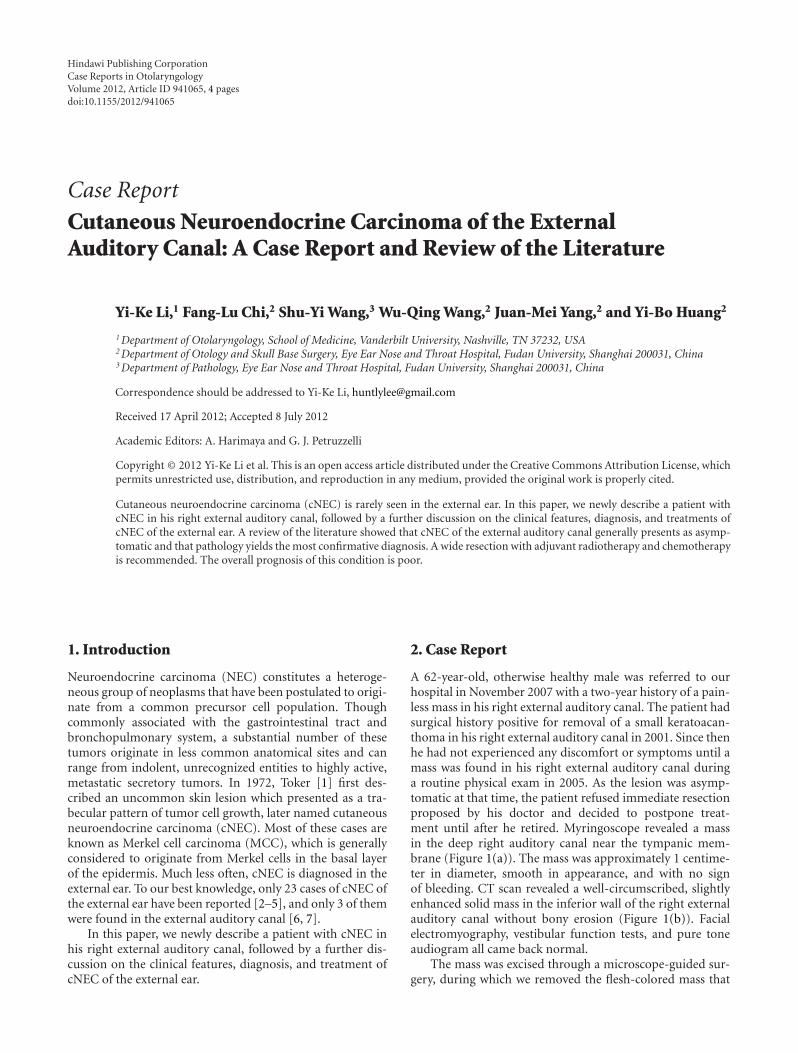
A 62-year-old, otherwise healthy male was referred to ourhospital in November 2007 with a two-year history of a pain-less mass in his right external auditory canal. The patient hadsurgical history positive for removal of a small keratoacan-thoma in his right external auditory canal in 2001. Since thenhe had not experienced any discomfort or symptoms until amass was found in his right external auditory canal duringa routine physical exam in 2005. As the lesion was asymp-tomatic at that time, the patient refused immediate resectionproposed by his doctor and decided to postpone treat-ment until after he retired. Myringoscope revealed a massin the deep right auditory canal near the tympanic mem-brane (Figure 1(a)). The mass was approximately 1 centime-ter in diameter, smooth in appearance, and with no signof bleeding. CT scan revealed a well-circumscribed, slightlyenhanced solid mass in the inferior wall of the right externalauditory canal without bony erosion (Figure 1(b)). Facialelectromyography, vestibular function tests, and pure toneaudiogram all came back normal.
The mass was excised through a microscope-guided sur-gery, during which we removed the flesh-colored mass that
2 Case Reports in Otolaryngology
(a)
(b)
Figure 1: Auxiliary examinations including (a) myringoscopewhich showed a flesh-colored mass in the anterior and inferior wallsof deep right external auditory canal, near the tympanic membrane.(b) Axial cranial CT which presented a well-circumscribed soft tis-sue mass with homogeneous density (white arrow) in the rightexternal auditory canal.
was adherent to the surface of tympanic membrane andpreserved the canal wall and the tympanic membrane. Theexcised mass was then sent to pathology.
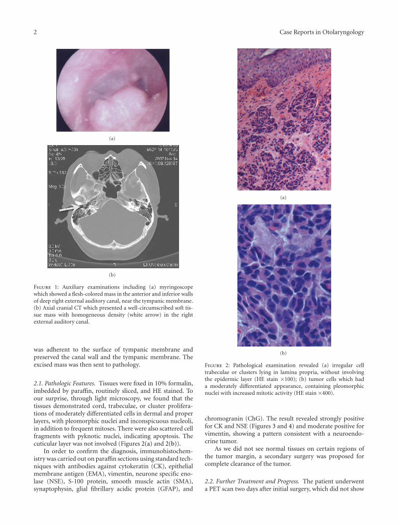
2.1. Pathologic Features. Tissues were fixed in 10% formalin,imbedded by paraffin, routinely sliced, and HE stained. Toour surprise, through light microscopy, we found that thetissues demonstrated cord, trabeculae, or cluster prolifera-tions of moderately differentiated cells in dermal and properlayers, with pleomorphic nuclei and inconspicuous nucleoli,in addition to frequent mitoses. There were also scattered cellfragments with pyknotic nuclei, indicating apoptosis. Thecuticular layer was not involved (Figures 2(a) and 2(b)).
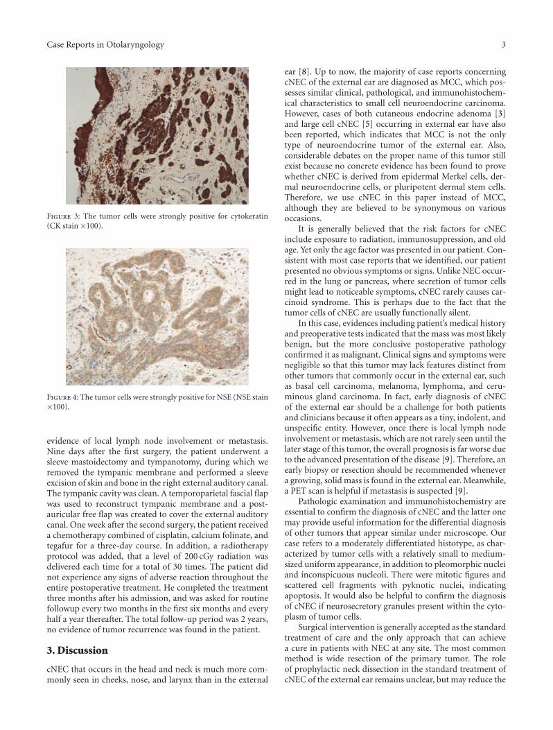
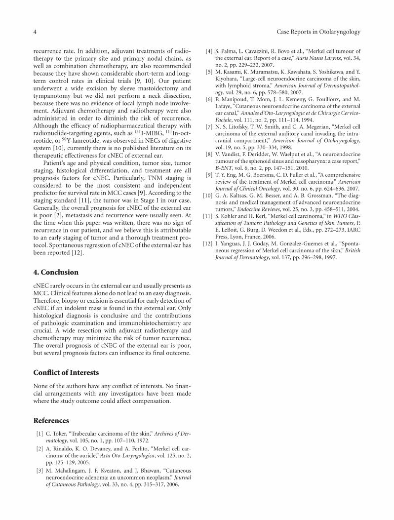
In order to confirm the diagnosis, immunohistochem-istry was carried out on paraffin sections using standard tech-niques with antibodies against cytokeratin (CK), epithelialmembrane antigen (EMA), vimentin, neurone specific eno-lase (NSE), S-100 protein, smooth muscle actin (SMA),synaptophysin, glial fibrillary acidic protein (GFAP), and
(a)
(b)
Figure 2: Pathological examination revealed (a) irregular celltrabeculae or clusters lying in lamina propria, without involvingthe epidermic layer (HE stain ×100); (b) tumor cells which hada moderately differentiated appearance, containing pleomorphicnuclei with increased mitotic activity (HE stain ×400).
chromogranin (ChG). The result revealed strongly positivefor CK and NSE (Figures 3 and 4) and moderate positive forvimentin, showing a pattern consistent with a neuroendo-crine tumor.
As we did not see normal tissues on certain regions ofthe tumor margin, a secondary surgery was proposed forcomplete clearance of the tumor.
2.2. Further Treatment and Progress. The patient underwenta PET scan two days after initial surgery, which did not show
Case Reports in Otolaryngology 3
Figure 3: The tumor cells were strongly positive for cytokeratin(CK stain ×100).
Figure 4: The tumor cells were strongly positive for NSE (NSE stain×100).
evidence of local lymph node involvement or metastasis.Nine days after the first surgery, the patient underwent asleeve mastoidectomy and tympanotomy, during which weremoved the tympanic membrane and performed a sleeveexcision of skin and bone in the right external auditory canal.The tympanic cavity was clean. A temporoparietal fascial flapwas used to reconstruct tympanic membrane and a post-auricular free flap was created to cover the external auditorycanal. One week after the second surgery, the patient receiveda chemotherapy combined of cisplatin, calcium folinate, andtegafur for a three-day course. In addition, a radiotherapyprotocol was added, that a level of 200 cGy radiation wasdelivered each time for a total of 30 times. The patient didnot experience any signs of adverse reaction throughout theentire postoperative treatment. He completed the treatmentthree months after his admission, and was asked for routinefollowup every two months in the first six months and everyhalf a year thereafter. The total follow-up period was 2 years,no evidence of tumor recurrence was found in the patient.
3. Discussion
cNEC that occurs in the head and neck is much more com-monly seen in cheeks, nose, and larynx than in the external
ear [8]. Up to now, the majority of case reports concerningcNEC of the external ear are diagnosed as MCC, which pos-sesses similar clinical, pathological, and immunohistochem-ical characteristics to small cell neuroendocrine carcinoma.However, cases of both cutaneous endocrine adenoma [3]and large cell cNEC [5] occurring in external ear have alsobeen reported, which indicates that MCC is not the onlytype of neuroendocrine tumor of the external ear. Also,considerable debates on the proper name of this tumor stillexist because no concrete evidence has been found to provewhether cNEC is derived from epidermal Merkel cells, der-mal neuroendocrine cells, or pluripotent dermal stem cells.Therefore, we use cNEC in this paper instead of MCC,although they are believed to be synonymous on variousoccasions.
It is generally believed that the risk factors for cNECinclude exposure to radiation, immunosuppression, and oldage. Yet only the age factor was presented in our patient. Con-sistent with most case reports that we identified, our patientpresented no obvious symptoms or signs. Unlike NEC occur-red in the lung or pancreas, where secretion of tumor cellsmight lead to noticeable symptoms, cNEC rarely causes car-cinoid syndrome. This is perhaps due to the fact that thetumor cells of cNEC are usually functionally silent.
In this case, evidences including patient’s medical historyand preoperative tests indicated that the mass was most likelybenign, but the more conclusive postoperative pathologyconfirmed it as malignant. Clinical signs and symptoms werenegligible so that this tumor may lack features distinct fromother tumors that commonly occur in the external ear, suchas basal cell carcinoma, melanoma, lymphoma, and ceru-minous gland carcinoma. In fact, early diagnosis of cNECof the external ear should be a challenge for both patientsand clinicians because it often appears as a tiny, indolent, andunspecific entity. However, once there is local lymph nodeinvolvement or metastasis, which are not rarely seen until thelater stage of this tumor, the overall prognosis is far worse dueto the advanced presentation of the disease [9]. Therefore, anearly biopsy or resection should be recommended whenevera growing, solid mass is found in the external ear. Meanwhile,a PET scan is helpful if metastasis is suspected [9].
Pathologic examination and immunohistochemistry areessential to confirm the diagnosis of cNEC and the latter onemay provide useful information for the differential diagnosisof other tumors that appear similar under microscope. Ourcase refers to a moderately differentiated histotype, as char-acterized by tumor cells with a relatively small to medium-sized uniform appearance, in addition to pleomorphic nucleiand inconspicuous nucleoli. There were mitotic figures andscattered cell fragments with pyknotic nuclei, indicatingapoptosis. It would also be helpful to confirm the diagnosisof cNEC if neurosecretory granules present within the cyto-plasm of tumor cells.
Surgical intervention is generally accepted as the standardtreatment of care and the only approach that can achievea cure in patients with NEC at any site. The most commonmethod is wide resection of the primary tumor. The roleof prophylactic neck dissection in the standard treatment ofcNEC of the external ear remains unclear, but may reduce the
4 Case Reports in Otolaryngology
recurrence rate. In addition, adjuvant treatments of radio-therapy to the primary site and primary nodal chains, aswell as combination chemotherapy, are also recommendedbecause they have shown considerable short-term and long-term control rates in clinical trials [9, 10]. Our patientunderwent a wide excision by sleeve mastoidectomy andtympanotomy but we did not perform a neck dissection,because there was no evidence of local lymph node involve-ment. Adjuvant chemotherapy and radiotherapy were alsoadministered in order to diminish the risk of recurrence.Although the efficacy of radiopharmaceutical therapy withradionuclide-targeting agents, such as 131I-MIBG, 111In-oct-reotide, or 90Y-lanreotide, was observed in NECs of digestivesystem [10], currently there is no published literature on itstherapeutic effectiveness for cNEC of external ear.
Patient’s age and physical condition, tumor size, tumorstaging, histological differentiation, and treatment are allprognosis factors for cNEC. Particularly, TNM staging isconsidered to be the most consistent and independentpredictor for survival rate in MCC cases [9]. According to thestaging standard [11], the tumor was in Stage I in our case.Generally, the overall prognosis for cNEC of the external earis poor [2], metastasis and recurrence were usually seen. Atthe time when this paper was written, there was no sign ofrecurrence in our patient, and we believe this is attributableto an early staging of tumor and a thorough treatment pro-tocol. Spontaneous regression of cNEC of the external ear hasbeen reported [12].
4. Conclusion
cNEC rarely occurs in the external ear and usually presents asMCC. Clinical features alone do not lead to an easy diagnosis.Therefore, biopsy or excision is essential for early detection ofcNEC if an indolent mass is found in the external ear. Onlyhistological diagnosis is conclusive and the contributionsof pathologic examination and immunohistochemistry arecrucial. A wide resection with adjuvant radiotherapy andchemotherapy may minimize the risk of tumor recurrence.The overall prognosis of cNEC of the external ear is poor,but several prognosis factors can influence its final outcome.
Conflict of Interests
None of the authors have any conflict of interests. No finan-cial arrangements with any investigators have been madewhere the study outcome could affect compensation.
References
[1] C. Toker, “Trabecular carcinoma of the skin,” Archives of Der-matology, vol. 105, no. 1, pp. 107–110, 1972.
[2] A. Rinaldo, K. O. Devaney, and A. Ferlito, “Merkel cell car-cinoma of the auricle,” Acta Oto-Laryngologica, vol. 125, no. 2,pp. 125–129, 2005.
[3] M. Mahalingam, J. F. Kveaton, and J. Bhawan, “Cutaneousneuroendocrine adenoma: an uncommon neoplasm,” Journalof Cutaneous Pathology, vol. 33, no. 4, pp. 315–317, 2006.
[4] S. Palma, L. Cavazzini, R. Bovo et al., “Merkel cell tumour ofthe external ear. Report of a case,” Auris Nasus Larynx, vol. 34,no. 2, pp. 229–232, 2007.
[5] M. Kasami, K. Muramatsu, K. Kawahata, S. Yoshikawa, and Y.Kiyohara, “Large-cell neuroendocrine carcinoma of the skin,with lymphoid stroma,” American Journal of Dermatopathol-ogy, vol. 29, no. 6, pp. 578–580, 2007.
[6] P. Manipoud, T. Mom, J. L. Kemeny, G. Fouilloux, and M.Lafaye, “Cutaneous neuroendocrine carcinoma of the externalear canal,” Annales d’Oto-Laryngologie et de Chirurgie Cervico-Faciale, vol. 111, no. 2, pp. 111–114, 1994.
[7] N. S. Litofsky, T. W. Smith, and C. A. Megerian, “Merkel cellcarcinoma of the external auditory canal invading the intra-cranial compartment,” American Journal of Otolaryngology,vol. 19, no. 5, pp. 330–334, 1998.
[8] V. Vandist, F. Deridder, W. Waelput et al., “A neuroendocrinetumour of the sphenoid sinus and nasopharynx: a case report,”B-ENT, vol. 6, no. 2, pp. 147–151, 2010.
[9] T. Y. Eng, M. G. Boersma, C. D. Fuller et al., “A comprehensivereview of the treatment of Merkel cell carcinoma,” AmericanJournal of Clinical Oncology, vol. 30, no. 6, pp. 624–636, 2007.
[10] G. A. Kaltsas, G. M. Besser, and A. B. Grossman, “The diag-nosis and medical management of advanced neuroendocrinetumors,” Endocrine Reviews, vol. 25, no. 3, pp. 458–511, 2004.
[11] S. Kohler and H. Kerl, “Merkel cell carcinoma,” in WHO Clas-sification of Tumors: Pathology and Genetics of Skin Tumors, P.E. LeBoit, G. Burg, D. Weedon et al., Eds., pp. 272–273, IARCPress, Lyon, France, 2006.
[12] I. Yanguas, J. J. Goday, M. Gonzalez-Guemes et al., “Sponta-neous regression of Merkel cell carcinoma of the sikn,” BritishJournal of Dermatology, vol. 137, pp. 296–298, 1997.